Empyema, Subdural
Thoracostomy
Pleural Effusion
Pleural Diseases
Chest Tubes
Thoracic Surgery, Video-Assisted
Bronchial Fistula
Pleura
Thoracoplasty
Lung Abscess
Fistula
Respiratory Tract Fistula
Pleural Cavity
Instillation, Drug
Primary distension of the guttural pouch lateral compartment secondary to empyema. (1/139)
A 6-year-old, 420-kg quarter horse gelding was presented with a 2-month history of difficulty swallowing and dyspnea. The horse was diagnosed with a right guttural pouch empyema with many large chondroids. Two surgeries were required to completely remove all the chondroids from what proved to be a primary distension of the guttural pouch lateral compartment. (+info)Effect of zinc-reversible growth-inhibitory activity in human empyema fluid on antibiotic microbicidal activity. (2/139)
Abscess fluid supernatants have zinc-reversible microbial growth-inhibitory activity that is mediated by calprotectin, a zinc-binding protein. Because it inhibits microbial growth, this activity might interfere with killing by antibiotics that require their target organisms to be proliferating. In the present study, we cultured bacteria in human empyema fluid and used zinc to overcome the growth-inhibitory effect of calprotectin. We then compared the effect of zinc on killing by the beta-lactams ampicillin and cefazolin with that of the fluoroquinolone trovafloxacin, since the latter may be better able to kill nonproliferating organisms. In empyema fluid diluted 1:5 in normal saline, addition of zinc (30 microM) increased growth of two strains of Staphyloccocus aureus and two strains of Escherichia coli but did not affect the MICs or MBCs of the three antibiotics in Mueller-Hinton broth. For one strain of S. aureus, no effect of zinc was found on killing by either ampicillin or cefazolin. However, with the other strain of S. aureus and both strains of E. coli, significant enhancement of killing by both drugs was observed with zinc addition. On the other hand, no effect on the killing of any of the organisms was observed for trovafloxacin when zinc was added. These results suggest that the zinc-reversible growth-inhibitory activity of abscess fluid may interfere with the microbicidal activity of antibiotics requiring proliferating target organisms, although antibiotics better able to kill nonproliferating organisms may be less affected by this phenomenon. (+info)Pharmacokinetics of trimethoprim-sulfamethoxazole in children. (3/139)
The present report extends experience with the use of trimethoprim-sulfamethoxazole (TMP-SMX) in children aged 3 months to 10 years. The regimen was TMP (200 mg)--SMX (1000 mg)/m-2d given in two equal doses. The drug was easily administered, well tolerated and efficacious in the treatment of a variety of infections in 12 children. A steady state had been achieved by the third dose of medication and accumulation of either component during days 1 through 4 did not occur. Serum concentrations of TMP were slightly lower in children aged less than 3 years compared with those aged 3 to 6 years but the differences were small and these results are preliminary. Peak mean serum TMP concentration was highest at day 3 when it reached 1.63 mug/ml. It is concluded that this regimen may be suboptimal for some major parenchymal infections even though the therapeutic result was excellent in most children. (+info)Rapid diagnosis of infectious pleural effusions by use of reagent strips. (4/139)
Reagent strips have not yet been tested for use in the diagnosis of infectious pleural effusions. A reagent strip was used to evaluate 82 patients with pleural effusions: 20 patients had transudative effusions, 35 had infectious exudative effusions (empyema in 14 and parapneumonic effusion in 21), and 27 had noninfectious exudative effusions. Pleural fluid protein, as evaluated by the reagent strip, proved accurate for the detection of exudative effusions (sensitivity, 93.1%; specificity, 50%; positive predictive value, 84.3%; negative predictive value, 71.5%; odds ratio [OR], 6.77; and 95% confidence interval [CI], 1.87-24). The reagent strip leukocyte esterase test effectively detected infectious exudative effusions (sensitivity, 42.8%; specificity, 91.3%; positive predictive value, 88.2%; negative predictive value, 51.2%; OR, 4.46; and 95% CI, 1.2-16.4). Pleural pH was significantly predicted by the reagent strip but was of no assistance in categorization of exudative effusions as infectious or noninfectious. Compared with physical, laboratory, and microbiological data, the reagent strip was as accurate for estimation of percentages of infectious and noninfectious exudative effusions. Thus, reagent strips may be a rapid, easy-to-use, and inexpensive technique for discriminating transudative from exudative pleural effusions and for categorizing exudative pleural effusions as infectious or noninfectious. (+info)Empyema in rheumatoid arthritis. (5/139)
Case notes of the last 67 patients to present at the Brompton Hospital with nontuberculous empyemas, and without malignant disease, have been examined. Three cases of empyema in association with rheumatoid arthritis (RA) were found, and these cases are reported. Previous literature concerning this association is reviewed. It is concluded that two types of empyema may occur in patients with RA. Some develop in association with nodular pleuropulmonary disease and the formation of pyopneumothoraces; in other cases large, recurrent, primary empyemas build up in the presence of active rheumatoid disease alone. As with rheumatoid pleural effusions, middle-aged men seem to be particularly susceptible. (+info)Evarts Ambrose Graham, empyema, and the dawn of clinical understanding of negative intrapleural pressure. (6/139)
The concept of negative intrapleural pressure is fairly new. Although the phenomenon had already been described, Wirz provided the first definitive analysis of its significance to the mechanics of breathing in 1923. By contrast, empyema has been known since antiquity; from the time of Hippocrates, treatment has consisted of open drainage. Open drainage was often successful and did not result in pneumothorax, because most cases of empyema were associated with adhesions and thickened visceral pleura that prevented the lung from collapsing. The epidemic of group A streptococcal pneumonia in military camps in 1917-1918 was associated with the rapid and early accumulation of empyema fluid and was the catalyst for renewed study of empyema. Use of open drainage to manage this illness resulted in a high immediate mortality rate, probably because patients developed pneumothorax. The work of Evarts Graham and the Empyema Commission married physiological understanding of pleural mechanics with rational clinical treatment and paved the way for further advances in thoracic surgery. (+info)An epidemiological investigation of a sustained high rate of pediatric parapneumonic empyema: risk factors and microbiological associations. (7/139)
We investigated the increasing incidence of pediatric empyema during the 1990s at Primary Children's Medical Center in Salt Lake City. Of 540 children hospitalized with community-acquired bacterial pneumonia (CAP) who were discharged from 1 July 1993 through 1 July 1999, 153 (28.3%) had empyema. The annual population incidence of empyema increased during the study period from 1 to 5 cases per 100,000 population aged <19 years. Streptococcus pneumoniae was identified as the most common cause of CAP with or without empyema; serotype 1 accounted for 50% of the cases of pneumococcal empyema. Patients with empyema were more likely to be >3 years old, to have > or =7 days of fever, to have varicella, and to have received antibiotics and ibuprofen before admission to the hospital, compared with patients without empyema (P<.0001 for each factor). The increasing incidence of empyema was associated with infection due to S. pneumoniae serotype 1, outpatient treatment with certain antibiotics, ibuprofen use, and varicella. (+info)Randomised trial of intrapleural urokinase in the treatment of childhood empyema. (8/139)
BACKGROUND: The role of intrapleural fibrinolytic agents in the treatment of childhood empyema has not been established. A randomised double blind placebo controlled trial of intrapleural urokinase was performed in children with parapneumonic empyema. METHODS: Sixty children (median age 3.3 years) were recruited from 10 centres and randomised to receive either intrapleural urokinase 40 000 units in 40 ml or saline 12 hourly for 3 days. The primary outcome measure was length of hospital stay after entry to the trial. RESULTS: Treatment with urokinase resulted in a significantly shorter hospital stay (7.4 v 9.5 days; ratio of geometric means 1.28, CI 1.16 to 1.41 p=0.027). A post hoc analysis showed that the use of small percutaneous drains was also associated with shorter hospital stay. Children treated with a combination of urokinase and a small drain had the shortest stay (6.0 days, CI 4.6 to 7.8). CONCLUSION: Intrapleural urokinase is effective in treating empyema in children and significantly shortens hospital stay. (+info)Empyema is a medical condition characterized by the accumulation of pus in a body cavity, most commonly in the pleural space surrounding the lungs. It is usually caused by a bacterial infection that spreads from the lung tissue to the pleural space. The buildup of pus can cause chest pain, cough, fever, and difficulty breathing. Empyema can be a complication of pneumonia or other respiratory infections, and it may require treatment with antibiotics, drainage of the pus, and sometimes surgery.
Empyema is a collection of pus in a body cavity. Pleural empyema refers to the presence of pus in the pleural space, which is the thin fluid-filled space that surrounds the lungs. This condition usually develops as a complication of pneumonia or lung infection, and it can cause symptoms such as chest pain, cough, fever, and difficulty breathing. Treatment typically involves antibiotics to treat the underlying infection, as well as drainage of the pus from the pleural space through procedures such as thoracentesis or chest tube placement. In severe cases, surgery may be necessary to remove the infected pleura and prevent recurrence.
Empyema subdural is a medical condition characterized by the presence of pus (purulent material) in the potential space between the dura mater and the arachnoid membrane of the brain. This space is called the subdural space. Empyema subdural can result from an infection that spreads from nearby areas such as the skull, face, or sinuses, or it can occur as a complication of neurosurgical procedures.
The symptoms of empyema subdural may include headache, altered mental status, fever, seizures, and neurological deficits depending on the severity and location of the infection. Diagnosis is usually made with the help of imaging studies such as CT or MRI scans, and treatment typically involves surgical drainage of the pus along with antibiotic therapy to eliminate the underlying infection. If left untreated, empyema subdural can lead to serious complications such as brain abscess, meningitis, or even death.
Tuberculous empyema is a specific type of empyema, which is a collection of pus in the pleural space (the space between the lungs and the chest wall). It is caused by the Mycobacterium tuberculosis bacterium, which is the same bacterium that causes tuberculosis (TB) of the lungs.
In tuberculous empyema, the bacteria spread from the lungs to the pleural space, where they cause an infection and inflammation. This can lead to the accumulation of pus and the development of a chronic empyema. Symptoms may include chest pain, cough, fever, and difficulty breathing. Treatment typically involves a prolonged course of multiple antibiotics to kill the bacteria, as well as drainage of the pus from the pleural space. In some cases, surgery may be necessary to remove the infected tissue and prevent recurrence.
Thoracostomy is a surgical procedure that involves the creation of an opening into the chest cavity to relieve excessive pressure, drain fluid or air accumulation, or provide access for surgery. It is commonly performed to treat conditions such as pneumothorax (collapsed lung), hemothorax (blood in the chest cavity), pleural effusion (excess fluid in the pleural space), and empyema (pus in the pleural space).
During a thoracostomy, a healthcare professional makes an incision on the chest wall and inserts a tube called a thoracostomy tube or chest tube. The tube is connected to a drainage system that helps remove the air, fluid, or blood from the chest cavity. This procedure can be performed as an emergency treatment or as a planned surgical intervention.
The medical definition of thoracostomy includes the following key components:
1. A surgical procedure
2. Involving the creation of an opening
3. Into the chest cavity (thorax)
4. To relieve pressure, drain fluids or air, or provide access for surgery
5. Often performed with the insertion of a thoracostomy tube or chest tube
6. Used to treat various conditions related to the pleural space and lungs
Pleural effusion is a medical condition characterized by the abnormal accumulation of fluid in the pleural space, which is the thin, fluid-filled space that surrounds the lungs and lines the inside of the chest wall. This space typically contains a small amount of fluid to allow for smooth movement of the lungs during breathing. However, when an excessive amount of fluid accumulates, it can cause symptoms such as shortness of breath, coughing, and chest pain.
Pleural effusions can be caused by various underlying medical conditions, including pneumonia, heart failure, cancer, pulmonary embolism, and autoimmune disorders. The fluid that accumulates in the pleural space can be transudative or exudative, depending on the cause of the effusion. Transudative effusions are caused by increased pressure in the blood vessels or decreased protein levels in the blood, while exudative effusions are caused by inflammation, infection, or cancer.
Diagnosis of pleural effusion typically involves a physical examination, chest X-ray, and analysis of the fluid in the pleural space. Treatment depends on the underlying cause of the effusion and may include medications, drainage of the fluid, or surgery.
Pleural diseases refer to conditions that affect the pleura, which is the thin, double-layered membrane that surrounds the lungs and lines the inside of the chest wall. The space between these two layers contains a small amount of fluid that helps the lungs move smoothly during breathing. Pleural diseases can cause inflammation, infection, or abnormal collections of fluid in the pleural space, leading to symptoms such as chest pain, cough, and difficulty breathing.
Some common examples of pleural diseases include:
1. Pleurisy: Inflammation of the pleura that causes sharp chest pain, often worsened by breathing or coughing.
2. Pleural effusion: An abnormal accumulation of fluid in the pleural space, which can be caused by various underlying conditions such as heart failure, pneumonia, cancer, or autoimmune disorders.
3. Empyema: A collection of pus in the pleural space, usually resulting from a bacterial infection.
4. Pleural thickening: Scarring and hardening of the pleura, which can restrict lung function and cause breathlessness.
5. Mesothelioma: A rare form of cancer that affects the pleura, often caused by exposure to asbestos.
6. Pneumothorax: A collection of air in the pleural space, which can result from trauma or a rupture of the lung tissue.
Proper diagnosis and treatment of pleural diseases require a thorough evaluation by a healthcare professional, often involving imaging tests such as chest X-rays or CT scans, as well as fluid analysis or biopsy if necessary.
Drainage, in medical terms, refers to the removal of excess fluid or accumulated collections of fluids from various body parts or spaces. This is typically accomplished through the use of medical devices such as catheters, tubes, or drains. The purpose of drainage can be to prevent the buildup of fluids that may cause discomfort, infection, or other complications, or to treat existing collections of fluid such as abscesses, hematomas, or pleural effusions. Drainage may also be used as a diagnostic tool to analyze the type and composition of the fluid being removed.
Chest tubes are medical devices that are inserted into the chest cavity to drain fluid, air, or blood. They are typically used to treat conditions such as pneumothorax (collapsed lung), hemothorax (blood in the chest cavity), pleural effusion (excess fluid in the chest cavity), and chylothorax (milky fluid in the chest cavity).
Chest tubes are usually inserted between the ribs and directed into the chest cavity, allowing for drainage of the affected area. The tubes are connected to a collection system that creates negative pressure, which helps to remove the air or fluid from the chest cavity.
The size and number of chest tubes used may vary depending on the severity and location of the condition being treated. Chest tubes are typically removed once the underlying condition has been resolved and the drainage has decreased to a minimal amount.
Thoracic surgery, video-assisted (VATS) is a minimally invasive surgical technique used to diagnose and treat various conditions related to the chest cavity, including the lungs, pleura, mediastinum, esophagus, and diaphragm. In VATS, a thoracoscope, a type of endoscope with a camera and light source, is inserted through small incisions in the chest wall to provide visualization of the internal structures. The surgeon then uses specialized instruments to perform the necessary surgical procedures, such as biopsies, lung resections, or esophageal repairs. Compared to traditional open thoracic surgery, VATS typically results in less postoperative pain, shorter hospital stays, and quicker recoveries for patients.
A bronchial fistula is an abnormal connection or passage between the bronchial tree (the airways in the lungs) and the surrounding tissues, such as the pleural space (the space between the lungs and the chest wall), blood vessels, or other organs. This condition can result from various causes, including lung injury, infection, surgery, or certain diseases such as cancer or tuberculosis.
Bronchial fistulas can lead to symptoms like coughing, wheezing, shortness of breath, and chest pain. They may also cause air leaks, pneumothorax (collapsed lung), or chronic infections. Treatment for bronchial fistulas depends on the underlying cause and severity of the condition but often involves surgical repair or closure of the abnormal connection.
The pleura is the medical term for the double-layered serous membrane that surrounds the lungs and lines the inside of the chest cavity. The two layers of the pleura are called the parietal pleura, which lines the chest cavity, and the visceral pleura, which covers the surface of the lungs.
The space between these two layers is called the pleural cavity, which contains a small amount of lubricating fluid that allows the lungs to move smoothly within the chest during breathing. The main function of the pleura is to protect the lungs and facilitate their movement during respiration.
Thoracotomy is a surgical procedure that involves making an incision on the chest wall to gain access to the thoracic cavity, which contains the lungs, heart, esophagus, trachea, and other vital organs. The incision can be made on the side (lateral thoracotomy), back (posterolateral thoracotomy), or front (median sternotomy) of the chest wall, depending on the specific surgical indication.
Thoracotomy is performed for various indications, including lung biopsy, lung resection, esophagectomy, heart surgery, and mediastinal mass removal. The procedure allows the surgeon to directly visualize and access the organs within the thoracic cavity, perform necessary procedures, and control bleeding if needed.
After the procedure, the incision is typically closed with sutures or staples, and a chest tube may be placed to drain any accumulated fluid or air from the pleural space around the lungs. The patient will require postoperative care and monitoring in a hospital setting until their condition stabilizes.
Thoracoplasty is a surgical procedure that involves the removal or collapse of one or more ribs and the attached muscles from the chest wall. This procedure is typically performed to correct a deformity or to remove infected tissue in the chest cavity, such as in cases of chronic empyema (a collection of pus in the pleural space) or tuberculosis.
The removal of ribs can also help to reduce the size of an overexpanded lung, which can occur due to conditions like COPD (chronic obstructive pulmonary disease) or a bronchopleural fistula (an abnormal connection between the airways and the pleural space). Thoracoplasty can also be used for cosmetic purposes, such as in the treatment of pectus excavatum (a deformity where the breastbone is sunken into the chest).
A lung abscess is a localized collection of pus in the lung parenchyma caused by an infectious process, often due to bacterial infection. It's characterized by necrosis and liquefaction of pulmonary tissue, resulting in a cavity filled with purulent material. The condition can develop as a complication of community-acquired or nosocomial pneumonia, aspiration of oral secretions containing anaerobic bacteria, septic embolism, or contiguous spread from a nearby infected site.
Symptoms may include cough with foul-smelling sputum, chest pain, fever, weight loss, and fatigue. Diagnosis typically involves imaging techniques such as chest X-ray or CT scan, along with microbiological examination of the sputum to identify the causative organism(s). Treatment often includes antibiotic therapy tailored to the identified pathogen(s), as well as supportive care such as bronchoscopy, drainage, or surgery in severe cases.
Thoracoscopy is a surgical procedure in which a thoracoscope, a type of endoscope, is inserted through a small incision between the ribs to examine the lungs and pleural space (the space surrounding the lungs). It allows the surgeon to directly view the chest cavity, take biopsies, and perform various operations. This procedure is often used in the diagnosis and treatment of pleural effusions, lung cancer, and other chest conditions.
A fistula is an abnormal connection or passage between two organs, vessels, or body parts that usually do not connect. It can form as a result of injury, infection, surgery, or disease. A fistula can occur anywhere in the body but commonly forms in the digestive system, genital area, or urinary system. The symptoms and treatment options for a fistula depend on its location and underlying cause.
A respiratory tract fistula is an abnormal connection or passage between the respiratory tract (which includes the nose, throat, windpipe, and lungs) and another organ or structure, such as the skin, digestive tract, or blood vessels. This condition can lead to complications such as air leakage, infection, and difficulty breathing. The causes of respiratory tract fistulas vary and can include trauma, surgery, infection, or cancer. Treatment depends on the location and severity of the fistula and may involve surgical repair, antibiotics, or other therapies.
The pleural cavity is the potential space between the visceral and parietal pleura, which are the two membranes that surround the lungs. The visceral pleura covers the outside of the lungs, while the parietal pleura lines the inside of the chest wall. Under normal conditions, these two layers are in contact with each other, and the space between them is virtually nonexistent. However, when air, fluid or inflammation accumulates within this space, it results in the formation of a pleural effusion, which can cause discomfort and difficulty breathing.
A pneumonectomy is a surgical procedure in which an entire lung is removed. This type of surgery is typically performed as a treatment for certain types of lung cancer, although it may also be used to treat other conditions such as severe damage or infection in the lung that does not respond to other treatments. The surgery requires general anesthesia and can be quite complex, with potential risks including bleeding, infection, pneumonia, and air leaks. Recovery from a pneumonectomy can take several weeks, and patients may require ongoing rehabilitation to regain strength and mobility.
Instillation, in the context of drug administration, refers to the process of introducing a medication or therapeutic agent into a body cavity or onto a mucous membrane surface using gentle, steady pressure. This is typically done with the help of a device such as an eyedropper, pipette, or catheter. The goal is to ensure that the drug is distributed evenly over the surface or absorbed through the mucous membrane for localized or systemic effects. Instillation can be used for various routes of administration including ocular (eye), nasal, auricular (ear), vaginal, and intra-articular (joint space) among others. The choice of instillation as a route of administration depends on the drug's properties, the desired therapeutic effect, and the patient's overall health status.
Hemothorax is a medical condition characterized by the presence of blood in the pleural space, which is the area between the lungs and the chest wall. This accumulation of blood can occur due to various reasons such as trauma, rupture of a blood vessel, or complications from lung or heart surgery.
The buildup of blood in the pleural space can cause the affected lung to collapse, leading to symptoms such as shortness of breath, chest pain, and cough. In severe cases, hemothorax can be life-threatening if not promptly diagnosed and treated. Treatment options may include chest tube drainage, blood transfusion, or surgery, depending on the severity and underlying cause of the condition.
A gastric fistula is an abnormal connection or passage between the stomach and another organ or the skin surface. This condition can occur as a result of complications from surgery, injury, infection, or certain diseases such as cancer. Symptoms may include persistent drainage from the site of the fistula, pain, malnutrition, and infection. Treatment typically involves surgical repair of the fistula and management of any underlying conditions.
 Empyema - Wikipedia
 Empyema
 Empyema: MedlinePlus Medical Encyclopedia
 Gallbladder Empyema: Background, Pathophysiology, Etiology
 Empyema, Tuberculous - Medical Dictionary online-medical-dictionary.org
 empyema | Taber's Medical Dictionary
 Empyema - Symptoms, diagnosis and treatment | BMJ Best Practice US
 Intracranial Epidural Abscess and Subdural Empyema - Brain, Spinal Cord, and Nerve Disorders - Merck Manuals Consumer Version
 Empyema: the use of broad range 16S rDNA PCR for pathogen detection | Archives of Disease in Childhood
 Role of Video-Assisted Thoracoscopic Surgery in the Management of Pleural Empyema
| Journal of Armed Forces Medical...
Role of Video-Assisted Thoracoscopic Surgery in the Management of Pleural Empyema
| Journal of Armed Forces Medical... A&E; Emergency Medicine; Accident and Emergency; Air leak; Pneumothorax; Pleural effusion; Empyema Archives - Brighton and...
 Cost-Effectiveness Analysis of Fibrinolysis vs Thoracoscopic Decortication for Early Empyema.
 and Empyema Archives - MedQuizzes
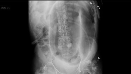
 Ruptured gallbladder empyema - Radiology Cases
 Empyema and Bronchopleural Fistula | Pearson's General Thoracic
 Empyema and Parapneumonic Effusions | Johns Hopkins ABX Guide