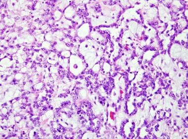
Endodermal Sinus Tumor
Mesonephroma
Dysgerminoma
alpha-Fetoproteins
Ovarian Neoplasms
Teratoma
Primary yolk sac tumour of the liver in adulthood. (1/81)
Primary yolk sac tumour of the liver is exceedingly rare. A 28 year old woman presented with a cystic liver mass and a markedly raised serum alpha-fetoprotein concentration. She underwent a partial hepatectomy for a suspected hepatocellular carcinoma but histological examination of the tumour revealed the classical morphological and immunohistochemical features of a yolk sac tumour. There was no evidence of an extrahepatic primary source. Review of this case, together with the six previously reported adult cases of primary yolk sac tumours of the liver, revealed several features of the tumour that may aid differentiation from hepatocellular carcinoma, with potential therapeutic implications. (+info)Human germ cell tumours: expression of gamma-glutamyl transpeptidase and sensitivity to cisplatin. (2/81)
Previous studies have shown that the enzyme-glutamyl transpeptidase (GGT) is essential for the nephrotoxicity of cisplatin. This study was designed to determine whether GGT activity is necessary for the therapeutic effect of the drug. The relationship between GGT expression and clinical response to platinum-based chemotherapy was examined in 41 human germ cell tumours. Sections of formalin-fixed, paraffin-embedded tumours were immunohistochemically stained with an antibody directed against human GGT. There was no expression of GGT in any of the 17 seminomas or four dysgerminomas; whereas, 12/12 ovarian yolk sac tumours and 4/4 embryonal carcinomas of the testis were GGT-positive. In stage I tumours fewer tumour cells expressed GGT than in later stage tumours. In four germ cell tumours of mixed histology, the seminomatous and dysgerminoma areas were GGT-negative while the areas of the tumour with yolk sac or embryonal histology contained GGT-positive tumour cells. The patients with seminomas or dysgerminomas who were treated with cisplatin-based chemotherapy, all had a complete response despite the absence of GGT expression in these tumours. Fifteen of the 16 patients with yolk sac or embryonal carcinomas received cisplatin-based chemotherapy following surgery. Twelve had a complete response, while three failed to respond to platinum-based therapy. There was no correlation between the level of GGT-expression and response to therapy in this group. Three of the four patients with tumours of mixed histology were treated with cisplatin-based therapy, and had a complete response. Therefore, expression of GGT is not necessary for the therapeutic effect of cisplatin in germ cell tumours. The results from this study suggest that systemic inhibition of GGT would inhibit the nephrotoxic side-effect of cisplatin without interfering with its activity towards germ cell tumours. (+info)Transcription factor GATA-4 is expressed in pediatric yolk sac tumors. (3/81)
Yolk sac tumors (YSTs) are malignant tumors that occur in the gonads of children and young adults, and at extragonadal sites in young children. The histological features of YSTs are variable and can be superimposed on other germ cell tumor histologies. Malignant endodermal cells within YSTs express alpha-fetoprotein, which can be detected in tumor tissue or serum. However, additional markers of endoderm differentiation would be beneficial for the classification of these tumors. Transcription factor GATA-4 regulates the differentiation and function of murine yolk sac endoderm, and its expression correlates with proliferation and cell survival in certain tissues. To see whether GATA-4 plays a role in human YSTs, we surveyed its expression in human germ cell tumors and cell lines. Northern analysis demonstrated expression of GATA-4 mRNA in four human germ cell tumor lines exhibiting yolk sac endoderm differentiation. GATA-4 protein was detected in eight of nine pediatric YSTs by immunohistochemistry. Three of five immature teratomas exhibited GATA-4 in neural blastematous cells and in cylindrical epithelium, whereas all 16 mature teratomas were devoid of GATA-4. We conclude that GATA-4 is a clinically useful marker of human YSTs and speculate that it may play a role in the maintenance of the malignant phenotype. (+info)Hematologic disorders associated with primary mediastinal nonseminomatous germ cell tumors. (4/81)
BACKGROUND: The association between primary germ cell tumors of the mediastinum (the space between the lung pleura that contains the heart and other chest viscera) and hematologic malignancies has been described by retrospective analysis of patients treated at individual clinical centers. To better characterize the risk of hematologic disorders in patients with extragonadal germ cell tumors and to describe the clinical and biologic features of the disorders, we studied an unselected population in a large, international, multicenter database. METHODS: Six hundred thirty-five patients treated at 11 centers in the United States and Europe from 1975 through 1996 were evaluated retrospectively. RESULTS: A hematologic disorder was observed in 17 patients with germ cell tumors. All cases developed among the 287 patients with primary mediastinal nonseminomatous germ cell tumors, giving an incidence rate in this group of 2.0% (95% confidence interval [CI] = 1.1%-3.1%) per year over a median follow-up time of 3 years. The risk of developing hematologic disorders was statistically significantly increased in patients with primary mediastinal nonseminomatous germ cell tumors in comparison with the age-matched general population (standardized incidence ratio = 250; 95% CI = 140-405). The median time to onset of hematologic neoplasia was 6 months (range, 0-47 months), and the median survival after diagnosis of the hematologic disorder was 5 months (range, 0-16 months) (two-sided P<.0001, comparing survival from the time of diagnosis of the germ cell tumor of patients with and without hematologic disorders). CONCLUSION: In our study, approximately one in 17 patients with primary mediastinal nonseminomatous germ cell tumors was affected by a hematologic disorder, whereas no cases were seen among 334 patients with other extragonadal germ cell tumors. The hematologic disorder had a statistically significant impact on prognosis, with none of the 17 reported patients surviving for more than 2 years. (+info)DNA copy number changes in malignant ovarian germ cell tumors. (5/81)
Malignant ovarian germ cell tumors (OGCTs) include immature teratomas (ITs), dysgerminomas (DGs), endodermal sinus tumors (ESTs), choriocarcinomas, and embryonal carcinomas. Knowledge about the genetic changes associated with malignant OGCT development is sparse. We therefore analyzed 25 OGCTs (12 DGs, 4 ESTs, and 9 ITs) for gains and losses by comparative genomic hybridization. In total, more gains than losses were observed, and the number of alterations ranged from 0-20 per tumor. The average number of changes among DGs, ESTs, and ITs was 10, 6, and 1.4, respectively. The most common changes in DGs were gains from chromosome arms 1p (33%), 6p (33%), 12p (67%), 12q (75%), 15q (42%), 20q (50%), 21q (67%), and 22q (58%); gains of the whole of chromosomes 7 (42%), 8 (42%), 17 (42%), and 19 (50%); and losses from 13q (58%). Two of three DGs with a gonadoblastoma component showed gains of 3p21 and loss of 5p, whereas none of the nine pure DGs had these changes, suggesting that they might be characteristic either of gonadoblastoma or of DG developing from a gonadoblastoma. Gain of 12p and gain from 1q were seen in three of four ESTs, whereas gains from 3p, 11q, and Xp and loss from 18q were each found in two tumors. Five of the ITs revealed changes (range, 1-4 changes/tumor), with gains from 1p, 16p, 19, and 22q each being found in two tumors. We conclude that ovarian DGs and ESTs seem to develop via the same genetic pathways that are already known for testicular germ cell tumors. On the other hand, ITs do not exhibit gain of 12p and also typically show fewer changes than other malignant OGCTs, indicating that they arise via different pathogenetic mechanisms. (+info)Expression of hMLH1 and hMSH2 and assessment of microsatellite instability in testicular and mediastinal germ cell tumours. (6/81)
The aim of this study was to investigate DNA mismatch repair deficiency in male germ cell tumours. We analysed the expression of two mismatch repair proteins, human mutL homologue 1 (hMLH1) and human mutS homologue 2 (hMSH2), and evaluated the frequency of microsatellite instability with 10 mononucleotide and two dinucleotide repeat sequences, in 39 paired tumour/normal DNA samples obtained from 17 testicular and two mediastinal germ cell tumours. In all 19 cases, hMLH1 and hMSH2 both showed nuclear immunolocalization in invasive and testicular in-situ tumours. In non-neoplastic seminiferous tubules, hMLH1 was expressed only in premeiotic germ cells, while hMSH2 was seen in all stages of spermatogenesis. Genetic analysis of dinucleotide markers revealed loss of heterozygosity in one of two testicular yolk sac tumours at D18S58 and an allelic shift at D2S123 in two of three testicular embryonal carcinomas, while none of the 12 seminomas exhibited a genetic abnormality at these loci. No abnormalities were demonstrated with the 10 mononucleotide markers. The two mediastinal germ cell tumours showed no genetic instability or allelic loss with all 12 markers. We suggest that genetic alterations as assessed by microsatellite analysis in germ cell tumours may reflect tissue maturation and phenotypic differentiation rather than tumour progression. In addition, we suggest that hMLH1 and hMSH2 genes may not be implicated in the genesis of germ cell tumours. (+info)Gonadal malignant germ cell tumors express immunoreactive inhibin/activin subunits. (7/81)
OBJECTIVE: Inhibin and activin are proteins produced by ovarian granulosa cells and testicular Sertoli cells and are members of the transforming growth factor-beta superfamily. Since increased circulating levels of immunoreactive inhibin were detected in women with malignant ovarian tumors, they were proposed as tumor markers for ovarian carcinoma. Immunohistochemical studies later confirmed the presence of inhibin and activin subunits in granulosa cell tumors and epithelial ovarian cancer, as well as in Sertoli and Leydig cell testicular cancer. However, there is discrepant information on the detection of inhibin and activin in malignant germ cell tumors (MGCT). The aim of the present study was to evaluate the immunohistochemical expression of the inhibin/activin alpha, betaA and betaB subunits in ovarian and testicular MGCT specimens using polyclonal antisera. METHODS: The ovarian tissue samples were composed of 19 MGCT, including dysgerminoma (n=18) and yolk sac tumor (n=1). The testis specimens included classic seminomas (n=20), embryonal carcinomas (n=7), choriocarcinomas (n=2), and yolk sac tumor (n=1). RESULTS: Ovarian and testicular malignant germ cell tumors expressed positive staining for inhibin/activin alpha, betaA and betaB subunits, with some variations between and within individual tumors: while ovarian dysgerminomas were diffusely positive for alpha, betaA and betaB, testicular tumors expressed alpha and betaB subunits, whereas betaA staining was weak. CONCLUSIONS: The present results show positive staining for inhibin/activin subunits in ovarian and testicular MGCT, suggesting a possible role in tumorigenesis with the resultant clinical implication. (+info)Mixed germ cell tumor in the eye of a dog. (8/81)
A 3-year-old female neutered Staffordshire Bull Terrier presented with a mixed germ cell tumor involving the base of the iris and the ciliary body of the right eye. The tumor mass was composed primarily of packeted vacuolated, polygonal (hepatoid) cells and small round cells; epithelial cells lining tubuloacinar structures were a less prominent component. The hepatoid and round cells stained positively for alpha-fetoprotein and cytokeratin. The epithelial cells stained positively for cytokeratin only, and some contained cytoplasmic mucin droplets. The polygonal cells were interpreted as a hepatoid variant of yolk sac tumor, and the epithelial cells were considered a teratomatous component. Trabeculae of bone were observed within the mass and may have been metaplastic or a teratomatous element. Extragonadal germ cell tumors are rare in dogs and have previously been reported only in the suprasellar region. This is the first report of this tumor type in the eye of a nonhuman species. (+info)An Endodermal Sinus Tumor (EST) is a type of germ cell tumor, which is a rare cancer that occurs most frequently in the ovaries or testicles but can also occur in other parts of the body. EST is also known as a yolk sac tumor because it resembles the yolk sac of an embryo.
ESTs are highly aggressive and fast-growing tumors that typically affect children and young adults, with a peak incidence in the first decade of life. These tumors can produce various proteins and substances, such as alpha-fetoprotein (AFP), which can be used as markers for diagnosis and monitoring treatment response.
The symptoms of EST depend on the location of the tumor but may include abdominal pain or swelling, constipation, nausea, vomiting, and irregular menstrual periods in females. Treatment typically involves surgical removal of the tumor, followed by chemotherapy to kill any remaining cancer cells. The prognosis for EST depends on several factors, including the stage of the disease at diagnosis, the patient's age, and the response to treatment.
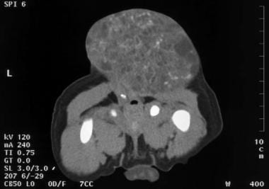
Mesonephroma is a very rare type of kidney tumor that originates from the mesonephric duct remnants, which are the embryonic precursors of the male reproductive system. This tumor typically affects older adults and is more common in men than women.
Mesonephromas are usually slow-growing and asymptomatic, making them difficult to detect at an early stage. When symptoms do occur, they may include flank pain, hematuria (blood in the urine), a palpable abdominal mass, and weight loss.
On imaging studies such as CT or MRI scans, mesonephromas typically appear as well-circumscribed masses within the kidney. The diagnosis is usually confirmed through a biopsy or surgical excision of the tumor.
Mesonephromas are composed of tubular structures lined with cuboidal to low columnar epithelial cells, often with clear cytoplasm. They may also contain areas of necrosis and hemorrhage. The treatment of mesonephroma typically involves surgical excision, and the prognosis is generally favorable, with a low risk of recurrence or metastasis. However, long-term follow-up is recommended due to the rarity and limited data on this type of tumor.
Dysgerminoma is a type of germ cell tumor that develops in the ovaries. It is a malignant (cancerous) tumor that primarily affects girls and women of reproductive age, although it can occur at any age. Dysgerminomas are composed of large, round, or polygonal cells with clear cytoplasm and distinct cell borders, arranged in nests or sheets. They may also contain lymphoid aggregates and may produce hormones such as estrogen or testosterone.
Dysgerminomas are usually unilateral (affecting one ovary), but they can be bilateral (affecting both ovaries) in about 10-15% of cases. They tend to grow and spread rapidly, so early detection and treatment are crucial for a favorable prognosis.
The standard treatment for dysgerminoma is surgical removal of the affected ovary or ovaries, followed by chemotherapy with agents such as bleomycin, etoposide, and cisplatin (BEP). With appropriate treatment, the five-year survival rate for patients with dysgerminoma is high, ranging from 80% to 95%.
Alpha-fetoprotein (AFP) is a protein produced by the yolk sac and the liver during fetal development. In adults, AFP is normally present in very low levels in the blood. However, abnormal production of AFP can occur in certain medical conditions, such as:
* Liver cancer or hepatocellular carcinoma (HCC)
* Germ cell tumors, including non-seminomatous testicular cancer and ovarian cancer
* Hepatitis or liver inflammation
* Certain types of benign liver disease, such as cirrhosis or hepatic adenomas
Elevated levels of AFP in the blood can be detected through a simple blood test. This test is often used as a tumor marker to help diagnose and monitor certain types of cancer, particularly HCC. However, it's important to note that an elevated AFP level alone is not enough to diagnose cancer, and further testing is usually needed to confirm the diagnosis. Additionally, some non-cancerous conditions can also cause elevated AFP levels, so it's important to interpret the test results in the context of the individual's medical history and other diagnostic tests.
Ovarian neoplasms refer to abnormal growths or tumors in the ovary, which can be benign (non-cancerous) or malignant (cancerous). These growths can originate from various cell types within the ovary, including epithelial cells, germ cells, and stromal cells. Ovarian neoplasms are often classified based on their cell type of origin, histological features, and potential for invasive or metastatic behavior.
Epithelial ovarian neoplasms are the most common type and can be further categorized into several subtypes, such as serous, mucinous, endometrioid, clear cell, and Brenner tumors. Some of these epithelial tumors have a higher risk of becoming malignant and spreading to other parts of the body.
Germ cell ovarian neoplasms arise from the cells that give rise to eggs (oocytes) and can include teratomas, dysgerminomas, yolk sac tumors, and embryonal carcinomas. Stromal ovarian neoplasms develop from the connective tissue cells supporting the ovary and can include granulosa cell tumors, thecomas, and fibromas.
It is essential to diagnose and treat ovarian neoplasms promptly, as some malignant forms can be aggressive and potentially life-threatening if not managed appropriately. Regular gynecological exams, imaging studies, and tumor marker tests are often used for early detection and monitoring of ovarian neoplasms. Treatment options may include surgery, chemotherapy, or radiation therapy, depending on the type, stage, and patient's overall health condition.
The pyriform (or piriform) sinus refers to a pair of narrow, funnel-shaped spaces located at the base of the tongue, near the epiglottis, in the upper part of the larynx. These sinuses are lined with respiratory epithelium and are part of the digestive tract, as they connect to the esophagus through the upper esophageal sphincter. The pyriform sinuses play a role in the initial stages of swallowing, directing food and liquids into the esophagus. They can also serve as a potential site for the entrapment and growth of foreign bodies or abnormal tissue, such as in the case of a pyriform sinus fistula or diverticulum.
A teratoma is a type of germ cell tumor, which is a broad category of tumors that originate from the reproductive cells. A teratoma contains developed tissues from all three embryonic germ layers: ectoderm, mesoderm, and endoderm. This means that a teratoma can contain various types of tissue such as hair, teeth, bone, and even more complex organs like eyes, thyroid, or neural tissue.
Teratomas are usually benign (non-cancerous), but they can sometimes be malignant (cancerous) and can spread to other parts of the body. They can occur anywhere in the body, but they're most commonly found in the ovaries and testicles. When found in these areas, they are typically removed surgically.
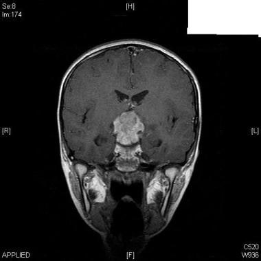
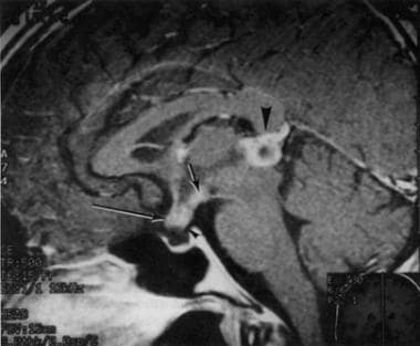
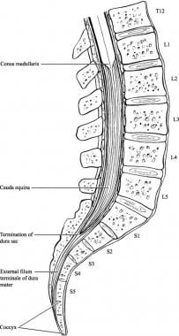
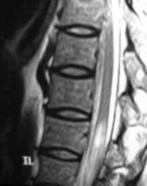
Teratomas can also occur in other locations such as the sacrum, coccyx (tailbone), mediastinum (the area between the lungs), and pineal gland (a small gland in the brain). These types of teratomas can be more complex to treat due to their location and potential to cause damage to nearby structures.
An encyclopedia is a comprehensive reference work containing articles on various topics, usually arranged in alphabetical order. In the context of medicine, a medical encyclopedia is a collection of articles that provide information about a wide range of medical topics, including diseases and conditions, treatments, tests, procedures, and anatomy and physiology. Medical encyclopedias may be published in print or electronic formats and are often used as a starting point for researching medical topics. They can provide reliable and accurate information on medical subjects, making them useful resources for healthcare professionals, students, and patients alike. Some well-known examples of medical encyclopedias include the Merck Manual and the Stedman's Medical Dictionary.
 Endodermal sinus tumor - Wikipedia
 Yolk Sac Tumor (Endodermal Sinus Tumors, Primitive Endodermal Tumors) Pathology: Definition, Epidemiology, Etiology
 Mediastinal endodermal sinus tumors - About the Disease - Genetic and Rare Diseases Information Center
 Ovarian cancer in young females: Causes and treatment
 Understanding the Different Kinds of Ovarian Cancer • The Fashionable Housewife
 Germ Cell Tumors (for Parents) - Children's Health System - Alabama (iFrame)
 A. Lindsay Frazier, MD - Dana-Farber Cancer Institute | Boston, MA
 Serous fluid: Metastatic sarcomas, melanoma, and other non-epithelial neoplasms - CytoJournal
 Krukenberg tumor | Radiology Reference Article | Radiopaedia.org
 14 Symptoms Of Ovarian Cancer In Teens And Survival Rate
 "Insulin-like growth factor-1 receptor expression in pediatric tumors: " by RESUL KARAKUŞ, ESRA KARAKUŞ et al.
![Ovarian Germ Cell Tumors Treatment (PDQ®): Treatment - Health Professional Information [NCI] | Kaiser Permanente](data:image/png;base64,iVBORw0KGgoAAAANSUhEUgAAABAAAAAQCAYAAAAf8/9hAAABuElEQVQ4jXXTTWyMURQG4Gc+n5/4GRNK/EQ0RJSkIpE0kWDDTqwQ3YidjVljx6IRKwuRsJLYjQhRKxvJNF36W4hYSFPtaOuvFKXtlGFxz0RNuMmXe7+bc97zvu85t+Afq1SuwBIcxjr04gVMXOn+KzaPYFiABn7E/xFcwyLsxTFMthbLY9+D0/iIcxjC1kiGLShislSuFFCIYrL4TuIQTuBgJFXxCl9xH++wHOdxA11NBg0Mxz6NDwEwgDvBpBe/sA9nQ26O400Jt7AMM1G5Cz1YiXbMw048xGesir2RYxeuhq4ZXAq6HXgW2uejG7dxAdtwvQlwKlh04B7240GAvQmAWXzCxaDfiU04k+EoxvAcbXgaxg7hfRj5XWphZ3jVFneXM2lAdqNPatvjCHoShr5EDRPox0bcxHq050GvGiB3sSJYjKEeYEVsCPr9KOELBnNpVPPQXQynx4P+tzkAw1F1cfg1igN5SHgblfuk/g9iKpKFmSNxvwNrsQbVXJquejCoBcAUfx5OqVxpSBM5jaVYjc3YnklDMhp0a63JLedZvJZaOoLxDI+kF7gQP/1nzQGpR1wBA78B8tOFmyZbu5oAAAAASUVORK5CYII=) Ovarian Germ Cell Tumors Treatment (PDQ®): Treatment - Health Professional Information [NCI] | Kaiser Permanente
Ovarian Germ Cell Tumors Treatment (PDQ®): Treatment - Health Professional Information [NCI] | Kaiser Permanente WikiGenes - Decompression, Surgical
 Nepalese Journal of Cancer
 Integrated Analysis of Serum Lymphocytes Develops a Nomogram for Predicting Prognosis of Intracranial Germ Cell Tumors to Chemo...
 Rare Cancer Support Forum • Index page
 Daniel von Allmen, MD
 Vaginal Cancer Symptoms, Doctors, Treatments, Advances & More | MediFind