Endolymph
Labyrinthine Fluids
Perilymph
Stria Vascularis
Otolithic Membrane
Ear, Inner
Saccule and Utricle
Semicircular Canals
Endolymphatic Sac
Cochlea
Endolymphatic Duct
Endolymphatic Hydrops
Cochlear Microphonic Potentials
Meniere Disease
Batrachoidiformes
Cochlear Duct
Hair Cells, Auditory
Scala Vestibuli
Nystagmus, Physiologic
Oncorhynchus mykiss
Labyrinth Diseases
Vestibule, Labyrinth
Organ of Corti
Ethacrynic Acid
Air Sacs
Rana esculenta
Vertigo
Chemical composition of saccular endolymph and otolith in fish inner ear: lack of spatial uniformity. (1/92)
Fish otoliths provide a record of age, growth, and environmental influences. In both trout and turbot, spatial chemical investigation of the endolymph surrounding the otolith (sagitta) showed a lack of uniformity. Proteins, PO(3-)(4), and Mg(2+) were significantly more concentrated in the proximal (facing the macula) than distal zone, whereas the opposite was observed for K(+) and total CO(2) (totCO(2)). Na(+) concentration ([Na(+)]) was 20% higher in the proximal zone in trout but not in turbot. Total Ca and Cl(-) contents were uniformly distributed in both species. We propose that the endolymphatic gradients of protein and totCO(2) concentration within the endolymph are involved in the otolithic biocalcification process. Microchemical analyses of otolith sections by wavelength dispersive spectrometry showed a lack of spatial uniformity in the K/Ca and Na/Ca ratios, whereas the Sr/Ca ratio was uniform. There is a clear relationship between endolymph and otolith [K(+)], but the interpretation of the results for [Na(+)] needs further investigation. Thus the lack of uniformity in the otolith composition must be taken into account when investigating otolith microchemistry. (+info)Altered cochlear fibrocytes in a mouse model of DFN3 nonsyndromic deafness. (2/92)
DFN3, an X chromosome-linked nonsyndromic mixed deafness, is caused by mutations in the BRN-4 gene, which encodes a POU transcription factor. Brn-4-deficient mice were created and found to exhibit profound deafness. No gross morphological changes were observed in the conductive ossicles or cochlea, although there was a dramatic reduction in endocochlear potential. Electron microscopy revealed severe ultrastructural alterations in cochlear spiral ligament fibrocytes. The findings suggest that these fibrocytes, which are mesenchymal in origin and for which a role in potassium ion homeostasis has been postulated, may play a critical role in auditory function. (+info)The effects of tone exposure on the inner ear functions in the guinea pig: impact tone vs. steady state tone. (3/92)
The damage-risk criterion (DRC) for hearing supposes that sound exposure with equal energy implies equal risk for noise-induced hearing loss (NIHL). We measured cochlear microphonics (CM), compound action potential (CAP), endocochlear potential (EP) and K+ ion concentration in the scala media, to see if the same level of Leq24h (impact tone and steady state tone) induced the same physiological changes in the inner ear function or not. Regarding the equal energy principle (EEP), we also examined if the EEP is appropriate or not at exposure of moderate level tone. We also checked how the time interval between impact tones affects or not the inner ear functions at the same Leq24h tone exposure. Therefore we used exposure at 1 pulse/second or 1 pulse/3 seconds and steady state tone exposure at Leq24h=90, 85 and 80 dB. The results are the following. Both steady state and impact tone exposure causes change of the electrophysiological data. First, CM maximum output voltage after exposure to impact tone of 115 dB (Leq24h=90 dB) was lower than after exposure to a 8 kHz steady state tone of 90 dB. CAP threshold (below 10 microV) obtained after the 115 and 110 dB exposure of impact tone were 5-10 dB higher than that of steady state tone of 90 dB. The negative EP induced by impact tone exposures showed the same tendency as the CM experiments. Having more frequent pulses (1 pulse/second vs. to 1 pulse/3 seconds) showed more inhibition. The K+ concentration time course remained similar to the control when the Leq24h was low (80 dB). Impact tone exposure induced stronger effects to the inner ear at exposure of moderate level tone than that of steady state tone of Leq24h. (+info)Comparative study of effects of impact tone and steady state tone exposure: EP and concentration of K+ ion and Na+ ion. (4/92)
To test the adequacy of equal energy principle (EEP), guinea pigs were exposed to impact tone. The changes in electrophysiological data, namely endocochlear potential (EP) and the change in K+ ion and Na+ ion concentrations in the endolymph were investigated. The frequency of impact tone was 1 pulse/second or 1 pulse/3 seconds. The steady state tone had Leq24h = 100, 95, 90 or 85 dB, and impact tone had Leq24h = 95, 90 or 85 dB. The results are the following. Both steady state and impact tone exposure cause changes of electrophysiological data. The effects on the absolute value of negative EP induced by impact tone exposures were smaller than that of steady state tone of the same Leq. The rate of pulses was also an important factor for impact tone exposure. Impact tone exposure of 1 pulse/second caused smaller absolute value of negative EP than that of 1 pulse/3 seconds. The K+ ion concentration time course in the endolymph remained similar to the control (Exp. 1) only in Exp. 8 (85 dB; the lowest steady state noise exposure in our experiments), but no decrease in the K+ ion concentration was detected in the other experiments, suggesting an alteration in the K+ ion flow. The Na+ ion concentration time course was also influenced showing no increase in Na+ ion concentration compared to the control (Exp. 1c) and the lowest steady-state exposure experiment (Exp. 8c). Our experimental results suggest that both the K+ ion and Na+ ion movement are altered by tone exposure. We found also that the different types of noise exposure with the same Leq value does not exhibit the same changes. Leq24h is not an accurate damage risk criteria. (+info)Effects of cholera toxin on cochlear endolymph production: model for endolymphatic hydrops. (5/92)
To evaluate a possible role for adenylate cyclase [ATP pyrophosphate-lyase (cyclizing), EC 4.6.1.1] and adenosine 3':5'-cyclic monophosphate in the secretion of endolymph, we studied the effect of an intra-scala media injection of purified cholera toxin (an adenylate cyclase stimulant) on cochlear endolymph volume, endolymphatic potential, and endolymphatic Na and K concentrations. (+info)EphB2 guides axons at the midline and is necessary for normal vestibular function. (6/92)
Mice lacking the EphB2 receptor tyrosine kinase display a cell-autonomous, strain-specific circling behavior that is associated with vestibular phenotypes. In mutant embryos, the contralateral inner ear efferent growth cones exhibit inappropriate pathway selection at the midline, while in mutant adults, the endolymph-filled lumen of the semicircular canals is severely reduced. EphB2 is expressed in the endolymph-producing dark cells in the inner ear epithelium, and these cells show ultrastructural defects in the mutants. A molecular link to fluid regulation is provided by demonstrating that PDZ domain-containing proteins that bind the C termini of EphB2 and B-ephrins can also recognize the cytoplasmic tails of anion exchangers and aquaporins. This suggests EphB2 may regulate ionic homeostasis and endolymph fluid production through macromolecular associations with membrane channels that transport chloride, bicarbonate, and water. (+info)Mechanism generating endocochlear potential: role played by intermediate cells in stria vascularis. (7/92)
The endocochlear DC potential (EP) is generated by the stria vascularis, and essential for the normal function of hair cells. Intermediate cells are melanocytes in the stria vascularis. To examine the contribution of the membrane potential of intermediate cells (E(m)) to the EP, a comparison was made between the effects of K(+) channel blockers on the E(m) and those on the EP. The E(m) of dissociated guinea pig intermediate cells was measured in the zero-current clamp mode of the whole-cell patch clamp configuration. The E(m) changed by 55.1 mV per 10-fold changes in extracellular K(+) concentration. Ba(2+), Cs(+), and quinine depressed the E(m) in a dose-dependent manner, whereas tetraethylammonium at 30 mM and 4-aminopyridine at 10 mM had no effect. The reduction of the E(m) by Ba(2+) and Cs(+) was enhanced by lowering the extracellular K(+) concentration from 3.6 mM to 1.2 mM. To examine the effect of the K(+) channel blockers on the EP, the EP of guinea pigs was maintained by vascular perfusion, and K(+) channel blockers were administered to the artificial blood. Ba(2+), Cs(+) and quinine depressed the EP in a dose-dependent manner, whereas tetraethylammonium at 30 mM and 4-aminopyridine at 10 mM did not change the EP. A 10-fold increase in the K(+) concentration in the artificial blood caused a minor decrease in the EP of only 10.6 mV. The changes in the EP were similar to those seen in the E(m) obtained at the lower extracellular K(+) concentration of 1.2 mM. On the basis of these results, we propose that the EP is critically dependent on the voltage jump across the plasma membrane of intermediate cells, and that K(+) concentration in the intercellular space in the stria vascularis may be actively controlled at a concentration lower than the plasma level. (+info)Location and function of the epithelial Na channel in the cochlea. (8/92)
In the cochlea, endolymph is a K-rich and Na-poor fluid. The purpose of the present study was to check the presence and to assess the role of epithelial Na channel (ENaC) in this organ. alpha-, beta-, and gamma-ENaC subunit mRNA, and proteins were detected in rat cochlea by RT-PCR and Western blot. alpha-ENaC subunit mRNA was localized by in situ hybridization in both epithelial (stria vascularis, spiral prominence, spiral limbus) and nonepithelial structures (spiral ligament, spiral ganglion). The alpha-ENaC-positive tissues were also positive for beta-subunit mRNA (except spiral ganglion) or for gamma-subunit mRNA (spiral limbus, spiral ligament, and spiral ganglion), but the signals of beta- and gamma-subunits were weaker than those observed for alpha-subunit. In vivo, the endocochlear potential was recorded in guinea pigs under normoxic and hypoxic conditions after endolymphatic perfusion of ENaC inhibitors (amiloride, benzamil) dissolved either in K-rich or Na-rich solutions. ENaC inhibitors altered the endocochlear potential when Na-rich but not when K-rich solutions were perfused. In conclusion, ENaC subunits are expressed in epithelial and nonepithelial cochlear structures. One of its functions is probably to maintain the low concentration of Na in endolymph. (+info)Endolymph is a specific type of fluid that is found within the inner ear, more specifically in the membranous labyrinth of the inner ear. This fluid plays a crucial role in maintaining balance and hearing functions. It helps in the stimulation of hair cells present in the inner ear which then transmit signals to the brain, enabling us to hear and maintain our balance. Any disturbance or changes in the composition or flow of endolymph can lead to various vestibular disorders and hearing problems.
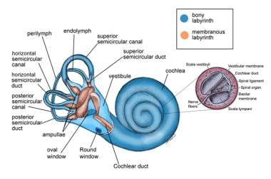
Labyrinthine fluids, also known as endolymph and perilymph, are fluids that fill the inner ear structures, specifically the bony labyrinth. The bony labyrinth is divided into two main parts: the cochlea, responsible for hearing, and the vestibular system, responsible for balance.
Endolymph is a clear, plasma-like fluid found within the membranous labyrinth, which is a series of interconnected tubes and sacs that lie inside the bony labyrinth. Endolymph plays a crucial role in the functioning of both the cochlea and vestibular system by creating an electrochemical gradient necessary for the conversion of mechanical sound vibrations into electrical signals in the cochlea, as well as facilitating the detection of head movements and maintaining balance in the vestibular system.
Perilymph, on the other hand, is a clear, colorless fluid that fills the space between the bony labyrinth and the membranous labyrinth. It is similar in composition to cerebrospinal fluid (CSF) and serves as a protective cushion for the delicate inner ear structures. Perilymph also helps maintain the electrochemical gradient required for sound transduction in the cochlea.
Disorders related to these labyrinthine fluids, such as endolymphatic hydrops or perilymph fistula, can lead to hearing and balance problems.
Perilymph is a type of fluid found in the inner ear, more specifically within the bony labyrinth of the inner ear. It fills the space between the membranous labyrinth and the bony labyrinth in the cochlea and vestibular system. Perilymph is similar in composition to cerebrospinal fluid (CSF) and contains sodium, chloride, and protein ions. Its main function is to protect the inner ear from damage, maintain hydrostatic pressure, and facilitate the transmission of sound waves to the hair cells in the cochlea for hearing.
Stria vascularis is a highly vascularized (rich in blood vessels) structure located in the cochlea of the inner ear. It plays a crucial role in the process of hearing by maintaining the endocochlear potential, which is essential for the conversion of sound waves into electrical signals that can be interpreted by the brain. The stria vascularis is composed of three layers: the marginal cells, intermediate cells, and basal cells, which work together to maintain the ionic balance and generate the endocochlear potential. Damage to the stria vascularis can result in hearing loss.
The otolithic membrane is a part of the inner ear's vestibular system, which contributes to our sense of balance and spatial orientation. It is composed of a gelatinous material containing tiny calcium carbonate crystals called otoconia or otoliths. These crystals provide weight to the membrane, allowing it to detect linear acceleration and gravity-induced head movements.
There are two otolithic membranes in each inner ear, located within the utricle and saccule, two of the three main vestibular organs. The utricle is primarily responsible for detecting horizontal movement and head tilts, while the saccule senses vertical motion and linear acceleration.
Damage to the otolithic membrane can result in balance disorders, vertigo, or dizziness.
The inner ear is the innermost part of the ear that contains the sensory organs for hearing and balance. It consists of a complex system of fluid-filled tubes and sacs called the vestibular system, which is responsible for maintaining balance and spatial orientation, and the cochlea, a spiral-shaped organ that converts sound vibrations into electrical signals that are sent to the brain.
The inner ear is located deep within the temporal bone of the skull and is protected by a bony labyrinth. The vestibular system includes the semicircular canals, which detect rotational movements of the head, and the otolith organs (the saccule and utricle), which detect linear acceleration and gravity.
Damage to the inner ear can result in hearing loss, tinnitus (ringing in the ears), vertigo (a spinning sensation), and balance problems.
The saccule and utricle are components of the vestibular system, which is responsible for maintaining balance and spatial orientation within the inner ear. Here are the medical definitions:
1. Saccule: A small sac-like structure located in the vestibular labyrinth of the inner ear. It is one of the two otolith organs (the other being the utricle) that detect linear acceleration and gravity. The saccule contains hair cells with stereocilia, which are embedded in a gelatinous matrix containing calcium carbonate crystals called otoconia. When the head changes position or moves linearly, the movement of these otoconia stimulates the hair cells, sending signals to the brain about the direction and speed of the motion.
2. Utricle: Another sac-like structure in the vestibular labyrinth, similar to the saccule but slightly larger. The utricle is also an otolith organ that detects linear acceleration and head tilts. It contains hair cells with stereocilia embedded in a gelatinous matrix filled with otoconia. When the head tilts or moves linearly, the movement of the otoconia stimulates the hair cells, providing information about the position and motion of the head to the brain.
In summary, both the saccule and utricle are essential for maintaining balance and spatial orientation by detecting linear acceleration and gravity through the movement of otoconia on their hair cell receptors.
The semicircular canals are part of the vestibular system in the inner ear that contributes to the sense of balance and spatial orientation. They are composed of three fluid-filled tubes, each located in a different plane (anterior, posterior, and horizontal) and arranged at approximately right angles to each other. The semicircular canals detect rotational movements of the head, enabling us to maintain our equilibrium during movement.
When the head moves, the fluid within the semicircular canals moves in response to that motion. At the end of each canal is a structure called the ampulla, which contains hair cells with hair-like projections (stereocilia) embedded in a gelatinous substance. As the fluid moves, it bends the stereocilia, stimulating the hair cells and sending signals to the brain via the vestibular nerve. The brain then interprets these signals to determine the direction and speed of head movement, allowing us to maintain our balance and orientation in space.
The endolymphatic sac is a small, fluid-filled structure that is part of the inner ear. It is located near the vestibular aqueduct and is responsible for maintaining the balance of fluids in the inner ear. The endolymphatic sac also plays a role in the resorption of endolymph, which is the fluid that fills the membranous labyrinth of the inner ear. Disorders of the endolymphatic sac can lead to conditions such as Meniere's disease, which is characterized by vertigo, hearing loss, and tinnitus.
The cochlea is a part of the inner ear that is responsible for hearing. It is a spiral-shaped structure that looks like a snail shell and is filled with fluid. The cochlea contains hair cells, which are specialized sensory cells that convert sound vibrations into electrical signals that are sent to the brain.
The cochlea has three main parts: the vestibular canal, the tympanic canal, and the cochlear duct. Sound waves enter the inner ear and cause the fluid in the cochlea to move, which in turn causes the hair cells to bend. This bending motion stimulates the hair cells to generate electrical signals that are sent to the brain via the auditory nerve.
The brain then interprets these signals as sound, allowing us to hear and understand speech, music, and other sounds in our environment. Damage to the hair cells or other structures in the cochlea can lead to hearing loss or deafness.
The endolymphatic duct is a narrow canal in the inner ear that is part of the membranous labyrinth. It connects the utricle and saccule (two sensory structures in the vestibular system responsible for detecting changes in head position and movement) to the endolymphatic sac (a dilated portion of the duct that helps regulate the volume and pressure of endolymph, a fluid found within the membranous labyrinth).
The endolymphatic duct plays a crucial role in maintaining the balance and homeostasis of the inner ear by allowing the absorption and circulation of endolymph. Disorders or abnormalities in this region can lead to various vestibular and hearing dysfunctions, such as Meniere's disease, endolymphatic hydrops, and other inner ear disorders.
Endolymphatic hydrops is a term used to describe the abnormal accumulation of fluid (endolymph) within the inner ear. This condition is most commonly associated with Meniere's disease, but can also be seen in other disorders that affect the inner ear.
The inner ear is made up of two main parts: the cochlea, which is responsible for hearing, and the vestibular system, which helps to control balance. Both of these systems are filled with fluid, including endolymph, which is a watery fluid that bathes the sensory hair cells in these structures.
In endolymphatic hydrops, there is an overproduction or decreased absorption of endolymph, leading to an abnormal buildup of fluid within the inner ear. This can cause a variety of symptoms, including vertigo (a spinning sensation), tinnitus (ringing in the ears), hearing loss, and a feeling of fullness or pressure in the affected ear.
The exact cause of endolymphatic hydrops is not fully understood, but it is thought to be related to changes in the inner ear's fluid balance. Treatment options may include medications to help control symptoms, as well as surgical procedures to relieve pressure on the inner ear.
Cochlear microphonic potentials (CMs) are electrical responses that originate from the hair cells in the cochlea, which is a part of the inner ear responsible for hearing. These potentials can be recorded using an electrode placed near the cochlea in response to sound stimulation.
The CMs are considered to be a passive response of the hair cells to the mechanical deflection caused by sound waves. They represent the receptor potential of the outer hair cells and are directly proportional to the sound pressure level. Unlike other electrical responses in the cochlea, such as the action potentials generated by the auditory nerve fibers, CMs do not require the presence of neurotransmitters or synaptic transmission.
Cochlear microphonic potentials have been used in research to study the biophysical properties of hair cells and their response to different types of sound stimuli. However, they are not typically used in clinical audiology due to their small amplitude and susceptibility to interference from other electrical signals in the body.
Menière disease is an inner ear disorder that is characterized by episodes of vertigo (a spinning sensation), tinnitus (ringing or buzzing in the ear), hearing loss, and aural fullness (a feeling of pressure or blockage in the ear). It is caused by an abnormal accumulation of endolymphatic fluid in the inner ear, which can lead to damage of the vestibular system and cochlea. The exact cause of this fluid buildup is not known, but it may be related to genetics, allergies, or autoimmune disorders. Menière disease is typically a chronic condition, with symptoms that can vary in frequency and severity over time. Treatment options include dietary modifications, diuretics, vestibular rehabilitation therapy, and, in some cases, surgery.
Batrachoidiformes is an order of primarily marine ray-finned fish that includes the genera Batrachoides, Halophryne, Porichthys, and Thalassophryne. These fish are characterized by having a stout body, large head, and strong, bony mouthparts. They are often called "toadfish" due to their warty skin and toad-like appearance. Some species have the ability to produce sounds, which they use for communication and mating. They are found in tropical and subtropical waters of the Atlantic and Pacific Oceans, as well as in the Mediterranean Sea.
Deafness is a hearing loss that is so severe that it results in significant difficulty in understanding or comprehending speech, even when using hearing aids. It can be congenital (present at birth) or acquired later in life due to various causes such as disease, injury, infection, exposure to loud noises, or aging. Deafness can range from mild to profound and may affect one ear (unilateral) or both ears (bilateral). In some cases, deafness may be accompanied by tinnitus, which is the perception of ringing or other sounds in the ears.
Deaf individuals often use American Sign Language (ASL) or other forms of sign language to communicate. Some people with less severe hearing loss may benefit from hearing aids, cochlear implants, or other assistive listening devices. Deafness can have significant social, educational, and vocational implications, and early intervention and appropriate support services are critical for optimal development and outcomes.
The cochlear duct, also known as the scala media, is a membranous duct located within the cochlea of the inner ear. It is one of three fluid-filled compartments in the cochlea, along with the vestibular duct (scala vestibuli) and the tympanic duct (scala tympani).
The cochlear duct contains endolymph, a specialized fluid that carries electrical signals to the auditory nerve. The organ of Corti, which is responsible for converting sound vibrations into electrical signals, is located within the cochlear duct.
The cochlear duct runs along the length of the cochlea and is separated from the vestibular duct by Reissner's membrane and from the tympanic duct by the basilar membrane. These membranes help to create a highly sensitive and selective environment for sound perception, allowing us to hear and distinguish different frequencies and intensities of sound.
Auditory hair cells are specialized sensory receptor cells located in the inner ear, more specifically in the organ of Corti within the cochlea. They play a crucial role in hearing by converting sound vibrations into electrical signals that can be interpreted by the brain.
These hair cells have hair-like projections called stereocilia on their apical surface, which are embedded in a gelatinous matrix. When sound waves reach the inner ear, they cause the fluid within the cochlea to move, which in turn causes the stereocilia to bend. This bending motion opens ion channels at the tips of the stereocilia, allowing positively charged ions (such as potassium) to flow into the hair cells and trigger a receptor potential.
The receptor potential then leads to the release of neurotransmitters at the base of the hair cells, which activate afferent nerve fibers that synapse with these cells. The electrical signals generated by this process are transmitted to the brain via the auditory nerve, where they are interpreted as sound.
There are two types of auditory hair cells: inner hair cells and outer hair cells. Inner hair cells are the primary sensory receptors responsible for transmitting information about sound to the brain. They make direct contact with afferent nerve fibers and are more sensitive to mechanical stimulation than outer hair cells.
Outer hair cells, on the other hand, are involved in amplifying and fine-tuning the mechanical response of the inner ear to sound. They have a unique ability to contract and relax in response to electrical signals, which allows them to adjust the stiffness of their stereocilia and enhance the sensitivity of the cochlea to different frequencies.
Damage or loss of auditory hair cells can lead to hearing impairment or deafness, as these cells cannot regenerate spontaneously in mammals. Therefore, understanding the structure and function of hair cells is essential for developing therapies aimed at treating hearing disorders.
Scala Vestibuli is a term used in anatomy, particularly in the field of otology (the study of the ear and its diseases). It refers to one of the three bony canals that make up the inner ear's complex system of fluid-filled channels known as the vestibular system.
More specifically, Scala Vestibuli is the uppermost of the three scalae (singular: scala) in the cochlea, a snail-shaped structure in the inner ear responsible for hearing. The other two scalae are Scala Tympani and Scala Media.
Scala Vestibuli and Scala Tympani are connected at the apex of the cochlea through an opening called the helicotrema. The Scala Vestibuli is filled with perilymph, a fluid that helps transmit sound waves to the inner ear.
Please note that while I strive to provide accurate and detailed information, it's always best to consult with a healthcare professional or medical textbook for definitive medical definitions and explanations.
Physiologic nystagmus is a type of normal, involuntary eye movement that occurs in certain situations. It is characterized by rhythmical to-and-fro movements of the eyes, which can be horizontal, vertical, or rotatory. The most common form of physiologic nystagmus is called "optokinetic nystagmus," which occurs when a person looks at a moving pattern, such as stripes on a rotating drum or scenery passing by a car window.
Optokinetic nystagmus helps to stabilize the image of the environment on the retina and allows the brain to perceive motion accurately. Another form of physiologic nystagmus is "pursuit nystagmus," which occurs when the eyes attempt to follow a slowly moving target. In this case, the eyes may overshoot the target and then make a corrective movement in the opposite direction.
Physiologic nystagmus is different from pathological nystagmus, which can be caused by various medical conditions such as brain damage, inner ear disorders, or medications that affect the nervous system. Pathological nystagmus may indicate a serious underlying condition and should be evaluated by a healthcare professional.
Oncorhynchus mykiss is the scientific name for a species of fish that is commonly known as the Rainbow Trout. According to the medical or clinical definition provided by the US National Library of Medicine, Oncorhynchus mykiss is "a freshwater fish that is widely cultured and an important food source in many parts of the world." It is also a popular game fish and is often stocked in lakes and rivers for recreational fishing. Rainbow trout are native to cold-water tributaries that flow into the Pacific Ocean in Asia and North America. They have been introduced widely throughout the world and can now be found in freshwater systems on every continent except Antarctica. Rainbow trout are a valuable species for both commercial and recreational fisheries, and they also play an important role in the food web as both predators and prey.
Labyrinth diseases refer to conditions that affect the inner ear's labyrinth, which is the complex system of fluid-filled channels and sacs responsible for maintaining balance and hearing. These diseases can cause symptoms such as vertigo (a spinning sensation), dizziness, nausea, hearing loss, and tinnitus (ringing in the ears). Examples of labyrinth diseases include Meniere's disease, labyrinthitis, vestibular neuronitis, and benign paroxysmal positional vertigo. Treatment for these conditions varies depending on the specific diagnosis but may include medications, physical therapy, or surgery.
The vestibular system is a part of the inner ear that contributes to our sense of balance and spatial orientation. It is made up of two main components: the vestibule and the labyrinth.
The vestibule is a bony chamber in the inner ear that contains two important structures called the utricle and saccule. These structures contain hair cells and fluid-filled sacs that help detect changes in head position and movement, allowing us to maintain our balance and orientation in space.
The labyrinth, on the other hand, is a more complex structure that includes the vestibule as well as three semicircular canals. These canals are also filled with fluid and contain hair cells that detect rotational movements of the head. Together, the vestibule and labyrinth work together to provide us with information about our body's position and movement in space.
Overall, the vestibular system plays a crucial role in maintaining our balance, coordinating our movements, and helping us navigate through our environment.
The Organ of Corti is the sensory organ of hearing within the cochlea of the inner ear. It is a structure in the inner spiral sulcus of the cochlear duct and is responsible for converting sound vibrations into electrical signals that are sent to the brain via the auditory nerve.
The Organ of Corti consists of hair cells, which are sensory receptors with hair-like projections called stereocilia on their apical surfaces. These stereocilia are embedded in a gelatinous matrix and are arranged in rows of different heights. When sound vibrations cause the fluid in the cochlea to move, the stereocilia bend, which opens ion channels and triggers nerve impulses that are sent to the brain.
Damage or loss of hair cells in the Organ of Corti can result in hearing loss, making it a critical structure for maintaining normal auditory function.
Ethacrynic acid is a loop diuretic drug that is primarily used to treat edema (swelling) associated with heart failure, liver cirrhosis, and kidney disease. It works by increasing the excretion of water and sodium in the urine, which helps reduce fluid buildup in the body. Ethacrynic acid is also known as a "high-ceiling" diuretic because it has a strong effect on urine production.
The drug is available in oral form and is typically taken once or twice a day, depending on the severity of the edema and the patient's response to treatment. Ethacrynic acid can have side effects, including hearing loss, kidney damage, and electrolyte imbalances, so it is important for patients to be monitored closely by their healthcare provider while taking this medication.
It is worth noting that ethacrynic acid is not as commonly used as other loop diuretics, such as furosemide or torsemide, due to its higher risk of side effects and the availability of safer alternatives.
Air sacs, also known as alveoli, are tiny air-filled sacs in the lungs where the exchange of oxygen and carbon dioxide occurs during respiration. They are a part of the respiratory system in mammals and birds. In humans, the lungs contain about 300 million alveoli, which are clustered together in small groups called alveolar sacs. The walls of the air sacs are extremely thin, allowing for the easy diffusion of oxygen and carbon dioxide between the air in the sacs and the blood in the capillaries that surround them.
"Rana esculenta" is not a medical term. It is the scientific name for a species of frog, also known as the edible frog or the common water frog. This species is native to Europe and has been introduced to other parts of the world. They are often farmed for their meat, which is considered a delicacy in some cultures.
If you have any confusion with a medical term or a topic, please provide it so I can give you an accurate information.
Vertigo is a specific type of dizziness characterized by the sensation that you or your surroundings are spinning or moving, even when you're perfectly still. It's often caused by issues with the inner ear or the balance-sensing systems of the body. Vertigo can be brought on by various conditions, such as benign paroxysmal positional vertigo (BPPV), labyrinthitis, vestibular neuritis, Meniere's disease, and migraines. In some cases, vertigo may also result from head or neck injuries, brain disorders like stroke or tumors, or certain medications. Treatment for vertigo depends on the underlying cause and can include specific exercises, medication, or surgery in severe cases.
Labyrinthitis is a medical condition characterized by inflammation of the labyrinth, which is the inner ear's balance- and hearing-sensitive system. It is often caused by an infection, such as a viral or bacterial infection, that spreads to the inner ear. The inflammation can affect the delicate structures of the labyrinth, leading to symptoms such as vertigo (a spinning sensation), dizziness, imbalance, hearing loss, and tinnitus (ringing in the ears). Labyrinthitis can be a serious condition that requires medical attention and treatment.
 Endolymph
Ear
Basilar membrane
Membranous labyrinth
Sense of balance
Ampullary cupula
Rabbit
Antonio Scarpa
Stereocilia (inner ear)
Perilymph
Stria vascularis of cochlear duct
Cochlea
Cochlear hydrops
Short-term effects of alcohol consumption
Claudius cell
Auditory system
Ototoxic medication
Inner ear
Oral skills
Semicircular canals
Etacrynic acid
Noise-induced hearing loss
Dark cell
Chlortalidone
Vestibular membrane
Endolymphatic hydrops
Causes of hearing loss
Reticular membrane
Otolith microchemical analysis
Benign paroxysmal positional vertigo
Endolymph - Wikipedia
 endolymph Archives - VetSci
Glossary of communication disorders - Wikipedia
 Vestibular System Anatomy: Overview, Membranous Labyrinth, Vestibular Sensory Epithelium
 MeSH Browser
Plus it
 PPT - Principles of Sensory Systems PowerPoint presentation | free to view - id: 1e36b8-OThiN
 Ménière's Disease | HealthLink BC
Plus it
 A Few Feet Above the Earth - Ridgeline issue 011
View source for Microbiota of the Human Ear - microbewiki
Plus it
 Figures and data in Size control of the inner ear via hydraulic feedback | eLife
 Frontiers | Discriminating Natal Source Populations of a Temperate Marine Fish Using Larval Otolith Chemistry
NIOSHTIC-2 Search Results - Full View
 Clinical aspects of hereditary hearing loss | Genetics in Medicine
Labyrinthitis: Background, Etiology, Epidemiology
 scala vestibuli
Plus it
 Jana Goyens - Google Scholar
 Ménière's Disease
 2022 in Review: The Year's Top Discoveries by Museum Researchers | Smithsonian Voices | National Museum of Natural History...
 Alan Shepard, his ear surgery and flight status - collectSPACE: Messages
 ATP6V1B1 gene: MedlinePlus Genetics
 Flashcards - physio 1
 Inner-Ear Balance by Tao Of Herbs, IEB9
 Post-COVID-19 Benign Paroxysmal Positional Vertigo
 Meniere's disease - Proxim
 Mutation:genetics<...
 head and neck anatomy.ppt
Perilymph8
- Perilymph and endolymph have unique ionic compositions suited to their functions in regulating electrochemical impulses of hair cells. (wikipedia.org)
- The electric potential of endolymph is ~80-90 mV more positive than perilymph due to a higher concentration of K compared to Na. (wikipedia.org)
- Endolymph has a high positive potential (80-120 mV in the cochlea), relative to other nearby fluids such as perilymph, due to its high concentration of positively charged ions. (wikipedia.org)
- The endolymph and perilymph differ based on the potassium and sodium concentration. (kenyon.edu)
- It was hypothesized that these gaps allow an intermixing of endolymph in the ES with perilymph in the fluid spaces of the OC (Bohne & Rabbitt, 1982). (cdc.gov)
- The inside of the hair cell has a negative intracellular potential of -60 millivolts with respect to the perilymph and -140 millivolts with respect to the endolymph. (britannica.com)
- The endocochlear potential is established through the development of tight cellular junctions between local networks of epithelial cells, connective tissue and supporting cells that completely partition the endolymph from the surrounding perilymph. (kyoto2.org)
- In the Traumatic Injuries DVD, Dr. Deshaw lectures on traumatic brain injuries, inner ear injuries (perilymph fistulas, endolymph hydrops, BPPV), C1 ligament injuries, and Alar and Transverse ligament damages, and how what seems like a simple neck injury can lead to severe symptoms such as blindness or death. (trialguides.com)
Potassium5
- The major cation in endolymph is potassium, with the values of sodium and potassium concentration in the endolymph being 0.91 mM and 154 mM, respectively. (wikipedia.org)
- The high potassium content of the endolymph means that potassium, not sodium, is carried as the de-polarizing electric current in the hair cells. (wikipedia.org)
- Endolymph, a potassium heavy fluid, oozes inside the so-called bony and membranous labyrinthine canals of the inner ear. (craigmod.com)
- The endolymph contains higher concentration of potassium ions than sodium ions [13]. (kenyon.edu)
- In the internal ear, pendrin transports bicarbonates to the endolymph, taking in this way an active part in pH control of the endolymph and providing proper functioning of KCNJ10 potassium channels and TRP5 calcium channels. (nel.edu)
Cochlea3
- When symptoms occur, the endolymph in one ear puts pressure on the cochlea, damaging hearing in that ear and adding pressure to the rest of the labyrinth, which causes the congested feeling. (healthdigest.com)
- Endolymph is the fluid present in the cochlea. (meritnation.com)
- The stria vascularis is a type of epithelium that is uniquely able to produce endolymph in the cochlea. (medscape.com)
Semicircular canals5
- Balance: Semicircular canals: angular acceleration of the endolymph in the semicircular canals stimulate the vestibular receptors of the endolymph. (wikipedia.org)
- The vestibule and semicircular canals sense the motion of the endolymph with specialized hair cells and assess the bodies position with respect to gravity. (kenyon.edu)
- The semicircular canals of miniaturized frogs are the smallest recorded for adult vertebrates, resulting in low sensitivity to angular acceleration due to insufficient displacement of endolymph. (boingboing.net)
- BPPV is believed to be caused by detached otoconia from the utricular maculae, which migrate into the semicircular canals (SCCs) and may either move freely in the endolymph (canalithiasis) or become attached to the cupula (cupulolithiasis). (frontiersin.org)
- Chow noted one limitation of the current technology - the implants, which target the three semicircular canals, tiny tubes in the inner ear that sense head rotations through the movement of endolymph within, do not fully account for the changes in vestibular sensitivity associated with linear acceleration and changes in gravity. (jhunewsletter.com)
Vestibular2
- Inside of the vestibular membrane is endolymph fluid that conducts sound to the basilar membrane. (kenyon.edu)
- Disorders of homeostasis in labyrinth fluids are responsible for abnormalities of its structure, such as enlargement of the vestibular aqueduct and of the endolymph sac. (nel.edu)
Cupula1
- It's inside there - those crescent-shaped canals - that gelatinous bulbs we've decided to call cupula, attached to stereocilia, detect the sloshing of our endolymph. (craigmod.com)
Membranous labyrinth4
- Endolymph is the fluid contained in the membranous labyrinth of the inner ear. (wikipedia.org)
- The membranous labyrinth is contained within the bony labyrinth, and within the membranous labyrinth is a fluid called endolymph. (wikipedia.org)
- Inside the membranous labyrinth is a fluid called endolymph. (healthdigest.com)
- The symptoms of Ménière's disease all trace back to the fluid in the membranous labyrinth, endolymph. (healthdigest.com)
Fluid called1
- But it may be related to a fluid called endolymph in the inner ear. (healthlinkbc.ca)
Meniere's2
- Meniere's disease is characterized by a surplus of endolymph, one of the fluids found in the inner ear. (groupeproxim.ca)
- Excessive endolymph is responsible for Meniere's syndrome. (medscape.com)
Endolymphatic1
- A condition where the volume of the endolymph is greatly enlarged is called endolymphatic hydrops and has been linked to Ménière's disease. (wikipedia.org)
Hearing1
- citation needed] Hearing: Cochlear duct: fluid waves in the endolymph of the cochlear duct stimulate the receptor cells, which in turn translate their movement into nerve impulses that the brain perceives as sound. (wikipedia.org)
Sense1
- AMPULLARY HAIR CELLS of the crests sense the movement of ENDOLYMPH resulting from rotation of the head. (bvsalud.org)
Hair2
- Because the hair cells are at a negative potential of about -50 mV, the potential difference from endolymph to hair cell is on the order of 150 mV, which is the largest electrical potential difference found in the body. (wikipedia.org)
- These results support the hypothesis that during or shortly after a damaging exposure, endolymph entered the OC & injured additional hair cells, supporting cells & nerve fibers. (cdc.gov)
Proper1
- Researchers aren't sure how and they're not entirely sure how much endolymph is a proper amount. (healthdigest.com)
Motion1
- Disruption of the endolymph due to jerky movements (like spinning around or driving over bumps while riding in a car) can cause motion sickness. (wikipedia.org)
Fluid called endolymph3
- The membranous labyrinth is contained within the bony labyrinth, and within the membranous labyrinth is a fluid called endolymph. (wikipedia.org)
- The membranous labyrinth is filled with a fluid called endolymph that, in the balance organs, stimulates receptors as the body moves. (nih.gov)
- This could be due to an overabundance of fluid called endolymph. (uppercervicalawareness.com)
Regulation of endolymph2
- Because Ménière's disease appears to run in families, it could also be the result of genetic variations that cause abnormalities in the volume or regulation of endolymph fluid. (nih.gov)
- 2 , 4 Ménière disease manifests as paroxysmal attacks of whirling vertigo due to failure of regulation of endolymph. (ajnr.org)
Fluids3
- Endolymph has a high positive potential (80-120 mV in the cochlea), relative to other nearby fluids such as perilymph, due to its high concentration of positively charged ions. (wikipedia.org)
- We were able to show that the rate of flow of these fluids is extremely slow, with endolymph flow shown to be less than 1 nanoliter per minute. (efluids.com)
- Inner Ear In temporal bone Labyrinth boney maze inside the bone Perilymph & Endolymph fluids inside the inner ear Inner Ear d. (powershow.com)
Perilymph and endolymph1
- Perilymph and endolymph have unique ionic compositions suited to their functions in regulating electrochemical impulses of hair cells. (wikipedia.org)
Cochlear2
- citation needed] Hearing: Cochlear duct: fluid waves in the endolymph of the cochlear duct stimulate the receptor cells, which in turn translate their movement into nerve impulses that the brain perceives as sound. (wikipedia.org)
- Aminoglycosides enter inner ear hair cells across their apical membranes via endocytosis, or through the mechanoelectrical transduction channels in vitro, suggesting that these drugs enter cochlear hair cells from endolymph to exert their cytotoxic effect. (elsevierpure.com)
Semicircular4
- Balance: Semicircular canals: angular acceleration of the endolymph in the semicircular canals stimulate the vestibular receptors of the endolymph. (wikipedia.org)
- Semicircular canals normally detect angular acceleration of the head by sensing motion of the endolymph. (neuroophthalmology.ca)
- Heavy debris within the canal generates inappropriate movement of the endolymph in response to linear accelerations, converting the canal into a gravity-sensing device - hence the erroneous sensation of spinning when there is a shift in the gravity vector with respect to the posterior semicircular canal. (neuroophthalmology.ca)
- They are perpendicular, semicircular channels that are filled with a fluid, the endolymph. (healthandmedicineinfo.com)
Homeostasis2
Labyrinth4
- Endolymph is the fluid contained in the membranous labyrinth of the inner ear. (wikipedia.org)
- Inside their walls (bony labyrinth) are thin, pliable tubes and sacs (membranous labyrinth) filled with endolymph. (nih.gov)
- In Ménière's disease, the endolymph buildup in the labyrinth interferes with the normal balance and hearing signals between the inner ear and the brain. (nih.gov)
- Endolymph' is the fluid that can be found in the labyrinth of the inner ear. (menieres-help.com)
Hydrops2
- This pathology strongly correlates with a dilatation of the fluidcompartment of the endolymph, so-called hydrops. (ens-lyon.fr)
- In the Traumatic Injuries DVD, Dr. Deshaw lectures on traumatic brain injuries, inner ear injuries (perilymph fistulas, endolymph hydrops, BPPV), C1 ligament injuries, and Alar and Transverse ligament damages, and how what seems like a simple neck injury can lead to severe symptoms such as blindness or death. (trialguides.com)
Disruption1
- Disruption of the endolymph due to jerky movements (like spinning around or driving over bumps while riding in a car) can cause motion sickness. (wikipedia.org)
Transduction1
- The capillaries of the stria vascularis provide this structure, an organ in its own right, so that it can maintain the high potassium concentration and positive potential of endolymph which are necessary for the transduction process. (nih.gov)
Sacs1
- Their ears are filled with ducts and sacs that are filled with endolymph - a clear fluid is mainly made of seawater that fills the ducts of a sharks' ear. (sharkdivingunlimited.com)
Concentration2
- The electric potential of endolymph is ~80-90 mV more positive than perilymph due to a higher concentration of K compared to Na. (wikipedia.org)
- Endolymph has high concentration of K+ and low concentration of Na+. (medicoapps.org)
Zebrafish1
- Endolymph collapse in the lte/nkcc1 mutant shows distinct parallels to that seen in mouse Nkcc1 mutants, validating zebrafish as a model for the study of endolymph disorders. (biologists.com)
Spaces2
- For years it was believed that endolymph and perilymph flowed along the fluid spaces. (efluids.com)
- It was hypothesized that these gaps allow an intermixing of endolymph in the ES with perilymph in the fluid spaces of the OC (Bohne & Rabbitt, 1982). (cdc.gov)
Exposure1
- These results support the hypothesis that during or shortly after a damaging exposure, endolymph entered the OC & injured additional hair cells, supporting cells & nerve fibers. (cdc.gov)
Flow1
- To promote normal arterial blood flow to the brain, enhance memory, increase excitability of the cerebral cortex, balance the endolymph and maintain healthy hearing, eye sight, and healthy functions of the neck. (taoofherbs.com)
Balance1
- Whenever a shark is thrown off balance, the endolymph fluid and otoliths slow so help the cells detect any lag, thus sending the correct signals needed for the shark to right itself. (sharkdivingunlimited.com)
Current1
- The duration of the stimulation of the hair cells of crista ampullaris compared with the duration of the endolymph current, and the resulting nystagmus. (nih.gov)
Hair cells2
- Because the hair cells are at a negative potential of about -50 mV, the potential difference from endolymph to hair cell is on the order of 150 mV, which is the largest electrical potential difference found in the body. (wikipedia.org)
- If so, this will facilitate electrophoretically driven aminoglycoside entry into hair cells from endolymph. (elsevierpure.com)
Cells1
- These data suggest that systemically administered aminoglycosides are trafficked from strial capillaries into marginal cells and clear into endolymph. (elsevierpure.com)