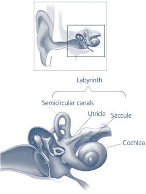
Endolymphatic Sac
Ear Neoplasms
Endolymphatic Duct
Endolymph
Vestibular Aqueduct
Meniere Disease
Endolymphatic Hydrops
von Hippel-Lindau Disease
Temporal Bone
Adenocarcinoma, Papillary
Ear, Inner
Yolk Sac
Vertigo
Anion Transport Proteins
Cerebellar Neoplasms
3D MRI of the membranous labyrinth. An age related comparison of MR findings in patients with labyrinthine fibrosis and in persons without inner ear symptoms. (1/52)
PURPOSE: We compared MRI of the membranous labyrinth in patients with chronic non-neoplastic inner ear disease and MR signs of labyrinthine fibrosis and controls depending on their age, in order to establish whether there were any MR differences regarding patient age groups, control age groups and between the patients and controls themselves. MATERIALS AND METHODS: Clinical ENT examinations as well as a T2* weighted 3D CISS (Constructive Interference in Steady State) sequence with a slice thickness of 0.7 mm were performed. Our collective was subdivided as follows: 0-19 years (10 controls, 3 patients with chronic non-neoplastic inner ear disease), 20-49 years (55 controls, 8 patients), 50 years and older (40 controls, 22 patients). Detectability of labyrinthine structures (e.g. cochlea, vestibule, semicircular canals) and filling defects were evaluated. RESULTS: In the 3 age-groups of the control collective no significant differences were observed in the membranous labyrinth. However differences concerning labyrinthine detectability emerged between controls and patients in both the 20-49 years and 50 years and older age groups. In the patient collective the 3 age groups showed no significant discrepancy in the mean number of lesions. CONCLUSION: Filling defects of the membranous labyrinth on 3D CISS MR images are pathological even in older persons. We would therefore recommend high resolution T2* weighted MRI in the case of suspected labyrinthine fibrosis. (+info)Extracellular ATP-induced inward current in isolated epithelial cells of the endolymphatic sac. (2/52)
Using whole-cell patch-clamp technique and Fura-2 fluorescence measurement, the presence of ATP-activated ion channels and its dependence on intracellular Ca2+ concentration ([Ca2+]i) in the epithelial cells of the endolymphatic sac were investigated. In zero current-clamp configuration, the average resting membrane potential was -66.8+/-1.3 mV (n=18). Application of 30 microM ATP to the bath induced a rapid membrane depolarization by 43.1+/-2.4 mV (n=18). In voltage-clamp configuration, ATP-induced inward current at holding potential (VH) of -60 mV was 169.7+/-6.3 pA (n=18). The amplitude of ATP-induced currents increased in sigmoidal fashion over the concentration range between 0.3 and 300 microM with a Hill coefficient (n) of 1.2 and a dissociation constant (Kd) of 11.7 microM. The potency order of purinergic analogues in ATP-induced current, which was 2MeSATP>ATPgammas>/=ATP>alpha, beta-ATP>ADP=AMP>/=adenosine=UTP, was consistent with the properties of the P2Y receptor. The independence of the reversal potential of the ATP-induced current from Cl- concentration suggests that the current is carried by a cation channel. The relative ionic permeability ratio of the channel modulated by ATP for cations was Ca2+>Na+>Li+>Ba2+>Cs+=K+. ATP (10 microM) increased [Ca2+]i in an external Ca2+-free solution to a lesser degree than that in the external solution containing 1.13 mM CaCl2. ATP-induced increase in [Ca2+]i can be mimicked by application of ionomycin in a Ca2+-free solution. These results indicate that ATP increases [Ca2+]i through the P2Y receptor with a subsequent activation of the non-selective cation channel, and that these effects of ATP are dependent on [Ca2+]i and extracellular Ca2+. (+info)MR evaluation of vestibulocochlear anomalies associated with large endolymphatic duct and sac. (3/52)
BACKGROUND AND PURPOSE: Large endolymphatic duct and sac (LEDS) is one of the most common anomalies seen in patients with congenital sensorineural hearing loss (SNHL), and is known to occur with other inner ear findings. Our purpose was to use high-resolution T2-weighted fast spin-echo (FSE) MR imaging to describe the features and prevalence of specific anomalies that occur in association with LEDS. METHODS: We retrospectively reviewed MR images of the inner ear obtained in 63 patients with LEDS and in 60 control subjects. We evaluated each image for features of cochlear and vestibular dysplasia, including deficiency of the cochlear modiolus, gross cochlear dysmorphism, asymmetry of the cochlear scalar chambers, enlargement of the membranous vestibule, gross vestibular dysmorphism, and abnormality of the semicircular canals (SCC). RESULTS: Cochlear anomalies were present in 76% of ears with LEDS. Modiolar deficiency, gross dysmorphism, and scalar asymmetry were seen in 94%, 71%, and 65% of abnormal cochleas, respectively. Vestibular abnormalities were present in 40% of ears with LEDS. Simple enlargement, gross dysmorphism, and distortion of the lateral SCC were seen in 84%, 16%, and 32% of abnormal vestibules, respectively. CONCLUSION: Coexistent cochlear anomalies, vestibular anomalies, or both are present in most ears with LEDS, and appear as a spectrum of lesions, ranging from subtle dymorphism to overt dysplasia. The presence of coexistent anomalies in LEDS affects treatment decisions and prognosis. Newer techniques of high-resolution FSE MR imaging provide a means of exquisite characterization of LEDS, as well as more sensitive detection of associated vestibulocochlear anomalies. (+info)MR imaging of the enlarged endolymphatic duct and sac syndrome by use of a 3D fast asymmetric spin-echo sequence: volume and signal-intensity measurement of the endolymphatic duct and sac and area measurement of the cochlear modiolus. (4/52)
BACKGROUND AND PURPOSE: In enlarged endolymphatic duct (EED) and sac (EES) syndrome, deformity of the EED and EES is congenital; however, hearing loss is acquired. To investigate the pathophysiology of progressive sensorineural hearing loss in EED and EES syndrome, we measured the volume of the EED and EES, the diameter of the EED and EES, the area of the cochlear modiolus, and the signal intensity of the EES and compared our findings against degree of hearing loss. METHODS: Thin-section MR images of 33 ears in 17 patients with EED and EES syndrome were studied. All studies were obtained on a 1.5-T MR unit using a quadrature surface phased-array coil. Heavily T2-weighted 3D fast asymmetric spin-echo images were obtained with a voxel size of 0.3 x 0.3 x 0.8 mm without zero-fill interpolation. Two radiologists traced the areas of the EED and EES manually, and the volume was calculated. The area of the cochlear modiolus, diameter of the EED and EES, and signal intensity of the EES were also measured by drawing regions of interest manually. The signal intensity ratio of EES/CSF was calculated. These measured values were compared against audiographic data, and the degree of linear correlation was determined. RESULTS: The volume of the EED and EES, the area of the modiolus, the diameter of the EED and EES, and the signal intensity of the EES did not show significant correlation with degree of hearing loss. CONCLUSION: These findings suggest that there is a microscopic area of damage or fragility in the inner ear not visible even with thin-section heavily T2-weighted MR imaging. (+info)Somatic von Hippel-Lindau gene mutations detected in sporadic endolymphatic sac tumors. (5/52)
Endolymphatic sac tumors (ELSTs) occur sporadically or in association with an autosomal dominantly inherited tumor syndrome, von Hippel-Lindau (VHL) disease. In VHL disease, a germline mutation of the VHL tumor suppressor gene is inherited, and loss of function of the wild-type allele occurs through genetic deletion with subsequent development of neoplastic growth. Genetic alterations associated with sporadic ELSTs are less well understood. In this study, we used tissue microdissection to selectively analyze neoplastic cells from four sporadic ELSTs. In two cases, we detected somatic mutations involving VHL gene exons 1 and 2, respectively. Additionally, one of these cases revealed deletion of the VHL gene locus. Two cases did not reveal VHL gene mutation; one of these two cases showed VHL gene deletion. These results suggest that mutations and allelic deletions of the VHL tumor suppressor gene play a role in the tumorigenesis of sporadic ELSTs. (+info)Endolymphatic sac tumor associated with a von Hippel-Lindau disease patient: an immunohistochemical study. (6/52)
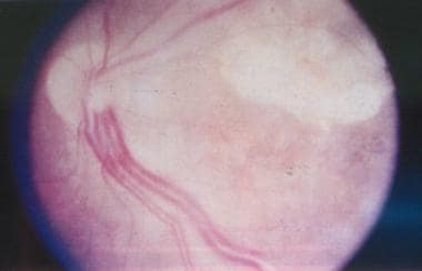
The authors report a case of endolymphatic sac tumor (ELST) associated with Von Hippel-Lindau disease (VHL). A 20-year-old female VHL patient received a resection of a cerebellar hemangioblastoma 3 years ago and she had a co-existing of left petrous tumor. The petrous tumor showed a remarkable progression in 3 years and was resected subtotally. Histologically, the resected petrous tumor showed a papillary structure containing cuboidal or columnar cells with fibrous stroma and numerous microvessels and destructed temporal bone, all of which are consistent with ELST. We studied the expression of various kinds of cytokeratins (CKs) immunohistochemically and found distinct expression of CKs (CAM 5.2, 34betaE-12, CK7, CK8 and CK19), but not for CK10/13 or CK20. Vascular endothelial growth factor and neuron specific enolase showed strong immunoreactivity in the tumor cells. CD34 also had weak expression. Ki-67 antigen (MIB-1) immunoreactivity was found in focal areas, and the labeling index in the highest-density area was 48.9%. These findings suggest that vascular endothelial growth factor overexpression is an important factor for angiogenesis in ELST, much like other VHL-associated tumors, and that ELST may have a more highly aggressive component than the low-grade malignancy noted in previous reports. (+info)Cytology of endolymphatic sac tumor. (7/52)
A 77-year-old man presented with decreased mental status and an enhancing partially cystic tumor along the left tentorium on magnetic resonance imaging after mastoidectomy and petrosectomy for an "auditory canal tumor." Smears of the aspirated cyst fluid revealed rare epithelial cell clusters, some with papillary features, foamy macrophages, and blood. The cells were orderly, with fairly bland nuclei and well-defined cell borders. The cell block contained similar epithelium, with cells containing eosinophilic and focally vacuolated cytoplasm, some with pigmented granules resembling hemosiderin. Numerous foam cells were also present. Review of the patient's previous and concurrent resection material showed an endolymphatic sac tumor, a rare neoplasm that arises in the endolymphatic sac in the temporal bone. The previously undescribed cytologic features of this rare neoplasm are discussed. (+info)Phenotypes associated with replacement of His by Arg in the Pendred syndrome gene. (8/52)
BACKGROUND: Pendred syndrome is often associated with inner ear malformations, especially enlarged vestibular aqueduct (EVA). Recently, mutations in the Pendred syndrome gene (PDS) have been reported in patients with EVA, in addition to those with classical Pendred syndrome. OBJECTIVE: The aim of this study was to investigate the genotype-phenotype correlations of PDS. METHODS: Each of the 21 exons and flanking splice regions of PDS was analysed by direct DNA sequencing in nine patients with EVA; allele-specific amplification was performed to confirm the mutation. Genetic analyses were compared with thyroid function tests, perchlorate discharge tests, thyroid volume and pure-tone audiogram. Magnetic resonance imaging was used to determine the volume of the endolymphatic duct and sac of each patient. RESULTS: A missense mutation, H723R, was identified in the homozygous state in three patients and in the heterozygous state in another three. Although none of the patients had goitre, increased serum thyroglobulin and an abnormal degree of iodide release were correlated with the number of mutant alleles identified. However, there was no relationship between the degree of hearing loss and the number of mutant alleles. CONCLUSION: The present study reveals that the number of mutant alleles correlates with the degree of subclinical thyroid abnormality, but not with the degree of hearing loss in Japanese patients with the PDS missense mutation H723R. (+info)The endolymphatic sac is a small, fluid-filled structure that is part of the inner ear. It is located near the vestibular aqueduct and is responsible for maintaining the balance of fluids in the inner ear. The endolymphatic sac also plays a role in the resorption of endolymph, which is the fluid that fills the membranous labyrinth of the inner ear. Disorders of the endolymphatic sac can lead to conditions such as Meniere's disease, which is characterized by vertigo, hearing loss, and tinnitus.
Ear neoplasms refer to abnormal growths or tumors that occur in the ear. These growths can be benign (non-cancerous) or malignant (cancerous) and can affect any part of the ear, including the outer ear, middle ear, inner ear, and the ear canal.
Benign ear neoplasms are typically slow-growing and do not spread to other parts of the body. Examples include exostoses, osteomas, and ceruminous adenomas. These types of growths are usually removed surgically for cosmetic reasons or if they cause discomfort or hearing problems.
Malignant ear neoplasms, on the other hand, can be aggressive and may spread to other parts of the body. Examples include squamous cell carcinoma, basal cell carcinoma, and adenoid cystic carcinoma. These types of tumors often require more extensive treatment, such as surgery, radiation therapy, and chemotherapy.
It is important to note that any new growth or change in the ear should be evaluated by a healthcare professional to determine the nature of the growth and develop an appropriate treatment plan.
The endolymphatic duct is a narrow canal in the inner ear that is part of the membranous labyrinth. It connects the utricle and saccule (two sensory structures in the vestibular system responsible for detecting changes in head position and movement) to the endolymphatic sac (a dilated portion of the duct that helps regulate the volume and pressure of endolymph, a fluid found within the membranous labyrinth).
The endolymphatic duct plays a crucial role in maintaining the balance and homeostasis of the inner ear by allowing the absorption and circulation of endolymph. Disorders or abnormalities in this region can lead to various vestibular and hearing dysfunctions, such as Meniere's disease, endolymphatic hydrops, and other inner ear disorders.
Endolymph is a specific type of fluid that is found within the inner ear, more specifically in the membranous labyrinth of the inner ear. This fluid plays a crucial role in maintaining balance and hearing functions. It helps in the stimulation of hair cells present in the inner ear which then transmit signals to the brain, enabling us to hear and maintain our balance. Any disturbance or changes in the composition or flow of endolymph can lead to various vestibular disorders and hearing problems.
The vestibular aqueduct is a bony canal that runs from the inner ear to the brain. It contains a membranous duct, called the endolymphatic duct, which is filled with a fluid called endolymph. The vestibular aqueduct plays a role in the maintenance of balance and hearing by regulating the pressure and composition of the endolymph. Abnormalities or damage to the vestibular aqueduct can lead to conditions such as endolymphatic hydrops, which can cause symptoms like vertigo, dizziness, and hearing loss.
Menière disease is an inner ear disorder that is characterized by episodes of vertigo (a spinning sensation), tinnitus (ringing or buzzing in the ear), hearing loss, and aural fullness (a feeling of pressure or blockage in the ear). It is caused by an abnormal accumulation of endolymphatic fluid in the inner ear, which can lead to damage of the vestibular system and cochlea. The exact cause of this fluid buildup is not known, but it may be related to genetics, allergies, or autoimmune disorders. Menière disease is typically a chronic condition, with symptoms that can vary in frequency and severity over time. Treatment options include dietary modifications, diuretics, vestibular rehabilitation therapy, and, in some cases, surgery.
Labyrinthitis is a medical condition characterized by inflammation of the labyrinth, which is the inner ear's balance- and hearing-sensitive system. It is often caused by an infection, such as a viral or bacterial infection, that spreads to the inner ear. The inflammation can affect the delicate structures of the labyrinth, leading to symptoms such as vertigo (a spinning sensation), dizziness, imbalance, hearing loss, and tinnitus (ringing in the ears). Labyrinthitis can be a serious condition that requires medical attention and treatment.
Endolymphatic hydrops is a term used to describe the abnormal accumulation of fluid (endolymph) within the inner ear. This condition is most commonly associated with Meniere's disease, but can also be seen in other disorders that affect the inner ear.
The inner ear is made up of two main parts: the cochlea, which is responsible for hearing, and the vestibular system, which helps to control balance. Both of these systems are filled with fluid, including endolymph, which is a watery fluid that bathes the sensory hair cells in these structures.
In endolymphatic hydrops, there is an overproduction or decreased absorption of endolymph, leading to an abnormal buildup of fluid within the inner ear. This can cause a variety of symptoms, including vertigo (a spinning sensation), tinnitus (ringing in the ears), hearing loss, and a feeling of fullness or pressure in the affected ear.
The exact cause of endolymphatic hydrops is not fully understood, but it is thought to be related to changes in the inner ear's fluid balance. Treatment options may include medications to help control symptoms, as well as surgical procedures to relieve pressure on the inner ear.
Von Hippel-Lindau (VHL) disease is a rare genetic disorder characterized by the development of tumors and cysts in various parts of the body. It is caused by mutations in the VHL gene, which leads to the abnormal growth of blood vessels, resulting in the formation of these tumors.
The tumors associated with VHL disease can develop in several organs, including the eyes (in the form of retinal hemangioblastomas), the brain and spinal cord (in the form of cerebellar hemangioblastomas and spinal cord hemangioblastomas), the adrenal glands (in the form of pheochromocytomas or paragangliomas), the kidneys (in the form of clear cell renal cell carcinomas), and the pancreas (in the form of serous cystadenomas or neuroendocrine tumors).
Individuals with VHL disease are at risk for developing multiple tumors over their lifetime, and the severity of the disease can vary widely from person to person. The diagnosis of VHL disease is typically made through genetic testing, family history, and imaging studies to detect the presence of tumors. Treatment may involve surgical removal of the tumors, radiation therapy, or other interventions depending on the location and size of the tumors. Regular monitoring and follow-up are essential for individuals with VHL disease to manage their condition effectively.
The cerebellopontine angle (CPA) is a narrow space located at the junction of the brainstem and the cerebellum, where the pons and cerebellum meet. This region is filled with several important nerves, blood vessels, and membranous coverings called meninges. The CPA is a common site for various neurological disorders because it contains critical structures such as:
1. Cerebellum: A part of the brain responsible for coordinating muscle movements, maintaining balance, and fine-tuning motor skills.
2. Pons: A portion of the brainstem that plays a role in several vital functions, including facial movements, taste sensation, sleep regulation, and respiration.
3. Cranial nerves: The CPA is home to the following cranial nerves:
* Vestibulocochlear nerve (CN VIII): This nerve has two components - cochlear and vestibular. The cochlear part is responsible for hearing, while the vestibular part contributes to balance and eye movement.
* Facial nerve (CN VII): This nerve controls facial expressions, taste sensation in the anterior two-thirds of the tongue, salivary gland function, and lacrimation (tear production).
4. Blood vessels: The CPA contains critical blood vessels like the anterior inferior cerebellar artery (AICA), which supplies blood to various parts of the brainstem, cerebellum, and cranial nerves.
5. Meninges: These are protective membranes surrounding the brain and spinal cord. In the CPA, the meninges include the dura mater, arachnoid mater, and pia mater.
Disorders that can affect the structures in the cerebellopontine angle include acoustic neuromas (vestibular schwannomas), meningiomas, epidermoids, and arteriovenous malformations. These conditions may cause symptoms such as hearing loss, tinnitus (ringing in the ears), vertigo (dizziness), facial weakness or numbness, difficulty swallowing, and imbalance.
The temporal bone is a paired bone that is located on each side of the skull, forming part of the lateral and inferior walls of the cranial cavity. It is one of the most complex bones in the human body and has several important structures associated with it. The main functions of the temporal bone include protecting the middle and inner ear, providing attachment for various muscles of the head and neck, and forming part of the base of the skull.
The temporal bone is divided into several parts, including the squamous part, the petrous part, the tympanic part, and the styloid process. The squamous part forms the lateral portion of the temporal bone and articulates with the parietal bone. The petrous part is the most medial and superior portion of the temporal bone and contains the inner ear and the semicircular canals. The tympanic part forms the lower and anterior portions of the temporal bone and includes the external auditory meatus or ear canal. The styloid process is a long, slender projection that extends downward from the inferior aspect of the temporal bone and serves as an attachment site for various muscles and ligaments.
The temporal bone plays a crucial role in hearing and balance, as it contains the structures of the middle and inner ear, including the oval window, round window, cochlea, vestibule, and semicircular canals. The stapes bone, one of the three bones in the middle ear, is entirely encased within the petrous portion of the temporal bone. Additionally, the temporal bone contains important structures for facial expression and sensation, including the facial nerve, which exits the skull through the stylomastoid foramen, a small opening in the temporal bone.
Skull neoplasms refer to abnormal growths or tumors that develop within the skull. These growths can be benign (non-cancerous) or malignant (cancerous). They can originate from various types of cells, such as bone cells, nerve cells, or soft tissues. Skull neoplasms can cause various symptoms depending on their size and location, including headaches, seizures, vision problems, hearing loss, and neurological deficits. Treatment options include surgery, radiation therapy, and chemotherapy. It is important to note that a neoplasm in the skull can also refer to metastatic cancer, which has spread from another part of the body to the skull.
Adenocarcinoma, papillary is a type of cancer that begins in the glandular cells and grows in a finger-like projection (called a papilla). This type of cancer can occur in various organs, including the lungs, pancreas, thyroid, and female reproductive system. The prognosis and treatment options for papillary adenocarcinoma depend on several factors, such as the location and stage of the tumor, as well as the patient's overall health. It is important to consult with a healthcare professional for an accurate diagnosis and personalized treatment plan.
The inner ear is the innermost part of the ear that contains the sensory organs for hearing and balance. It consists of a complex system of fluid-filled tubes and sacs called the vestibular system, which is responsible for maintaining balance and spatial orientation, and the cochlea, a spiral-shaped organ that converts sound vibrations into electrical signals that are sent to the brain.
The inner ear is located deep within the temporal bone of the skull and is protected by a bony labyrinth. The vestibular system includes the semicircular canals, which detect rotational movements of the head, and the otolith organs (the saccule and utricle), which detect linear acceleration and gravity.
Damage to the inner ear can result in hearing loss, tinnitus (ringing in the ears), vertigo (a spinning sensation), and balance problems.
The yolk sac is a structure that forms in the early stages of an embryo's development. It is a extra-embryonic membrane, which means it exists outside of the developing embryo, and it plays a critical role in providing nutrients to the growing embryo during the initial stages of development.
In more detail, the yolk sac is responsible for producing blood cells, contributing to the formation of the early circulatory system, and storing nutrients that are absorbed from the yolk material inside the egg or uterus. The yolk sac also has a role in the development of the gut and the immune system.
As the embryo grows and the placenta develops, the yolk sac's function becomes less critical, and it eventually degenerates. However, remnants of the yolk sac can sometimes persist and may be found in the developing fetus or newborn baby. In some cases, abnormalities in the development or regression of the yolk sac can lead to developmental problems or congenital disorders.
Vertigo is a specific type of dizziness characterized by the sensation that you or your surroundings are spinning or moving, even when you're perfectly still. It's often caused by issues with the inner ear or the balance-sensing systems of the body. Vertigo can be brought on by various conditions, such as benign paroxysmal positional vertigo (BPPV), labyrinthitis, vestibular neuritis, Meniere's disease, and migraines. In some cases, vertigo may also result from head or neck injuries, brain disorders like stroke or tumors, or certain medications. Treatment for vertigo depends on the underlying cause and can include specific exercises, medication, or surgery in severe cases.
Anion transport proteins are specialized membrane transport proteins that facilitate the movement of negatively charged ions, known as anions, across biological membranes. These proteins play a crucial role in maintaining ionic balance and regulating various physiological processes within the body.
There are several types of anion transport proteins, including:
1. Cl-/HCO3- exchangers (also known as anion exchangers or band 3 proteins): These transporters facilitate the exchange of chloride (Cl-) and bicarbonate (HCO3-) ions across the membrane. They are widely expressed in various tissues, including the red blood cells, gastrointestinal tract, and kidneys, where they help regulate pH, fluid balance, and electrolyte homeostasis.
2. Sulfate permeases: These transporters facilitate the movement of sulfate ions (SO42-) across membranes. They are primarily found in the epithelial cells of the kidneys, intestines, and choroid plexus, where they play a role in sulfur metabolism and absorption.
3. Cl- channels: These proteins form ion channels that allow chloride ions to pass through the membrane. They are involved in various physiological processes, such as neuronal excitability, transepithelial fluid transport, and cell volume regulation.
4. Cation-chloride cotransporters: These transporters move both cations (positively charged ions) and chloride anions together across the membrane. They are involved in regulating neuronal excitability, cell volume, and ionic balance in various tissues.
Dysfunction of anion transport proteins has been implicated in several diseases, such as cystic fibrosis (due to mutations in the CFTR Cl- channel), distal renal tubular acidosis (due to defects in Cl-/HCO3- exchangers), and some forms of epilepsy (due to abnormalities in cation-chloride cotransporters).
Cerebellar neoplasms refer to abnormal growths or tumors that develop in the cerebellum, which is the part of the brain responsible for coordinating muscle movements and maintaining balance. These tumors can be benign (non-cancerous) or malignant (cancerous), and they can arise from various types of cells within the cerebellum.
The most common type of cerebellar neoplasm is a medulloblastoma, which arises from primitive nerve cells in the cerebellum. Other types of cerebellar neoplasms include astrocytomas, ependymomas, and brain stem gliomas. Symptoms of cerebellar neoplasms may include headaches, vomiting, unsteady gait, coordination problems, and visual disturbances. Treatment options depend on the type, size, and location of the tumor, as well as the patient's overall health and age. Treatment may involve surgery, radiation therapy, chemotherapy, or a combination of these approaches.
 Endolymphatic sac
Endolymphatic sac tumor
Arthur Böttcher
Ménière's disease
Inner ear
Cochlear hydrops
Utriculosaccular duct
Alan Shepard
Endolymphatic hydrops
Phelsuma
Benign tumor
Eurydactylodes
Pendrin
Giuseppe Sterzi
Enlarged vestibular aqueduct
Ceruminous adenoma
Cellular senescence
Subarcuate fossa
Phakomatosis
Standing's day gecko
Endolymphatic duct
STS-65
Hereditary cancer syndrome
Saccule
Vestibular aqueduct
List of MeSH codes (A09)
Von Hippel-Lindau disease
Neurotology
Elst
Pendred syndrome
Endolymphatic sac - Wikipedia
 Surgical management of endolymphatic sac tumors - PubMed
 Single-Cell RNA-Seq Analysis of the Endolymphatic Sac of the Mouse Inner Ear | NIH Research Festival
 Meniere Disease (Idiopathic Endolymphatic Hydrops) Treatment & Management: Approach Considerations, Principles of Medical...
 PUBLISHED: Endolymphatic Sac Decompression | JOMI News
Endolymphatic Sac Tumor - New York Ear
von Hippel-Lindau Disease: Overview, Physiology, Prognosis
What Is Ménière's Disease? - Diagnosis and Treatment
Glomus Jugulare Tumors Differential Diagnoses
 Ole Winther
- Research outputs
- Staff
Ole Winther
- Research outputs
- Staff
von Hippel-Lindau Disease: Overview, Physiology, Prognosis
NIH Clinical Center Search the Studies: Study Number, Study Title
 Classifications of Meniere's Disease - American Academy of Audiology
 Meniere disease: MedlinePlus Medical Encyclopedia
Biomarkers Search
Publikationen
 Von Hippel-Lindau disease - About the Disease - Genetic and Rare Diseases Information Center
 Medifocus Guidebook on Meniere's Disease
Von Hippel-Lindau Syndrome
 RFA-DC-11-002: Identification of Immune-Mediated Causes of Sensorineural Hearing Loss (R21/R33)
 Histology Atlas of the Developing Prenatal and Postnatal Mouse Central Nervous System, with Emphasis on Prenatal Days E7.5 to...
 Value of CT and three-dimensional reconstruction revealing specific radiological signs for screening causative high jugular...
Plus it
 Vestibular and Non-Vestibular Causes of Dizziness
IndexCat
 Meniere Disease - Ear, Nose, and Throat Disorders - Merck Manuals Consumer Version
 Frontiers | Brain Reconstruction Across the Fish-Tetrapod Transition; Insights From Modern Amphibians
Hydrops10
- If endolymphatic hydrops is attributable to a given disease process-that is, if it is Ménière syndrome rather than Ménière disease-the first-line management is diagnosis and treatment of the primary disease (eg, syphilis, thyroid disease). (medscape.com)
- Steroids have also been helpful in treating endolymphatic hydrops because of their anti-inflammatory properties. (medscape.com)
- 10. Experimental endolymphatic hydrops in sharks. (nih.gov)
- 13. Endolymphatic sac amputation without hydrops. (nih.gov)
- 14. Otosclerosis and endolymphatic hydrops. (nih.gov)
- Meniere's disease is also called idiopathic endolymphatic hydrops . (medifocus.com)
- It is also not clear whether endolymphatic hydrops is the cause or the result of Meniere's disease, since there are known cases of individuals with endolymphatic hydrops who otherwise have no symptoms of Meniere's disease. (medifocus.com)
- The aim of the present study was to investigate the pathological features of vestibular aqueduct (VA) related high jugular bulb (HJB) and explore the possible cause-consequence relation between HJB and endolymphatic hydrops (EH), and the potential specific radiological signs for screening causative HJB in Meniere's disease (MD). (researchsquare.com)
- Endolymphatic hydrops (EH) is the pathological substrate of Meniere's disease (MD), which may be caused by deficient absorption in the sac or obstruction of the endolymphatic duct (1-4). (researchsquare.com)
- High jugular bulb (HJB), one of the most common anatomical variant in temporal bone, has been reported to be more common in MD, and it has been suggested to put pressure on the endolymphatic sac (ES) and distal vestibular aqueduct (VA), which contains the endolymphatic duct and the infratemporal endolymphatic sac, resulting in endolymphatic hydrops and Meniere-like symptoms (5,6). (researchsquare.com)
Tumors11
- Neoplasms of the endolymphatic sac are very rare tumors. (wikipedia.org)
- Objective To review the cumulative experience of surgical management of endolymphatic sac tumors (ELSTs) in the literature. (nih.gov)
- Endolymphatic sac tumors are rare tumors that can result in the destruction of the ear and involvement of close by neurological structures. (newyorkear.com)
- Many Endolymphatic sac tumors are benign (non-cancerous), but can show up on MRI or CAT scans and appear like acoustic neuromas (vestibular schwannomas) or meningiomas. (newyorkear.com)
- 1. Evaluation and management of endolymphatic sac and duct tumors. (nih.gov)
- 5. Endolymphatic sac tumors. (nih.gov)
- 20. Endolymphatic sac tumors: histopathologic confirmation, clinical characterization, and implication in von Hippel-Lindau disease. (nih.gov)
- Endolymphatic sac tumors (ELST) are also been found (up to 10%) and may cause hearing loss. (orpha.net)
- Imaging studies can be used to detect CNS tumors, pheochromocytoma/paraganglioma, endolymphatic sac tumors, renal tumors, and renal and pancreatic cysts. (orpha.net)
- Von Hippel-Lindau syndrome is an inherited disorder characterized by the formation of tumors and fluid-filled sacs (cysts) in many different parts of the body. (nih.gov)
- About 10 percent of people with von Hippel-Lindau syndrome develop endolymphatic sac tumors, which are noncancerous tumors in the inner ear . (nih.gov)
Tumor7
- 2. Endolymphatic sac tumor located around semicircular canals. (nih.gov)
- 3. Endolymphatic sac tumor of temporal bone. (nih.gov)
- 8. [Papillary tumor of the endolymphatic sac]. (nih.gov)
- 11. Endolymphatic sac tumor: unique features of two cases and review of the literature. (nih.gov)
- 15. Gamma knife treatment of an endolymphatic sac tumor: unique features of a case and review of the literature. (nih.gov)
- 16. Endolymphatic Sac Tumor as a Ménière-Like Vertiginous Syndrome: A Case Report. (nih.gov)
- Surgical management of endolymphatic sac tumor: classification, outcomes and strategy. (bvsalud.org)
Vestibular aqueduct3
- this duct is joined by the utriculosaccular duct, and then passes along the vestibular aqueduct and ends in a blind pouch, the endolymphatic sac, on the posterior surface of the petrous portion of the temporal bone, where it is in contact with the dura mater. (wikipedia.org)
- 18. Massive endolymphatic sac and vestibular aqueduct in Mondini dysplasia. (nih.gov)
- CT or MR imaging, or both, may be used to evaluate the vestibular aqueduct, endolymphatic duct, and sac and to rule out associated infectious or neoplastic disease. (ajnr.org)
Epithelial cells in the endolymphatic sac2
- 100:148-156 Fukuzawa K, Sakagami M, Matsunaga T, Fujita H. Endocytotic activity of the free floating cells and epithelial cells in the endolymphatic sac: an electron microscopic study. (wikipedia.org)
- SLC26A4 encodes a transmembrane anion exchanger, pendrin, which is expressed in a subset of nonsensory epithelial cells in the endolymphatic sac (ES). (nih.gov)
Cochlear2
- 9. MR imaging of the cochlear modiolus: area measurement in healthy subjects and in patients with a large endolymphatic duct and sac. (nih.gov)
- The pathophysiological findings of Ménière disease are associated with an accumulation of endolymph in the cochlear duct, which occurs at the expense of the perilymphatic space, and inadequate absorption of the endolymph by the endolymphatic sac [ 2 , 4 ]. (e-jyms.org)
Endolymph2
- Inside their walls (bony labyrinth) are thin, pliable tubes and sacs (membranous labyrinth) filled with endolymph. (nih.gov)
- In mammals, disruptions to endolymph homeostasis can result in either collapse or distension of the endolymphatic compartment in the cochlea, with concomitant hearing loss. (biologists.com)
Tumours3
- Non-cancerous tumours can form in the endolymphatic duct of the inner ear, which regulates the balance of an individual. (nccs.com.sg)
- These tumours are called endolymphatic sac tumours , which can cause problems such as hearing loss or deafness. (nccs.com.sg)
- Endolymphatic sac tumours are observed in up to. (entandaudiologynews.com)
Duct and sac1
- 7. Advanced techniques in magnetic resonance imaging in the evaluation of the large endolymphatic duct and sac syndrome. (nih.gov)
Fluid4
- It is thought that otosclerotic formations may cause malformation in the endolymphatic sac or may change the chemical properties of the perilymphatic and endolymphatic fluid. (medifocus.com)
- Fluid in the ear is held in a pouch-like structure called the endolymphatic sac. (merckmanuals.com)
- The zebrafish little ears ( lte )mutant shows a collapse of the otic vesicle in the larva, apparently owing to a loss of endolymphatic fluid in the ear, together with an over-inflation of the swim bladder. (biologists.com)
- After all the whole concept of an endolymphatic shunt is 'drain' the excess fluid away. (menieres-help.com)
Decompression2
- Endolymphatic sac (ELS) decompression can be performed for patients with Meniere's disease who have failed conservative treatment such as dietary changes and medical therapy. (jomi.com)
- Decompression of the sac can be accomplished by removing overlying bone, incising the dura, or stenting the dura open. (jomi.com)
Inner ear1
- Mutations of the SLC26A4 gene are a common cause of hearing loss associated with enlargement of the endolymphatic sac and duct (EES) of the inner ear. (nih.gov)
Distal1
- The distal projection of the endolymphatic sac in human embryos. (nih.gov)
Blind pouch1
- The blind pouch at the end of the endolymphatic duct. (nih.gov)
Secretion1
- Ultrastructural evidence of a merocrine secretion in the human endolymphatic sac. (wikipedia.org)
Posterior1
- 4. Risk of damage to the endolymphatic sac and duct during removal of the posterior meatal wall: an anatomic study. (nih.gov)
Development1
- Although numerous radiological studies have shown smaller VA dimensions and high rate of JB abnormalities among affected individuals, suggesting that HJB is likely to interfere with VA, causing endolymphatic sac dysfunction and the development of EH (7,8). (researchsquare.com)
Human1
- Human endolymphatic sac: morphologic evidence of immunologic function. (wikipedia.org)
Literature1
- 6. Endolymphatic sac tumour: case report and literature review. (nih.gov)
Studies1
- Studies suggest that the endolymphatic duct and endolymphatic sac perform both absorptive and secretory, as well as phagocytic and immunodefensive, functions. (wikipedia.org)