Enterocolitis
Enterocolitis, Necrotizing
Enterocolitis, Neutropenic
Enterocolitis, Pseudomembranous
Infant, Premature, Diseases
Cronobacter sakazakii
Infant, Very Low Birth Weight
Enteral Nutrition
Hirschsprung Disease
Enterocytes
Antifungal Agents
Mycoses
Infant, Newborn, Diseases
Intestines
Amphotericin B
Intestinal Mucosa
Infant, Extremely Low Birth Weight
Lung Diseases, Fungal
Opportunistic Infections
Candidiasis
Intensive Care Units, Neonatal
Hematologic Neoplasms
Probiotics
Bacteremia
Milk, Human
Immunocompromised Host
Disease Models, Animal
Colitis, Lymphocytic
Echinocandins
Granulocyte Colony-Stimulating Factor
Ileum
Sepsis
Invasive Pulmonary Aspergillosis
Gestational Age
Cecum
Fungemia
Dysbiosis
Treatment Outcome
Neoplasms
Aspergillus fumigatus
Amikacin
Bifidobacterium
Piperacillin
Ceftazidime
Mice, Inbred ICR
Intestine, Small
Itraconazole
Gram-Negative Bacterial Infections
Vancomycin
Retrospective Studies
Parenteral Nutrition
Infant Nutritional Physiological Phenomena
Antibiotic Prophylaxis
Asphyxia
Feces
Mucin-3
Gram-Positive Bacterial Infections
Ductus Arteriosus, Patent
Prospective Studies
Anti-Infective Agents
Mucin-2
Short Bowel Syndrome
Fluconazole
Lactobacillus reuteri
Neutrophils
Paneth Cells
Antineoplastic Combined Chemotherapy Protocols
Birth Weight
Colon
Infant, Low Birth Weight
Butyrivibrio
Bacterial Translocation
Pulmonary Aspergillosis
Term Birth
Microbial Sensitivity Tests
Penicillanic Acid
Risk Factors
Candida tropicalis
Triazoles
Parenteral Nutrition, Total
Intensive Care, Neonatal
Leukocyte Transfusion
Colitis, Ischemic
Cephalosporins
Toll-Like Receptor 4
Miliaria
Clostridium
Mucormycosis
Gastrointestinal Tract
Drug Therapy, Combination
Bronchopulmonary Dysplasia
Metagenome
Neutropenic enteropathy. (1/12)
Neutropenic enteropathy (NE) is used to describe the inflammation of the bowel in neutropenic patients under aggressive chemotherapy, mainly for lymphoproliferative and hematologic malignancies. Surgical intervention may be required in patients with the advent of the disease. We report our experience in 7 children with NE who had to be treated surgically. Absolute neutrophil counts were less than 1000/mm3 in all, with positive blood cultures in five patients. Four patients recovered with rapid resolution of neutropenia, while three patients died with persistent neutropenia. (+info)Neutropenic enterocolitis in lung cancer: a report of two cases and a review of the literature. (2/12)
The first patient, a 68-year-old woman, presented neutropenic fever and hemorrhagic diarrhea on the sixth day of a combination chemotherapy of carboplatin and paclitaxel. The second patient, a 30-year-old man, presented neutropenia and diarrhea on the tenth day of the second cycle of a combination chemotherapy of cisplatin and vinorelbine. In both patients, abdominal computed tomography scan showed thickening of the colon wall and pericolic edema, and the ultrasonography revealed echogenic thickening of the colon walls. These findings confirmed the diagnosis of neutropenic enterocolitis. After the treatments, we changed the anticancer drug regimen; and successfully achieved partial responses. (+info)Neutropenic enterocolitis in an advanced epithelial ovarian cancer patient treated with paclitaxel/platinum-based chemotherapy: a case report and review of the literature. (3/12)
BACKGROUND: Literature data show that neutropenic enterocolitis is a rare but severe complication that can occur in cancer patients treated with chemotherapy and especially with taxanes. CASE REPORT: A 60-year-old woman with stage Illc epithelial ovarian cancer developed neutropenic fever, abdominal pain, severe diarrhoea, nausea, vomiting and oral mucositis one week after the first postoperative cycle of paclitaxel (175 mg/m2 3-hour infusion) plus carboplatin-based chemotherapy. Abdominal X-ray showed diffuse dilatation of the ileal and colonic loops with air/fluid. The patient soon recovered after intensive supportive care. For the second cycle the dose of paclitaxel was reduced by 20%, but nine days later the patient again developed severe neutropenia with fever, abdominal colicky pain, diarrhoea and vomiting. The culture of blood samples collected on admission was found to be positive for Escherichia coli, whereas stools resulted negative for both enteric rods and Clostridium difficile toxin. The patient recovered with intensive supportive care, and chemotherapy was continued with single-agent carboplatin. DISCUSSION: The increasing use of paclitaxel in first-line as well as in the salvage treatment of epithelial ovarian cancer could increase the occurrence of neutropenic enterocolitis in patients with this malignancy. The importance of symptoms such as neutropenic fever, abdominal pain and tenderness and severe diarrhoea should be stressed in patients who receive taxane-based chemotherapy, and intensive supportive care management should be started immediately. (+info)Neutropenic colitis during standard dose combination chemotherapy with nedaplatin and irinotecan for testicular cancer. (4/12)
A 54-year-old man received combination chemotherapy with nedaplatin and irinotecan as salvage chemotherapy for refractory non-seminomatous testicular cancer. The patient developed abdominal pain and high fever on Day 21 after the initiation of chemotherapy. Computed tomography revealed thickening of the terminal ileum wall and paralytic ileus. The patient recovered with intensive supportive management including broad-spectrum antibiotics, bowel rest with gastric intubation and intravenous gamma-globulin. Neutropenic colitis has been thought to be a serious gastrointestinal complication associated with chemotherapy for hematological malignancy. The mortality rate is as high as 21-48% according to a recent review. The present case indicates that the neutropenic colitis can occur under neutropenic conditions induced by the standard-dose chemotherapy for solid cancer. (+info)Invasive fungal infections in neutropenic enterocolitis: a systematic analysis of pathogens, incidence, treatment and mortality in adult patients. (5/12)
BACKGROUND: Neutropenic enterocolitis is a life-threatening complication most frequently occurring after intensive chemotherapy in acute leukaemias. Gramnegative bacteria constitute the most important group of causative pathogens. Fungi have also been reported, but their practical relevance remains unclear. The guidelines do not address concrete treatment recommendations for fungal neutropenic enterocolitis. METHODS: Here, we conducted a metaanalysis to answer the questions: What are frequency and mortality of fungal neutropenic enterocolitis? Do frequencies and microbiological distribution of causative fungi support empirical antimycotic therapy? Do reported results of antimycotic therapy in documented fungal neutropenic enterocolitis help with the selection of appropriate drugs? Following a systematic search, we extracted and summarised all detail data from the complete literature. RESULTS: Among 186 articles describing patients with neutropenic enterocolitis, we found 29 reports describing 53 patients with causative fungal pathogens. We found no randomised controlled trial, no good quality cohort study and no good quality case control study on the role of antifungal treatment. The pooled frequency of fungal neutropenic enterocolitis was 6.2% calculated from all 860 reported patients and 3.4% calculated from selected representative studies only. In 94% of the patients, Candida spp. were involved. The pooled mortality rate was 81.8%. Most authors did not report or perform antifungal therapy. CONCLUSION: In patients with neutropenic enterocolitis, fungal pathogens play a relevant, but secondary role compared to bacteria. Evidence concerning therapy is very poor, but epidemiological data from this study may provide helpful clues to select empiric antifungal therapy in neutropenic enterocolitis. (+info)Sonographic signs of neutropenic enterocolitis. (6/12)
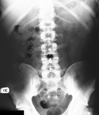
AIM: To investigate the sonographic features at time of diagnosis and follow-up in patients with neutropenic enterocolitis. METHODS: The sonographic findings in 14 patients with neutropenic enterocolitis were described and evaluated regarding symptoms and clinical outcome. RESULTS: In all patients with neutropenic enterocolitis, the ileocoecal region was involved with wall thickening >10 mm. A transmural inflammatory pattern, hypervascularity of the thickened bowel wall and free abdominal fluid were the common findings. The sonographically revealed thickness of the bowel wall was associated with lethal outcome (P<0.03). In the 11 surviving patients,the improvement of clinical symptoms was accompanied by progressive reduction of intestinal wall thickness. CONCLUSION: High-end sonography of the bowel is a helpful tool for diagnosis,assessment of prognosis and follow-up of patients with neutropenic enterocolitis.The ultrasonographically revealed bowel thickness reflects the severity and the course of the disease, and seems to be predictive for the clinical outcome. (+info)Diarrhea in neutropenic patients: a prospective cohort study with emphasis on neutropenic enterocolitis. (7/12)
BACKGROUND: Although diarrhea is a frequent complication in neutropenic patients, its true incidence, risk factors and clinical course have not been investigated prospectively. PATIENTS AND METHODS: The study was carried out at Hacettepe University Hospital for Adults and involved patients over 16 years of age. Patients with malignant diseases who were neutropenic on admission or who became neutropenic during their stay in the wards between January 2001 and February 2003 were included. They were monitored daily until discharge, exitus, or recovery from neutropenia-whichever occurred earlier-to monitor the presence of diarrhea and other infections. RESULTS: A total of 317 neutropenic episodes in 215 patients were followed. Diarrhea was observed in 18.6% episodes, and the incidence of NEC was 3.5%. The etiology in 27% episodes of diarrhea could not be identified. The use of anthracyclines and mitoxantrone increased the incidence of diarrhea. Prior use of penicillin derivatives plus beta-lactam inhibitors and N-imidazoline derivatives was associated with decreased incidence of diarrhea. CONCLUSIONS: Diarrhea is a common complication in neutropenic patients. Not only specific conditions like NEC, but also nonspecific diseases like parasitosis may be the cause of diarrhea in this patient population. (+info)Neutropenic enterocolitis. (8/12)
We report a case of neutropenic enterocolitis diagnosed on computerized tomography abdomen in a 56-year-old man having high-grade non-Hodgkin's lymphoma. After appropriate management, the patient recovered completely. (+info)Enterocolitis is a medical condition that involves inflammation of the small intestine (enteritis) and large intestine (colitis). This condition can affect people of all ages, but it is most commonly seen in infants and young children. The symptoms of enterocolitis may include diarrhea, abdominal cramps, bloating, nausea, vomiting, fever, and dehydration.
There are several types of enterocolitis, including:
1. Infectious Enterocolitis: This type is caused by a bacterial, viral, or parasitic infection in the intestines. Common causes include Salmonella, Shigella, Escherichia coli (E. coli), and norovirus.
2. Antibiotic-Associated Enterocolitis: This type is caused by an overgrowth of harmful bacteria in the intestines following the use of antibiotics that kill off beneficial gut bacteria.
3. Pseudomembranous Enterocolitis: This is a severe form of antibiotic-associated enterocolitis caused by the bacterium Clostridioides difficile (C. diff).
4. Necrotizing Enterocolitis: This is a serious condition that primarily affects premature infants, causing inflammation and damage to the intestinal tissue, which can lead to perforations and sepsis.
5. Ischemic Enterocolitis: This type is caused by reduced blood flow to the intestines, often due to conditions such as mesenteric ischemia or vasculitis.
6. Radiation Enterocolitis: This type occurs as a complication of radiation therapy for cancer treatment, which can damage the intestinal lining and lead to inflammation.
7. Eosinophilic Enterocolitis: This is a rare condition characterized by an excessive buildup of eosinophils (a type of white blood cell) in the intestinal tissue, leading to inflammation and symptoms similar to those seen in inflammatory bowel disease.
Treatment for enterocolitis depends on the underlying cause and severity of the condition. It may include antibiotics, antiparasitic medications, probiotics, or surgery in severe cases.
Necrotizing enterocolitis (NEC) is a serious gastrointestinal condition that primarily affects premature infants. It is characterized by the inflammation and death of intestinal tissue, which can lead to perforations (holes) in the bowel wall. Here's a brief medical definition:
Necrotizing enterocolitis (NEK-roh-tiz-ing en-ter-koh-li-TIE-tis): A gastrointestinal emergency in which the inner lining of the intestinal wall undergoes necrosis (tissue death) due to inflammation, often affecting premature infants. The condition may result in bowel perforations, sepsis, and other systemic complications, requiring surgical intervention and intensive care management.
The exact cause of NEC is not fully understood, but it's thought to be associated with factors such as prematurity, formula feeding, intestinal immaturity or injury, and disturbed blood flow in the intestines. Symptoms may include abdominal distention, bloody stools, feeding intolerance, lethargy, and temperature instability. Early recognition and prompt treatment are crucial for improving outcomes in affected infants.
Neutropenic enterocolitis is a serious and potentially life-threatening complication that can occur in individuals with severely compromised immune systems, such as those undergoing chemotherapy or radiation therapy for cancer treatment. It is also known as typhlitis or neutropenic colitis.
The condition is characterized by inflammation of the inner lining of the small intestine and colon (enterocolitis), which occurs in the absence of adequate numbers of white blood cells, particularly neutrophils, that are necessary to fight off infection. As a result, the intestinal tract becomes vulnerable to bacterial or fungal invasion, leading to inflammation, tissue damage, and potentially necrosis (tissue death).
Symptoms of neutropenic enterocolitis may include fever, abdominal pain, nausea, vomiting, diarrhea, and bloody stools. The condition can progress rapidly and lead to sepsis, a systemic inflammatory response that can be fatal if not treated promptly.
Diagnosis of neutropenic enterocolitis typically involves a combination of clinical symptoms, imaging studies such as CT scans or MRI, and laboratory tests to assess the severity of neutropenia and identify any underlying infectious agents. Treatment usually involves administering broad-spectrum antibiotics and antifungal medications to treat or prevent infection, as well as supportive care to manage symptoms and maintain hydration and nutrition. In severe cases, surgery may be necessary to remove necrotic tissue and prevent further complications.
Neutropenia is a condition characterized by an abnormally low concentration (less than 1500 cells/mm3) of neutrophils, a type of white blood cell that plays a crucial role in fighting off bacterial and fungal infections. Neutrophils are essential components of the innate immune system, and their main function is to engulf and destroy microorganisms that can cause harm to the body.
Neutropenia can be classified as mild, moderate, or severe based on the severity of the neutrophil count reduction:
* Mild neutropenia: Neutrophil count between 1000-1500 cells/mm3
* Moderate neutropenia: Neutrophil count between 500-1000 cells/mm3
* Severe neutropenia: Neutrophil count below 500 cells/mm3
Severe neutropenia significantly increases the risk of developing infections, as the body's ability to fight off microorganisms is severely compromised. Common causes of neutropenia include viral infections, certain medications (such as chemotherapy or antibiotics), autoimmune disorders, and congenital conditions affecting bone marrow function. Treatment for neutropenia typically involves addressing the underlying cause, administering granulocyte-colony stimulating factors to boost neutrophil production, and providing appropriate antimicrobial therapy to prevent or treat infections.
Pseudomembranous enterocolitis is a medical condition characterized by inflammation of the inner lining of the small intestine (enteritis) and large intestine (colitis), resulting in the formation of pseudomembranes – raised, yellowish-white plaques composed of fibrin, mucus, and inflammatory cells. The condition is most commonly caused by a toxin produced by the bacterium Clostridioides difficile (C. difficile), which can overgrow in the gut following disruption of the normal gut microbiota, often after antibiotic use. Symptoms may include diarrhea, abdominal cramps, fever, nausea, and dehydration. Severe cases can lead to complications such as sepsis, toxic megacolon, or even death if left untreated. Treatment typically involves discontinuing the offending antibiotic, administering oral metronidazole or vancomycin to eliminate C. difficile, and managing symptoms with supportive care. In some cases, fecal microbiota transplantation (FMT) may be considered as a treatment option.
A "premature infant" is a newborn delivered before 37 weeks of gestation. They are at greater risk for various health complications and medical conditions compared to full-term infants, due to their immature organ systems and lower birth weight. Some common diseases and health issues that premature infants may face include:
1. Respiratory Distress Syndrome (RDS): A lung disorder caused by the lack of surfactant, a substance that helps keep the lungs inflated. Premature infants, especially those born before 34 weeks, are at higher risk for RDS.
2. Intraventricular Hemorrhage (IVH): Bleeding in the brain's ventricles, which can lead to developmental delays or neurological issues. The risk of IVH is inversely proportional to gestational age, meaning that the earlier the infant is born, the higher the risk.
3. Necrotizing Enterocolitis (NEC): A gastrointestinal disease where the intestinal tissue becomes inflamed and can die. Premature infants are at greater risk for NEC due to their immature digestive systems.
4. Jaundice: A yellowing of the skin and eyes caused by an accumulation of bilirubin, a waste product from broken-down red blood cells. Premature infants may have higher rates of jaundice due to their liver's immaturity.
5. Infections: Premature infants are more susceptible to infections because of their underdeveloped immune systems. Common sources of infection include the mother's genital tract, bloodstream, or hospital environment.
6. Anemia: A condition characterized by a low red blood cell count or insufficient hemoglobin. Premature infants may develop anemia due to frequent blood sampling, rapid growth, or inadequate erythropoietin production.
7. Retinopathy of Prematurity (ROP): An eye disorder affecting premature infants, where abnormal blood vessel growth occurs in the retina. Severe ROP can lead to vision loss or blindness if not treated promptly.
8. Developmental Delays: Premature infants are at risk for developmental delays due to their immature nervous systems and environmental factors such as sensory deprivation or separation from parents.
9. Patent Ductus Arteriosus (PDA): A congenital heart defect where the ductus arteriosus, a blood vessel that connects two major arteries in the fetal heart, fails to close after birth. Premature infants are at higher risk for PDA due to their immature cardiovascular systems.
10. Hypothermia: Premature infants have difficulty maintaining body temperature and are at risk for hypothermia, which can lead to increased metabolic demands, poor feeding, and infection.
Agranulocytosis is a medical condition characterized by an abnormally low concentration of granulocytes (a type of white blood cells) in the peripheral blood. Granulocytes, which include neutrophils, eosinophils, and basophils, play a crucial role in the body's defense against infections. A significant reduction in their numbers can make an individual highly susceptible to various bacterial and fungal infections.
The condition is typically defined as having fewer than 150 granulocytes per microliter of blood or less than 1% of the total white blood cell count. Symptoms of agranulocytosis may include fever, fatigue, sore throat, mouth ulcers, and susceptibility to infections. The condition can be caused by various factors, including certain medications, medical treatments (such as chemotherapy or radiation therapy), autoimmune disorders, and congenital conditions. Immediate medical attention is required for individuals diagnosed with agranulocytosis to prevent and treat potential infections and restore the normal granulocyte count.
Fever, also known as pyrexia or febrile response, is a common medical sign characterized by an elevation in core body temperature above the normal range of 36.5-37.5°C (97.7-99.5°F) due to a dysregulation of the body's thermoregulatory system. It is often a response to an infection, inflammation, or other underlying medical conditions, and it serves as a part of the immune system's effort to combat the invading pathogens or to repair damaged tissues.
Fevers can be classified based on their magnitude:
* Low-grade fever: 37.5-38°C (99.5-100.4°F)
* Moderate fever: 38-39°C (100.4-102.2°F)
* High-grade or severe fever: above 39°C (102.2°F)
It is important to note that a single elevated temperature reading does not necessarily indicate the presence of a fever, as body temperature can fluctuate throughout the day and can be influenced by various factors such as physical activity, environmental conditions, and the menstrual cycle in females. The diagnosis of fever typically requires the confirmation of an elevated core body temperature on at least two occasions or a consistently high temperature over a period of time.
While fevers are generally considered beneficial in fighting off infections and promoting recovery, extremely high temperatures or prolonged febrile states may necessitate medical intervention to prevent potential complications such as dehydration, seizures, or damage to vital organs.
A premature infant is a baby born before 37 weeks of gestation. They may face various health challenges because their organs are not fully developed. The earlier a baby is born, the higher the risk of complications. Prematurity can lead to short-term and long-term health issues, such as respiratory distress syndrome, jaundice, anemia, infections, hearing problems, vision problems, developmental delays, and cerebral palsy. Intensive medical care and support are often necessary for premature infants to ensure their survival and optimal growth and development.
A newborn infant is a baby who is within the first 28 days of life. This period is also referred to as the neonatal period. Newborns require specialized care and attention due to their immature bodily systems and increased vulnerability to various health issues. They are closely monitored for signs of well-being, growth, and development during this critical time.
'Cronobacter sakazakii' is a gram-negative, rod-shaped bacterium that is part of the Enterobacteriaceae family. It is an opportunistic pathogen capable of causing severe invasive infections such as meningitis and sepsis, particularly in newborns, infants, and immunocompromised individuals. The bacterium has been found in various environmental sources, including dried foods like powdered infant formula, herbs, and spices. Proper hygiene practices and the safe handling, preparation, and storage of food and feeding utensils can help prevent Cronobacter sakazakii infections.
A very low birth weight (VLBW) infant is a baby born weighing less than 1500 grams (3 pounds, 5 ounces). This category includes babies who are extremely preterm (born at or before 28 weeks of gestation) and/or have intrauterine growth restriction. VLBW infants often face significant health challenges, including respiratory distress syndrome, brain bleeds, infections, and feeding difficulties. They may require extended hospital stays in the neonatal intensive care unit (NICU) and have a higher risk of long-term neurodevelopmental impairments compared to infants with normal birth weights.
Enteral nutrition refers to the delivery of nutrients to a person through a tube that is placed into the gastrointestinal tract, specifically into the stomach or small intestine. This type of nutrition is used when a person is unable to consume food or liquids by mouth due to various medical conditions such as swallowing difficulties, malabsorption, or gastrointestinal disorders.
Enteral nutrition can be provided through different types of feeding tubes, including nasogastric tubes, which are inserted through the nose and down into the stomach, and gastrostomy or jejunostomy tubes, which are placed directly into the stomach or small intestine through a surgical incision.
The nutrients provided through enteral nutrition may include commercially prepared formulas that contain a balance of carbohydrates, proteins, fats, vitamins, and minerals, or blenderized whole foods that are pureed and delivered through the feeding tube. The choice of formula or type of feed depends on the individual's nutritional needs, gastrointestinal function, and medical condition.
Enteral nutrition is a safe and effective way to provide nutrition support to people who are unable to meet their nutritional needs through oral intake alone. It can help prevent malnutrition, promote wound healing, improve immune function, and enhance overall health and quality of life.
Hirschsprung disease is a gastrointestinal disorder that affects the large intestine, specifically the section known as the colon. This condition is congenital, meaning it is present at birth. It occurs due to the absence of ganglion cells (nerve cells) in the bowel's muscular wall, which are responsible for coordinating muscle contractions that move food through the digestive tract.
The affected segment of the colon cannot relax and propel the contents within it, leading to various symptoms such as constipation, intestinal obstruction, or even bowel perforation in severe cases. Common diagnostic methods include rectal suction biopsy, anorectal manometry, and contrast enema studies. Treatment typically involves surgical removal of the aganglionic segment and reattachment of the normal colon to the anus (known as a pull-through procedure).
Enterocytes are the absorptive cells that line the villi of the small intestine. They are a type of epithelial cell and play a crucial role in the absorption of nutrients from food into the bloodstream. Enterocytes have finger-like projections called microvilli on their apical surface, which increases their surface area and enhances their ability to absorb nutrients. They also contain enzymes that help digest and break down carbohydrates, proteins, and fats into smaller molecules that can be absorbed. Additionally, enterocytes play a role in the absorption of ions, water, and vitamins.
Antifungal agents are a type of medication used to treat and prevent fungal infections. These agents work by targeting and disrupting the growth of fungi, which include yeasts, molds, and other types of fungi that can cause illness in humans.
There are several different classes of antifungal agents, including:
1. Azoles: These agents work by inhibiting the synthesis of ergosterol, a key component of fungal cell membranes. Examples of azole antifungals include fluconazole, itraconazole, and voriconazole.
2. Echinocandins: These agents target the fungal cell wall, disrupting its synthesis and leading to fungal cell death. Examples of echinocandins include caspofungin, micafungin, and anidulafungin.
3. Polyenes: These agents bind to ergosterol in the fungal cell membrane, creating pores that lead to fungal cell death. Examples of polyene antifungals include amphotericin B and nystatin.
4. Allylamines: These agents inhibit squalene epoxidase, a key enzyme in ergosterol synthesis. Examples of allylamine antifungals include terbinafine and naftifine.
5. Griseofulvin: This agent disrupts fungal cell division by binding to tubulin, a protein involved in fungal cell mitosis.
Antifungal agents can be administered topically, orally, or intravenously, depending on the severity and location of the infection. It is important to use antifungal agents only as directed by a healthcare professional, as misuse or overuse can lead to resistance and make treatment more difficult.
Febrile neutropenia is a medical condition characterized by a fever (temperature over 101°F or 38.3°C) and a low count of neutrophils, a type of white blood cell that helps fight infections. Neutropenia is defined as an absolute neutrophil count (ANC) of less than 1500 cells/mm3, but in the case of febrile neutropenia, the ANC is typically less than 500 cells/mm3 or is expected to fall below this level. This condition is often a complication of chemotherapy or radiation therapy used to treat cancer, as these treatments can suppress the immune system and lead to a decrease in white blood cell counts. Febrile neutropenia increases the risk of developing severe and potentially life-threatening infections.
Aspergillosis is a medical condition that is caused by the infection of the Aspergillus fungi. This fungus is commonly found in decaying organic matter, such as leaf litter and compost piles, and can also be found in some indoor environments like air conditioning systems and old buildings with water damage.
There are several types of aspergillosis, including:
1. Allergic bronchopulmonary aspergillosis (ABPA): This type of aspergillosis occurs when a person's immune system overreacts to the Aspergillus fungi, causing inflammation in the airways and lungs. ABPA is often seen in people with asthma or cystic fibrosis.
2. Invasive aspergillosis: This is a serious and potentially life-threatening condition that occurs when the Aspergillus fungi invade the bloodstream and spread to other organs, such as the brain, heart, or kidneys. Invasive aspergillosis typically affects people with weakened immune systems, such as those undergoing chemotherapy or organ transplantation.
3. Aspergilloma: Also known as a "fungus ball," an aspergilloma is a growth of the Aspergillus fungi that forms in a preexisting lung cavity, such as one caused by previous lung disease or injury. While an aspergilloma itself is not typically harmful, it can cause symptoms like coughing up blood or chest pain if it grows too large or becomes infected.
Symptoms of aspergillosis can vary depending on the type and severity of the infection. Treatment may include antifungal medications, surgery to remove the fungal growth, or management of underlying conditions that increase the risk of infection.
Intestinal perforation is a medical condition that refers to a hole or tear in the lining of the intestine. This can occur anywhere along the gastrointestinal tract, including the small intestine, large intestine (colon), or stomach. Intestinal perforation allows the contents of the intestines, such as digestive enzymes and bacteria, to leak into the abdominal cavity, which can lead to a serious inflammatory response known as peritonitis.
Intestinal perforation can be caused by various factors, including:
* Mechanical trauma (e.g., gunshot wounds, stab wounds)
* Inflammatory bowel disease (e.g., Crohn's disease, ulcerative colitis)
* Diverticulitis
* Appendicitis
* Intestinal obstruction
* Infections (e.g., typhoid fever, tuberculosis)
* Certain medications (e.g., nonsteroidal anti-inflammatory drugs, corticosteroids)
* Radiation therapy
* Ischemic bowel disease (lack of blood flow to the intestines)
Symptoms of intestinal perforation may include sudden abdominal pain, nausea, vomiting, fever, and decreased bowel movements. Treatment typically involves surgery to repair the perforation and remove any damaged tissue. Antibiotics are also administered to prevent infection. In severe cases, a temporary or permanent colostomy or ileostomy may be necessary.
Infant formula is a manufactured food designed and marketed for feeding to babies and infants under 12 months of age, but may also be used as a supplementary feedings for older children. It is usually derived from cow's milk, but can also be made from soy or other proteins. Infant formulas are designed to provide a well-balanced diet with appropriate amounts of protein, fat, carbohydrate, vitamins, and minerals to support growth and development in infants who are not breastfed. They come in various forms such as powder, concentrate, or ready-to-feed liquid and must meet strict nutritional and safety standards set by regulatory agencies like the U.S. Food and Drug Administration (FDA) and the European Commission (EC).
Mycoses are a group of diseases caused by fungal infections. These infections can affect various parts of the body, including the skin, nails, hair, lungs, and internal organs. The severity of mycoses can range from superficial, mild infections to systemic, life-threatening conditions, depending on the type of fungus and the immune status of the infected individual. Some common types of mycoses include candidiasis, dermatophytosis, histoplasmosis, coccidioidomycosis, and aspergillosis. Treatment typically involves antifungal medications, which can be topical or systemic, depending on the location and severity of the infection.
A "newborn infant" refers to a baby in the first 28 days of life outside of the womb. This period is crucial for growth and development, but also poses unique challenges as the infant's immune system is not fully developed, making them more susceptible to various diseases.
"Newborn diseases" are health conditions that specifically affect newborn infants. These can be categorized into three main types:
1. Congenital disorders: These are conditions that are present at birth and may be inherited or caused by factors such as infection, exposure to harmful substances during pregnancy, or chromosomal abnormalities. Examples include Down syndrome, congenital heart defects, and spina bifida.
2. Infectious diseases: Newborn infants are particularly vulnerable to infections due to their immature immune systems. Common infectious diseases in newborns include sepsis (bloodstream infection), pneumonia, and meningitis. These can be acquired from the mother during pregnancy or childbirth, or from the environment after birth.
3. Developmental disorders: These are conditions that affect the normal growth and development of the newborn infant. Examples include cerebral palsy, intellectual disabilities, and vision or hearing impairments.
It is important to note that many newborn diseases can be prevented or treated with appropriate medical care, including prenatal care, proper hygiene practices, and timely vaccinations. Regular check-ups and monitoring of the newborn's health by a healthcare provider are essential for early detection and management of any potential health issues.
The intestines, also known as the bowel, are a part of the digestive system that extends from the stomach to the anus. They are responsible for the further breakdown and absorption of nutrients from food, as well as the elimination of waste products. The intestines can be divided into two main sections: the small intestine and the large intestine.
The small intestine is a long, coiled tube that measures about 20 feet in length and is lined with tiny finger-like projections called villi, which increase its surface area and enhance nutrient absorption. The small intestine is where most of the digestion and absorption of nutrients takes place.
The large intestine, also known as the colon, is a wider tube that measures about 5 feet in length and is responsible for absorbing water and electrolytes from digested food, forming stool, and eliminating waste products from the body. The large intestine includes several regions, including the cecum, colon, rectum, and anus.
Together, the intestines play a critical role in maintaining overall health and well-being by ensuring that the body receives the nutrients it needs to function properly.
Amphotericin B is an antifungal medication used to treat serious and often life-threatening fungal infections. It works by binding to the ergosterol in the fungal cell membrane, creating pores that lead to the loss of essential cell components and ultimately cell death.
The medical definition of Amphotericin B is:
A polyene antifungal agent derived from Streptomyces nodosus, with a broad spectrum of activity against various fungi, including Candida, Aspergillus, Cryptococcus, and Histoplasma capsulatum. Amphotericin B is used to treat systemic fungal infections, such as histoplasmosis, cryptococcosis, candidiasis, and aspergillosis, among others. It may be administered intravenously or topically, depending on the formulation and the site of infection.
Adverse effects associated with Amphotericin B include infusion-related reactions (such as fever, chills, and hypotension), nephrotoxicity, electrolyte imbalances, and anemia. These side effects are often dose-dependent and may be managed through careful monitoring and adjustment of the dosing regimen.
The intestinal mucosa is the innermost layer of the intestines, which comes into direct contact with digested food and microbes. It is a specialized epithelial tissue that plays crucial roles in nutrient absorption, barrier function, and immune defense. The intestinal mucosa is composed of several cell types, including absorptive enterocytes, mucus-secreting goblet cells, hormone-producing enteroendocrine cells, and immune cells such as lymphocytes and macrophages.
The surface of the intestinal mucosa is covered by a single layer of epithelial cells, which are joined together by tight junctions to form a protective barrier against harmful substances and microorganisms. This barrier also allows for the selective absorption of nutrients into the bloodstream. The intestinal mucosa also contains numerous lymphoid follicles, known as Peyer's patches, which are involved in immune surveillance and defense against pathogens.
In addition to its role in absorption and immunity, the intestinal mucosa is also capable of producing hormones that regulate digestion and metabolism. Dysfunction of the intestinal mucosa can lead to various gastrointestinal disorders, such as inflammatory bowel disease, celiac disease, and food allergies.
An "Extremely Low Birth Weight" (ELBW) infant is a newborn with a birth weight below 1000 grams (2 pounds, 3 ounces), according to the World Health Organization (WHO). This classification is part of the broader category of low birth weight infants, which includes those born weighing less than 2500 grams (about 5.5 pounds). ELBW infants often face significant health challenges due to their prematurity and small size, which can include issues with breathing, feeding, temperature regulation, and potential long-term neurodevelopmental impairments. It is crucial for these infants to receive specialized care in a neonatal intensive care unit (NICU) to optimize their chances of survival and promote healthy development.
Fungal lung diseases, also known as fungal pneumonia or mycoses, refer to a group of respiratory disorders caused by the infection of fungi in the lungs. These fungi are commonly found in the environment, such as soil, decaying organic matter, and contaminated materials. People can develop lung diseases from fungi after inhaling spores or particles that contain fungi.
There are several types of fungal lung diseases, including:
1. Aspergillosis: This is caused by the Aspergillus fungus and can affect people with weakened immune systems. It can cause allergic reactions, lung infections, or invasive aspergillosis, which can spread to other organs.
2. Cryptococcosis: This is caused by the Cryptococcus fungus and is usually found in soil contaminated with bird droppings. It can cause pneumonia, meningitis, or skin lesions.
3. Histoplasmosis: This is caused by the Histoplasma capsulatum fungus and is commonly found in the Ohio and Mississippi River valleys. It can cause flu-like symptoms, lung infections, or disseminated histoplasmosis, which can spread to other organs.
4. Blastomycosis: This is caused by the Blastomyces dermatitidis fungus and is commonly found in the southeastern and south-central United States. It can cause pneumonia, skin lesions, or disseminated blastomycosis, which can spread to other organs.
5. Coccidioidomycosis: This is caused by the Coccidioides immitis fungus and is commonly found in the southwestern United States. It can cause flu-like symptoms, lung infections, or disseminated coccidioidomycosis, which can spread to other organs.
Fungal lung diseases can range from mild to severe, depending on the type of fungus and the person's immune system. Treatment may include antifungal medications, surgery, or supportive care. Prevention measures include avoiding exposure to contaminated soil or dust, wearing protective masks in high-risk areas, and promptly seeking medical attention if symptoms develop.
Opportunistic infections (OIs) are infections that occur more frequently or are more severe in individuals with weakened immune systems, often due to a underlying condition such as HIV/AIDS, cancer, or organ transplantation. These infections are caused by microorganisms that do not normally cause disease in people with healthy immune function, but can take advantage of an opportunity to infect and cause damage when the body's defense mechanisms are compromised. Examples of opportunistic infections include Pneumocystis pneumonia, tuberculosis, candidiasis (thrush), and cytomegalovirus infection. Preventive measures, such as antimicrobial medications and vaccinations, play a crucial role in reducing the risk of opportunistic infections in individuals with weakened immune systems.
Anti-bacterial agents, also known as antibiotics, are a type of medication used to treat infections caused by bacteria. These agents work by either killing the bacteria or inhibiting their growth and reproduction. There are several different classes of anti-bacterial agents, including penicillins, cephalosporins, fluoroquinolones, macrolides, and tetracyclines, among others. Each class of antibiotic has a specific mechanism of action and is used to treat certain types of bacterial infections. It's important to note that anti-bacterial agents are not effective against viral infections, such as the common cold or flu. Misuse and overuse of antibiotics can lead to antibiotic resistance, which is a significant global health concern.
Feeding methods refer to the various ways that infants and young children receive nutrition. The most common feeding methods are breastfeeding and bottle-feeding, although some infants may require more specialized feeding methods due to medical conditions or developmental delays.
Breastfeeding is the act of providing human milk to an infant directly from the breast. It is the natural and normal way for infants to receive nutrition and has numerous benefits for both the mother and the baby, including improved immunity, reduced risk of infections, and enhanced bonding between parent and child.
Bottle-feeding involves providing an infant with expressed human milk or formula in a bottle with a rubber nipple. This method can be useful for mothers who are unable to breastfeed due to medical reasons, work commitments, or personal preference. However, it is important to ensure that the bottle and nipple are properly sterilized and that the infant is held in an upright position during feeding to reduce the risk of ear infections and other complications.
For infants who have difficulty breastfeeding or bottle-feeding due to medical conditions such as cleft lip or palate, gastroesophageal reflux disease (GERD), or neurological impairments, specialized feeding methods may be necessary. These may include the use of specially designed bottles, nipples, or feeding tubes that deliver nutrition directly to the stomach or small intestine.
In all cases, it is important to ensure that infants and young children receive adequate nutrition for healthy growth and development. Parents should consult with their healthcare provider to determine the most appropriate feeding method for their child based on their individual needs and circumstances.
Candidiasis is a fungal infection caused by Candida species, most commonly Candida albicans. It can affect various parts of the body, including the skin, mucous membranes (such as the mouth and vagina), and internal organs (like the esophagus, lungs, or blood).
The symptoms of candidiasis depend on the location of the infection:
1. Oral thrush: White patches on the tongue, inner cheeks, gums, or roof of the mouth. These patches may be painful and can bleed slightly when scraped.
2. Vaginal yeast infection: Itching, burning, redness, and swelling of the vagina and vulva; thick, white, odorless discharge from the vagina.
3. Esophageal candidiasis: Difficulty swallowing, pain when swallowing, or feeling like food is "stuck" in the throat.
4. Invasive candidiasis: Fever, chills, and other signs of infection; multiple organ involvement may lead to various symptoms depending on the affected organs.
Risk factors for developing candidiasis include diabetes, HIV/AIDS, use of antibiotics or corticosteroids, pregnancy, poor oral hygiene, and wearing tight-fitting clothing that traps moisture. Treatment typically involves antifungal medications, such as fluconazole, nystatin, or clotrimazole, depending on the severity and location of the infection.
A Neonatal Intensive Care Unit (NICU) is a specialized hospital unit that provides advanced, intensive care for newborn babies who are born prematurely, critically ill, or have complex medical conditions. The NICU staff includes neonatologists, neonatal nurses, respiratory therapists, and other healthcare professionals trained to provide specialized care for these vulnerable infants.
The NICU is equipped with advanced technology and monitoring systems to support the babies' breathing, heart function, temperature regulation, and nutrition. The unit may include incubators or radiant warmers to maintain the baby's body temperature, ventilators to assist with breathing, and intravenous lines to provide fluids and medications.
NICUs are typically classified into levels based on the complexity of care provided, ranging from Level I (basic care for healthy newborns) to Level IV (the highest level of care for critically ill newborns). The specific services and level of care provided in a NICU may vary depending on the hospital and geographic location.
Hematologic neoplasms, also known as hematological malignancies, are a group of diseases characterized by the uncontrolled growth and accumulation of abnormal blood cells or bone marrow cells. These disorders can originate from the myeloid or lymphoid cell lines, which give rise to various types of blood cells, including red blood cells, white blood cells, and platelets.
Hematologic neoplasms can be broadly classified into three categories:
1. Leukemias: These are cancers that primarily affect the bone marrow and blood-forming tissues. They result in an overproduction of abnormal white blood cells, which interfere with the normal functioning of the blood and immune system. There are several types of leukemia, including acute lymphoblastic leukemia (ALL), chronic lymphocytic leukemia (CLL), acute myeloid leukemia (AML), and chronic myeloid leukemia (CML).
2. Lymphomas: These are cancers that develop from the lymphatic system, which is a part of the immune system responsible for fighting infections. Lymphomas can affect lymph nodes, spleen, bone marrow, and other organs. The two main types of lymphoma are Hodgkin lymphoma (HL) and non-Hodgkin lymphoma (NHL).
3. Myelomas: These are cancers that arise from the plasma cells, a type of white blood cell responsible for producing antibodies. Multiple myeloma is the most common type of myeloma, characterized by an excessive proliferation of malignant plasma cells in the bone marrow, leading to the production of abnormal amounts of monoclonal immunoglobulins (M proteins) and bone destruction.
Hematologic neoplasms can have various symptoms, such as fatigue, weakness, frequent infections, easy bruising or bleeding, weight loss, swollen lymph nodes, and bone pain. The diagnosis typically involves a combination of medical history, physical examination, laboratory tests, imaging studies, and sometimes bone marrow biopsy. Treatment options depend on the type and stage of the disease and may include chemotherapy, radiation therapy, targeted therapy, immunotherapy, stem cell transplantation, or a combination of these approaches.
Probiotics are defined by the World Health Organization (WHO) as "live microorganisms which when administered in adequate amounts confer a health benefit on the host." They are often referred to as "good" or "friendly" bacteria because they help keep your gut healthy. Probiotics are naturally found in certain foods such as fermented foods like yogurt, sauerkraut, and some cheeses, or they can be taken as dietary supplements.
The most common groups of probiotics are lactic acid bacteria (like Lactobacillus) and bifidobacteria. They can help restore the balance of bacteria in your gut when it's been disrupted by things like illness, medication (such as antibiotics), or poor diet. Probiotics have been studied for their potential benefits in a variety of health conditions, including digestive issues, skin conditions, and even mental health disorders, although more research is needed to fully understand their effects and optimal uses.
Bacterial infections are caused by the invasion and multiplication of bacteria in or on tissues of the body. These infections can range from mild, like a common cold, to severe, such as pneumonia, meningitis, or sepsis. The symptoms of a bacterial infection depend on the type of bacteria invading the body and the area of the body that is affected.
Bacteria are single-celled microorganisms that can live in many different environments, including in the human body. While some bacteria are beneficial to humans and help with digestion or protect against harmful pathogens, others can cause illness and disease. When bacteria invade the body, they can release toxins and other harmful substances that damage tissues and trigger an immune response.
Bacterial infections can be treated with antibiotics, which work by killing or inhibiting the growth of bacteria. However, it is important to note that misuse or overuse of antibiotics can lead to antibiotic resistance, making treatment more difficult. It is also essential to complete the full course of antibiotics as prescribed, even if symptoms improve, to ensure that all bacteria are eliminated and reduce the risk of recurrence or development of antibiotic resistance.
Bacteremia is the presence of bacteria in the bloodstream. It is a medical condition that occurs when bacteria from another source, such as an infection in another part of the body, enter the bloodstream. Bacteremia can cause symptoms such as fever, chills, and rapid heart rate, and it can lead to serious complications such as sepsis if not treated promptly with antibiotics.
Bacteremia is often a result of an infection elsewhere in the body that allows bacteria to enter the bloodstream. This can happen through various routes, such as during medical procedures, intravenous (IV) drug use, or from infected wounds or devices that come into contact with the bloodstream. In some cases, bacteremia may also occur without any obvious source of infection.
It is important to note that not all bacteria in the bloodstream cause harm, and some people may have bacteria in their blood without showing any symptoms. However, if bacteria in the bloodstream multiply and cause an immune response, it can lead to bacteremia and potentially serious complications.
An enterostomy is a surgical procedure that creates an opening from the intestine to the abdominal wall, which allows for the elimination of waste from the body. This opening is called a stoma and can be temporary or permanent, depending on the individual's medical condition. There are several types of enterostomies, including colostomy, ileostomy, and jejunostomy, which differ based on the specific location in the intestine where the stoma is created.
The purpose of an enterostomy may vary, but it is often performed to divert the flow of waste away from a diseased or damaged section of the intestine, allowing it to heal. Common reasons for an enterostomy include inflammatory bowel disease, cancer, trauma, and birth defects.
After the surgery, patients will need to wear a pouching system over the stoma to collect waste. They will also require specialized care and education on how to manage their stoma and maintain their overall health. With proper care and support, individuals with an enterostomy can lead active and fulfilling lives.
Human milk, also known as breast milk, is the nutrient-rich fluid produced by the human female mammary glands to feed and nourish their infants. It is the natural and species-specific first food for human babies, providing all the necessary nutrients in a form that is easily digestible and absorbed. Human milk contains a balance of proteins, carbohydrates, fats, vitamins, minerals, and other bioactive components that support the growth, development, and immunity of newborns and young infants. Its composition changes over time, adapting to meet the changing needs of the growing infant.
Fever of Unknown Origin (FUO) is a medical condition defined as a fever that remains undiagnosed after one week of inpatient evaluation or three days of outpatient evaluation, with temperatures repeatedly measuring at or above 38.3°C (101°F). The fevers can be continuous or intermittent and are often associated with symptoms such as fatigue, weight loss, and general malaise.
The causes of FUO can be broadly categorized into four groups: infections, inflammatory diseases, neoplasms (cancers), and miscellaneous conditions. Infections account for a significant proportion of cases, particularly in immunocompromised individuals. Other possible causes include connective tissue disorders, vasculitides, drug reactions, and factitious fever.
The diagnostic approach to FUO involves a thorough history and physical examination, laboratory tests, and imaging studies. The goal is to identify the underlying cause of the fever and provide appropriate treatment. In some cases, despite extensive evaluation, the cause may remain undiagnosed, and management focuses on supportive care and monitoring for any new symptoms or complications.
An immunocompromised host refers to an individual who has a weakened or impaired immune system, making them more susceptible to infections and decreased ability to fight off pathogens. This condition can be congenital (present at birth) or acquired (developed during one's lifetime).
Acquired immunocompromised states may result from various factors such as medical treatments (e.g., chemotherapy, radiation therapy, immunosuppressive drugs), infections (e.g., HIV/AIDS), chronic diseases (e.g., diabetes, malnutrition, liver disease), or aging.
Immunocompromised hosts are at a higher risk for developing severe and life-threatening infections due to their reduced immune response. Therefore, they require special consideration when it comes to prevention, diagnosis, and treatment of infectious diseases.
Animal disease models are specialized animals, typically rodents such as mice or rats, that have been genetically engineered or exposed to certain conditions to develop symptoms and physiological changes similar to those seen in human diseases. These models are used in medical research to study the pathophysiology of diseases, identify potential therapeutic targets, test drug efficacy and safety, and understand disease mechanisms.
The genetic modifications can include knockout or knock-in mutations, transgenic expression of specific genes, or RNA interference techniques. The animals may also be exposed to environmental factors such as chemicals, radiation, or infectious agents to induce the disease state.
Examples of animal disease models include:
1. Mouse models of cancer: Genetically engineered mice that develop various types of tumors, allowing researchers to study cancer initiation, progression, and metastasis.
2. Alzheimer's disease models: Transgenic mice expressing mutant human genes associated with Alzheimer's disease, which exhibit amyloid plaque formation and cognitive decline.
3. Diabetes models: Obese and diabetic mouse strains like the NOD (non-obese diabetic) or db/db mice, used to study the development of type 1 and type 2 diabetes, respectively.
4. Cardiovascular disease models: Atherosclerosis-prone mice, such as ApoE-deficient or LDLR-deficient mice, that develop plaque buildup in their arteries when fed a high-fat diet.
5. Inflammatory bowel disease models: Mice with genetic mutations affecting intestinal barrier function and immune response, such as IL-10 knockout or SAMP1/YitFc mice, which develop colitis.
Animal disease models are essential tools in preclinical research, but it is important to recognize their limitations. Differences between species can affect the translatability of results from animal studies to human patients. Therefore, researchers must carefully consider the choice of model and interpret findings cautiously when applying them to human diseases.
Lymphocytic colitis is a type of microscopic colitis, which is a chronic inflammatory condition that affects the large intestine (colon). In lymphocytic colitis, there is an increased number of lymphocytes (a type of white blood cell) in the lining of the colon. This inflammation can cause symptoms such as chronic watery diarrhea, abdominal cramps, and urgency. The exact cause of lymphocytic colitis is not known, but it is thought to be related to an immune response to an environmental trigger in genetically susceptible individuals. It is more common in women than men and typically affects people over the age of 40. Treatment may include medications such as anti-diarrheal agents, corticosteroids, or immunosuppressive drugs. In some cases, dietary modifications or elimination of certain foods from the diet may also be helpful in managing symptoms.
Clostridium infections are caused by bacteria of the genus Clostridium, which are gram-positive, rod-shaped, spore-forming, and often anaerobic organisms. These bacteria can be found in various environments, including soil, water, and the human gastrointestinal tract. Some Clostridium species can cause severe and potentially life-threatening infections in humans. Here are some of the most common Clostridium infections with their medical definitions:
1. Clostridioides difficile infection (CDI): An infection caused by the bacterium Clostridioides difficile, previously known as Clostridium difficile. It typically occurs after antibiotic use disrupts the normal gut microbiota, allowing C. difficile to overgrow and produce toxins that cause diarrhea, colitis, and other gastrointestinal symptoms. Severe cases can lead to sepsis, toxic megacolon, or even death.
2. Clostridium tetani infection: Also known as tetanus, this infection is caused by the bacterium Clostridium tetani. The spores of this bacterium are commonly found in soil and animal feces. They can enter the body through wounds, cuts, or punctures, germinate, and produce a potent exotoxin called tetanospasmin. This toxin causes muscle stiffness and spasms, particularly in the neck and jaw (lockjaw), which can lead to difficulty swallowing, breathing, and potentially fatal complications.
3. Clostridium botulinum infection: This infection is caused by the bacterium Clostridium botulinum and results in botulism, a rare but severe paralytic illness. The bacteria produce neurotoxins (botulinum toxins) that affect the nervous system, causing symptoms such as double vision, drooping eyelids, slurred speech, difficulty swallowing, dry mouth, and muscle weakness. In severe cases, botulism can lead to respiratory failure and death.
4. Gas gangrene (Clostridium perfringens infection): A rapidly progressing soft tissue infection caused by Clostridium perfringens or other clostridial species. The bacteria produce potent exotoxins that cause tissue destruction, gas production, and widespread necrosis. Gas gangrene is characterized by severe pain, swelling, discoloration, and a foul-smelling discharge. If left untreated, it can lead to sepsis, multi-organ failure, and death.
5. Clostridioides difficile infection (C. difficile infection): Although not caused by a typical clostridial species, C. difficile is a gram-positive, spore-forming bacterium that can cause severe diarrhea and colitis, particularly in hospitalized patients or those who have recently taken antibiotics. The bacteria produce toxins A and B, which damage the intestinal lining and contribute to inflammation and diarrhea. C. difficile infection can range from mild to life-threatening, with complications such as sepsis, toxic megacolon, and bowel perforation.
In the context of human anatomy, the thigh is the part of the lower limb that extends from the hip to the knee. It is the upper and largest portion of the leg and is primarily composed of the femur bone, which is the longest and strongest bone in the human body, as well as several muscles including the quadriceps femoris (front thigh), hamstrings (back thigh), and adductors (inner thigh). The major blood vessels and nerves that supply the lower limb also pass through the thigh.
Echinocandins are a class of antifungal medications that inhibit the synthesis of 1,3-β-D-glucan, a key component of the fungal cell wall. This results in osmotic instability and ultimately leads to fungal cell death. Echinocandins are commonly used to treat invasive fungal infections caused by Candida species and Aspergillus species. The three drugs in this class that are approved for use in humans are caspofungin, micafungin, and anidulafungin.
Here's a brief overview of each drug:
1. Caspofungin (Cancidas, Cancidas-W): This is the first echinocandin to be approved for use in humans. It is indicated for the treatment of invasive candidiasis, including candidemia, acute disseminated candidiasis, and other forms of Candida infections. Caspofungin is also approved for the prevention of Candida infections in patients undergoing hematopoietic stem cell transplantation.
2. Micafungin (Mycamine): This echinocandin is approved for the treatment of candidemia, esophageal candidiasis, and other forms of Candida infections. It is also used for the prevention of Candida infections in patients undergoing hematopoietic stem cell transplantation.
3. Anidulafungin (Eraxis): This echinocandin is approved for the treatment of esophageal candidiasis and candidemia, as well as other forms of Candida infections. It is also used for the prevention of Candida infections in patients undergoing hematopoietic stem cell transplantation.
Echinocandins have a broad spectrum of activity against many fungal species, including those that are resistant to other classes of antifungal medications. They are generally well-tolerated and have a low incidence of drug interactions. However, they should be used with caution in patients with hepatic impairment, as their metabolism may be affected by liver dysfunction.
Granulocyte Colony-Stimulating Factor (G-CSF) is a type of growth factor that specifically stimulates the production and survival of granulocytes, a type of white blood cell crucial for fighting off infections. G-CSF works by promoting the proliferation and differentiation of hematopoietic stem cells into mature granulocytes, primarily neutrophils, in the bone marrow.
Recombinant forms of G-CSF are used clinically as a medication to boost white blood cell production in patients undergoing chemotherapy or radiation therapy for cancer, those with congenital neutropenia, and those who have had a bone marrow transplant. By increasing the number of circulating neutrophils, G-CSF helps reduce the risk of severe infections during periods of intense immune suppression.
Examples of recombinant G-CSF medications include filgrastim (Neupogen), pegfilgrastim (Neulasta), and lipegfilgrastim (Lonquex).
Cronobacter is a genus of facultatively anaerobic, gram-negative bacteria that are motile by means of peritrichous flagella. These bacteria were previously known as Enterobacter sakazakii and can be found in various environments such as water, soil, and dry food products.
Cronobacter species are known to cause severe invasive infections in newborns and infants, including meningitis, sepsis, and necrotizing enterocolitis. They have also been associated with rare cases of bacteremia, wound infections, and pneumonia in adults with weakened immune systems.
The bacteria can be transmitted through contaminated food or water, and powdered infant formula has been identified as a significant source of infection. To reduce the risk of Cronobacter infection, it is recommended to follow strict hygiene practices during preparation and handling of infant formula and other susceptible foods.
The ileum is the third and final segment of the small intestine, located between the jejunum and the cecum (the beginning of the large intestine). It plays a crucial role in nutrient absorption, particularly for vitamin B12 and bile salts. The ileum is characterized by its thin, lined walls and the presence of Peyer's patches, which are part of the immune system and help surveil for pathogens.
Sepsis is a life-threatening condition that arises when the body's response to an infection injures its own tissues and organs. It is characterized by a whole-body inflammatory state (systemic inflammation) that can lead to blood clotting issues, tissue damage, and multiple organ failure.
Sepsis happens when an infection you already have triggers a chain reaction throughout your body. Infections that lead to sepsis most often start in the lungs, urinary tract, skin, or gastrointestinal tract.
Sepsis is a medical emergency. If you suspect sepsis, seek immediate medical attention. Early recognition and treatment of sepsis are crucial to improve outcomes. Treatment usually involves antibiotics, intravenous fluids, and may require oxygen, medication to raise blood pressure, and corticosteroids. In severe cases, surgery may be required to clear the infection.
Invasive pulmonary aspergillosis (IPA) is a severe and often life-threatening fungal infection caused by the mold Aspergillus fumigatus or other Aspergillus species. It primarily affects immunocompromised individuals, such as those with hematologic malignancies, solid organ transplant recipients, or those receiving high-dose corticosteroids or other immunosuppressive therapies.
In IPA, the fungal hyphae invade the pulmonary blood vessels and surrounding lung tissue, leading to the formation of nodular lesions, infarcts, and cavities in the lungs. The infection can also spread to other organs, causing disseminated aspergillosis.
Symptoms of IPA include fever, cough, chest pain, hemoptysis (coughing up blood), and shortness of breath. Diagnosis typically involves a combination of radiologic imaging, such as computed tomography (CT) scans, and microbiological or molecular testing of respiratory specimens, blood, or tissue samples.
Treatment usually includes systemic antifungal therapy with agents such as voriconazole, isavuconazole, or liposomal amphotericin B. The prognosis of IPA is generally poor, with high mortality rates ranging from 30% to 90%, depending on the underlying condition and severity of the infection.
Gestational age is the length of time that has passed since the first day of the last menstrual period (LMP) in pregnant women. It is the standard unit used to estimate the age of a pregnancy and is typically expressed in weeks. This measure is used because the exact date of conception is often not known, but the start of the last menstrual period is usually easier to recall.
It's important to note that since ovulation typically occurs around two weeks after the start of the LMP, gestational age is approximately two weeks longer than fetal age, which is the actual time elapsed since conception. Medical professionals use both gestational and fetal age to track the development and growth of the fetus during pregnancy.
The cecum is the first part of the large intestine, located at the junction of the small and large intestines. It is a pouch-like structure that connects to the ileum (the last part of the small intestine) and the ascending colon (the first part of the large intestine). The cecum is where the appendix is attached. Its function is to absorb water and electrolytes, and it also serves as a site for the fermentation of certain types of dietary fiber by gut bacteria. However, the exact functions of the cecum are not fully understood.
Fungemia is the presence of fungi (fungal organisms) in the blood. It's a type of bloodstream infection, which can be serious and life-threatening, particularly for people with weakened immune systems. The fungi that cause fungemia often enter the bloodstream through medical devices like catheters or from a fungal infection somewhere else in the body.
Fungemia is often associated with conditions like candidemia (caused by Candida species) and aspergillemia (caused by Aspergillus species). Symptoms can vary widely but often include fever, chills, and other signs of infection. It's important to diagnose and treat fungemia promptly to prevent serious complications like sepsis.
Dysbiosis is a term used to describe an imbalance in the microbiota, or the community of microorganisms, that normally live on and inside the body. These microorganisms include bacteria, viruses, fungi, and other microbes. In a healthy state, these microorganisms exist in a balanced relationship with each other and with their human host. However, when this balance is disrupted, it can lead to an overgrowth of harmful microbes and a decrease in the number of beneficial ones. This imbalance can occur in different parts of the body, such as the gut, skin, or mouth, and can contribute to various health problems.
In medical terms, dysbiosis is often used to describe an alteration in the composition of the gut microbiota that has been associated with a variety of diseases, including inflammatory bowel disease, irritable bowel syndrome, obesity, diabetes, and even some neurological disorders. The exact mechanisms by which dysbiosis contributes to these conditions are not fully understood, but it is thought to involve changes in the metabolic activities of the microbiota, as well as their interactions with the host's immune system.
It's important to note that while dysbiosis has been linked to various health issues, it does not necessarily mean that it is the cause of those conditions. More research is needed to fully understand the role of dysbiosis in human health and disease.
Treatment outcome is a term used to describe the result or effect of medical treatment on a patient's health status. It can be measured in various ways, such as through symptoms improvement, disease remission, reduced disability, improved quality of life, or survival rates. The treatment outcome helps healthcare providers evaluate the effectiveness of a particular treatment plan and make informed decisions about future care. It is also used in clinical research to compare the efficacy of different treatments and improve patient care.
Mannans are a type of complex carbohydrate, specifically a heteropolysaccharide, that are found in the cell walls of certain plants, algae, and fungi. They consist of chains of mannose sugars linked together, often with other sugar molecules such as glucose or galactose.
Mannans have various biological functions, including serving as a source of energy for microorganisms that can break them down. In some cases, mannans can also play a role in the immune response and are used as a component of vaccines to stimulate an immune response.
In the context of medicine, mannans may be relevant in certain conditions such as gut dysbiosis or allergic reactions to foods containing mannans. Additionally, some research has explored the potential use of mannans as a delivery vehicle for drugs or other therapeutic agents.
Neoplasms are abnormal growths of cells or tissues in the body that serve no physiological function. They can be benign (non-cancerous) or malignant (cancerous). Benign neoplasms are typically slow growing and do not spread to other parts of the body, while malignant neoplasms are aggressive, invasive, and can metastasize to distant sites.
Neoplasms occur when there is a dysregulation in the normal process of cell division and differentiation, leading to uncontrolled growth and accumulation of cells. This can result from genetic mutations or other factors such as viral infections, environmental exposures, or hormonal imbalances.
Neoplasms can develop in any organ or tissue of the body and can cause various symptoms depending on their size, location, and type. Treatment options for neoplasms include surgery, radiation therapy, chemotherapy, immunotherapy, and targeted therapy, among others.
'Aspergillus fumigatus' is a species of fungi that belongs to the genus Aspergillus. It is a ubiquitous mold that is commonly found in decaying organic matter, such as leaf litter, compost, and rotting vegetation. This fungus is also known to be present in indoor environments, including air conditioning systems, dust, and water-damaged buildings.
Aspergillus fumigatus is an opportunistic pathogen, which means that it can cause infections in people with weakened immune systems. It can lead to a range of conditions known as aspergillosis, including allergic reactions, lung infections, and invasive infections that can spread to other parts of the body.
The fungus produces small, airborne spores that can be inhaled into the lungs, where they can cause infection. In healthy individuals, the immune system is usually able to eliminate the spores before they can cause harm. However, in people with weakened immune systems, such as those undergoing chemotherapy or organ transplantation, or those with certain underlying medical conditions like asthma or cystic fibrosis, the fungus can establish an infection.
Infections caused by Aspergillus fumigatus can be difficult to treat, and treatment options may include antifungal medications, surgery, or a combination of both. Prompt diagnosis and treatment are essential for improving outcomes in people with aspergillosis.
Amikacin is a type of antibiotic known as an aminoglycoside, which is used to treat various bacterial infections. It works by binding to the 30S subunit of the bacterial ribosome, inhibiting protein synthesis and ultimately leading to bacterial cell death. Amikacin is often used to treat serious infections caused by Gram-negative bacteria, including Pseudomonas aeruginosa, Escherichia coli, and Klebsiella pneumoniae. It may be given intravenously or intramuscularly, depending on the severity and location of the infection. As with all antibiotics, amikacin should be used judiciously to prevent the development of antibiotic resistance.
Bifidobacterium is a genus of Gram-positive, non-motile, often branching anaerobic bacteria that are commonly found in the gastrointestinal tracts of humans and other animals, as well as in fermented foods. These bacteria play an important role in maintaining the health and balance of the gut microbiota by aiding in digestion, producing vitamins, and preventing the growth of harmful bacteria.
Bifidobacteria are also known for their probiotic properties and are often used as dietary supplements to improve digestive health, boost the immune system, and alleviate symptoms of various gastrointestinal disorders such as irritable bowel syndrome and inflammatory bowel disease.
There are over 50 species of Bifidobacterium, with some of the most common ones found in the human gut being B. bifidum, B. longum, B. breve, and B. adolescentis. These bacteria are characterized by their ability to ferment a variety of carbohydrates, including dietary fibers, oligosaccharides, and sugars, producing short-chain fatty acids (SCFAs) such as acetate, lactate, and formate as end products.
Bifidobacteria have a complex cell wall structure that contains unique polysaccharides called exopolysaccharides (EPS), which have been shown to have prebiotic properties and can stimulate the growth of other beneficial bacteria in the gut. Additionally, some strains of Bifidobacterium produce antimicrobial compounds that inhibit the growth of pathogenic bacteria, further contributing to their probiotic effects.
Overall, Bifidobacterium is an important genus of beneficial bacteria that play a crucial role in maintaining gut health and promoting overall well-being.
Piperacillin is a type of antibiotic known as a semisynthetic penicillin that is used to treat a variety of infections caused by bacteria. It works by interfering with the ability of bacteria to form a cell wall, which is necessary for their survival. This causes the bacterial cells to become unstable and eventually die.
Piperacillin has a broad spectrum of activity against both gram-positive and gram-negative bacteria, including many strains that are resistant to other antibiotics. It is often used in combination with other antibiotics, such as tazobactam, to increase its effectiveness against certain types of bacteria.
Piperacillin is typically administered intravenously in a hospital setting and is used to treat serious infections such as pneumonia, sepsis, and abdominal or urinary tract infections. As with all antibiotics, it should be used only when necessary and under the guidance of a healthcare professional to reduce the risk of antibiotic resistance.
"Newborn animals" refers to the very young offspring of animals that have recently been born. In medical terminology, newborns are often referred to as "neonates," and they are classified as such from birth until about 28 days of age. During this time period, newborn animals are particularly vulnerable and require close monitoring and care to ensure their survival and healthy development.
The specific needs of newborn animals can vary widely depending on the species, but generally, they require warmth, nutrition, hydration, and protection from harm. In many cases, newborns are unable to regulate their own body temperature or feed themselves, so they rely heavily on their mothers for care and support.
In medical settings, newborn animals may be examined and treated by veterinarians to ensure that they are healthy and receiving the care they need. This can include providing medical interventions such as feeding tubes, antibiotics, or other treatments as needed to address any health issues that arise. Overall, the care and support of newborn animals is an important aspect of animal medicine and conservation efforts.
Lipopeptides are a type of molecule that consists of a lipid (fatty acid) tail attached to a small peptide (short chain of amino acids). They are produced naturally by various organisms, including bacteria, and play important roles in cell-to-cell communication, signaling, and as components of bacterial membranes. Some lipopeptides have also been found to have antimicrobial properties and are being studied for their potential use as therapeutic agents.
Ceftazidime is a third-generation cephalosporin antibiotic, which is used to treat a variety of bacterial infections. It works by interfering with the bacteria's ability to form a cell wall, leading to bacterial cell death. Ceftazidime has a broad spectrum of activity and is effective against many Gram-negative and some Gram-positive bacteria.
It is often used to treat serious infections such as pneumonia, urinary tract infections, and sepsis, particularly when they are caused by antibiotic-resistant bacteria. Ceftazidime is also commonly used in combination with other antibiotics to treat complicated abdominal infections, bone and joint infections, and hospital-acquired pneumonia.
Like all antibiotics, ceftazidime can cause side effects, including diarrhea, nausea, vomiting, and allergic reactions. It may also affect the kidneys and should be used with caution in patients with impaired renal function. Ceftazidime is available in both intravenous (IV) and oral forms.
ICR (Institute of Cancer Research) is a strain of albino Swiss mice that are widely used in scientific research. They are an outbred strain, which means that they have been bred to maintain maximum genetic heterogeneity. However, it is also possible to find inbred strains of ICR mice, which are genetically identical individuals produced by many generations of brother-sister mating.
Inbred ICR mice are a specific type of ICR mouse that has been inbred for at least 20 generations. This means that they have a high degree of genetic uniformity and are essentially genetically identical to one another. Inbred strains of mice are often used in research because their genetic consistency makes them more reliable models for studying biological phenomena and testing new therapies or treatments.
It is important to note that while inbred ICR mice may be useful for certain types of research, they do not necessarily represent the genetic diversity found in human populations. Therefore, it is important to consider the limitations of using any animal model when interpreting research findings and applying them to human health.
The small intestine is the portion of the gastrointestinal tract that extends from the pylorus of the stomach to the beginning of the large intestine (cecum). It plays a crucial role in the digestion and absorption of nutrients from food. The small intestine is divided into three parts: the duodenum, jejunum, and ileum.
1. Duodenum: This is the shortest and widest part of the small intestine, approximately 10 inches long. It receives chyme (partially digested food) from the stomach and begins the process of further digestion with the help of various enzymes and bile from the liver and pancreas.
2. Jejunum: The jejunum is the middle section, which measures about 8 feet in length. It has a large surface area due to the presence of circular folds (plicae circulares), finger-like projections called villi, and microvilli on the surface of the absorptive cells (enterocytes). These structures increase the intestinal surface area for efficient absorption of nutrients, electrolytes, and water.
3. Ileum: The ileum is the longest and final section of the small intestine, spanning about 12 feet. It continues the absorption process, mainly of vitamin B12, bile salts, and any remaining nutrients. At the end of the ileum, there is a valve called the ileocecal valve that prevents backflow of contents from the large intestine into the small intestine.
The primary function of the small intestine is to absorb the majority of nutrients, electrolytes, and water from ingested food. The mucosal lining of the small intestine contains numerous goblet cells that secrete mucus, which protects the epithelial surface and facilitates the movement of chyme through peristalsis. Additionally, the small intestine hosts a diverse community of microbiota, which contributes to various physiological functions, including digestion, immunity, and protection against pathogens.
Itraconazole is an antifungal medication used to treat various fungal infections, including blastomycosis, histoplasmosis, aspergillosis, and candidiasis. It works by inhibiting the synthesis of ergosterol, a vital component of fungal cell membranes, thereby disrupting the integrity and function of these membranes. Itraconazole is available in oral and intravenous forms for systemic use and as a topical solution or cream for localized fungal infections.
Medical Definition:
Itraconazole (i-tra-KON-a-zole): A synthetic triazole antifungal agent used to treat various fungal infections, such as blastomycosis, histoplasmosis, aspergillosis, and candidiasis. It inhibits the synthesis of ergosterol, a critical component of fungal cell membranes, leading to disruption of their integrity and function. Itraconazole is available in oral (capsule and solution) and intravenous forms for systemic use and as a topical solution or cream for localized fungal infections.
Gram-negative bacterial infections refer to illnesses or diseases caused by Gram-negative bacteria, which are a group of bacteria that do not retain crystal violet dye during the Gram staining procedure used in microbiology. This characteristic is due to the structure of their cell walls, which contain a thin layer of peptidoglycan and an outer membrane composed of lipopolysaccharides (LPS), proteins, and phospholipids.
The LPS component of the outer membrane is responsible for the endotoxic properties of Gram-negative bacteria, which can lead to severe inflammatory responses in the host. Common Gram-negative bacterial pathogens include Escherichia coli (E. coli), Klebsiella pneumoniae, Pseudomonas aeruginosa, Acinetobacter baumannii, and Proteus mirabilis, among others.
Gram-negative bacterial infections can cause a wide range of clinical syndromes, such as pneumonia, urinary tract infections, bloodstream infections, meningitis, and soft tissue infections. The severity of these infections can vary from mild to life-threatening, depending on the patient's immune status, the site of infection, and the virulence of the bacterial strain.
Effective antibiotic therapy is crucial for treating Gram-negative bacterial infections, but the increasing prevalence of multidrug-resistant strains has become a significant global health concern. Therefore, accurate diagnosis and appropriate antimicrobial stewardship are essential to ensure optimal patient outcomes and prevent further spread of resistance.
Vancomycin is an antibiotic that belongs to the glycopeptide class. It is primarily used to treat severe infections caused by Gram-positive bacteria, including methicillin-resistant Staphylococcus aureus (MRSA) and vancomycin-resistant Enterococci (VRE). Vancomycin works by inhibiting the synthesis of bacterial cell walls. It is usually administered intravenously in a hospital setting due to its potential nephrotoxicity and ototoxicity. The medical definition of 'Vancomycin' can be summarized as:
"A glycopeptide antibiotic used to treat severe infections caused by Gram-positive bacteria, particularly those that are resistant to other antibiotics. It inhibits bacterial cell wall synthesis and is administered intravenously due to its potential nephrotoxicity and ototoxicity."
Retrospective studies, also known as retrospective research or looking back studies, are a type of observational study that examines data from the past to draw conclusions about possible causal relationships between risk factors and outcomes. In these studies, researchers analyze existing records, medical charts, or previously collected data to test a hypothesis or answer a specific research question.
Retrospective studies can be useful for generating hypotheses and identifying trends, but they have limitations compared to prospective studies, which follow participants forward in time from exposure to outcome. Retrospective studies are subject to biases such as recall bias, selection bias, and information bias, which can affect the validity of the results. Therefore, retrospective studies should be interpreted with caution and used primarily to generate hypotheses for further testing in prospective studies.
Parenteral nutrition (PN) is a medical term used to describe the delivery of nutrients directly into a patient's bloodstream through a vein, bypassing the gastrointestinal tract. It is a specialized medical treatment that is typically used when a patient cannot receive adequate nutrition through enteral feeding, which involves the ingestion and digestion of food through the mouth or a feeding tube.
PN can be used to provide essential nutrients such as carbohydrates, proteins, fats, vitamins, minerals, and electrolytes to patients who have conditions that prevent them from absorbing nutrients through their gut, such as severe gastrointestinal tract disorders, malabsorption syndromes, or short bowel syndrome.
PN is administered through a catheter that is inserted into a vein, typically in the chest or arm. The nutrient solution is prepared under sterile conditions and delivered through an infusion pump to ensure accurate and controlled delivery of the solution.
While PN can be a life-saving intervention for some patients, it also carries risks such as infection, inflammation, and organ damage. Therefore, it should only be prescribed and administered by healthcare professionals with specialized training in this area.
I'm sorry for any confusion, but "Infant Nutritional Physiological Phenomena" is not a standard or widely recognized medical term. It seems to be a very specific phrase that may relate to various physiological processes and phenomena related to infant nutrition.
To try and provide some clarity, I can offer a brief explanation of the individual terms:
1. Infant: A young child, typically under one year of age.
2. Nutritional: Relating to food or nourishment, particularly in relation to energy and the balance of essential nutrients required for growth, repair, and maintenance of bodily functions.
3. Physiological: Describing processes and functions that occur within a living organism as part of normal bodily function, including biochemical reactions, organ function, and responses to environmental stimuli.
4. Phenomena: Observable events or occurrences.
So, "Infant Nutritional Physiological Phenomena" could refer to observable events or processes related to an infant's nutrition and physiology. However, without further context, it is difficult to provide a more precise definition. Examples of such phenomena might include the development of feeding skills, growth patterns, or changes in metabolism related to dietary intake.
Antibiotic prophylaxis refers to the use of antibiotics to prevent infection from occurring in the first place, rather than treating an existing infection. This practice is commonly used before certain medical procedures or surgeries that have a high risk of infection, such as joint replacements, heart valve surgery, or organ transplants. The goal of antibiotic prophylaxis is to reduce the risk of infection by introducing antibiotics into the body before bacteria have a chance to multiply and cause an infection.
The choice of antibiotic for prophylaxis depends on several factors, including the type of procedure being performed, the patient's medical history and allergies, and the most common types of bacteria that can cause infection in that particular situation. The antibiotic is typically given within one hour before the start of the procedure, and may be continued for up to 24 hours afterward, depending on the specific guidelines for that procedure.
It's important to note that antibiotic prophylaxis should only be used when it is truly necessary, as overuse of antibiotics can contribute to the development of antibiotic-resistant bacteria. Therefore, the decision to use antibiotic prophylaxis should be made carefully and in consultation with a healthcare provider.
Asphyxia is a medical condition that occurs when there is insufficient oxygen supply or excessive carbon dioxide buildup in the body, leading to impaired respiration and oxygenation of organs. This can result in unconsciousness, damage to internal organs, and potentially death if not treated promptly.
Asphyxia can be caused by various factors such as strangulation, choking, smoke inhalation, chemical exposure, or drowning. Symptoms of asphyxia may include shortness of breath, coughing, wheezing, cyanosis (bluish discoloration of the skin and mucous membranes), rapid heartbeat, confusion, and eventually loss of consciousness.
Immediate medical attention is required for individuals experiencing symptoms of asphyxia. Treatment may involve providing supplemental oxygen, removing the source of obstruction or exposure to harmful substances, and supporting respiratory function with mechanical ventilation if necessary. Prevention measures include avoiding hazardous environments, using proper safety equipment, and seeking prompt medical attention in case of suspected asphyxiation.
Feces are the solid or semisolid remains of food that could not be digested or absorbed in the small intestine, along with bacteria and other waste products. After being stored in the colon, feces are eliminated from the body through the rectum and anus during defecation. Feces can vary in color, consistency, and odor depending on a person's diet, health status, and other factors.
A milk bank, also known as a human milk bank or breastmilk bank, is a service that collects, screens, pasteurizes, and stores donated human breast milk. The milk is then distributed to hospitals, outpatient facilities, or directly to individuals in need, such as premature infants or those with medical conditions that prevent them from receiving their own mother's milk. Milk banks follow strict protocols to ensure the safety and quality of the donated milk, including blood tests for disease screening and pasteurization to kill any potential viruses or bacteria. The goal of a milk bank is to provide a safe and reliable source of human breast milk to promote the health and well-being of vulnerable infants.
Mucin-3, also known as MUC3A or CA15-3, is a type of mucin protein that is heavily glycosylated and found on the apical surface of epithelial cells in the gastrointestinal tract. It is a transmembrane protein that plays a role in protecting the epithelial surface from damage, infection, and inflammation. Mucin-3 has been identified as a tumor antigen and its expression is often upregulated in various types of cancer, including colon, pancreatic, and ovarian cancers. The soluble form of Mucin-3 can be measured in the blood and used as a tumor marker to monitor the progression of certain cancers.
Gram-positive bacterial infections refer to illnesses or diseases caused by Gram-positive bacteria, which are a group of bacteria that turn purple when stained using the Gram stain method. This staining technique is used in microbiology to differentiate between two main types of bacteria based on their cell wall composition.
Gram-positive bacteria have a thick layer of peptidoglycan in their cell walls, which retains the crystal violet stain used in the Gram staining process. Some common examples of Gram-positive bacteria include Staphylococcus aureus, Streptococcus pyogenes, and Enterococcus faecalis.
Gram-positive bacterial infections can range from mild skin infections to severe and life-threatening conditions such as pneumonia, meningitis, and sepsis. The symptoms of these infections depend on the type of bacteria involved and the location of the infection in the body. Treatment typically involves the use of antibiotics that are effective against Gram-positive bacteria, such as penicillin, vancomycin, or clindamycin. However, the emergence of antibiotic resistance among Gram-positive bacteria is a growing concern and can complicate treatment in some cases.
Patent Ductus Arteriosus (PDA) is a congenital heart defect in which the ductus arteriosus, a normal fetal blood vessel that connects the pulmonary artery and the aorta, fails to close after birth. The ductus arteriosus allows blood to bypass the lungs while the fetus is still in the womb, but it should close shortly after birth as the newborn begins to breathe and oxygenate their own blood.
If the ductus arteriosus remains open or "patent," it can result in abnormal blood flow between the pulmonary artery and aorta. This can lead to various cardiovascular complications, such as:
1. Pulmonary hypertension (high blood pressure in the lungs)
2. Congestive heart failure
3. Increased risk of respiratory infections
The severity of the symptoms and the need for treatment depend on the size of the PDA and the amount of blood flow that is shunted from the aorta to the pulmonary artery. Small PDAs may close on their own over time, while larger PDAs typically require medical intervention, such as medication or surgical closure.
Prospective studies, also known as longitudinal studies, are a type of cohort study in which data is collected forward in time, following a group of individuals who share a common characteristic or exposure over a period of time. The researchers clearly define the study population and exposure of interest at the beginning of the study and follow up with the participants to determine the outcomes that develop over time. This type of study design allows for the investigation of causal relationships between exposures and outcomes, as well as the identification of risk factors and the estimation of disease incidence rates. Prospective studies are particularly useful in epidemiology and medical research when studying diseases with long latency periods or rare outcomes.
Anti-infective agents are a class of medications that are used to treat infections caused by various microorganisms such as bacteria, viruses, fungi, and parasites. These agents work by either killing the microorganism or inhibiting its growth, thereby helping to control the infection and alleviate symptoms.
There are several types of anti-infective agents, including:
1. Antibiotics: These are medications that are used to treat bacterial infections. They work by either killing bacteria (bactericidal) or inhibiting their growth (bacteriostatic).
2. Antivirals: These are medications that are used to treat viral infections. They work by interfering with the replication of the virus, preventing it from spreading and causing further damage.
3. Antifungals: These are medications that are used to treat fungal infections. They work by disrupting the cell membrane of the fungus, killing it or inhibiting its growth.
4. Antiparasitics: These are medications that are used to treat parasitic infections. They work by either killing the parasite or inhibiting its growth and reproduction.
It is important to note that anti-infective agents are not effective against all types of infections, and it is essential to use them appropriately to avoid the development of drug-resistant strains of microorganisms.
Mucin-2, also known as MUC2, is a type of mucin that is primarily produced by the goblet cells in the mucous membranes lining the gastrointestinal tract. It is a large, heavily glycosylated protein that forms the gel-like structure of mucus, which provides lubrication and protection to the epithelial surfaces. Mucin-2 is the major component of intestinal mucus and plays an important role in maintaining the integrity of the gut barrier by preventing the adhesion and colonization of harmful microorganisms. Additionally, it has been shown to have anti-inflammatory properties and may play a role in regulating immune responses in the gut.
Short Bowel Syndrome (SBS) is a malabsorption disorder that occurs when a significant portion of the small intestine has been removed or is functionally lost due to surgical resection, congenital abnormalities, or other diseases. The condition is characterized by an inability to absorb sufficient nutrients, water, and electrolytes from food, leading to diarrhea, malnutrition, dehydration, and weight loss.
The small intestine plays a crucial role in digestion and absorption of nutrients, and when more than 50% of its length is affected, the body's ability to absorb essential nutrients becomes compromised. The severity of SBS depends on the extent of the remaining small intestine, the presence or absence of the ileocecal valve (a sphincter that separates the small and large intestines), and the functionality of the residual intestinal segments.
Symptoms of Short Bowel Syndrome include:
1. Chronic diarrhea
2. Steatorrhea (fatty stools)
3. Dehydration
4. Weight loss
5. Fat-soluble vitamin deficiencies (A, D, E, and K)
6. Electrolyte imbalances
7. Malnutrition
8. Anemia
9. Bacterial overgrowth in the small intestine
10. Osteoporosis due to calcium and vitamin D deficiencies
Treatment for Short Bowel Syndrome typically involves a combination of nutritional support, medication, and sometimes surgical interventions. Nutritional management includes oral or enteral feeding with specially formulated elemental or semi-elemental diets, as well as parenteral nutrition (intravenous feeding) to provide essential nutrients that cannot be absorbed through the gastrointestinal tract. Medications such as antidiarrheals, H2 blockers, proton pump inhibitors, and antibiotics may also be used to manage symptoms and prevent complications. In some cases, intestinal transplantation might be considered for severe SBS patients who do not respond to other treatments.
Fluconazole is an antifungal medication used to treat and prevent various fungal infections, such as candidiasis (yeast infections), cryptococcal meningitis, and other fungal infections that affect the mouth, throat, blood, lungs, genital area, and other parts of the body. It works by inhibiting the growth of fungi that cause these infections. Fluconazole is available in various forms, including tablets, capsules, and intravenous (IV) solutions, and is typically prescribed to be taken once daily.
The medical definition of Fluconazole can be found in pharmacological or medical dictionaries, which describe it as a triazole antifungal agent that inhibits fungal cytochrome P450-dependent synthesis of ergosterol, a key component of the fungal cell membrane. This results in increased permeability and leakage of cellular contents, ultimately leading to fungal death. Fluconazole has a broad spectrum of activity against various fungi, including Candida, Cryptococcus, Aspergillus, and others.
It is important to note that while Fluconazole is an effective antifungal medication, it may have side effects and interactions with other medications. Therefore, it should only be used under the guidance of a healthcare professional.
Lactobacillus reuteri is a species of gram-positive, facultatively anaerobic bacteria that belongs to the lactic acid bacteria group. It is commonly found in the gastrointestinal tract of humans and other animals, as well as in some fermented foods.
Lactobacillus reuteri has been studied for its potential probiotic benefits, including its ability to inhibit the growth of harmful bacteria, stimulate the immune system, and promote digestive health. It produces several antimicrobial compounds, such as lactic acid, reuterin, and bacteriocins, which help maintain a healthy balance of microorganisms in the gut.
Lactobacillus reuteri has also been shown to have anti-inflammatory effects, which may be beneficial in treating conditions such as inflammatory bowel disease, irritable bowel syndrome, and eczema. Additionally, it may help prevent dental cavities by inhibiting the growth of harmful oral bacteria.
It's worth noting that while Lactobacillus reuteri has shown promise in various studies, more research is needed to fully understand its potential health benefits and safety.
Neutrophils are a type of white blood cell that are part of the immune system's response to infection. They are produced in the bone marrow and released into the bloodstream where they circulate and are able to move quickly to sites of infection or inflammation in the body. Neutrophils are capable of engulfing and destroying bacteria, viruses, and other foreign substances through a process called phagocytosis. They are also involved in the release of inflammatory mediators, which can contribute to tissue damage in some cases. Neutrophils are characterized by the presence of granules in their cytoplasm, which contain enzymes and other proteins that help them carry out their immune functions.
Paneth cells are specialized epithelial cells located in the small intestine, specifically in the crypts of Lieberkühn. They play an essential role in the immune function and maintenance of the intestinal environment. Paneth cells are characterized by their large, granulated secretory vesicles that contain antimicrobial peptides and proteins, such as defensins and lysozyme. These substances help to control the growth of bacteria in the small intestine and maintain a balanced microbiota. Additionally, Paneth cells secrete other factors that support the function and survival of stem cells located in the crypts. They are also involved in the inflammatory response by producing cytokines and chemokines, which help to recruit immune cells to the site of infection or injury.
Antineoplastic combined chemotherapy protocols refer to a treatment plan for cancer that involves the use of more than one antineoplastic (chemotherapy) drug given in a specific sequence and schedule. The combination of drugs is used because they may work better together to destroy cancer cells compared to using a single agent alone. This approach can also help to reduce the likelihood of cancer cells becoming resistant to the treatment.
The choice of drugs, dose, duration, and frequency are determined by various factors such as the type and stage of cancer, patient's overall health, and potential side effects. Combination chemotherapy protocols can be used in various settings, including as a primary treatment, adjuvant therapy (given after surgery or radiation to kill any remaining cancer cells), neoadjuvant therapy (given before surgery or radiation to shrink the tumor), or palliative care (to alleviate symptoms and prolong survival).
It is important to note that while combined chemotherapy protocols can be effective in treating certain types of cancer, they can also cause significant side effects, including nausea, vomiting, hair loss, fatigue, and an increased risk of infection. Therefore, patients undergoing such treatment should be closely monitored and managed by a healthcare team experienced in administering chemotherapy.
Birth weight refers to the first weight of a newborn infant, usually taken immediately after birth. It is a critical vital sign that indicates the baby's health status and is used as a predictor for various short-term and long-term health outcomes.
Typically, a full-term newborn's weight ranges from 5.5 to 8.8 pounds (2.5 to 4 kg), although normal birth weights can vary significantly based on factors such as gestational age, genetics, maternal health, and nutrition. Low birth weight is defined as less than 5.5 pounds (2.5 kg), while high birth weight is greater than 8.8 pounds (4 kg).
Low birth weight babies are at a higher risk for various medical complications, including respiratory distress syndrome, jaundice, infections, and developmental delays. High birth weight babies may face challenges with delivery, increased risk of obesity, and potential metabolic issues later in life. Regular prenatal care is essential to monitor fetal growth and ensure a healthy pregnancy and optimal birth weight for the baby.
The colon, also known as the large intestine, is a part of the digestive system in humans and other vertebrates. It is an organ that eliminates waste from the body and is located between the small intestine and the rectum. The main function of the colon is to absorb water and electrolytes from digested food, forming and storing feces until they are eliminated through the anus.
The colon is divided into several regions, including the cecum, ascending colon, transverse colon, descending colon, sigmoid colon, rectum, and anus. The walls of the colon contain a layer of muscle that helps to move waste material through the organ by a process called peristalsis.
The inner surface of the colon is lined with mucous membrane, which secretes mucus to lubricate the passage of feces. The colon also contains a large population of bacteria, known as the gut microbiota, which play an important role in digestion and immunity.
Low birth weight is a term used to describe babies who are born weighing less than 5 pounds, 8 ounces (2,500 grams). It's often defined as a birth weight of 2,499 grams or less. This can be further categorized into very low birth weight (less than 1,500 grams) and extremely low birth weight (less than 1,000 grams). Low birth weight is most commonly caused by premature birth, but it can also be caused by growth restriction in the womb. These babies are at risk for numerous health complications, both in the short and long term.
Butyrivibrio is a genus of gram-positive, anaerobic bacteria that are commonly found in the gastrointestinal tracts of animals, including ruminants and humans. These bacteria play an important role in the digestion of plant material by producing enzymes that break down complex carbohydrates into simpler sugars, which can then be fermented to produce butyrate, a short-chain fatty acid that serves as an energy source for the host animal.
The name Butyrivibrio is derived from the Latin word "butyrum," meaning butter, and the Greek word "vibrios," meaning rod-shaped. This reflects the fact that these bacteria are known to produce butyrate, which is a fatty acid that is commonly found in butter and other dairy products.
Butyrivibrio species are generally considered to be beneficial members of the gut microbiota, as they help to maintain a healthy balance of microorganisms in the digestive tract and contribute to the breakdown and absorption of nutrients from food. However, like all bacteria, they can potentially cause disease if they enter other parts of the body or if they overgrow and disrupt the normal balance of the gut microbiota.
Bacterial translocation is a medical condition that refers to the migration and establishment of bacteria from the gastrointestinal tract to normally sterile sites inside the body, such as the mesenteric lymph nodes, bloodstream, or other organs. This phenomenon is most commonly associated with impaired intestinal barrier function, which can occur in various clinical settings, including severe trauma, burns, sepsis, major surgery, and certain gastrointestinal diseases like inflammatory bowel disease (IBD) and liver cirrhosis.
The translocation of bacteria from the gut to other sites can lead to systemic inflammation, sepsis, and multiple organ dysfunction syndrome (MODS), which can be life-threatening in severe cases. The underlying mechanisms of bacterial translocation are complex and involve several factors, such as changes in gut microbiota, increased intestinal permeability, impaired immune function, and altered intestinal motility.
Preventing bacterial translocation is an important goal in the management of patients at risk for this condition, and strategies may include optimizing nutritional support, maintaining adequate fluid and electrolyte balance, using probiotics or antibiotics to modulate gut microbiota, and promoting intestinal barrier function through various pharmacological interventions.
Pulmonary aspergillosis is a respiratory infection caused by the fungus Aspergillus. It mainly affects the lungs, but it can also spread to other parts of the body. There are several forms of pulmonary aspergillosis, including:
1. Allergic bronchopulmonary aspergillosis (ABPA): This form occurs in people with asthma or cystic fibrosis. The immune system overreacts to the presence of Aspergillus, causing inflammation and damage to the airways.
2. Aspergilloma: Also known as a fungus ball, this is a growth of Aspergillus that develops in a preexisting lung cavity, usually caused by old tuberculosis or scarring from previous lung infections.
3. Invasive pulmonary aspergillosis (IPA): This is the most severe form and occurs when the fungus invades the lung tissue, blood vessels, and other organs. It primarily affects people with weakened immune systems due to conditions like cancer, HIV/AIDS, organ transplants, or long-term use of corticosteroids.
Symptoms of pulmonary aspergillosis can vary depending on the form and severity of the infection. They may include cough, chest pain, shortness of breath, fever, fatigue, weight loss, and bloody sputum. Diagnosis typically involves imaging tests like chest X-rays or CT scans, along with laboratory tests to detect Aspergillus antigens or DNA in blood or respiratory samples. Treatment options include antifungal medications, surgery to remove fungal growths, and management of underlying conditions that weaken the immune system.
A "term birth" is a medical term that refers to a delivery or pregnancy that has reached 37 weeks or more. It is the normal length of a full-term pregnancy and is considered a healthy and low-risk period for childbirth. Babies born at term have the best chance of being healthy and not experiencing any significant medical issues, compared to those born preterm (before 37 weeks) or postterm (after 42 weeks). The different types of term births are:
* Early Term: Between 37 weeks and 38 weeks, 6 days.
* Full Term: Between 39 weeks and 40 weeks, 6 days.
* Late Term: Between 41 weeks and 41 weeks, 6 days.
* Postterm: 42 weeks or later.
It is important to note that while a term birth is generally considered low-risk, there can still be variations in the health of babies born at different points within this range. For example, research has shown that babies born at 39 weeks have better outcomes than those born at 37 or 38 weeks. Therefore, it is always best to consult with a healthcare provider for individualized guidance and recommendations regarding pregnancy and childbirth.
Microbial sensitivity tests, also known as antibiotic susceptibility tests (ASTs) or bacterial susceptibility tests, are laboratory procedures used to determine the effectiveness of various antimicrobial agents against specific microorganisms isolated from a patient's infection. These tests help healthcare providers identify which antibiotics will be most effective in treating an infection and which ones should be avoided due to resistance. The results of these tests can guide appropriate antibiotic therapy, minimize the potential for antibiotic resistance, improve clinical outcomes, and reduce unnecessary side effects or toxicity from ineffective antimicrobials.
There are several methods for performing microbial sensitivity tests, including:
1. Disk diffusion method (Kirby-Bauer test): A standardized paper disk containing a predetermined amount of an antibiotic is placed on an agar plate that has been inoculated with the isolated microorganism. After incubation, the zone of inhibition around the disk is measured to determine the susceptibility or resistance of the organism to that particular antibiotic.
2. Broth dilution method: A series of tubes or wells containing decreasing concentrations of an antimicrobial agent are inoculated with a standardized microbial suspension. After incubation, the minimum inhibitory concentration (MIC) is determined by observing the lowest concentration of the antibiotic that prevents visible growth of the organism.
3. Automated systems: These use sophisticated technology to perform both disk diffusion and broth dilution methods automatically, providing rapid and accurate results for a wide range of microorganisms and antimicrobial agents.
The interpretation of microbial sensitivity test results should be done cautiously, considering factors such as the site of infection, pharmacokinetics and pharmacodynamics of the antibiotic, potential toxicity, and local resistance patterns. Regular monitoring of susceptibility patterns and ongoing antimicrobial stewardship programs are essential to ensure optimal use of these tests and to minimize the development of antibiotic resistance.
Penicillanic acid is not a term that has a widely accepted or established medical definition in the context of human medicine or clinical practice. It is a chemical compound that is a derivative of penicillin, an antibiotic produced by certain types of mold. Penicillanic acid is a breakdown product of penicillin and is not itself used as a medication.
In chemistry, penicillanic acid is a organic compound with the formula (CH3)2C6H5COOH. It is a derivative of benzene and has a carboxylic acid group and a five-membered ring containing a sulfur atom and a double bond, which is a characteristic feature of penicillin and its derivatives.
It's important to note that while penicillanic acid may have relevance in the context of chemistry or microbiology research, it does not have a direct medical definition or application in clinical medicine.
Medical Definition:
"Risk factors" are any attribute, characteristic or exposure of an individual that increases the likelihood of developing a disease or injury. They can be divided into modifiable and non-modifiable risk factors. Modifiable risk factors are those that can be changed through lifestyle choices or medical treatment, while non-modifiable risk factors are inherent traits such as age, gender, or genetic predisposition. Examples of modifiable risk factors include smoking, alcohol consumption, physical inactivity, and unhealthy diet, while non-modifiable risk factors include age, sex, and family history. It is important to note that having a risk factor does not guarantee that a person will develop the disease, but rather indicates an increased susceptibility.
Oral administration is a route of giving medications or other substances by mouth. This can be in the form of tablets, capsules, liquids, pastes, or other forms that can be swallowed. Once ingested, the substance is absorbed through the gastrointestinal tract and enters the bloodstream to reach its intended target site in the body. Oral administration is a common and convenient route of medication delivery, but it may not be appropriate for all substances or in certain situations, such as when rapid onset of action is required or when the patient has difficulty swallowing.
'Candida tropicalis' is a species of yeast that can be found normally in certain environments, including the human body (such as the skin, mouth, and digestive system). However, it can also cause infections in people with weakened immune systems or underlying medical conditions. These infections can occur in various parts of the body, including the bloodstream, urinary tract, and skin.
Like other Candida species, C. tropicalis is a type of fungus that reproduces by budding, forming oval-shaped cells. It is often resistant to certain antifungal medications, which can make infections more difficult to treat. Proper diagnosis and treatment, usually with antifungal drugs, are essential for managing C. tropicalis infections.
Triazoles are a class of antifungal medications that have broad-spectrum activity against various fungi, including yeasts, molds, and dermatophytes. They work by inhibiting the synthesis of ergosterol, an essential component of fungal cell membranes, leading to increased permeability and disruption of fungal growth. Triazoles are commonly used in both systemic and topical formulations for the treatment of various fungal infections, such as candidiasis, aspergillosis, cryptococcosis, and dermatophytoses. Some examples of triazole antifungals include fluconazole, itraconazole, voriconazole, and posaconazole.
Enterobacteriaceae are a large family of gram-negative bacteria that are commonly found in the human gut and surrounding environment. Infections caused by Enterobacteriaceae can occur when these bacteria enter parts of the body where they are not normally present, such as the bloodstream, urinary tract, or abdominal cavity.
Enterobacteriaceae infections can cause a range of symptoms depending on the site of infection. For example:
* Urinary tract infections (UTIs) caused by Enterobacteriaceae may cause symptoms such as frequent urination, pain or burning during urination, and lower abdominal pain.
* Bloodstream infections (bacteremia) caused by Enterobacteriaceae can cause fever, chills, and sepsis, a potentially life-threatening condition characterized by a whole-body inflammatory response to infection.
* Pneumonia caused by Enterobacteriaceae may cause cough, chest pain, and difficulty breathing.
* Intra-abdominal infections (such as appendicitis or diverticulitis) caused by Enterobacteriaceae can cause abdominal pain, fever, and changes in bowel habits.
Enterobacteriaceae infections are typically treated with antibiotics, but the increasing prevalence of antibiotic-resistant strains of these bacteria has made treatment more challenging in recent years. Preventing the spread of Enterobacteriaceae in healthcare settings and promoting good hygiene practices can help reduce the risk of infection.
Total Parenteral Nutrition (TPN) is a medical term used to describe a specialized nutritional support system that is delivered through a vein (intravenously). It provides all the necessary nutrients that a patient needs, such as carbohydrates, proteins, fats, vitamins, and minerals. TPN is typically used when a patient cannot eat or digest food through their gastrointestinal tract for various reasons, such as severe malabsorption, intestinal obstruction, or inflammatory bowel disease. The term "total" indicates that the nutritional support is complete and meets all of the patient's nutritional needs.
Neonatal Intensive Care (NIC) is a specialized medical care for newborn babies who are born prematurely, have low birth weight, or have medical conditions that require advanced medical intervention. This can include monitoring and support for breathing, heart function, temperature regulation, and nutrition. NICUs are staffed with healthcare professionals trained in neonatology, nursing, respiratory therapy, and other specialized areas to provide the highest level of care for these vulnerable infants.
The goal of NICU is to stabilize the newborn's condition, treat medical problems, promote growth and development, and support the family throughout the hospitalization and transition to home. The level of care provided in a NICU can vary depending on the severity of the infant's condition, ranging from basic monitoring and support to complex treatments such as mechanical ventilation, surgery, and medication therapy.
In general, NICUs are classified into different levels based on the complexity of care they can provide. Level I NICUs provide basic care for infants born at or near term who require minimal medical intervention. Level II NICUs provide more advanced care for premature or sick newborns who require specialized monitoring and treatment but do not need surgery or complex therapies. Level III NICUs provide the highest level of care, including advanced respiratory support, surgical services, and critical care for critically ill infants with complex medical conditions.
Leukocyte transfusion, also known as white blood cell (WBC) transfusion, involves the intravenous administration of leukocytes (white blood cells) from a donor to a recipient. This procedure is typically used in patients with severe immunodeficiency or those undergoing bone marrow transplantation, where they are unable to produce sufficient white blood cells to fight off infections.
Leukocyte transfusions can help boost the recipient's immune system and provide them with temporary protection against infections. However, this procedure carries some risks, including febrile non-hemolytic transfusion reactions, allergic reactions, transmission of infectious diseases, and the potential for transfusion-associated graft-versus-host disease (TA-GVHD). Therefore, leukocyte transfusions are usually reserved for specific clinical situations where the benefits outweigh the risks.
Ischemic colitis is a condition characterized by inflammation of the large intestine (colon) due to reduced blood flow to the area. This reduction in blood flow, also known as ischemia, can be caused by various factors such as narrowing or blockage of the blood vessels that supply the colon, low blood pressure, or certain medications.
Symptoms of ischemic colitis may include sudden abdominal pain, bloody diarrhea, nausea, vomiting, and fever. In severe cases, it can lead to tissue death, perforation of the colon, and sepsis. Treatment typically involves supportive care such as fluid replacement, bowel rest, and antibiotics. In some cases, surgery may be necessary to remove damaged tissue or restore blood flow to the area.
Cephalosporins are a class of antibiotics that are derived from the fungus Acremonium, originally isolated from seawater and cow dung. They have a similar chemical structure to penicillin and share a common four-membered beta-lactam ring in their molecular structure.
Cephalosporins work by inhibiting the synthesis of bacterial cell walls, which ultimately leads to bacterial death. They are broad-spectrum antibiotics, meaning they are effective against a wide range of bacteria, including both Gram-positive and Gram-negative organisms.
There are several generations of cephalosporins, each with different spectra of activity and pharmacokinetic properties. The first generation cephalosporins have a narrow spectrum of activity and are primarily used to treat infections caused by susceptible Gram-positive bacteria, such as Staphylococcus aureus and Streptococcus pneumoniae.
Second-generation cephalosporins have an expanded spectrum of activity that includes some Gram-negative organisms, such as Escherichia coli and Haemophilus influenzae. Third-generation cephalosporins have even broader spectra of activity and are effective against many resistant Gram-negative bacteria, such as Pseudomonas aeruginosa and Klebsiella pneumoniae.
Fourth-generation cephalosporins have activity against both Gram-positive and Gram-negative organisms, including some that are resistant to other antibiotics. They are often reserved for the treatment of serious infections caused by multidrug-resistant bacteria.
Cephalosporins are generally well tolerated, but like penicillin, they can cause allergic reactions in some individuals. Cross-reactivity between cephalosporins and penicillin is estimated to occur in 5-10% of patients with a history of penicillin allergy. Other potential adverse effects include gastrointestinal symptoms (such as nausea, vomiting, and diarrhea), neurotoxicity, and nephrotoxicity.
Toll-Like Receptor 4 (TLR4) is a type of protein found on the surface of some cells in the human body, including immune cells like macrophages and dendritic cells. It belongs to a class of proteins called pattern recognition receptors (PRRs), which play a crucial role in the innate immune system's response to infection.
TLR4 recognizes and responds to specific molecules found on gram-negative bacteria, such as lipopolysaccharide (LPS), also known as endotoxin. When TLR4 binds to LPS, it triggers a signaling cascade that leads to the activation of immune cells, production of pro-inflammatory cytokines and chemokines, and initiation of the adaptive immune response.
TLR4 is an essential component of the body's defense against gram-negative bacterial infections, but its overactivation can also contribute to the development of various inflammatory diseases, such as sepsis, atherosclerosis, and certain types of cancer.
Pseudomonas infections are infections caused by the bacterium Pseudomonas aeruginosa or other species of the Pseudomonas genus. These bacteria are gram-negative, opportunistic pathogens that can cause various types of infections, including respiratory, urinary tract, gastrointestinal, dermatological, and bloodstream infections.
Pseudomonas aeruginosa is a common cause of healthcare-associated infections, particularly in patients with weakened immune systems, chronic lung diseases, or those who are hospitalized for extended periods. The bacteria can also infect wounds, burns, and medical devices such as catheters and ventilators.
Pseudomonas infections can be difficult to treat due to the bacteria's resistance to many antibiotics. Treatment typically involves the use of multiple antibiotics that are effective against Pseudomonas aeruginosa. In severe cases, intravenous antibiotics or even hospitalization may be necessary.
Prevention measures include good hand hygiene, contact precautions for patients with known Pseudomonas infections, and proper cleaning and maintenance of medical equipment.
Miliaria, also known as heat rash or prickly heat, is a common skin condition that typically occurs in hot and humid environments. It's caused by the blockage of sweat ducts in the skin, leading to the accumulation of sweat beneath the skin surface. This results in the development of small, red, itchy bumps or blisters.
There are two main types of miliaria:
1. Miliaria crystallina: This is the mildest form and occurs when sweat ducts near the surface of the skin become blocked. It's characterized by clear, fluid-filled bumps that easily break and leave no mark.
2. Miliaria rubra: Also known as prickly heat, this type occurs when sweat becomes trapped deeper within the skin. The bumps are redder, more inflamed, and can be itchy or prickly. In severe cases, they may become painful or infected.
Miliaria commonly affects infants, young children, and adults who live in hot and humid climates or those who experience excessive sweating due to physical activity or illness. Treatment typically involves avoiding overheating, wearing loose-fitting clothing, and using topical treatments to alleviate symptoms.
'Clostridium' is a genus of gram-positive, rod-shaped bacteria that are widely distributed in nature, including in soil, water, and the gastrointestinal tracts of animals and humans. Many species of Clostridium are anaerobic, meaning they can grow and reproduce in environments with little or no oxygen. Some species of Clostridium are capable of producing toxins that can cause serious and sometimes life-threatening illnesses in humans and animals.
Some notable species of Clostridium include:
* Clostridium tetani, which causes tetanus (also known as lockjaw)
* Clostridium botulinum, which produces botulinum toxin, the most potent neurotoxin known and the cause of botulism
* Clostridium difficile, which can cause severe diarrhea and colitis, particularly in people who have recently taken antibiotics
* Clostridium perfringens, which can cause food poisoning and gas gangrene.
It is important to note that not all species of Clostridium are harmful, and some are even beneficial, such as those used in the production of certain fermented foods like sauerkraut and natto. However, due to their ability to produce toxins and cause illness, it is important to handle and dispose of materials contaminated with Clostridium species carefully, especially in healthcare settings.
An extremely premature infant is a baby born alive before 28 weeks of gestation. This group of infants is at the highest risk for morbidity and mortality among preterm infants. They often require extensive medical support in the neonatal intensive care unit, including mechanical ventilation, surfactant replacement therapy, and total parenteral nutrition. Extremely premature infants are also at increased risk for long-term neurodevelopmental impairments, such as cerebral palsy, intellectual disability, and vision and hearing problems. The survival rate for extremely premature infants has improved in recent decades due to advances in medical technology and care, but these infants remain a vulnerable population that requires close monitoring and specialized treatment.
Mucormycosis is a serious and often life-threatening invasive fungal infection caused by the Mucorales family of fungi. It primarily affects people with weakened immune systems, such as those with uncontrolled diabetes, cancer, organ transplant recipients, or those who have been treated with high doses of corticosteroids.
The infection typically begins in the respiratory tract after inhaling spores from the environment, but it can also occur through skin wounds or gastrointestinal exposure to the fungi. The infection can quickly spread to other parts of the body, including the sinuses, brain, and lungs, causing tissue damage and necrosis.
Symptoms of mucormycosis depend on the site of infection but may include fever, cough, shortness of breath, chest pain, headache, sinus congestion, facial swelling, and blackened areas of skin or tissue. Treatment typically involves a combination of antifungal medications, surgical debridement of infected tissue, and management of underlying medical conditions that increase the risk of infection.
The gastrointestinal (GI) tract, also known as the digestive tract, is a continuous tube that starts at the mouth and ends at the anus. It is responsible for ingesting, digesting, absorbing, and excreting food and waste materials. The GI tract includes the mouth, esophagus, stomach, small intestine (duodenum, jejunum, ileum), large intestine (cecum, colon, rectum, anus), and accessory organs such as the liver, gallbladder, and pancreas. The primary function of this system is to process and extract nutrients from food while also protecting the body from harmful substances, pathogens, and toxins.
Combination drug therapy is a treatment approach that involves the use of multiple medications with different mechanisms of action to achieve better therapeutic outcomes. This approach is often used in the management of complex medical conditions such as cancer, HIV/AIDS, and cardiovascular diseases. The goal of combination drug therapy is to improve efficacy, reduce the risk of drug resistance, decrease the likelihood of adverse effects, and enhance the overall quality of life for patients.
In combining drugs, healthcare providers aim to target various pathways involved in the disease process, which may help to:
1. Increase the effectiveness of treatment by attacking the disease from multiple angles.
2. Decrease the dosage of individual medications, reducing the risk and severity of side effects.
3. Slow down or prevent the development of drug resistance, a common problem in chronic diseases like HIV/AIDS and cancer.
4. Improve patient compliance by simplifying dosing schedules and reducing pill burden.
Examples of combination drug therapy include:
1. Antiretroviral therapy (ART) for HIV treatment, which typically involves three or more drugs from different classes to suppress viral replication and prevent the development of drug resistance.
2. Chemotherapy regimens for cancer treatment, where multiple cytotoxic agents are used to target various stages of the cell cycle and reduce the likelihood of tumor cells developing resistance.
3. Cardiovascular disease management, which may involve combining medications such as angiotensin-converting enzyme (ACE) inhibitors, beta-blockers, diuretics, and statins to control blood pressure, heart rate, fluid balance, and cholesterol levels.
4. Treatment of tuberculosis, which often involves a combination of several antibiotics to target different aspects of the bacterial life cycle and prevent the development of drug-resistant strains.
When prescribing combination drug therapy, healthcare providers must carefully consider factors such as potential drug interactions, dosing schedules, adverse effects, and contraindications to ensure safe and effective treatment. Regular monitoring of patients is essential to assess treatment response, manage side effects, and adjust the treatment plan as needed.
Bronchopulmonary dysplasia (BPD) is a chronic lung disease that primarily affects premature infants. It is defined as the need for supplemental oxygen at 28 days of life or beyond, due to abnormal development and injury to the lungs.
The condition was first described in the 1960s, following the introduction of mechanical ventilation and high concentrations of oxygen therapy for premature infants with respiratory distress syndrome (RDS). These treatments, while lifesaving, can also cause damage to the delicate lung tissue, leading to BPD.
The pathogenesis of BPD is complex and involves an interplay between genetic factors, prenatal exposures, and postnatal injury from mechanical ventilation and oxygen toxicity. Inflammation, oxidative stress, and impaired lung development contribute to the development of BPD.
Infants with BPD typically have abnormalities in their airways, alveoli (air sacs), and blood vessels in the lungs. These changes can lead to symptoms such as difficulty breathing, wheezing, coughing, and poor growth. Treatment may include oxygen therapy, bronchodilators, corticosteroids, diuretics, and other medications to support lung function and minimize complications.
The prognosis for infants with BPD varies depending on the severity of the disease and associated medical conditions. While some infants recover completely, others may have long-term respiratory problems that require ongoing management.
A metagenome is the collective genetic material contained within a sample taken from a specific environment, such as soil or water, or within a community of organisms, like the microbiota found in the human gut. It includes the genomes of all the microorganisms present in that environment or community, including bacteria, archaea, fungi, viruses, and other microbes, whether they can be cultured in the lab or not. By analyzing the metagenome, scientists can gain insights into the diversity, abundance, and functional potential of the microbial communities present in that environment.
A premature birth is defined as the delivery of a baby before 37 weeks of gestation. This can occur spontaneously or as a result of medical intervention due to maternal or fetal complications. Premature babies, also known as preemies, may face various health challenges depending on how early they are born and their weight at birth. These challenges can include respiratory distress syndrome, jaundice, anemia, issues with feeding and digestion, developmental delays, and vision problems. With advancements in medical care and neonatal intensive care units (NICUs), many premature babies survive and go on to lead healthy lives.
 Neutropenic enterocolitis
Chemotherapy
Cecum
Clostridial necrotizing enteritis
List of MeSH codes (C06)
Small intestine
Clostridium tertium
Neutropenia
Procalcitonin
Neutropenic enterocolitis - Wikipedia
 Neutropenic Enterocolitis: Background, Pathophysiology, Etiology
 Neutropenic Enterocolitis and Sepsis | Encyclopedia MDPI
Fatal Neutropenic Enterocolitis With Docetaxel in France
 Neutropenic Enterocolitis: What to Expect - Cancer Therapy Advisor
Clostridium tertium in Necrotizing Fasciitis and Gangrene - Volume 9, Number 10-October 2003 - Emerging Infectious Diseases...
 RX List database - use generic or medication brand name - GlobalRPH
 Seagen to Highlight ADCETRIS® (brentuximab vedotin) Clinical Progress at the 2022 American Society of Hematology (ASH) Annual...
Hycamtin (topotecan) dosing, indications, interactions, adverse effects, and more
 Shane Cross, PharmD, BCPS - St. Jude Children's Research Hospital
 Courtney Moreno, MD
 Journal of Pediatric Infectious Diseases - Volume 6, issue 1 - Journals - IOS Press
 Clostridial Necrotizing Enteritis - Infectious Diseases - MSD Manual Professional Edition
 Taxotere
 Clolar - Drug Information from Guideline Central
 For Healthcare Professionals | Mucormycosis | CDC
6 Abdomen and Pelvis | Radiology Key
SurgPath4U Case Viewer
 Neutropenic Fever - REBEL EM - Emergency Medicine Blog
 Nachum Werbin - Fingerprint
- Tel Aviv University
Nachum Werbin - Fingerprint
- Tel Aviv University
Taxotere (docetaxel) dosing, indications, interactions, adverse effects, and more
 Pesquisa | Portal Regional da BVS
 urofacial syndrome - Ontology Browser - Rat Genome Database
 Faculty Profile | School of Medicine | University of Colorado Denver
 chemotherapy and Weed | Mixing Weed and chemotherapy
 Taxotere Permanent Hair Loss Lawsuit | Kishish Law Group
Infectious Diseases - MSD Manual Professional Edition
 Question: Heme-Onc | PEM Source
 Takeda to Present Data During 60th American Society of Hematology Annual Meeting on Wide Array of Treatment Needs for Blood...
Typhlitis9
- Neutropenic enterocolitis, also known as typhlitis, is an inflammation of the cecum (part of the large intestine) that may be associated with infection. (wikipedia.org)
- Neutropenic enterocolitis (NE), which in the past was also known as typhlitis or ileocecal syndrome for the segment of the gastrointestinal tract most affected, is a nosological entity that is difficult to diagnose and whose pathogenesis is not fully known to date. (encyclopedia.pub)
- Cross SJ , Patel JR, Wolf J. Diagnosis and Management of Typhlitis and Neutropenic Enterocolitis in Children with Cancer. (stjude.org)
- Neutropenic enterocolitis (NEC) or typhlitis represents a clinical syndrome in which the cecum becomes inflamed in the setting of hematologic malignancies or chemotherapy-induced neutropenia. (surgpath4u.com)
- There is usually a lack of neutrophils or other inflammatory cells due to the overall neutropenic state of the patient, however, some sources assert that the presence of neutrophils can be found in cases of typhlitis and does not rule out the diagnosis. (surgpath4u.com)
- Necrotizing enterocolitis (typhlitis) in adults: UpToDate.com. (surgpath4u.com)
- For which of the following organisms should one empirically treat when there is high concern for typhlitis (neutropenic enterocolitis)? (pemsource.org)
- Typhlitis, also known as neutropenic enterocolitis, which typically occurs in neutropenic oncology patients undergoing chemotherapy, but can occur in other disease processes causing neutropenia. (pemsource.org)
- See "Abdominal tuberculosis" and "Neutropenic enterocolitis (typhlitis)" and "Clinical features of carcinoid syndrome" and "NSAIDs: Adverse effects on the distal small bowel and colon" and "Clinical manifestations, diagnosis, and prognosis of Crohn disease in adults" and "Clinical presentation, diagnosis, and staging of colorectal cancer" . (medilib.ir)
Infection6
- Successful treatment hinges on:[citation needed] Early diagnosis provided by a high index of suspicion and the use of CT scanning Nonoperative treatment for uncomplicated cases Empiric antibiotics, particularly if the patient is neutropenic or at other risk of infection. (wikipedia.org)
- Patients with febrile neutropenia and/or neutropenic infection should have their docetaxel dose reduced to 60 mg/m 2 for all subsequent cycles. (medscape.com)
- One such infection is neutropenic enterocolitis (NEC), which, if not diagnosed early, can lead to significant morbidity and even death. (cancertherapyadvisor.com)
- The focus of this review will be the multidisciplinary approach to management of anorectal infection, neutropenic enterocolitis, appendicitis, and cholecystitis in the neutropenic cancer patient. (cancernetwork.com)
- Disseminated mucormycosis may follow any of the forms of mucormycosis described above but is usually seen in neutropenic patients with a pulmonary infection. (cdc.gov)
- The most common clinical Y pseudotuberculosis syndromes are self-limited enterocolitis and mesenteric lymphadenitis (pseudoappendicitis), but septicemia may occur in immunocompromised hosts, resulting in metastatic infection. (medscape.com)
Inflammation4
- Five fatal cases of neutropenic enterocolitis , inflammation of the mucosa of the small intestine and colon, have been reported in patients treated with docetaxel , French oncologists were informed in a letter dated February 15 by the ANSM (French National Drug and Health Product Safety Agency). (medscape.com)
- Neutropenic enterocolitis - Another potentially serious side effect of Taxotere includes inflammation of a small pouch located at the head of the large intestine. (charlesboyk-law.com)
- Taxotere can cause permanent alopecia (baldness), neutropenic enterocolitis (inflammation of the intestine) and other unwanted effects. (kishishlaw.com)
- Taxotere is also associated with neutropenic enterocolitis , an inflammation of the intestine that usually occurs with a low white blood cell count, common in patients with compromised immune systems. (kishishlaw.com)
Sepsis1
- Neutropenic Enterocolitis and Sepsis" Encyclopedia , https://encyclopedia.pub/entry/11060 (accessed December 01, 2023). (encyclopedia.pub)
Cecum3
- The pathologic process of neutropenic enterocolitis may involve the cecum alone, or it may extend to the ileum, the ascending colon, or both. (medscape.com)
- This similar life-threatening syndrome develops in the cecum of neutropenic patients (eg, those with leukemia or receiving cancer chemotherapy). (msdmanuals.com)
- Neutropenic enterocolitis has a predilection for the cecum, but may involve nearly any segment of the gastrointestinal tract. (surgpath4u.com)
Neutropenia1
- Although the exact pathogenesis and progression of neutropenic enterocolitis are unknown, profound neutropenia appears to be the common denominator, in conjunction with intestinal mucosal injury and immune compromise. (medscape.com)
Occurs2
- Neutropenic enterocolitis (NE), as the phrase used to identify it suggests, is a severe inflammatory bowel disease that occurs in neutropenic patients. (encyclopedia.pub)
- 4 It also occurs in neutropenic cancer patients and hematopoietic stem cell transplant or solid organ transplant recipients. (cdc.gov)
Clinical2
- The clinical presentation of neutropenic enterocolitis can be dramatic, and the outcome may be devastating. (medscape.com)
- It is the most common form of mucormycosis among neonates and is challenging to diagnose partly because of its clinical resemblance to necrotizing enterocolitis, a far more common disease. (cdc.gov)
Patients6
- Although cytotoxic chemotherapeutic agents account for most cases of neutropenic enterocolitis, other conditions may also predispose some patients to develop this condition. (medscape.com)
- The exact incidence and prevalence of neutropenic enterocolitis are unknown, because many patients survive and are never diagnosed with this condition. (medscape.com)
- the incidence rate of 21 studies was 5.3% in patients hospitalized for hematologic malignancies, high-dose chemotherapy for solid tumors, or aplastic anemia, while another cohort study found it in 3.5% of 317 severely neutropenic patients [ 4 ] [ 5 ] . (encyclopedia.pub)
- 2017;23:42-47 ) notes that neutropenic enterocolitis is a severe condition that usually affects immunocompromised patients. (medscape.com)
- In patients who are receiving chemotherapy, certain agents may contribute to the increased permeability of the gut wall in already neutropenic patients and facilitate the translocation of bacteria. (cancertherapyadvisor.com)
- In 2017, Medscape reported that five patients who had taken generic forms of Taxotere had died from neutropenic enterocolitis . (kishishlaw.com)
Clostridium2
- Clostridium difficile is a species of Gram-positive bacteria that commonly causes severe diarrhea and other intestinal diseases when competing bacteria are wiped out by antibiotics, causing pseudomembranous colitis, whereas Clostridium septicum is responsible for most cases of neutropenic enterocolitis. (wikipedia.org)
- Neutropenic enterocolitis must be distinguished from Clostridioides (formerly Clostridium ) difficile -induced diarrhea, graft-vs-host disease, and colitis due to cytomegalovirus. (msdmanuals.com)
Diagnosis2
- In addition, because there is no gold standard of diagnosis for neutropenic enterocolitis, the inclusion criteria differ among studies. (medscape.com)
- There should be a low threshold to rule out NEC in a neutropenic patient complaining of these symptoms, including the prompt ordering of radiologic studies to confirm the diagnosis. (cancertherapyadvisor.com)
Adults3
- [ 10 ] neutropenic enterocolitis has increasingly been reported in adults with a variety of myeloproliferative disorders or solid malignant tumors, as well as in the setting of immunosuppression with solid organ and bone marrow transplantation . (medscape.com)
- [ 20 ] Data regarding neutropenic enterocolitis in adults are sparse. (medscape.com)
- Neutropenic enterocolitis in adults: systematic analysis of evidence quality. (cancertherapyadvisor.com)
Incidence1
- An even greater paucity of information regarding the international incidence and prevalence rates of neutropenic enterocolitis exists in the published literature. (medscape.com)
Antibiotics1
- Treatment of neutropenic enterocolitis is with antibiotics, but surgery may be necessary. (msdmanuals.com)
Infections1
- Once a patient is neutropenic, they are susceptible to myriad infections that can quickly become lethal. (cancertherapyadvisor.com)
Colitis1
- Neutropenic colitis in children. (childrensmercy.org)
Fever1
- The pathogens responsible for neutropenic fever have changed over time. (rebelem.com)
Typically1
- Yersinia pseudotuberculosis is a cold-tolerant bacterial species in the family Enterobacteriaceae that most commonly causes foodborne illness, typically enterocolitis or mesenteric lymphadenitis (pseudoappendicitis) in children. (medscape.com)
Septic1
- The organism has been implicated in bacteremia ( 3 , 4 ), meningitis ( 5 ), septic arthritis ( 6 ), enterocolitis ( 7 ), spontaneous bacterial peritonitis ( 8 ), posttraumatic brain abscess ( 9 ), and pneumonia ( 4 ). (cdc.gov)
Inflammatory1
- In neutropenic enterocolitics, there is often a paucity of inflammatory cells and neutrophils -- but damage to the bowel is evidenced by mucosal and submucosal edema, epithelial sloughing, hemorrhage, and patchy-to-complete epithelial necrosis. (surgpath4u.com)
Risk1
- Primary prophylaxis with granulocyte colony-stimulating factor (G- CSF ), a growth factor, should be considered to reduce the risk of neutropenic complications. (medscape.com)
Pediatric3
- 8. Diagnosis and outcome of neutropenic enterocolitis: experience in a single tertiary pediatric surgical center in China. (nih.gov)
- Recurrent neutropenic enterocolitis in a pediatric patient. (texasvascularassociates.com)
- Neutropenic fever is one of the most common reasons for ED visits among pediatric cancer patients. (uchicago.edu)
Febrile1
- Consider neutropenic enterocolitis in the setting of critically ill neutropenic febrile patients with an acute abdomen, particularly when chemotherapeutic agents with known oral or gastrointestinal toxicity have been administered (eg, high-dose cytarabine). (medscape.com)
Complication2
- Neutropenic enterocolitis: an unusual complication of HCV combination therapy with PEG-IFN and ribavirin. (medscape.com)
- 12. Neutropenic enterocolitis: a rare presenting complication of acute leukemia. (nih.gov)
Syndrome1
- This similar life-threatening syndrome develops in the cecum of neutropenic patients (eg, those with leukemia or receiving cancer chemotherapy). (msdmanuals.com)
Abdominal5
- 1. The surgical choice in neutropenic patients with hematological disorders and acute abdominal complications. (nih.gov)
- 2. Acute illnesses necessitating urgent abdominal surgery in neutropenic cancer patients: description of 14 cases and review of the literature. (nih.gov)
- 4. [The medical and surgical management of acute abdominal complications in neutropenic cancer patient: description of 21 cases]. (nih.gov)
- 7. Severe abdominal infections in neutropenic patients. (nih.gov)
- Los síntomas consisten en DOLOR ABDOMINAL y DIARREA. (bvsalud.org)
Acute1
- Jain Y, Arya LS, Kataria R. Neutropenic enterocolitis in children with acute lymphoblastic leukemia. (medscape.com)
Rectum1
- There is one reported case in the available English literature of neutropenic enterocolitis involving the rectum. (abstractarchives.com)
Systematic1
- Neutropenic enterocolitis in adults: systematic analysis of evidence quality. (medscape.com)
Clinical1
- It is the most common form of mucormycosis among neonates and is challenging to diagnose partly because of its clinical resemblance to necrotizing enterocolitis, a far more common disease. (cdc.gov)
Bacteremia1
- The organism has been implicated in bacteremia ( 3 , 4 ), meningitis ( 5 ), septic arthritis ( 6 ), enterocolitis ( 7 ), spontaneous bacterial peritonitis ( 8 ), posttraumatic brain abscess ( 9 ), and pneumonia ( 4 ). (cdc.gov)
Treatment1
- Treatment of neutropenic enterocolitis is with antibiotics, but surgery may be necessary. (msdmanuals.com)
Typical1
- The caecum and ascending colon showed changes typical of neutropenic enterocolitis. (nih.gov)
Management1
- 5. Surgical management of neutropenic enterocolitis. (nih.gov)
Disease1
- Necrotizing enterocolitis (NEC) is a serious gastrointestinal disease of neonates and is a leading cause of death and disability in preterm newborns. (medscape.com)