Eosinophils
Eosinophil Peroxidase
Eosinophil Granule Proteins
Chemotactic Factors, Eosinophil
Interleukin-5
Eosinophil Cationic Protein
Chemokine CCL11
Eosinophil Major Basic Protein
Eosinophil-Derived Neurotoxin
Receptors, CCR3
Pulmonary Eosinophilia
Chemokine CCL24
Chemokines, CC
Asthma
Leukocyte Count
Cell Degranulation
Blood Proteins
Chemotaxis, Leukocyte
Hypereosinophilic Syndrome
Leukotriene C4
Peroxidases
Allergens
Hypersensitivity
Bronchoalveolar Lavage Fluid
Bronchial Hyperreactivity
Platelet Activating Factor
Neutrophils
Immunoglobulin E
Respiratory Hypersensitivity
Granulocyte-Macrophage Colony-Stimulating Factor
Basophils
Lung
Receptors, Interleukin-5
Leukotriene B4
Cytokines
Pleurisy
Antigens, CD18
Th2 Cells
Sputum
Interleukin-5 Receptor alpha Subunit
Integrin alpha4
Interleukin-3
Chemokine CCL5
Mice, Inbred BALB C
Lysophospholipase
Cells, Cultured
Hypersensitivity, Immediate
Guinea Pigs
Nasal Polyps
Bronchi
Monocyte Chemoattractant Proteins
Mast Cells
Interleukin-4
Inflammation
Bronchial Provocation Tests
Receptors, Chemokine
Complement C5a
Cell Movement
Schistosoma mansoni
SRS-A
Chemotactic Factors
Integrin alpha4beta1
Interleukin-13
Flow Cytometry
Rhinitis, Allergic, Perennial
Anti-Allergic Agents
Methacholine Chloride
Eosinophilic Esophagitis
Cell Separation
Nasal Lavage Fluid
Macrophage-1 Antigen
Chemokine CCL7
Leukotrienes
Trichinellosis
Dermatitis, Atopic
Leukotriene Antagonists
Antigens, CD
N-Formylmethionine Leucyl-Phenylalanine
Vascular Cell Adhesion Molecule-1
Zymosan
Mice, Inbred C57BL
Rhinitis, Allergic, Seasonal
Granulocytes
Prolonged eosinophil accumulation in allergic lung interstitium of ICAM-2 deficient mice results in extended hyperresponsiveness. (1/3575)
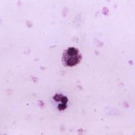
ICAM-2-deficient mice exhibit prolonged accumulation of eosinophils in lung interstitium concomitant with a delayed increase in eosinophil numbers in the airway lumen during the development of allergic lung inflammation. The ICAM-2-dependent increased and prolonged accumulation of eosinophils in lung interstitium results in prolonged, heightened airway hyperresponsiveness. These findings reveal an essential role for ICAM-2 in the development of the inflammatory and respiratory components of allergic lung disease. This phenotype is caused by the lack of ICAM-2 expression on non-hematopoietic cells. ICAM-2 deficiency on endothelial cells causes reduced eosinophil transmigration in vitro. ICAM-2 is not essential for lymphocyte homing or the development of leukocytes, with the exception of megakaryocyte progenitors, which are significantly reduced. (+info)Clonality of isolated eosinophils in the hypereosinophilic syndrome. (2/3575)
The idiopathic hypereosinophilic syndrome (IHES) is a rare disorder characterized by unexplained, persistent eosinophilia associated with multiple organ dysfunction due to eosinophilic tissue infiltration. In the absence of karyotypic abnormalities, there is no specific test to detect clonal eosinophilia in IHES. Analysis of X-chromosome inactivation patterns can be used to determine whether proliferative disorders are clonal in origin. Methylation of HpaII and Hha I sites near the polymorphic trinucleotide repeat of the human androgen receptor gene (HUMARA) has been shown to correlate with X-inactivation. In this study, we have used the polymerase chain reaction (PCR) with nested primers to analyze X-inactivation patterns of the HUMARA loci in purified eosinophils from female patients with eosinophilia. Peripheral blood eosinophils were isolated by their autofluoresence using flow cytometric sorting. Eosinophils purified from a female patient presenting with IHES were found to show a clonal pattern of X-inactivation. Eosinophil-depleted leukocytes from this patient were polyclonal by HUMARA analysis, thus excluding skewedness of random X-inactivation. After corticosteroid suppression of her blood eosinophilia, a clonal population of eosinophils could no longer be detected in purified eosinophils. In contrast, eosinophils purified from a patient with Churg-Strauss syndrome and from six patients with reactive eosinophilias attributed to allergy, parasitic infection, or drug reaction showed a polyclonal pattern of X-inactivation by HUMARA analysis. The finding of clonal eosinophilia in a patient presenting with IHES indicates that such patients may have, in reality, a low-grade clonal disorder that can be distinguished from reactive eosinophilias by HUMARA analysis. Further, the method described can be used to monitor disease progression. (+info)Structural determinants of the eosinophil: chemotactic activity of the acidic tetrapeptides of eosinophil chemotactic factor of anaphylaxis. (3/3575)
The acidic tetrapeptides of ECF-A, Ala/Val-Gly-Ser-Glu, exhibit peak in vitro chemotactic activity for human eosinophils at concentrations of 3 X 10(-8) M to 10(-6) M, and rapidly deactivate eosinophils to homologous and other stimuli at concentrations as low as 10(-10) M. The analogue Leu-Gly-Ser-Glu reaches peak activity at 10(-8)M-10(-7)M, while Phe-Gly-Ser-Glu requires 10(-4)M to elicit a peak response. Although inversion of the order of glycine and serine does not alter the eosinophil chemotactic activity of the tetrapeptides, deletion of glycine increases by 10-fold the concentration required for peak chemotactic activity, indicating the critical nature of the spacing between NH2- and COOH-terminal residues. The substituent COOH-terminal tripeptide, which is only marginally chemotactic, irreversibly suppresses eosinophil chemotactic responsiveness at a concentration 10,000-fold higher than concentrations necessary for deactivation by the intact tetrapeptide. The high concentration of tripeptide required for this cell directed effect, which is assumed to be analogous to deactivation, is attributed to the absence of the NH2-terminal residue which would facilitate effective interaction with the eosinophil. A substituent NH2-terminal tripeptide and amides of the NH2-terminal amino acids, which are devoid of chemotactic and deactivating activities, reversibly inhibit the tetrapeptide stimulus in a dose-response fashion. The additional finding that the NH2-terminal tripeptide protects the eosinophil from deactivation by the intact tetrapeptide confirms that the competitive interaction is stimulus specific. (+info)Selective eosinophil transendothelial migration triggered by eotaxin via modulation of Mac-1/ICAM-1 and VLA-4/VCAM-1 interactions. (4/3575)
We have recently cloned eotaxin, a highly efficacious eosinophilic chemokine involved in the development of lung eosinophilia during allergic inflammatory reactions. To understand more precisely how eotaxin facilitates the specific migration of eosinophils, we have studied which adhesion receptors are essential for eotaxin action both in vivo and in vitro. Experiments using mice genetically deficient in adhesion receptors demonstrated that molecules previously reported to be involved in both leukocyte tethering/rolling (P-selectin and E-selectin) and in sticking/ transmigration (ICAM-1 and VCAM-1) are required for eotaxin action in vivo. To further elucidate the mechanism(s) involved in this process, we have used an in vitro transendothelial chemotaxis model. mAb neutralization studies performed in this system suggest that the integrins Mac-1 (CD11b/18), VLA-4 (alpha4beta1) and LFA-1 (CD11a/18) are involved in the transendothelial chemotaxis of eosinophils to eotaxin. Accordingly, the expression of these integrins on eosinophils is elevated by direct action of this chemokine in a concentration-dependent manner. Taken together, our results suggest that eotaxin-induced eosinophil transendothelial migration in vivo and in vitro relies on Mac-1/ICAM-1 and VLA-4NCAM-1 interactions, the latter ones becoming more relevant at later time points of the eotaxin-induced recruitment process. (+info)Airway inflammatory response to ozone in subjects with different asthma severity. (5/3575)
The aim of this study was to evaluate whether ozone exposure induces a similar airway inflammatory response in subjects with different degrees of asthma severity. Two groups of asthmatic subjects were studied: seven with intermittent mild asthma not requiring regular treatment (group A); and seven with persistent mild asthma requiring regular treatment with inhaled corticosteroids and long-acting beta2-agonists (group B). All subjects were exposed, in a randomized cross-over design, to air or O3 (0.26 parts per million (ppm) for 2 h with intermittent exercise); subjects in group B withdrew from regular treatment 72 h before each exposure. Before the exposure, and 1 and 2 h after the beginning of the exposure they performed a pulmonary function test, and a questionnaire was completed to obtain a total symptom score (TSS). Six hours after the end of the exposure, hypertonic saline (HS) sputum induction was conducted. Sputum cell percentages, eosinophil cationic protein (ECP) and interleukin (IL)-8 concentrations in the sputum supernatant were measured. TSS significantly increased and forced vital capacity (FVC) and forced expiratory volume in one second (FEV1) significantly decreased after O3 exposure in comparison with air exposure in group A, whereas no changes were observed in group B except for a significant decrement of FEV1 2 h after the beginning of O3 exposure. Sputum neutrophil percentage was significantly higher after O3 exposure than after air exposure in both groups (Group A: 70.2% (28-87) versus 26.6% (8.6-73.2); Group B: 62.1% (25-82.4) versus 27.9% (14.4-54)). IL-8 was higher in sputum supernatant collected 6 h after O3 exposure than after air, only in group A. No change due to O3 has been found in sputum eosinophil percentage and ECP concentration in both groups. In conclusion, the degree of airway response to a short-term exposure to ozone is different in subjects with asthma of different severity. The available data do not allow elucidation of whether this difference depends on the severity of the disease or on the regular anti-inflammatory treatment. (+info)Orally exhaled nitric oxide levels are related to the degree of blood eosinophilia in atopic children with mild-intermittent asthma. (6/3575)
Increased levels of nitric oxide have been found in expired air of patients with asthma, and these are thought to be related to the airway inflammatory events that characterize this disorder. Since, in adults, bronchial inflammatory changes are present even in mild disease, the present study was designed to evaluate whether a significant proportion of children with mild-intermittent asthma could have increased exhaled air NO concentrations. Twenty-two atopic children (aged 11.1+/-0.8 yrs) with mild-intermittent asthma, treated only with inhaled beta2-adrenoreceptor agonists on demand and 22 age-matched controls were studied. NO concentrations in orally exhaled air, measured by chemiluminescence, were significantly higher in asthmatics, as compared to controls (19.4+/-3.3 parts per billion (ppb) and 4.0+/-0.5 ppb, respectively; p<0.01). Interestingly, 14 out of 22 asthmatic children had NO levels >8.8 ppb (i.e. >2 standard deviations of the mean in controls). In asthmatic patients, but not in control subjects, statistically significant correlations were found between exhaled NO levels and absolute number or percentage of blood eosinophils (r=0.63 and 0.56, respectively; p<0.01, each comparison). In contrast, exhaled NO levels were not correlated with forced expiratory volume in one second (FEV1) or forced expiratory flows at 25-75% of vital capacity (FEF25-75%) or forced vital capacity (FVC), either in control subjects, or in asthmatic patients (p>0.1, each correlation). These results suggest that a significant proportion of children with mild-intermittent asthma may have airway inflammation, as shown by the presence of elevated levels of nitric oxide in the exhaled air. The clinical relevance of this observation remains to be established. (+info)A genome-wide screen for asthma-associated quantitative trait loci in a mouse model of allergic asthma. (7/3575)
Asthma is the most common illness of childhood, affecting one child in seven in the UK. Asthma has a genetic basis, but genetic studies of asthma in humans are confounded by uncontrolled environmental factors, varying penetrance and phenotypic pleiotropy. An animal model of asthma would offer controlled exposure, limited and consistent genetic variation, and unlimited size of sibships. Following immunization and subsequent challenge with ovalbumin, the Biozzi BP2 mouse shows features of asthma, including airway inflammation, eosinophil infiltration and non-specific bronchial responsiveness. In order to identify genetic loci influencing these traits, a cross was made between BP2 and BALB/c mice, and a genome-wide screen carried out in the F2progeny of the F1intercross. Five potentially linked loci were identified, four of which corresponded to human regions of syntenic homology that previously have shown linkage to asthma-associated traits. (+info)Effects of Th2 cytokines on chemokine expression in the lung: IL-13 potently induces eotaxin expression by airway epithelial cells. (8/3575)
Airway inflammation associated with asthma is characterized by massive infiltration of eosinophils, mediated in part by specific chemoattractant factors produced in the lung. Allergen-specific Th2 cells appear to play a central role in asthma; for example, adoptively transferred Th2 cells induced lung eosinophilia associated with induction of specific chemokines. Interestingly, Th2 supernatant alone administered intranasally to naive mice induced eotaxin, RANTES, monocyte-chemotactic protein-1, and KC expression along with lung eosinophilia. We tested the major cytokines individually and found that IL-4 and IL-5 induced higher levels of macrophage-inflammatory protein-1alpha and KC; IL-4 also increased the production of monocyte-chemotactic protein-1; IL-13 and IL-4 induced eotaxin. IL-13 was by far the most potent inducer of eotaxin; indeed, a neutralizing anti-IL-13 Ab removed most of the eotaxin-inducing activity from Th2 supernatants, although it did not entirely block the recruitment of eosinophils. While TNF-alpha did not stimulate eotaxin production by itself, it markedly augmented eotaxin induction by IL-13. IL-13 was able to induce eotaxin in the lung of JAK3-deficient mice, suggesting that JAK3 is not required for IL-13 signaling in airway epithelial cells; however, eosinophilia was not induced in this situation, suggesting that JAK3 transduces other IL-13-mediated mechanisms critical for eosinophil recruitment. Our study suggests that IL-13 is an important mediator in the pathogenesis of asthma and therefore a potential target for asthma therapy. (+info)Eosinophils are a type of white blood cell that play an important role in the body's immune response. They are produced in the bone marrow and released into the bloodstream, where they can travel to different tissues and organs throughout the body. Eosinophils are characterized by their granules, which contain various proteins and enzymes that are toxic to parasites and can contribute to inflammation.
Eosinophils are typically associated with allergic reactions, asthma, and other inflammatory conditions. They can also be involved in the body's response to certain infections, particularly those caused by parasites such as worms. In some cases, elevated levels of eosinophils in the blood or tissues (a condition called eosinophilia) can indicate an underlying medical condition, such as a parasitic infection, autoimmune disorder, or cancer.
Eosinophils are named for their staining properties - they readily take up eosin dye, which is why they appear pink or red under the microscope. They make up only about 1-6% of circulating white blood cells in healthy individuals, but their numbers can increase significantly in response to certain triggers.
Eosinophil peroxidase (EPO) is an enzyme that is primarily found in the granules of eosinophils, which are a type of white blood cell that plays a role in the immune response. EPO is involved in the destruction of certain types of parasites and also contributes to the inflammatory response in allergic reactions and other diseases.
EPO catalyzes the conversion of hydrogen peroxide to hypochlorous acid, which is a potent oxidizing agent that can kill or inhibit the growth of microorganisms. EPO also plays a role in the production of other reactive oxygen species, which can contribute to tissue damage and inflammation in certain conditions.
Elevated levels of EPO in tissues or bodily fluids may be indicative of eosinophil activation and degranulation, which can occur in various diseases such as asthma, allergies, parasitic infections, and some types of cancer. Measuring EPO levels can be useful in the diagnosis and monitoring of these conditions.
Eosinophil granule proteins are a group of biologically active molecules that are stored within the granules of eosinophils, which are types of white blood cells. These proteins include:
1. Eosinophil cationic protein (ECP): A protein with potent ribonuclease activity and the ability to disrupt cell membranes. It is involved in the immune response against parasites and has been implicated in the pathogenesis of several inflammatory diseases, such as asthma and allergies.
2. Eosinophil peroxidase (EPO): An enzyme that generates hypohalous acids, which can cause oxidative damage to cells and tissues. It contributes to the microbicidal activity of eosinophils and has been implicated in the pathogenesis of various inflammatory diseases.
3. Major basic protein (MBP): A highly cationic protein that can disrupt cell membranes, leading to cell lysis. MBP is involved in the immune response against parasites and has been linked to tissue damage in several inflammatory conditions, such as asthma, chronic rhinosinusitis, and eosinophilic esophagitis.
4. Eosinophil-derived neurotoxin (EDN): A protein with ribonuclease activity that can induce histamine release from mast cells and contribute to the inflammatory response. EDN is also involved in the immune response against parasites and has been implicated in the pathogenesis of asthma, allergies, and other inflammatory diseases.
These eosinophil granule proteins are released during eosinophil activation and degranulation, which can occur in response to various stimuli, such as immune complexes, cytokines, and infectious agents. Their release contributes to the inflammatory response and can lead to tissue damage in various diseases.
Chemotactic factors are substances that attract and guide cells, particularly immune cells, to specific locations in the body. Eosinophils are a type of white blood cell that play a role in the immune response, particularly against parasites and in allergic reactions. Therefore, chemotactic factors for eosinophils are substances that attract eosinophils to specific sites in the body.
These factors can be produced by various cells, including mast cells, basophils, and T-lymphocytes, in response to an infection or inflammation. They work by binding to receptors on the surface of eosinophils and activating signaling pathways that cause the eosinophils to migrate towards the source of the chemotactic factor.
Examples of chemotactic factors for eosinophils include:
1. Eotaxins: These are a group of chemokines (a type of signaling protein) that specifically attract eosinophils. They are produced by various cells, including endothelial cells, epithelial cells, and immune cells.
2. Leukotrienes: These are lipid mediators produced by mast cells and basophils in response to an allergic reaction or infection. They can attract eosinophils to the site of inflammation.
3. Platelet-activating factor (PAF): This is a lipid mediator produced by various cells, including endothelial cells and immune cells. It can attract eosinophils and activate them, leading to degranulation and release of their contents.
4. Complement components: The complement system is a group of proteins that play a role in the immune response. Some complement components, such as C3a and C5a, can act as chemotactic factors for eosinophils.
Overall, chemotactic factors for eosinophils play an important role in the immune response by recruiting these cells to sites of infection or inflammation. However, excessive activation of eosinophils and production of chemotactic factors can contribute to the development of various diseases, such as asthma and allergies.
Eosinophilia is a medical condition characterized by an abnormally high concentration of eosinophils in the circulating blood. Eosinophils are a type of white blood cell that play an important role in the immune system, particularly in fighting off parasitic infections and regulating allergic reactions. However, when their numbers become excessively high, they can contribute to tissue damage and inflammation.
Eosinophilia is typically defined as a count of more than 500 eosinophils per microliter of blood. Mild eosinophilia (up to 1,500 cells/μL) may not cause any symptoms and may be discovered during routine blood tests. However, higher levels of eosinophilia can lead to various symptoms such as coughing, wheezing, skin rashes, and organ damage, depending on the underlying cause.
The causes of eosinophilia are varied and can include allergic reactions, parasitic infections, autoimmune disorders, certain medications, and some types of cancer. Accurate diagnosis and treatment of eosinophilia require identification and management of the underlying cause.
Interleukin-5 (IL-5) is a type of cytokine, which is a small signaling protein that mediates and regulates immunity, inflammation, and hematopoiesis. IL-5 is primarily produced by activated T cells, especially Th2 cells, as well as mast cells, eosinophils, and innate lymphoid cells (ILCs).
The primary function of IL-5 is to regulate the growth, differentiation, activation, and survival of eosinophils, a type of white blood cell that plays a crucial role in the immune response against parasitic infections. IL-5 also enhances the ability of eosinophils to migrate from the bone marrow into the bloodstream and then into tissues, where they can participate in immune responses.
In addition to its effects on eosinophils, IL-5 has been shown to have a role in the regulation of B cell function, including promoting the survival and differentiation of B cells into antibody-secreting plasma cells. Dysregulation of IL-5 production and activity has been implicated in several diseases, including asthma, allergies, and certain parasitic infections.
Eosinophil Cationic Protein (ECP) is a protein found in the granules of eosinophils, which are a type of white blood cell that plays a role in the immune response, particularly against parasitic infections and allergens. ECP is released from eosinophils during degranulation, a process that occurs when these cells are activated and release their granules' contents.
Elevated levels of ECP in body fluids, such as blood or sputum, can indicate eosinophil activation and may be associated with various inflammatory conditions, including asthma, allergies, and some parasitic infections. Measuring ECP levels can help monitor disease activity and assess the effectiveness of treatment in these conditions.
Chemokine CCL11, also known as eotaxin-1, is a small chemotactic cytokine that belongs to the CC subfamily of chemokines. Chemokines are a group of proteins that play crucial roles in immunity and inflammation by recruiting immune cells to sites of infection or tissue injury.
CCL11 specifically attracts eosinophils, a type of white blood cell that is involved in allergic reactions and the immune response to parasitic worm infections. It does this by binding to its specific receptor, CCR3, which is expressed on the surface of eosinophils and other cells.
CCL11 is produced by a variety of cells, including epithelial cells, endothelial cells, fibroblasts, and immune cells such as macrophages and Th2 lymphocytes. It has been implicated in the pathogenesis of several diseases, including asthma, allergies, and certain neurological disorders.
Eosinophil Major Basic Protein (eMBP) is a cytotoxic protein found in the granules of eosinophils, which are a type of white blood cell that plays a role in the immune response, particularly against parasitic infections. eMBP is one of the four major basic proteins (MBPs) and is released during degranulation of eosinophils, a process that occurs in response to certain stimuli such as allergens or parasites.
eMBP has been found to have several biological activities, including direct toxicity to various cells, including parasites, mast cells, and airway epithelial cells. It can also induce the production of pro-inflammatory cytokines and chemokines, contributing to the inflammation observed in diseases such as asthma and allergies.
It is important to note that while eMBP has been extensively studied for its role in immunity and disease, further research is needed to fully understand its mechanisms of action and potential therapeutic applications.
Eosinophil-Derived Neurotoxin (EDN) is a protein that is released from the granules of eosinophils, which are a type of white blood cell involved in the immune response. EDN has both neurotoxic and ribonucleolytic activities, meaning it can damage nerve cells and also degrade RNA. It is thought to play a role in the pathogenesis of certain diseases such as asthma and some forms of inflammatory bowel disease. EDN is also known as eosinophil cationic protein or ECP.
Ribonucleases (RNases) are a group of enzymes that catalyze the degradation of ribonucleic acid (RNA) molecules by hydrolyzing the phosphodiester bonds. These enzymes play crucial roles in various biological processes, such as RNA processing, turnover, and quality control. They can be classified into several types based on their specificities, mechanisms, and cellular localizations.
Some common classes of ribonucleases include:
1. Endoribonucleases: These enzymes cleave RNA internally, at specific sequences or structural motifs. Examples include RNase A, which targets single-stranded RNA; RNase III, which cuts double-stranded RNA at specific stem-loop structures; and RNase T1, which recognizes and cuts unpaired guanosine residues in RNA molecules.
2. Exoribonucleases: These enzymes remove nucleotides from the ends of RNA molecules. They can be further divided into 5'-3' exoribonucleases, which degrade RNA starting from the 5' end, and 3'-5' exoribonucleases, which start at the 3' end. Examples include Xrn1, a 5'-3' exoribonuclease involved in mRNA decay; and Dis3/RRP6, a 3'-5' exoribonuclease that participates in ribosomal RNA processing and degradation.
3. Specific ribonucleases: These enzymes target specific RNA molecules or regions with high precision. For example, RNase P is responsible for cleaving the 5' leader sequence of precursor tRNAs (pre-tRNAs) during their maturation; and RNase MRP is involved in the processing of ribosomal RNA and mitochondrial RNA molecules.
Dysregulation or mutations in ribonucleases have been implicated in various human diseases, such as neurological disorders, cancer, and viral infections. Therefore, understanding their functions and mechanisms is crucial for developing novel therapeutic strategies.
CCR3 (C-C chemokine receptor type 3) is a type of cell surface receptor that binds to specific chemokines, which are a group of small signaling proteins involved in immune responses and inflammation. CCR3 is primarily expressed on the surface of certain types of immune cells, including eosinophils, basophils, and Th2 lymphocytes.
The binding of chemokines to CCR3 triggers a series of intracellular signaling events that regulate various cellular functions, such as chemotaxis (directed migration), activation, and degranulation. CCR3 plays an important role in the pathophysiology of several diseases, including asthma, allergies, and inflammatory bowel disease, where it contributes to the recruitment and activation of immune cells that mediate tissue damage and inflammation.
Therefore, CCR3 is a potential target for the development of therapies aimed at modulating immune responses and reducing inflammation in these conditions.
Pulmonary eosinophilia is a condition characterized by an increased number of eosinophils, a type of white blood cell, in the lungs or pulmonary tissues. Eosinophils play a role in the body's immune response to parasites and allergens, but an overabundance can contribute to inflammation and damage in the lungs.
The condition may be associated with various underlying causes, such as:
1. Asthma or allergic bronchopulmonary aspergillosis (ABPA)
2. Eosinophilic lung diseases, like eosinophilic pneumonia or idiopathic hypereosinophilic syndrome
3. Parasitic infections, such as ascariasis or strongyloidiasis
4. Drug reactions, including certain antibiotics and anti-inflammatory drugs
5. Connective tissue disorders, like rheumatoid arthritis or Churg-Strauss syndrome
6. Malignancies, such as lymphoma or leukemia
7. Other less common conditions, like tropical pulmonary eosinophilia or cryptogenic organizing pneumonia
Symptoms of pulmonary eosinophilia can vary but often include cough, shortness of breath, wheezing, and chest discomfort. Diagnosis typically involves a combination of clinical evaluation, imaging studies, and laboratory tests, such as complete blood count (CBC) with differential, bronchoalveolar lavage (BAL), or lung biopsy. Treatment depends on the underlying cause and may include corticosteroids, antibiotics, or antiparasitic medications.
Chemokine CCL24, also known as Eotaxin-2, is a type of small signaling protein that belongs to the CC chemokine family. Chemokines are involved in immune responses and inflammation, and they help direct the movement of cells around the body by interacting with specific receptors on their surfaces.
CCL24 is primarily produced by epithelial cells, fibroblasts, and endothelial cells, and it plays a crucial role in recruiting eosinophils, a type of white blood cell that is involved in allergic reactions and inflammatory responses, to sites of injury or infection. CCL24 exerts its effects by binding to the CCR3 receptor on the surface of eosinophils and other immune cells.
Abnormal levels of CCL24 have been implicated in several diseases, including asthma, allergies, and certain types of cancer. For example, increased levels of CCL24 have been found in the airways of people with asthma, and they have been associated with more severe disease and poorer lung function. Similarly, elevated levels of CCL24 have been detected in the tumor microenvironment of several cancers, where they may contribute to the recruitment of immune cells that promote tumor growth and metastasis.
Chemokines are a family of small proteins that are involved in immune responses and inflammation. They mediate the chemotaxis (directed migration) of various cells, including leukocytes (white blood cells). Chemokines are classified into four major subfamilies based on the arrangement of conserved cysteine residues near the amino terminus: CXC, CC, C, and CX3C.
CC chemokines, also known as β-chemokines, are characterized by the presence of two adjacent cysteine residues near their N-terminal end. There are 27 known human CC chemokines, including MCP-1 (monocyte chemoattractant protein-1), RANTES (regulated on activation, normal T cell expressed and secreted), and eotaxin.
CC chemokines play important roles in the recruitment of immune cells to sites of infection or injury, as well as in the development and maintenance of immune responses. They bind to specific G protein-coupled receptors (GPCRs) on the surface of target cells, leading to the activation of intracellular signaling pathways that regulate cell migration, proliferation, and survival.
Dysregulation of CC chemokines and their receptors has been implicated in various inflammatory and autoimmune diseases, as well as in cancer. Therefore, targeting CC chemokine-mediated signaling pathways has emerged as a promising therapeutic strategy for the treatment of these conditions.
Asthma is a chronic respiratory disease characterized by inflammation and narrowing of the airways, leading to symptoms such as wheezing, coughing, shortness of breath, and chest tightness. The airway obstruction in asthma is usually reversible, either spontaneously or with treatment.
The underlying cause of asthma involves a combination of genetic and environmental factors that result in hypersensitivity of the airways to certain triggers, such as allergens, irritants, viruses, exercise, and emotional stress. When these triggers are encountered, the airways constrict due to smooth muscle spasm, swell due to inflammation, and produce excess mucus, leading to the characteristic symptoms of asthma.
Asthma is typically managed with a combination of medications that include bronchodilators to relax the airway muscles, corticosteroids to reduce inflammation, and leukotriene modifiers or mast cell stabilizers to prevent allergic reactions. Avoiding triggers and monitoring symptoms are also important components of asthma management.
There are several types of asthma, including allergic asthma, non-allergic asthma, exercise-induced asthma, occupational asthma, and nocturnal asthma, each with its own set of triggers and treatment approaches. Proper diagnosis and management of asthma can help prevent exacerbations, improve quality of life, and reduce the risk of long-term complications.
A leukocyte count, also known as a white blood cell (WBC) count, is a laboratory test that measures the number of leukocytes in a sample of blood. Leukocytes are a vital part of the body's immune system and help fight infection and inflammation. A high or low leukocyte count may indicate an underlying medical condition, such as an infection, inflammation, or a bone marrow disorder. The normal range for a leukocyte count in adults is typically between 4,500 and 11,000 cells per microliter (mcL) of blood. However, the normal range can vary slightly depending on the laboratory and the individual's age and sex.
Cell degranulation is the process by which cells, particularly immune cells like mast cells and basophils, release granules containing inflammatory mediators in response to various stimuli. These mediators include histamine, leukotrienes, prostaglandins, and other chemicals that play a role in allergic reactions, inflammation, and immune responses. The activation of cell surface receptors triggers a signaling cascade that leads to the exocytosis of these granules, resulting in degranulation. This process is important for the immune system's response to foreign invaders and for the development of allergic reactions.
Blood proteins, also known as serum proteins, are a group of complex molecules present in the blood that are essential for various physiological functions. These proteins include albumin, globulins (alpha, beta, and gamma), and fibrinogen. They play crucial roles in maintaining oncotic pressure, transporting hormones, enzymes, vitamins, and minerals, providing immune defense, and contributing to blood clotting.
Albumin is the most abundant protein in the blood, accounting for about 60% of the total protein mass. It functions as a transporter of various substances, such as hormones, fatty acids, and drugs, and helps maintain oncotic pressure, which is essential for fluid balance between the blood vessels and surrounding tissues.
Globulins are divided into three main categories: alpha, beta, and gamma globulins. Alpha and beta globulins consist of transport proteins like lipoproteins, hormone-binding proteins, and enzymes. Gamma globulins, also known as immunoglobulins or antibodies, are essential for the immune system's defense against pathogens.
Fibrinogen is a protein involved in blood clotting. When an injury occurs, fibrinogen is converted into fibrin, which forms a mesh to trap platelets and form a clot, preventing excessive bleeding.
Abnormal levels of these proteins can indicate various medical conditions, such as liver or kidney disease, malnutrition, infections, inflammation, or autoimmune disorders. Blood protein levels are typically measured through laboratory tests like serum protein electrophoresis (SPE) and immunoelectrophoresis (IEP).
Chemotaxis, Leukocyte is the movement of leukocytes (white blood cells) towards a higher concentration of a particular chemical substance, known as a chemotactic factor. This process plays a crucial role in the immune system's response to infection and injury.
When there is an infection or tissue damage, certain cells release chemotactic factors, which are small molecules or proteins that can attract leukocytes to the site of inflammation. Leukocytes have receptors on their surface that can detect these chemotactic factors and move towards them through a process called chemotaxis.
Once they reach the site of inflammation, leukocytes can help eliminate pathogens or damaged cells by phagocytosis (engulfing and destroying) or releasing toxic substances that kill the invading microorganisms. Chemotaxis is an essential part of the immune system's defense mechanisms and helps to maintain tissue homeostasis and prevent the spread of infection.
Hypereosinophilic Syndrome (HES) is a group of disorders characterized by persistent eosinophilia (an abnormal increase in the number of eosinophils, a type of white blood cell) leading to organ damage. The eosinophil count in the peripheral blood is typically greater than 1500 cells/μL. HES can affect various organs, including the heart, skin, nervous system, and digestive tract, causing symptoms such as shortness of breath, cough, fatigue, skin rashes, muscle weakness, and abdominal pain. The exact cause of HES is not fully understood, but it is thought to be related to abnormal production or activation of eosinophils. Treatment may include corticosteroids, immunosuppressive drugs, and targeted therapies that reduce eosinophil levels.
Leukotriene C4 (LTC4) is a type of lipid mediator called a cysteinyl leukotriene, which is derived from arachidonic acid through the 5-lipoxygenase pathway. It is primarily produced by activated mast cells and basophils, and to a lesser extent by eosinophils, during an allergic response or inflammation.
LTC4 plays a crucial role in the pathogenesis of asthma and other allergic diseases by causing bronchoconstriction, increased vascular permeability, mucus secretion, and recruitment of inflammatory cells to the site of inflammation. It exerts its effects by binding to cysteinyl leukotriene receptors (CysLT1 and CysLT2) found on various cell types, including airway smooth muscle cells, bronchial epithelial cells, and immune cells.
LTC4 is rapidly metabolized to Leukotriene D4 (LTD4) and then to Leukotriene E4 (LTE4) by enzymes such as gamma-glutamyl transpeptidase and dipeptidases, which are present in the extracellular space. These metabolites also have biological activity and contribute to the inflammatory response.
Inhibitors of 5-lipoxygenase or leukotriene receptor antagonists are used as therapeutic agents for the treatment of asthma, allergies, and other inflammatory conditions.
Ovalbumin is the major protein found in egg white, making up about 54-60% of its total protein content. It is a glycoprotein with a molecular weight of around 45 kDa and has both hydrophilic and hydrophobic regions. Ovalbumin is a single polypeptide chain consisting of 385 amino acids, including four disulfide bridges that contribute to its structure.
Ovalbumin is often used in research as a model antigen for studying immune responses and allergies. In its native form, ovalbumin is not allergenic; however, when it is denatured or degraded into smaller peptides through cooking or digestion, it can become an allergen for some individuals.
In addition to being a food allergen, ovalbumin has been used in various medical and research applications, such as vaccine development, immunological studies, and protein structure-function analysis.
Peroxidases are a group of enzymes that catalyze the oxidation of various substrates using hydrogen peroxide (H2O2) as the electron acceptor. These enzymes contain a heme prosthetic group, which plays a crucial role in their catalytic activity. Peroxidases are widely distributed in nature and can be found in plants, animals, and microorganisms. They play important roles in various biological processes, including defense against oxidative stress, lignin degradation, and host-pathogen interactions. Some common examples of peroxidases include glutathione peroxidase, which helps protect cells from oxidative damage, and horseradish peroxidase, which is often used in laboratory research.
An allergen is a substance that can cause an allergic reaction in some people. These substances are typically harmless to most people, but for those with allergies, the immune system mistakenly identifies them as threats and overreacts, leading to the release of histamines and other chemicals that cause symptoms such as itching, sneezing, runny nose, rashes, hives, and difficulty breathing. Common allergens include pollen, dust mites, mold spores, pet dander, insect venom, and certain foods or medications. When a person comes into contact with an allergen, they may experience symptoms that range from mild to severe, depending on the individual's sensitivity to the substance and the amount of exposure.
Hypersensitivity is an exaggerated or inappropriate immune response to a substance that is generally harmless to most people. It's also known as an allergic reaction. This abnormal response can be caused by various types of immunological mechanisms, including antibody-mediated reactions (types I, II, and III) and cell-mediated reactions (type IV). The severity of the hypersensitivity reaction can range from mild discomfort to life-threatening conditions. Common examples of hypersensitivity reactions include allergic rhinitis, asthma, atopic dermatitis, food allergies, and anaphylaxis.
Bronchoalveolar lavage (BAL) fluid is a type of clinical specimen obtained through a procedure called bronchoalveolar lavage. This procedure involves inserting a bronchoscope into the lungs and instilling a small amount of saline solution into a specific area of the lung, then gently aspirating the fluid back out. The fluid that is recovered is called bronchoalveolar lavage fluid.
BAL fluid contains cells and other substances that are present in the lower respiratory tract, including the alveoli (the tiny air sacs where gas exchange occurs). By analyzing BAL fluid, doctors can diagnose various lung conditions, such as pneumonia, interstitial lung disease, and lung cancer. They can also monitor the effectiveness of treatments for these conditions by comparing the composition of BAL fluid before and after treatment.
BAL fluid is typically analyzed for its cellular content, including the number and type of white blood cells present, as well as for the presence of bacteria, viruses, or other microorganisms. The fluid may also be tested for various proteins, enzymes, and other biomarkers that can provide additional information about lung health and disease.
Bronchial hyperresponsiveness (BHR) or bronchial hyperreactivity (BH) is a medical term that refers to the increased sensitivity and exaggerated response of the airways to various stimuli. In people with BHR, the airways narrow (constrict) more than usual in response to certain triggers such as allergens, cold air, exercise, or irritants like smoke or fumes. This narrowing can cause symptoms such as wheezing, coughing, chest tightness, and shortness of breath.
BHR is often associated with asthma and other respiratory conditions, including chronic obstructive pulmonary disease (COPD) and bronchiectasis. It is typically diagnosed through a series of tests that measure the degree of airway narrowing in response to various stimuli. These tests may include spirometry, methacholine challenge test, or histamine challenge test.
BHR can be managed with medications such as bronchodilators and anti-inflammatory drugs, which help to relax the muscles around the airways and reduce inflammation. It is also important to avoid triggers that can exacerbate symptoms and make BHR worse.
Platelet-activating factor (PAF) is a potent phospholipid mediator that plays a significant role in various inflammatory and immune responses. It is a powerful lipid signaling molecule released mainly by activated platelets, neutrophils, monocytes, endothelial cells, and other cell types during inflammation or injury.
PAF has a molecular structure consisting of an alkyl chain linked to a glycerol moiety, a phosphate group, and an sn-2 acetyl group. This unique structure allows PAF to bind to its specific G protein-coupled receptor (PAF-R) on the surface of target cells, triggering various intracellular signaling cascades that result in cell activation, degranulation, and aggregation.
The primary functions of PAF include:
1. Platelet activation and aggregation: PAF stimulates platelets to aggregate, release their granules, and activate the coagulation cascade, which can lead to thrombus formation.
2. Neutrophil and monocyte activation: PAF activates these immune cells, leading to increased adhesion, degranulation, and production of reactive oxygen species (ROS) and pro-inflammatory cytokines.
3. Vasodilation and increased vascular permeability: PAF can cause vasodilation by acting on endothelial cells, leading to an increase in blood flow and facilitating the extravasation of immune cells into inflamed tissues.
4. Bronchoconstriction: In the respiratory system, PAF can induce bronchoconstriction and recruitment of inflammatory cells, contributing to asthma symptoms.
5. Neurotransmission modulation: PAF has been implicated in neuroinflammation and may play a role in neuronal excitability, synaptic plasticity, and cognitive functions.
Dysregulated PAF signaling has been associated with several pathological conditions, including atherosclerosis, sepsis, acute respiratory distress syndrome (ARDS), ischemia-reperfusion injury, and neuroinflammatory disorders. Therefore, targeting the PAF pathway may provide therapeutic benefits in these diseases.
Neutrophils are a type of white blood cell that are part of the immune system's response to infection. They are produced in the bone marrow and released into the bloodstream where they circulate and are able to move quickly to sites of infection or inflammation in the body. Neutrophils are capable of engulfing and destroying bacteria, viruses, and other foreign substances through a process called phagocytosis. They are also involved in the release of inflammatory mediators, which can contribute to tissue damage in some cases. Neutrophils are characterized by the presence of granules in their cytoplasm, which contain enzymes and other proteins that help them carry out their immune functions.
Immunoglobulin E (IgE) is a type of antibody that plays a key role in the immune response to parasitic infections and allergies. It is produced by B cells in response to stimulation by antigens, such as pollen, pet dander, or certain foods. Once produced, IgE binds to receptors on the surface of mast cells and basophils, which are immune cells found in tissues and blood respectively. When an individual with IgE antibodies encounters the allergen again, the cross-linking of IgE molecules bound to the FcεRI receptor triggers the release of mediators such as histamine, leukotrienes, prostaglandins, and various cytokines from these cells. These mediators cause the symptoms of an allergic reaction, such as itching, swelling, and redness. IgE also plays a role in protecting against certain parasitic infections by activating eosinophils, which can kill the parasites.
In summary, Immunoglobulin E (IgE) is a type of antibody that plays a crucial role in the immune response to allergens and parasitic infections, it binds to receptors on the surface of mast cells and basophils, when an individual with IgE antibodies encounters the allergen again, it triggers the release of mediators from these cells causing the symptoms of an allergic reaction.
Respiratory hypersensitivity, also known as respiratory allergies or hypersensitive pneumonitis, refers to an exaggerated immune response in the lungs to inhaled substances or allergens. This condition occurs when the body's immune system overreacts to harmless particles, leading to inflammation and damage in the airways and alveoli (air sacs) of the lungs.
There are two types of respiratory hypersensitivity: immediate and delayed. Immediate hypersensitivity, also known as type I hypersensitivity, is mediated by immunoglobulin E (IgE) antibodies and results in symptoms such as sneezing, runny nose, and asthma-like symptoms within minutes to hours of exposure to the allergen. Delayed hypersensitivity, also known as type III or type IV hypersensitivity, is mediated by other immune mechanisms and can take several hours to days to develop after exposure to the allergen.
Common causes of respiratory hypersensitivity include mold spores, animal dander, dust mites, pollen, and chemicals found in certain occupations. Symptoms may include coughing, wheezing, shortness of breath, chest tightness, and fatigue. Treatment typically involves avoiding the allergen, if possible, and using medications such as corticosteroids, bronchodilators, or antihistamines to manage symptoms. In severe cases, immunotherapy (allergy shots) may be recommended to help desensitize the immune system to the allergen.
Granulocyte-Macrophage Colony-Stimulating Factor (GM-CSF) is a type of cytokine, which is a small signaling protein involved in immune response and hematopoiesis (the formation of blood cells). GM-CSF's specific role is to stimulate the production, proliferation, and activation of granulocytes (a type of white blood cell that fights against infection) and macrophages (large white blood cells that eat foreign substances, bacteria, and dead or dying cells).
In medical terms, GM-CSF is often used in therapeutic settings to boost the production of white blood cells in patients undergoing chemotherapy or radiation treatment for cancer. This can help to reduce the risk of infection during these treatments. It can also be used to promote the growth and differentiation of stem cells in bone marrow transplant procedures.
Basophils are a type of white blood cell that are part of the immune system. They are granulocytes, which means they contain granules filled with chemicals that can be released in response to an infection or inflammation. Basophils are relatively rare, making up less than 1% of all white blood cells.
When basophils become activated, they release histamine and other chemical mediators that can contribute to allergic reactions, such as itching, swelling, and redness. They also play a role in inflammation, helping to recruit other immune cells to the site of an infection or injury.
Basophils can be identified under a microscope based on their characteristic staining properties. They are typically smaller than other granulocytes, such as neutrophils and eosinophils, and have a multi-lobed nucleus with dark purple-staining granules in the cytoplasm.
While basophils play an important role in the immune response, abnormal levels of basophils can be associated with various medical conditions, such as allergies, infections, and certain types of leukemia.
A lung is a pair of spongy, elastic organs in the chest that work together to enable breathing. They are responsible for taking in oxygen and expelling carbon dioxide through the process of respiration. The left lung has two lobes, while the right lung has three lobes. The lungs are protected by the ribcage and are covered by a double-layered membrane called the pleura. The trachea divides into two bronchi, which further divide into smaller bronchioles, leading to millions of tiny air sacs called alveoli, where the exchange of gases occurs.
Cytoplasmic granules are small, membrane-bound organelles or inclusions found within the cytoplasm of cells. They contain various substances such as proteins, lipids, carbohydrates, and genetic material. Cytoplasmic granules have diverse functions depending on their specific composition and cellular location. Some examples include:
1. Secretory granules: These are found in secretory cells and store hormones, neurotransmitters, or enzymes before they are released by exocytosis.
2. Lysosomes: These are membrane-bound organelles that contain hydrolytic enzymes for intracellular digestion of waste materials, foreign substances, and damaged organelles.
3. Melanosomes: Found in melanocytes, these granules produce and store the pigment melanin, which is responsible for skin, hair, and eye color.
4. Weibel-Palade bodies: These are found in endothelial cells and store von Willebrand factor and P-selectin, which play roles in hemostasis and inflammation.
5. Peroxisomes: These are single-membrane organelles that contain enzymes for various metabolic processes, such as β-oxidation of fatty acids and detoxification of harmful substances.
6. Lipid bodies (also called lipid droplets): These are cytoplasmic granules that store neutral lipids, such as triglycerides and cholesteryl esters. They play a role in energy metabolism and intracellular signaling.
7. Glycogen granules: These are cytoplasmic inclusions that store glycogen, a polysaccharide used for energy storage in animals.
8. Protein bodies: Found in plants, these granules store excess proteins and help regulate protein homeostasis within the cell.
9. Electron-dense granules: These are found in certain immune cells, such as mast cells and basophils, and release mediators like histamine during an allergic response.
10. Granules of unknown composition or function may also be present in various cell types.
Interleukin-5 (IL-5) receptors are a type of cell surface receptor that bind to and respond to the cytokine IL-5. These receptors are found on the surface of certain immune cells, including eosinophils, basophils, and some types of T cells.
The IL-5 receptor is a heterodimer, meaning it is composed of two different subunits: the alpha (IL-5Rα) and beta (IL-5Rβ) chains. The alpha chain is specific to IL-5 and confers binding specificity, while the beta chain is shared with other cytokine receptors and mediates signal transduction.
Activation of the IL-5 receptor leads to a variety of cellular responses, including proliferation, differentiation, and survival of eosinophils and basophils. These cells play important roles in the immune response, particularly in the defense against parasitic infections and in allergic reactions. Dysregulation of IL-5 signaling has been implicated in several diseases, including asthma, chronic obstructive pulmonary disease (COPD), and eosinophilic disorders.
Leukotriene B4 (LTB4) is a type of lipid mediator called eicosanoid, which is derived from arachidonic acid through the 5-lipoxygenase pathway. It is primarily produced by neutrophils, eosinophils, monocytes, and macrophages in response to various stimuli such as infection, inflammation, or injury. LTB4 acts as a potent chemoattractant and activator of these immune cells, playing a crucial role in the recruitment and activation of neutrophils during acute inflammatory responses. It also enhances the adhesion of leukocytes to endothelial cells, contributing to the development of tissue damage and edema. Dysregulation of LTB4 production has been implicated in several pathological conditions, including asthma, atherosclerosis, and cancer.
Cytokines are a broad and diverse category of small signaling proteins that are secreted by various cells, including immune cells, in response to different stimuli. They play crucial roles in regulating the immune response, inflammation, hematopoiesis, and cellular communication.
Cytokines mediate their effects by binding to specific receptors on the surface of target cells, which triggers intracellular signaling pathways that ultimately result in changes in gene expression, cell behavior, and function. Some key functions of cytokines include:
1. Regulating the activation, differentiation, and proliferation of immune cells such as T cells, B cells, natural killer (NK) cells, and macrophages.
2. Coordinating the inflammatory response by recruiting immune cells to sites of infection or tissue damage and modulating their effector functions.
3. Regulating hematopoiesis, the process of blood cell formation in the bone marrow, by controlling the proliferation, differentiation, and survival of hematopoietic stem and progenitor cells.
4. Modulating the development and function of the nervous system, including neuroinflammation, neuroprotection, and neuroregeneration.
Cytokines can be classified into several categories based on their structure, function, or cellular origin. Some common types of cytokines include interleukins (ILs), interferons (IFNs), tumor necrosis factors (TNFs), chemokines, colony-stimulating factors (CSFs), and transforming growth factors (TGFs). Dysregulation of cytokine production and signaling has been implicated in various pathological conditions, such as autoimmune diseases, chronic inflammation, cancer, and neurodegenerative disorders.
Pleurisy is a medical condition characterized by inflammation of the pleura, which are the thin membranes that surround the lungs and line the inside of the chest cavity. The pleura normally produce a small amount of lubricating fluid that allows for smooth movement of the lungs during breathing. However, when they become inflamed (a condition known as pleuritis), this can cause pain and difficulty breathing.
The symptoms of pleurisy may include sharp chest pain that worsens with deep breathing or coughing, shortness of breath, cough, fever, and muscle aches. The pain may be localized to one area of the chest or may radiate to other areas such as the shoulders or back.
Pleurisy can have many different causes, including bacterial or viral infections, autoimmune disorders, pulmonary embolism (a blood clot that travels to the lungs), and certain medications or chemicals. Treatment typically involves addressing the underlying cause of the inflammation, as well as managing symptoms such as pain and breathing difficulties with medications such as nonsteroidal anti-inflammatory drugs (NSAIDs) or opioids. In some cases, more invasive treatments such as thoracentesis (removal of fluid from the chest cavity) may be necessary.
CD18 is a type of protein called an integrin that is found on the surface of many different types of cells in the human body, including white blood cells (leukocytes). It plays a crucial role in the immune system by helping these cells to migrate through blood vessel walls and into tissues where they can carry out their various functions, such as fighting infection and inflammation.
CD18 forms a complex with another protein called CD11b, and together they are known as Mac-1 or CR3 (complement receptor 3). This complex is involved in the recognition and binding of various molecules, including bacterial proteins and fragments of complement proteins, which help to trigger an immune response.
CD18 has been implicated in a number of diseases, including certain types of cancer, inflammatory bowel disease, and rheumatoid arthritis. Mutations in the gene that encodes CD18 can lead to a rare disorder called leukocyte adhesion deficiency (LAD) type 1, which is characterized by recurrent bacterial infections and impaired wound healing.
Th2 cells, or T helper 2 cells, are a type of CD4+ T cell that plays a key role in the immune response to parasites and allergens. They produce cytokines such as IL-4, IL-5, IL-13 which promote the activation and proliferation of eosinophils, mast cells, and B cells, leading to the production of antibodies such as IgE. Th2 cells also play a role in the pathogenesis of allergic diseases such as asthma, atopic dermatitis, and allergic rhinitis.
It's important to note that an imbalance in Th1/Th2 response can lead to immune dysregulation and disease states. For example, an overactive Th2 response can lead to allergic reactions while an underactive Th2 response can lead to decreased ability to fight off parasitic infections.
It's also worth noting that there are other subsets of CD4+ T cells such as Th1, Th17, Treg and others, each with their own specific functions and cytokine production profiles.
Sputum is defined as a mixture of saliva and phlegm that is expelled from the respiratory tract during coughing, sneezing or deep breathing. It can be clear, mucoid, or purulent (containing pus) depending on the underlying cause of the respiratory issue. Examination of sputum can help diagnose various respiratory conditions such as infections, inflammation, or other lung diseases.
The Interleukin-5 Receptor alpha Subunit (IL-5Rα) is a protein that forms part of the Type I cytokine receptor, specifically for the interleukin-5 (IL-5) cytokine. This receptor is found on the surface of hematopoietic cells, such as eosinophils and basophils. The binding of IL-5 to the IL-5Rα subunit initiates intracellular signaling cascades that regulate the growth, activation, differentiation, and survival of eosinophils and basophils, which are crucial in the immune response against parasitic infections and allergic reactions. Mutations in the gene encoding IL-5Rα can lead to altered immune responses and diseases such as hypereosinophilic syndromes.
Integrin α4 (also known as CD49d or ITGA4) is a subunit of integrin proteins, which are heterodimeric transmembrane receptors that mediate cell-cell and cell-extracellular matrix interactions. Integrin α4 typically pairs with β1 (CD29 or ITGB1) or β7 (ITGB7) subunits to form integrins α4β1 and α4β7, respectively.
Integrin α4β1, also known as very late antigen-4 (VLA-4), is widely expressed on various hematopoietic cells, including lymphocytes, monocytes, eosinophils, and basophils. It plays crucial roles in the adhesion, migration, and homing of these cells to secondary lymphoid organs, as well as in the recruitment of immune cells to inflammatory sites. Integrin α4β1 binds to its ligands, vascular cell adhesion molecule-1 (VCAM-1) and fibronectin, via the arginine-glycine-aspartic acid (RGD) motif.
Integrin α4β7, on the other hand, is primarily expressed on gut-homing lymphocytes and interacts with mucosal addressin cell adhesion molecule-1 (MAdCAM-1), a protein mainly found in the high endothelial venules of intestinal Peyer's patches and mesenteric lymph nodes. This interaction facilitates the trafficking of immune cells to the gastrointestinal tract, where they participate in immune responses against pathogens and maintain gut homeostasis.
In summary, Integrin α4 is a crucial subunit of integrins that mediates cell adhesion, migration, and homing to specific tissues through its interactions with various ligands. Dysregulation of integrin α4 has been implicated in several pathological conditions, including inflammatory diseases, autoimmune disorders, and cancer metastasis.
Interleukin-3 (IL-3) is a type of cytokine, which is a small signaling protein that modulates the immune response, cell growth, and differentiation. IL-3 is primarily produced by activated T cells and mast cells. It plays an essential role in the survival, proliferation, and differentiation of hematopoietic stem cells, which give rise to all blood cell types. Specifically, IL-3 supports the development of myeloid lineage cells, including basophils, eosinophils, mast cells, megakaryocytes, and erythroid progenitors.
IL-3 binds to its receptor, the interleukin-3 receptor (IL-3R), which consists of two subunits: CD123 (the alpha chain) and CD131 (the beta chain). The binding of IL-3 to its receptor triggers a signaling cascade within the cell that ultimately leads to changes in gene expression, promoting cell growth and differentiation. Dysregulation of IL-3 production or signaling has been implicated in several hematological disorders, such as leukemia and myelodysplastic syndromes.
Chemokine (C-C motif) ligand 5, also known as RANTES (Regulated on Activation, Normal T cell Expressed and Secreted), is a chemokine that plays a crucial role in the immune system. It is a small signaling protein that attracts and activates immune cells, such as leukocytes, to the sites of infection or inflammation. Chemokine CCL5 binds to specific receptors on the surface of target cells, including CCR1, CCR3, and CCR5, and triggers a cascade of intracellular signaling events that result in cell migration and activation.
Chemokine CCL5 is involved in various physiological and pathological processes, such as wound healing, immune surveillance, and inflammation. It has been implicated in the pathogenesis of several diseases, including HIV infection, rheumatoid arthritis, multiple sclerosis, and cancer. In HIV infection, Chemokine CCL5 can bind to and inhibit the entry of the virus into CD4+ T cells by blocking the interaction between the viral envelope protein gp120 and the chemokine receptor CCR5. However, in advanced stages of HIV infection, the virus may develop resistance to this inhibitory effect, leading to increased viral replication and disease progression.
BALB/c is an inbred strain of laboratory mouse that is widely used in biomedical research. The strain was developed at the Institute of Cancer Research in London by Henry Baldwin and his colleagues in the 1920s, and it has since become one of the most commonly used inbred strains in the world.
BALB/c mice are characterized by their black coat color, which is determined by a recessive allele at the tyrosinase locus. They are also known for their docile and friendly temperament, making them easy to handle and work with in the laboratory.
One of the key features of BALB/c mice that makes them useful for research is their susceptibility to certain types of tumors and immune responses. For example, they are highly susceptible to developing mammary tumors, which can be induced by chemical carcinogens or viral infection. They also have a strong Th2-biased immune response, which makes them useful models for studying allergic diseases and asthma.
BALB/c mice are also commonly used in studies of genetics, neuroscience, behavior, and infectious diseases. Because they are an inbred strain, they have a uniform genetic background, which makes it easier to control for genetic factors in experiments. Additionally, because they have been bred in the laboratory for many generations, they are highly standardized and reproducible, making them ideal subjects for scientific research.
Lysophospholipase is an enzyme that catalyzes the hydrolysis of a single fatty acid from lysophospholipids, producing a glycerophosphocholine and free fatty acid. This enzyme plays a role in the metabolism of lipids and membrane homeostasis. There are several types of lysophospholipases that differ based on their specificity for the head group of the lysophospholipid substrate, such as lysophosphatidylcholine-specific phospholipase or lysophospholipase 1 (LPLA1), and lysophosphatidic acid-specific phospholipase D or autotaxin (ATX).
Deficiency or mutations in lysophospholipases can lead to various diseases, such as LPI (lysophosphatidylinositol lipidosis) caused by a deficiency of the lysophospholipase superfamily member called Ptdlns-specific phospholipase C (PLC).
Note: This definition is for general information purposes only and may not include all the latest findings or medical terminologies. For accurate and comprehensive understanding, it's recommended to consult authoritative medical textbooks or resources.
"Cells, cultured" is a medical term that refers to cells that have been removed from an organism and grown in controlled laboratory conditions outside of the body. This process is called cell culture and it allows scientists to study cells in a more controlled and accessible environment than they would have inside the body. Cultured cells can be derived from a variety of sources, including tissues, organs, or fluids from humans, animals, or cell lines that have been previously established in the laboratory.
Cell culture involves several steps, including isolation of the cells from the tissue, purification and characterization of the cells, and maintenance of the cells in appropriate growth conditions. The cells are typically grown in specialized media that contain nutrients, growth factors, and other components necessary for their survival and proliferation. Cultured cells can be used for a variety of purposes, including basic research, drug development and testing, and production of biological products such as vaccines and gene therapies.
It is important to note that cultured cells may behave differently than they do in the body, and results obtained from cell culture studies may not always translate directly to human physiology or disease. Therefore, it is essential to validate findings from cell culture experiments using additional models and ultimately in clinical trials involving human subjects.
Hypersensitivity, Immediate: Also known as Type I hypersensitivity, it is an exaggerated and abnormal immune response that occurs within minutes to a few hours after exposure to a second dose of an allergen (a substance that triggers an allergic reaction). This type of hypersensitivity is mediated by immunoglobulin E (IgE) antibodies, which are produced by the immune system in response to the first exposure to the allergen. Upon subsequent exposures, these IgE antibodies bind to mast cells and basophils, leading to their degranulation and the release of mediators such as histamine, leukotrienes, and prostaglandins. These mediators cause a variety of symptoms, including itching, swelling, redness, and pain at the site of exposure, as well as systemic symptoms such as difficulty breathing, wheezing, and hypotension (low blood pressure). Examples of immediate hypersensitivity reactions include allergic asthma, hay fever, anaphylaxis, and some forms of food allergy.
I must clarify that the term "Guinea Pigs" is not typically used in medical definitions. However, in colloquial or informal language, it may refer to people who are used as the first to try out a new medical treatment or drug. This is known as being a "test subject" or "in a clinical trial."
In the field of scientific research, particularly in studies involving animals, guinea pigs are small rodents that are often used as experimental subjects due to their size, cost-effectiveness, and ease of handling. They are not actually pigs from Guinea, despite their name's origins being unclear. However, they do not exactly fit the description of being used in human medical experiments.
Nasal polyps are benign (noncancerous) growths that originate from the lining of your nasal passages or sinuses. They most often occur in the area where the sinuses open into the nasal cavity. Small nasal polyps may not cause any problems. But if they grow large enough, they can block your nasal passages and lead to breathing issues, frequent infections and loss of smell.
Nasal polyps are associated with chronic inflammation due to conditions such as asthma, allergic rhinitis or chronic sinusitis. Treatment typically includes medication to reduce the size of the polyps or surgery to remove them. Even after successful treatment, nasal polyps often return.
"Bronchi" are a pair of airways in the respiratory system that branch off from the trachea (windpipe) and lead to the lungs. They are responsible for delivering oxygen-rich air to the lungs and removing carbon dioxide during exhalation. The right bronchus is slightly larger and more vertical than the left, and they further divide into smaller branches called bronchioles within the lungs. Any abnormalities or diseases affecting the bronchi can impact lung function and overall respiratory health.
Monocyte chemoattractant proteins (MCPs) are a group of chemokines, which are small signaling proteins that attract immune cells to sites of infection or inflammation. Specifically, MCPs are responsible for recruiting monocytes and other immune cells to areas of tissue damage or infection.
There are several subtypes of MCPs, including MCP-1 (CCL2), MCP-2 (CCL8), MCP-3 (CCL7), and MCP-4 (CCL13). These proteins bind to specific receptors on the surface of monocytes and other immune cells, triggering a series of intracellular signaling events that result in cell migration towards the site of injury or infection.
MCPs play an important role in the pathogenesis of various inflammatory diseases, such as atherosclerosis, rheumatoid arthritis, and cancer. For example, elevated levels of MCP-1 have been associated with increased monocyte recruitment to the arterial wall, leading to the formation of plaques that can cause heart attacks and strokes. Similarly, high levels of MCPs have been found in the synovial fluid of patients with rheumatoid arthritis, contributing to joint inflammation and damage.
Overall, Monocyte chemoattractant proteins are crucial components of the immune system's response to injury and infection, but their dysregulation can contribute to the development of various diseases.
Mast cells are a type of white blood cell that are found in connective tissues throughout the body, including the skin, respiratory tract, and gastrointestinal tract. They play an important role in the immune system and help to defend the body against pathogens by releasing chemicals such as histamine, heparin, and leukotrienes, which help to attract other immune cells to the site of infection or injury. Mast cells also play a role in allergic reactions, as they release histamine and other chemicals in response to exposure to an allergen, leading to symptoms such as itching, swelling, and redness. They are derived from hematopoietic stem cells in the bone marrow and mature in the tissues where they reside.
Interleukin-4 (IL-4) is a type of cytokine, which is a cell signaling molecule that mediates communication between cells in the immune system. Specifically, IL-4 is produced by activated T cells and mast cells, among other cells, and plays an important role in the differentiation and activation of immune cells called Th2 cells.
Th2 cells are involved in the immune response to parasites, as well as in allergic reactions. IL-4 also promotes the growth and survival of B cells, which produce antibodies, and helps to regulate the production of certain types of antibodies. In addition, IL-4 has anti-inflammatory effects and can help to downregulate the immune response in some contexts.
Defects in IL-4 signaling have been implicated in a number of diseases, including asthma, allergies, and certain types of cancer.
Inflammation is a complex biological response of tissues to harmful stimuli, such as pathogens, damaged cells, or irritants. It is characterized by the following signs: rubor (redness), tumor (swelling), calor (heat), dolor (pain), and functio laesa (loss of function). The process involves the activation of the immune system, recruitment of white blood cells, and release of inflammatory mediators, which contribute to the elimination of the injurious stimuli and initiation of the healing process. However, uncontrolled or chronic inflammation can also lead to tissue damage and diseases.
Bronchial provocation tests are a group of medical tests used to assess the airway responsiveness of the lungs by challenging them with increasing doses of a specific stimulus, such as methacholine or histamine, which can cause bronchoconstriction (narrowing of the airways) in susceptible individuals. These tests are often performed to diagnose and monitor asthma and other respiratory conditions that may be associated with heightened airway responsiveness.
The most common type of bronchial provocation test is the methacholine challenge test, which involves inhaling increasing concentrations of methacholine aerosol via a nebulizer. The dose response is measured by monitoring lung function (usually through spirometry) before and after each exposure. A positive test is indicated when there is a significant decrease in forced expiratory volume in one second (FEV1) or other measures of airflow, which suggests bronchial hyperresponsiveness.
Other types of bronchial provocation tests include histamine challenges, exercise challenges, and mannitol challenges. These tests have specific indications, contraindications, and protocols that should be followed to ensure accurate results and patient safety. Bronchial provocation tests are typically conducted in a controlled clinical setting under the supervision of trained healthcare professionals.
Chemokine receptors are a type of G protein-coupled receptor (GPCR) that bind to chemokines, which are small signaling proteins involved in immune cell trafficking and inflammation. These receptors play a crucial role in the regulation of immune responses, hematopoiesis, and development. Chemokine receptors are expressed on the surface of various cells, including leukocytes, endothelial cells, and fibroblasts. Upon binding to their respective chemokines, these receptors activate intracellular signaling pathways that lead to cell migration, activation, or proliferation. There are several subfamilies of chemokine receptors, including CXCR, CCR, CX3CR, and XCR, each with distinct specificities for different chemokines. Dysregulation of chemokine receptor signaling has been implicated in various pathological conditions, such as autoimmune diseases, cancer, and viral infections.
Complement C5a is a protein fragment that is generated during the activation of the complement system, which is a part of the immune system. The complement system helps to eliminate pathogens and damaged cells from the body by tagging them for destruction and attracting immune cells to the site of infection or injury.
C5a is formed when the fifth component of the complement system (C5) is cleaved into two smaller fragments, C5a and C5b, during the complement activation cascade. C5a is a potent pro-inflammatory mediator that can attract and activate various immune cells, such as neutrophils, monocytes, and eosinophils, to the site of infection or injury. It can also increase vascular permeability, promote the release of histamine, and induce the production of reactive oxygen species, all of which contribute to the inflammatory response.
However, excessive or uncontrolled activation of the complement system and generation of C5a can lead to tissue damage and inflammation, contributing to the pathogenesis of various diseases, such as sepsis, acute respiratory distress syndrome (ARDS), and autoimmune disorders. Therefore, targeting C5a or its receptors has been explored as a potential therapeutic strategy for these conditions.
Cell movement, also known as cell motility, refers to the ability of cells to move independently and change their location within tissue or inside the body. This process is essential for various biological functions, including embryonic development, wound healing, immune responses, and cancer metastasis.
There are several types of cell movement, including:
1. **Crawling or mesenchymal migration:** Cells move by extending and retracting protrusions called pseudopodia or filopodia, which contain actin filaments. This type of movement is common in fibroblasts, immune cells, and cancer cells during tissue invasion and metastasis.
2. **Amoeboid migration:** Cells move by changing their shape and squeezing through tight spaces without forming protrusions. This type of movement is often observed in white blood cells (leukocytes) as they migrate through the body to fight infections.
3. **Pseudopodial extension:** Cells extend pseudopodia, which are temporary cytoplasmic projections containing actin filaments. These protrusions help the cell explore its environment and move forward.
4. **Bacterial flagellar motion:** Bacteria use a whip-like structure called a flagellum to propel themselves through their environment. The rotation of the flagellum is driven by a molecular motor in the bacterial cell membrane.
5. **Ciliary and ependymal movement:** Ciliated cells, such as those lining the respiratory tract and fallopian tubes, have hair-like structures called cilia that beat in coordinated waves to move fluids or mucus across the cell surface.
Cell movement is regulated by a complex interplay of signaling pathways, cytoskeletal rearrangements, and adhesion molecules, which enable cells to respond to environmental cues and navigate through tissues.
"Schistosoma mansoni" is a specific species of parasitic flatworm, also known as a blood fluke, that causes the disease schistosomiasis (also known as snail fever). This trematode has a complex life cycle involving both freshwater snails and humans. The adult worms live in the blood vessels of the human host, particularly in the venous plexus of the intestines, where they lay eggs that are excreted through feces. These eggs can hatch in fresh water and infect specific snail species, which then release a free-swimming form called cercariae. These cercariae can penetrate the skin of humans who come into contact with infested water, leading to infection and subsequent health complications if left untreated.
The medical definition of "Schistosoma mansoni" is: A species of trematode parasitic flatworm that causes schistosomiasis in humans through its complex life cycle involving freshwater snails as an intermediate host. Adult worms reside in the blood vessels of the human host, particularly those surrounding the intestines, and release eggs that are excreted through feces. Infection occurs when cercariae, released by infected snails, penetrate human skin during contact with infested water.
"SRS-A" is an older abbreviation for "Slow-Reacting Substance of Anaphylaxis," which refers to a group of molecules called "leukotrienes." Leukotrienes are mediators of inflammation and play a key role in the pathogenesis of asthma and other allergic diseases. They are produced by mast cells and basophils upon activation, and cause bronchoconstriction, increased vascular permeability, and mucus production.
The term "SRS-A" is not commonly used in modern medical literature, as it has been largely replaced by the more specific names of its individual components: LTC4, LTD4, and LTE4. These leukotrienes are now collectively referred to as the "cysteinyl leukotrienes."
Allergic conjunctivitis is a type of conjunctivitis (inflammation of the conjunctiva, the membrane that covers the white part of the eye and the inner surface of the eyelids) caused by an allergic reaction to substances such as pollen, dust mites, or pet dander. It is often characterized by redness, itching, watering, and swelling of the eyes. In some cases, the eyes may also become sensitive to light. Allergic conjunctivitis is not contagious and can be treated with medications such as antihistamines, decongestants, or mast cell stabilizers.
Cell adhesion refers to the binding of cells to extracellular matrices or to other cells, a process that is fundamental to the development, function, and maintenance of multicellular organisms. Cell adhesion is mediated by various cell surface receptors, such as integrins, cadherins, and immunoglobulin-like cell adhesion molecules (Ig-CAMs), which interact with specific ligands in the extracellular environment. These interactions lead to the formation of specialized junctions, such as tight junctions, adherens junctions, and desmosomes, that help to maintain tissue architecture and regulate various cellular processes, including proliferation, differentiation, migration, and survival. Disruptions in cell adhesion can contribute to a variety of diseases, including cancer, inflammation, and degenerative disorders.
Chemotactic factors are substances that attract or repel cells, particularly immune cells, by stimulating directional movement in response to a chemical gradient. These factors play a crucial role in the body's immune response and inflammation process. They include:
1. Chemokines: A family of small signaling proteins that direct the migration of immune cells to sites of infection or tissue damage.
2. Cytokines: A broad category of signaling molecules that mediate and regulate immunity, inflammation, and hematopoiesis. Some cytokines can also act as chemotactic factors.
3. Complement components: Cleavage products of the complement system can attract immune cells to the site of infection or tissue injury.
4. Growth factors: Certain growth factors, like colony-stimulating factors (CSFs), can stimulate the migration and proliferation of specific cell types.
5. Lipid mediators: Products derived from arachidonic acid metabolism, such as leukotrienes and prostaglandins, can also act as chemotactic factors.
6. Formyl peptides: Bacterial-derived formylated peptides can attract and activate neutrophils during an infection.
7. Extracellular matrix (ECM) components: Fragments of ECM proteins, like collagen and fibronectin, can serve as chemotactic factors for immune cells.
These factors help orchestrate the immune response by guiding the movement of immune cells to specific locations in the body where they are needed.
Integrin α4β1, also known as Very Late Antigen-4 (VLA-4), is a heterodimeric transmembrane receptor protein composed of two subunits, α4 and β1. It is involved in various cellular activities such as adhesion, migration, and signaling. This integrin plays a crucial role in the immune system by mediating the interaction between leukocytes (white blood cells) and the endothelial cells that line blood vessels. The activation of Integrin α4β1 allows leukocytes to roll along and then firmly adhere to the endothelium, followed by their migration into surrounding tissues, particularly during inflammation and immune responses. Additionally, Integrin α4β1 also interacts with extracellular matrix proteins such as fibronectin and helps regulate cell survival, proliferation, and differentiation in various cell types.
Interleukin-13 (IL-13) is a cytokine that plays a crucial role in the immune response, particularly in the development of allergic inflammation and hypersensitivity reactions. It is primarily produced by activated Th2 cells, mast cells, basophils, and eosinophils. IL-13 mediates its effects through binding to the IL-13 receptor complex, which consists of the IL-13Rα1 and IL-4Rα chains.
IL-13 has several functions in the body, including:
* Regulation of IgE production by B cells
* Induction of eosinophil differentiation and activation
* Inhibition of proinflammatory cytokine production by macrophages
* Promotion of mucus production and airway hyperresponsiveness in the lungs, contributing to the pathogenesis of asthma.
Dysregulation of IL-13 has been implicated in various diseases, such as allergic asthma, atopic dermatitis, and chronic rhinosinusitis. Therefore, targeting IL-13 with biologic therapies has emerged as a promising approach for the treatment of these conditions.
Flow cytometry is a medical and research technique used to measure physical and chemical characteristics of cells or particles, one cell at a time, as they flow in a fluid stream through a beam of light. The properties measured include:
* Cell size (light scatter)
* Cell internal complexity (granularity, also light scatter)
* Presence or absence of specific proteins or other molecules on the cell surface or inside the cell (using fluorescent antibodies or other fluorescent probes)
The technique is widely used in cell counting, cell sorting, protein engineering, biomarker discovery and monitoring disease progression, particularly in hematology, immunology, and cancer research.
Allergic rhinitis, perennial type, is a medical condition characterized by inflammation of the nasal passages caused by an allergic response to environmental allergens that are present throughout the year. Unlike seasonal allergic rhinitis, which is triggered by specific pollens or molds during certain times of the year, perennial allergic rhinitis is a persistent condition that occurs year-round.
Common allergens responsible for perennial allergic rhinitis include dust mites, cockroaches, pet dander, and indoor mold spores. Symptoms may include sneezing, runny or stuffy nose, itchy eyes, ears, throat, or roof of the mouth. Treatment options typically involve avoiding exposure to the offending allergens, if possible, as well as medications such as antihistamines, nasal corticosteroids, and leukotriene receptor antagonists to manage symptoms. Immunotherapy (allergy shots) may also be recommended for long-term management in some cases.
Anti-allergic agents, also known as antihistamines, are a class of medications used to treat allergies. They work by blocking the action of histamine, a substance in the body that is released during an allergic reaction and causes symptoms such as itching, sneezing, runny nose, and watery eyes.
There are two main types of antihistamines: first-generation and second-generation. First-generation antihistamines, such as diphenhydramine (Benadryl) and chlorpheniramine (Chlor-Trimeton), can cause drowsiness and other side effects, such as dry mouth and blurred vision. They are typically used for the treatment of short-term symptoms, such as those caused by seasonal allergies or a mild reaction to an insect bite.
Second-generation antihistamines, such as loratadine (Claritin) and cetirizine (Zyrtec), are less likely to cause drowsiness and other side effects. They are often used for the long-term treatment of chronic allergies, such as those caused by dust mites or pet dander.
In addition to their use in treating allergies, antihistamines may also be used to treat symptoms of motion sickness, insomnia, and anxiety. It is important to follow the instructions on the label when taking antihistamines and to talk to a healthcare provider if you have any questions or concerns about using these medications.
Methacholine chloride is a medication that is used as a diagnostic tool to help identify and assess the severity of asthma or other respiratory conditions that cause airway hyperresponsiveness. It is a synthetic derivative of acetylcholine, which is a neurotransmitter that causes smooth muscle contraction in the body.
When methacholine chloride is inhaled, it stimulates the muscarinic receptors in the airways, causing them to constrict or narrow. This response is measured and used to determine the degree of airway hyperresponsiveness, which can help diagnose asthma and assess its severity.
The methacholine challenge test involves inhaling progressively higher doses of methacholine chloride until a significant decrease in lung function is observed or until a maximum dose is reached. The test results are then used to guide treatment decisions and monitor the effectiveness of therapy. It's important to note that this test should be conducted under the supervision of a healthcare professional, as it carries some risks, including bronchoconstriction and respiratory distress.
Eosinophilic esophagagitis (EE) is a chronic, immune-mediated disorder characterized by symptoms related to esophageal dysfunction and eosinophil-predominant inflammation. It's typically diagnosed through endoscopic biopsy that reveals more than 15 eosinophils per high power field in the esophagus, despite treatment for gastroesophageal reflux disease (GERD).
Eosinophils are a type of white blood cell that play an important role in the body's immune response. In EE, these cells accumulate in the esophagus and cause inflammation, leading to symptoms such as difficulty swallowing (dysphagia), food impaction, chest pain, heartburn, and regurgitation.
The disorder is often associated with other atopic conditions, such as asthma, allergic rhinitis, and eczema. Treatment typically involves a combination of dietary modifications, medications (such as proton pump inhibitors or corticosteroids), and esophageal dilation in cases where there is stricture formation.
Cell separation is a process used to separate and isolate specific cell types from a heterogeneous mixture of cells. This can be accomplished through various physical or biological methods, depending on the characteristics of the cells of interest. Some common techniques for cell separation include:
1. Density gradient centrifugation: In this method, a sample containing a mixture of cells is layered onto a density gradient medium and then centrifuged. The cells are separated based on their size, density, and sedimentation rate, with denser cells settling closer to the bottom of the tube and less dense cells remaining near the top.
2. Magnetic-activated cell sorting (MACS): This technique uses magnetic beads coated with antibodies that bind to specific cell surface markers. The labeled cells are then passed through a column placed in a magnetic field, which retains the magnetically labeled cells while allowing unlabeled cells to flow through.
3. Fluorescence-activated cell sorting (FACS): In this method, cells are stained with fluorochrome-conjugated antibodies that recognize specific cell surface or intracellular markers. The stained cells are then passed through a laser beam, which excites the fluorophores and allows for the detection and sorting of individual cells based on their fluorescence profile.
4. Filtration: This simple method relies on the physical size differences between cells to separate them. Cells can be passed through filters with pore sizes that allow smaller cells to pass through while retaining larger cells.
5. Enzymatic digestion: In some cases, cells can be separated by enzymatically dissociating tissues into single-cell suspensions and then using various separation techniques to isolate specific cell types.
These methods are widely used in research and clinical settings for applications such as isolating immune cells, stem cells, or tumor cells from biological samples.
Rolipram is not a medical term per se, but it is the name of a pharmaceutical compound. Rolipram is a selective inhibitor of phosphodiesterase-4 (PDE4), an enzyme that plays a role in regulating the body's inflammatory response and is involved in various cellular signaling pathways.
Rolipram has been investigated as a potential therapeutic agent for several medical conditions, including depression, asthma, chronic obstructive pulmonary disease (COPD), and Alzheimer's disease. However, its development as a drug has been hindered by issues related to its pharmacokinetics, such as poor bioavailability and a short half-life, as well as side effects like nausea and emesis.
Therefore, while Rolipram is an important compound in the field of pharmacology and has contributed significantly to our understanding of PDE4's role in various physiological processes, it is not typically used as a medical term to describe a specific disease or condition.
Nasal lavage fluid refers to the fluid that is obtained through a process called nasal lavage or nasal washing. This procedure involves instilling a saline solution into the nose and then allowing it to drain out, taking with it any mucus, debris, or other particles present in the nasal passages. The resulting fluid can be collected and analyzed for various purposes, such as diagnosing sinus infections, allergies, or other conditions affecting the nasal cavity and surrounding areas.
It is important to note that the term "nasal lavage fluid" may also be used interchangeably with "nasal wash fluid," "nasal irrigation fluid," or "sinus rinse fluid." These terms all refer to the same basic concept of using a saline solution to clean out the nasal passages and collect the resulting fluid for analysis.
The Macrophage-1 Antigen (also known as Macrophage Antigen-1 or CD14) is a glycoprotein found on the surface of various cells, including monocytes, macrophages, and some dendritic cells. It functions as a receptor for complexes formed by lipopolysaccharides (LPS) and LPS-binding protein (LBP), which are involved in the immune response to gram-negative bacteria. CD14 plays a crucial role in activating immune cells and initiating the release of proinflammatory cytokines upon recognizing bacterial components.
In summary, Macrophage-1 Antigen is a cell surface receptor that contributes to the recognition and response against gram-negative bacteria by interacting with LPS-LBP complexes.
Chemokine (C-C motif) ligand 7 (CCL7), also known as monocyte chemotactic protein 3 (MCP-3), is a small signaling protein that belongs to the CC-chemokine family. Chemokines are a group of cytokines, or cell signaling molecules, that play crucial roles in immune responses and inflammation by recruiting various immune cells to the sites of infection or injury.
CCL7 is produced by different types of cells, including monocytes, macrophages, fibroblasts, endothelial cells, and certain tumor cells. It exerts its functions by binding to specific chemokine receptors found on the surface of target cells, primarily CCR1, CCR2, and CCR3. The primary role of CCL7 is to attract monocytes, memory T cells, and dendritic cells to the site of inflammation or injury, thereby contributing to the initiation and progression of immune responses.
CCL7 has been implicated in several pathological conditions, such as atherosclerosis, rheumatoid arthritis, cancer, and HIV infection. Its expression is often upregulated during these conditions, leading to excessive recruitment of immune cells, which can result in tissue damage and further exacerbate the disease process. Understanding the role of CCL7 in various diseases may provide insights into developing novel therapeutic strategies for their treatment.
Leukotrienes are a type of lipid mediator derived from arachidonic acid, which is a fatty acid found in the cell membranes of various cells in the body. They are produced by the 5-lipoxygenase (5-LO) pathway and play an essential role in the inflammatory response. Leukotrienes are involved in several physiological and pathophysiological processes, including bronchoconstriction, increased vascular permeability, and recruitment of immune cells to sites of injury or infection.
There are four main types of leukotrienes: LTB4, LTC4, LTD4, and LTE4. These molecules differ from each other based on the presence or absence of specific chemical groups attached to their core structure. Leukotrienes exert their effects by binding to specific G protein-coupled receptors (GPCRs) found on the surface of various cells.
LTB4 is primarily involved in neutrophil chemotaxis and activation, while LTC4, LTD4, and LTE4 are collectively known as cysteinyl leukotrienes (CysLTs). CysLTs cause bronchoconstriction, increased mucus production, and vascular permeability in the airways, contributing to the pathogenesis of asthma and other respiratory diseases.
In summary, leukotrienes are potent lipid mediators that play a crucial role in inflammation and immune responses. Their dysregulation has been implicated in several disease states, making them an important target for therapeutic intervention.
Trichinellosis is a parasitic disease caused by the roundworm Trichinella spiralis. The infection typically occurs when contaminated raw or undercooked meat, often pork, is consumed. After ingestion, the larvae of the worm are released from the cysts in the meat and migrate to the small intestine, where they mature into adults.
The adult females then lay new larvae that penetrate the intestinal wall and travel through the bloodstream to striated muscle tissue (such as skeletal muscles), where they encapsulate and form new cysts. The symptoms of trichinellosis can vary widely, depending on the number of worms ingested and the intensity of infection. Early symptoms may include diarrhea, abdominal pain, nausea, vomiting, and fever. As the larvae migrate to muscle tissue, additional symptoms such as muscle pain, weakness, swelling of the face, eyelids, or tongue, and skin rashes can occur. Severe infections may lead to life-threatening complications, including heart and respiratory failure.
Prevention measures include cooking meat thoroughly (to an internal temperature of at least 160°F or 71°C), freezing meat properly (at -15°F or -26°C for several days) to kill the parasites, and avoiding consumption of raw or undercooked meat, especially from wild animals.
Monoclonal antibodies are a type of antibody that are identical because they are produced by a single clone of cells. They are laboratory-produced molecules that act like human antibodies in the immune system. They can be designed to attach to specific proteins found on the surface of cancer cells, making them useful for targeting and treating cancer. Monoclonal antibodies can also be used as a therapy for other diseases, such as autoimmune disorders and inflammatory conditions.
Monoclonal antibodies are produced by fusing a single type of immune cell, called a B cell, with a tumor cell to create a hybrid cell, or hybridoma. This hybrid cell is then able to replicate indefinitely, producing a large number of identical copies of the original antibody. These antibodies can be further modified and engineered to enhance their ability to bind to specific targets, increase their stability, and improve their effectiveness as therapeutic agents.
Monoclonal antibodies have several mechanisms of action in cancer therapy. They can directly kill cancer cells by binding to them and triggering an immune response. They can also block the signals that promote cancer growth and survival. Additionally, monoclonal antibodies can be used to deliver drugs or radiation directly to cancer cells, increasing the effectiveness of these treatments while minimizing their side effects on healthy tissues.
Monoclonal antibodies have become an important tool in modern medicine, with several approved for use in cancer therapy and other diseases. They are continuing to be studied and developed as a promising approach to treating a wide range of medical conditions.
Atopic dermatitis is a chronic, inflammatory skin condition that is commonly known as eczema. It is characterized by dry, itchy, and scaly patches on the skin that can become red, swollen, and cracked over time. The condition often affects the skin on the face, hands, feet, and behind the knees, and it can be triggered or worsened by exposure to certain allergens, irritants, stress, or changes in temperature and humidity. Atopic dermatitis is more common in people with a family history of allergies, such as asthma or hay fever, and it often begins in infancy or early childhood. The exact cause of atopic dermatitis is not fully understood, but it is thought to involve a combination of genetic and environmental factors that affect the immune system and the skin's ability to maintain a healthy barrier function.
Leukotriene antagonists are a class of medications that work by blocking the action of leukotrienes, which are chemicals released by the immune system in response to an allergen or irritant. Leukotrienes cause airway muscles to tighten and inflammation in the airways, leading to symptoms such as wheezing, shortness of breath, and coughing. By blocking the action of leukotrienes, leukotriene antagonists can help relieve these symptoms and improve lung function. These medications are often used to treat asthma and allergic rhinitis (hay fever). Examples of leukotriene antagonists include montelukast, zafirlukast, and pranlukast.
CD (cluster of differentiation) antigens are cell-surface proteins that are expressed on leukocytes (white blood cells) and can be used to identify and distinguish different subsets of these cells. They are important markers in the field of immunology and hematology, and are commonly used to diagnose and monitor various diseases, including cancer, autoimmune disorders, and infectious diseases.
CD antigens are designated by numbers, such as CD4, CD8, CD19, etc., which refer to specific proteins found on the surface of different types of leukocytes. For example, CD4 is a protein found on the surface of helper T cells, while CD8 is found on cytotoxic T cells.
CD antigens can be used as targets for immunotherapy, such as monoclonal antibody therapy, in which antibodies are designed to bind to specific CD antigens and trigger an immune response against cancer cells or infected cells. They can also be used as markers to monitor the effectiveness of treatments and to detect minimal residual disease (MRD) after treatment.
It's important to note that not all CD antigens are exclusive to leukocytes, some can be found on other cell types as well, and their expression can vary depending on the activation state or differentiation stage of the cells.
N-Formylmethionine Leucyl-Phenylalanine (fMLP) is not a medical condition, but rather a synthetic peptide that is often used in laboratory settings for research purposes. It is a formylated methionine residue linked to a leucine and phenylalanine tripeptide.
fMLP is a potent chemoattractant for certain types of white blood cells, including neutrophils and monocytes. When these cells encounter fMLP, they are stimulated to migrate towards the source of the peptide and release various inflammatory mediators. As such, fMLP is often used in studies of inflammation, immune cell function, and signal transduction pathways.
It's important to note that while fMLP has important research applications, it is not a substance that would be encountered or used in clinical medicine.
Vascular Cell Adhesion Molecule-1 (VCAM-1) is a glycoprotein expressed on the surface of endothelial cells that plays a crucial role in the inflammatory response. It is involved in the recruitment and adhesion of leukocytes to the site of inflammation. VCAM-1 interacts with integrins on the surface of leukocytes, particularly very late antigen-4 (VLA-4), to facilitate this adhesion process. This interaction leads to the activation of signaling pathways that promote the migration of leukocytes across the endothelial barrier and into the surrounding tissue, where they can contribute to the immune response and resolution of inflammation. Increased expression of VCAM-1 has been associated with various inflammatory diseases, including atherosclerosis, rheumatoid arthritis, and multiple sclerosis.
Zymosan is a type of substance that is derived from the cell walls of yeast and some types of fungi. It's often used in laboratory research as an agent to stimulate inflammation, because it can activate certain immune cells (such as neutrophils) and cause them to release pro-inflammatory chemicals.
In medical terms, Zymosan is sometimes used as a tool for studying the immune system and inflammation in experimental settings. It's important to note that Zymosan itself is not a medical condition or disease, but rather a research reagent with potential applications in understanding human health and disease.
C57BL/6 (C57 Black 6) is an inbred strain of laboratory mouse that is widely used in biomedical research. The term "inbred" refers to a strain of animals where matings have been carried out between siblings or other closely related individuals for many generations, resulting in a population that is highly homozygous at most genetic loci.
The C57BL/6 strain was established in 1920 by crossing a female mouse from the dilute brown (DBA) strain with a male mouse from the black strain. The resulting offspring were then interbred for many generations to create the inbred C57BL/6 strain.
C57BL/6 mice are known for their robust health, longevity, and ease of handling, making them a popular choice for researchers. They have been used in a wide range of biomedical research areas, including studies of cancer, immunology, neuroscience, cardiovascular disease, and metabolism.
One of the most notable features of the C57BL/6 strain is its sensitivity to certain genetic modifications, such as the introduction of mutations that lead to obesity or impaired glucose tolerance. This has made it a valuable tool for studying the genetic basis of complex diseases and traits.
Overall, the C57BL/6 inbred mouse strain is an important model organism in biomedical research, providing a valuable resource for understanding the genetic and molecular mechanisms underlying human health and disease.
Allergic rhinitis, seasonal (also known as hay fever) is a type of inflammation in the nose which occurs when an individual breathes in allergens such as pollen or mold spores. The immune system identifies these substances as harmful and releases histamine and other chemicals, causing symptoms such as sneezing, runny or stuffy nose, red, watery, and itchy eyes, cough, and fatigue. Unlike perennial allergic rhinitis, seasonal allergic rhinitis is worse during specific times of the year when certain plants pollinate.
Granulocytes are a type of white blood cell that plays a crucial role in the body's immune system. They are called granulocytes because they contain small granules in their cytoplasm, which are filled with various enzymes and proteins that help them fight off infections and destroy foreign substances.
There are three types of granulocytes: neutrophils, eosinophils, and basophils. Neutrophils are the most abundant type and are primarily responsible for fighting bacterial infections. Eosinophils play a role in defending against parasitic infections and regulating immune responses. Basophils are involved in inflammatory reactions and allergic responses.
Granulocytes are produced in the bone marrow and released into the bloodstream, where they circulate and patrol for any signs of infection or foreign substances. When they encounter a threat, they quickly move to the site of infection or injury and release their granules to destroy the invading organisms or substances.
Abnormal levels of granulocytes in the blood can indicate an underlying medical condition, such as an infection, inflammation, or a bone marrow disorder.
Cell survival refers to the ability of a cell to continue living and functioning normally, despite being exposed to potentially harmful conditions or treatments. This can include exposure to toxins, radiation, chemotherapeutic drugs, or other stressors that can damage cells or interfere with their normal processes.
In scientific research, measures of cell survival are often used to evaluate the effectiveness of various therapies or treatments. For example, researchers may expose cells to a particular drug or treatment and then measure the percentage of cells that survive to assess its potential therapeutic value. Similarly, in toxicology studies, measures of cell survival can help to determine the safety of various chemicals or substances.
It's important to note that cell survival is not the same as cell proliferation, which refers to the ability of cells to divide and multiply. While some treatments may promote cell survival, they may also inhibit cell proliferation, making them useful for treating diseases such as cancer. Conversely, other treatments may be designed to specifically target and kill cancer cells, even if it means sacrificing some healthy cells in the process.
 Eosinophil
Eosinophil peroxidase
Eosinophil cationic protein
Eosinophil-derived neurotoxin
Sialadenitis
Eosinophilic myocarditis
Loeffler endocarditis
Prostaglandin DP2 receptor
Diamine oxidase
Interleukin 5
Granulomatous meningoencephalitis
Granulocyte
Toll-like receptor 1
Chronic eosinophilic leukemia
Adaptive immune system
Dorothea Zucker-Franklin
Basophil
CCR3 (gene)
IgG4-related disease
FCER1
Urinary cast
Animal heme-dependent peroxidases
PTPRJ
Fragment crystallizable region
CCL16
Severe cutaneous adverse reactions
CCL12
Parasitic worm
OXGR1
Bromide
Eosinophil - Wikipedia
 Eosinophil News, Research
 Prognostic role of eosinophils in pulmonary fibrosis
 Eosinophil count - absolute: MedlinePlus Medical Encyclopedia
The role of eosinophils in immunotherapy
 eosinophil quantity - Ontology Report - Rat Genome Database
 Eosinophils and tissue remodeling : Relevance to airway disease | Lund University Publications
 Quantitative Assessment of Eosinophils in Dermatomyositis Skin Biopsies With Correlation of Eosinophils to Pruritus and Other...
 Medical Science Monitor | Comparison of N-epsilon-(Carboxymethyl)Lysine levels and percentage of eosinophils in induced sputum...
 Eosinophil Peroxidase Assay - Cell Technology
 "T2-high" in severe asthma related to blood eosinophil, exhaled nitric oxide and serum periostin | European Respiratory Society
 National Eosinophil Awareness Week (May 19-25, 2024) - Apfed
Pneumotox » Drug » Natalizumab » XIX.c - BAL: An excess proportion of eosinophils
 REVIEW ARTICLE Eosinophils
 Boer goats appear to lack a functional IgA and eosinophil response against natural nematode infection - Enlighten Publications
 COPD treatment choices based on blood eosinophils: are we there yet?
 TWiP 102: Nursing eosinophils | This Week in Parasitism
Eosinophils and effusion: a clinical conundrum | European Respiratory Society
 BioLINCC: AsthmaNet Steroids in Eosinophil Negative Asthma (SIENA)
Eosinophils
 Radiation-induced tumor immune microenvironments and potential targets for combination therapy | Signal Transduction and...
 Look to eosinophils to guide acute COPD treatment • The Medical Republic
Granulocytic sarcoma of the small intestine preceding acute myelomonocytic leukemia with abnormal eosinophils and inv(16)<...
 Eosinophils Test : Bloodoxy
EOSINOPHILS AND EOSINOPHIL PRODUCTS IN ASTHMA | Journal of Ayub Medical College Abbottabad
NHANES 1999-2000: Complete Blood Count with 5-part Differential - Whole Blood Data Documentation, Codebook, and...
high eosinophil count..
Eosinophil granulocyte - wikidoc
 What is Absolute Eosinophils in Blood Test
Neutrophils6
- Idiopathic pulmonary fibrosis (IPF) and pulmonary fibrosis associated with a collagen vascular disorder (PF-CVD) are chronic inflammatory lung disorders which may be characterized in various subgroups of patients by increased numbers of macrophages, neutrophils, lymphocytes, and/or eosinophils. (nih.gov)
- Airway inflammation is present during an acute exacerbation of asthma, and is characterized by infiltration and activation of both eosinophils and neutrophils 2 . (edu.pk)
- Lundahl J, Halldén G, Hed J. Differences in intracellular pool and receptor-dependent mobilization of the adhesion-promoting glycoprotein Mac-1 between eosinophils and neutrophils. (edu.pk)
- Eosinophils are one of three types of granulocytes, along with neutrophils and basophils. (healthmatters.io)
- Eosinophils are granulocytes (white blood cells that contain granules in their cytoplasm) derived from the same progenitor cells as monocytes-macrophages, neutrophils, and basophils. (msdmanuals.com)
- Although they are phagocytic, eosinophils are less efficient than neutrophils in killing intracellular bacteria. (msdmanuals.com)
Absolute eosinophil11
- An absolute eosinophil count is a blood test that measures the number of one type of white blood cells called eosinophils. (medlineplus.gov)
- The percentage of eosinophils is multiplied by the white blood cell count to give the absolute eosinophil count. (medlineplus.gov)
- Eosinophilia is defined as the presence of an absolute eosinophil count of more than 500 per microliter (uL) of blood. (clinlabnavigator.com)
- Absolute eosinophil count refers to the number of circulating eosinophils in the peripheral blood expressed as cells per uL. (clinlabnavigator.com)
- An absolute eosinophil count greater than or equal to 5x10 9 /L indicates hypereosinophilia. (medscape.com)
- The absolute eosinophil count is different from the percentage of eosinophils, which is another value that may be reported on a blood test. (healthchek.in)
- What Is a Normal Absolute Eosinophil Count? (healthchek.in)
- The normal range for absolute eosinophil count is typically between 0 and 450 cells/μL, although this can vary slightly depending on the laboratory that performs the test. (healthchek.in)
- If your absolute eosinophil count falls within this range, it is considered to be within normal limits. (healthchek.in)
- What Does a High Absolute Eosinophil Count Mean? (healthchek.in)
- A high absolute eosinophil count, also known as eosinophilia, can be a sign of several different conditions. (healthchek.in)
Eosinophilia9
- A high number of eosinophils (eosinophilia) are often linked to a variety of disorders. (medlineplus.gov)
- Eosinophilia and eosinophil-related disorders. (medlineplus.gov)
- Routine blood tests revealed an eosinophilia of 9×10 9 eosinophils per L and a C-reactive protein concentration of 106 mg⋅L −1 . (ersjournals.com)
- Acute eosinophilic pneumonia is not a likely differential diagnosis as typically this presents with a normal blood eosinophil count but sputum or BAL eosinophilia. (ersjournals.com)
- In 1994, Jennette et al published "The Chapel Hill consensus conference" where they came up with a definition of EGPA as "eosinophil-rich and granulomatous inflammation involving respiratory tract and necrotizing vasculitis affecting small to medium-sized vessels associated with asthma and eosinophilia. (medscape.com)
- The high eosinophil count causes include allergy, parasitic infection, skin conditions and pulmonary eosinophilia, etc. (icliniq.com)
- However, although eosinophilia commonly accompanies helminthic infections and eosinophils are toxic to helminths in vitro, there is no direct evidence that they kill parasites in vivo. (msdmanuals.com)
- Murine eosinophil development and allergic lung eosinophilia are largely dependent on the signaling adaptor GRB2. (bvsalud.org)
- Based on these findings pharmacologic GRB2 inhibitors may have the potential to dampen tissue eosinophilia in various eosinophil -associated diseases . (bvsalud.org)
Peroxidase14
- The staining is concentrated in small granules within the cellular cytoplasm, which contain many chemical mediators, such as eosinophil peroxidase, ribonuclease (RNase), deoxyribonucleases (DNase), lipase, plasminogen, and major basic protein. (wikipedia.org)
- Following activation, eosinophils effector functions include production of the following: Cationic granule proteins and their release by degranulation Reactive oxygen species such as hypobromite, superoxide, and peroxide (hypobromous acid, which is preferentially produced by eosinophil peroxidase) Lipid mediators like the eicosanoids from the leukotriene (e.g. (wikipedia.org)
- The primary granules contain lysophospholipase, associated with eosinophilic infiltrations 14 , and the secondary, or specific granules, contain major basic protein (MBP), eosinophils cationic protein (ECP), eosinophil peroxidase (EPO) and eosinophil protein X or eosinophil derived neurotoxin (EPX/EDN) 15,16 . (edu.pk)
- Role of eosinophil peroxidase in the origins of protein oxidation in asthma. (edu.pk)
- Takenaka T, Okuda M, Kawabori S, Kubo K. Extracellular release of peroxidase from eosinophils by interaction with immune complexes. (edu.pk)
- Major basic protein, eosinophil peroxidase , and eosinophil cationic protein are toxic to many tissues. (wikidoc.org)
- Eosinophil Peroxidase" is a descriptor in the National Library of Medicine's controlled vocabulary thesaurus, MeSH (Medical Subject Headings) . (wakehealth.edu)
- A 66-kDa peroxidase found in EOSINOPHIL granules. (wakehealth.edu)
- Eosinophil peroxidase is a cationic protein with a pI of 10.8 and is comprised of a heavy chain subunit and a light chain subunit. (wakehealth.edu)
- This graph shows the total number of publications written about "Eosinophil Peroxidase" by people in this website by year, and whether "Eosinophil Peroxidase" was a major or minor topic of these publications. (wakehealth.edu)
- Below are the most recent publications written about "Eosinophil Peroxidase" by people in Profiles. (wakehealth.edu)
- IL-6 was found to coelute with eosinophil crystalloid granule marker proteins, including eosinophil peroxidase (EPO), major basic protein (MBP), arylsulfatase B, end β-hexosaminidase. (huji.ac.il)
- Eosinophil peroxidase, which differs significantly from peroxidase of other granulocytes, generates oxidizing radicals in the presence of hydrogen peroxide and a halide. (msdmanuals.com)
- The Release Kinetics of Eosinophil Peroxidase and Mitochondrial DNA Is Different in Association with Eosinophil Extracellular Trap Formation. (unil.ch)
Bone marrow7
- IL-5 controls the development of eosinophils in the bone marrow, as they differentiate from myeloid precursor cells. (wikipedia.org)
- Eosinophils produce and store many secondary granule proteins prior to their exit from the bone marrow. (wikipedia.org)
- Eosinophils are developed in the bone marrow. (medicalhealthtests.com)
- When Th2 lymphocytes are activated to produce cytokines, such as IL-5, this increases the production of eosinophils in the bone marrow and promotes release of these cells into the circulation 8 . (edu.pk)
- Human eosinophil hematopoiesis studied in vitro by means of murine eosinophil differentiation factor (IL5): production of functionally active eosinophils from normal human bone marrow. (edu.pk)
- Eosinophils develop and mature in bone marrow . (wikidoc.org)
- By using GRB2 inhibitors and inducible deletion of the Grb2 gene in mouse eosinophils we demonstrate that GRB2 plays a critical role for development of eosinophils from bone marrow precursors. (bvsalud.org)
Role of eosinophils2
- This review provides an overview of tissue remodeling in both health and airway disease with a particular focus on eosinophilic asthma and chronic rhinosinusitis with nasal polyps, as well as the role of eosinophils in these processes and the implications for therapeutic interventions. (lu.se)
- The TWiPyzoites solve the case of the Uncommon Parasite, and discuss the role of eosinophils in promoting the growth of Trichinella in skeletal muscle. (microbe.tv)
Cationic5
- Kokuludag A, Sin A, Terzioglu E, Saydam G, Sebik F. Elevation of serum eosinophil cationic protein, soluble tumor necrosis factor receptors and soluble intercellular adhesion molecule-1 levels in acute bronchial asthma. (edu.pk)
- Venge P. Serum measurement of eosinophil cationic protein (ECP) in bronchial asthma. (edu.pk)
- release of the eosinophil cationic protein. (edu.pk)
- Following activation by an immune stimulus, eosinophils degranulate to release an array of cytotoxic granule cationic proteins that are capable of inducing tissue damage and dysfunction. (wikidoc.org)
- [11] Eosinophil cationic protein and eosinophil-derived neurotoxin are ribonucleases with antiviral activity. (wikidoc.org)
Granules12
- When eosinophils are activated, they undergo cytolysis, where the breaking of the cell releases eosinophilic granules found in extracellular DNA traps. (wikipedia.org)
- This causes eosinophils to show up as orange-red granules. (medlineplus.gov)
- A prominent feature of the eosinophil is the presence of many spherical or ovoid granules in their cytoplasm. (edu.pk)
- Four distinct populations of granule (primary granules, secondary or specific granules, small granules, lipid bodies) have been recognized in eosinophil 13 . (edu.pk)
- In addition, eosinophils play a role in fighting viral infections, which is evident from the abundance of RNAses they contain within their granules, and in fibrin removal during inflammation . (wikidoc.org)
- Eosinophil cells contain small sand-like granules that release a toxic protein to destroy and consume invading organisms. (healthmatters.io)
- A 2.5-fold enhancement of IL-6 immunoreactivity in eosinophil granules was observed within 10 minutes of IFNγ treatment (500 U/mL), as determined by subcellular fractionation and CLSM. (huji.ac.il)
- Eosinophils contain different populations of mediator-storage organelles, including small secretory vesicles as well as crystalloid granules. (biomedcentral.com)
- In cytolysis, eosinophil cell membrane loses its integrity and crystalloid granules are released to extracellular space. (biomedcentral.com)
- We hypothesized that eosinophil crystalloid granules are equipped to function independently in extracellular space. (biomedcentral.com)
- Our findings indicate that both DNA and RNA localize to human and rabbit eosinophil crystalloid granules and that RNA seems to be synthesized in intra-granular space further suggesting the presence of functional transcription machinery inside the granules. (biomedcentral.com)
- Taken together our findings define a new potential role for eosinophil crystalloid granules as independent extracellular functional units in inflammatory events and may reveal a novel target in modulating the inflammatory events. (biomedcentral.com)
High eosinophil count1
- In some cases, a high eosinophil count may be a sign of a more serious condition, such as a blood cancer or a rare autoimmune disease called hypereosinophilic syndrome. (healthchek.in)
Asthma35
- For instance, patients with eosinophilic asthma have high levels of eosinophils that lead to inflammation and tissue damage, making it more difficult for patients to breathe. (wikipedia.org)
- Eosinophils, along with basophils and mast cells, are important mediators of allergic responses and asthma pathogenesis and are associated with disease severity. (wikipedia.org)
- Physiological consequences associated with eosinophil-driven remodeling include impaired lung function and reduced bronchodilator reversibility in asthma, and obstructed airflow in chronic rhinosinusitis with nasal polyps. (lu.se)
- Indeed, there is early evidence that eosinophil attenuation may reduce remodeling and disease progression in asthma. (lu.se)
- Eosinophils are also responsible for asthma related symptoms. (medicalhealthtests.com)
- F eNO (≥30 ppb) and blood eosinophils (≥300 cells·µL −1 ) gave a moderate prediction of T2-high asthma. (ersjournals.com)
- T2-high severe asthma can be predicted to some extent from raised levels of F eNO , blood and sputum eosinophil counts, but serum IgE or serum periostin were poor predictors. (ersjournals.com)
- Eosinophils are known to be an indirect marker of airway inflammation in asthma. (edu.pk)
- Eosinophils are currently regarded as the effector cells responsible for much of the pathology of asthma. (edu.pk)
- Eosinophil-mediated damage to the respiratory epithelium is a major pathogenetic mechanism in asthma. (edu.pk)
- This article is a review of the latest works about the relationship of eosinophil and eosinophil products with asthma. (edu.pk)
- Impact of blood eosinophil variability in asthma: a real-life population study. (ers-education.org)
- To compare an inhaled glucocorticoid with placebo and a long-acting muscarinic antagonist (LAMA) with placebo in patients with mild, persistent asthma, according to the patient's sputum eosinophil level at baseline. (nih.gov)
- Interleukin-8 secretion and neutrophil recruitment accompanies induced sputum eosinophil activation in children with acute asthma. (edu.pk)
- Labbe A, Aublet-Cuvelier B, Jouaville L, Beaugeon G, Fiani L, Petit I, Ouchchane L, Doly M. Prospective longitudinal study of urinary eosinophil protein X in children with asthma and chronic cough. (edu.pk)
- Interleukin-5 induces CD34(+) eosinophil progenitor mobilization and eosinophil CCR3 expression in asthma. (edu.pk)
- Eosinophils in asthma. (edu.pk)
- Gleich GJ (1990): The eosinophil and bronchial asthma: current understanding. (edu.pk)
- Eotaxin represents the principal eosinophil chemoattractant in a novel murine asthma model induced by house dust containing cockroach allergens. (edu.pk)
- Role of interleukin-13 in eosinophil accumulation and airway remodelling in a mouse model of chronic asthma. (edu.pk)
- Koh YI, Choi S. Blood eosinophil counts for the prediction of the severity of exercise-induced bronchospasm in asthma. (edu.pk)
- Horn BR, Robin ED, Theodore J, Van Kessel A. Total eosinophil counts in the management of bronchial asthma. (edu.pk)
- Eosinophils are considered the main effector cells in allergic responses and asthma pathogenesis and are associated with disease severity. (wikidoc.org)
- Eosinophils are most commonly associated with allergies and asthma, as they are involved in the immune response to allergens. (healthchek.in)
- In addition to their role in allergies and asthma, eosinophils are also involved in the immune response to parasitic infections. (healthchek.in)
- Eosinophils significantly contribute to inflammation related to allergies, eczema, and asthma. (healthmatters.io)
- Eosinophils are a type of white blood cell that controls immune responses to allergies and asthma. (kellavangsness.com)
- The potential role of platelet-activating factor (PAF)-acether and of IL-5 as an eosinophil-proliferating, activating, and/or recruiting mediator in asthma led us to study the effects of human (h) rIL-5 (hrIL-5) and PAF-acether, alone or combined, on isolated guinea pig eosinophils. (pasteur.fr)
- Because glucocorticoids are a mainstay in the treatment of asthma and other allergic diseases, we tested the effect of various steroid hormones on secretory IgA- and IgG-induced eosinophil degranulation in vitro. (elsevierpure.com)
- Association between blood eosinophil count and risk of readmission for patients with asthma: Historical cohort study. (ox.ac.uk)
- BACKGROUND: Recent studies have demonstrated an association between high blood eosinophil counts and greater risk of asthma exacerbations. (ox.ac.uk)
- We sought to determine whether patients hospitalized for an asthma exacerbation were at greater risk of readmission if they had a high blood eosinophil count documented before the first hospitalization. (ox.ac.uk)
- METHODS: This historical cohort study drew on 2 years of medical record data (Clinical Practice Research Datalink with Hospital Episode Statistics linkage) of patients (aged ≥5 years) admitted to hospital in England for asthma, with recorded blood eosinophil count within 1 baseline year before admission. (ox.ac.uk)
- CONCLUSIONS: A high blood eosinophil count in the year before an asthma-related hospitalization is associated with increased risk of readmission within the following year. (ox.ac.uk)
- These findings suggest that patients with asthma and preadmission high blood eosinophil count require careful follow-up, with treatment optimization, after discharge. (ox.ac.uk)
Percentage of eosinophils7
- In this study, we compared the validity of measuring CML levels and percentage of eosinophils in induced sputum for assessment of airway functions, and evaluated the clinical implications of sputum CML levels in the asthmatic airways. (medscimonit.com)
- Material/Methods: We examined CML levels and percentage of eosinophils in induced sputum from 37 asthmatic patients and 15 normal controls, and evaluated the relationships between these parameters and clinical profiles of asthmatic patients. (medscimonit.com)
- Results: The percentage of eosinophils in induced sputum was significantly higher in asthmatic patients than in normal controls. (medscimonit.com)
- However, there was no significant correlation between CML level and percentage of eosinophils. (medscimonit.com)
- In asthmatic patients, percentage of eosinophils was significantly correlated with FEV1/FVC and degree of airway hyperreactivity to methacholine. (medscimonit.com)
- In contrast, CML level, but not percentage of eosinophils, was significantly correlated with degree of small airways dysfunction. (medscimonit.com)
- The percentage of eosinophils is the proportion of white blood cells that are eosinophils, and it is reported as a percentage of the total white blood cell count. (healthchek.in)
Granulocytes2
- Pioneering work in the 1980s elucidated that eosinophils were unique granulocytes, having the capacity to survive for extended periods of time after their maturation as demonstrated by ex-vivo culture experiments. (wikipedia.org)
- Eosinophil granulocytes , usually called eosinophils (or, less commonly, acidophils ), are white blood cells of the immune system that are responsible for combating infection and parasites in vertebrates . (wikidoc.org)
Lymphocytes1
- The wall thickening is characterised by submucosal oedema, vasodilatation, with cellular infiltrates, predominantly of lymphocytes and eosinophils. (edu.pk)
Degranulation9
- These mediators are released by a process called degranulation following activation of the eosinophil, and are toxic to both parasite and host tissues. (wikipedia.org)
- The regulatory mechanisms of activation and degranulation of eosinophils occur in three stages. (edu.pk)
- Since its discovery in 2000, initial in vitro studies have found that the engagement of Siglec-8 with a monoclonal antibody or with selective polyvalent sialoglycan ligands induced the cell death of eosinophils and inhibited mast cell degranulation. (mdpi.com)
- Eosinophils were incubated with various steroids, including dexamethasone, hydrocortisone, methylprednisolone, estradiol, or dihydrotestosterone at concentrations from 10 -9 to 10 -4 mol/L. Sepharose 4B beads coupled to ovalbumin, secretory IgA, or IgG were added as targets of degranulation and incubated at 37 ° C for 4 hours. (elsevierpure.com)
- The release of eosinophil-derived neurotoxin was measured by radioimmunoassay as an index of degranulation. (elsevierpure.com)
- Dexamethasone (10 -9 to 10 -4 mol/L), hydrocortisone (10 -9 to 10 -4 mol/L), estradiol (10 -9 to 10 -7 mol/L), and dihydrotestosterone (10 -9 to 10 -4 mol/L) had no effect on normodense eosinophil degranulation. (elsevierpure.com)
- Methylprednisolone, 10 -5 mol/L, inhibited degranulation of normodense eosinophils up to 20%, whereas 10 -4 mol/L inhibited degranulation of hypodense eosinophils, up to 30%, Overall, no difference in inhibition by steroids was observed between normodense and hypodense eosinophils. (elsevierpure.com)
- rIL-5 enhanced immunoglobulin-induced eosinophil degranulation, but this effect of rIL-5 was not blocked by any of the steroids tested. (elsevierpure.com)
- These results suggest that eosinophil degranulation and rIL-5-mediated eosinophil activation are not direct targets of glucocorticoids and that the beneficial effects of glucocorticoids on allergic inflammation in vivo are not likely due to direct effects on eosinophil degranulation. (elsevierpure.com)
Counts8
- The technician then counts how many eosinophils are present per 100 cells. (medlineplus.gov)
- We will then focus on targeted immunotherapies for atopic disease, their effects on absolute peripheral eosinophil counts, and use of peripheral eosinophils as a predictor of treatment response. (nih.gov)
- In atopic disease, lower absolute peripheral eosinophil counts are typically associated with improved outcomes. (nih.gov)
- Are peripheral blood eosinophil counts a guideline for omalizumab treatment? (ers-education.org)
- Peripheral blood eosinophil counts in COPD patients and its relationship with prescription of inhaled corticosteroid in real world setting in Japan. (ers-education.org)
- Statistical analyses failed to find any association in naturally infected goats between high IgA or eosinophil responses and low faecal egg counts. (gla.ac.uk)
- Serum eosinophil counts can effectively be used to direct steroid treatment for COPD exacerbations, lowering the need for systemic glucocorticoids, according to new UK research. (medicalrepublic.com.au)
- c-e) Spearman correlation testing comparing the enrichment score of PRISE #1 with the c) time to exacerbation, d) sputum eosinophil counts (log 10 ), and e) macrophage cell counts (log 10 ). (ersjournals.com)
Major basic p3
- The lumen contains mucus plugs, shed epithelium (Creola bodies) and Charcot-Leyden crystals (crystallised eosinophil derived major basic protein). (edu.pk)
- Lavage fluid from the lumen of the asthmatic airway contains numerous mediators including histamine, prostaglandins (predominantly PGD2 and PGF2), leukotrienes, kinins, kallikrein, and eosinophil derived major basic protein. (edu.pk)
- Eosinophilic vasculitis is indicated by angiocentric eosinophil major basic protein (eMBP) staining. (medscape.com)
Neurotoxin1
- Eosinophil-derived neurotoxin can severely damage myelinated neurons. (msdmanuals.com)
Protein2
- There is evidence to suggest that eosinophil granule protein expression is regulated by the non-coding RNA EGOT. (wikipedia.org)
- Ultrastructural localization of the Charcot-Leyden crystal protein (lysophospholipase) to a distinct crystalloid-free granule population in mature human eosinophils. (edu.pk)
Exacerbations2
- It is known since long that the total eosinophil count reflects asthmatic activity and is useful for regulating steroid dosage and for early detection of exacerbations. (edu.pk)
- Taken together with the two previous randomised trials, the STARR2 trial shows that the treatment of COPD exacerbations should be guided by a blood eosinophil biomarker," the authors said. (medicalrepublic.com.au)
Monocytes1
- My blood reports show high monocytes and eosinophils for over two years of span. (icliniq.com)
Proteins1
- The third step comprises the release of granule proteins, which occurs when eosinophils become exposed to soluble mediators for example PAF 10 , immuncomplexes 11 or solid particles 12 . (edu.pk)
Peripheral2
- Cytolytic mAbs AK002 and benralizumab completely reduce peripheral and tissue eosinophil numbers. (nih.gov)
- In this report, we show that IL-6 immunofluorescence localizes to the matrix of the crystalloid granule in peripheral blood eosinophils from atopic asthmatics using confocal laser scanning microscopy (CLSM). (huji.ac.il)
Tissues7
- After maturation, eosinophils circulate in blood and migrate to inflammatory sites in tissues, or to sites of helminth infection in response to chemokines like CCL11 (eotaxin-1), CCL24 (eotaxin-2), CCL5 (RANTES), 5-hydroxyicosatetraenoic acid and 5-oxo-eicosatetraenoic acid, and certain leukotrienes like leukotriene B4 (LTB4) and MCP1/4. (wikipedia.org)
- The good news: a monoclonal antibody treatment called benralizumab proved quite effective in a clinical trial at depleting the number of eosinophils found in the blood and digestive tract tissues of patients with eosinophilic gastritis. (news-medical.net)
- The second step is the migration of eosinophils from the circulation to various tissues. (edu.pk)
- An essential mediator of eosinophil trafficking into mucosal tissues. (edu.pk)
- After maturation, eosinophils circulate in blood and migrate to inflammatory sites in tissues, or to sites of helminth infection in response to chemokines like CCL11 (eotaxin-1), CCL24 (eotaxin-2), CCL5 ( RANTES ), and certain leukotrienes like leukotriene B4 (LTB4). (wikidoc.org)
- Eosinophils are a type of white blood cell that lives in your body's tissues. (healthmatters.io)
- The circulating half-life of eosinophils is 6 to 12 hours, with most eosinophils residing in tissues (eg, the upper respiratory tract, gastrointestinal tract, skin, uterus). (msdmanuals.com)
Tissue4
- While eosinophils are released into the bloodstream, they reside in tissue. (wikipedia.org)
- Eosinophils persist in the circulation for 8-12 hours, and can survive in tissue for an additional 8-12 days in the absence of stimulation. (wikipedia.org)
- Areas for future research are also noted, to help improve our understanding of the homeostatic and pathological roles of eosinophils in tissue remodeling, which should aid the development of targeted and effective treatments for eosinophilic diseases of the airways. (lu.se)
- But having too many eosinophils for a long period of time can cause chronic inflammation, which may lead to tissue damage and/or chronic health conditions. (healthmatters.io)
Presence of eosinophils2
- The presence of eosinophils in these latter organs is associated with disease. (wikipedia.org)
- The objective of this retrospective study was to analyze dermatomyositis skin biopsies for the presence of eosinophils and correlate this finding with other histopathologic and clinical characteristics. (iasp-pain.org)
Development of eosinophils1
- A novel mechanism for the effects of indomethacin in upregulating the development of eosinophils has also been documented. (fiocruz.br)
Mediators2
- Cysteinyl leukotrienes (cys-LTs), mainly synthesized in eosinophils, are a class of inflammatory lipid mediators produced via multiple enzymatic reactions from arachidonic acid. (elsevierpure.com)
- The effects of a variety of widely used anti-inflammatory agents (dexamethasone, indomethacin, and montelu-kast) as well as ubiquitous mediators of inflammation (prostaglandin E 2and nitric oxide) on the development of murine eosinophils ex vivo and in vivo have been studied over the last decade. (fiocruz.br)
Basophils1
- Among these, Siglec-8 is a CD33-related family member selectively expressed on human mast cells and eosinophils, and at low levels on basophils. (mdpi.com)
Active eosinophils1
- Bronchoscopic biopsies show presence of significantly more active eosinophils in the submucosae of mild and even asymptomatic asthmatics compared to controls. (edu.pk)
Parasites4
- Eosinophils, sometimes called eosinophiles or, less commonly, acidophils, are a variety of white blood cells and one of the immune system components responsible for combating multicellular parasites and certain infections in vertebrates. (wikipedia.org)
- Eosinophils are a type of white blood cell that are responsible for fighting off infections and parasites. (healthchek.in)
- Eosinophils are a type of white blood cell (leukocytes) that protect your body from parasites, allergens, foreign bacteria and outside organisms. (healthmatters.io)
- Eosinophils prevent foreign organisms from growing inside of host cells (parasites). (healthmatters.io)
Inflammation8
- Atopic inflammation is complex largely due to the various cytokines which affect eosinophils activation, proliferation, differentiation, and survival. (nih.gov)
- When an allergen enters the body, eosinophils are activated and release substances that cause inflammation and mucus production. (healthchek.in)
- Eosinophils increase inflammation to help the body fight off infection and disease. (healthmatters.io)
- Eosinophils are a type of white blood cell that helps to fight the disease.These inherited actions are said to be linked to certain infections and allergic diseases.The other functions include the movement of the areas of inflammation, trapping substances, killing cells, and bactericidal and anti-parasitic activity. (orangehealth.in)
- Eosinophils are multifaceted immune cells with diverse functions that enhance allergic inflammation. (elsevierpure.com)
- Eosinophils, prominent cells in asthmatic inflammation, have been shown to synthesize, store, and release an array of up to 18 cytokines and growth factors, including interleukin-6 (IL-6). (huji.ac.il)
- Eosinophils are innate effector cells associated with allergic inflammation . (bvsalud.org)
- Our results indicate that GRB2 is important for eosinophil development in steady- state conditions and during allergic inflammation . (bvsalud.org)
Granule2
- Granule localization of IL-6 was confirmed using dot-blot analysis and enzyme-linked immunosorbent essay (ELISA) on subcellular fractions of highly purified eosinophils produced from density centrifugation across a 0% to 45% Nycodenz gradient. (huji.ac.il)
- Immunoreactivity to IL-6 colocalized with granule- associated IL-2 end IL-5 in subfractionated eosinophils. (huji.ac.il)
Human eosinophils2
- Human eosinophils express, relative to other circulating leukocytes, large amounts of secretory 14-kD phospholipase A2. (edu.pk)
- Here, we review the regulation of fatty acid metabolism, especially cys-LT metabolism, in human eosinophils toward allergic inflammatory status. (elsevierpure.com)
Normodense eosinophil1
- hrIL-5 dose-dependently stimulated normodense eosinophil chemotaxis, reaching a peak at 500 ng/ml (98 +/- 21 migrating eosinophils, n = 5, p less than 0.05). (pasteur.fr)
Monocyte2
- What Is The Reason For High Monocyte and Eosinophil? (icliniq.com)
- iCliniq / Answers / Hematology / Higher Eosinophil Count / What Is The Reason For High Monocyte and Eosinophil? (icliniq.com)
Count31
- Normal eosinophil count is less than 500 cells per microliter (cells/mcL). (medlineplus.gov)
- The eosinophil count is used to help confirm a diagnosis. (medlineplus.gov)
- Analysis of IMPACT: is one blood eosinophil count measurement sufficient to predict ICS treatment response in COPD? (ers-education.org)
- Lab tests: White count of 9000, 12% eosinophils (elevated). (microbe.tv)
- Elevated blood eosinophil count is typical of allergic or hypersensitivity disease, parasitic infections, and cancer. (ersjournals.com)
- The blood eosinophil count identifies patients who would benefit from systemic glucocorticoids and helps reduce the systemic exposure and toxicity of universal prednisolone therapy. (medicalrepublic.com.au)
- Eosinophil blood count (absolute): 30-350. (medscape.com)
- Absolute eosinophils, also known as the eosinophil count, is a measurement of the number of eosinophils in a sample of blood. (healthchek.in)
- In some cases, a slightly elevated eosinophil count may still be within the normal range for that person. (healthchek.in)
- If you have a parasitic infection, your eosinophil count may be elevated. (healthchek.in)
- An Eosinophil count (the absolute number) is a blood test typically ordered as part of a white blood cell count (WBC) with a differential. (healthmatters.io)
- Knowing your eosinophil count can help doctors determine how many eosinophils, a type of white blood cell, are in your bloodstream. (healthmatters.io)
- Why do I need an Eosinophil count? (healthmatters.io)
- What is an Eosinophil Count test? (orangehealth.in)
- The Eosinophil Count is the number of eosinophils present in the body. (orangehealth.in)
- A person may be advised to have the Eosinophil Count test if he or she shows signs of certain infections or allergic reactions. (orangehealth.in)
- When is the Eosinophil Count test recommended? (orangehealth.in)
- The Eosinophil Count test :Checks the number of eosinophils present in the blood. (orangehealth.in)
- What is the cost of the Eosinophil Count test in Gurgaon? (orangehealth.in)
- The cost of the Eosinophil Count test in Gurgaon is Rs. (orangehealth.in)
- How often should I get tested for the Eosinophil Count test? (orangehealth.in)
- The Eosinophil Count test is prescribed by your doctor after a complete assessment of your symptoms and conditions. (orangehealth.in)
- What are the normal values of the Eosinophil Count test? (orangehealth.in)
- Is fasting required for an Eosinophil Count test? (orangehealth.in)
- Any other precautions to be taken before getting myself tested for an Eosinophil Count test? (orangehealth.in)
- How can I book an Eosinophil Count test in Gurgaon? (orangehealth.in)
- Why should I book an Eosinophil Count test with Orange Health Labs in Gurgaon? (orangehealth.in)
- The eosinophil count can decrease with stress, with the use of beta-blockers or corticosteroids, and sometimes during bacterial or viral infections. (msdmanuals.com)
- The condition most commonly associated with a low eosinophil count is hypercortisolism. (msdmanuals.com)
- We analyzed the association between high blood eosinophil count (≥0.35x10⁹ cells/L) and readmission risk during 1 year of follow-up after hospital discharge, with adjustment for predefined, relevant confounders using forward selection. (ox.ac.uk)
- Overall, 835/2,613 (32.0%) had a preadmission high blood eosinophil count. (ox.ac.uk)
Vitro1
- Anti-Siglec-8 antibody administration in vivo to humanized and transgenic mice selectively expressing Siglec-8 on mouse eosinophils and mast cells confirmed the in vitro findings, and identified additional anti-inflammatory effects. (mdpi.com)
Neutrophil1
- Could eosinophil / neutrophil ratio be used to determine treatment in COPD patients? (ers-education.org)
Serum1
- They were more symptomatic with higher exhaled nitric oxide fraction ( F eNO ) and blood and sputum eosinophils, but not serum IgE or periostin. (ersjournals.com)
Glucocorticoids2
- SIENA was a prospective study to determine if patients with low sputum eosinophil levels benefit from inhaled glucocorticoids and/or an alternative treatment with a LAMA. (nih.gov)
- Some, but not all, of the developmental effects of glucocorticoids on eosinophils could be accounted for their ability to prevent generation of nitric oxide through inducible NO synthase, which leads to apoptosis through the CD95-CD95L pathway. (fiocruz.br)
Eosinophilic2
- Esophageal biopsy demonstrating more than 20 epithelial eosinophils per high-power field indicates eosinophilic esophagitis. (medscape.com)
- Nasal polyp-derived eosinophils from patients with eosinophilic rhinosinusitis present a characteristic fatty acid metabolism with selectively higher production of leukotriene D 4 . (elsevierpure.com)
Inflammatory3
- Eosinophils are a specialized type of cell within the immune system that are involved in anti-parasitic and inflammatory responses. (news-medical.net)
- Eosinophils serve many functions in the body, including mediation of allergic responses, eradication of parasitic infections, and down-regulation of the inflammatory response. (clinlabnavigator.com)
- Eosinophils are major effector cells in allergic inflammatory response. (biomedcentral.com)
Acute1
- We report a case of preleukemic granulocytic sarcoma of the small intestine preceding the development of acute myelomonocytic leukemia with abnormal eosinophils and inversion of chromosome 16, inv(16)(p13q22). (northwestern.edu)
Blood17
- In normal individuals, eosinophils make up about 1-3% of white blood cells, and are about 12-17 micrometres in size with bilobed nuclei. (wikipedia.org)
- An eosinophil is a type of white blood cell that is an integral part of the human immune system. (medicalhealthtests.com)
- Excess concentration of eosinophils can be detected by eosinophils blood test or urine eosinophils test which can confirm this. (medicalhealthtests.com)
- There are some malignant diseases that will result in an elevated level of eosinophils in the blood stream. (medicalhealthtests.com)
- The most common way of testing the eosinophil level is a blood test. (medicalhealthtests.com)
- COPD treatment choices based on blood eosinophils: are we there yet? (ers-education.org)
- Can blood eosinophils predict sputum eosinophils in stable COPD? (ers-education.org)
- Patients with COPD prescribed inhaled corticosteroid in primary care - Time for re-assessment based on exacerbation rate and blood eosinophils? (ers-education.org)
- Eosinophils blood (%): 0.0-6.0 (This range may vary slightly in different laboratories. (medscape.com)
- Eosinophils are white blood cells that play a role in fighting parasitic, viral, and bacterial infections. (medscape.com)
- Eosinophils make up about 1-6% of white blood cells, and are about 12-17 micrometers in size. (wikidoc.org)
- One of these values is absolute eosinophils, which is a type of white blood cell that plays an important role in the immune system. (healthchek.in)
- In this blog post, we'll take a closer look at what absolute eosinophils are, what they indicate in a blood test, and what factors can affect their levels. (healthchek.in)
- This value is reported as the number of eosinophils per microliter of blood (cells/μL). (healthchek.in)
- Eosinophils are larger than most cells and make up less than 5% of all white blood cells in your body. (healthmatters.io)
- If your healthcare provider suspects you have a certain condition or disease, they may order the Eos blood test to check eosinophil levels. (healthmatters.io)
- Eosinophils are a type of white blood cell (WBC) that helps to fight bacterial and viral infections. (orangehealth.in)
Significantly1
- Eosinophil preincubation with hrIL-5 increased significantly the migration by PAF-acether (173 +/- 23 migrating eosinophils with PAF-acether 10 nM after preincubation with hrIL-5 500 ng/ml vs 69 +/- 10 after preincubation with buffer alone, p less than 0.01) and failed to enhance migration by LTB4 or to uncover an activity for FMLP. (pasteur.fr)
Immune2
- Allergies: Allergic reactions can cause eosinophils to increase in number as part of the immune response. (healthchek.in)
- Parasitic infections: Eosinophils play a key role in the immune response to parasitic infections. (healthchek.in)
Extracellular2
- Eosinophils play a cardinal role in airway remodeling both in health and disease, driving epithelial homeostasis and extracellular matrix turnover. (lu.se)
- Biopsy staining typically reveals extracellular eMBP, often out of proportion to the numbers of intact eosinophils. (medscape.com)
Infection1
- An increased number of eosinophils may mean your body is fighting an infection or allergic reaction. (healthmatters.io)
Typically1
- Under the light microscope, a bilobed nucleus is typically seen in a normal healthy eosinophil 6 . (edu.pk)
Maturation1
- IL-5, GM-CSF, and IL-3 are important for eosinophil activation as well as maturation. (wikidoc.org)
Diseases7
- Eosinophils become active when you have certain allergic diseases, infections, and other medical conditions. (medlineplus.gov)
- The purpose of this special week is to create awareness and educate both the general public and medical community about eosinophil-associated diseases. (apfed.org)
- We invite you to celebrate National Eosinophil Awareness Week by showing your support of those who live with eosinophil-associated diseases. (apfed.org)
- The conference is a terrific opportunity to learn about eosinophil-associated diseases, gain practical management strategies, and hear research updates from experts in the field. (apfed.org)
- Purchase wearable awareness items to show off your support for eosinophil-associated diseases all throughout the year. (apfed.org)
- There are discussion boards for different types of eosinophil-associated diseases. (apfed.org)
- Eosinophil-associated diseases occur in all epithelial organs, including the gastrointestinal tract, urinary tract, upper and lower respiratory tracts, the skin, and the heart. (medscape.com)
Cytokines1
- In eosinophils, various stimuli (including cytokines, chemokines, and pathogen-derived factors) prime and/or induce leukotriene generation and secretion. (elsevierpure.com)
Myeloid Cells1
- Although GM-CSF and IL-3 also increase the production of other myeloid cells, IL-5 increases eosinophil production exclusively. (msdmanuals.com)
Disorders2
Biomarker1
- Much of the current evidence on eosinophils as a biomarker comes from post-hoc analyses in therapeutic immunotherapy. (nih.gov)
Granulocyte1
- Eosinophil production appears to be regulated by T cells through the secretion of the hematopoietic growth factors granulocyte-macrophage colony-stimulating factor (GM-CSF), interleukin-3 (IL-3), and interleukin-5 (IL-5). (msdmanuals.com)
Vivo1
- The relative contribution of these signaling pathways for eosinophil development and homeostasis in vivo are poorly understood. (bvsalud.org)