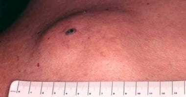
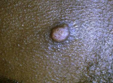
Epidermal Cyst
Pilomatrixoma
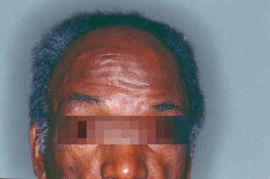
Gardner's syndrome and steatocystoma multiplex. Two unusual genetically determined conditions occurring in same patient. (1/183)
A 43-year-old man is described who had Gardner's syndrome and steatocystoma multiplex. These two unusual genetically determined conditions were associated because he had inherited the Gardner's syndrome from his father and the steatocystoma multiplex from his mother. (+info)Contralateral deafness following unilateral suboccipital brain tumor surgery in a patient with large vestibular aqueduct--case report. (2/183)
A 68-year-old female developed contralateral deafness following extirpation of a left cerebellopontine angle epidermoid cyst. Computed tomography showed that large vestibular aqueduct was present. This unusual complication may have been caused by an abrupt pressure change after cerebrospinal fluid release, which was transmitted through the large vestibular aqueduct and resulted in cochlear damage. (+info)Primary intracranial squamous cell carcinoma--case report. (3/183)
A 50-year-old female presented with primary intracranial squamous cell carcinoma (SCC) at the right cerebellopontine angle manifesting as right facial nerve paresis. She had undergone gross total removal of a right cerebellopontine angle epidermoid cyst 10 years before and had done well until recently. Magnetic resonance imaging showed a heterogeneous tumor with markedly enhanced irregular margin. Subtotal removal of the tumor was achieved. Histological examination showed moderately differentiated SCC. After surgery, she underwent chemotherapy and gamma radiosurgery. She is now well 5 years after the diagnosis of SCC. (+info)Unusual sonographic appearance of an epidermoid cyst of the testis. (4/183)
Epidermoid cyst of the testis is a rare benign testicular tumor with varied sonographic appearances. We present a case in which two specific ultrasonographic patterns were seen: (1) an "onion ring" configuration of alternating hyperechoic and hypoechoic regions, described previously as being characteristic of this lesion, and (2) a heterogeneous region with multiple punctate hyperechoic foci. (+info)Epidermoid tumor within Meckel's cave--case report. (5/183)
A rare case of an epidermoid tumor lying within Meckel's cave is reported. A 27-year-old housewife presented with complaints of right facial hypesthesia for two and a half years. On examination she had partial loss of touch sensation in the right trigeminal nerve distribution. Magnetic resonance imaging revealed a tumor located at the right petrous apex and cavernous sinus. The epidermoid tumor was excised through a lateral basal subtemporal approach. The symptoms resolved following surgery. (+info)Intramedullary spinal epidermoid cyst. (6/183)
Intramedullary epidermoid cysts of the spinal cord are rare tumours, especially those not associated with spinal dysraphism. Around 50 cases have been reported in the literature. Of these, only seven cases have had magnetic resonance imaging studies. We report two cases of spinal intramedullary epidermoid cysts with MR imaging. Both were not associated with spina bifida. In one patient the tumour was located at D4 vertebral level, while in other within the conus medullaris. The clinical features, MR imaging characteristics and surgical treatment of such rare intramedullary benign tumours are discussed, and the relevant literature reviewed. (+info)The utility of diffusion-weighted imaging with navigator-echo technique for the diagnosis of spinal epidermoid cysts. (7/183)
We report a case of a spinal epidermoid cyst in which diffusion-weighted imaging with a navigator-echo technique was useful for the differential diagnosis from other cystic tumors. Motion artifacts are inherent on diffusion-weighted images of the spinal region; however, the navigator-echo technique compensated for this problem and provided high-quality images. Diffusion-weighted imaging with navigator echoes is considered to be a potentially useful tool in the differential diagnosis of spinal cystic tumors. (+info)Hyperdense intracranial epidermoid: an uncommon presentation. (8/183)
A thirty year old female presented with sudden onset of severe headache, papilloedema and altered sensorium. Computerised tomography (CT) scan showed a hyperdense vermian mass in the posterior fossa. Operative findings and histological examination revealed spontaneous bleed into the epidermoid cyst. Difficulty in the preoperative diagnosis and uncommon presentation of the intracranial epidermoid cyst prompted us to report this case. (+info)An epidermal cyst is a common benign skin condition characterized by the growth of a sac-like structure filled with keratin, a protein found in the outermost layer of the skin (epidermis). These cysts typically appear as round, firm bumps just under the surface of the skin, often on the face, neck, trunk, or scalp. They can vary in size from a few millimeters to several centimeters in diameter.
Epidermal cysts usually develop as a result of the accumulation of dead skin cells that become trapped within a hair follicle or a pilosebaceous unit (a structure that contains a hair follicle and an oil gland). The keratin produced by the skin cells then collects inside the sac, causing it to expand gradually.
These cysts are generally slow-growing, painless, and rarely cause any symptoms. However, they may become infected or inflamed, leading to redness, tenderness, pain, or pus formation. In such cases, medical attention might be necessary to drain the cyst or administer antibiotics to treat the infection.
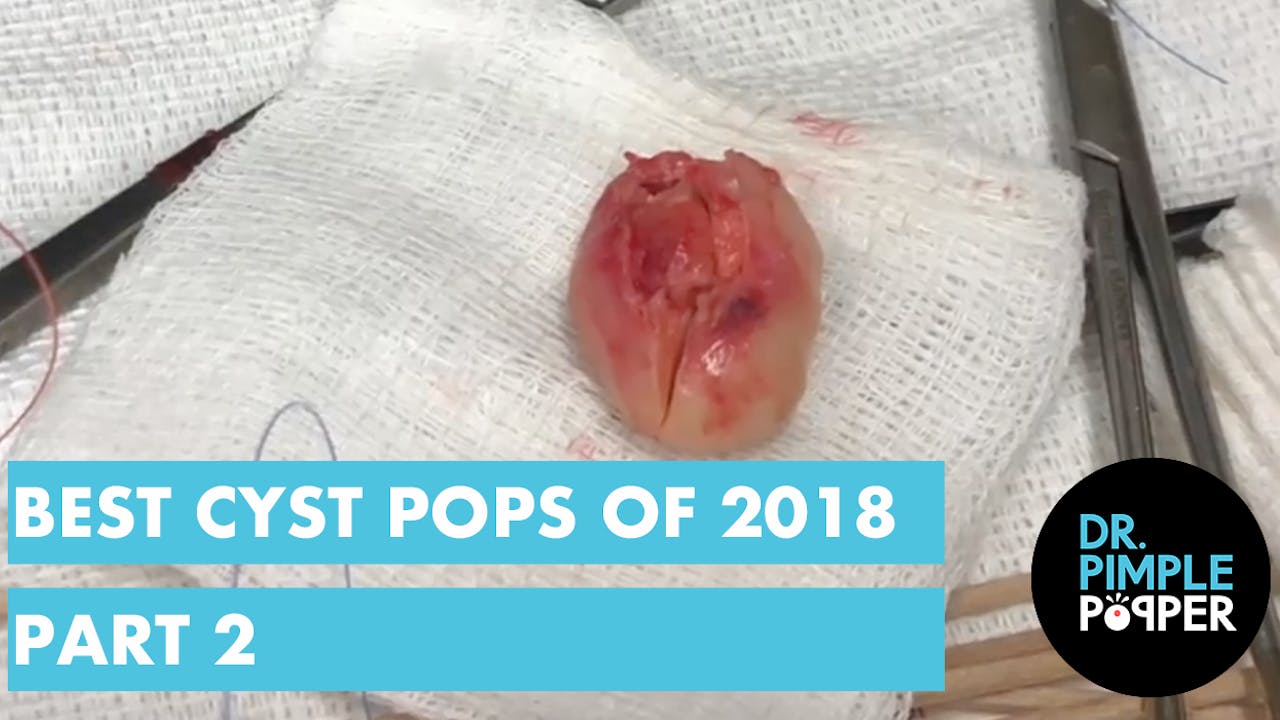
Epidermal cysts can be removed surgically if they cause cosmetic concerns or become frequently infected. The procedure typically involves making an incision in the skin and removing the entire sac along with its contents to prevent recurrence.
Neoplasms are abnormal growths of cells or tissues in the body that can be benign (non-cancerous) or malignant (cancerous). When referring to "Complex and Mixed Neoplasms," it is typically used in the context of histopathology, where it describes tumors with a mixture of different types of cells or growth patterns.
A complex neoplasm usually contains areas with various architectural patterns, cell types, or both, making its classification challenging. It may require extensive sampling and careful examination to determine its nature and behavior. These neoplasms can be either benign or malignant, depending on the specific characteristics of the tumor cells and their growth pattern.
A mixed neoplasm, on the other hand, is a tumor that contains more than one type of cell or tissue component, often arising from different germ layers (the three primary layers of embryonic development: ectoderm, mesoderm, and endoderm). A common example of a mixed neoplasm is a teratoma, which can contain tissues derived from all three germ layers, such as skin, hair, teeth, bone, and muscle. Mixed neoplasms can also be benign or malignant, depending on the specific components of the tumor.
It's important to note that the classification and behavior of complex and mixed neoplasms can vary significantly based on their location in the body, cellular composition, and other factors. Accurate diagnosis typically requires a thorough examination by an experienced pathologist and may involve additional tests, such as immunohistochemistry or molecular analysis, to determine the appropriate treatment and management strategies.
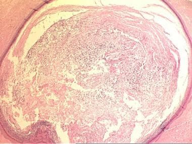
Pilomatrixoma is a benign skin tumor that originates from the hair follicle's matrix. It is also known as calcifying epithelioma of Malherbe. This slow-growing tumor typically appears as a hard, mobile, small nodule, often on the head or neck region. Pilomatrixomas are usually painless but can become inflamed or infected. They are more common in children and young adults and are slightly more prevalent in females than males. Histologically, pilomatrixoma is characterized by the presence of shadow cells, basaloid cells, and calcifications. Surgical excision is the standard treatment for this condition.
Foot dermatoses refer to various skin conditions that affect the feet. These can include inflammatory conditions like eczema and psoriasis, infectious diseases such as athlete's foot (tinea pedis), fungal infections, bacterial infections, viral infections (like plantar warts caused by HPV), and autoimmune blistering disorders. Additionally, contact dermatitis from irritants or allergens can also affect the feet. Proper diagnosis is essential to determine the best course of treatment for each specific condition.
A cyst is a closed sac, having a distinct membrane and division between the sac and its surrounding tissue, that contains fluid, air, or semisolid material. Cysts can occur in various parts of the body, including the skin, internal organs, and bones. They can be caused by various factors, such as infection, genetic predisposition, or blockage of a duct or gland. Some cysts may cause symptoms, such as pain or discomfort, while others may not cause any symptoms at all. Treatment for cysts depends on the type and location of the cyst, as well as whether it is causing any problems. Some cysts may go away on their own, while others may need to be drained or removed through a surgical procedure.
 Eruptive vellus hair cyst
Bordetella ansorpii
Vocal cord cyst
List of cutaneous neoplasms associated with systemic syndromes
Pilomatricoma
Cholesteatoma
Prevalence of female genital mutilation
Keratin implantation cyst
Epidermoid cyst
Fibrofolliculoma
Mantleoma
Reactional keratosis
Thermal keratosis
Brooke-Fordyce syndrome
Eccrine carcinoma
Microcystic adnexal carcinoma
Hidradenocarcinoma
Folliculosebaceous-apocrine hamartoma
Arsenical keratosis
Hydrocarbon keratosis
Syringocystadenoma papilliferum
Clear cell acanthoma
Trichoepithelioma
Sebaceous carcinoma
Porocarcinoma
Follicular hybrid cyst
Ciliated cyst of the vulva
Marjolin's ulcer
Chondroid syringoma
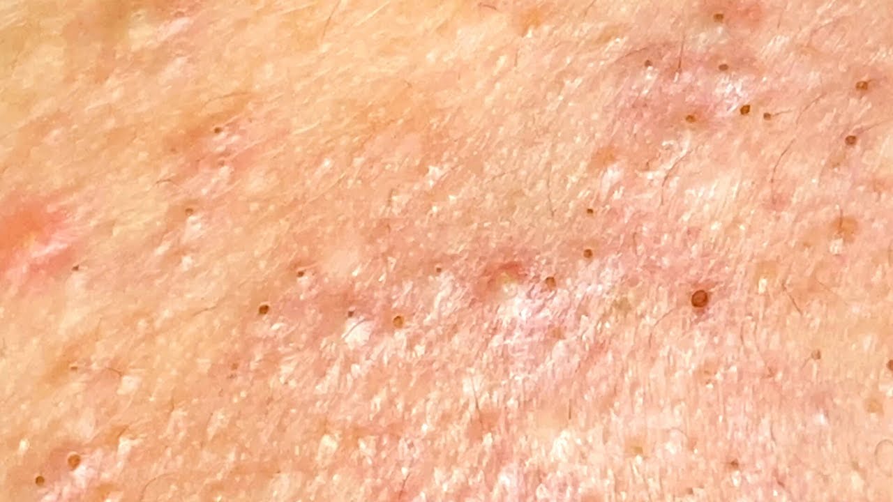
Acne
 Epidermal Inclusion Cyst: Background, Pathophysiology, Etiology
 Calcified Epidermal Cyst on the Neck : International Journal of Dermatology and Venereology
 Epidermal Cyst - MedicalRecords.com
 ROJoson PEP Talk: Epidermal Cysts in Skin | PPT
Epidermal Inclusion Cyst: Background, Pathophysiology, Epidemiology
Causes, Symptoms and Treatment of Epidermal Inclusion Cyst
Removal of an Epidermal Cyst on the Midback - Viral On The Web Now
 Penile Distortion Caused by a Large Epidermal Cyst after Augmentation Penoplasty: A Case Report
 Vulvar Inclusion and Epidermal Cysts - Women's Health Issues - MSD Manual Consumer Version
 Epidermoid cyst: MedlinePlus Medical Encyclopedia
 Plunging epidermal cyst of the floor of the mouth in a young child<...
Eruptive vellus hair cyst - Wikipedia
 Sexual and Reproductive Health Screening during the Domestic Medical Examination for Newly Arrived Refugees | Immigrant and...
 Bump on the back of the head: Causes and when to consult a doctor
Trichilemmal Cyst (Pilar Cyst) Medication
 Slow-Growing Thumb Nodule | AAFP
 ICD-10 Code for Disorder of penis, unspecified- N48.9- Codify by AAPC
Table 1 - Bordetella petrii Infection with Long-lasting Persistence in Human - Volume 17, Number 4-April 2011 - Emerging...
 couchtater
 Related Articles - Annals Singapore
Related Articles | Annals Singapore
 Male breast cancer case study | GPonline
Advanced Search Results - Public Health Image Library(PHIL)
 Journal of Clinical Immunology and Immunopathology Research - articles
 Infections
 General Surgery | Adventist Health
Cord
 Swollen Glands, Hernias, and Other Lumps Under the Skin | HealthLink BC
 Joerg Huelsken - People - EPFL
Inclusion21
- Historically, epidermoid cysts have been referred to by various terms, including follicular infundibular cysts, epidermal cysts, and epidermal inclusion cysts. (medscape.com)
- The term epidermal inclusion cyst refers specifically to an epidermoid cyst that is the result of the implantation of epidermal elements in the dermis. (medscape.com)
- True epidermal inclusion cysts result from the implantation of epithelial elements in the dermis. (medscape.com)
- Cameron DS, Hilsinger RL Jr. Squamous cell carcinoma in an epidermal inclusion cyst: case report. (medscape.com)
- Epidermal Inclusion Cysts are common cutaneous cysts. (simple-remedies.com)
- The terminology epidermal inclusion cyst particularly refers to an epidermoid cyst which occurs as a consequence to the lodging of epidermal elements in the dermis. (simple-remedies.com)
- Epidermal cysts, also called epidermal inclusion cysts, are one of the most common benign lesions and can be found on practically all regions of the body. (jwmr.org)
- Cysts that develop on the vulva include inclusion cysts and epidermal cysts. (msdmanuals.com)
- Vulvar inclusion cysts are small sacs that contain tissue from the surface of the vulva. (msdmanuals.com)
- Inclusion cysts are the most common cysts of the vulva. (msdmanuals.com)
- Inclusion cysts may also develop in the vagina. (msdmanuals.com)
- Some inclusion cysts develop on their own. (msdmanuals.com)
- Vaginal inclusion cysts are usually small and do not cause symptoms. (msdmanuals.com)
- The answer is A: epidermal inclusion cyst. (aafp.org)
- Also called epidermoid or epithelial inclusion cysts, these are benign, keratin-containing cysts with epidermal linings that can occur subcutaneously or be intratendinous or intraosseous. (aafp.org)
- On physical examination, epidermal inclusion cysts are mobile and firm and do not transilluminate. (aafp.org)
- Blind pimples can turn into epidermal inclusion cysts( EICs) if not treated properly. . (skintypesolutions.com)
- Hard pimples that do not go away and have a smelly discharge may be epidermal inclusion cysts (EIC). (skintypesolutions.com)
- Blind pimples, or epidermal inclusion cysts (EICs), occur when excess oil, dead skin cells, and bacteria clog the pores. (skintypesolutions.com)
- Nevi arise from nevus cells, incompletely differentiated melanocytes in the epidermis and dermis and in the junction zone between these 2 layers, and are the third most common benign lesions encountered in the periocular region (after papillomas and epidermal inclusion cysts). (aao.org)
- Presence of skin comorbidities (e.g. inverse psoriasis, epidermal inclusion cyst) that may interfere with study assessments for HS. (who.int)
Cutaneous5
- Epidermoid cysts represent the most common cutaneous cysts. (medscape.com)
- 6-9 Cutaneous calcified epidermal cysts have been reported on the scrotum, vulva, scalp, subungual area, breast, and heel, but are most commonly located on the scrotum. (lww.com)
- Although an epidermal cyst is a common cutaneous lesion that can present anywhere on the body, it is not easily found on the penis. (jwmr.org)
- Al-Khateeb TH, Al-Masri NM, Al-Zoubi F. Cutaneous cysts of the head and neck. (medscape.com)
- Cutaneous tumors derived from the outer root sheath of hair follicles, which show trichilemmal keratinization, are trichilemmal, proliferating trichilemmal, and malignant proliferating trichilemmal cysts. (scitechnol.com)
Keratin5
- The epidermal cysts are usually caused by an accumulation of keratin - a protein which naturally occurs in skin cells. (simple-remedies.com)
- The cysts form when keratin gets entrapped below the skin due to some disruption to the skin or hair follicle. (simple-remedies.com)
- Stratified-squamous epithelium with a granular layer that surrounding a cystic space filled with laminated keratin and a variable number of vellus hair cysts is seen to be present. (wikipedia.org)
- These cysts are smooth, dense lumps containing a buildup of keratin, the protein the body uses to make hair and nails. (medicalnewstoday.com)
- Immunoperoxidase anti-keratin staining of epidermal and pilar cysts. (medscape.com)
Sebaceous Cysts3
- Sometimes, epidermoid cysts are called sebaceous cysts. (medlineplus.gov)
- Epidermoid cysts are filled with dead skin cells, while true sebaceous cysts are filled with yellowish oily material. (medlineplus.gov)
- These types of cysts were commonly called sebaceous cysts, but the terms epidermal cyst and epidermoid cyst have come into fashion in recent years. (healthandnutritiontips.net)
Follicular infundibular cysts1
- Other names for this condition include - epidermoid cysts, follicular infundibular cysts, and epidermal cysts. (simple-remedies.com)
Lesion3
- Histological characteristics of the lesion of calcified epidermal cyst on the neck. (lww.com)
- The histopathology report of the lesion confirmed the diagnosis of epidermal cyst. (unair.ac.id)
- 973 lesion 7 × 6 cm (Figure 1), which showed follicles, multiple follicular cysts with poor vascularity on colour Doppler scan. (who.int)
Neoplasms1
- Epidermal nevi, neoplasms, and cysts. (medlineplus.gov)
Tumors2
- Kirkham N. Tumors and Cysts of the Epidermis. (medscape.com)
- Benign epidermal tumors and proliferations. (mountsinai.org)
Lesions3
- Because most lesions originate from the follicular infundibulum, the more general term epidermoid cyst is favored. (medscape.com)
- Epidermal (epidermoid) cysts are considered rare lesions in the oral cavity. (unair.ac.id)
- Eruptive vellus hair cysts (or EVHC) are small lesions that occur most often in the chest wall, abdomen and extremities, often with a crusted surface. (wikipedia.org)
Epidermoid cysts are not2
- Epidermoid cysts are not dangerous and do not need to be treated unless they cause symptoms or show signs of inflammation (redness or tenderness). (medlineplus.gov)
- Although epidermoid cysts are not harmful, your provider should examine you for signs of skin cancer. (medlineplus.gov)
Contained in epidermoid cysts1
- Inflammation is mediated in part by the horny material contained in epidermoid cysts. (medscape.com)
Congenital3
- Congenital epidermoid cysts of the anterior fontanelle or those that are orogenital in location presumably result from sequestration or trapping of epidermal rests along embryonic fusion planes during development. (medscape.com)
- Epidermal cysts of congenital origin, which are usually caused by abnormal embryologic closure of the median raphe, are easily discovered by parents [ 1 ]. (jwmr.org)
- Congenital cysts occur more frequently as solitary cysts but may be multiple. (bvsalud.org)
Types of cysts1
- This is not correct because the contents of the two types of cysts are different. (medlineplus.gov)
Pilar cysts7
- Pilar cysts are skin cysts that usually develop on a person's scalp but can also occur on the neck. (medicalnewstoday.com)
- Doctors sometimes refer to pilar cysts as trichilemmal cysts. (medicalnewstoday.com)
- Pilar cysts usually grow slowly, and they typically vary between several millimeters and a few centimeters in diameter. (medicalnewstoday.com)
- Pilar cysts are more common in females than in males and can sometimes run in families. (medicalnewstoday.com)
- Pilar cysts are generally harmless and asymptomatic, but they can sometimes be painful. (medicalnewstoday.com)
- Learn more about pilar cysts. (medicalnewstoday.com)
- Hereditary pilar cysts. (medscape.com)
Trichilemmal cyst10
- There are no medications, systemic or topical, that shrink or resolve a trichilemmal cyst. (medscape.com)
- Malignant proliferating trichilemmal cyst. (medscape.com)
- Mapping of hereditary trichilemmal cyst (TRICY1) to chromosome 3p24-p21.2 and exclusion of beta-CATENIN and MLH1. (medscape.com)
- Merkel cell tumor in a trichilemmal cyst: collision or association? (medscape.com)
- Merkel cell carcinoma in situ arising in a trichilemmal cyst: a case report and literature review. (medscape.com)
- Melikoglu C, Eren F, Keklik B, Aslan C, Sutcu M, Zeynep Tarini E. Trichilemmal cyst of the third fingertip: a case report. (medscape.com)
- Malignant proliferating trichilemmal cyst of the scalp: A case report]. (medscape.com)
- Hanau D, Grosshans E. Trichilemmal cyst with intrinsic parietal sebaceous and apocrine structures. (medscape.com)
- The proliferating trichilemmal cyst is more common in women compared to men and more than 80% of patients are elderly women. (scitechnol.com)
- The trichilemmal cyst is by far the most common form of them. (scitechnol.com)
Histological3
- Histological examination of a calcified epidermal cyst reveals a dermal cystic structure. (lww.com)
- In some instances a dermatologist may perform a histological evaluation of a cyst sac that has been removed, but cases of cancer associated with cysts are really very rare. (healthandnutritiontips.net)
- Recognition of OOC as a unique entity has long been due, yet its inexplicable radiographic presentation resembling dentigerous cyst, histological likeness to odontogenic keratocyst (OKC) and inconsistent cytokeratin expression profiles overlapping with both as well as with the epidermis, makes it rather confounding. (bvsalud.org)
Painful6
- Treatment is necessary if the cyst turns red and gets inflamed, painful, or infected. (simple-remedies.com)
- Infected cysts may be red and tender and make sexual activity painful. (msdmanuals.com)
- Epidermoid cysts may become infected and form painful abscesses . (medlineplus.gov)
- If you do have an epidermoid cyst, call your provider if it becomes red or painful. (medlineplus.gov)
- Vulvar endometriomas are rare, painful, blood-filled cysts that develop when tissue from the lining of the uterus (endometrial tissue) appears in the vulva. (merckmanuals.com)
- Most cysts eventually become large enough or painful enough to demand additional treatment. (midwestderm.com)
Occur1
- These cysts are the result of a hair follicle infection, which can occur if the follicle has for whatever reason become blocked. (healthandnutritiontips.net)
Dermatology2
- Minor office surgery - Your Midwest Dermatology doctor can excise the entire cyst. (midwestderm.com)
- If your cyst is inflamed, Midwest Dermatology may start you on antibiotics first. (midwestderm.com)
Malignant2
- Any benign or malignant process affecting or growing near the pilosebaceous unit may lead to occlusion or impingement of the follicular ostia with subsequent formation of a cyst. (medscape.com)
- Hardly ever, epidermoid cysts can become malignant. (simple-remedies.com)
Stratified squamous e2
- In addition to laminated layers of keratinous material, the cavity is filled with amorphous basophilic calcareous sediment ( Fig. 1 A). The cyst wall consists of thinned stratified squamous epithelium similar to the hair follicle infundibulum, which is surrounded by fibrosis ( Fig. 1 B). Active inflammation is sometimes seen around the cyst. (lww.com)
- B: The cyst wall consists of thinned stratified squamous epithelium surrounded by fibrosis (hematoxylin-eosin staining, ×200). (lww.com)
Epithelial4
- They may result from the sequestration of epidermal rests during embryonic life, occlusion of the pilosebaceous unit, or traumatic or surgical implantation of epithelial elements. (medscape.com)
- 3 In some cases the cyst wall and keratinous material are absorbed, leaving only areas of calcification, 4 suggesting that the pathological finding of the attenuated epithelial lining may ultimately disappear. (lww.com)
- Levine DJ, Robertson DB, Varma VA. Familial subconjunctival epithelial cysts associated with the nevoid basal cell carcinoma syndrome. (medscape.com)
- The cyst is usually preceded by minor trauma that results in epithelial cells being trapped deep in the dermis and forming the cyst. (aafp.org)
Scalp Cyst8
- A scalp cyst is fortunately usually quite small, and more often than not does not cause any particular problem. (healthandnutritiontips.net)
- A scalp cyst is normally what is referred to as an epidermal cyst, a skin cyst if you prefer. (healthandnutritiontips.net)
- If you have a scalp cyst it will generally be felt as a small bump, no bigger than a pimple, which often is tender to the touch. (healthandnutritiontips.net)
- When this fails, the scalp cyst may have to be surgically removed. (healthandnutritiontips.net)
- A scalp cyst should always be treated by a physician or dermatologist should removal be necessary, to ensure the sac is removed, and to lessen the chances of scarring or infection. (healthandnutritiontips.net)
- Maintaining a clean scalp will also go a long ways towards preventing future occurrences of the scalp cyst. (healthandnutritiontips.net)
- Hair Loss - One effect of the scalp cyst can be hair loss, especially is a number of cysts are present. (healthandnutritiontips.net)
- Summary - A scalp cyst is normally not of great concern, but the one thing to remember is not to try to physically remove it on your own. (healthandnutritiontips.net)
Lump6
- An epidermal cyst is a small, round lump in the top layer of skin called the epidermis. (medicalrecords.com)
- To diagnose the cysts, your dermatologist will inspect the lump and the surrounding skin, and ask you for a medical history. (simple-remedies.com)
- An epidermoid cyst is a closed sac under the skin, or a skin lump, filled with dead skin cells. (medlineplus.gov)
- A ganglion is a soft, rubbery lump (a type of cyst) on the front or back of the wrist. (healthlinkbc.ca)
- Here's the good word: Do not try to extract a lump, a bump, a cyst or a I-don't-know-what-it-is at home. (midwestderm.com)
- Lipomas differ from cysts in that they tend to be painless and do not get infected or inflamed - but they may feel like a lump of dough under your skin. (midwestderm.com)
Surgically3
- At times the epidermal cyst may need to be excised surgically. (simple-remedies.com)
- However, if a pilar cyst is leading to discomfort or other problems, a doctor may recommend surgically removing it. (medicalnewstoday.com)
- Once the cysts have been effectively treated, or surgically removed, normal hair growth will usually resume. (healthandnutritiontips.net)
Milia4
- Finally, the term milia refers to very small, superficial epidermoid cysts. (medscape.com)
- Small epidermoid cysts known as milia are common in the neonatal period. (medscape.com)
- The cysts appear similar clinically to steatocystoma multiplex, as well as acneiform eruptions and milia. (wikipedia.org)
- Milia are not cysts but they can turn into EICs if picked or removed improperly. (skintypesolutions.com)
Growths3
- It remains unclear whether calcified epidermal cysts are the result of dystrophic calcification of preceding epidermal cysts that are affected by inflammation followed by cystic dilation, calcification, cyst wall rupture, and granulomatous reactions, 1 or if they are truly idiopathic growths without any triggering factor such as foreign body infiltration, metabolic or hormonal disorder, or trauma. (lww.com)
- A bump on the back of the head has many possible causes, including injuries, cysts, fatty growths, inflamed hair follicles, and more. (medicalnewstoday.com)
- Epidermal growths. (mountsinai.org)
Epidermis2
- In addition, epidermoid cysts express cytokeratins 1 and 10, which are constituents of the suprabasilar layers of the epidermis. (medscape.com)
- Oxidative and nitrosative stress caused by drug metabolism may be a trigger for keratinocyte apoptosis in the epidermis seen in toxic epidermal necrolysis (TEN) and Stevens Johnson syndrome (SJS). (e-ijd.org)
Lipomas1
- Cysts are in general benign, but in some cases, if associated with fibromas or lipomas of the skin, may fall into the category of being premalignant. (healthandnutritiontips.net)
Hair follicles2
- Vulvar epidermal cysts are similar but contain secretions from oil-producing (sebaceous) glands near hair follicles. (msdmanuals.com)
- Most of the time the cyst simply pushes against the wall of one or more hair follicles, and when this happens hair will not emerge. (healthandnutritiontips.net)
Clinically1
- The entity can also be clinically similar to a common epidermal cyst ( Fig. 2 ). (lww.com)
Terminal phalanx2
- Certain injuries, especially of the crushing type, have been associated with subungual or terminal phalanx epidermoid cysts. (medscape.com)
- Handa U, Kumar S, Mohan H. Aspiration cytology of epidermoid cyst of terminal phalanx. (medscape.com)
Large epidermoid1
- A case of a large epidermoid cyst of the floor of the mouth extending extraorally in a 3-year-old boy is presented. (unair.ac.id)
Diagnosis4
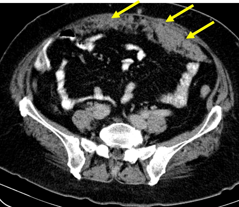
- While epidermal cysts can be evaluated by sonography, computed tomography (CT), or magnetic resonance imaging (MRI), definitive diagnosis is based on microscopic analysis of the excised biopsy specimen. (jwmr.org)
- Ultrasonography and fine needle aspiration cytology suggested the diagnosis of either dermoid or epidermoid cyst. (unair.ac.id)
- Ibrahim AE, Barikian A, Janom H, Kaddoura I. Numerous recurrent trichilemmal cysts of the scalp: differential diagnosis and surgical management. (medscape.com)
- Based on the clinical and radiographic evaluation, a preliminary diagnosis of dentigerous cyst was made. (bvsalud.org)
Breast1
- Reports describe the formation of multiple epidermoid cysts after rhinoplasty, breast augmentation, and liposuction. (medscape.com)
Skin8
- Pigmentation of epidermoid cysts is common in individuals with dark skin. (medscape.com)
- The cysts are tiny, hard masses which form underneath the skin. (simple-remedies.com)
- The cysts are formed when elements of the surface skin get under the skin surface. (medlineplus.gov)
- The cyst then becomes filled with dead skin because as the skin grows, it can't be shed as it can elsewhere on the body. (medlineplus.gov)
- 1 The only other possible complication is local infection following traumatic rupture of the cyst or disruption of the overlying skin. (aafp.org)
- Imagine the feeling of a large pea growing under your skin and you can understand what a cyst feels like. (midwestderm.com)
- But, instead, a small sac forms under the skin and fills up with dead cells, as well as pus or liquid, and voila, you've got a cyst! (midwestderm.com)
- This is often a temporary solution because the cyst sac remains in the skin. (midwestderm.com)
Neck1
- Branchial cleft cysts are found in the neck and do not usually cause problems unless they become infected. (healthlinkbc.ca)
Steatocystoma1
- A true sebaceous cyst is called a steatocystoma. (medlineplus.gov)
Penile3
- Here, we introduce an extremely rare case of a penile epidermal cyst causing penile distortion. (jwmr.org)
- Nevertheless, penile epidermal cysts are relatively rare [ 1 , 2 ]. (jwmr.org)
- This report presents an extremely rare case of a large epidermal cyst causing penile distortion that occurred years after augmentation penoplasty with dermal fat graft. (jwmr.org)
Antibiotics2
- Your health care provider will prescribe antibiotics and drainage of the cyst. (simple-remedies.com)
- In those instances where a cyst has become inflamed or infected, a doctor will usually treat it with antibiotics and wait for the condition to subside before attempting to remove the cyst, to avoid spreading of the infection. (healthandnutritiontips.net)
Carcinoma4
- In a series of epidermoid cysts with carcinoma, immunohistochemical results for HPV were negative, suggesting that HPV is not likely to play a role in the development in squamous cell carcinoma (SCC) in epidermoid cysts. (medscape.com)
- Bauer B. Carcinoma arising in a sebaceous cyst. (medscape.com)
- Delacretaz J. Keratotic basal-cell carcinoma arising from an epidermoid cyst. (medscape.com)
- Punch biopsy revealed a grade 3 (poorly differentiated) invasive ductal carcinoma, which was estrogen receptor (ER) positive and human epidermal growth factor receptor (HER2) negative. (gponline.com)
Common8
- Epidermoid cysts are approximately twice as common in men as in women. (medscape.com)
- Epidermoid cysts are very common. (medlineplus.gov)
- These cysts are more common in adults than in children. (medlineplus.gov)
- Ganglion cysts are common and can arise from various joints and tendons, but they most often affect the wrists and fingers. (aafp.org)
- Ganglion cysts are most common in women younger than 40 years. (aafp.org)
- These cysts are most common in teenagers. (healthlinkbc.ca)
- These cysts are mainly seen on the scalp and are relatively common. (scitechnol.com)
- A true cyst of the PANCREAS, distinguished from the much more common PANCREATIC PSEUDOCYST by possessing a lining of mucous EPITHELIUM. (bvsalud.org)
Recur2
- If needed, treatment consists of surgical excision, but up to 11% of cysts may recur after excision. (aafp.org)
- This is a fairly quick and easy method, but cysts may also recur after this treatment. (midwestderm.com)
Eruptive1
- Aloi F, Tomasini C, Pippione M. Mycosis fungoides and eruptive epidermoid cysts: a unique response of follicular and eccrine structures. (medscape.com)
Ovarian1
- Roadmap to evaluate ovarian cysts. (radiologyassistant.nl)
Removal1
- In many cases however, removal of the cyst is needed. (healthandnutritiontips.net)
Eccrine2
- HPV infection, ultraviolet exposure, and eccrine duct occlusion may be additional factors in the development of palmoplantar epidermoid cysts. (medscape.com)
- 2 Calcified epidermal cysts reportedly have an eccrine origin. (lww.com)
Hereditary3
- Certain hereditary syndromes are associated with epidermoid cysts. (medscape.com)
- Hereditary trichilemmal cysts. (medscape.com)
- Hereditary trichilemmal cysts: a proposal for the assessment of diagnostic clinical criteria. (medscape.com)
Trauma1
- If epithelium remains enclosed during wound healing after trauma or surgery, keratinization occurs and leads to cyst formation [ 3 ]. (jwmr.org)
Ganglion2
Infection1
- Squeezing out the contents of the cyst on your own can lead to infection, so leave the cyst well alone or discuss with your doctor. (simple-remedies.com)
Symptoms3
- Cysts that do not become infected usually cause no symptoms, but they occasionally cause irritation. (msdmanuals.com)
- If cysts cause symptoms, they are removed. (msdmanuals.com)
- If a cyst is not causing symptoms, treatment may not be necessary. (medicalnewstoday.com)
Clinical1
- Based on the clinical, radiographic and histopathological features, the cyst was diagnosed as orthokeratinized odontogenic cyst. (bvsalud.org)
Unclear1
- The manner in which carcinomas may arise within epidermoid cysts is unclear. (medscape.com)
Squeezing1
- Attempting the remove the cyst by squeezing it is more often than not an exercise in futility, as the cyst contains a sac, and unless the sac is removed, the cyst will most likely return. (healthandnutritiontips.net)


.jpg)