Escherichia coli
Escherichia coli O157
Shiga-Toxigenic Escherichia coli
Hemolytic-Uremic Syndrome
Shiga Toxin 1
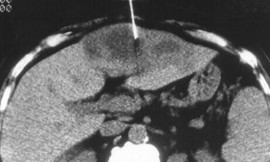
Diarrhea
Enteropathogenic Escherichia coli
Shiga Toxin 2
Shiga Toxins
Feces
Shiga Toxin
Molecular Sequence Data
Uropathogenic Escherichia coli
Bacterial Toxins
Base Sequence
Escherichia coli K12
Plasmids
Adhesins, Escherichia coli
Amino Acid Sequence
Cloning, Molecular
Bacterial Adhesion
Mutation
Enterotoxins
Fimbriae, Bacterial
Gene Expression Regulation, Bacterial
Fimbriae Proteins
Serotyping
Urinary Tract Infections
Operon
Adhesins, Bacterial
Balantidiasis
Enterotoxigenic Escherichia coli
Bacterial Outer Membrane Proteins
Chromosomes, Bacterial
RNA, Bacterial
Lipopolysaccharides
Enterohemorrhagic Escherichia coli
Sepsis
Cattle
Swine
Disease Outbreaks
Intestinal Mucosa
Balantidium
Intestines
Genes
Pyelonephritis
Genetic Complementation Test
Sequence Homology, Amino Acid
Restriction Mapping
Neutrophils
Binding Sites
Rabbits
Injections, Intraperitoneal
Culture Media
Temperature
Receptors, Antigen, T-Cell, gamma-delta
Substrate Specificity
Conjugation, Genetic
Electrophoresis, Polyacrylamide Gel
Escherichia
Transcription, Genetic
Virulence
Mice, Inbred C3H
Mice, Inbred C57BL
Recombinant Fusion Proteins
Drug Resistance, Microbial
Virulence Factors
Protein Conformation
beta-Galactosidase
Chromosome Mapping
Escherichia coli Vaccines
Models, Molecular
Ampicillin
Microbial Sensitivity Tests
Mutagenesis, Site-Directed
Bacteriophage lambda
Chloramphenicol
Protein Binding
Drug Resistance, Bacterial
Polymerase Chain Reaction
DNA-Directed RNA Polymerases
Immunoglobulin G
Carrier Proteins
Campylobacter coli
Membrane Transport Proteins
Disease Models, Animal
Transduction, Genetic
Sequence Analysis, DNA
Hydrogen-Ion Concentration
Sequence Alignment
Food Microbiology
Colicins
Species Specificity
DNA Restriction Enzymes
Genetics, Microbial
DNA Transposable Elements
Membrane Proteins
Galactosidases
Lac Operon
Meningitis
Adenosine Triphosphatases
SOS Response (Genetics)
Promoter Regions, Genetic
Transformation, Bacterial
Recombination, Genetic
Nucleic Acid Conformation
Mutagenesis
Cytokines
Genes, Regulator
Cell Membrane
Ribosomes
beta-Lactamases
DNA Primers
Lysogeny
Periplasmic Binding Proteins
Rec A Recombinases
Anaerobiosis
Incidence
Suppression, Genetic
Sequence Homology, Nucleic Acid
Maltose-Binding Proteins
Enterobacteriaceae
DNA, Recombinant
Meningitis, Escherichia coli
Salmonella typhimurium
RNA, Transfer
Citrobacter rodentium
Lactose
Sigma Factor
Cattle Diseases
Structure-Activity Relationship
Amino Acids
Ribosomal Proteins
Oxidoreductases
Protein Biosynthesis
Mutagenesis, Insertional
Gene Expression
Phenotype
Crystallography, X-Ray
Poultry Diseases
F Factor
Phosphotransferases
Colony Count, Microbial
Protein Structure, Tertiary
Thymine
DNA-Binding Proteins
Macromolecular Substances
Adenosine Triphosphate
Open Reading Frames
Ultraviolet Rays
R Factors
Isopropyl Thiogalactoside
Periplasm
Nalidixic Acid
Gene Deletion
Bacteria
Streptomycin
Codon
Trihexosylceramides
Biological Transport
O Antigens
Enzyme Stability
Catalysis
Nucleic Acid Hybridization
Hemolysin Proteins
DNA Repair
Protein Structure, Secondary
Arabinose
Carbon Isotopes
Uracil
Spheroplasts
Repressor Proteins
Porins
Protective effect of bactericidal/permeability-increasing protein (rBPI21) in baboon sepsis is related to its antibacterial, not antiendotoxin, properties. (1/5328)
OBJECTIVE AND SUMMARY BACKGROUND DATA: The recombinant fragment of bactericidal/permeability-increasing protein, rBPI21, has potent bactericidal activity against gram-negative bacteria as well as antiendotoxin (lipopolysaccharide [LPS]) action. On the basis of these activities, the authors sought to discover whether rBPI21 would be protective in baboons with live Escherichia coli-induced sepsis and whether the potential protective effects of rBPI21 (together with antibiotics) would be more closely related to its antibacterial or LPS-neutralizing effects. METHODS: In a prospective, randomized, placebo-controlled subchronic laboratory study, the efficacy of rBPI21 or placebo was studied over 72 hours in chronically instrumented male baboons infused with live E. coli under antibiotic therapy. RESULTS: Intravenous rBPI21 attenuated sepsis-related organ failure and increased survival significantly. Bacteremia was significantly reduced in the rBPI21 group at 2 hours after the start of the E. coli infusion, whereas circulating LPS was less affected. The in vivo formation of tumor necrosis factor was significantly suppressed by the rBPI21 treatment regimen. Microcirculation and organ function were improved. CONCLUSIONS: In baboon live E. coli sepsis, the salutary effect of rBPI21 results from a more prevalent antibacterial than antiendotoxin activity. (+info)In vitro activities of cephalosporins and quinolones against Escherichia coli strains isolated from diarrheic dairy calves. (2/5328)
The in vitro activities of several cephalosporins and quinolones against 195 strains of Escherichia coli isolated from diary calves affected by neonatal diarrhea were determined. One hundred thirty-seven of these strains produced one or more potential virulence factors (F5, F41, F17, cytotoxic necrotizing factor, verotoxin, and the eae gene), but the remaining 58 strains did not produce any of these factors. From 11 to 18% of the E. coli strains were resistant to cephalothin, nalidixic acid, enoxacin, and enrofloxacin. However, cefuroxime, cefotaxime, and cefquinome were highly effective against the E. coli isolates tested. Some significant differences (P < 0.05) in resistance to quinolones between the strains producing potential virulence factors and nonfimbriated, nontoxigenic, eae-negative strains were found. Thus, eae-positive, necrotoxigenic, and verotoxigenic (except for nalidixic acid) E. coli strains were significantly more sensitive to nalidixic acid, enoxacin, and enrofloxacin than nonfimbriated, nontoxigenic, eae-negative strains. Moreover, eae-positive strains were significantly more sensitive to enoxacin and enrofloxacin than F5-positive strains. Thus, the result of this study suggest that the bovine E. coli strains that produce some potential virulence factors are more sensitive to quinolones than those that do not express these factors. (+info)Augmentation of killing of Escherichia coli O157 by combinations of lactate, ethanol, and low-pH conditions. (3/5328)
The acid tolerance of Escherichia coli O157:H7 strains can be overcome by addition of lactate, ethanol, or a combination of the two agents. Killing can be increased by as much as 4 log units in the first 5 min of incubation at pH 3 even for the most acid-tolerant isolates. Exponential-phase, habituated, and stationary-phase cells are all sensitive to incubation with lactate and ethanol. Killing correlates with disruption of the capacity for pH homeostasis. Habituated and stationary-phase cells can partially offset the effects of the lowering of cytoplasmic pH. (+info)Listeria monocytogenes and Escherichia coli septicemia and meningoencephalitis in a 7-day-old llama. (4/5328)
Listeria monocytogenes and Escherichia coli were isolated from blood collected on presentation and tissues samples taken postmortem. Listeria monocytogenes was isolated from cerebrospinal fluid collected antemortem. The importance of passive transfer of immunity, the subtlety of neurologic signs in early meningitis, and considering blood-CSF penetration in antimicrobial selection are discussed. (+info)A murine model of renal abscess formation. (5/5328)
We developed a murine model of kidney abscess by direct renal injection of either Escherichia coli (1 x 10(6) to 7 x 10(6) organisms) or sterile medium. Bacterial infection produced renal abscesses, bacteremia, and late-onset leukocytosis in all animals. Controls were unaffected. This model may be useful for the study of various sequelae of kidney infection. (+info)Enteropathogenic E. coli attenuates secretagogue-induced net intestinal ion transport but not Cl- secretion. (6/5328)
Enteric bacterial pathogens often increase intestinal Cl- secretion. Enteropathogenic Escherichia coli (EPEC) does not stimulate active ion secretion. In fact, EPEC infection decreases net ion transport in response to classic secretagogues. This has been presumed to reflect diminished Cl- secretion. The aim of this study was to investigate the influence of EPEC infection on specific intestinal epithelial ion transport processes. T84 cell monolayers infected with EPEC were used for these studies. EPEC infection significantly decreased short-circuit current (Isc) in response to carbachol and forskolin, yet 125I efflux studies revealed no difference in Cl- channel activity. There was also no alteration in basolateral K+ channel or Na+-K+-2Cl- cotransport activity. Furthermore, net 36Cl- flux was not decreased by EPEC. No alterations in either K+ or Na+ transport could be demonstrated. Instead, removal of basolateral bicarbonate from uninfected monolayers yielded an Isc response approximating that observed with EPEC infection, whereas bicarbonate removal from EPEC-infected monolayers further diminished Isc. These studies suggest that the reduction in stimulated Isc is not secondary to diminished Cl- secretion. Alternatively, bicarbonate-dependent transport processes appear to be perturbed. (+info)Organization of biogenesis genes for aggregative adherence fimbria II defines a virulence gene cluster in enteroaggregative Escherichia coli. (7/5328)
Several virulence-related genes have been described for prototype enteroaggregative Escherichia coli (EAEC) strain 042, which has been shown to cause diarrhea in human volunteers. Among these factors are the enterotoxins Pet and EAST and the fimbrial antigen aggregative adherence fimbria II (AAF/II), all of which are encoded on the 65-MDa virulence plasmid pAA2. Using nucleotide sequence analysis and insertional mutagenesis, we have found that the genes required for the expression of each of these factors, as well as the transcriptional activator of fimbrial expression AggR, map to a distinct cluster on the pAA2 plasmid map. The cluster is 23 kb in length and includes two regions required for expression of the AAF/II fimbria. These fimbrial biogenesis genes feature a unique organization in which the chaperone, subunit, and transcriptional activator lie in one cluster, whereas the second, unlinked cluster comprises a silent chaperone gene, usher, and invasin reminiscent of Dr family fimbrial clusters. This plasmid-borne virulence locus may represent an important set of virulence determinants in EAEC strains. (+info)Drosophila melanogaster transferrin. Cloning, deduced protein sequence, expression during the life cycle, gene localization and up-regulation on bacterial infection. (8/5328)
Drosophila melanogaster transferrin cDNA was cloned from an ovarian cDNA library by using a PCR fragment amplified by two primers designed from other dipteran transferrin sequences. The clone (2035 bp) encodes a protein of 641 amino acids containing a signal peptide of 29 amino acids. Like other insect transferrins, Drosophila transferrin appears to have a functional iron-binding site only in the N-terminal lobe. The C-terminal lobe lacks iron-binding residues found in other transferrins, and has large deletions which make it much smaller than functional C-terminal lobes in other transferrins. In-situ hybridization using a digoxigenin labeled transferrin cDNA probe revealed that the gene is located at position 17B1-2 on the X chromosome. Northern blot analysis showed that transferrin mRNA was present in the larval, pupal and adult stages, but was not detectable in the embryo. Iron supplementation of the diet resulted in lower levels of transferrin mRNA. When adult flies were inoculated with bacteria (Escherichia coli), transferrin mRNA synthesis was markedly increased relative to controls. (+info)Escherichia coli (E. coli) infections refer to illnesses caused by the bacterium E. coli, which can cause a range of symptoms depending on the specific strain and site of infection. The majority of E. coli strains are harmless and live in the intestines of healthy humans and animals. However, some strains, particularly those that produce Shiga toxins, can cause severe illness.
E. coli infections can occur through various routes, including contaminated food or water, person-to-person contact, or direct contact with animals or their environments. Common symptoms of E. coli infections include diarrhea (often bloody), abdominal cramps, nausea, and vomiting. In severe cases, complications such as hemolytic uremic syndrome (HUS) can occur, which may lead to kidney failure and other long-term health problems.
Preventing E. coli infections involves practicing good hygiene, cooking meats thoroughly, avoiding cross-contamination of food during preparation, washing fruits and vegetables before eating, and avoiding unpasteurized dairy products and juices. Prompt medical attention is necessary if symptoms of an E. coli infection are suspected to prevent potential complications.
'Escherichia coli (E. coli) proteins' refer to the various types of proteins that are produced and expressed by the bacterium Escherichia coli. These proteins play a critical role in the growth, development, and survival of the organism. They are involved in various cellular processes such as metabolism, DNA replication, transcription, translation, repair, and regulation.
E. coli is a gram-negative, facultative anaerobe that is commonly found in the intestines of warm-blooded organisms. It is widely used as a model organism in scientific research due to its well-studied genetics, rapid growth, and ability to be easily manipulated in the laboratory. As a result, many E. coli proteins have been identified, characterized, and studied in great detail.
Some examples of E. coli proteins include enzymes involved in carbohydrate metabolism such as lactase, sucrase, and maltose; proteins involved in DNA replication such as the polymerases, single-stranded binding proteins, and helicases; proteins involved in transcription such as RNA polymerase and sigma factors; proteins involved in translation such as ribosomal proteins, tRNAs, and aminoacyl-tRNA synthetases; and regulatory proteins such as global regulators, two-component systems, and transcription factors.
Understanding the structure, function, and regulation of E. coli proteins is essential for understanding the basic biology of this important organism, as well as for developing new strategies for combating bacterial infections and improving industrial processes involving bacteria.
'Escherichia coli' (E. coli) is a type of gram-negative, facultatively anaerobic, rod-shaped bacterium that commonly inhabits the intestinal tract of humans and warm-blooded animals. It is a member of the family Enterobacteriaceae and one of the most well-studied prokaryotic model organisms in molecular biology.
While most E. coli strains are harmless and even beneficial to their hosts, some serotypes can cause various forms of gastrointestinal and extraintestinal illnesses in humans and animals. These pathogenic strains possess virulence factors that enable them to colonize and damage host tissues, leading to diseases such as diarrhea, urinary tract infections, pneumonia, and sepsis.
E. coli is a versatile organism with remarkable genetic diversity, which allows it to adapt to various environmental niches. It can be found in water, soil, food, and various man-made environments, making it an essential indicator of fecal contamination and a common cause of foodborne illnesses. The study of E. coli has contributed significantly to our understanding of fundamental biological processes, including DNA replication, gene regulation, and protein synthesis.
Escherichia coli (E. coli) O157 is a serotype of the bacterium E. coli that is associated with foodborne illness. This strain is pathogenic and produces Shiga toxins, which can cause severe damage to the lining of the small intestine and potentially lead to hemorrhagic diarrhea and kidney failure. E. coli O157 is often transmitted through contaminated food, particularly undercooked ground beef, as well as raw or unpasteurized dairy products, fruits, and vegetables. It can also be spread through contact with infected individuals or animals, especially in settings like farms, petting zoos, and swimming pools. Proper food handling, cooking, and hygiene practices are crucial to preventing E. coli O157 infections.
Shiga-toxigenic Escherichia coli (STEC) are strains of the bacterium E. coli that produce one or both of two potent toxins called Shiga toxin or Shiga-like toxin. These toxins are named after Shigella dysenteriae type 1, from which the STEC Shiga toxin was originally isolated. The Shiga toxins cause severe damage to the lining of intestines and can lead to a range of symptoms such as diarrhea (often bloody), stomach cramps, vomiting, and fever. In severe cases, it can progress to hemolytic uremic syndrome (HUS), a serious complication that can cause kidney failure, brain damage, and even death, particularly in young children, the elderly, and immunocompromised individuals.
STEC is often found in the intestines of healthy animals, especially ruminants like cattle, goats, and sheep, and can be transmitted to humans through contaminated food or water, or direct contact with infected animals or their feces. Common sources of STEC include undercooked ground beef, raw milk, contaminated vegetables, and unpasteurized dairy products. It's important to note that not all strains of E. coli are Shiga-toxigenic, and only a small percentage of STEC infections result in severe illness or HUS.
Hemolytic-Uremic Syndrome (HUS) is a serious condition that affects the blood and kidneys. It is characterized by three major features: the breakdown of red blood cells (hemolysis), the abnormal clotting of small blood vessels (microthrombosis), and acute kidney failure.
The breakdown of red blood cells leads to the release of hemoglobin into the bloodstream, which can cause anemia. The microthrombi can obstruct the flow of blood in the kidneys' filtering system (glomeruli), leading to damaged kidney function and potentially acute kidney failure.
HUS is often caused by a bacterial infection, most commonly Escherichia coli (E. coli) that produces Shiga toxins. This form of HUS is known as STEC-HUS or Stx-HUS. Other causes include infections with other bacteria, viruses, medications, pregnancy complications, and certain medical conditions such as autoimmune diseases.
Symptoms of HUS may include fever, fatigue, decreased urine output, blood in the stool, swelling in the face, hands, or feet, and irritability or confusion. Treatment typically involves supportive care, including dialysis for kidney failure, transfusions to replace lost red blood cells, and managing high blood pressure. In severe cases, a kidney transplant may be necessary.
Serositis is a medical term that refers to inflammation of the serous membranes, which are thin layers of tissue that line the inner surfaces of body cavities and surround organs such as the heart, lungs, and abdomen. The serous membranes produce a lubricating fluid called serous fluid that helps reduce friction between internal organs and enables them to move smoothly against each other.
Inflammation of these membranes can result in excessive production of serous fluid, leading to the accumulation of fluid in the surrounding body cavities. This accumulation can cause symptoms such as chest pain, coughing, difficulty breathing, or abdominal swelling and discomfort.
Serositis is often associated with various medical conditions, including autoimmune diseases like rheumatoid arthritis, lupus, and Sjogren's syndrome. Infections, cancers, and certain medications may also cause serositis. Treatment typically involves addressing the underlying condition causing the inflammation and managing symptoms with medications such as nonsteroidal anti-inflammatory drugs (NSAIDs), corticosteroids, or immunosuppressive agents.
Shiga toxin 1 (Stx1) is a protein toxin produced by certain strains of the bacterium Escherichia coli (E. coli), specifically those that belong to serotype O157:H7 and some other Shiga toxin-producing E. coli (STEC) or enterohemorrhagic E. coli (EHEC).
Shiga toxins are named after Kiyoshi Shiga, who discovered the first strain of E. coli that produces this toxin in 1897. These toxins inhibit protein synthesis in eukaryotic cells and cause damage to the endothelial cells lining blood vessels, which can lead to various clinical manifestations such as hemorrhagic colitis (bloody diarrhea) and hemolytic uremic syndrome (HUS), a severe complication that can result in kidney failure.
Shiga toxin 1 is composed of two subunits, A and B. The B subunit binds to specific glycolipid receptors on the surface of target cells, facilitating the uptake of the toxin into the cell. Once inside the cell, the A subunit inhibits protein synthesis by removing an adenine residue from a specific region of the 28S rRNA molecule in the ribosome, thereby preventing peptide bond formation and leading to cell death.
Shiga toxin 1 is highly toxic and can cause significant morbidity and mortality, particularly in children, the elderly, and immunocompromised individuals. Antibiotics are generally not recommended for the treatment of Shiga toxin-producing E. coli infections because they may increase the risk of developing HUS by inducing bacterial lysis and releasing more toxins into the circulation. Supportive care, hydration, and close monitoring are essential for managing these infections.
Diarrhea is a condition in which an individual experiences loose, watery stools frequently, often exceeding three times a day. It can be acute, lasting for several days, or chronic, persisting for weeks or even months. Diarrhea can result from various factors, including viral, bacterial, or parasitic infections, food intolerances, medications, and underlying medical conditions such as inflammatory bowel disease or irritable bowel syndrome. Dehydration is a potential complication of diarrhea, particularly in severe cases or in vulnerable populations like young children and the elderly.
Enteropathogenic Escherichia coli (EPEC) are a type of bacteria that can cause diarrheal illness in humans, particularly in children under the age of 2. These bacteria colonize and infect the small intestine, causing inflammation and damage to the intestinal lining. This results in a variety of symptoms, including watery diarrhea, abdominal cramps, vomiting, and fever.
EPEC are characterized by their ability to form attaching and effacing (A/E) lesions on intestinal cells. These lesions cause the cells to reorganize and form a structure called a pedestal, which helps the bacteria attach to the cell surface and evade the host's immune system. EPEC also produce toxins that can damage the intestinal lining and contribute to the development of diarrhea.
EPEC are transmitted through contaminated food and water, as well as person-to-person contact. They are a common cause of traveler's diarrhea and have been associated with outbreaks in child care centers and other settings where people are in close proximity to each other. Prevention measures include good hygiene practices, such as handwashing and proper food handling and preparation, as well as avoiding contaminated food and water sources.
Shiga toxin 2 (Stx2) is a protein toxin produced by certain strains of the bacterium Escherichia coli (E. coli), specifically those that belong to serotype O157:H7 and some other Shiga toxin-producing E. coli (STEC) or enterohemorrhagic E. coli (EHEC).
Stx2 is named after Dr. Kiyoshi Shiga, who first discovered the related Shiga toxin in 1898. It is a powerful cytotoxin that can cause damage to cells lining the intestines and other organs. The toxin inhibits protein synthesis in the cells by removing an adenine residue from the 28S rRNA of the 60S ribosomal subunit, leading to cell death.
Exposure to Stx2 can occur through ingestion of contaminated food or water, or direct contact with infected animals or their feces. In severe cases, it can lead to hemorrhagic colitis, which is characterized by bloody diarrhea and abdominal cramps, and hemolytic uremic syndrome (HUS), a serious complication that can cause kidney failure, anemia, and neurological problems.
It's important to note that Stx2 has two major subtypes, Stx2a and Stx2b, which differ in their biological activities and clinical significance. Stx2a is considered more potent than Stx2b and is associated with a higher risk of developing HUS.
Shiga toxins are a type of protein toxin produced by certain strains of bacteria, including some types of Escherichia coli (E. coli) and Shigella dysenteriae. These toxins get their name from Dr. Kiyoshi Shiga, who first discovered them in the late 19th century.
Shiga toxins are classified into two main types: Shiga toxin 1 (Stx1) and Shiga toxin 2 (Stx2). Both types of toxins are similar in structure and function, but they differ in their potency and genetic makeup. Shiga toxins inhibit protein synthesis in cells by removing an adenine residue from a specific region of the 28S rRNA molecule in the ribosome, which ultimately leads to cell death.
These toxins can cause severe damage to the lining of the intestines and are associated with hemorrhagic colitis, a potentially life-threatening condition characterized by bloody diarrhea, abdominal cramps, and fever. In some cases, Shiga toxins can also enter the bloodstream and cause systemic complications such as hemolytic uremic syndrome (HUS), which is characterized by kidney failure, anemia, and thrombocytopenia.
Exposure to Shiga toxins typically occurs through ingestion of contaminated food or water, or through direct contact with infected individuals or animals. Preventive measures include good hygiene practices, such as thorough handwashing, cooking meats thoroughly, and avoiding unpasteurized dairy products and untreated water.
Bacterial proteins are a type of protein that are produced by bacteria as part of their structural or functional components. These proteins can be involved in various cellular processes, such as metabolism, DNA replication, transcription, and translation. They can also play a role in bacterial pathogenesis, helping the bacteria to evade the host's immune system, acquire nutrients, and multiply within the host.
Bacterial proteins can be classified into different categories based on their function, such as:
1. Enzymes: Proteins that catalyze chemical reactions in the bacterial cell.
2. Structural proteins: Proteins that provide structural support and maintain the shape of the bacterial cell.
3. Signaling proteins: Proteins that help bacteria to communicate with each other and coordinate their behavior.
4. Transport proteins: Proteins that facilitate the movement of molecules across the bacterial cell membrane.
5. Toxins: Proteins that are produced by pathogenic bacteria to damage host cells and promote infection.
6. Surface proteins: Proteins that are located on the surface of the bacterial cell and interact with the environment or host cells.
Understanding the structure and function of bacterial proteins is important for developing new antibiotics, vaccines, and other therapeutic strategies to combat bacterial infections.
A bacterial gene is a segment of DNA (or RNA in some viruses) that contains the genetic information necessary for the synthesis of a functional bacterial protein or RNA molecule. These genes are responsible for encoding various characteristics and functions of bacteria such as metabolism, reproduction, and resistance to antibiotics. They can be transmitted between bacteria through horizontal gene transfer mechanisms like conjugation, transformation, and transduction. Bacterial genes are often organized into operons, which are clusters of genes that are transcribed together as a single mRNA molecule.
It's important to note that the term "bacterial gene" is used to describe genetic elements found in bacteria, but not all genetic elements in bacteria are considered genes. For example, some DNA sequences may not encode functional products and are therefore not considered genes. Additionally, some bacterial genes may be plasmid-borne or phage-borne, rather than being located on the bacterial chromosome.
Feces are the solid or semisolid remains of food that could not be digested or absorbed in the small intestine, along with bacteria and other waste products. After being stored in the colon, feces are eliminated from the body through the rectum and anus during defecation. Feces can vary in color, consistency, and odor depending on a person's diet, health status, and other factors.
Shiga toxins are a type of protein toxin produced by certain strains of bacteria, including some types of Escherichia coli (E. coli) and Shigella dysenteriae. These toxins get their name from Kiyoshi Shiga, the scientist who discovered them in 1897.
Shiga toxins are potent cytotoxins that can cause damage to cells by inhibiting protein synthesis. They consist of two main components: an enzymatically active A subunit and several B subunits that bind to specific receptors on the surface of target cells, facilitating the entry of the A subunit into the cell.
Once inside the cell, the A subunit cleaves a crucial component of the protein synthesis machinery called ribosome, leading to cell death or dysfunction. Shiga toxins can cause severe illnesses such as hemorrhagic colitis and hemolytic uremic syndrome (HUS), which can be life-threatening in some cases.
It's worth noting that Shiga toxin-producing E. coli (STEC) infections are often foodborne, and they can cause a range of symptoms from mild diarrhea to severe abdominal cramps, bloody diarrhea, and kidney failure. Prevention measures include proper food handling, cooking meat thoroughly, washing fruits and vegetables, and practicing good hygiene.
Molecular sequence data refers to the specific arrangement of molecules, most commonly nucleotides in DNA or RNA, or amino acids in proteins, that make up a biological macromolecule. This data is generated through laboratory techniques such as sequencing, and provides information about the exact order of the constituent molecules. This data is crucial in various fields of biology, including genetics, evolution, and molecular biology, allowing for comparisons between different organisms, identification of genetic variations, and studies of gene function and regulation.
Uropathogenic Escherichia coli (UPEC) are a subgroup of E. coli bacteria that have developed the ability to cause urinary tract infections (UTIs). These infections can affect any part of the urinary system, including the kidneys, ureters, bladder, and urethra. UPEC are responsible for the majority of uncomplicated UTIs in otherwise healthy individuals.
UPEC possess various virulence factors that allow them to adhere to and colonize the urinary tract, evade host immune responses, and cause tissue damage. Some of these virulence factors include fimbriae, which are hair-like structures that help the bacteria attach to host cells; toxins such as hemolysin, which can damage host cells; and polysaccharide capsules, which protect the bacteria from phagocytosis by host immune cells.
UPEC can cause a range of UTI symptoms, including frequent urination, pain or burning during urination, strong-smelling or cloudy urine, and fever. If left untreated, UTIs caused by UPEC can lead to more serious complications, such as kidney damage or bloodstream infections. Treatment typically involves antibiotics that are effective against UPEC, such as trimethoprim-sulfamethoxazole, nitrofurantoin, or fluoroquinolones. However, the increasing prevalence of antibiotic resistance among UPEC isolates is a growing concern and highlights the need for ongoing research into new treatment strategies.
Bacterial toxins are poisonous substances produced and released by bacteria. They can cause damage to the host organism's cells and tissues, leading to illness or disease. Bacterial toxins can be classified into two main types: exotoxins and endotoxins.
Exotoxins are proteins secreted by bacterial cells that can cause harm to the host. They often target specific cellular components or pathways, leading to tissue damage and inflammation. Some examples of exotoxins include botulinum toxin produced by Clostridium botulinum, which causes botulism; diphtheria toxin produced by Corynebacterium diphtheriae, which causes diphtheria; and tetanus toxin produced by Clostridium tetani, which causes tetanus.
Endotoxins, on the other hand, are components of the bacterial cell wall that are released when the bacteria die or divide. They consist of lipopolysaccharides (LPS) and can cause a generalized inflammatory response in the host. Endotoxins can be found in gram-negative bacteria such as Escherichia coli and Pseudomonas aeruginosa.
Bacterial toxins can cause a wide range of symptoms depending on the type of toxin, the dose, and the site of infection. They can lead to serious illnesses or even death if left untreated. Vaccines and antibiotics are often used to prevent or treat bacterial infections and reduce the risk of severe complications from bacterial toxins.
A base sequence in the context of molecular biology refers to the specific order of nucleotides in a DNA or RNA molecule. In DNA, these nucleotides are adenine (A), guanine (G), cytosine (C), and thymine (T). In RNA, uracil (U) takes the place of thymine. The base sequence contains genetic information that is transcribed into RNA and ultimately translated into proteins. It is the exact order of these bases that determines the genetic code and thus the function of the DNA or RNA molecule.
Escherichia coli (E. coli) K12 is a strain of the bacterium E. coli that is commonly used in scientific research. It was originally isolated from the human intestine and has been well-studied due to its relatively harmless nature compared to other strains of E. coli that can cause serious illness.
The "K12" designation refers to a specific set of genetic characteristics that distinguish this strain from others. It is a non-pathogenic, or non-harmful, strain that is often used as a model organism in molecular biology and genetics research. Researchers have developed many tools and resources for studying E. coli K12, including a complete genome sequence and extensive collections of mutant strains.
E. coli K12 is not typically found in the environment and is not associated with disease in healthy individuals. However, it can be used as an indicator organism to detect fecal contamination in water supplies, since it is commonly present in the intestines of warm-blooded animals.
Bacterial DNA refers to the genetic material found in bacteria. It is composed of a double-stranded helix containing four nucleotide bases - adenine (A), thymine (T), guanine (G), and cytosine (C) - that are linked together by phosphodiester bonds. The sequence of these bases in the DNA molecule carries the genetic information necessary for the growth, development, and reproduction of bacteria.
Bacterial DNA is circular in most bacterial species, although some have linear chromosomes. In addition to the main chromosome, many bacteria also contain small circular pieces of DNA called plasmids that can carry additional genes and provide resistance to antibiotics or other environmental stressors.
Unlike eukaryotic cells, which have their DNA enclosed within a nucleus, bacterial DNA is present in the cytoplasm of the cell, where it is in direct contact with the cell's metabolic machinery. This allows for rapid gene expression and regulation in response to changing environmental conditions.
A plasmid is a small, circular, double-stranded DNA molecule that is separate from the chromosomal DNA of a bacterium or other organism. Plasmids are typically not essential for the survival of the organism, but they can confer beneficial traits such as antibiotic resistance or the ability to degrade certain types of pollutants.
Plasmids are capable of replicating independently of the chromosomal DNA and can be transferred between bacteria through a process called conjugation. They often contain genes that provide resistance to antibiotics, heavy metals, and other environmental stressors. Plasmids have also been engineered for use in molecular biology as cloning vectors, allowing scientists to replicate and manipulate specific DNA sequences.
Plasmids are important tools in genetic engineering and biotechnology because they can be easily manipulated and transferred between organisms. They have been used to produce vaccines, diagnostic tests, and genetically modified organisms (GMOs) for various applications, including agriculture, medicine, and industry.
Adhesins in Escherichia coli (E. coli) refer to proteins or structures on the surface of E. coli bacteria that allow them to adhere to host cells or surfaces. These adhesins play a crucial role in the initial attachment and colonization of the bacterium to the host, which can lead to infection and disease.
There are several types of adhesins found in E. coli, including fimbrial and non-fimbrial adhesins. Fimbrial adhesins, also known as pili, are hair-like structures that extend from the surface of the bacterium and can bind to specific receptors on host cells. Non-fimbrial adhesins, on the other hand, are proteins located on the outer membrane of the bacterium that can mediate adherence to host cells or surfaces.
One well-known example of an E. coli adhesin is the P fimbriae, which is associated with urinary tract infections (UTIs). The P fimbriae bind to galabiose receptors on the surface of uroepithelial cells, allowing the bacterium to colonize and infect the urinary tract. Other types of E. coli adhesins have been implicated in various extraintestinal infections, such as meningitis, sepsis, and neonatal meningitis.
Understanding the mechanisms of E. coli adhesion is important for developing strategies to prevent and treat infections caused by this bacterium.
An amino acid sequence is the specific order of amino acids in a protein or peptide molecule, formed by the linking of the amino group (-NH2) of one amino acid to the carboxyl group (-COOH) of another amino acid through a peptide bond. The sequence is determined by the genetic code and is unique to each type of protein or peptide. It plays a crucial role in determining the three-dimensional structure and function of proteins.
Molecular cloning is a laboratory technique used to create multiple copies of a specific DNA sequence. This process involves several steps:
1. Isolation: The first step in molecular cloning is to isolate the DNA sequence of interest from the rest of the genomic DNA. This can be done using various methods such as PCR (polymerase chain reaction), restriction enzymes, or hybridization.
2. Vector construction: Once the DNA sequence of interest has been isolated, it must be inserted into a vector, which is a small circular DNA molecule that can replicate independently in a host cell. Common vectors used in molecular cloning include plasmids and phages.
3. Transformation: The constructed vector is then introduced into a host cell, usually a bacterial or yeast cell, through a process called transformation. This can be done using various methods such as electroporation or chemical transformation.
4. Selection: After transformation, the host cells are grown in selective media that allow only those cells containing the vector to grow. This ensures that the DNA sequence of interest has been successfully cloned into the vector.
5. Amplification: Once the host cells have been selected, they can be grown in large quantities to amplify the number of copies of the cloned DNA sequence.
Molecular cloning is a powerful tool in molecular biology and has numerous applications, including the production of recombinant proteins, gene therapy, functional analysis of genes, and genetic engineering.
Bacterial adhesion is the initial and crucial step in the process of bacterial colonization, where bacteria attach themselves to a surface or tissue. This process involves specific interactions between bacterial adhesins (proteins, fimbriae, or pili) and host receptors (glycoproteins, glycolipids, or extracellular matrix components). The attachment can be either reversible or irreversible, depending on the strength of interaction. Bacterial adhesion is a significant factor in initiating biofilm formation, which can lead to various infectious diseases and medical device-associated infections.
A mutation is a permanent change in the DNA sequence of an organism's genome. Mutations can occur spontaneously or be caused by environmental factors such as exposure to radiation, chemicals, or viruses. They may have various effects on the organism, ranging from benign to harmful, depending on where they occur and whether they alter the function of essential proteins. In some cases, mutations can increase an individual's susceptibility to certain diseases or disorders, while in others, they may confer a survival advantage. Mutations are the driving force behind evolution, as they introduce new genetic variability into populations, which can then be acted upon by natural selection.
Infantile diarrhea is a medical condition characterized by loose, watery stools in infants and young children. It can be caused by various factors such as viral or bacterial infections, food intolerances, allergies, or malabsorption disorders. In some cases, it may also be associated with certain medications or underlying medical conditions.
Infantile diarrhea can lead to dehydration and other complications if not treated promptly and properly. It is important to monitor the infant's hydration status by checking for signs of dehydration such as dry mouth, sunken eyes, and decreased urine output. If diarrhea persists or is accompanied by vomiting, fever, or other concerning symptoms, it is recommended to seek medical attention promptly.
Treatment for infantile diarrhea typically involves rehydration with oral electrolyte solutions, as well as addressing the underlying cause of the diarrhea if possible. In severe cases, hospitalization and intravenous fluids may be necessary.
Enterotoxins are types of toxic substances that are produced by certain microorganisms, such as bacteria. These toxins are specifically designed to target and affect the cells in the intestines, leading to symptoms such as diarrhea, vomiting, and abdominal cramps. One well-known example of an enterotoxin is the toxin produced by Staphylococcus aureus bacteria, which can cause food poisoning. Another example is the cholera toxin produced by Vibrio cholerae, which can cause severe diarrhea and dehydration. Enterotoxins work by interfering with the normal functioning of intestinal cells, leading to fluid accumulation in the intestines and subsequent symptoms.
Bacterial fimbriae are thin, hair-like protein appendages that extend from the surface of many types of bacteria. They are involved in the attachment of bacteria to surfaces, other cells, or extracellular structures. Fimbriae enable bacteria to adhere to host tissues and form biofilms, which contribute to bacterial pathogenicity and survival in various environments. These protein structures are composed of several thousand subunits of a specific protein called pilin. Some fimbriae can recognize and bind to specific receptors on host cells, initiating the process of infection and colonization.
Anti-bacterial agents, also known as antibiotics, are a type of medication used to treat infections caused by bacteria. These agents work by either killing the bacteria or inhibiting their growth and reproduction. There are several different classes of anti-bacterial agents, including penicillins, cephalosporins, fluoroquinolones, macrolides, and tetracyclines, among others. Each class of antibiotic has a specific mechanism of action and is used to treat certain types of bacterial infections. It's important to note that anti-bacterial agents are not effective against viral infections, such as the common cold or flu. Misuse and overuse of antibiotics can lead to antibiotic resistance, which is a significant global health concern.
Gene expression regulation in bacteria refers to the complex cellular processes that control the production of proteins from specific genes. This regulation allows bacteria to adapt to changing environmental conditions and ensure the appropriate amount of protein is produced at the right time.
Bacteria have a variety of mechanisms for regulating gene expression, including:
1. Operon structure: Many bacterial genes are organized into operons, which are clusters of genes that are transcribed together as a single mRNA molecule. The expression of these genes can be coordinately regulated by controlling the transcription of the entire operon.
2. Promoter regulation: Transcription is initiated at promoter regions upstream of the gene or operon. Bacteria have regulatory proteins called sigma factors that bind to the promoter and recruit RNA polymerase, the enzyme responsible for transcribing DNA into RNA. The binding of sigma factors can be influenced by environmental signals, allowing for regulation of transcription.
3. Attenuation: Some operons have regulatory regions called attenuators that control transcription termination. These regions contain hairpin structures that can form in the mRNA and cause transcription to stop prematurely. The formation of these hairpins is influenced by the concentration of specific metabolites, allowing for regulation of gene expression based on the availability of those metabolites.
4. Riboswitches: Some bacterial mRNAs contain regulatory elements called riboswitches that bind small molecules directly. When a small molecule binds to the riboswitch, it changes conformation and affects transcription or translation of the associated gene.
5. CRISPR-Cas systems: Bacteria use CRISPR-Cas systems for adaptive immunity against viruses and plasmids. These systems incorporate short sequences from foreign DNA into their own genome, which can then be used to recognize and cleave similar sequences in invading genetic elements.
Overall, gene expression regulation in bacteria is a complex process that allows them to respond quickly and efficiently to changing environmental conditions. Understanding these regulatory mechanisms can provide insights into bacterial physiology and help inform strategies for controlling bacterial growth and behavior.
Fimbriae proteins are specialized protein structures found on the surface of certain bacteria, including some pathogenic species. Fimbriae, also known as pili, are thin, hair-like appendages that extend from the bacterial cell wall and play a role in the attachment of the bacterium to host cells or surfaces.
Fimbrial proteins are responsible for the assembly and structure of these fimbriae. They are produced by the bacterial cell and then self-assemble into long, thin fibers that extend from the surface of the bacterium. The proteins have a highly conserved sequence at their carboxy-terminal end, which is important for their polymerization and assembly into fimbriae.
Fimbrial proteins can vary widely between different species of bacteria, and even between strains of the same species. Some fimbrial proteins are adhesins, meaning they bind to specific receptors on host cells, allowing the bacterium to attach to and colonize tissues. Other fimbrial proteins may play a role in biofilm formation or other aspects of bacterial pathogenesis.
Understanding the structure and function of fimbrial proteins is important for developing new strategies to prevent or treat bacterial infections, as these proteins can be potential targets for vaccines or therapeutic agents.
Serotyping is a laboratory technique used to classify microorganisms, such as bacteria and viruses, based on the specific antigens or proteins present on their surface. It involves treating the microorganism with different types of antibodies and observing which ones bind to its surface. Each distinct set of antigens corresponds to a specific serotype, allowing for precise identification and characterization of the microorganism. This technique is particularly useful in epidemiology, vaccine development, and infection control.
Urinary Tract Infections (UTIs) are defined as the presence of pathogenic microorganisms, typically bacteria, in any part of the urinary system, which includes the kidneys, ureters, bladder, and urethra, resulting in infection and inflammation. The majority of UTIs are caused by Escherichia coli (E. coli) bacteria, but other organisms such as Klebsiella, Proteus, Staphylococcus saprophyticus, and Enterococcus can also cause UTIs.
UTIs can be classified into two types based on the location of the infection:
1. Lower UTI or bladder infection (cystitis): This type of UTI affects the bladder and urethra. Symptoms may include a frequent and urgent need to urinate, pain or burning during urination, cloudy or strong-smelling urine, and discomfort in the lower abdomen or back.
2. Upper UTI or kidney infection (pyelonephritis): This type of UTI affects the kidneys and can be more severe than a bladder infection. Symptoms may include fever, chills, nausea, vomiting, and pain in the flanks or back.
UTIs are more common in women than men due to their shorter urethra, which makes it easier for bacteria to reach the bladder. Other risk factors for UTIs include sexual activity, use of diaphragms or spermicides, urinary catheterization, diabetes, and weakened immune systems.
UTIs are typically diagnosed through a urinalysis and urine culture to identify the causative organism and determine the appropriate antibiotic treatment. In some cases, imaging studies such as ultrasound or CT scan may be necessary to evaluate for any underlying abnormalities in the urinary tract.
An operon is a genetic unit in prokaryotic organisms (like bacteria) consisting of a cluster of genes that are transcribed together as a single mRNA molecule, which then undergoes translation to produce multiple proteins. This genetic organization allows for the coordinated regulation of genes that are involved in the same metabolic pathway or functional process. The unit typically includes promoter and operator regions that control the transcription of the operon, as well as structural genes encoding the proteins. Operons were first discovered in bacteria, but similar genetic organizations have been found in some eukaryotic organisms, such as yeast.
Bacterial adhesins are proteins or structures on the surface of bacterial cells that allow them to attach to other cells or surfaces. This ability to adhere to host tissues is an important first step in the process of bacterial infection and colonization. Adhesins can recognize and bind to specific receptors on host cells, such as proteins or sugars, enabling the bacteria to establish a close relationship with the host and evade immune responses.
There are several types of bacterial adhesins, including fimbriae, pili, and non-fimbrial adhesins. Fimbriae and pili are thin, hair-like structures that extend from the bacterial surface and can bind to a variety of host cell receptors. Non-fimbrial adhesins are proteins that are directly embedded in the bacterial cell wall and can also mediate attachment to host cells.
Bacterial adhesins play a crucial role in the pathogenesis of many bacterial infections, including urinary tract infections, respiratory tract infections, and gastrointestinal infections. Understanding the mechanisms of bacterial adhesion is important for developing new strategies to prevent and treat bacterial infections.
Balantidiasis is a medical condition caused by the protozoan parasite Balantidium coli. This parasite typically infects the large intestine, causing symptoms such as diarrhea, abdominal pain, and bloody stools in severe cases. The infection can occur through ingesting contaminated food or water, and it is more common in areas with poor sanitation and among people who have close contact with animals, particularly pigs.
Balantidium coli is a large ciliated protozoan that can exist as both an active trophozoite form and a dormant cyst form. The trophozoites colonize the large intestine and can cause damage to the intestinal lining, leading to symptoms of balantidiasis.
Diagnosis of balantidiasis typically involves identifying the parasite in stool samples using microscopy or other laboratory tests. Treatment usually involves medications such as tetracyclines, metronidazole, or nitroimidazoles, which can help to eliminate the infection and alleviate symptoms.
Preventing balantidiasis involves practicing good hygiene and sanitation, including washing hands thoroughly after using the bathroom and before handling food, as well as avoiding contaminated water sources and uncooked or undercooked meat.
Swine diseases refer to a wide range of infectious and non-infectious conditions that affect pigs. These diseases can be caused by viruses, bacteria, fungi, parasites, or environmental factors. Some common swine diseases include:
1. Porcine Reproductive and Respiratory Syndrome (PRRS): a viral disease that causes reproductive failure in sows and respiratory problems in piglets and grower pigs.
2. Classical Swine Fever (CSF): also known as hog cholera, is a highly contagious viral disease that affects pigs of all ages.
3. Porcine Circovirus Disease (PCVD): a group of diseases caused by porcine circoviruses, including Porcine CircoVirus Associated Disease (PCVAD) and Postweaning Multisystemic Wasting Syndrome (PMWS).
4. Swine Influenza: a respiratory disease caused by type A influenza viruses that can infect pigs and humans.
5. Mycoplasma Hyopneumoniae: a bacterial disease that causes pneumonia in pigs.
6. Actinobacillus Pleuropneumoniae: a bacterial disease that causes severe pneumonia in pigs.
7. Salmonella: a group of bacteria that can cause food poisoning in humans and a variety of diseases in pigs, including septicemia, meningitis, and abortion.
8. Brachyspira Hyodysenteriae: a bacterial disease that causes dysentery in pigs.
9. Erysipelothrix Rhusiopathiae: a bacterial disease that causes erysipelas in pigs.
10. External and internal parasites, such as lice, mites, worms, and flukes, can also cause diseases in swine.
Prevention and control of swine diseases rely on good biosecurity practices, vaccination programs, proper nutrition, and management practices. Regular veterinary check-ups and monitoring are essential to detect and treat diseases early.
Enterotoxigenic Escherichia coli (ETEC) is a type of diarrheagenic E. coli that causes traveler's diarrhea and diarrheal diseases in infants in developing countries. It produces one or two enterotoxins, known as heat-labile toxin (LT) and heat-stable toxin (ST), which cause the intestinal lining to secrete large amounts of water and electrolytes, resulting in watery diarrhea. ETEC is often transmitted through contaminated food or water and is a common cause of traveler's diarrhea in people traveling to areas with poor sanitation. It can also cause outbreaks in refugee camps, nursing homes, and other institutional settings. Prevention measures include avoiding consumption of untreated water and raw or undercooked foods, as well as practicing good personal hygiene.
In the context of medicine and pharmacology, "kinetics" refers to the study of how a drug moves throughout the body, including its absorption, distribution, metabolism, and excretion (often abbreviated as ADME). This field is called "pharmacokinetics."
1. Absorption: This is the process of a drug moving from its site of administration into the bloodstream. Factors such as the route of administration (e.g., oral, intravenous, etc.), formulation, and individual physiological differences can affect absorption.
2. Distribution: Once a drug is in the bloodstream, it gets distributed throughout the body to various tissues and organs. This process is influenced by factors like blood flow, protein binding, and lipid solubility of the drug.
3. Metabolism: Drugs are often chemically modified in the body, typically in the liver, through processes known as metabolism. These changes can lead to the formation of active or inactive metabolites, which may then be further distributed, excreted, or undergo additional metabolic transformations.
4. Excretion: This is the process by which drugs and their metabolites are eliminated from the body, primarily through the kidneys (urine) and the liver (bile).
Understanding the kinetics of a drug is crucial for determining its optimal dosing regimen, potential interactions with other medications or foods, and any necessary adjustments for special populations like pediatric or geriatric patients, or those with impaired renal or hepatic function.
Bacterial outer membrane proteins (OMPs) are a type of protein found in the outer membrane of gram-negative bacteria. The outer membrane is a unique characteristic of gram-negative bacteria, and it serves as a barrier that helps protect the bacterium from hostile environments. OMPs play a crucial role in maintaining the structural integrity and selective permeability of the outer membrane. They are involved in various functions such as nutrient uptake, transport, adhesion, and virulence factor secretion.
OMPs are typically composed of beta-barrel structures that span the bacterial outer membrane. These proteins can be classified into several groups based on their size, function, and structure. Some of the well-known OMP families include porins, autotransporters, and two-partner secretion systems.
Porins are the most abundant type of OMPs and form water-filled channels that allow the passive diffusion of small molecules, ions, and nutrients across the outer membrane. Autotransporters are a diverse group of OMPs that play a role in bacterial pathogenesis by secreting virulence factors or acting as adhesins. Two-partner secretion systems involve the cooperation between two proteins to transport effector molecules across the outer membrane.
Understanding the structure and function of bacterial OMPs is essential for developing new antibiotics and therapies that target gram-negative bacteria, which are often resistant to conventional treatments.
Bacterial chromosomes are typically circular, double-stranded DNA molecules that contain the genetic material of bacteria. Unlike eukaryotic cells, which have their DNA housed within a nucleus, bacterial chromosomes are located in the cytoplasm of the cell, often associated with the bacterial nucleoid.
Bacterial chromosomes can vary in size and structure among different species, but they typically contain all of the genetic information necessary for the survival and reproduction of the organism. They may also contain plasmids, which are smaller circular DNA molecules that can carry additional genes and can be transferred between bacteria through a process called conjugation.
One important feature of bacterial chromosomes is their ability to replicate rapidly, allowing bacteria to divide quickly and reproduce in large numbers. The replication of the bacterial chromosome begins at a specific origin point and proceeds in opposite directions until the entire chromosome has been copied. This process is tightly regulated and coordinated with cell division to ensure that each daughter cell receives a complete copy of the genetic material.
Overall, the study of bacterial chromosomes is an important area of research in microbiology, as understanding their structure and function can provide insights into bacterial genetics, evolution, and pathogenesis.
Bacterial RNA refers to the genetic material present in bacteria that is composed of ribonucleic acid (RNA). Unlike higher organisms, bacteria contain a single circular chromosome made up of DNA, along with smaller circular pieces of DNA called plasmids. These bacterial genetic materials contain the information necessary for the growth and reproduction of the organism.
Bacterial RNA can be divided into three main categories: messenger RNA (mRNA), ribosomal RNA (rRNA), and transfer RNA (tRNA). mRNA carries genetic information copied from DNA, which is then translated into proteins by the rRNA and tRNA molecules. rRNA is a structural component of the ribosome, where protein synthesis occurs, while tRNA acts as an adapter that brings amino acids to the ribosome during protein synthesis.
Bacterial RNA plays a crucial role in various cellular processes, including gene expression, protein synthesis, and regulation of metabolic pathways. Understanding the structure and function of bacterial RNA is essential for developing new antibiotics and other therapeutic strategies to combat bacterial infections.
Bacterial antibodies are a type of antibodies produced by the immune system in response to an infection caused by bacteria. These antibodies are proteins that recognize and bind to specific antigens on the surface of the bacterial cells, marking them for destruction by other immune cells. Bacterial antibodies can be classified into several types based on their structure and function, including IgG, IgM, IgA, and IgE. They play a crucial role in the body's defense against bacterial infections and provide immunity to future infections with the same bacteria.
Lipopolysaccharides (LPS) are large molecules found in the outer membrane of Gram-negative bacteria. They consist of a hydrophilic polysaccharide called the O-antigen, a core oligosaccharide, and a lipid portion known as Lipid A. The Lipid A component is responsible for the endotoxic activity of LPS, which can trigger a powerful immune response in animals, including humans. This response can lead to symptoms such as fever, inflammation, and septic shock, especially when large amounts of LPS are introduced into the bloodstream.
Recombinant proteins are artificially created proteins produced through the use of recombinant DNA technology. This process involves combining DNA molecules from different sources to create a new set of genes that encode for a specific protein. The resulting recombinant protein can then be expressed, purified, and used for various applications in research, medicine, and industry.
Recombinant proteins are widely used in biomedical research to study protein function, structure, and interactions. They are also used in the development of diagnostic tests, vaccines, and therapeutic drugs. For example, recombinant insulin is a common treatment for diabetes, while recombinant human growth hormone is used to treat growth disorders.
The production of recombinant proteins typically involves the use of host cells, such as bacteria, yeast, or mammalian cells, which are engineered to express the desired protein. The host cells are transformed with a plasmid vector containing the gene of interest, along with regulatory elements that control its expression. Once the host cells are cultured and the protein is expressed, it can be purified using various chromatography techniques.
Overall, recombinant proteins have revolutionized many areas of biology and medicine, enabling researchers to study and manipulate proteins in ways that were previously impossible.
Enterohemorrhagic Escherichia coli (EHEC) are a type of Shiga toxin-producing E. coli (STEC). They are characterized by their ability to cause hemorrhagic diarrhea and the presence of a virulence factor known as Shiga toxin or Verocytotoxin. The most well-known serotype of EHEC is O157:H7, but there are other non-O157 serotypes that can also cause human illness.
EHEC infection typically occurs through the consumption of contaminated food or water, or direct contact with infected animals or their environment. Once ingested, EHEC colonize the intestines and produce Shiga toxins, which can damage the lining of the intestine and cause bloody diarrhea. In severe cases, Shiga toxins can also enter the bloodstream and cause hemolytic uremic syndrome (HUS), a serious complication that can lead to kidney failure and other long-term health problems.
Preventing EHEC infection involves practicing good food safety habits, such as washing hands thoroughly before preparing or eating food, cooking meats to the recommended internal temperature, avoiding unpasteurized dairy products and juices, and washing fruits and vegetables thoroughly before eating. It is also important to handle and store food properly to prevent cross-contamination with EHEC bacteria.
Sepsis is a life-threatening condition that arises when the body's response to an infection injures its own tissues and organs. It is characterized by a whole-body inflammatory state (systemic inflammation) that can lead to blood clotting issues, tissue damage, and multiple organ failure.
Sepsis happens when an infection you already have triggers a chain reaction throughout your body. Infections that lead to sepsis most often start in the lungs, urinary tract, skin, or gastrointestinal tract.
Sepsis is a medical emergency. If you suspect sepsis, seek immediate medical attention. Early recognition and treatment of sepsis are crucial to improve outcomes. Treatment usually involves antibiotics, intravenous fluids, and may require oxygen, medication to raise blood pressure, and corticosteroids. In severe cases, surgery may be required to clear the infection.
"Cattle" is a term used in the agricultural and veterinary fields to refer to domesticated animals of the genus *Bos*, primarily *Bos taurus* (European cattle) and *Bos indicus* (Zebu). These animals are often raised for meat, milk, leather, and labor. They are also known as bovines or cows (for females), bulls (intact males), and steers/bullocks (castrated males). However, in a strict medical definition, "cattle" does not apply to humans or other animals.
"Swine" is a common term used to refer to even-toed ungulates of the family Suidae, including domestic pigs and wild boars. However, in a medical context, "swine" often appears in the phrase "swine flu," which is a strain of influenza virus that typically infects pigs but can also cause illness in humans. The 2009 H1N1 pandemic was caused by a new strain of swine-origin influenza A virus, which was commonly referred to as "swine flu." It's important to note that this virus is not transmitted through eating cooked pork products; it spreads from person to person, mainly through respiratory droplets produced when an infected person coughs or sneezes.
A disease outbreak is defined as the occurrence of cases of a disease in excess of what would normally be expected in a given time and place. It may affect a small and localized group or a large number of people spread over a wide area, even internationally. An outbreak may be caused by a new agent, a change in the agent's virulence or host susceptibility, or an increase in the size or density of the host population.
Outbreaks can have significant public health and economic impacts, and require prompt investigation and control measures to prevent further spread of the disease. The investigation typically involves identifying the source of the outbreak, determining the mode of transmission, and implementing measures to interrupt the chain of infection. This may include vaccination, isolation or quarantine, and education of the public about the risks and prevention strategies.
Examples of disease outbreaks include foodborne illnesses linked to contaminated food or water, respiratory infections spread through coughing and sneezing, and mosquito-borne diseases such as Zika virus and West Nile virus. Outbreaks can also occur in healthcare settings, such as hospitals and nursing homes, where vulnerable populations may be at increased risk of infection.
Coliphages are viruses that infect and replicate within certain species of bacteria that belong to the coliform group, particularly Escherichia coli (E. coli). These viruses are commonly found in water and soil environments and are frequently used as indicators of fecal contamination in water quality testing. Coliphages are not harmful to humans or animals, but their presence in water can suggest the potential presence of pathogenic bacteria or other microorganisms that may pose a health risk. There are two main types of coliphages: F-specific RNA coliphages and somatic (or non-F specific) DNA coliphages.
The intestinal mucosa is the innermost layer of the intestines, which comes into direct contact with digested food and microbes. It is a specialized epithelial tissue that plays crucial roles in nutrient absorption, barrier function, and immune defense. The intestinal mucosa is composed of several cell types, including absorptive enterocytes, mucus-secreting goblet cells, hormone-producing enteroendocrine cells, and immune cells such as lymphocytes and macrophages.
The surface of the intestinal mucosa is covered by a single layer of epithelial cells, which are joined together by tight junctions to form a protective barrier against harmful substances and microorganisms. This barrier also allows for the selective absorption of nutrients into the bloodstream. The intestinal mucosa also contains numerous lymphoid follicles, known as Peyer's patches, which are involved in immune surveillance and defense against pathogens.
In addition to its role in absorption and immunity, the intestinal mucosa is also capable of producing hormones that regulate digestion and metabolism. Dysfunction of the intestinal mucosa can lead to various gastrointestinal disorders, such as inflammatory bowel disease, celiac disease, and food allergies.
'Balantidium' is a genus of large protozoan parasites belonging to the family Balantidiidae. The most common and clinically significant species is Balantidium coli, which is the causative agent of balantidiasis, a zoonotic intestinal disease. B. coli primarily infects domestic pigs, but it can also infect humans, particularly those who have close contact with pigs or consume contaminated food or water.
Balantidium coli has a complex life cycle that includes both trophozoite and cyst stages. The trophozoites are the active, feeding stage that lives within the intestines of the host, while the cysts are the dormant, infective stage that can be shed in the feces and transmitted to a new host through ingestion.
In humans, B. coli infection can cause symptoms such as diarrhea, abdominal pain, bloating, and weight loss. In severe cases, it can lead to intestinal ulcers, perforations, and even death in immunocompromised individuals. Proper sanitation, hygiene, and avoidance of contaminated food and water are critical measures for preventing the spread of balantidiasis.
The intestines, also known as the bowel, are a part of the digestive system that extends from the stomach to the anus. They are responsible for the further breakdown and absorption of nutrients from food, as well as the elimination of waste products. The intestines can be divided into two main sections: the small intestine and the large intestine.
The small intestine is a long, coiled tube that measures about 20 feet in length and is lined with tiny finger-like projections called villi, which increase its surface area and enhance nutrient absorption. The small intestine is where most of the digestion and absorption of nutrients takes place.
The large intestine, also known as the colon, is a wider tube that measures about 5 feet in length and is responsible for absorbing water and electrolytes from digested food, forming stool, and eliminating waste products from the body. The large intestine includes several regions, including the cecum, colon, rectum, and anus.
Together, the intestines play a critical role in maintaining overall health and well-being by ensuring that the body receives the nutrients it needs to function properly.
A gene is a specific sequence of nucleotides in DNA that carries genetic information. Genes are the fundamental units of heredity and are responsible for the development and function of all living organisms. They code for proteins or RNA molecules, which carry out various functions within cells and are essential for the structure, function, and regulation of the body's tissues and organs.
Each gene has a specific location on a chromosome, and each person inherits two copies of every gene, one from each parent. Variations in the sequence of nucleotides in a gene can lead to differences in traits between individuals, including physical characteristics, susceptibility to disease, and responses to environmental factors.
Medical genetics is the study of genes and their role in health and disease. It involves understanding how genes contribute to the development and progression of various medical conditions, as well as identifying genetic risk factors and developing strategies for prevention, diagnosis, and treatment.
Pyelonephritis is a type of urinary tract infection (UTI) that involves the renal pelvis and the kidney parenchyma. It's typically caused by bacterial invasion, often via the ascending route from the lower urinary tract. The most common causative agent is Escherichia coli (E. coli), but other bacteria such as Klebsiella, Proteus, and Pseudomonas can also be responsible.
Acute pyelonephritis can lead to symptoms like fever, chills, flank pain, nausea, vomiting, and frequent or painful urination. If left untreated, it can potentially cause permanent kidney damage, sepsis, or other complications. Chronic pyelonephritis, on the other hand, is usually associated with underlying structural or functional abnormalities of the urinary tract.
Diagnosis typically involves a combination of clinical evaluation, urinalysis, and imaging studies, while treatment often consists of antibiotics tailored to the identified pathogen and the patient's overall health status.
A genetic complementation test is a laboratory procedure used in molecular genetics to determine whether two mutated genes can complement each other's function, indicating that they are located at different loci and represent separate alleles. This test involves introducing a normal or wild-type copy of one gene into a cell containing a mutant version of the same gene, and then observing whether the presence of the normal gene restores the normal function of the mutated gene. If the introduction of the normal gene results in the restoration of the normal phenotype, it suggests that the two genes are located at different loci and can complement each other's function. However, if the introduction of the normal gene does not restore the normal phenotype, it suggests that the two genes are located at the same locus and represent different alleles of the same gene. This test is commonly used to map genes and identify genetic interactions in a variety of organisms, including bacteria, yeast, and animals.
Sequence homology, amino acid, refers to the similarity in the order of amino acids in a protein or a portion of a protein between two or more species. This similarity can be used to infer evolutionary relationships and functional similarities between proteins. The higher the degree of sequence homology, the more likely it is that the proteins are related and have similar functions. Sequence homology can be determined through various methods such as pairwise alignment or multiple sequence alignment, which compare the sequences and calculate a score based on the number and type of matching amino acids.
Restriction mapping is a technique used in molecular biology to identify the location and arrangement of specific restriction endonuclease recognition sites within a DNA molecule. Restriction endonucleases are enzymes that cut double-stranded DNA at specific sequences, producing fragments of various lengths. By digesting the DNA with different combinations of these enzymes and analyzing the resulting fragment sizes through techniques such as agarose gel electrophoresis, researchers can generate a restriction map - a visual representation of the locations and distances between recognition sites on the DNA molecule. This information is crucial for various applications, including cloning, genome analysis, and genetic engineering.
Neutrophils are a type of white blood cell that are part of the immune system's response to infection. They are produced in the bone marrow and released into the bloodstream where they circulate and are able to move quickly to sites of infection or inflammation in the body. Neutrophils are capable of engulfing and destroying bacteria, viruses, and other foreign substances through a process called phagocytosis. They are also involved in the release of inflammatory mediators, which can contribute to tissue damage in some cases. Neutrophils are characterized by the presence of granules in their cytoplasm, which contain enzymes and other proteins that help them carry out their immune functions.
In the context of medical and biological sciences, a "binding site" refers to a specific location on a protein, molecule, or cell where another molecule can attach or bind. This binding interaction can lead to various functional changes in the original protein or molecule. The other molecule that binds to the binding site is often referred to as a ligand, which can be a small molecule, ion, or even another protein.
The binding between a ligand and its target binding site can be specific and selective, meaning that only certain ligands can bind to particular binding sites with high affinity. This specificity plays a crucial role in various biological processes, such as signal transduction, enzyme catalysis, or drug action.
In the case of drug development, understanding the location and properties of binding sites on target proteins is essential for designing drugs that can selectively bind to these sites and modulate protein function. This knowledge can help create more effective and safer therapeutic options for various diseases.
I believe there may be some confusion in your question. "Rabbits" is a common name used to refer to the Lagomorpha species, particularly members of the family Leporidae. They are small mammals known for their long ears, strong legs, and quick reproduction.
However, if you're referring to "rabbits" in a medical context, there is a term called "rabbit syndrome," which is a rare movement disorder characterized by repetitive, involuntary movements of the fingers, resembling those of a rabbit chewing. It is also known as "finger-chewing chorea." This condition is usually associated with certain medications, particularly antipsychotics, and typically resolves when the medication is stopped or adjusted.
In the field of medicine, "time factors" refer to the duration of symptoms or time elapsed since the onset of a medical condition, which can have significant implications for diagnosis and treatment. Understanding time factors is crucial in determining the progression of a disease, evaluating the effectiveness of treatments, and making critical decisions regarding patient care.
For example, in stroke management, "time is brain," meaning that rapid intervention within a specific time frame (usually within 4.5 hours) is essential to administering tissue plasminogen activator (tPA), a clot-busting drug that can minimize brain damage and improve patient outcomes. Similarly, in trauma care, the "golden hour" concept emphasizes the importance of providing definitive care within the first 60 minutes after injury to increase survival rates and reduce morbidity.
Time factors also play a role in monitoring the progression of chronic conditions like diabetes or heart disease, where regular follow-ups and assessments help determine appropriate treatment adjustments and prevent complications. In infectious diseases, time factors are crucial for initiating antibiotic therapy and identifying potential outbreaks to control their spread.
Overall, "time factors" encompass the significance of recognizing and acting promptly in various medical scenarios to optimize patient outcomes and provide effective care.
"Intraperitoneal injection" is a medical term that refers to the administration of a substance or medication directly into the peritoneal cavity, which is the space between the lining of the abdominal wall and the organs contained within it. This type of injection is typically used in clinical settings for various purposes, such as delivering chemotherapy drugs, anesthetics, or other medications directly to the abdominal organs.
The procedure involves inserting a needle through the abdominal wall and into the peritoneal cavity, taking care to avoid any vital structures such as blood vessels or nerves. Once the needle is properly positioned, the medication can be injected slowly and carefully to ensure even distribution throughout the cavity.
It's important to note that intraperitoneal injections are typically reserved for situations where other routes of administration are not feasible or effective, as they carry a higher risk of complications such as infection, bleeding, or injury to surrounding organs. As with any medical procedure, it should only be performed by trained healthcare professionals under appropriate clinical circumstances.
Culture media is a substance that is used to support the growth of microorganisms or cells in an artificial environment, such as a petri dish or test tube. It typically contains nutrients and other factors that are necessary for the growth and survival of the organisms being cultured. There are many different types of culture media, each with its own specific formulation and intended use. Some common examples include blood agar, which is used to culture bacteria; Sabouraud dextrose agar, which is used to culture fungi; and Eagle's minimum essential medium, which is used to culture animal cells.
Temperature, in a medical context, is a measure of the degree of hotness or coldness of a body or environment. It is usually measured using a thermometer and reported in degrees Celsius (°C), degrees Fahrenheit (°F), or kelvin (K). In the human body, normal core temperature ranges from about 36.5-37.5°C (97.7-99.5°F) when measured rectally, and can vary slightly depending on factors such as time of day, physical activity, and menstrual cycle. Elevated body temperature is a common sign of infection or inflammation, while abnormally low body temperature can indicate hypothermia or other medical conditions.
1. Receptors: In the context of physiology and medicine, receptors are specialized proteins found on the surface of cells or inside cells that detect and respond to specific molecules, known as ligands. They play a crucial role in various biological processes, including signal transduction, cell communication, and regulation of physiological functions.
2. Antigen: An antigen is a foreign substance (usually a protein) that triggers an immune response when introduced into the body. Antigens can be derived from various sources, such as bacteria, viruses, fungi, or parasites. They are recognized by the immune system as non-self and stimulate the production of antibodies and activation of immune cells, like T-cells, to eliminate the threat.
3. T-Cell: T-cells, also known as T-lymphocytes, are a type of white blood cell that plays a central role in cell-mediated immunity. They are produced in the bone marrow and mature in the thymus gland. T-cells have receptors on their surface called T-cell receptors (TCRs) that enable them to recognize and respond to specific antigens presented by antigen-presenting cells (APCs). There are several types of T-cells, including CD4+ helper T-cells, CD8+ cytotoxic T-cells, and regulatory T-cells.
4. gamma-delta (γδ) T-Cell: Gamma-delta (γδ) T-cells are a subset of T-cells that possess a distinct T-cell receptor (TCR) composed of gamma and delta chains. Unlike conventional T-cells, which typically recognize peptide antigens presented by major histocompatibility complex (MHC) molecules, γδ T-cells can directly recognize various non-peptide antigens, such as lipids, glycolipids, and small metabolites. They are involved in the early stages of immune responses, tissue homeostasis, and cancer surveillance.
Substrate specificity in the context of medical biochemistry and enzymology refers to the ability of an enzyme to selectively bind and catalyze a chemical reaction with a particular substrate (or a group of similar substrates) while discriminating against other molecules that are not substrates. This specificity arises from the three-dimensional structure of the enzyme, which has evolved to match the shape, charge distribution, and functional groups of its physiological substrate(s).
Substrate specificity is a fundamental property of enzymes that enables them to carry out highly selective chemical transformations in the complex cellular environment. The active site of an enzyme, where the catalysis takes place, has a unique conformation that complements the shape and charge distribution of its substrate(s). This ensures efficient recognition, binding, and conversion of the substrate into the desired product while minimizing unwanted side reactions with other molecules.
Substrate specificity can be categorized as:
1. Absolute specificity: An enzyme that can only act on a single substrate or a very narrow group of structurally related substrates, showing no activity towards any other molecule.
2. Group specificity: An enzyme that prefers to act on a particular functional group or class of compounds but can still accommodate minor structural variations within the substrate.
3. Broad or promiscuous specificity: An enzyme that can act on a wide range of structurally diverse substrates, albeit with varying catalytic efficiencies.
Understanding substrate specificity is crucial for elucidating enzymatic mechanisms, designing drugs that target specific enzymes or pathways, and developing biotechnological applications that rely on the controlled manipulation of enzyme activities.
Genetic conjugation is a type of genetic transfer that occurs between bacterial cells. It involves the process of one bacterium (the donor) transferring a piece of its DNA to another bacterium (the recipient) through direct contact or via a bridge-like connection called a pilus. This transferred DNA may contain genes that provide the recipient cell with new traits, such as antibiotic resistance or virulence factors, which can make the bacteria more harmful or difficult to treat. Genetic conjugation is an important mechanism for the spread of antibiotic resistance and other traits among bacterial populations.
Electrophoresis, polyacrylamide gel (EPG) is a laboratory technique used to separate and analyze complex mixtures of proteins or nucleic acids (DNA or RNA) based on their size and electrical charge. This technique utilizes a matrix made of cross-linked polyacrylamide, a type of gel, which provides a stable and uniform environment for the separation of molecules.
In this process:
1. The polyacrylamide gel is prepared by mixing acrylamide monomers with a cross-linking agent (bis-acrylamide) and a catalyst (ammonium persulfate) in the presence of a buffer solution.
2. The gel is then poured into a mold and allowed to polymerize, forming a solid matrix with uniform pore sizes that depend on the concentration of acrylamide used. Higher concentrations result in smaller pores, providing better resolution for separating smaller molecules.
3. Once the gel has set, it is placed in an electrophoresis apparatus containing a buffer solution. Samples containing the mixture of proteins or nucleic acids are loaded into wells on the top of the gel.
4. An electric field is applied across the gel, causing the negatively charged molecules to migrate towards the positive electrode (anode) while positively charged molecules move toward the negative electrode (cathode). The rate of migration depends on the size, charge, and shape of the molecules.
5. Smaller molecules move faster through the gel matrix and will migrate farther from the origin compared to larger molecules, resulting in separation based on size. Proteins and nucleic acids can be selectively stained after electrophoresis to visualize the separated bands.
EPG is widely used in various research fields, including molecular biology, genetics, proteomics, and forensic science, for applications such as protein characterization, DNA fragment analysis, cloning, mutation detection, and quality control of nucleic acid or protein samples.
"Escherichia" is a genus of gram-negative, facultatively anaerobic, rod-shaped bacteria that are commonly found in the intestines of warm-blooded organisms. The most well-known species in this genus is "Escherichia coli," or "E. coli," which is a normal inhabitant of the human gut and is often used as an indicator of fecal contamination in water and food. Some strains of E. coli can cause illness, however, including diarrhea, urinary tract infections, and meningitis. Other species in the genus "Escherichia" are less well-known and are not typically associated with disease.
Genetic transcription is the process by which the information in a strand of DNA is used to create a complementary RNA molecule. This process is the first step in gene expression, where the genetic code in DNA is converted into a form that can be used to produce proteins or functional RNAs.
During transcription, an enzyme called RNA polymerase binds to the DNA template strand and reads the sequence of nucleotide bases. As it moves along the template, it adds complementary RNA nucleotides to the growing RNA chain, creating a single-stranded RNA molecule that is complementary to the DNA template strand. Once transcription is complete, the RNA molecule may undergo further processing before it can be translated into protein or perform its functional role in the cell.
Transcription can be either "constitutive" or "regulated." Constitutive transcription occurs at a relatively constant rate and produces essential proteins that are required for basic cellular functions. Regulated transcription, on the other hand, is subject to control by various intracellular and extracellular signals, allowing cells to respond to changing environmental conditions or developmental cues.
Virulence, in the context of medicine and microbiology, refers to the degree or severity of damage or harm that a pathogen (like a bacterium, virus, fungus, or parasite) can cause to its host. It is often associated with the ability of the pathogen to invade and damage host tissues, evade or suppress the host's immune response, replicate within the host, and spread between hosts.
Virulence factors are the specific components or mechanisms that contribute to a pathogen's virulence, such as toxins, enzymes, adhesins, and capsules. These factors enable the pathogen to establish an infection, cause tissue damage, and facilitate its transmission between hosts. The overall virulence of a pathogen can be influenced by various factors, including host susceptibility, environmental conditions, and the specific strain or species of the pathogen.
'C3H' is the name of an inbred strain of laboratory mice that was developed at the Jackson Laboratory in Bar Harbor, Maine. The mice are characterized by their uniform genetic background and have been widely used in biomedical research for many decades.
The C3H strain is particularly notable for its susceptibility to certain types of cancer, including mammary tumors and lymphomas. It also has a high incidence of age-related macular degeneration and other eye diseases. The strain is often used in studies of immunology, genetics, and carcinogenesis.
Like all inbred strains, the C3H mice are the result of many generations of brother-sister matings, which leads to a high degree of genetic uniformity within the strain. This makes them useful for studying the effects of specific genes or environmental factors on disease susceptibility and other traits. However, it also means that they may not always be representative of the genetic diversity found in outbred populations, including humans.
C57BL/6 (C57 Black 6) is an inbred strain of laboratory mouse that is widely used in biomedical research. The term "inbred" refers to a strain of animals where matings have been carried out between siblings or other closely related individuals for many generations, resulting in a population that is highly homozygous at most genetic loci.
The C57BL/6 strain was established in 1920 by crossing a female mouse from the dilute brown (DBA) strain with a male mouse from the black strain. The resulting offspring were then interbred for many generations to create the inbred C57BL/6 strain.
C57BL/6 mice are known for their robust health, longevity, and ease of handling, making them a popular choice for researchers. They have been used in a wide range of biomedical research areas, including studies of cancer, immunology, neuroscience, cardiovascular disease, and metabolism.
One of the most notable features of the C57BL/6 strain is its sensitivity to certain genetic modifications, such as the introduction of mutations that lead to obesity or impaired glucose tolerance. This has made it a valuable tool for studying the genetic basis of complex diseases and traits.
Overall, the C57BL/6 inbred mouse strain is an important model organism in biomedical research, providing a valuable resource for understanding the genetic and molecular mechanisms underlying human health and disease.
Recombinant fusion proteins are artificially created biomolecules that combine the functional domains or properties of two or more different proteins into a single protein entity. They are generated through recombinant DNA technology, where the genes encoding the desired protein domains are linked together and expressed as a single, chimeric gene in a host organism, such as bacteria, yeast, or mammalian cells.
The resulting fusion protein retains the functional properties of its individual constituent proteins, allowing for novel applications in research, diagnostics, and therapeutics. For instance, recombinant fusion proteins can be designed to enhance protein stability, solubility, or immunogenicity, making them valuable tools for studying protein-protein interactions, developing targeted therapies, or generating vaccines against infectious diseases or cancer.
Examples of recombinant fusion proteins include:
1. Etaglunatide (ABT-523): A soluble Fc fusion protein that combines the heavy chain fragment crystallizable region (Fc) of an immunoglobulin with the extracellular domain of the human interleukin-6 receptor (IL-6R). This fusion protein functions as a decoy receptor, neutralizing IL-6 and its downstream signaling pathways in rheumatoid arthritis.
2. Etanercept (Enbrel): A soluble TNF receptor p75 Fc fusion protein that binds to tumor necrosis factor-alpha (TNF-α) and inhibits its proinflammatory activity, making it a valuable therapeutic option for treating autoimmune diseases like rheumatoid arthritis, ankylosing spondylitis, and psoriasis.
3. Abatacept (Orencia): A fusion protein consisting of the extracellular domain of cytotoxic T-lymphocyte antigen 4 (CTLA-4) linked to the Fc region of an immunoglobulin, which downregulates T-cell activation and proliferation in autoimmune diseases like rheumatoid arthritis.
4. Belimumab (Benlysta): A monoclonal antibody that targets B-lymphocyte stimulator (BLyS) protein, preventing its interaction with the B-cell surface receptor and inhibiting B-cell activation in systemic lupus erythematosus (SLE).
5. Romiplostim (Nplate): A fusion protein consisting of a thrombopoietin receptor agonist peptide linked to an immunoglobulin Fc region, which stimulates platelet production in patients with chronic immune thrombocytopenia (ITP).
6. Darbepoetin alfa (Aranesp): A hyperglycosylated erythropoiesis-stimulating protein that functions as a longer-acting form of recombinant human erythropoietin, used to treat anemia in patients with chronic kidney disease or cancer.
7. Palivizumab (Synagis): A monoclonal antibody directed against the F protein of respiratory syncytial virus (RSV), which prevents RSV infection and is administered prophylactically to high-risk infants during the RSV season.
8. Ranibizumab (Lucentis): A recombinant humanized monoclonal antibody fragment that binds and inhibits vascular endothelial growth factor A (VEGF-A), used in the treatment of age-related macular degeneration, diabetic retinopathy, and other ocular disorders.
9. Cetuximab (Erbitux): A chimeric monoclonal antibody that binds to epidermal growth factor receptor (EGFR), used in the treatment of colorectal cancer and head and neck squamous cell carcinoma.
10. Adalimumab (Humira): A fully humanized monoclonal antibody that targets tumor necrosis factor-alpha (TNF-α), used in the treatment of various inflammatory diseases, including rheumatoid arthritis, psoriasis, and Crohn's disease.
11. Bevacizumab (Avastin): A recombinant humanized monoclonal antibody that binds to VEGF-A, used in the treatment of various cancers, including colorectal, lung, breast, and kidney cancer.
12. Trastuzumab (Herceptin): A humanized monoclonal antibody that targets HER2/neu receptor, used in the treatment of breast cancer.
13. Rituximab (Rituxan): A chimeric monoclonal antibody that binds to CD20 antigen on B cells, used in the treatment of non-Hodgkin's lymphoma and rheumatoid arthritis.
14. Palivizumab (Synagis): A humanized monoclonal antibody that binds to the F protein of respiratory syncytial virus, used in the prevention of respiratory syncytial virus infection in high-risk infants.
15. Infliximab (Remicade): A chimeric monoclonal antibody that targets TNF-α, used in the treatment of various inflammatory diseases, including Crohn's disease, ulcerative colitis, rheumatoid arthritis, and ankylosing spondylitis.
16. Natalizumab (Tysabri): A humanized monoclonal antibody that binds to α4β1 integrin, used in the treatment of multiple sclerosis and Crohn's disease.
17. Adalimumab (Humira): A fully human monoclonal antibody that targets TNF-α, used in the treatment of various inflammatory diseases, including rheumatoid arthritis, psoriatic arthritis, ankylosing spondylitis, Crohn's disease, and ulcerative colitis.
18. Golimumab (Simponi): A fully human monoclonal antibody that targets TNF-α, used in the treatment of rheumatoid arthritis, psoriatic arthritis, ankylosing spondylitis, and ulcerative colitis.
19. Certolizumab pegol (Cimzia): A PEGylated Fab' fragment of a humanized monoclonal antibody that targets TNF-α, used in the treatment of rheumatoid arthritis, psoriatic arthritis, ankylosing spondylitis, and Crohn's disease.
20. Ustekinumab (Stelara): A fully human monoclonal antibody that targets IL-12 and IL-23, used in the treatment of psoriasis, psoriatic arthritis, and Crohn's disease.
21. Secukinumab (Cosentyx): A fully human monoclonal antibody that targets IL-17A, used in the treatment of psoriasis, psoriatic arthritis, and ankylosing spondylitis.
22. Ixekizumab (Taltz): A fully human monoclonal antibody that targets IL-17A, used in the treatment of psoriasis and psoriatic arthritis.
23. Brodalumab (Siliq): A fully human monoclonal antibody that targets IL-17 receptor A, used in the treatment of psoriasis.
24. Sarilumab (Kevzara): A fully human monoclonal antibody that targets the IL-6 receptor, used in the treatment of rheumatoid arthritis.
25. Tocilizumab (Actemra): A humanized monoclonal antibody that targets the IL-6 receptor, used in the treatment of rheumatoid arthritis, systemic juvenile idiopathic arthritis, polyarticular juvenile idiopathic arthritis, giant cell arteritis, and chimeric antigen receptor T-cell-induced cytokine release syndrome.
26. Siltuximab (Sylvant): A chimeric monoclonal antibody that targets IL-6, used in the treatment of multicentric Castleman disease.
27. Satralizumab (Enspryng): A humanized monoclonal antibody that targets IL-6 receptor alpha, used in the treatment of neuromyelitis optica spectrum disorder.
28. Sirukumab (Plivensia): A human monoclonal antibody that targets IL-6, used in the treatment
Molecular weight, also known as molecular mass, is the mass of a molecule. It is expressed in units of atomic mass units (amu) or daltons (Da). Molecular weight is calculated by adding up the atomic weights of each atom in a molecule. It is a useful property in chemistry and biology, as it can be used to determine the concentration of a substance in a solution, or to calculate the amount of a substance that will react with another in a chemical reaction.
Microbial drug resistance is a significant medical issue that refers to the ability of microorganisms (such as bacteria, viruses, fungi, or parasites) to withstand or survive exposure to drugs or medications designed to kill them or limit their growth. This phenomenon has become a major global health concern, particularly in the context of bacterial infections, where it is also known as antibiotic resistance.
Drug resistance arises due to genetic changes in microorganisms that enable them to modify or bypass the effects of antimicrobial agents. These genetic alterations can be caused by mutations or the acquisition of resistance genes through horizontal gene transfer. The resistant microbes then replicate and multiply, forming populations that are increasingly difficult to eradicate with conventional treatments.
The consequences of drug-resistant infections include increased morbidity, mortality, healthcare costs, and the potential for widespread outbreaks. Factors contributing to the emergence and spread of microbial drug resistance include the overuse or misuse of antimicrobials, poor infection control practices, and inadequate surveillance systems.
To address this challenge, it is crucial to promote prudent antibiotic use, strengthen infection prevention and control measures, develop new antimicrobial agents, and invest in research to better understand the mechanisms underlying drug resistance.
Virulence factors are characteristics or components of a microorganism, such as bacteria, viruses, fungi, or parasites, that contribute to its ability to cause damage or disease in a host organism. These factors can include various structures, enzymes, or toxins that allow the pathogen to evade the host's immune system, attach to and invade host tissues, obtain nutrients from the host, or damage host cells directly.
Examples of virulence factors in bacteria include:
1. Endotoxins: lipopolysaccharides found in the outer membrane of Gram-negative bacteria that can trigger a strong immune response and inflammation.
2. Exotoxins: proteins secreted by some bacteria that have toxic effects on host cells, such as botulinum toxin produced by Clostridium botulinum or diphtheria toxin produced by Corynebacterium diphtheriae.
3. Adhesins: structures that help the bacterium attach to host tissues, such as fimbriae or pili in Escherichia coli.
4. Capsules: thick layers of polysaccharides or proteins that surround some bacteria and protect them from the host's immune system, like those found in Streptococcus pneumoniae or Klebsiella pneumoniae.
5. Invasins: proteins that enable bacteria to invade and enter host cells, such as internalins in Listeria monocytogenes.
6. Enzymes: proteins that help bacteria obtain nutrients from the host by breaking down various molecules, like hemolysins that lyse red blood cells to release iron or hyaluronidases that degrade connective tissue.
Understanding virulence factors is crucial for developing effective strategies to prevent and treat infectious diseases caused by these microorganisms.
Protein conformation refers to the specific three-dimensional shape that a protein molecule assumes due to the spatial arrangement of its constituent amino acid residues and their associated chemical groups. This complex structure is determined by several factors, including covalent bonds (disulfide bridges), hydrogen bonds, van der Waals forces, and ionic bonds, which help stabilize the protein's unique conformation.
Protein conformations can be broadly classified into two categories: primary, secondary, tertiary, and quaternary structures. The primary structure represents the linear sequence of amino acids in a polypeptide chain. The secondary structure arises from local interactions between adjacent amino acid residues, leading to the formation of recurring motifs such as α-helices and β-sheets. Tertiary structure refers to the overall three-dimensional folding pattern of a single polypeptide chain, while quaternary structure describes the spatial arrangement of multiple folded polypeptide chains (subunits) that interact to form a functional protein complex.
Understanding protein conformation is crucial for elucidating protein function, as the specific three-dimensional shape of a protein directly influences its ability to interact with other molecules, such as ligands, nucleic acids, or other proteins. Any alterations in protein conformation due to genetic mutations, environmental factors, or chemical modifications can lead to loss of function, misfolding, aggregation, and disease states like neurodegenerative disorders and cancer.
Beta-galactosidase is an enzyme that catalyzes the hydrolysis of beta-galactosides into monosaccharides. It is found in various organisms, including bacteria, yeast, and mammals. In humans, it plays a role in the breakdown and absorption of certain complex carbohydrates, such as lactose, in the small intestine. Deficiency of this enzyme in humans can lead to a disorder called lactose intolerance. In scientific research, beta-galactosidase is often used as a marker for gene expression and protein localization studies.
Chromosome mapping, also known as physical mapping, is the process of determining the location and order of specific genes or genetic markers on a chromosome. This is typically done by using various laboratory techniques to identify landmarks along the chromosome, such as restriction enzyme cutting sites or patterns of DNA sequence repeats. The resulting map provides important information about the organization and structure of the genome, and can be used for a variety of purposes, including identifying the location of genes associated with genetic diseases, studying evolutionary relationships between organisms, and developing genetic markers for use in breeding or forensic applications.
Escherichia coli (E. coli) vaccines are designed to protect against infections caused by various strains of the E. coli bacterium. These vaccines typically contain inactivated or attenuated (weakened) forms of the bacteria, which stimulate an immune response when introduced into the body. The immune system learns to recognize and fight off the specific strain of E. coli used in the vaccine, providing protection against future infections with that strain.
There are several types of E. coli vaccines available or in development, including:
1. Shiga toxin-producing E. coli (STEC) vaccines: These vaccines protect against STEC strains, such as O157:H7 and non-O157 STECs, which can cause severe illness, including hemorrhagic colitis and hemolytic uremic syndrome (HUS).
2. Enterotoxigenic E. coli (ETEC) vaccines: These vaccines target ETEC strains that are a common cause of traveler's diarrhea in people visiting areas with poor sanitation.
3. Enteropathogenic E. coli (EPEC) vaccines: EPEC strains can cause persistent diarrhea, especially in young children in developing countries. Vaccines against these strains are still in the research and development stage.
4. Extraintestinal pathogenic E. coli (ExPEC) vaccines: These vaccines aim to protect against ExPEC strains that can cause urinary tract infections, sepsis, and meningitis.
It is important to note that different E. coli vaccines are designed for specific purposes and may not provide cross-protection against other strains or types of E. coli infections.
Molecular models are three-dimensional representations of molecular structures that are used in the field of molecular biology and chemistry to visualize and understand the spatial arrangement of atoms and bonds within a molecule. These models can be physical or computer-generated and allow researchers to study the shape, size, and behavior of molecules, which is crucial for understanding their function and interactions with other molecules.
Physical molecular models are often made up of balls (representing atoms) connected by rods or sticks (representing bonds). These models can be constructed manually using materials such as plastic or wooden balls and rods, or they can be created using 3D printing technology.
Computer-generated molecular models, on the other hand, are created using specialized software that allows researchers to visualize and manipulate molecular structures in three dimensions. These models can be used to simulate molecular interactions, predict molecular behavior, and design new drugs or chemicals with specific properties. Overall, molecular models play a critical role in advancing our understanding of molecular structures and their functions.
Ampicillin is a penicillin-type antibiotic used to treat a wide range of bacterial infections. It works by interfering with the ability of bacteria to form cell walls, which are essential for their survival. This causes the bacterial cells to become unstable and eventually die.
The medical definition of Ampicillin is:
"A semi-synthetic penicillin antibiotic, derived from the Penicillium mold. It is used to treat a variety of infections caused by susceptible gram-positive and gram-negative bacteria. Ampicillin is effective against both aerobic and anaerobic organisms. It is commonly used to treat respiratory tract infections, urinary tract infections, meningitis, and endocarditis."
It's important to note that Ampicillin is not effective against infections caused by methicillin-resistant Staphylococcus aureus (MRSA) or other bacteria that have developed resistance to penicillins. Additionally, overuse of antibiotics like Ampicillin can lead to the development of antibiotic resistance, which is a significant public health concern.
Microbial sensitivity tests, also known as antibiotic susceptibility tests (ASTs) or bacterial susceptibility tests, are laboratory procedures used to determine the effectiveness of various antimicrobial agents against specific microorganisms isolated from a patient's infection. These tests help healthcare providers identify which antibiotics will be most effective in treating an infection and which ones should be avoided due to resistance. The results of these tests can guide appropriate antibiotic therapy, minimize the potential for antibiotic resistance, improve clinical outcomes, and reduce unnecessary side effects or toxicity from ineffective antimicrobials.
There are several methods for performing microbial sensitivity tests, including:
1. Disk diffusion method (Kirby-Bauer test): A standardized paper disk containing a predetermined amount of an antibiotic is placed on an agar plate that has been inoculated with the isolated microorganism. After incubation, the zone of inhibition around the disk is measured to determine the susceptibility or resistance of the organism to that particular antibiotic.
2. Broth dilution method: A series of tubes or wells containing decreasing concentrations of an antimicrobial agent are inoculated with a standardized microbial suspension. After incubation, the minimum inhibitory concentration (MIC) is determined by observing the lowest concentration of the antibiotic that prevents visible growth of the organism.
3. Automated systems: These use sophisticated technology to perform both disk diffusion and broth dilution methods automatically, providing rapid and accurate results for a wide range of microorganisms and antimicrobial agents.
The interpretation of microbial sensitivity test results should be done cautiously, considering factors such as the site of infection, pharmacokinetics and pharmacodynamics of the antibiotic, potential toxicity, and local resistance patterns. Regular monitoring of susceptibility patterns and ongoing antimicrobial stewardship programs are essential to ensure optimal use of these tests and to minimize the development of antibiotic resistance.
Site-directed mutagenesis is a molecular biology technique used to introduce specific and targeted changes to a specific DNA sequence. This process involves creating a new variant of a gene or a specific region of interest within a DNA molecule by introducing a planned, deliberate change, or mutation, at a predetermined site within the DNA sequence.
The methodology typically involves the use of molecular tools such as PCR (polymerase chain reaction), restriction enzymes, and/or ligases to introduce the desired mutation(s) into a plasmid or other vector containing the target DNA sequence. The resulting modified DNA molecule can then be used to transform host cells, allowing for the production of large quantities of the mutated gene or protein for further study.
Site-directed mutagenesis is a valuable tool in basic research, drug discovery, and biotechnology applications where specific changes to a DNA sequence are required to understand gene function, investigate protein structure/function relationships, or engineer novel biological properties into existing genes or proteins.
Bacteriophage lambda, often simply referred to as phage lambda, is a type of virus that infects the bacterium Escherichia coli (E. coli). It is a double-stranded DNA virus that integrates its genetic material into the bacterial chromosome as a prophage when it infects the host cell. This allows the phage to replicate along with the bacterium until certain conditions trigger the lytic cycle, during which new virions are produced and released by lysing, or breaking open, the host cell.
Phage lambda is widely studied in molecular biology due to its well-characterized life cycle and genetic structure. It has been instrumental in understanding various fundamental biological processes such as gene regulation, DNA recombination, and lysis-lysogeny decision.
Chloramphenicol is an antibiotic medication that is used to treat a variety of bacterial infections. It works by inhibiting the ability of bacteria to synthesize proteins, which essential for their growth and survival. This helps to stop the spread of the infection and allows the body's immune system to clear the bacteria from the body.
Chloramphenicol is a broad-spectrum antibiotic, which means that it is effective against many different types of bacteria. It is often used to treat serious infections that have not responded to other antibiotics. However, because of its potential for serious side effects, including bone marrow suppression and gray baby syndrome, chloramphenicol is usually reserved for use in cases where other antibiotics are not effective or are contraindicated.
Chloramphenicol can be given by mouth, injection, or applied directly to the skin in the form of an ointment or cream. It is important to take or use chloramphenicol exactly as directed by a healthcare provider, and to complete the full course of treatment even if symptoms improve before all of the medication has been taken. This helps to ensure that the infection is fully treated and reduces the risk of antibiotic resistance.
Protein binding, in the context of medical and biological sciences, refers to the interaction between a protein and another molecule (known as the ligand) that results in a stable complex. This process is often reversible and can be influenced by various factors such as pH, temperature, and concentration of the involved molecules.
In clinical chemistry, protein binding is particularly important when it comes to drugs, as many of them bind to proteins (especially albumin) in the bloodstream. The degree of protein binding can affect a drug's distribution, metabolism, and excretion, which in turn influence its therapeutic effectiveness and potential side effects.
Protein-bound drugs may be less available for interaction with their target tissues, as only the unbound or "free" fraction of the drug is active. Therefore, understanding protein binding can help optimize dosing regimens and minimize adverse reactions.
Bacterial drug resistance is a type of antimicrobial resistance that occurs when bacteria evolve the ability to survive and reproduce in the presence of drugs (such as antibiotics) that would normally kill them or inhibit their growth. This can happen due to various mechanisms, including genetic mutations or the acquisition of resistance genes from other bacteria.
As a result, bacterial infections may become more difficult to treat, requiring higher doses of medication, alternative drugs, or longer treatment courses. In some cases, drug-resistant infections can lead to serious health complications, increased healthcare costs, and higher mortality rates.
Examples of bacterial drug resistance include methicillin-resistant Staphylococcus aureus (MRSA), vancomycin-resistant Enterococci (VRE), and multidrug-resistant tuberculosis (MDR-TB). Preventing the spread of bacterial drug resistance is crucial for maintaining effective treatments for infectious diseases.
Polymerase Chain Reaction (PCR) is a laboratory technique used to amplify specific regions of DNA. It enables the production of thousands to millions of copies of a particular DNA sequence in a rapid and efficient manner, making it an essential tool in various fields such as molecular biology, medical diagnostics, forensic science, and research.
The PCR process involves repeated cycles of heating and cooling to separate the DNA strands, allow primers (short sequences of single-stranded DNA) to attach to the target regions, and extend these primers using an enzyme called Taq polymerase, resulting in the exponential amplification of the desired DNA segment.
In a medical context, PCR is often used for detecting and quantifying specific pathogens (viruses, bacteria, fungi, or parasites) in clinical samples, identifying genetic mutations or polymorphisms associated with diseases, monitoring disease progression, and evaluating treatment effectiveness.
DNA-directed RNA polymerases are enzymes that synthesize RNA molecules using a DNA template in a process called transcription. These enzymes read the sequence of nucleotides in a DNA molecule and use it as a blueprint to construct a complementary RNA strand.
The RNA polymerase moves along the DNA template, adding ribonucleotides one by one to the growing RNA chain. The synthesis is directional, starting at the promoter region of the DNA and moving towards the terminator region.
In bacteria, there is a single type of RNA polymerase that is responsible for transcribing all types of RNA (mRNA, tRNA, and rRNA). In eukaryotic cells, however, there are three different types of RNA polymerases: RNA polymerase I, II, and III. Each type is responsible for transcribing specific types of RNA.
RNA polymerases play a crucial role in gene expression, as they link the genetic information encoded in DNA to the production of functional proteins. Inhibition or mutation of these enzymes can have significant consequences for cellular function and survival.
Immunoglobulin G (IgG) is a type of antibody, which is a protective protein produced by the immune system in response to foreign substances like bacteria or viruses. IgG is the most abundant type of antibody in human blood, making up about 75-80% of all antibodies. It is found in all body fluids and plays a crucial role in fighting infections caused by bacteria, viruses, and toxins.
IgG has several important functions:
1. Neutralization: IgG can bind to the surface of bacteria or viruses, preventing them from attaching to and infecting human cells.
2. Opsonization: IgG coats the surface of pathogens, making them more recognizable and easier for immune cells like neutrophils and macrophages to phagocytose (engulf and destroy) them.
3. Complement activation: IgG can activate the complement system, a group of proteins that work together to help eliminate pathogens from the body. Activation of the complement system leads to the formation of the membrane attack complex, which creates holes in the cell membranes of bacteria, leading to their lysis (destruction).
4. Antibody-dependent cellular cytotoxicity (ADCC): IgG can bind to immune cells like natural killer (NK) cells and trigger them to release substances that cause target cells (such as virus-infected or cancerous cells) to undergo apoptosis (programmed cell death).
5. Immune complex formation: IgG can form immune complexes with antigens, which can then be removed from the body through various mechanisms, such as phagocytosis by immune cells or excretion in urine.
IgG is a critical component of adaptive immunity and provides long-lasting protection against reinfection with many pathogens. It has four subclasses (IgG1, IgG2, IgG3, and IgG4) that differ in their structure, function, and distribution in the body.
Carrier proteins, also known as transport proteins, are a type of protein that facilitates the movement of molecules across cell membranes. They are responsible for the selective and active transport of ions, sugars, amino acids, and other molecules from one side of the membrane to the other, against their concentration gradient. This process requires energy, usually in the form of ATP (adenosine triphosphate).
Carrier proteins have a specific binding site for the molecule they transport, and undergo conformational changes upon binding, which allows them to move the molecule across the membrane. Once the molecule has been transported, the carrier protein returns to its original conformation, ready to bind and transport another molecule.
Carrier proteins play a crucial role in maintaining the balance of ions and other molecules inside and outside of cells, and are essential for many physiological processes, including nerve impulse transmission, muscle contraction, and nutrient uptake.
'Campylobacter coli' is a species of bacteria that can cause gastrointestinal illness in humans. It is one of the several species within the genus Campylobacter, which are gram-negative, microaerophilic, spiral or curved rods. 'Campylobacter coli' is commonly found in the intestines of animals, particularly swine and cattle, and can be transmitted to humans through contaminated food or water.
The most common symptom of infection with 'Campylobacter coli' is diarrhea, which can range from mild to severe and may be accompanied by abdominal cramps, fever, nausea, and vomiting. The illness, known as campylobacteriosis, typically lasts for about a week and resolves on its own without specific treatment in most cases. However, in some cases, the infection can lead to more serious complications, such as bacteremia (bacterial infection of the blood) or Guillain-Barré syndrome, a rare neurological disorder that can cause muscle weakness and paralysis.
Prevention measures include cooking food thoroughly, washing hands and surfaces frequently, and avoiding cross-contamination between raw and cooked foods. 'Campylobacter coli' infections are also reportable to public health authorities in many jurisdictions, as they are considered a significant cause of foodborne illness worldwide.
Membrane transport proteins are specialized biological molecules, specifically integral membrane proteins, that facilitate the movement of various substances across the lipid bilayer of cell membranes. They are responsible for the selective and regulated transport of ions, sugars, amino acids, nucleotides, and other molecules into and out of cells, as well as within different cellular compartments. These proteins can be categorized into two main types: channels and carriers (or pumps). Channels provide a passive transport mechanism, allowing ions or small molecules to move down their electrochemical gradient, while carriers actively transport substances against their concentration gradient, requiring energy usually in the form of ATP. Membrane transport proteins play a crucial role in maintaining cell homeostasis, signaling processes, and many other physiological functions.
Animal disease models are specialized animals, typically rodents such as mice or rats, that have been genetically engineered or exposed to certain conditions to develop symptoms and physiological changes similar to those seen in human diseases. These models are used in medical research to study the pathophysiology of diseases, identify potential therapeutic targets, test drug efficacy and safety, and understand disease mechanisms.
The genetic modifications can include knockout or knock-in mutations, transgenic expression of specific genes, or RNA interference techniques. The animals may also be exposed to environmental factors such as chemicals, radiation, or infectious agents to induce the disease state.
Examples of animal disease models include:
1. Mouse models of cancer: Genetically engineered mice that develop various types of tumors, allowing researchers to study cancer initiation, progression, and metastasis.
2. Alzheimer's disease models: Transgenic mice expressing mutant human genes associated with Alzheimer's disease, which exhibit amyloid plaque formation and cognitive decline.
3. Diabetes models: Obese and diabetic mouse strains like the NOD (non-obese diabetic) or db/db mice, used to study the development of type 1 and type 2 diabetes, respectively.
4. Cardiovascular disease models: Atherosclerosis-prone mice, such as ApoE-deficient or LDLR-deficient mice, that develop plaque buildup in their arteries when fed a high-fat diet.
5. Inflammatory bowel disease models: Mice with genetic mutations affecting intestinal barrier function and immune response, such as IL-10 knockout or SAMP1/YitFc mice, which develop colitis.
Animal disease models are essential tools in preclinical research, but it is important to recognize their limitations. Differences between species can affect the translatability of results from animal studies to human patients. Therefore, researchers must carefully consider the choice of model and interpret findings cautiously when applying them to human diseases.
Genetic transduction is a process in molecular biology that describes the transfer of genetic material from one bacterium to another by a viral vector called a bacteriophage (or phage). In this process, the phage infects one bacterium and incorporates a portion of the bacterial DNA into its own genetic material. When the phage then infects a second bacterium, it can transfer the incorporated bacterial DNA to the new host. This can result in the horizontal gene transfer (HGT) of traits such as antibiotic resistance or virulence factors between bacteria.
There are two main types of transduction: generalized and specialized. In generalized transduction, any portion of the bacterial genome can be packaged into the phage particle, leading to a random assortment of genetic material being transferred. In specialized transduction, only specific genes near the site where the phage integrates into the bacterial chromosome are consistently transferred.
It's important to note that genetic transduction is not to be confused with transformation or conjugation, which are other mechanisms of HGT in bacteria.
DNA Sequence Analysis is the systematic determination of the order of nucleotides in a DNA molecule. It is a critical component of modern molecular biology, genetics, and genetic engineering. The process involves determining the exact order of the four nucleotide bases - adenine (A), guanine (G), cytosine (C), and thymine (T) - in a DNA molecule or fragment. This information is used in various applications such as identifying gene mutations, studying evolutionary relationships, developing molecular markers for breeding, and diagnosing genetic diseases.
The process of DNA Sequence Analysis typically involves several steps, including DNA extraction, PCR amplification (if necessary), purification, sequencing reaction, and electrophoresis. The resulting data is then analyzed using specialized software to determine the exact sequence of nucleotides.
In recent years, high-throughput DNA sequencing technologies have revolutionized the field of genomics, enabling the rapid and cost-effective sequencing of entire genomes. This has led to an explosion of genomic data and new insights into the genetic basis of many diseases and traits.
Hydrogen-ion concentration, also known as pH, is a measure of the acidity or basicity of a solution. It is defined as the negative logarithm (to the base 10) of the hydrogen ion activity in a solution. The standard unit of measurement is the pH unit. A pH of 7 is neutral, less than 7 is acidic, and greater than 7 is basic.
In medical terms, hydrogen-ion concentration is important for maintaining homeostasis within the body. For example, in the stomach, a high hydrogen-ion concentration (low pH) is necessary for the digestion of food. However, in other parts of the body such as blood, a high hydrogen-ion concentration can be harmful and lead to acidosis. Conversely, a low hydrogen-ion concentration (high pH) in the blood can lead to alkalosis. Both acidosis and alkalosis can have serious consequences on various organ systems if not corrected.
In genetics, sequence alignment is the process of arranging two or more DNA, RNA, or protein sequences to identify regions of similarity or homology between them. This is often done using computational methods to compare the nucleotide or amino acid sequences and identify matching patterns, which can provide insight into evolutionary relationships, functional domains, or potential genetic disorders. The alignment process typically involves adjusting gaps and mismatches in the sequences to maximize the similarity between them, resulting in an aligned sequence that can be visually represented and analyzed.
Food microbiology is the study of the microorganisms that are present in food, including bacteria, viruses, fungi, and parasites. This field examines how these microbes interact with food, how they affect its safety and quality, and how they can be controlled during food production, processing, storage, and preparation. Food microbiology also involves the development of methods for detecting and identifying pathogenic microorganisms in food, as well as studying the mechanisms of foodborne illnesses and developing strategies to prevent them. Additionally, it includes research on the beneficial microbes found in certain fermented foods and their potential applications in improving food quality and safety.
Colicins are a type of protein produced by certain strains of bacteria, specifically Escherichia coli (E. coli). They have antibacterial properties and function by punching holes in the membranes of other bacterial cells, leading to their death. Colicins are plasmid-encoded bacteriocins, which means they are encoded on plasmids, small circular DNA molecules that can exist independently of the chromosomal DNA.
Colicins are produced by E. coli as a defense mechanism against other competing bacteria in their environment. They are released when the producing cell dies or undergoes programmed cell death (PCD), also known as bacterial suicide. Once released, colicins can bind to specific receptors on the surface of sensitive target cells and enter them through the membrane.
Once inside the target cell, colicins disrupt the cell's functions by interacting with essential proteins or nucleic acids. They can act in various ways, such as cleaving DNA, inhibiting protein synthesis, or creating pores in the membrane that allow for the leakage of essential molecules and ions, ultimately leading to the death of the target cell.
It is important to note that colicins are not harmful to humans or animals and have been studied as potential therapeutic agents against bacterial infections. However, their use as antibiotics has not yet been approved for clinical use due to various challenges, such as developing effective delivery systems and addressing concerns about promoting bacterial resistance.
Species specificity is a term used in the field of biology, including medicine, to refer to the characteristic of a biological entity (such as a virus, bacterium, or other microorganism) that allows it to interact exclusively or preferentially with a particular species. This means that the biological entity has a strong affinity for, or is only able to infect, a specific host species.
For example, HIV is specifically adapted to infect human cells and does not typically infect other animal species. Similarly, some bacterial toxins are species-specific and can only affect certain types of animals or humans. This concept is important in understanding the transmission dynamics and host range of various pathogens, as well as in developing targeted therapies and vaccines.
DNA restriction enzymes, also known as restriction endonucleases, are a type of enzyme that cut double-stranded DNA at specific recognition sites. These enzymes are produced by bacteria and archaea as a defense mechanism against foreign DNA, such as that found in bacteriophages (viruses that infect bacteria).
Restriction enzymes recognize specific sequences of nucleotides (the building blocks of DNA) and cleave the phosphodiester bonds between them. The recognition sites for these enzymes are usually palindromic, meaning that the sequence reads the same in both directions when facing the opposite strands of DNA.
Restriction enzymes are widely used in molecular biology research for various applications such as genetic engineering, genome mapping, and DNA fingerprinting. They allow scientists to cut DNA at specific sites, creating precise fragments that can be manipulated and analyzed. The use of restriction enzymes has been instrumental in the development of recombinant DNA technology and the Human Genome Project.
Microbial genetics is the study of heredity and variation in microorganisms, including bacteria, viruses, fungi, and parasites. It involves the investigation of their genetic material (DNA and RNA), genes, gene expression, genetic regulation, mutations, genetic recombination, and genome organization. This field is crucial for understanding the mechanisms of microbial pathogenesis, evolution, ecology, and biotechnological applications. Research in microbial genetics has led to significant advancements in areas such as antibiotic resistance, vaccine development, and gene therapy.
A newborn infant is a baby who is within the first 28 days of life. This period is also referred to as the neonatal period. Newborns require specialized care and attention due to their immature bodily systems and increased vulnerability to various health issues. They are closely monitored for signs of well-being, growth, and development during this critical time.
DNA transposable elements, also known as transposons or jumping genes, are mobile genetic elements that can change their position within a genome. They are composed of DNA sequences that include genes encoding the enzymes required for their own movement (transposase) and regulatory elements. When activated, the transposase recognizes specific sequences at the ends of the element and catalyzes the excision and reintegration of the transposable element into a new location in the genome. This process can lead to genetic variation, as the insertion of a transposable element can disrupt the function of nearby genes or create new combinations of gene regulatory elements. Transposable elements are widespread in both prokaryotic and eukaryotic genomes and are thought to play a significant role in genome evolution.
Membrane proteins are a type of protein that are embedded in the lipid bilayer of biological membranes, such as the plasma membrane of cells or the inner membrane of mitochondria. These proteins play crucial roles in various cellular processes, including:
1. Cell-cell recognition and signaling
2. Transport of molecules across the membrane (selective permeability)
3. Enzymatic reactions at the membrane surface
4. Energy transduction and conversion
5. Mechanosensation and signal transduction
Membrane proteins can be classified into two main categories: integral membrane proteins, which are permanently associated with the lipid bilayer, and peripheral membrane proteins, which are temporarily or loosely attached to the membrane surface. Integral membrane proteins can further be divided into three subcategories based on their topology:
1. Transmembrane proteins, which span the entire width of the lipid bilayer with one or more alpha-helices or beta-barrels.
2. Lipid-anchored proteins, which are covalently attached to lipids in the membrane via a glycosylphosphatidylinositol (GPI) anchor or other lipid modifications.
3. Monotopic proteins, which are partially embedded in the membrane and have one or more domains exposed to either side of the bilayer.
Membrane proteins are essential for maintaining cellular homeostasis and are targets for various therapeutic interventions, including drug development and gene therapy. However, their structural complexity and hydrophobicity make them challenging to study using traditional biochemical methods, requiring specialized techniques such as X-ray crystallography, nuclear magnetic resonance (NMR) spectroscopy, and single-particle cryo-electron microscopy (cryo-EM).
Bacterial antigens are substances found on the surface or produced by bacteria that can stimulate an immune response in a host organism. These antigens can be proteins, polysaccharides, teichoic acids, lipopolysaccharides, or other molecules that are recognized as foreign by the host's immune system.
When a bacterial antigen is encountered by the host's immune system, it triggers a series of responses aimed at eliminating the bacteria and preventing infection. The host's immune system recognizes the antigen as foreign through the use of specialized receptors called pattern recognition receptors (PRRs), which are found on various immune cells such as macrophages, dendritic cells, and neutrophils.
Once a bacterial antigen is recognized by the host's immune system, it can stimulate both the innate and adaptive immune responses. The innate immune response involves the activation of inflammatory pathways, the recruitment of immune cells to the site of infection, and the production of antimicrobial peptides.
The adaptive immune response, on the other hand, involves the activation of T cells and B cells, which are specific to the bacterial antigen. These cells can recognize and remember the antigen, allowing for a more rapid and effective response upon subsequent exposures.
Bacterial antigens are important in the development of vaccines, as they can be used to stimulate an immune response without causing disease. By identifying specific bacterial antigens that are associated with virulence or pathogenicity, researchers can develop vaccines that target these antigens and provide protection against infection.
Galactosidases are a group of enzymes that catalyze the hydrolysis of galactose-containing sugars, specifically at the beta-glycosidic bond. There are several types of galactosidases, including:
1. Beta-galactosidase: This is the most well-known type of galactosidase and it catalyzes the hydrolysis of lactose into glucose and galactose. It has important roles in various biological processes, such as lactose metabolism in animals and cell wall biosynthesis in plants.
2. Alpha-galactosidase: This enzyme catalyzes the hydrolysis of alpha-galactosides, which are found in certain plant-derived foods like legumes. A deficiency in this enzyme can lead to a genetic disorder called Fabry disease.
3. N-acetyl-beta-glucosaminidase: This enzyme is also known as hexosaminidase and it catalyzes the hydrolysis of N-acetyl-beta-D-glucosamine residues from glycoproteins, glycolipids, and other complex carbohydrates.
Galactosidases are widely used in various industrial applications, such as food processing, biotechnology, and biofuel production. They also have potential therapeutic uses, such as in the treatment of lysosomal storage disorders like Fabry disease.
The lac operon is a genetic regulatory system found in the bacteria Escherichia coli that controls the expression of genes responsible for the metabolism of lactose as a source of energy. It consists of three structural genes (lacZ, lacY, and lacA) that code for enzymes involved in lactose metabolism, as well as two regulatory elements: the lac promoter and the lac operator.
The lac repressor protein, produced by the lacI gene, binds to the lac operator sequence when lactose is not present, preventing RNA polymerase from transcribing the structural genes. When lactose is available, it is converted into allolactose, which acts as an inducer and binds to the lac repressor protein, causing a conformational change that prevents it from binding to the operator sequence. This allows RNA polymerase to bind to the promoter and transcribe the structural genes, leading to the production of enzymes necessary for lactose metabolism.
In summary, the lac operon is a genetic regulatory system in E. coli that controls the expression of genes involved in lactose metabolism based on the availability of lactose as a substrate.
Meningitis is a medical condition characterized by the inflammation of the meninges, which are the membranes that cover the brain and spinal cord. This inflammation can be caused by various infectious agents, such as bacteria, viruses, fungi, or parasites, or by non-infectious causes like autoimmune diseases, cancer, or certain medications.
The symptoms of meningitis may include fever, headache, stiff neck, nausea, vomiting, confusion, and sensitivity to light. In severe cases, it can lead to seizures, coma, or even death if not treated promptly and effectively. Bacterial meningitis is usually more severe and requires immediate medical attention, while viral meningitis is often less severe and may resolve on its own without specific treatment.
It's important to note that meningitis can be a serious and life-threatening condition, so if you suspect that you or someone else has symptoms of meningitis, you should seek medical attention immediately.
Adenosine triphosphatases (ATPases) are a group of enzymes that catalyze the conversion of adenosine triphosphate (ATP) into adenosine diphosphate (ADP) and inorganic phosphate. This reaction releases energy, which is used to drive various cellular processes such as muscle contraction, transport of ions across membranes, and synthesis of proteins and nucleic acids.
ATPases are classified into several types based on their structure, function, and mechanism of action. Some examples include:
1. P-type ATPases: These ATPases form a phosphorylated intermediate during the reaction cycle and are involved in the transport of ions across membranes, such as the sodium-potassium pump and calcium pumps.
2. F-type ATPases: These ATPases are found in mitochondria, chloroplasts, and bacteria, and are responsible for generating a proton gradient across the membrane, which is used to synthesize ATP.
3. V-type ATPases: These ATPases are found in vacuolar membranes and endomembranes, and are involved in acidification of intracellular compartments.
4. A-type ATPases: These ATPases are found in the plasma membrane and are involved in various functions such as cell signaling and ion transport.
Overall, ATPases play a crucial role in maintaining the energy balance of cells and regulating various physiological processes.
Promoter regions in genetics refer to specific DNA sequences located near the transcription start site of a gene. They serve as binding sites for RNA polymerase and various transcription factors that regulate the initiation of gene transcription. These regulatory elements help control the rate of transcription and, therefore, the level of gene expression. Promoter regions can be composed of different types of sequences, such as the TATA box and CAAT box, and their organization and composition can vary between different genes and species.
In a medical context, "hot temperature" is not a standard medical term with a specific definition. However, it is often used in relation to fever, which is a common symptom of illness. A fever is typically defined as a body temperature that is higher than normal, usually above 38°C (100.4°F) for adults and above 37.5-38°C (99.5-101.3°F) for children, depending on the source.
Therefore, when a medical professional talks about "hot temperature," they may be referring to a body temperature that is higher than normal due to fever or other causes. It's important to note that a high environmental temperature can also contribute to an elevated body temperature, so it's essential to consider both the body temperature and the environmental temperature when assessing a patient's condition.
Bacterial transformation is a natural process by which exogenous DNA is taken up and incorporated into the genome of a bacterial cell. This process was first discovered in 1928 by Frederick Griffith, who observed that dead virulent bacteria could transfer genetic material to live avirulent bacteria, thereby conferring new properties such as virulence to the recipient cells.
The uptake of DNA by bacterial cells typically occurs through a process called "competence," which can be either naturally induced under certain environmental conditions or artificially induced in the laboratory using various methods. Once inside the cell, the exogenous DNA may undergo recombination with the host genome, resulting in the acquisition of new genes or the alteration of existing ones.
Bacterial transformation has important implications for both basic research and biotechnology. It is a powerful tool for studying gene function and for engineering bacteria with novel properties, such as the ability to produce valuable proteins or degrade environmental pollutants. However, it also poses potential risks in the context of genetic engineering and biocontainment, as transformed bacteria may be able to transfer their newly acquired genes to other organisms in the environment.
Genetic recombination is the process by which genetic material is exchanged between two similar or identical molecules of DNA during meiosis, resulting in new combinations of genes on each chromosome. This exchange occurs during crossover, where segments of DNA are swapped between non-sister homologous chromatids, creating genetic diversity among the offspring. It is a crucial mechanism for generating genetic variability and facilitating evolutionary change within populations. Additionally, recombination also plays an essential role in DNA repair processes through mechanisms such as homologous recombinational repair (HRR) and non-homologous end joining (NHEJ).
Nucleic acid conformation refers to the three-dimensional structure that nucleic acids (DNA and RNA) adopt as a result of the bonding patterns between the atoms within the molecule. The primary structure of nucleic acids is determined by the sequence of nucleotides, while the conformation is influenced by factors such as the sugar-phosphate backbone, base stacking, and hydrogen bonding.
Two common conformations of DNA are the B-form and the A-form. The B-form is a right-handed helix with a diameter of about 20 Å and a pitch of 34 Å, while the A-form has a smaller diameter (about 18 Å) and a shorter pitch (about 25 Å). RNA typically adopts an A-form conformation.
The conformation of nucleic acids can have significant implications for their function, as it can affect their ability to interact with other molecules such as proteins or drugs. Understanding the conformational properties of nucleic acids is therefore an important area of research in molecular biology and medicine.
Mutagenesis is the process by which the genetic material (DNA or RNA) of an organism is changed in a way that can alter its phenotype, or observable traits. These changes, known as mutations, can be caused by various factors such as chemicals, radiation, or viruses. Some mutations may have no effect on the organism, while others can cause harm, including diseases and cancer. Mutagenesis is a crucial area of study in genetics and molecular biology, with implications for understanding evolution, genetic disorders, and the development of new medical treatments.
Cytokines are a broad and diverse category of small signaling proteins that are secreted by various cells, including immune cells, in response to different stimuli. They play crucial roles in regulating the immune response, inflammation, hematopoiesis, and cellular communication.
Cytokines mediate their effects by binding to specific receptors on the surface of target cells, which triggers intracellular signaling pathways that ultimately result in changes in gene expression, cell behavior, and function. Some key functions of cytokines include:
1. Regulating the activation, differentiation, and proliferation of immune cells such as T cells, B cells, natural killer (NK) cells, and macrophages.
2. Coordinating the inflammatory response by recruiting immune cells to sites of infection or tissue damage and modulating their effector functions.
3. Regulating hematopoiesis, the process of blood cell formation in the bone marrow, by controlling the proliferation, differentiation, and survival of hematopoietic stem and progenitor cells.
4. Modulating the development and function of the nervous system, including neuroinflammation, neuroprotection, and neuroregeneration.
Cytokines can be classified into several categories based on their structure, function, or cellular origin. Some common types of cytokines include interleukins (ILs), interferons (IFNs), tumor necrosis factors (TNFs), chemokines, colony-stimulating factors (CSFs), and transforming growth factors (TGFs). Dysregulation of cytokine production and signaling has been implicated in various pathological conditions, such as autoimmune diseases, chronic inflammation, cancer, and neurodegenerative disorders.
Regulator genes are a type of gene that regulates the activity of other genes in an organism. They do not code for a specific protein product but instead control the expression of other genes by producing regulatory proteins such as transcription factors, repressors, or enhancers. These regulatory proteins bind to specific DNA sequences near the target genes and either promote or inhibit their transcription into mRNA. This allows regulator genes to play a crucial role in coordinating complex biological processes, including development, differentiation, metabolism, and response to environmental stimuli.
There are several types of regulator genes, including:
1. Constitutive regulators: These genes are always active and produce regulatory proteins that control the expression of other genes in a consistent manner.
2. Inducible regulators: These genes respond to specific signals or environmental stimuli by producing regulatory proteins that modulate the expression of target genes.
3. Negative regulators: These genes produce repressor proteins that bind to DNA and inhibit the transcription of target genes, thereby reducing their expression.
4. Positive regulators: These genes produce activator proteins that bind to DNA and promote the transcription of target genes, thereby increasing their expression.
5. Master regulators: These genes control the expression of multiple downstream target genes involved in specific biological processes or developmental pathways.
Regulator genes are essential for maintaining proper gene expression patterns and ensuring normal cellular function. Mutations in regulator genes can lead to various diseases, including cancer, developmental disorders, and metabolic dysfunctions.
A cell membrane, also known as the plasma membrane, is a thin semi-permeable phospholipid bilayer that surrounds all cells in animals, plants, and microorganisms. It functions as a barrier to control the movement of substances in and out of the cell, allowing necessary molecules such as nutrients, oxygen, and signaling molecules to enter while keeping out harmful substances and waste products. The cell membrane is composed mainly of phospholipids, which have hydrophilic (water-loving) heads and hydrophobic (water-fearing) tails. This unique structure allows the membrane to be flexible and fluid, yet selectively permeable. Additionally, various proteins are embedded in the membrane that serve as channels, pumps, receptors, and enzymes, contributing to the cell's overall functionality and communication with its environment.
Ribosomes are complex macromolecular structures composed of ribonucleic acid (RNA) and proteins that play a crucial role in protein synthesis within cells. They serve as the site for translation, where messenger RNA (mRNA) is translated into a specific sequence of amino acids to create a polypeptide chain, which eventually folds into a functional protein.
Ribosomes consist of two subunits: a smaller subunit and a larger subunit. These subunits are composed of ribosomal RNA (rRNA) molecules and proteins. In eukaryotic cells, the smaller subunit is denoted as the 40S subunit, while the larger subunit is referred to as the 60S subunit. In prokaryotic cells, these subunits are named the 30S and 50S subunits, respectively. The ribosome's overall structure resembles a "doughnut" or a "cotton reel," with grooves and binding sites for various factors involved in protein synthesis.
Ribosomes can be found floating freely within the cytoplasm of cells or attached to the endoplasmic reticulum (ER) membrane, forming part of the rough ER. Membrane-bound ribosomes are responsible for synthesizing proteins that will be transported across the ER and ultimately secreted from the cell or inserted into the membrane. In contrast, cytoplasmic ribosomes synthesize proteins destined for use within the cytoplasm or organelles.
In summary, ribosomes are essential components of cells that facilitate protein synthesis by translating mRNA into functional polypeptide chains. They can be found in various cellular locations and exist as either free-floating entities or membrane-bound structures.
Beta-lactamases are enzymes produced by certain bacteria that can break down and inactivate beta-lactam antibiotics, such as penicillins, cephalosporins, and carbapenems. This enzymatic activity makes the bacteria resistant to these antibiotics, limiting their effectiveness in treating infections caused by these organisms.
Beta-lactamases work by hydrolyzing the beta-lactam ring, a structural component of these antibiotics that is essential for their antimicrobial activity. By breaking down this ring, the enzyme renders the antibiotic ineffective against the bacterium, allowing it to continue growing and potentially causing harm.
There are different classes of beta-lactamases (e.g., Ambler Class A, B, C, and D), each with distinct characteristics and mechanisms for breaking down various beta-lactam antibiotics. The emergence and spread of bacteria producing these enzymes have contributed to the growing problem of antibiotic resistance, making it increasingly challenging to treat infections caused by these organisms.
To overcome this issue, researchers have developed beta-lactamase inhibitors, which are drugs that can bind to and inhibit the activity of these enzymes, thus restoring the effectiveness of certain beta-lactam antibiotics. Examples of such combinations include amoxicillin/clavulanate (Augmentin) and piperacillin/tazobactam (Zosyn).
DNA primers are short single-stranded DNA molecules that serve as a starting point for DNA synthesis. They are typically used in laboratory techniques such as the polymerase chain reaction (PCR) and DNA sequencing. The primer binds to a complementary sequence on the DNA template through base pairing, providing a free 3'-hydroxyl group for the DNA polymerase enzyme to add nucleotides and synthesize a new strand of DNA. This allows for specific and targeted amplification or analysis of a particular region of interest within a larger DNA molecule.
Lysogeny is a process in the life cycle of certain viruses, known as bacteriophages or phages, which can infect bacteria. In lysogeny, the viral DNA integrates into the chromosome of the host bacterium and replicates along with it, remaining dormant and not producing any new virus particles. This state is called lysogeny or the lysogenic cycle.
The integrated viral DNA is known as a prophage. The bacterial cell that contains a prophage is called a lysogen. The lysogen can continue to grow and divide normally, passing the prophage onto its daughter cells during reproduction. This dormant state can last for many generations of the host bacterium.
However, under certain conditions such as DNA damage or exposure to UV radiation, the prophage can be induced to excise itself from the bacterial chromosome and enter the lytic cycle. In the lytic cycle, the viral DNA replicates rapidly, producing many new virus particles, which eventually leads to the lysis (breaking open) of the host cell and the release of the newly formed virions.
Lysogeny is an important mechanism for the spread and survival of bacteriophages in bacterial populations. It also plays a role in horizontal gene transfer between bacteria, as genes carried by prophages can be transferred to other bacteria during transduction.
Periplasmic binding proteins (PBPs) are a type of water-soluble protein found in the periplasmic space of gram-negative bacteria. They play a crucial role in the bacterial uptake of specific nutrients, such as amino acids, sugars, and ions, through a process known as active transport.
PBPs function by specifically binding to their target substrates in the extracellular environment and then shuttling them across the inner membrane into the cytoplasm. This is achieved through a complex series of interactions with other proteins, including transmembrane permeases and ATP-binding cassette (ABC) transporters.
The binding of PBPs to their substrates typically results in a conformational change that allows for the transport of the substrate across the inner membrane. Once inside the cytoplasm, the substrate can be used for various metabolic processes, such as energy production or biosynthesis.
PBPs are often used as targets for the development of new antibiotics, as they play a critical role in bacterial survival and virulence. Inhibiting their function can disrupt essential physiological processes and lead to bacterial death.
Campylobacter infections are illnesses caused by the bacterium *Campylobacter jejuni* or other species of the genus *Campylobacter*. These bacteria are commonly found in the intestines of animals, particularly birds, and can be transmitted to humans through contaminated food, water, or contact with infected animals.
The most common symptom of Campylobacter infection is diarrhea, which can range from mild to severe and may be bloody. Other symptoms may include abdominal cramps, fever, nausea, and vomiting. The illness usually lasts about a week, but in some cases, it can lead to serious complications such as bacteremia (bacteria in the bloodstream), meningitis, or Guillain-Barré syndrome, a neurological disorder that can cause muscle weakness and paralysis.
Campylobacter infections are typically treated with antibiotics, but in mild cases, they may resolve on their own without treatment. Prevention measures include cooking meat thoroughly, washing hands and surfaces that come into contact with raw meat, avoiding unpasteurized dairy products and untreated water, and handling pets, particularly birds and reptiles, with care.
Recombination is a natural process that occurs in cells to exchange genetic information between two similar or identical strands of DNA. This process helps to maintain the stability and diversity of the genome. RecA (RecA protein) is a type of recombinase enzyme found in bacteria, including Escherichia coli, that plays a crucial role in this process.
RecA recombinases are proteins that facilitate the exchange of genetic information between two DNA molecules by promoting homologous pairing and strand exchange. Homologous pairing is the alignment of similar or identical sequences of nucleotides on two different DNA molecules, while strand exchange refers to the physical transfer of one strand of DNA from one molecule to another.
RecA recombinases work by forming a nucleoprotein filament on single-stranded DNA (ssDNA) and then searching for complementary sequences on double-stranded DNA (dsDNA). Once a complementary sequence is found, the RecA protein facilitates the invasion of the ssDNA into the dsDNA, leading to strand exchange and the formation of a joint molecule. This joint molecule can then be used as a template for DNA replication or repair.
RecA recombinases have been extensively studied due to their importance in genetic recombination and DNA repair. They also have potential applications in biotechnology, such as in the development of genome engineering tools and methods for detecting and quantifying specific DNA sequences.
Anaerobiosis is a state in which an organism or a portion of an organism is able to live and grow in the absence of molecular oxygen (O2). In biological contexts, "anaerobe" refers to any organism that does not require oxygen for growth, and "aerobe" refers to an organism that does require oxygen for growth.
There are two types of anaerobes: obligate anaerobes, which cannot tolerate the presence of oxygen and will die if exposed to it; and facultative anaerobes, which can grow with or without oxygen but prefer to grow in its absence. Some organisms are able to switch between aerobic and anaerobic metabolism depending on the availability of oxygen, a process known as "facultative anaerobiosis."
Anaerobic respiration is a type of metabolic process that occurs in the absence of molecular oxygen. In this process, organisms use alternative electron acceptors other than oxygen to generate energy through the transfer of electrons during cellular respiration. Examples of alternative electron acceptors include nitrate, sulfate, and carbon dioxide.
Anaerobic metabolism is less efficient than aerobic metabolism in terms of energy production, but it allows organisms to survive in environments where oxygen is not available or is toxic. Anaerobic bacteria are important decomposers in many ecosystems, breaking down organic matter and releasing nutrients back into the environment. In the human body, anaerobic bacteria can cause infections and other health problems if they proliferate in areas with low oxygen levels, such as the mouth, intestines, or deep tissue wounds.
In epidemiology, the incidence of a disease is defined as the number of new cases of that disease within a specific population over a certain period of time. It is typically expressed as a rate, with the number of new cases in the numerator and the size of the population at risk in the denominator. Incidence provides information about the risk of developing a disease during a given time period and can be used to compare disease rates between different populations or to monitor trends in disease occurrence over time.
Genetic suppression is a concept in genetics that refers to the phenomenon where the expression or function of one gene is reduced or silenced by another gene. This can occur through various mechanisms such as:
* Allelic exclusion: When only one allele (version) of a gene is expressed, while the other is suppressed.
* Epigenetic modifications: Chemical changes to the DNA or histone proteins that package DNA can result in the suppression of gene expression.
* RNA interference: Small RNAs can bind to and degrade specific mRNAs (messenger RNAs), preventing their translation into proteins.
* Transcriptional repression: Proteins called transcription factors can bind to DNA and prevent the recruitment of RNA polymerase, which is necessary for gene transcription.
Genetic suppression plays a crucial role in regulating gene expression and maintaining proper cellular function. It can also contribute to diseases such as cancer when genes that suppress tumor growth are suppressed themselves.
Sequence homology in nucleic acids refers to the similarity or identity between the nucleotide sequences of two or more DNA or RNA molecules. It is often used as a measure of biological relationship between genes, organisms, or populations. High sequence homology suggests a recent common ancestry or functional constraint, while low sequence homology may indicate a more distant relationship or different functions.
Nucleic acid sequence homology can be determined by various methods such as pairwise alignment, multiple sequence alignment, and statistical analysis. The degree of homology is typically expressed as a percentage of identical or similar nucleotides in a given window of comparison.
It's important to note that the interpretation of sequence homology depends on the biological context and the evolutionary distance between the sequences compared. Therefore, functional and experimental validation is often necessary to confirm the significance of sequence homology.
Maltose-binding proteins (MBPs) are a type of protein that are capable of binding to maltose, a disaccharide made up of two glucose molecules. MBPs are found in many organisms, including bacteria and plants. In bacteria such as Escherichia coli, MBPs play a role in the transport and metabolism of maltose and maltodextrins, which are polymers of glucose.
MBPs are often used in laboratory research as model systems for studying protein folding and stability. They have a well-characterized three-dimensional structure and are relatively small, making them easy to produce and study. MBPs are also known for their high binding affinity and specificity for maltose, making them useful for purifying and detecting this sugar in various applications.
Enterobacteriaceae is a family of gram-negative, rod-shaped bacteria that are commonly found in the intestines of humans and animals. Many species within this family are capable of causing various types of infections, particularly in individuals with weakened immune systems. Some common examples of Enterobacteriaceae include Escherichia coli (E. coli), Klebsiella pneumoniae, Proteus mirabilis, and Salmonella enterica.
These bacteria are typically characterized by their ability to ferment various sugars and produce acid and gas as byproducts. They can also be distinguished by their biochemical reactions, such as their ability to produce certain enzymes or resist specific antibiotics. Infections caused by Enterobacteriaceae can range from mild to severe, depending on the species involved and the overall health of the infected individual.
Some infections caused by Enterobacteriaceae include urinary tract infections, pneumonia, bloodstream infections, and foodborne illnesses. Proper hygiene, such as handwashing and safe food handling practices, can help prevent the spread of these bacteria and reduce the risk of infection.
Recombinant DNA is a term used in molecular biology to describe DNA that has been created by combining genetic material from more than one source. This is typically done through the use of laboratory techniques such as molecular cloning, in which fragments of DNA are inserted into vectors (such as plasmids or viruses) and then introduced into a host organism where they can replicate and produce many copies of the recombinant DNA molecule.
Recombinant DNA technology has numerous applications in research, medicine, and industry, including the production of recombinant proteins for use as therapeutics, the creation of genetically modified organisms (GMOs) for agricultural or industrial purposes, and the development of new tools for genetic analysis and manipulation.
It's important to note that while recombinant DNA technology has many potential benefits, it also raises ethical and safety concerns, and its use is subject to regulation and oversight in many countries.
"Escherichia coli (E. coli) meningitis" is a specific type of bacterial meningitis, which is an inflammation of the membranes covering the brain and spinal cord (meninges). E. coli is a gram-negative, facultatively anaerobic, rod-shaped bacterium that is commonly found in the lower intestine of warm-blooded organisms. While most strains of E. coli are harmless and even beneficial to their hosts, some serotypes can cause severe food poisoning and other illnesses.
E. coli meningitis is relatively rare but can occur in newborns and young infants, particularly those who are premature or have underlying health conditions that weaken their immune systems. The bacteria can enter the bloodstream and travel to the brain, causing meningitis. Symptoms of E. coli meningitis may include fever, vomiting, irritability, lethargy, seizures, and a stiff neck.
E. coli meningitis is a serious medical emergency that requires prompt treatment with antibiotics to prevent complications such as brain damage or hearing loss. Infants who are diagnosed with E. coli meningitis may also require supportive care, such as fluid replacement and respiratory support, to help them recover.
"Salmonella enterica" serovar "Typhimurium" is a subspecies of the bacterial species Salmonella enterica, which is a gram-negative, facultatively anaerobic, rod-shaped bacterium. It is a common cause of foodborne illness in humans and animals worldwide. The bacteria can be found in a variety of sources, including contaminated food and water, raw meat, poultry, eggs, and dairy products.
The infection caused by Salmonella Typhimurium is typically self-limiting and results in gastroenteritis, which is characterized by symptoms such as diarrhea, abdominal cramps, fever, and vomiting. However, in some cases, the infection can spread to other parts of the body and cause more severe illness, particularly in young children, older adults, and people with weakened immune systems.
Salmonella Typhimurium is a major public health concern due to its ability to cause outbreaks of foodborne illness, as well as its potential to develop antibiotic resistance. Proper food handling, preparation, and storage practices can help prevent the spread of Salmonella Typhimurium and other foodborne pathogens.
Transfer RNA (tRNA) is a type of RNA molecule that plays a crucial role in protein synthesis, the process by which cells create proteins. In protein synthesis, tRNAs serve as adaptors, translating the genetic code present in messenger RNA (mRNA) into the corresponding amino acids required to build a protein.
Each tRNA molecule has a distinct structure, consisting of approximately 70-90 nucleotides arranged in a cloverleaf shape with several loops and stems. The most important feature of a tRNA is its anticodon, a sequence of three nucleotides located in one of the loops. This anticodon base-pairs with a complementary codon on the mRNA during translation, ensuring that the correct amino acid is added to the growing polypeptide chain.
Before tRNAs can participate in protein synthesis, they must be charged with their specific amino acids through an enzymatic process involving aminoacyl-tRNA synthetases. These enzymes recognize and bind to both the tRNA and its corresponding amino acid, forming a covalent bond between them. Once charged, the aminoacyl-tRNA complex is ready to engage in translation and contribute to protein formation.
In summary, transfer RNA (tRNA) is a small RNA molecule that facilitates protein synthesis by translating genetic information from messenger RNA into specific amino acids, ultimately leading to the creation of functional proteins within cells.
Citrobacter rodentium is a gram-negative, facultative anaerobic, rod-shaped bacterium that belongs to the family Enterobacteriaceae. It is a natural pathogen in mice and has been used as a model organism to study enteropathogenic and enterohemorrhagic Escherichia coli (EPEC and EHEC) infections in humans, due to its similar virulence mechanisms. C. rodentium primarily colonizes the large intestine, causing inflammation, diarrhea, and weight loss in mice. It is not considered a significant human pathogen, but there have been rare reports of Citrobacter species causing opportunistic infections in immunocompromised individuals.
Lactose is a disaccharide, a type of sugar, that is naturally found in milk and dairy products. It is made up of two simple sugars, glucose and galactose, linked together. In order for the body to absorb and use lactose, it must be broken down into these simpler sugars by an enzyme called lactase, which is produced in the lining of the small intestine.
People who have a deficiency of lactase are unable to fully digest lactose, leading to symptoms such as bloating, diarrhea, and abdominal cramps, a condition known as lactose intolerance.
A sigma factor is a type of protein in bacteria that plays an essential role in the initiation of transcription, which is the first step of gene expression. Sigma factors recognize and bind to specific sequences on DNA, known as promoters, enabling the attachment of RNA polymerase, the enzyme responsible for synthesizing RNA.
In bacteria, RNA polymerase is made up of several subunits, including a core enzyme and a sigma factor. The sigma factor confers specificity to the RNA polymerase by recognizing and binding to the promoter region of the DNA, allowing transcription to begin. Once transcription starts, the sigma factor is released from the RNA polymerase, which then continues to synthesize RNA until it reaches the end of the gene.
Bacteria have multiple sigma factors that allow them to respond to different environmental conditions and stresses by regulating the expression of specific sets of genes. For example, some sigma factors are involved in the regulation of genes required for growth and metabolism under normal conditions, while others are involved in the response to heat shock, starvation, or other stressors.
Overall, sigma factors play a crucial role in regulating gene expression in bacteria, allowing them to adapt to changing environmental conditions and maintain cellular homeostasis.
Cattle diseases are a range of health conditions that affect cattle, which include but are not limited to:
1. Bovine Respiratory Disease (BRD): Also known as "shipping fever," BRD is a common respiratory illness in feedlot cattle that can be caused by several viruses and bacteria.
2. Bovine Viral Diarrhea (BVD): A viral disease that can cause a variety of symptoms, including diarrhea, fever, and reproductive issues.
3. Johne's Disease: A chronic wasting disease caused by the bacterium Mycobacterium avium subspecies paratuberculosis. It primarily affects the intestines and can cause severe diarrhea and weight loss.
4. Digital Dermatitis: Also known as "hairy heel warts," this is a highly contagious skin disease that affects the feet of cattle, causing lameness and decreased productivity.
5. Infectious Bovine Keratoconjunctivitis (IBK): Also known as "pinkeye," IBK is a common and contagious eye infection in cattle that can cause blindness if left untreated.
6. Salmonella: A group of bacteria that can cause severe gastrointestinal illness in cattle, including diarrhea, dehydration, and septicemia.
7. Leptospirosis: A bacterial disease that can cause a wide range of symptoms in cattle, including abortion, stillbirths, and kidney damage.
8. Blackleg: A highly fatal bacterial disease that causes rapid death in young cattle. It is caused by Clostridium chauvoei and vaccination is recommended for prevention.
9. Anthrax: A serious infectious disease caused by the bacterium Bacillus anthracis. Cattle can become infected by ingesting spores found in contaminated soil, feed or water.
10. Foot-and-Mouth Disease (FMD): A highly contagious viral disease that affects cloven-hooved animals, including cattle. It is characterized by fever and blisters on the feet, mouth, and teats. FMD is not a threat to human health but can have serious economic consequences for the livestock industry.
It's important to note that many of these diseases can be prevented or controlled through good management practices, such as vaccination, biosecurity measures, and proper nutrition. Regular veterinary care and monitoring are also crucial for early detection and treatment of any potential health issues in your herd.
A Structure-Activity Relationship (SAR) in the context of medicinal chemistry and pharmacology refers to the relationship between the chemical structure of a drug or molecule and its biological activity or effect on a target protein, cell, or organism. SAR studies aim to identify patterns and correlations between structural features of a compound and its ability to interact with a specific biological target, leading to a desired therapeutic response or undesired side effects.
By analyzing the SAR, researchers can optimize the chemical structure of lead compounds to enhance their potency, selectivity, safety, and pharmacokinetic properties, ultimately guiding the design and development of novel drugs with improved efficacy and reduced toxicity.
Amino acids are organic compounds that serve as the building blocks of proteins. They consist of a central carbon atom, also known as the alpha carbon, which is bonded to an amino group (-NH2), a carboxyl group (-COOH), a hydrogen atom (H), and a variable side chain (R group). The R group can be composed of various combinations of atoms such as hydrogen, oxygen, sulfur, nitrogen, and carbon, which determine the unique properties of each amino acid.
There are 20 standard amino acids that are encoded by the genetic code and incorporated into proteins during translation. These include:
1. Alanine (Ala)
2. Arginine (Arg)
3. Asparagine (Asn)
4. Aspartic acid (Asp)
5. Cysteine (Cys)
6. Glutamine (Gln)
7. Glutamic acid (Glu)
8. Glycine (Gly)
9. Histidine (His)
10. Isoleucine (Ile)
11. Leucine (Leu)
12. Lysine (Lys)
13. Methionine (Met)
14. Phenylalanine (Phe)
15. Proline (Pro)
16. Serine (Ser)
17. Threonine (Thr)
18. Tryptophan (Trp)
19. Tyrosine (Tyr)
20. Valine (Val)
Additionally, there are several non-standard or modified amino acids that can be incorporated into proteins through post-translational modifications, such as hydroxylation, methylation, and phosphorylation. These modifications expand the functional diversity of proteins and play crucial roles in various cellular processes.
Amino acids are essential for numerous biological functions, including protein synthesis, enzyme catalysis, neurotransmitter production, energy metabolism, and immune response regulation. Some amino acids can be synthesized by the human body (non-essential), while others must be obtained through dietary sources (essential).
Ribosomal proteins are a type of protein that play a crucial role in the structure and function of ribosomes, which are complex molecular machines found within all living cells. Ribosomes are responsible for translating messenger RNA (mRNA) into proteins during the process of protein synthesis.
Ribosomal proteins can be divided into two categories based on their location within the ribosome:
1. Large ribosomal subunit proteins: These proteins are associated with the larger of the two subunits of the ribosome, which is responsible for catalyzing peptide bond formation during protein synthesis.
2. Small ribosomal subunit proteins: These proteins are associated with the smaller of the two subunits of the ribosome, which is responsible for binding to the mRNA and decoding the genetic information it contains.
Ribosomal proteins have a variety of functions, including helping to stabilize the structure of the ribosome, assisting in the binding of substrates and cofactors necessary for protein synthesis, and regulating the activity of the ribosome. Mutations in ribosomal proteins can lead to a variety of human diseases, including developmental disorders, neurological conditions, and cancer.
Aerobiosis is the process of living, growing, and functioning in the presence of oxygen. It refers to the metabolic processes that require oxygen to break down nutrients and produce energy in cells. This is in contrast to anaerobiosis, which is the ability to live and grow in the absence of oxygen.
In medical terms, aerobiosis is often used to describe the growth of microorganisms, such as bacteria and fungi, that require oxygen to survive and multiply. These organisms are called aerobic organisms, and they play an important role in many biological processes, including decomposition and waste breakdown.
However, some microorganisms are unable to grow in the presence of oxygen and are instead restricted to environments where oxygen is absent or limited. These organisms are called anaerobic organisms, and their growth and metabolism are referred to as anaerobiosis.
DNA replication is the biological process by which DNA makes an identical copy of itself during cell division. It is a fundamental mechanism that allows genetic information to be passed down from one generation of cells to the next. During DNA replication, each strand of the double helix serves as a template for the synthesis of a new complementary strand. This results in the creation of two identical DNA molecules. The enzymes responsible for DNA replication include helicase, which unwinds the double helix, and polymerase, which adds nucleotides to the growing strands.
Oxidoreductases are a class of enzymes that catalyze oxidation-reduction reactions, which involve the transfer of electrons from one molecule (the reductant) to another (the oxidant). These enzymes play a crucial role in various biological processes, including energy production, metabolism, and detoxification.
The oxidoreductase-catalyzed reaction typically involves the donation of electrons from a reducing agent (donor) to an oxidizing agent (acceptor), often through the transfer of hydrogen atoms or hydride ions. The enzyme itself does not undergo any permanent chemical change during this process, but rather acts as a catalyst to lower the activation energy required for the reaction to occur.
Oxidoreductases are classified and named based on the type of electron donor or acceptor involved in the reaction. For example, oxidoreductases that act on the CH-OH group of donors are called dehydrogenases, while those that act on the aldehyde or ketone groups are called oxidases. Other examples include reductases, peroxidases, and catalases.
Understanding the function and regulation of oxidoreductases is important for understanding various physiological processes and developing therapeutic strategies for diseases associated with impaired redox homeostasis, such as cancer, neurodegenerative disorders, and cardiovascular disease.
Protein biosynthesis is the process by which cells generate new proteins. It involves two major steps: transcription and translation. Transcription is the process of creating a complementary RNA copy of a sequence of DNA. This RNA copy, or messenger RNA (mRNA), carries the genetic information to the site of protein synthesis, the ribosome. During translation, the mRNA is read by transfer RNA (tRNA) molecules, which bring specific amino acids to the ribosome based on the sequence of nucleotides in the mRNA. The ribosome then links these amino acids together in the correct order to form a polypeptide chain, which may then fold into a functional protein. Protein biosynthesis is essential for the growth and maintenance of all living organisms.
Bovine mastitis is a common inflammatory condition that affects the mammary gland (udder) of dairy cows. It's primarily caused by bacterial infections, with Escherichia coli (E. coli), Streptococcus spp., and Staphylococcus aureus being some of the most common pathogens involved. The infection can lead to varying degrees of inflammation, which might result in decreased milk production, changes in milk composition, and, if left untreated, potentially severe systemic illness in the cow.
The clinical signs of bovine mastitis may include:
- Redness and heat in the affected quarter (or quarters) of the udder
- Swelling and pain upon palpation
- Decreased milk production or changes in milk appearance (such as flakes, clots, or watery consistency)
- Systemic signs like fever, loss of appetite, and depression in severe cases
Mastitis can be classified into two main types: clinical mastitis, which is characterized by visible signs of inflammation, and subclinical mastitis, where the infection might not present with obvious external symptoms but could still lead to decreased milk quality and production.
Prevention and control measures for bovine mastitis include good milking practices, maintaining a clean and dry environment for the cows, practicing proper udder hygiene, administering antibiotics or other treatments as necessary, and regularly monitoring milk for signs of infection through somatic cell count testing.
Insertional mutagenesis is a process of introducing new genetic material into an organism's genome at a specific location, which can result in a change or disruption of the function of the gene at that site. This technique is often used in molecular biology research to study gene function and regulation. The introduction of the foreign DNA is typically accomplished through the use of mobile genetic elements, such as transposons or viruses, which are capable of inserting themselves into the genome.
The insertion of the new genetic material can lead to a loss or gain of function in the affected gene, resulting in a mutation. This type of mutagenesis is called "insertional" because the mutation is caused by the insertion of foreign DNA into the genome. The effects of insertional mutagenesis can range from subtle changes in gene expression to the complete inactivation of a gene.
This technique has been widely used in genetic research, including the study of developmental biology, cancer, and genetic diseases. It is also used in the development of genetically modified organisms (GMOs) for agricultural and industrial applications.
Gene expression is the process by which the information encoded in a gene is used to synthesize a functional gene product, such as a protein or RNA molecule. This process involves several steps: transcription, RNA processing, and translation. During transcription, the genetic information in DNA is copied into a complementary RNA molecule, known as messenger RNA (mRNA). The mRNA then undergoes RNA processing, which includes adding a cap and tail to the mRNA and splicing out non-coding regions called introns. The resulting mature mRNA is then translated into a protein on ribosomes in the cytoplasm through the process of translation.
The regulation of gene expression is a complex and highly controlled process that allows cells to respond to changes in their environment, such as growth factors, hormones, and stress signals. This regulation can occur at various stages of gene expression, including transcriptional activation or repression, RNA processing, mRNA stability, and translation. Dysregulation of gene expression has been implicated in many diseases, including cancer, genetic disorders, and neurological conditions.
A phenotype is the physical or biochemical expression of an organism's genes, or the observable traits and characteristics resulting from the interaction of its genetic constitution (genotype) with environmental factors. These characteristics can include appearance, development, behavior, and resistance to disease, among others. Phenotypes can vary widely, even among individuals with identical genotypes, due to differences in environmental influences, gene expression, and genetic interactions.
X-ray crystallography is a technique used in structural biology to determine the three-dimensional arrangement of atoms in a crystal lattice. In this method, a beam of X-rays is directed at a crystal and diffracts, or spreads out, into a pattern of spots called reflections. The intensity and angle of each reflection are measured and used to create an electron density map, which reveals the position and type of atoms in the crystal. This information can be used to determine the molecular structure of a compound, including its shape, size, and chemical bonds. X-ray crystallography is a powerful tool for understanding the structure and function of biological macromolecules such as proteins and nucleic acids.
Poultry diseases refer to a wide range of infectious and non-infectious disorders that affect domesticated birds, particularly those raised for meat, egg, or feather production. These diseases can be caused by various factors including viruses, bacteria, fungi, parasites, genetic predisposition, environmental conditions, and management practices.
Infectious poultry diseases are often highly contagious and can lead to significant economic losses in the poultry industry due to decreased production, increased mortality, and reduced quality of products. Some examples of infectious poultry diseases include avian influenza, Newcastle disease, salmonellosis, colibacillosis, mycoplasmosis, aspergillosis, and coccidiosis.
Non-infectious poultry diseases can be caused by factors such as poor nutrition, environmental stressors, and management issues. Examples of non-infectious poultry diseases include ascites, fatty liver syndrome, sudden death syndrome, and various nutritional deficiencies.
Prevention and control of poultry diseases typically involve a combination of biosecurity measures, vaccination programs, proper nutrition, good management practices, and monitoring for early detection and intervention. Rapid and accurate diagnosis of poultry diseases is crucial to implementing effective treatment and prevention strategies, and can help minimize the impact of disease outbreaks on both individual flocks and the broader poultry industry.
I'm not aware of a widely recognized or established medical term called "F factor." It is possible that it could be a term specific to certain medical specialties, research, or publications. In order to provide an accurate and helpful response, I would need more context or information about where you encountered this term.
If you meant to ask about the F-plasmid, which is sometimes referred to as the "F factor" in bacteriology, it is a type of plasmid that can be found in certain strains of bacteria and carries genes related to conjugation (the process by which bacteria transfer genetic material between each other). The F-plasmid can exist as an independent circular DNA molecule or integrate into the chromosome of the host bacterium.
If this is not the term you were looking for, please provide more context so I can give a better answer.
Phosphotransferases are a group of enzymes that catalyze the transfer of a phosphate group from a donor molecule to an acceptor molecule. This reaction is essential for various cellular processes, including energy metabolism, signal transduction, and biosynthesis.
The systematic name for this group of enzymes is phosphotransferase, which is derived from the general reaction they catalyze: D-donor + A-acceptor = D-donor minus phosphate + A-phosphate. The donor molecule can be a variety of compounds, such as ATP or a phosphorylated protein, while the acceptor molecule is typically a compound that becomes phosphorylated during the reaction.
Phosphotransferases are classified into several subgroups based on the type of donor and acceptor molecules they act upon. For example, kinases are a subgroup of phosphotransferases that transfer a phosphate group from ATP to a protein or other organic compound. Phosphatases, another subgroup, remove phosphate groups from molecules by transferring them to water.
Overall, phosphotransferases play a critical role in regulating many cellular functions and are important targets for drug development in various diseases, including cancer and neurological disorders.
A "colony count" is a method used to estimate the number of viable microorganisms, such as bacteria or fungi, in a sample. In this technique, a known volume of the sample is spread onto the surface of a solid nutrient medium in a petri dish and then incubated under conditions that allow the microorganisms to grow and form visible colonies. Each colony that grows on the plate represents an individual cell (or small cluster of cells) from the original sample that was able to divide and grow under the given conditions. By counting the number of colonies that form, researchers can make a rough estimate of the concentration of microorganisms in the original sample.
The term "microbial" simply refers to microscopic organisms, such as bacteria, fungi, or viruses. Therefore, a "colony count, microbial" is a general term that encompasses the use of colony counting techniques to estimate the number of any type of microorganism in a sample.
Colony counts are used in various fields, including medical research, food safety testing, and environmental monitoring, to assess the levels of contamination or the effectiveness of disinfection procedures. However, it is important to note that colony counts may not always provide an accurate measure of the total number of microorganisms present in a sample, as some cells may be injured or unable to grow under the conditions used for counting. Additionally, some microorganisms may form clusters or chains that can appear as single colonies, leading to an overestimation of the true cell count.
Tertiary protein structure refers to the three-dimensional arrangement of all the elements (polypeptide chains) of a single protein molecule. It is the highest level of structural organization and results from interactions between various side chains (R groups) of the amino acids that make up the protein. These interactions, which include hydrogen bonds, ionic bonds, van der Waals forces, and disulfide bridges, give the protein its unique shape and stability, which in turn determines its function. The tertiary structure of a protein can be stabilized by various factors such as temperature, pH, and the presence of certain ions. Any changes in these factors can lead to denaturation, where the protein loses its tertiary structure and thus its function.
Thymine is a pyrimidine nucleobase that is one of the four nucleobases in the nucleic acid double helix of DNA (the other three being adenine, guanine, and cytosine). It is denoted by the letter T in DNA notation and pairs with adenine via two hydrogen bonds. Thymine is not typically found in RNA, where uracil takes its place pairing with adenine. The structure of thymine consists of a six-membered ring (pyrimidine) fused to a five-membered ring containing two nitrogen atoms and a ketone group.
DNA-binding proteins are a type of protein that have the ability to bind to DNA (deoxyribonucleic acid), the genetic material of organisms. These proteins play crucial roles in various biological processes, such as regulation of gene expression, DNA replication, repair and recombination.
The binding of DNA-binding proteins to specific DNA sequences is mediated by non-covalent interactions, including electrostatic, hydrogen bonding, and van der Waals forces. The specificity of binding is determined by the recognition of particular nucleotide sequences or structural features of the DNA molecule.
DNA-binding proteins can be classified into several categories based on their structure and function, such as transcription factors, histones, and restriction enzymes. Transcription factors are a major class of DNA-binding proteins that regulate gene expression by binding to specific DNA sequences in the promoter region of genes and recruiting other proteins to modulate transcription. Histones are DNA-binding proteins that package DNA into nucleosomes, the basic unit of chromatin structure. Restriction enzymes are DNA-binding proteins that recognize and cleave specific DNA sequences, and are widely used in molecular biology research and biotechnology applications.
Periplasmic proteins are a type of protein that are found in the periplasm, which is the compartment between the inner and outer membranes of gram-negative bacteria. This region contains a variety of enzymes and other proteins that play important roles in various cellular processes, including nutrient transport, metabolism, and protection against antibiotics.
Periplasmic proteins are synthesized on the cytoplasmic side of the inner membrane and are then translocated across the membrane into the periplasm through specialized protein channels. Once in the periplasm, these proteins can perform a variety of functions, such as binding to and transporting nutrients, breaking down toxic compounds, or participating in quality control processes that help ensure the proper folding and assembly of other proteins.
Periplasmic proteins are often involved in important bacterial processes, such as the production of antibiotics, the degradation of complex carbohydrates, and the resistance to environmental stresses. As a result, they have attracted interest as potential targets for new antibiotics and other therapeutic agents.
Macromolecular substances, also known as macromolecules, are large, complex molecules made up of repeating subunits called monomers. These substances are formed through polymerization, a process in which many small molecules combine to form a larger one. Macromolecular substances can be naturally occurring, such as proteins, DNA, and carbohydrates, or synthetic, such as plastics and synthetic fibers.
In the context of medicine, macromolecular substances are often used in the development of drugs and medical devices. For example, some drugs are designed to bind to specific macromolecules in the body, such as proteins or DNA, in order to alter their function and produce a therapeutic effect. Additionally, macromolecular substances may be used in the creation of medical implants, such as artificial joints and heart valves, due to their strength and durability.
It is important for healthcare professionals to have an understanding of macromolecular substances and how they function in the body, as this knowledge can inform the development and use of medical treatments.
A bacterial genome is the complete set of genetic material, including both DNA and RNA, found within a single bacterium. It contains all the hereditary information necessary for the bacterium to grow, reproduce, and survive in its environment. The bacterial genome typically includes circular chromosomes, as well as plasmids, which are smaller, circular DNA molecules that can carry additional genes. These genes encode various functional elements such as enzymes, structural proteins, and regulatory sequences that determine the bacterium's characteristics and behavior.
Bacterial genomes vary widely in size, ranging from around 130 kilobases (kb) in Mycoplasma genitalium to over 14 megabases (Mb) in Sorangium cellulosum. The complete sequencing and analysis of bacterial genomes have provided valuable insights into the biology, evolution, and pathogenicity of bacteria, enabling researchers to better understand their roles in various diseases and potential applications in biotechnology.
Bacterial load refers to the total number or concentration of bacteria present in a given sample, tissue, or body fluid. It is a measure used to quantify the amount of bacterial infection or colonization in a particular area. The bacterial load can be expressed as colony-forming units (CFU) per milliliter (ml), gram (g), or other units of measurement depending on the sample type. High bacterial loads are often associated with more severe infections and increased inflammation.
'Bacillus subtilis' is a gram-positive, rod-shaped bacterium that is commonly found in soil and vegetation. It is a facultative anaerobe, meaning it can grow with or without oxygen. This bacterium is known for its ability to form durable endospores during unfavorable conditions, which allows it to survive in harsh environments for long periods of time.
'Bacillus subtilis' has been widely studied as a model organism in microbiology and molecular biology due to its genetic tractability and rapid growth. It is also used in various industrial applications, such as the production of enzymes, antibiotics, and other bioproducts.
Although 'Bacillus subtilis' is generally considered non-pathogenic, there have been rare cases of infection in immunocompromised individuals. It is important to note that this bacterium should not be confused with other pathogenic species within the genus Bacillus, such as B. anthracis (causative agent of anthrax) or B. cereus (a foodborne pathogen).
Adenosine Triphosphate (ATP) is a high-energy molecule that stores and transports energy within cells. It is the main source of energy for most cellular processes, including muscle contraction, nerve impulse transmission, and protein synthesis. ATP is composed of a base (adenine), a sugar (ribose), and three phosphate groups. The bonds between these phosphate groups contain a significant amount of energy, which can be released when the bond between the second and third phosphate group is broken, resulting in the formation of adenosine diphosphate (ADP) and inorganic phosphate. This process is known as hydrolysis and can be catalyzed by various enzymes to drive a wide range of cellular functions. ATP can also be regenerated from ADP through various metabolic pathways, such as oxidative phosphorylation or substrate-level phosphorylation, allowing for the continuous supply of energy to cells.
An open reading frame (ORF) is a continuous stretch of DNA or RNA sequence that has the potential to be translated into a protein. It begins with a start codon (usually "ATG" in DNA, which corresponds to "AUG" in RNA) and ends with a stop codon ("TAA", "TAG", or "TGA" in DNA; "UAA", "UAG", or "UGA" in RNA). The sequence between these two points is called a coding sequence (CDS), which, when transcribed into mRNA and translated into amino acids, forms a polypeptide chain.
In eukaryotic cells, ORFs can be located in either protein-coding genes or non-coding regions of the genome. In prokaryotic cells, multiple ORFs may be present on a single strand of DNA, often organized into operons that are transcribed together as a single mRNA molecule.
It's important to note that not all ORFs necessarily represent functional proteins; some may be pseudogenes or result from errors in genome annotation. Therefore, additional experimental evidence is typically required to confirm the expression and functionality of a given ORF.
According to the medical definition, ultraviolet (UV) rays are invisible radiations that fall in the range of the electromagnetic spectrum between 100-400 nanometers. UV rays are further divided into three categories: UVA (320-400 nm), UVB (280-320 nm), and UVC (100-280 nm).
UV rays have various sources, including the sun and artificial sources like tanning beds. Prolonged exposure to UV rays can cause damage to the skin, leading to premature aging, eye damage, and an increased risk of skin cancer. UVA rays penetrate deeper into the skin and are associated with skin aging, while UVB rays primarily affect the outer layer of the skin and are linked to sunburns and skin cancer. UVC rays are the most harmful but fortunately, they are absorbed by the Earth's atmosphere and do not reach the surface.
Healthcare professionals recommend limiting exposure to UV rays, wearing protective clothing, using broad-spectrum sunscreen with an SPF of at least 30, and avoiding tanning beds to reduce the risk of UV-related health problems.
In the context of medical laboratory reporting, "R factors" refer to a set of values that describe the resistance of certain bacteria to different antibiotics. These factors are typically reported as R1, R2, R3, and so on, where each R factor corresponds to a specific antibiotic or class of antibiotics.
An R factor value of "1" indicates susceptibility to the corresponding antibiotic, while an R factor value of "R" (or "R-", depending on the laboratory's reporting practices) indicates resistance. An intermediate category may also be reported as "I" or "I-", indicating that the bacterium is intermediately sensitive to the antibiotic in question.
It's important to note that R factors are just one piece of information used to guide clinical decision-making around antibiotic therapy, and should be interpreted in conjunction with other factors such as the patient's clinical presentation, the severity of their infection, and any relevant guidelines or recommendations from infectious disease specialists.
IsoPROPYL THIO-galacto-side (IPTG) is a chemical compound used in molecular biology as an inducer of gene transcription. It is a synthetic analog of allolactose, which is the natural inducer of the lac operon in E. coli bacteria. The lac operon contains genes that code for enzymes involved in the metabolism of lactose, and its expression is normally repressed when lactose is not present. However, when lactose or IPTG is added to the growth medium, it binds to the repressor protein (lac repressor) and prevents it from binding to the operator region of the lac operon, thereby allowing transcription of the structural genes.
IPTG is often used in laboratory experiments to induce the expression of cloned genes that have been placed under the control of the lac promoter. When IPTG is added to the bacterial culture, it binds to the lac repressor and allows for the transcription and translation of the gene of interest. This can be useful for producing large quantities of a particular protein or for studying the regulation of gene expression in bacteria.
It's important to note that IPTG is not metabolized by E.coli, so it remains active in the growth medium throughout the experiment and can be added at any point during the growth cycle.
The periplasm is a term used in the field of microbiology, specifically in reference to gram-negative bacteria. It refers to the compartment or region located between the bacterial cell's inner membrane (cytoplasmic membrane) and its outer membrane. This space contains a unique mixture of proteins, ions, and other molecules that play crucial roles in various cellular processes, such as nutrient uptake, waste excretion, and the maintenance of cell shape.
The periplasm is characterized by its peptidoglycan layer, which provides structural support to the bacterial cell and protects it from external pressures. This layer is thinner in gram-negative bacteria compared to gram-positive bacteria, which do not have an outer membrane and thus lack a periplasmic space.
Understanding the periplasmic region of gram-negative bacteria is essential for developing antibiotics and other therapeutic agents that can target specific cellular processes or disrupt bacterial growth and survival.
Nalidixic acid is an antimicrobial agent, specifically a synthetic quinolone derivative. It is primarily used for the treatment of urinary tract infections caused by susceptible strains of gram-negative bacteria, such as Escherichia coli, Proteus mirabilis, and Klebsiella pneumoniae.
Nalidixic acid works by inhibiting bacterial DNA gyrase, an enzyme necessary for DNA replication. This leads to the prevention of DNA synthesis and ultimately results in bacterial cell death. However, its use has become limited due to the emergence of resistance and the availability of more effective antimicrobials.
It is essential to note that nalidixic acid is not typically used as a first-line treatment for urinary tract infections or any other type of infection. It should only be used when other antibiotics are not suitable due to resistance, allergies, or other factors. Additionally, the drug's potential side effects, such as gastrointestinal disturbances, headaches, and dizziness, may limit its use in some patients.
Gene deletion is a type of mutation where a segment of DNA, containing one or more genes, is permanently lost or removed from a chromosome. This can occur due to various genetic mechanisms such as homologous recombination, non-homologous end joining, or other types of genomic rearrangements.
The deletion of a gene can have varying effects on the organism, depending on the function of the deleted gene and its importance for normal physiological processes. If the deleted gene is essential for survival, the deletion may result in embryonic lethality or developmental abnormalities. However, if the gene is non-essential or has redundant functions, the deletion may not have any noticeable effects on the organism's phenotype.
Gene deletions can also be used as a tool in genetic research to study the function of specific genes and their role in various biological processes. For example, researchers may use gene deletion techniques to create genetically modified animal models to investigate the impact of gene deletion on disease progression or development.
Hydrolysis is a chemical process, not a medical one. However, it is relevant to medicine and biology.
Hydrolysis is the breakdown of a chemical compound due to its reaction with water, often resulting in the formation of two or more simpler compounds. In the context of physiology and medicine, hydrolysis is a crucial process in various biological reactions, such as the digestion of food molecules like proteins, carbohydrates, and fats. Enzymes called hydrolases catalyze these hydrolysis reactions to speed up the breakdown process in the body.
Bacteria are single-celled microorganisms that are among the earliest known life forms on Earth. They are typically characterized as having a cell wall and no membrane-bound organelles. The majority of bacteria have a prokaryotic organization, meaning they lack a nucleus and other membrane-bound organelles.
Bacteria exist in diverse environments and can be found in every habitat on Earth, including soil, water, and the bodies of plants and animals. Some bacteria are beneficial to their hosts, while others can cause disease. Beneficial bacteria play important roles in processes such as digestion, nitrogen fixation, and biogeochemical cycling.
Bacteria reproduce asexually through binary fission or budding, and some species can also exchange genetic material through conjugation. They have a wide range of metabolic capabilities, with many using organic compounds as their source of energy, while others are capable of photosynthesis or chemosynthesis.
Bacteria are highly adaptable and can evolve rapidly in response to environmental changes. This has led to the development of antibiotic resistance in some species, which poses a significant public health challenge. Understanding the biology and behavior of bacteria is essential for developing strategies to prevent and treat bacterial infections and diseases.
Streptomycin is an antibiotic drug derived from the actinobacterium Streptomyces griseus. It belongs to the class of aminoglycosides and works by binding to the 30S subunit of the bacterial ribosome, thereby inhibiting protein synthesis and leading to bacterial death.
Streptomycin is primarily used to treat a variety of infections caused by gram-negative and gram-positive bacteria, including tuberculosis, brucellosis, plague, tularemia, and certain types of bacterial endocarditis. It is also used as part of combination therapy for the treatment of multidrug-resistant tuberculosis (MDR-TB).
Like other aminoglycosides, streptomycin has a narrow therapeutic index and can cause ototoxicity (hearing loss) and nephrotoxicity (kidney damage) with prolonged use or high doses. Therefore, its use is typically limited to cases where other antibiotics are ineffective or contraindicated.
It's important to note that the use of streptomycin requires careful monitoring of drug levels and kidney function, as well as regular audiometric testing to detect any potential hearing loss.
A codon is a sequence of three adjacent nucleotides in DNA or RNA that specifies the insertion of a particular amino acid during protein synthesis, or signals the beginning or end of translation. In DNA, these triplets are read during transcription to produce a complementary mRNA molecule, which is then translated into a polypeptide chain during translation. There are 64 possible codons in the standard genetic code, with 61 encoding for specific amino acids and three serving as stop codons that signal the termination of protein synthesis.
Trihexosylceramides are a type of glycosphingolipids, which are complex lipids found in animal tissues. They consist of a ceramide molecule (a sphingosine and fatty acid) with three hexose sugars attached to it in a specific sequence, typically glucose-galactose-galactose.
Trihexosylceramides are further classified into two types based on the type of ceramide they contain: lactosylceramide (Gal-Glc-Cer) and isoglobotrihexosylceramide (GalNAcβ1-4Galβ1-4Glc-Cer).
These lipids are important components of the cell membrane and play a role in various biological processes, including cell recognition, signal transduction, and cell adhesion. Abnormal accumulation of trihexosylceramides has been implicated in certain diseases, such as Gaucher disease and Tay-Sachs disease, which are caused by deficiencies in enzymes involved in their breakdown.
Biological transport refers to the movement of molecules, ions, or solutes across biological membranes or through cells in living organisms. This process is essential for maintaining homeostasis, regulating cellular functions, and enabling communication between cells. There are two main types of biological transport: passive transport and active transport.
Passive transport does not require the input of energy and includes:
1. Diffusion: The random movement of molecules from an area of high concentration to an area of low concentration until equilibrium is reached.
2. Osmosis: The diffusion of solvent molecules (usually water) across a semi-permeable membrane from an area of lower solute concentration to an area of higher solute concentration.
3. Facilitated diffusion: The assisted passage of polar or charged substances through protein channels or carriers in the cell membrane, which increases the rate of diffusion without consuming energy.
Active transport requires the input of energy (in the form of ATP) and includes:
1. Primary active transport: The direct use of ATP to move molecules against their concentration gradient, often driven by specific transport proteins called pumps.
2. Secondary active transport: The coupling of the movement of one substance down its electrochemical gradient with the uphill transport of another substance, mediated by a shared transport protein. This process is also known as co-transport or counter-transport.
"O antigens" are a type of antigen found on the lipopolysaccharide (LPS) component of the outer membrane of Gram-negative bacteria. The "O" in O antigens stands for "outer" membrane. These antigens are composed of complex carbohydrates and can vary between different strains of the same species of bacteria, which is why they are also referred to as the bacterial "O" somatic antigens.
The O antigens play a crucial role in the virulence and pathogenesis of many Gram-negative bacteria, as they help the bacteria evade the host's immune system by changing the structure of the O antigen, making it difficult for the host to mount an effective immune response against the bacterial infection.
The identification and classification of O antigens are important in epidemiology, clinical microbiology, and vaccine development, as they can be used to differentiate between different strains of bacteria and to develop vaccines that provide protection against specific bacterial infections.
Enzyme stability refers to the ability of an enzyme to maintain its structure and function under various environmental conditions, such as temperature, pH, and the presence of denaturants or inhibitors. A stable enzyme retains its activity and conformation over time and across a range of conditions, making it more suitable for industrial and therapeutic applications.
Enzymes can be stabilized through various methods, including chemical modification, immobilization, and protein engineering. Understanding the factors that affect enzyme stability is crucial for optimizing their use in biotechnology, medicine, and research.
Catalysis is the process of increasing the rate of a chemical reaction by adding a substance known as a catalyst, which remains unchanged at the end of the reaction. A catalyst lowers the activation energy required for the reaction to occur, thereby allowing the reaction to proceed more quickly and efficiently. This can be particularly important in biological systems, where enzymes act as catalysts to speed up metabolic reactions that are essential for life.
Histidine is an essential amino acid, meaning it cannot be synthesized by the human body and must be obtained through dietary sources. Its chemical formula is C6H9N3O2. Histidine plays a crucial role in several physiological processes, including:
1. Protein synthesis: As an essential amino acid, histidine is required for the production of proteins, which are vital components of various tissues and organs in the body.
2. Hemoglobin synthesis: Histidine is a key component of hemoglobin, the protein in red blood cells responsible for carrying oxygen throughout the body. The imidazole side chain of histidine acts as a proton acceptor/donor, facilitating the release and uptake of oxygen by hemoglobin.
3. Acid-base balance: Histidine is involved in maintaining acid-base homeostasis through its role in the biosynthesis of histamine, which is a critical mediator of inflammatory responses and allergies. The decarboxylation of histidine results in the formation of histamine, which can increase vascular permeability and modulate immune responses.
4. Metal ion binding: Histidine has a high affinity for metal ions such as zinc, copper, and iron. This property allows histidine to participate in various enzymatic reactions and maintain the structural integrity of proteins.
5. Antioxidant defense: Histidine-containing dipeptides, like carnosine and anserine, have been shown to exhibit antioxidant properties by scavenging reactive oxygen species (ROS) and chelating metal ions. These compounds may contribute to the protection of proteins and DNA from oxidative damage.
Dietary sources of histidine include meat, poultry, fish, dairy products, and wheat germ. Histidine deficiency is rare but can lead to growth retardation, anemia, and impaired immune function.
Nucleic acid hybridization is a process in molecular biology where two single-stranded nucleic acids (DNA, RNA) with complementary sequences pair together to form a double-stranded molecule through hydrogen bonding. The strands can be from the same type of nucleic acid or different types (i.e., DNA-RNA or DNA-cDNA). This process is commonly used in various laboratory techniques, such as Southern blotting, Northern blotting, polymerase chain reaction (PCR), and microarray analysis, to detect, isolate, and analyze specific nucleic acid sequences. The hybridization temperature and conditions are critical to ensure the specificity of the interaction between the two strands.
Hemolysins are a type of protein toxin produced by certain bacteria, fungi, and plants that have the ability to damage and destroy red blood cells (erythrocytes), leading to their lysis or hemolysis. This results in the release of hemoglobin into the surrounding environment. Hemolysins can be classified into two main categories:
1. Exotoxins: These are secreted by bacteria and directly damage host cells. They can be further divided into two types:
* Membrane attack complex/perforin-like proteins (MACPF): These hemolysins create pores in the membrane of red blood cells, disrupting their integrity and causing lysis. Examples include alpha-hemolysin from Staphylococcus aureus and streptolysin O from Streptococcus pyogenes.
* Enzymatic hemolysins: These hemolysins are enzymes that degrade specific components of the red blood cell membrane, ultimately leading to lysis. An example is streptolysin S from Streptococcus pyogenes, which is a thiol-activated, oxygen-labile hemolysin.
2. Endotoxins: These are part of the outer membrane of Gram-negative bacteria and can cause indirect hemolysis by activating the complement system or by stimulating the release of inflammatory mediators from host cells.
Hemolysins play a significant role in bacterial pathogenesis, contributing to tissue damage, impaired immune responses, and disease progression.
Cysteine is a semi-essential amino acid, which means that it can be produced by the human body under normal circumstances, but may need to be obtained from external sources in certain conditions such as illness or stress. Its chemical formula is HO2CCH(NH2)CH2SH, and it contains a sulfhydryl group (-SH), which allows it to act as a powerful antioxidant and participate in various cellular processes.
Cysteine plays important roles in protein structure and function, detoxification, and the synthesis of other molecules such as glutathione, taurine, and coenzyme A. It is also involved in wound healing, immune response, and the maintenance of healthy skin, hair, and nails.
Cysteine can be found in a variety of foods, including meat, poultry, fish, dairy products, eggs, legumes, nuts, seeds, and some grains. It is also available as a dietary supplement and can be used in the treatment of various medical conditions such as liver disease, bronchitis, and heavy metal toxicity. However, excessive intake of cysteine may have adverse effects on health, including gastrointestinal disturbances, nausea, vomiting, and headaches.
DNA repair is the process by which cells identify and correct damage to the DNA molecules that encode their genome. DNA can be damaged by a variety of internal and external factors, such as radiation, chemicals, and metabolic byproducts. If left unrepaired, this damage can lead to mutations, which may in turn lead to cancer and other diseases.
There are several different mechanisms for repairing DNA damage, including:
1. Base excision repair (BER): This process repairs damage to a single base in the DNA molecule. An enzyme called a glycosylase removes the damaged base, leaving a gap that is then filled in by other enzymes.
2. Nucleotide excision repair (NER): This process repairs more severe damage, such as bulky adducts or crosslinks between the two strands of the DNA molecule. An enzyme cuts out a section of the damaged DNA, and the gap is then filled in by other enzymes.
3. Mismatch repair (MMR): This process repairs errors that occur during DNA replication, such as mismatched bases or small insertions or deletions. Specialized enzymes recognize the error and remove a section of the newly synthesized strand, which is then replaced by new nucleotides.
4. Double-strand break repair (DSBR): This process repairs breaks in both strands of the DNA molecule. There are two main pathways for DSBR: non-homologous end joining (NHEJ) and homologous recombination (HR). NHEJ directly rejoins the broken ends, while HR uses a template from a sister chromatid to repair the break.
Overall, DNA repair is a crucial process that helps maintain genome stability and prevent the development of diseases caused by genetic mutations.
Secondary protein structure refers to the local spatial arrangement of amino acid chains in a protein, typically described as regular repeating patterns held together by hydrogen bonds. The two most common types of secondary structures are the alpha-helix (α-helix) and the beta-pleated sheet (β-sheet). In an α-helix, the polypeptide chain twists around itself in a helical shape, with each backbone atom forming a hydrogen bond with the fourth amino acid residue along the chain. This forms a rigid rod-like structure that is resistant to bending or twisting forces. In β-sheets, adjacent segments of the polypeptide chain run parallel or antiparallel to each other and are connected by hydrogen bonds, forming a pleated sheet-like arrangement. These secondary structures provide the foundation for the formation of tertiary and quaternary protein structures, which determine the overall three-dimensional shape and function of the protein.
Arabinose is a simple sugar or monosaccharide that is a stereoisomer of xylose. It is a pentose, meaning it contains five carbon atoms, and is classified as a hexahydroxyhexital because it has six hydroxyl (-OH) groups attached to the carbon atoms. Arabinose is found in various plant polysaccharides, such as hemicelluloses, gums, and pectic substances. It can also be found in some bacteria and yeasts, where it plays a role in their metabolism. In humans, arabinose is not an essential nutrient and must be metabolized by specific enzymes if consumed.
Carbon isotopes are variants of the chemical element carbon that have different numbers of neutrons in their atomic nuclei. The most common and stable isotope of carbon is carbon-12 (^{12}C), which contains six protons and six neutrons. However, carbon can also come in other forms, known as isotopes, which contain different numbers of neutrons.
Carbon-13 (^{13}C) is a stable isotope of carbon that contains seven neutrons in its nucleus. It makes up about 1.1% of all carbon found on Earth and is used in various scientific applications, such as in tracing the metabolic pathways of organisms or in studying the age of fossilized materials.
Carbon-14 (^{14}C), also known as radiocarbon, is a radioactive isotope of carbon that contains eight neutrons in its nucleus. It is produced naturally in the atmosphere through the interaction of cosmic rays with nitrogen gas. Carbon-14 has a half-life of about 5,730 years, which makes it useful for dating organic materials, such as archaeological artifacts or fossils, up to around 60,000 years old.
Carbon isotopes are important in many scientific fields, including geology, biology, and medicine, and are used in a variety of applications, from studying the Earth's climate history to diagnosing medical conditions.
Uracil is not a medical term, but it is a biological molecule. Medically or biologically, uracil can be defined as one of the four nucleobases in the nucleic acid of RNA (ribonucleic acid) that is linked to a ribose sugar by an N-glycosidic bond. It forms base pairs with adenine in double-stranded RNA and DNA. Uracil is a pyrimidine derivative, similar to thymine found in DNA, but it lacks the methyl group (-CH3) that thymine has at the 5 position of its ring.
A spheroplast is a type of cell structure that is used in some scientific research and studies. It is created through the process of removing the cell wall from certain types of cells, such as bacteria or yeast, while leaving the cell membrane intact. This results in a round, spherical shape, hence the name "spheroplast."
Spheroplasts are often used in research because they allow scientists to study the properties and functions of the cell membrane more easily, without the interference of the rigid cell wall. They can also be used to introduce foreign DNA or other molecules into the cell, as the absence of a cell wall makes it easier for these substances to enter.
It is important to note that spheroplasts are not naturally occurring structures and must be created in a laboratory setting through specialized techniques.
Repressor proteins are a type of regulatory protein in molecular biology that suppress the transcription of specific genes into messenger RNA (mRNA) by binding to DNA. They function as part of gene regulation processes, often working in conjunction with an operator region and a promoter region within the DNA molecule. Repressor proteins can be activated or deactivated by various signals, allowing for precise control over gene expression in response to changing cellular conditions.
There are two main types of repressor proteins:
1. DNA-binding repressors: These directly bind to specific DNA sequences (operator regions) near the target gene and prevent RNA polymerase from transcribing the gene into mRNA.
2. Allosteric repressors: These bind to effector molecules, which then cause a conformational change in the repressor protein, enabling it to bind to DNA and inhibit transcription.
Repressor proteins play crucial roles in various biological processes, such as development, metabolism, and stress response, by controlling gene expression patterns in cells.
Porins are a type of protein found in the outer membrane of gram-negative bacteria. They form water-filled channels, or pores, that allow small molecules such as ions, nutrients, and waste products to pass through the otherwise impermeable outer membrane. Porins are important for the survival of gram-negative bacteria, as they enable the selective transport of essential molecules while providing a barrier against harmful substances.
There are different types of porins, classified based on their structure and function. Some examples include:
1. General porins (also known as nonspecific porins): These are the most common type of porins and form large, water-filled channels that allow passive diffusion of small molecules up to 600-700 Da in size. They typically have a trimeric structure, with three identical or similar subunits forming a pore in the membrane.
2. Specific porins: These porins are more selective in the molecules they allow to pass through and often have smaller pores than general porins. They can be involved in the active transport of specific molecules or ions, requiring energy from the cell.
3. Autotransporters: While not strictly considered porins, autotransporter proteins share some structural similarities with porins and are involved in the transport of protein domains across the outer membrane. They consist of an N-terminal passenger domain and a C-terminal translocator domain, which forms a β-barrel pore in the outer membrane through which the passenger domain is transported.
Porins have attracted interest as potential targets for antibiotic development, as they play crucial roles in bacterial survival and virulence. Inhibiting porin function or blocking the pores could disrupt essential processes in gram-negative bacteria, providing a new approach to treating infections caused by these organisms.
Shigella is a genus of Gram-negative, facultatively anaerobic, rod-shaped bacteria that are primarily responsible for causing shigellosis, also known as bacillary dysentery. These pathogens are highly infectious and can cause severe gastrointestinal illness in humans through the consumption of contaminated food or water, or direct contact with an infected person's feces.
There are four main species of Shigella: S. dysenteriae, S. flexneri, S. boydii, and S. sonnei. Each species has distinct serotypes that differ in their epidemiology, clinical presentation, and antibiotic susceptibility patterns. The severity of shigellosis can range from mild diarrhea to severe dysentery with abdominal cramps, fever, and tenesmus (the strong, frequent urge to defecate). In some cases, Shigella infections may lead to complications such as bacteremia, seizures, or hemolytic uremic syndrome.
Preventive measures include maintaining good personal hygiene, proper food handling and preparation, access to clean water, and adequate sanitation facilities. Antibiotic treatment is generally recommended for severe cases of shigellosis, but the emergence of antibiotic-resistant strains has become a growing concern in recent years.
A cell-free system is a biochemical environment in which biological reactions can occur outside of an intact living cell. These systems are often used to study specific cellular processes or pathways, as they allow researchers to control and manipulate the conditions in which the reactions take place. In a cell-free system, the necessary enzymes, substrates, and cofactors for a particular reaction are provided in a test tube or other container, rather than within a whole cell.
Cell-free systems can be derived from various sources, including bacteria, yeast, and mammalian cells. They can be used to study a wide range of cellular processes, such as transcription, translation, protein folding, and metabolism. For example, a cell-free system might be used to express and purify a specific protein, or to investigate the regulation of a particular metabolic pathway.
One advantage of using cell-free systems is that they can provide valuable insights into the mechanisms of cellular processes without the need for time-consuming and resource-intensive cell culture or genetic manipulation. Additionally, because cell-free systems are not constrained by the limitations of a whole cell, they offer greater flexibility in terms of reaction conditions and the ability to study complex or transient interactions between biological molecules.
Overall, cell-free systems are an important tool in molecular biology and biochemistry, providing researchers with a versatile and powerful means of investigating the fundamental processes that underlie life at the cellular level.
Ribosomal RNA (rRNA) is a type of RNA molecule that is a key component of ribosomes, which are the cellular structures where protein synthesis occurs in cells. In ribosomes, rRNA plays a crucial role in the process of translation, where genetic information from messenger RNA (mRNA) is translated into proteins.
Ribosomal RNA is synthesized in the nucleus and then transported to the cytoplasm, where it assembles with ribosomal proteins to form ribosomes. Within the ribosome, rRNA provides a structural framework for the assembly of the ribosome and also plays an active role in catalyzing the formation of peptide bonds between amino acids during protein synthesis.
There are several different types of rRNA molecules, including 5S, 5.8S, 18S, and 28S rRNA, which vary in size and function. These rRNA molecules are highly conserved across different species, indicating their essential role in protein synthesis and cellular function.
"Chickens" is a common term used to refer to the domesticated bird, Gallus gallus domesticus, which is widely raised for its eggs and meat. However, in medical terms, "chickens" is not a standard term with a specific definition. If you have any specific medical concern or question related to chickens, such as food safety or allergies, please provide more details so I can give a more accurate answer.
Deoxyribonucleic acid (DNA) is the genetic material present in the cells of organisms where it is responsible for the storage and transmission of hereditary information. DNA is a long molecule that consists of two strands coiled together to form a double helix. Each strand is made up of a series of four nucleotide bases - adenine (A), guanine (G), cytosine (C), and thymine (T) - that are linked together by phosphate and sugar groups. The sequence of these bases along the length of the molecule encodes genetic information, with A always pairing with T and C always pairing with G. This base-pairing allows for the replication and transcription of DNA, which are essential processes in the functioning and reproduction of all living organisms.
Heat-shock proteins (HSPs) are a group of conserved proteins that are produced by cells in response to stressful conditions, such as increased temperature, exposure to toxins, or infection. They play an essential role in protecting cells and promoting their survival under stressful conditions by assisting in the proper folding and assembly of other proteins, preventing protein aggregation, and helping to refold or degrade damaged proteins. HSPs are named according to their molecular weight, for example, HSP70 and HSP90. They are found in all living organisms, from bacteria to humans, indicating their fundamental importance in cellular function and survival.
Genetic engineering, also known as genetic modification, is a scientific process where the DNA or genetic material of an organism is manipulated to bring about a change in its characteristics. This is typically done by inserting specific genes into the organism's genome using various molecular biology techniques. These new genes may come from the same species (cisgenesis) or a different species (transgenesis). The goal is to produce a desired trait, such as resistance to pests, improved nutritional content, or increased productivity. It's widely used in research, medicine, and agriculture. However, it's important to note that the use of genetically engineered organisms can raise ethical, environmental, and health concerns.
Phylogeny is the evolutionary history and relationship among biological entities, such as species or genes, based on their shared characteristics. In other words, it refers to the branching pattern of evolution that shows how various organisms have descended from a common ancestor over time. Phylogenetic analysis involves constructing a tree-like diagram called a phylogenetic tree, which depicts the inferred evolutionary relationships among organisms or genes based on molecular sequence data or other types of characters. This information is crucial for understanding the diversity and distribution of life on Earth, as well as for studying the emergence and spread of diseases.
Peptidoglycan is a complex biological polymer made up of sugars and amino acids that forms a crucial component of the cell walls of bacteria. It provides structural support and protection to bacterial cells, contributing to their shape and rigidity. Peptidoglycan is unique to bacterial cell walls and is not found in the cells of other organisms, such as plants, animals, or fungi.
The polymer is composed of linear chains of alternating units of N-acetylglucosamine (NAG) and N-acetylmuramic acid (NAM), which are linked together by glycosidic bonds. The NAM residues contain short peptide side chains, typically consisting of four amino acids, that cross-link adjacent polysaccharide chains, forming a rigid layer around the bacterial cell.
The composition and structure of peptidoglycan can vary between different species of bacteria, which is one factor contributing to their diversity. The enzymes responsible for synthesizing and degrading peptidoglycan are important targets for antibiotics, as inhibiting these processes can weaken or kill the bacterial cells without affecting host organisms.
Multienzyme complexes are specialized protein structures that consist of multiple enzymes closely associated or bound together, often with other cofactors and regulatory subunits. These complexes facilitate the sequential transfer of substrates along a series of enzymatic reactions, also known as a metabolic pathway. By keeping the enzymes in close proximity, multienzyme complexes enhance reaction efficiency, improve substrate specificity, and maintain proper stoichiometry between different enzymes involved in the pathway. Examples of multienzyme complexes include the pyruvate dehydrogenase complex, the citrate synthase complex, and the fatty acid synthetase complex.
Serine endopeptidases are a type of enzymes that cleave peptide bonds within proteins (endopeptidases) and utilize serine as the nucleophilic amino acid in their active site for catalysis. These enzymes play crucial roles in various biological processes, including digestion, blood coagulation, and programmed cell death (apoptosis). Examples of serine endopeptidases include trypsin, chymotrypsin, thrombin, and elastase.
Maltose is a disaccharide made up of two glucose molecules joined by an alpha-1,4 glycosidic bond. It is commonly found in malted barley and is created during the germination process when amylase breaks down starches into simpler sugars. Maltose is less sweet than sucrose (table sugar) and is broken down into glucose by the enzyme maltase during digestion.
I'm sorry for any confusion, but "Hydro-Lyases" is not a recognized medical term or category in biochemistry. It seems like there might be a misunderstanding or a typo in the term.
In biochemistry, "lyases" are enzymes that catalyze the removal of groups from substrates by means other than hydrolysis or oxidation, often forming a double bond or a ring-forming reaction. They are classified and named based on the type of bond they break.
If you meant to ask about a specific enzyme or reaction, could you please provide more context or clarify the term? I'd be happy to help further with accurate information.
A lyase is a type of enzyme that catalyzes the breaking of various chemical bonds in a molecule, often resulting in the formation of two new molecules. Lyases differ from other types of enzymes, such as hydrolases and oxidoreductases, because they create double bonds or rings as part of their reaction mechanism.
In the context of medical terminology, lyases are not typically discussed on their own, but rather as a type of enzyme that can be involved in various biochemical reactions within the body. For example, certain lyases play a role in the metabolism of carbohydrates, lipids, and amino acids, among other molecules.
One specific medical application of lyase enzymes is in the diagnosis of certain genetic disorders. For instance, individuals with hereditary fructose intolerance (HFI) lack the enzyme aldolase B, which is a type of lyase that helps break down fructose in the liver. By measuring the activity of aldolase B in a patient's blood or tissue sample, doctors can diagnose HFI and recommend appropriate dietary restrictions to manage the condition.
Overall, while lyases are not a medical diagnosis or condition themselves, they play important roles in various biochemical processes within the body and can be useful in the diagnosis of certain genetic disorders.
Tryptophan is an essential amino acid, meaning it cannot be synthesized by the human body and must be obtained through dietary sources. Its chemical formula is C11H12N2O2. Tryptophan plays a crucial role in various biological processes as it serves as a precursor to several important molecules, including serotonin, melatonin, and niacin (vitamin B3). Serotonin is a neurotransmitter involved in mood regulation, appetite control, and sleep-wake cycles, while melatonin is a hormone that regulates sleep-wake patterns. Niacin is essential for energy production and DNA repair.
Foods rich in tryptophan include turkey, chicken, fish, eggs, cheese, milk, nuts, seeds, and whole grains. In some cases, tryptophan supplementation may be recommended to help manage conditions related to serotonin imbalances, such as depression or insomnia, but this should only be done under the guidance of a healthcare professional due to potential side effects and interactions with other medications.
Protein folding is the process by which a protein molecule naturally folds into its three-dimensional structure, following the synthesis of its amino acid chain. This complex process is determined by the sequence and properties of the amino acids, as well as various environmental factors such as temperature, pH, and the presence of molecular chaperones. The final folded conformation of a protein is crucial for its proper function, as it enables the formation of specific interactions between different parts of the molecule, which in turn define its biological activity. Protein misfolding can lead to various diseases, including neurodegenerative disorders such as Alzheimer's and Parkinson's disease.
Crystallization is a process in which a substance transitions from a liquid or dissolved state to a solid state, forming a crystal lattice. In the medical context, crystallization can refer to the formation of crystals within the body, which can occur under certain conditions such as changes in pH, temperature, or concentration of solutes. These crystals can deposit in various tissues and organs, leading to the formation of crystal-induced diseases or disorders.
For example, in patients with gout, uric acid crystals can accumulate in joints, causing inflammation, pain, and swelling. Similarly, in nephrolithiasis (kidney stones), minerals in the urine can crystallize and form stones that can obstruct the urinary tract. Crystallization can also occur in other medical contexts, such as in the formation of dental calculus or plaque, and in the development of cataracts in the eye.
Aminoacyl-tRNA synthetases (also known as aminoacyl-tRNA ligases) are a group of enzymes that play a crucial role in protein synthesis. They are responsible for attaching specific amino acids to their corresponding transfer RNAs (tRNAs), creating aminoacyl-tRNA complexes. These complexes are then used in the translation process to construct proteins according to the genetic code.
Each aminoacyl-tRNA synthetase is specific to a particular amino acid, and there are 20 different synthetases in total, one for each of the standard amino acids. The enzymes catalyze the reaction between an amino acid and ATP to form an aminoacyl-AMP intermediate, which then reacts with the appropriate tRNA to create the aminoacyl-tRNA complex. This two-step process ensures the fidelity of the translation process by preventing mismatching of amino acids with their corresponding tRNAs.
Defects in aminoacyl-tRNA synthetases can lead to various genetic disorders and diseases, such as Charcot-Marie-Tooth disease type 2D, distal spinal muscular atrophy, and leukoencephalopathy with brainstem and spinal cord involvement and lactate acidosis (LBSL).
A genetic vector is a vehicle, often a plasmid or a virus, that is used to introduce foreign DNA into a host cell as part of genetic engineering or gene therapy techniques. The vector contains the desired gene or genes, along with regulatory elements such as promoters and enhancers, which are needed for the expression of the gene in the target cells.
The choice of vector depends on several factors, including the size of the DNA to be inserted, the type of cell to be targeted, and the efficiency of uptake and expression required. Commonly used vectors include plasmids, adenoviruses, retroviruses, and lentiviruses.
Plasmids are small circular DNA molecules that can replicate independently in bacteria. They are often used as cloning vectors to amplify and manipulate DNA fragments. Adenoviruses are double-stranded DNA viruses that infect a wide range of host cells, including human cells. They are commonly used as gene therapy vectors because they can efficiently transfer genes into both dividing and non-dividing cells.
Retroviruses and lentiviruses are RNA viruses that integrate their genetic material into the host cell's genome. This allows for stable expression of the transgene over time. Lentiviruses, a subclass of retroviruses, have the advantage of being able to infect non-dividing cells, making them useful for gene therapy applications in post-mitotic tissues such as neurons and muscle cells.
Overall, genetic vectors play a crucial role in modern molecular biology and medicine, enabling researchers to study gene function, develop new therapies, and modify organisms for various purposes.
Oxidation-Reduction (redox) reactions are a type of chemical reaction involving a transfer of electrons between two species. The substance that loses electrons in the reaction is oxidized, and the substance that gains electrons is reduced. Oxidation and reduction always occur together in a redox reaction, hence the term "oxidation-reduction."
In biological systems, redox reactions play a crucial role in many cellular processes, including energy production, metabolism, and signaling. The transfer of electrons in these reactions is often facilitated by specialized molecules called electron carriers, such as nicotinamide adenine dinucleotide (NAD+/NADH) and flavin adenine dinucleotide (FAD/FADH2).
The oxidation state of an element in a compound is a measure of the number of electrons that have been gained or lost relative to its neutral state. In redox reactions, the oxidation state of one or more elements changes as they gain or lose electrons. The substance that is oxidized has a higher oxidation state, while the substance that is reduced has a lower oxidation state.
Overall, oxidation-reduction reactions are fundamental to the functioning of living organisms and are involved in many important biological processes.
Genotype, in genetics, refers to the complete heritable genetic makeup of an individual organism, including all of its genes. It is the set of instructions contained in an organism's DNA for the development and function of that organism. The genotype is the basis for an individual's inherited traits, and it can be contrasted with an individual's phenotype, which refers to the observable physical or biochemical characteristics of an organism that result from the expression of its genes in combination with environmental influences.
It is important to note that an individual's genotype is not necessarily identical to their genetic sequence. Some genes have multiple forms called alleles, and an individual may inherit different alleles for a given gene from each parent. The combination of alleles that an individual inherits for a particular gene is known as their genotype for that gene.
Understanding an individual's genotype can provide important information about their susceptibility to certain diseases, their response to drugs and other treatments, and their risk of passing on inherited genetic disorders to their offspring.
Endoribonucleases are enzymes that cleave RNA molecules internally, meaning they cut the phosphodiester bond between nucleotides within the RNA chain. These enzymes play crucial roles in various cellular processes, such as RNA processing, degradation, and quality control. Different endoribonucleases recognize specific sequences or structural features in RNA substrates, allowing them to target particular regions for cleavage. Some well-known examples of endoribonucleases include RNase III, RNase T1, and RNase A, each with distinct substrate preferences and functions.
Cyclic AMP (Adenosine Monophosphate) Receptor Protein, also known as Cyclic AMP-dependent Protein Kinase (PKA), is a crucial intracellular signaling molecule that mediates various cellular responses. PKA is a serine/threonine protein kinase that gets activated by the binding of cyclic AMP to its regulatory subunits, leading to the release and activation of its catalytic subunits.
Once activated, the catalytic subunit of PKA phosphorylates various target proteins, including enzymes, ion channels, and transcription factors, thereby modulating their activities. This process plays a vital role in regulating numerous physiological processes such as metabolism, gene expression, cell growth, differentiation, and apoptosis.
The dysregulation of PKA signaling has been implicated in various pathological conditions, including cancer, cardiovascular diseases, neurodegenerative disorders, and diabetes. Therefore, understanding the molecular mechanisms underlying PKA activation and regulation is essential for developing novel therapeutic strategies to treat these diseases.
I believe there might be a slight confusion in your question. T-phages are not a medical term, but rather a term used in the field of molecular biology and virology. T-phages refer to specific bacteriophages (viruses that infect bacteria) that belong to the family of Podoviridae and have a tail structure with a contractile sheath.
To be more specific, T-even phages are a group of T-phages that include well-studied bacteriophages like T2, T4, and T6. These phages infect Escherichia coli bacteria and have been extensively researched to understand their life cycles, genetic material packaging, and molecular mechanisms of infection.
In summary, T-phages are not a medical term but rather refer to specific bacteriophages used in scientific research.
Electron microscopy (EM) is a type of microscopy that uses a beam of electrons to create an image of the sample being examined, resulting in much higher magnification and resolution than light microscopy. There are several types of electron microscopy, including transmission electron microscopy (TEM), scanning electron microscopy (SEM), and reflection electron microscopy (REM).
In TEM, a beam of electrons is transmitted through a thin slice of the sample, and the electrons that pass through the sample are focused to form an image. This technique can provide detailed information about the internal structure of cells, viruses, and other biological specimens, as well as the composition and structure of materials at the atomic level.
In SEM, a beam of electrons is scanned across the surface of the sample, and the electrons that are scattered back from the surface are detected to create an image. This technique can provide information about the topography and composition of surfaces, as well as the structure of materials at the microscopic level.
REM is a variation of SEM in which the beam of electrons is reflected off the surface of the sample, rather than scattered back from it. This technique can provide information about the surface chemistry and composition of materials.
Electron microscopy has a wide range of applications in biology, medicine, and materials science, including the study of cellular structure and function, disease diagnosis, and the development of new materials and technologies.
Protein sorting signals, also known as sorting motifs or sorting determinants, are specific sequences or domains within a protein that determine its intracellular trafficking and localization. These signals can be found in the amino acid sequence of a protein and are recognized by various sorting machinery such as receptors, coat proteins, and transport vesicles. They play a crucial role in directing newly synthesized proteins to their correct destinations within the cell, including the endoplasmic reticulum (ER), Golgi apparatus, lysosomes, plasma membrane, or extracellular space.
There are several types of protein sorting signals, such as:
1. Signal peptides: These are short sequences of amino acids found at the N-terminus of a protein that direct it to the ER for translocation across the membrane and subsequent processing in the secretory pathway.
2. Transmembrane domains: Hydrophobic regions within a protein that span the lipid bilayer, often serving as anchors to tether proteins to specific organelle membranes or the plasma membrane.
3. Glycosylphosphatidylinositol (GPI) anchors: These are post-translational modifications added to the C-terminus of a protein, allowing it to be attached to the outer leaflet of the plasma membrane.
4. Endoplasmic reticulum retrieval signals: KDEL or KKXX-like sequences found at the C-terminus of proteins that direct their retrieval from the Golgi apparatus back to the ER.
5. Lysosomal targeting signals: Sequences within a protein, such as mannose 6-phosphate (M6P) residues or tyrosine-based motifs, that facilitate its recognition and transport to lysosomes.
6. Nuclear localization signals (NLS): Short sequences of basic amino acids that direct a protein to the nuclear pore complex for import into the nucleus.
7. Nuclear export signals (NES): Sequences rich in leucine residues that facilitate the export of proteins from the nucleus to the cytoplasm.
These various targeting and localization signals help ensure that proteins are delivered to their proper destinations within the cell, allowing for the coordinated regulation of cellular processes and functions.
Exodeoxyribonuclease V, also known as RecJ or ExoV, is an enzyme that belongs to the family of exodeoxyribonucleases. It functions by removing nucleotides from the 3'-end of a DNA strand in a stepwise manner, leaving 5'-phosphate and 3'-hydroxyl groups after each cleavage event. Exodeoxyribonuclease V plays a crucial role in various DNA metabolic processes, including DNA repair, recombination, and replication. It is highly specific for double-stranded DNA substrates and requires the presence of a 5'-phosphate group at the cleavage site. Exodeoxyribonuclease V has been identified in several organisms, including bacteria and archaea, and its activity is tightly regulated to ensure proper maintenance and protection of genomic integrity.
Magnesium is an essential mineral that plays a crucial role in various biological processes in the human body. It is the fourth most abundant cation in the body and is involved in over 300 enzymatic reactions, including protein synthesis, muscle and nerve function, blood glucose control, and blood pressure regulation. Magnesium also contributes to the structural development of bones and teeth.
In medical terms, magnesium deficiency can lead to several health issues, such as muscle cramps, weakness, heart arrhythmias, and seizures. On the other hand, excessive magnesium levels can cause symptoms like diarrhea, nausea, and muscle weakness. Magnesium supplements or magnesium-rich foods are often recommended to maintain optimal magnesium levels in the body.
Some common dietary sources of magnesium include leafy green vegetables, nuts, seeds, legumes, whole grains, and dairy products. Magnesium is also available in various forms as a dietary supplement, including magnesium oxide, magnesium citrate, magnesium chloride, and magnesium glycinate.
Isoleucine is an essential branched-chain amino acid, meaning it cannot be synthesized by the human body and must be obtained through dietary sources. Its chemical formula is C6H13NO2. Isoleucine is crucial for muscle protein synthesis, hemoglobin formation, and energy regulation during exercise or fasting. It is found in various foods such as meat, fish, eggs, dairy products, legumes, and nuts. Deficiency of isoleucine may lead to various health issues like muscle wasting, fatigue, and mental confusion.
Single-stranded DNA (ssDNA) is a form of DNA that consists of a single polynucleotide chain. In contrast, double-stranded DNA (dsDNA) consists of two complementary polynucleotide chains that are held together by hydrogen bonds.
In the double-helix structure of dsDNA, each nucleotide base on one strand pairs with a specific base on the other strand through hydrogen bonding: adenine (A) with thymine (T), and guanine (G) with cytosine (C). This base pairing provides stability to the double-stranded structure.
Single-stranded DNA, on the other hand, lacks this complementary base pairing and is therefore less stable than dsDNA. However, ssDNA can still form secondary structures through intrastrand base pairing, such as hairpin loops or cruciform structures.
Single-stranded DNA is found in various biological contexts, including viral genomes, transcription bubbles during gene expression, and in certain types of genetic recombination. It also plays a critical role in some laboratory techniques, such as polymerase chain reaction (PCR) and DNA sequencing.
N-Glycosyl hydrolases (or N-glycanases) are a class of enzymes that catalyze the hydrolysis of the glycosidic bond between an N-glycosyl group and an aglycon, which is typically another part of a larger molecule such as a protein or lipid. N-Glycosyl groups refer to carbohydrate moieties attached to an nitrogen atom, usually in the side chain of an amino acid such as asparagine (Asn) in proteins.
N-Glycosyl hydrolases play important roles in various biological processes, including the degradation and processing of glycoproteins, the modification of glycolipids, and the breakdown of complex carbohydrates. These enzymes are widely distributed in nature and have been found in many organisms, from bacteria to humans.
The classification and nomenclature of N-Glycosyl hydrolases are based on the type of glycosidic bond they cleave and the stereochemistry of the reaction they catalyze. They are grouped into different families in the Carbohydrate-Active enZymes (CAZy) database, which provides a comprehensive resource for the study of carbohydrate-active enzymes.
It is worth noting that N-Glycosyl hydrolases can have both beneficial and detrimental effects on human health. For example, they are involved in the normal turnover and degradation of glycoproteins in the body, but they can also contribute to the pathogenesis of certain diseases, such as lysosomal storage disorders, where mutations in N-Glycosyl hydrolases lead to the accumulation of undigested glycoconjugates and cellular damage.
Integration Host Factors (IHF) are small, DNA-binding proteins that play a crucial role in the organization and regulation of DNA in many bacteria. They function by binding to specific sequences of DNA and causing a bend or kink in the double helix. This bending of the DNA brings distant regions of the genome into close proximity, allowing for interactions between different regulatory elements and facilitating various DNA transactions such as transcription, replication, and repair. IHF also plays a role in protecting the genome from damage by preventing the invasion of foreign DNA and promoting the specific recognition of bacterial chromosomal sites during partitioning. Overall, IHF is an essential protein that helps regulate gene expression and maintain genomic stability in bacteria.
Bacterial polysaccharides are complex carbohydrates that consist of long chains of sugar molecules (monosaccharides) linked together by glycosidic bonds. They are produced and used by bacteria for various purposes such as:
1. Structural components: Bacterial polysaccharides, such as peptidoglycan and lipopolysaccharide (LPS), play a crucial role in maintaining the structural integrity of bacterial cells. Peptidoglycan is a major component of the bacterial cell wall, while LPS forms the outer layer of the outer membrane in gram-negative bacteria.
2. Nutrient storage: Some bacteria synthesize and store polysaccharides as an energy reserve, similar to how plants store starch. These polysaccharides can be broken down and utilized by the bacterium when needed.
3. Virulence factors: Bacterial polysaccharides can also function as virulence factors, contributing to the pathogenesis of bacterial infections. For example, certain bacteria produce capsular polysaccharides (CPS) that surround and protect the bacterial cells from host immune defenses, allowing them to evade phagocytosis and persist within the host.
4. Adhesins: Some polysaccharides act as adhesins, facilitating the attachment of bacteria to surfaces or host cells. This is important for biofilm formation, which helps bacteria resist environmental stresses and antibiotic treatments.
5. Antigenic properties: Bacterial polysaccharides can be highly antigenic, eliciting an immune response in the host. The antigenicity of these molecules can vary between different bacterial species or even strains within a species, making them useful as targets for vaccines and diagnostic tests.
In summary, bacterial polysaccharides are complex carbohydrates that serve various functions in bacteria, including structural support, nutrient storage, virulence factor production, adhesion, and antigenicity.
Salmonella is a genus of rod-shaped, Gram-negative bacteria that are facultative anaerobes and are motile due to peritrichous flagella. They are non-spore forming and often have a single polar flagellum when grown in certain conditions. Salmonella species are important pathogens in humans and other animals, causing foodborne illnesses known as salmonellosis.
Salmonella can be found in the intestinal tracts of humans, birds, reptiles, and mammals. They can contaminate various foods, including meat, poultry, eggs, dairy products, and fresh produce. The bacteria can survive and multiply in a wide range of temperatures and environments, making them challenging to control completely.
Salmonella infection typically leads to gastroenteritis, characterized by symptoms such as diarrhea, abdominal cramps, fever, and vomiting. In some cases, the infection may spread beyond the intestines, leading to more severe complications like bacteremia (bacterial infection of the blood) or focal infections in various organs.
There are two main species of Salmonella: S. enterica and S. bongori. S. enterica is further divided into six subspecies and numerous serovars, with over 2,500 distinct serotypes identified to date. Some well-known Salmonella serovars include S. Typhi (causes typhoid fever), S. Paratyphi A, B, and C (cause paratyphoid fever), and S. Enteritidis and S. Typhimurium (common causes of foodborne salmonellosis).
Microbial viability is the ability of a microorganism to grow, reproduce and maintain its essential life functions. It can be determined through various methods such as cell growth in culture media, staining techniques that detect metabolic activity, or direct observation of active movement. In contrast, non-viable microorganisms are those that have been killed or inactivated and cannot replicate or cause further harm. The measurement of microbial viability is important in various fields such as medicine, food safety, water quality, and environmental monitoring to assess the effectiveness of disinfection and sterilization procedures, and to determine the presence and concentration of harmful bacteria in different environments.
Messenger RNA (mRNA) is a type of RNA (ribonucleic acid) that carries genetic information copied from DNA in the form of a series of three-base code "words," each of which specifies a particular amino acid. This information is used by the cell's machinery to construct proteins, a process known as translation. After being transcribed from DNA, mRNA travels out of the nucleus to the ribosomes in the cytoplasm where protein synthesis occurs. Once the protein has been synthesized, the mRNA may be degraded and recycled. Post-transcriptional modifications can also occur to mRNA, such as alternative splicing and addition of a 5' cap and a poly(A) tail, which can affect its stability, localization, and translation efficiency.
Bacteriological techniques refer to the various methods and procedures used in the laboratory for the cultivation, identification, and study of bacteria. These techniques are essential in fields such as medicine, biotechnology, and research. Here are some common bacteriological techniques:
1. **Sterilization**: This is a process that eliminates or kills all forms of life, including bacteria, viruses, fungi, and spores. Common sterilization methods include autoclaving (using steam under pressure), dry heat (in an oven), chemical sterilants, and radiation.
2. **Aseptic Technique**: This refers to practices used to prevent contamination of sterile materials or environments with microorganisms. It includes the use of sterile equipment, gloves, and lab coats, as well as techniques such as flaming, alcohol swabbing, and using aseptic transfer devices.
3. **Media Preparation**: This involves the preparation of nutrient-rich substances that support bacterial growth. There are various types of media, including solid (agar), liquid (broth), and semi-solid (e.g., stab agar). The choice of medium depends on the type of bacteria being cultured and the purpose of the investigation.
4. **Inoculation**: This is the process of introducing a bacterial culture into a medium. It can be done using a loop, swab, or needle. The inoculum should be taken from a pure culture to avoid contamination.
5. **Incubation**: After inoculation, the bacteria are allowed to grow under controlled conditions of temperature, humidity, and atmospheric composition. This process is called incubation.
6. **Staining and Microscopy**: Bacteria are too small to be seen with the naked eye. Therefore, they need to be stained and observed under a microscope. Gram staining is a common method used to differentiate between two major groups of bacteria based on their cell wall composition.
7. **Biochemical Tests**: These are tests used to identify specific bacterial species based on their biochemical characteristics, such as their ability to ferment certain sugars, produce particular enzymes, or resist certain antibiotics.
8. **Molecular Techniques**: Advanced techniques like PCR and DNA sequencing can provide more precise identification of bacteria. They can also be used for genetic analysis and epidemiological studies.
Remember, handling microorganisms requires careful attention to biosafety procedures to prevent accidental infection or environmental contamination.
Oligodeoxyribonucleotides (ODNs) are relatively short, synthetic single-stranded DNA molecules. They typically contain 15 to 30 nucleotides, but can range from 2 to several hundred nucleotides in length. ODNs are often used as tools in molecular biology research for various applications such as:
1. Nucleic acid detection and quantification (e.g., real-time PCR)
2. Gene regulation (antisense, RNA interference)
3. Gene editing (CRISPR-Cas systems)
4. Vaccine development
5. Diagnostic purposes
Due to their specificity and affinity towards complementary DNA or RNA sequences, ODNs can be designed to target a particular gene or sequence of interest. This makes them valuable tools in understanding gene function, regulation, and interaction with other molecules within the cell.
Tritium is not a medical term, but it is a term used in the field of nuclear physics and chemistry. Tritium (symbol: T or 3H) is a radioactive isotope of hydrogen with two neutrons and one proton in its nucleus. It is also known as heavy hydrogen or superheavy hydrogen.
Tritium has a half-life of about 12.3 years, which means that it decays by emitting a low-energy beta particle (an electron) to become helium-3. Due to its radioactive nature and relatively short half-life, tritium is used in various applications, including nuclear weapons, fusion reactors, luminous paints, and medical research.
In the context of medicine, tritium may be used as a radioactive tracer in some scientific studies or medical research, but it is not a term commonly used to describe a medical condition or treatment.
Extrachromosomal inheritance refers to the transmission of genetic information that occurs outside of the chromosomes, which are the structures in the cell nucleus that typically contain and transmit genetic material. This type of inheritance is relatively rare and can involve various types of genetic elements, such as plasmids or transposons.
In extrachromosomal inheritance, these genetic elements can replicate independently of the chromosomes and be passed on to offspring through mechanisms other than traditional Mendelian inheritance. This can lead to non-Mendelian patterns of inheritance, where traits do not follow the expected dominant or recessive patterns.
One example of extrachromosomal inheritance is the transmission of mitochondrial DNA (mtDNA), which occurs in the cytoplasm of the cell rather than on the chromosomes. Mitochondria are organelles that produce energy for the cell, and they contain their own small circular genome that is inherited maternally. Mutations in mtDNA can lead to a variety of genetic disorders, including mitochondrial diseases.
Overall, extrachromosomal inheritance is an important area of study in genetics, as it can help researchers better understand the complex ways in which genetic information is transmitted and expressed in living organisms.
Nitrosoguanidines are a type of organic compound that contain a nitroso (NO) group and a guanidine group. They are known to be potent nitrosating agents, which means they can release nitrous acid or related nitrosating species. Nitrosation is a reaction that leads to the formation of N-nitroso compounds, some of which have been associated with an increased risk of cancer in humans. Therefore, nitrosoguanidines are often used in laboratory studies to investigate the mechanisms of nitrosation and the effects of N-nitroso compounds on biological systems. However, they are not typically used as therapeutic agents due to their potential carcinogenicity.
Bacterial typing techniques are methods used to identify and differentiate bacterial strains or isolates based on their unique characteristics. These techniques are essential in epidemiological studies, infection control, and research to understand the transmission dynamics, virulence, and antibiotic resistance patterns of bacterial pathogens.
There are various bacterial typing techniques available, including:
1. **Bacteriophage Typing:** This method involves using bacteriophages (viruses that infect bacteria) to identify specific bacterial strains based on their susceptibility or resistance to particular phages.
2. **Serotyping:** It is a technique that differentiates bacterial strains based on the antigenic properties of their cell surface components, such as capsules, flagella, and somatic (O) and flagellar (H) antigens.
3. **Biochemical Testing:** This method uses biochemical reactions to identify specific metabolic pathways or enzymes present in bacterial strains, which can be used for differentiation. Commonly used tests include the catalase test, oxidase test, and various sugar fermentation tests.
4. **Molecular Typing Techniques:** These methods use genetic markers to identify and differentiate bacterial strains at the DNA level. Examples of molecular typing techniques include:
* **Pulsed-Field Gel Electrophoresis (PFGE):** This method uses restriction enzymes to digest bacterial DNA, followed by electrophoresis in an agarose gel under pulsed electrical fields. The resulting banding patterns are analyzed and compared to identify related strains.
* **Multilocus Sequence Typing (MLST):** It involves sequencing specific housekeeping genes to generate unique sequence types that can be used for strain identification and phylogenetic analysis.
* **Whole Genome Sequencing (WGS):** This method sequences the entire genome of a bacterial strain, providing the most detailed information on genetic variation and relatedness between strains. WGS data can be analyzed using various bioinformatics tools to identify single nucleotide polymorphisms (SNPs), gene deletions or insertions, and other genetic changes that can be used for strain differentiation.
These molecular typing techniques provide higher resolution than traditional methods, allowing for more accurate identification and comparison of bacterial strains. They are particularly useful in epidemiological investigations to track the spread of pathogens and identify outbreaks.
Gel chromatography is a type of liquid chromatography that separates molecules based on their size or molecular weight. It uses a stationary phase that consists of a gel matrix made up of cross-linked polymers, such as dextran, agarose, or polyacrylamide. The gel matrix contains pores of various sizes, which allow smaller molecules to penetrate deeper into the matrix while larger molecules are excluded.
In gel chromatography, a mixture of molecules is loaded onto the top of the gel column and eluted with a solvent that moves down the column by gravity or pressure. As the sample components move down the column, they interact with the gel matrix and get separated based on their size. Smaller molecules can enter the pores of the gel and take longer to elute, while larger molecules are excluded from the pores and elute more quickly.
Gel chromatography is commonly used to separate and purify proteins, nucleic acids, and other biomolecules based on their size and molecular weight. It is also used in the analysis of polymers, colloids, and other materials with a wide range of applications in chemistry, biology, and medicine.
"Klebsiella pneumoniae" is a medical term that refers to a type of bacteria belonging to the family Enterobacteriaceae. It's a gram-negative, encapsulated, non-motile, rod-shaped bacterium that can be found in various environments, including soil, water, and the gastrointestinal tracts of humans and animals.
"Klebsiella pneumoniae" is an opportunistic pathogen that can cause a range of infections, particularly in individuals with weakened immune systems or underlying medical conditions. It's a common cause of healthcare-associated infections, such as pneumonia, urinary tract infections, bloodstream infections, and wound infections.
The bacterium is known for its ability to produce a polysaccharide capsule that makes it resistant to phagocytosis by white blood cells, allowing it to evade the host's immune system. Additionally, "Klebsiella pneumoniae" has developed resistance to many antibiotics, making infections caused by this bacterium difficult to treat and a growing public health concern.
Acyltransferases are a group of enzymes that catalyze the transfer of an acyl group (a functional group consisting of a carbon atom double-bonded to an oxygen atom and single-bonded to a hydrogen atom) from one molecule to another. This transfer involves the formation of an ester bond between the acyl group donor and the acyl group acceptor.
Acyltransferases play important roles in various biological processes, including the biosynthesis of lipids, fatty acids, and other metabolites. They are also involved in the detoxification of xenobiotics (foreign substances) by catalyzing the addition of an acyl group to these compounds, making them more water-soluble and easier to excrete from the body.
Examples of acyltransferases include serine palmitoyltransferase, which is involved in the biosynthesis of sphingolipids, and cholesteryl ester transfer protein (CETP), which facilitates the transfer of cholesteryl esters between lipoproteins.
Acyltransferases are classified based on the type of acyl group they transfer and the nature of the acyl group donor and acceptor molecules. They can be further categorized into subclasses based on their sequence similarities, three-dimensional structures, and evolutionary relationships.
'Campylobacter' is a genus of gram-negative, spiral-shaped bacteria that are commonly found in the intestinal tracts of animals, including birds and mammals. These bacteria are a leading cause of bacterial foodborne illness worldwide, with Campylobacter jejuni being the most frequently identified species associated with human infection.
Campylobacter infection, also known as campylobacteriosis, typically causes symptoms such as diarrhea (often bloody), abdominal cramps, fever, and vomiting. The infection is usually acquired through the consumption of contaminated food or water, particularly undercooked poultry, raw milk, and contaminated produce. It can also be transmitted through contact with infected animals or their feces.
While most cases of campylobacteriosis are self-limiting and resolve within a week without specific treatment, severe or prolonged infections may require antibiotic therapy. In rare cases, Campylobacter infection can lead to serious complications such as bacteremia (bacterial bloodstream infection), meningitis, or Guillain-Barré syndrome, a neurological disorder that can cause muscle weakness and paralysis.
Preventive measures include proper food handling and cooking techniques, thorough handwashing, and avoiding cross-contamination between raw and cooked foods.
Tetracycline is a broad-spectrum antibiotic, which is used to treat various bacterial infections. It works by preventing the growth and multiplication of bacteria. It is a part of the tetracycline class of antibiotics, which also includes doxycycline, minocycline, and others.
Tetracycline is effective against a wide range of gram-positive and gram-negative bacteria, as well as some atypical organisms such as rickettsia, chlamydia, mycoplasma, and spirochetes. It is commonly used to treat respiratory infections, skin infections, urinary tract infections, sexually transmitted diseases, and other bacterial infections.
Tetracycline is available in various forms, including tablets, capsules, and liquid solutions. It should be taken orally with a full glass of water, and it is recommended to take it on an empty stomach, at least one hour before or two hours after meals. The drug can cause tooth discoloration in children under the age of 8, so it is generally not recommended for use in this population.
Like all antibiotics, tetracycline should be used only to treat bacterial infections and not viral infections, such as the common cold or flu. Overuse or misuse of antibiotics can lead to antibiotic resistance, which makes it harder to treat infections in the future.
Carbohydrate epimerases are a group of enzymes that catalyze the interconversion of specific stereoisomers (epimers) of carbohydrates by the reversible oxidation and reduction of carbon atoms, usually at the fourth or fifth position. These enzymes play important roles in the biosynthesis and modification of various carbohydrate-containing molecules, such as glycoproteins, proteoglycans, and glycolipids, which are involved in numerous biological processes including cell recognition, signaling, and adhesion.
The reaction catalyzed by carbohydrate epimerases involves the transfer of a hydrogen atom and a proton between two adjacent carbon atoms, leading to the formation of new stereochemical configurations at these positions. This process can result in the conversion of one epimer into another, thereby expanding the structural diversity of carbohydrates and their derivatives.
Carbohydrate epimerases are classified based on the type of substrate they act upon and the specific stereochemical changes they induce. Some examples include UDP-glucose 4-epimerase, which interconverts UDP-glucose and UDP-galactose; UDP-N-acetylglucosamine 2-epimerase, which converts UDP-N-acetylglucosamine to UDP-N-acetylmannosamine; and GDP-fucose synthase, which catalyzes the conversion of GDP-mannose to GDP-fucose.
Understanding the function and regulation of carbohydrate epimerases is crucial for elucidating their roles in various biological processes and developing strategies for targeting them in therapeutic interventions.
The Phosphoenolpyruvate (PEP) sugar phosphotransferase system (PTS) is not exactly a "sugar," but rather a complex molecular machinery used by certain bacteria for the transport and phosphorylation of sugars. The PTS system is a major carbohydrate transport system in many gram-positive and gram-negative bacteria, which allows them to take up and metabolize various sugars for energy and growth.
The PTS system consists of several protein components, including the enzyme I (EI), histidine phosphocarrier protein (HPr), and sugar-specific enzymes II (EII). The process begins when PEP transfers a phosphate group to EI, which then passes it on to HPr. The phosphorylated HPr then interacts with the sugar-specific EII complex, which is composed of two domains: the membrane-associated domain (EIIA) and the periplasmic domain (EIIC).
When a sugar molecule binds to the EIIC domain, it induces a conformational change that allows the phosphate group from HPr to be transferred to the sugar. This phosphorylation event facilitates the translocation of the sugar across the membrane and into the cytoplasm, where it undergoes further metabolic reactions.
In summary, the Phosphoenolpyruvate Sugar Phosphotransferase System (PEP-PTS) is a bacterial transport system that utilizes phosphoryl groups from phosphoenolpyruvate to facilitate the uptake and phosphorylation of sugars, allowing bacteria to efficiently metabolize and utilize various carbon sources for energy and growth.
Centrifugation, Density Gradient is a medical laboratory technique used to separate and purify different components of a mixture based on their size, density, and shape. This method involves the use of a centrifuge and a density gradient medium, such as sucrose or cesium chloride, to create a stable density gradient within a column or tube.
The sample is carefully layered onto the top of the gradient and then subjected to high-speed centrifugation. During centrifugation, the particles in the sample move through the gradient based on their size, density, and shape, with heavier particles migrating faster and further than lighter ones. This results in the separation of different components of the mixture into distinct bands or zones within the gradient.
This technique is commonly used to purify and concentrate various types of biological materials, such as viruses, organelles, ribosomes, and subcellular fractions, from complex mixtures. It allows for the isolation of pure and intact particles, which can then be collected and analyzed for further study or use in downstream applications.
In summary, Centrifugation, Density Gradient is a medical laboratory technique used to separate and purify different components of a mixture based on their size, density, and shape using a centrifuge and a density gradient medium.
Methyltransferases are a class of enzymes that catalyze the transfer of a methyl group (-CH3) from a donor molecule to an acceptor molecule, which is often a protein, DNA, or RNA. This transfer of a methyl group can modify the chemical and physical properties of the acceptor molecule, playing a crucial role in various cellular processes such as gene expression, signal transduction, and DNA repair.
In biochemistry, methyltransferases are classified based on the type of donor molecule they use for the transfer of the methyl group. The most common methyl donor is S-adenosylmethionine (SAM), a universal methyl group donor found in many organisms. Methyltransferases that utilize SAM as a cofactor are called SAM-dependent methyltransferases.
Abnormal regulation or function of methyltransferases has been implicated in several diseases, including cancer and neurological disorders. Therefore, understanding the structure, function, and regulation of these enzymes is essential for developing targeted therapies to treat these conditions.
Transcription factors are proteins that play a crucial role in regulating gene expression by controlling the transcription of DNA to messenger RNA (mRNA). They function by binding to specific DNA sequences, known as response elements, located in the promoter region or enhancer regions of target genes. This binding can either activate or repress the initiation of transcription, depending on the properties and interactions of the particular transcription factor. Transcription factors often act as part of a complex network of regulatory proteins that determine the precise spatiotemporal patterns of gene expression during development, differentiation, and homeostasis in an organism.
Transferases are a class of enzymes that facilitate the transfer of specific functional groups (like methyl, acetyl, or phosphate groups) from one molecule (the donor) to another (the acceptor). This transfer of a chemical group can alter the physical or chemical properties of the acceptor molecule and is a crucial process in various metabolic pathways. Transferases play essential roles in numerous biological processes, such as biosynthesis, detoxification, and catabolism.
The classification of transferases is based on the type of functional group they transfer:
1. Methyltransferases - transfer a methyl group (-CH3)
2. Acetyltransferases - transfer an acetyl group (-COCH3)
3. Aminotransferases or Transaminases - transfer an amino group (-NH2 or -NHR, where R is a hydrogen atom or a carbon-containing group)
4. Glycosyltransferases - transfer a sugar moiety (a glycosyl group)
5. Phosphotransferases - transfer a phosphate group (-PO3H2)
6. Sulfotransferases - transfer a sulfo group (-SO3H)
7. Acyltransferases - transfer an acyl group (a fatty acid or similar molecule)
These enzymes are identified and named according to the systematic nomenclature of enzymes developed by the Nomenclature Committee of the International Union of Biochemistry and Molecular Biology (IUBMB). The naming convention includes the class of enzyme, the specific group being transferred, and the molecules involved in the transfer reaction. For example, the enzyme that transfers a phosphate group from ATP to glucose is named "glucokinase."
Klebsiella is a genus of Gram-negative, facultatively anaerobic, encapsulated, non-motile, rod-shaped bacteria that are part of the family Enterobacteriaceae. They are commonly found in the normal microbiota of the mouth, skin, and intestines, but can also cause various types of infections, particularly in individuals with weakened immune systems.
Klebsiella pneumoniae is the most common species and can cause pneumonia, urinary tract infections, bloodstream infections, and wound infections. Other Klebsiella species, such as K. oxytoca, can also cause similar types of infections. These bacteria are resistant to many antibiotics, making them difficult to treat and a significant public health concern.
Circular dichroism (CD) is a technique used in physics and chemistry to study the structure of molecules, particularly large biological molecules such as proteins and nucleic acids. It measures the difference in absorption of left-handed and right-handed circularly polarized light by a sample. This difference in absorption can provide information about the three-dimensional structure of the molecule, including its chirality or "handedness."
In more technical terms, CD is a form of spectroscopy that measures the differential absorption of left and right circularly polarized light as a function of wavelength. The CD signal is measured in units of millidegrees (mdeg) and can be positive or negative, depending on the type of chromophore and its orientation within the molecule.
CD spectra can provide valuable information about the secondary and tertiary structure of proteins, as well as the conformation of nucleic acids. For example, alpha-helical proteins typically exhibit a strong positive band near 190 nm and two negative bands at around 208 nm and 222 nm, while beta-sheet proteins show a strong positive band near 195 nm and two negative bands at around 217 nm and 175 nm.
CD spectroscopy is a powerful tool for studying the structural changes that occur in biological molecules under different conditions, such as temperature, pH, or the presence of ligands or other molecules. It can also be used to monitor the folding and unfolding of proteins, as well as the binding of drugs or other small molecules to their targets.
Biological models, also known as physiological models or organismal models, are simplified representations of biological systems, processes, or mechanisms that are used to understand and explain the underlying principles and relationships. These models can be theoretical (conceptual or mathematical) or physical (such as anatomical models, cell cultures, or animal models). They are widely used in biomedical research to study various phenomena, including disease pathophysiology, drug action, and therapeutic interventions.
Examples of biological models include:
1. Mathematical models: These use mathematical equations and formulas to describe complex biological systems or processes, such as population dynamics, metabolic pathways, or gene regulation networks. They can help predict the behavior of these systems under different conditions and test hypotheses about their underlying mechanisms.
2. Cell cultures: These are collections of cells grown in a controlled environment, typically in a laboratory dish or flask. They can be used to study cellular processes, such as signal transduction, gene expression, or metabolism, and to test the effects of drugs or other treatments on these processes.
3. Animal models: These are living organisms, usually vertebrates like mice, rats, or non-human primates, that are used to study various aspects of human biology and disease. They can provide valuable insights into the pathophysiology of diseases, the mechanisms of drug action, and the safety and efficacy of new therapies.
4. Anatomical models: These are physical representations of biological structures or systems, such as plastic models of organs or tissues, that can be used for educational purposes or to plan surgical procedures. They can also serve as a basis for developing more sophisticated models, such as computer simulations or 3D-printed replicas.
Overall, biological models play a crucial role in advancing our understanding of biology and medicine, helping to identify new targets for therapeutic intervention, develop novel drugs and treatments, and improve human health.
Bacteriocin plasmids are autonomously replicating extrachromosomal genetic elements that carry the genes required for the biosynthesis, immunity, and regulation of bacteriocins. Bacteriocins are ribosomally synthesized antimicrobial peptides produced by bacteria to inhibit the growth of competing or closely related strains. These plasmids play a crucial role in the ecology and evolution of bacterial communities by providing a competitive advantage to the producing strain and promoting genetic diversity through horizontal gene transfer. Bacteriocin plasmids can be conjugative, mobilizable, or non-mobilizable, depending on their ability to self-transfer or require helper plasmids for transfer. They often contain additional genes encoding various functions, such as resistance to heavy metals, antibiotics, or other bacteriocins, which contribute to the fitness and adaptability of the host strain in diverse environments.
A regulon is a group of genes that are regulated together in response to a specific signal or stimulus, often through the action of a single transcription factor or regulatory protein. This means that when the transcription factor binds to specific DNA sequences called operators, it can either activate or repress the transcription of all the genes within the regulon.
This type of gene regulation is important for coordinating complex biological processes, such as cellular metabolism, stress responses, and developmental programs. By regulating a group of genes together, cells can ensure that they are all turned on or off in a coordinated manner, allowing for more precise control over the overall response to a given signal.
It's worth noting that the term "regulon" is not commonly used in clinical medicine, but rather in molecular biology and genetics research.
Monosaccharide transport proteins are a type of membrane transport protein that facilitate the passive or active transport of monosaccharides, such as glucose, fructose, and galactose, across cell membranes. These proteins play a crucial role in the absorption, distribution, and metabolism of carbohydrates in the body.
There are two main types of monosaccharide transport proteins: facilitated diffusion transporters and active transporters. Facilitated diffusion transporters, also known as glucose transporters (GLUTs), passively transport monosaccharides down their concentration gradient without the need for energy. In contrast, active transporters, such as the sodium-glucose cotransporter (SGLT), use energy in the form of ATP to actively transport monosaccharides against their concentration gradient.
Monosaccharide transport proteins are found in various tissues throughout the body, including the intestines, kidneys, liver, and brain. They play a critical role in maintaining glucose homeostasis by regulating the uptake and release of glucose into and out of cells. Dysfunction of these transporters has been implicated in several diseases, such as diabetes, cancer, and neurological disorders.
Leucine is an essential amino acid, meaning it cannot be produced by the human body and must be obtained through the diet. It is one of the three branched-chain amino acids (BCAAs), along with isoleucine and valine. Leucine is critical for protein synthesis and muscle growth, and it helps to regulate blood sugar levels, promote wound healing, and produce growth hormones.
Leucine is found in various food sources such as meat, dairy products, eggs, and certain plant-based proteins like soy and beans. It is also available as a dietary supplement for those looking to increase their intake for athletic performance or muscle recovery purposes. However, it's important to consult with a healthcare professional before starting any new supplement regimen.
Iron-sulfur proteins are a group of metalloproteins that contain iron and sulfur atoms in their active centers. These clusters of iron and sulfur atoms, also known as iron-sulfur clusters, can exist in various forms, including Fe-S, 2Fe-2S, 3Fe-4S, and 4Fe-4S structures. The iron atoms are coordinated to the protein through cysteine residues, while the sulfur atoms can be in the form of sulfide (S2-) or sulfane (-S-).
These proteins play crucial roles in many biological processes, such as electron transfer, redox reactions, and enzyme catalysis. They are found in various organisms, from bacteria to humans, and are involved in a wide range of cellular functions, including energy metabolism, photosynthesis, nitrogen fixation, and DNA repair.
Iron-sulfur proteins can be classified into several categories based on their structure and function, such as ferredoxins, Rieske proteins, high-potential iron-sulfur proteins (HiPIPs), and radical SAM enzymes. Dysregulation or mutations in iron-sulfur protein genes have been linked to various human diseases, including neurodegenerative disorders, cancer, and mitochondrial disorders.
Magnetic Resonance Spectroscopy (MRS) is a non-invasive diagnostic technique that provides information about the biochemical composition of tissues, including their metabolic state. It is often used in conjunction with Magnetic Resonance Imaging (MRI) to analyze various metabolites within body tissues, such as the brain, heart, liver, and muscles.
During MRS, a strong magnetic field, radio waves, and a computer are used to produce detailed images and data about the concentration of specific metabolites in the targeted tissue or organ. This technique can help detect abnormalities related to energy metabolism, neurotransmitter levels, pH balance, and other biochemical processes, which can be useful for diagnosing and monitoring various medical conditions, including cancer, neurological disorders, and metabolic diseases.
There are different types of MRS, such as Proton (^1^H) MRS, Phosphorus-31 (^31^P) MRS, and Carbon-13 (^13^C) MRS, each focusing on specific elements or metabolites within the body. The choice of MRS technique depends on the clinical question being addressed and the type of information needed for diagnosis or monitoring purposes.
Hexosyltransferases are a group of enzymes that catalyze the transfer of a hexose (a type of sugar molecule made up of six carbon atoms) from a donor molecule to an acceptor molecule. This transfer results in the formation of a glycosidic bond between the two molecules.
Hexosyltransferases are involved in various biological processes, including the biosynthesis of complex carbohydrates, such as glycoproteins and glycolipids, which play important roles in cell recognition, signaling, and communication. These enzymes can transfer a variety of hexose sugars, including glucose, galactose, mannose, fucose, and N-acetylglucosamine, to different acceptor molecules, such as proteins, lipids, or other carbohydrates.
Hexosyltransferases are classified based on the type of donor molecule they use, the type of sugar they transfer, and the type of glycosidic bond they form. Some examples of hexosyltransferases include:
* Glycosyltransferases (GTs): These enzymes transfer a sugar from an activated donor molecule, such as a nucleotide sugar, to an acceptor molecule. GTs are involved in the biosynthesis of various glycoconjugates, including proteoglycans, glycoproteins, and glycolipids.
* Fucosyltransferases (FUTs): These enzymes transfer fucose, a type of hexose sugar, to an acceptor molecule. FUTs are involved in the biosynthesis of various glycoconjugates, including blood group antigens and Lewis antigens.
* Galactosyltransferases (GALTs): These enzymes transfer galactose, another type of hexose sugar, to an acceptor molecule. GALTs are involved in the biosynthesis of various glycoconjugates, including lactose in milk and gangliosides in the brain.
* Mannosyltransferases (MTs): These enzymes transfer mannose, a type of hexose sugar, to an acceptor molecule. MTs are involved in the biosynthesis of various glycoconjugates, including N-linked glycoproteins and yeast cell walls.
Hexosyltransferases play important roles in many biological processes, including cell recognition, signaling, and adhesion. Dysregulation of these enzymes has been implicated in various diseases, such as cancer, inflammation, and neurodegenerative disorders. Therefore, understanding the mechanisms of hexosyltransferases is crucial for developing new therapeutic strategies.
Galactose is a simple sugar or monosaccharide that is a constituent of lactose, the disaccharide found in milk and dairy products. It's structurally similar to glucose but with a different chemical structure, and it plays a crucial role in various biological processes.
Galactose can be metabolized in the body through the action of enzymes such as galactokinase, galactose-1-phosphate uridylyltransferase, and UDP-galactose 4'-epimerase. Inherited deficiencies in these enzymes can lead to metabolic disorders like galactosemia, which can cause serious health issues if not diagnosed and treated promptly.
In summary, Galactose is a simple sugar that plays an essential role in lactose metabolism and other biological processes.
Bacterial infections are caused by the invasion and multiplication of bacteria in or on tissues of the body. These infections can range from mild, like a common cold, to severe, such as pneumonia, meningitis, or sepsis. The symptoms of a bacterial infection depend on the type of bacteria invading the body and the area of the body that is affected.
Bacteria are single-celled microorganisms that can live in many different environments, including in the human body. While some bacteria are beneficial to humans and help with digestion or protect against harmful pathogens, others can cause illness and disease. When bacteria invade the body, they can release toxins and other harmful substances that damage tissues and trigger an immune response.
Bacterial infections can be treated with antibiotics, which work by killing or inhibiting the growth of bacteria. However, it is important to note that misuse or overuse of antibiotics can lead to antibiotic resistance, making treatment more difficult. It is also essential to complete the full course of antibiotics as prescribed, even if symptoms improve, to ensure that all bacteria are eliminated and reduce the risk of recurrence or development of antibiotic resistance.
"Pseudomonas aeruginosa" is a medically important, gram-negative, rod-shaped bacterium that is widely found in the environment, such as in soil, water, and on plants. It's an opportunistic pathogen, meaning it usually doesn't cause infection in healthy individuals but can cause severe and sometimes life-threatening infections in people with weakened immune systems, burns, or chronic lung diseases like cystic fibrosis.
P. aeruginosa is known for its remarkable ability to resist many antibiotics and disinfectants due to its intrinsic resistance mechanisms and the acquisition of additional resistance determinants. It can cause various types of infections, including respiratory tract infections, urinary tract infections, gastrointestinal infections, dermatitis, and severe bloodstream infections known as sepsis.
The bacterium produces a variety of virulence factors that contribute to its pathogenicity, such as exotoxins, proteases, and pigments like pyocyanin and pyoverdine, which aid in iron acquisition and help the organism evade host immune responses. Effective infection control measures, appropriate use of antibiotics, and close monitoring of high-risk patients are crucial for managing P. aeruginosa infections.
Bacteriolysis is the breaking down or destruction of bacterial cells. This process can occur naturally or as a result of medical treatment, such as when antibiotics target and destroy bacteria by disrupting their cell walls. The term "bacteriolysis" specifically refers to the breakdown of the bacterial cell membrane, which can lead to the release of the contents of the bacterial cell and ultimately result in the death of the organism.
Hemagglutination is a medical term that refers to the agglutination or clumping together of red blood cells (RBCs) in the presence of an agglutinin, which is typically a protein or a polysaccharide found on the surface of certain viruses, bacteria, or incompatible blood types.
In simpler terms, hemagglutination occurs when the agglutinin binds to specific antigens on the surface of RBCs, causing them to clump together and form visible clumps or aggregates. This reaction is often used in diagnostic tests to identify the presence of certain viruses or bacteria, such as influenza or HIV, by mixing a sample of blood or other bodily fluid with a known agglutinin and observing whether hemagglutination occurs.
Hemagglutination inhibition (HI) assays are also commonly used to measure the titer or concentration of antibodies in a serum sample, by adding serial dilutions of the serum to a fixed amount of agglutinin and observing the highest dilution that still prevents hemagglutination. This can help determine whether a person has been previously exposed to a particular pathogen and has developed immunity to it.
 1992-1993 Jack in the Box E. coli outbreak
Safe sex
AB5 toxin
Pathogenic Escherichia coli
Gerald Domingue
Bacteriuria
Chaperone-usher fimbriae
Complication (medicine)
ICA meat repackaging controversy
Enteroaggregative Escherichia coli
Intimin
Bacterial morphological plasticity
Deaths in November 2009
Salpingitis
Raw milk
P fimbriae
Biofilm
Claire M. Fraser
Pyelonephritis
RNA therapeutics
William H. McClain
Bacterial adhesin
Ann Marie McNamara
1996 Odwalla E. coli outbreak
Sara Cody
OmpT
Milk borne diseases
Diarrhea
Edward Thomas Ryan
Salami
O157:H737
- Most reported STEC infections in the United States are caused by E. coli O157:H7, with an estimated 73,000 cases occurring each year ( 2 ). (cdc.gov)
- Infection with Escherichia coli O157:H7 causes an estimated 20,000 cases of diarrhea in the United States each year. (cdc.gov)
- Although E. coli O157:H7 can be isolated using commercially available media, many clinical laboratories do not routinely test stool samples for the organism. (cdc.gov)
- In 1993, the Council of State and Territorial Epidemiologists recommended that clinical laboratories begin culturing all bloody stools -- and optimally all diarrheal stools -- for E. coli O157:H7 (1). (cdc.gov)
- This report describes the investigation of a pseudo-outbreak of E. coli O157:H7 infection that occurred in New Jersey during July 1994 after a year-long increase in the number of laboratories culturing all diarrheal specimens for this pathogen. (cdc.gov)
- From June 1 through July 27, 1994, a total of 46 culture-confirmed cases of E. coli O157:H7 infection were reported to the New Jersey Department of Health (NJDOH). (cdc.gov)
- A case was defined as a stool culture positive for E. coli O157:H7 in a New Jersey resident with onset of diarrhea during July 1994. (cdc.gov)
- This finding was verified by laboratory tests that identified 17 different strains of E. coli O157:H7 among the 23 clinical isolates. (cdc.gov)
- To assess the role of enhanced laboratory surveillance in generating the increase in case reports, NJDOH surveyed 20 clinical laboratories that had reported at least one E. coli O157:H7 isolate during 1994. (cdc.gov)
- The number of laboratories culturing all diarrheal specimens for E. coli O157:H7 had increased from two (10%) in July 1993 to 18 (90%) in July 1994. (cdc.gov)
- Editorial Note: Since 1993, several outbreaks of E. coli O157:H7 infection have been detected as a result of increased laboratory testing for this organism (2,3). (cdc.gov)
- A primary strategy for preventing infection with E. coli O157:H7 is reducing risk behaviors through consumer education. (cdc.gov)
- In New Jersey, the sudden increase in E. coli O157:H7 case reports was reported widely by the news media. (cdc.gov)
- Although traceback investigations can be important in preventing E. coli O157:H7 infections, they should be undertaken selectively. (cdc.gov)
- Pork is a known, although infrequent, source of human Escherichia coli O157:H7 infection. (cdc.gov)
- E. coli O157:H7 infections often result in clinically severe illness with serious complications in humans. (cdc.gov)
- During July-October 2014, an outbreak of 119 cases of E. coli O157:H7 infections associated with exposure to contaminated pork products occurred in Alberta, Canada. (cdc.gov)
- E. coli O157:H7-contaminated pork and pork production environments and mishandling of pork products were identified at all key points in the implicated pork distribution chain. (cdc.gov)
- Pork should be considered in public health E. coli O157:H7 investigations and prevention messaging, and pork handling and cooking practices should be carefully assessed during regulatory food facility inspections. (cdc.gov)
- During July-October 2014, an outbreak of 119 Escherichia coli O157:H7 infections in Alberta, Canada was identified through notifiable disease surveillance and investigated by local, provincial, and federal public health and food regulatory agencies. (cdc.gov)
- E. coli O157:H7-contaminated pork and pork production environments and mishandling of pork products were identified at all key points in the chain, including slaughter, processor, retail, and restaurant facilities. (cdc.gov)
- An outbreak-specific pulsed-field gel electrophoresis (PFGE) cluster pattern was found in clinical and pork E. coli O157:H7 isolates. (cdc.gov)
- Pork should be considered a potential source in E. coli O157:H7 investigations and prevention messaging, and pork handling and cooking practices should be carefully assessed during regulatory food facility inspections. (cdc.gov)
- For this outbreak, a case was defined as a laboratory culture-confirmed E. coli O157:H7 infection with one of 16 PFGE cluster patterns identified in a resident of or visitor to Canada during July-October 2014. (cdc.gov)
- The most common cause of HUS is infection with a strain of Escherichia coli (E. coli) bacterium called E. coli O157:H7 , although other bacteria and viruses may also cause this condition. (nih.gov)
- The 1992-1993 Jack in the Box E. coli outbreak occurred when the Escherichia coli O157:H7 bacterium (originating from contaminated beef patties) killed four children and infected 732 people across four states. (wikipedia.org)
- The wide media coverage and scale of the outbreak were responsible for "bringing the exotic-sounding bacterium out of the lab and into the public consciousness," but it was not the first E. coli O157:H7 outbreak resulting from undercooked patties. (wikipedia.org)
- On January 12, 1993, Phil Tarr, then a pediatric gastroenterologist at the University of Washington and Seattle's Children's Hospital, filed a report with the Washington State Department of Health (DOH) about a perceived cluster of children with bloody diarrhea and Hemolytic Uremic Syndrome (HUS) likely caused by E. coli O157:H7. (wikipedia.org)
- Four children died: Six-year-old Lauren Beth Rudolph of southern California died on December 28, 1992, due to complications of an E. coli O157:H7 infection later tied to the same outbreak. (wikipedia.org)
- Two-year-old Brenda Nole of Tacoma, Washington, who died on January 22, 1993, at Children's Hospital Medical Center in Seattle of heart failure stemming from kidney failure caused by the bacteria E. coli O157:H7. (wikipedia.org)
- An outbreak of Escherichia coli O157:H7 infections following a dairy education school field trip in Washington state, 2015. (tci-thaijo.org)
- Scientists at the National Institute of Allergy and Infectious Diseases (NIAID), part of the National Institutes of Health, have shown how the O157:H7 strain of Escherichia coli causes infection and thrives by manipulating the host immune response. (eurekalert.org)
- While most E. coli strains help check the growth of harmful bacteria in the guts of animals and humans, a few E. coli strains, such as O157:H7, can cause severe diarrhea, abdominal cramps and, in rare cases, death. (eurekalert.org)
- Human cases of E. coli O157:H7 have been linked to consumption of raw, undercooked, or spoiled meat. (eurekalert.org)
- NIAID researchers plan to use the new information to further study how the host immune system mounts a response to E. coli O157:H7 when infection begins and how the bacterium selectively blocks these defenses. (eurekalert.org)
- IKK-beta phosphorylation regulates RPS3 nuclear translocation and NF-kappa B function during infection with Escherichia coli strain O157:H7. (eurekalert.org)
- In the United States, Escherichia coli O157:H7 causes ≈73,000 infections and 60 deaths annually (1). (foodsafetynews.com)
Isolates17
- Median duration of E. coli BSI was similar for infants with ampicillin-resistant [1 day (range: 1, 10)] and ampicillin-susceptible isolates [1 day (1, 6), P = 0.43]. (medscape.com)
- Among ampicillin-resistant E. coli isolates, unadjusted odds of death at 30 days were not significantly lower for infants treated with ≥1 active empiric antimicrobial agent [10 of 100 (10%)] versus those receiving no active empiric antimicrobial agent [1 of 23 (4%), P = 0.69]. (medscape.com)
- Conclusion: The proportion of E. coli and K. pneumoniae isolates producing ESBL have increased considerably in the North Denmark Region. (aau.dk)
- The phylogenetic analysis indicates majority of uropathogenic E. coli isolates were phylogroup B2 followed by phylogroup D. Phylogroup B2 carries more virulence genes. (biomedcentral.com)
- Objective: This study aimed to assess the current status of drug resistance among urinary Escherichia coli isolates. (alliedacademies.org)
- Escherichia coli isolates from different diagnostic specimens including blood , tracheal/bronchial secretions and others were collected. (bvsalud.org)
- Among E. coli isolates, 17.7% were classified as multidrug resistant. (bvsalud.org)
- Escherichia coli isolates showed different pulsotypes and dominant epidemic clones were not detected. (bvsalud.org)
- We compare our findings to a UK-wide collection of invasive E. coli isolates (n=1509) with publicly available phenotypic and genotypic data. (ox.ac.uk)
- The TZP-R/3GC-S phenotype was displayed in a broad range of sequence types, which was mirrored in the same phenotype from the UK-wide collection, and the overall diversity of invasive E. coli isolates. (ox.ac.uk)
- Methods We conducted unselected, population-level genomic surveillance of bloodstream E. coli and Klebsiella pneumoniae isolates from 2008 to 2018 in Oxfordshire, United Kingdom. (ox.ac.uk)
- Results We sequenced 3478 E. coli isolates (3278 passed quality control) and 556 K. pneumoniae isolates (535 [K-antigen] and 549 [O-antigen] passed quality control). (ox.ac.uk)
- For E. coli, based on serotyping, the ExPEC4V and ExPEC10V vaccines under investigation would cover 46% and 72% of Oxfordshire isolates respectively, and 47% and 71% of MDR isolates. (ox.ac.uk)
- We determined the prevalence and antimicrobial susceptibilities of Escherichia coli (E. coli) isolates from beef and assessed beef seller's practices and awareness of antimicrobial resistance (AMR) from Jos metropolis, Plateau State, Nigeria. (afenet-journal.net)
- Thirty E. coli isolates (26.3%) were obtained of which 29 (92.8%) exhibited Multi Drug Resistance (MDR) with 4 (13.3%) isolates being positive for extended spectrum beta lactamase production. (afenet-journal.net)
- Escherichia coli was prevalent on fresh beef and the E. coli isolates exhibited multidrug resistance phenotype. (afenet-journal.net)
- Isolates have been collected from patients with complicated intra-abdominal infections since 2002 and from patients with complicated urinary tract infections since 2010. (merck.com)
Strains11
- Indicated for E coli (enterotoxigenic and enteroaggregative strains) associated with traveler's diarrhea. (medscape.com)
- Most strains of E. coli are harmless however, specific strains such as enterohaemorrhagic E. coli , can cause severe foodborne disease. (who.int)
- that normally reside in the intestine of healthy people, but some strains can cause infection in the digestive tract, urinary tract, or many other parts of the body. (msdmanuals.com)
- Some strains of E. coli normally inhabit the digestive tract of healthy people. (msdmanuals.com)
- However, some strains of E. coli have acquired genes that enable them to cause infection. (msdmanuals.com)
- Uropathogenic E. coli strains possess an arsenal of virulence factors that contribute to their ability to overcome different defense mechanisms cause disease. (biomedcentral.com)
- The animal model chosen for the in vivo experiments was edema disease of swine, a naturally occurring STEC disease of weaned swine caused by host-adapted strains of E. coli that shares a similar pathogenesis with HUS. (iastate.edu)
- Recent reports have identified E. coli strains with phenotypic resistance to piperacillin/tazobactam but susceptibility to third-generation cephalosporins (TZP-R/3GC-S). In this study we sought to determine the genetic diversity of this phenotype in E. coli (n=58) isolated between 2014-2017 at a single tertiary hospital in Liverpool, UK, as well as the associated resistance mechanisms. (ox.ac.uk)
- The antimicrobial susceptibility patterns of uropathogenic Escherichia coli strains to the common antibimcrobials ampicillin, cotrimoxazole, coamoxyclav, ceftazidime, ceftriaxone, nitrofurantoin, and gentamycin were determined in 177 children aged from 2 to 36 months. (biomedcentral.com)
- We studied the susceptibility to antimicrobials of Escherichia coli strains that cause symptomatic community-acquired urinary tract infections in small children treated at our institution. (biomedcentral.com)
- Therefore, it is extremely important to study the genetic characteristics of the most prevalent strains causing bacteremia to more effectively target the strategies for controlling these infections. (mdpi.com)
Uropathogenic4
- Uropathogenic Escherichia coli (UPEC) bacteria are the major cause of urinary tract infections. (biomedcentral.com)
- To the best of our knowledge, there is no information on phylogenetics and genes that encode virulence factors of uropathogenic E. coli in Ethiopia. (biomedcentral.com)
- METHODS: Uropathogenic E. coli 1677 was instilled transurethrally into adult C57BL/6J male mice. (nih.gov)
- High rates of resistance to trimethoprim-sulfamethoxazole (TMP-SMX) among uropathogenic Escherichia coli are recognized, and concerns exist about emerging fluoroquinolone resistance. (wustl.edu)
Virulence factors3
- To review studies that improve the current knowledge on the epidemiology, virulence factors, detection, and chronic complications of enteroaggregative Escherichia coli (EAEC) infection. (nih.gov)
- in order to identify differences in the immune and biochemical response from older adults with UTI by Escherichia coli (E. coli) to major virulence factors in the pathogenesis of UTI. (edu.pe)
- So, knowing the phylogroup and virulence factors of E. coli responsible for UTI is important for proper management, prevention and control of urinary tract infection. (biomedcentral.com)
Humans2
- Escherichia coli (abbreviated as E. coli ) are a large and diverse group of bacteria commonly found in the gut of humans and warm-blooded animals. (who.int)
- 50% resistance) for treatment of E. coli infection in humans. (afenet-journal.net)
STEC14
- Prompt, accurate diagnosis of STEC infection is important because appropriate treatment early in the course of infection might decrease the risk for serious complications such as renal damage and improve overall patient outcome. (cdc.gov)
- and clinical considerations and recommendations for management of patients with STEC infection. (cdc.gov)
- Improving the diagnostic accuracy of STEC infection by clinical laboratories should ensure prompt diagnosis and treatment of these infections in patients and increase detection of STEC outbreaks in the community. (cdc.gov)
- Shiga toxin--producing E. coli (STEC) cause approximately 100,000 illnesses, 3,000 hospitalizations, and 90 deaths annually in the United States, according to the last estimate in 1999 ( 2 ). (cdc.gov)
- In the United States, six non-O157 serogroups (O26, O45, O103, O111, O121, and O145) account for the majority of reported non-O157 STEC infections ( 5 ). (cdc.gov)
- STEC that cause human illness are also referred to as enterohemorrhagic E. coli . (cdc.gov)
- In this report, all E. coli that produce a Shiga toxin are referred to as STEC. (cdc.gov)
- Hemolytic uremic syndrome (HUS), a systemic complication of Shiga toxin-producing Escherichia coli (STEC) infection, is the number one cause of renal failure in children in the United States. (iastate.edu)
- At present, there is no known method to predict which patients with STEC infection will develop HUS and no effective treatment for the disorder once it is diagnosed. (iastate.edu)
- The goals of the studies outlined in this thesis were to: 1) clarify the role of Stx in modulating inflammation during STEC infection 2) identify specific interactions between Stx and neutrophils to assess the potential role of this leukocyte in Stx transport during STEC infection and 3) assess the efficacy of an oral Stx-binding agent at preventing systemic disease following STEC inoculation. (iastate.edu)
- To accomplish these goals, in vitro experiments, as well as in vivo experiments in an animal model of STEC infection, were used. (iastate.edu)
- Shiga toxin-producing Escherichia coli (STEC) infection - 29 EU and EEA countries reported 8,313 confirmed cases of Shiga toxin-producing Escherichia coli (STEC) infection, with the overall notification rate being 2.2 cases per 100,000 of the population. (scot.nhs.uk)
- The majority of HUS in children is secondary to Shiga toxin induced Escherichia Coli (STEC). (ac.ir)
- Lettuce is associated with recurrent outbreaks of Shiga toxin-producing E. coli (STEC) infections following seasonal trends. (usda.gov)
Pathogens2
- Preliminary FoodNet data on the incidence of infection with pathogens transmitted commonly through food--10 states, 2007. (medscape.com)
- Methicillin-resistant Staphylococcus aureus (MRSA) is considered one of the most important and common pathogens causing hospital-acquired bloodstream infections. (mdpi.com)
Susceptibility4
- Antimicrobial susceptibility testing provides information that allows physicians to select the most appropriate antimicrobial agents for treating a specific infection. (alliedacademies.org)
- Escherichia coli susceptibility testing was performed using the disk diffusion technique according to the guidelines of the Clinical and Laboratory Standards Institute. (biomedcentral.com)
- The engine uses natural language processing and other methods to parse key data elements needed for infection control alerts: organism, antibiotics tested, minimum inhibitory concentrations, susceptibility interpretation, body source of the culture, and health care facility where drawn. (ahrq.gov)
- For example, allergies to foods can irritate the bladder wall and increase susceptibility to urinary tract infections. (bionity.com)
Diarrhea9
- Escherichia coli diarrhea. (medscape.com)
- EAEC infection is an important cause of diarrhea in outbreak and non-outbreak settings in developing and developed countries. (nih.gov)
- Intestinal infections can cause diarrhea, sometimes severe or bloody, and abdominal pain. (msdmanuals.com)
- Escherichia coli is one of the most frequent causes of many common bacterial infections, including cholecystitis , bacteremia , cholangitis , urinary tract infection (UTI), and traveler's diarrhea, and other clinical infections such as neonatal meningitis and pneumonia. (medscape.com)
- Enterotoxigenic E coli (ETEC) is a cause of traveler's diarrhea. (medscape.com)
- Enteropathogenic E coli (EPEC) is a cause of childhood diarrhea . (medscape.com)
- Enteroaggregative E coli (EAggEC) is primarily associated with persistent diarrhea in children in developing countries, and enteroadherent E coli (EAEC) is a cause of childhood diarrhea and traveler's diarrhea in Mexico and North Africa. (medscape.com)
- The worst type of E. coli causes bloody diarrhea, and can sometimes cause kidney failure and even death. (nih.gov)
- Enterotoxigenic Escherichia coli (ETEC) is an important gastrointestinal pathogen responsible for bacterial diarrhea throughout the world. (nature.com)
Bacteria16
- Antibiotics can effectively treat E. coli infections outside the digestive tract and most intestinal infections but are not used to treat intestinal infections by one strain of these bacteria. (msdmanuals.com)
- E. coli bacteria were isolated from UTI patients using culture and conventional biochemical tests. (biomedcentral.com)
- E. coli is the name of a type of bacteria that lives in your intestines. (nih.gov)
- You can get E. coli infections by eating foods containing the bacteria. (nih.gov)
- To reduce the development of drug-resistant bacteria and maintain the effectiveness of Polymyxin and other antibacterial drugs, Polymyxin B for Injection should be used only to treat or prevent infections that are proven or strongly suspected to be caused by bacteria. (nih.gov)
- This paper describes a new method to detect the interaction between Escherichia coli and bacteriophage T7 in a sensitive and quantitative way, using the nanomechanical motion of bacteria adhered to a cantilever surface. (biofisica.info)
- The device was able to determine the viability of a low amount of living bacteria attached to the cantilever, and was used to monitor T7 interaction with E. coli over a wide range of virus concentrations up to 109 PFU ml-1. (biofisica.info)
- All significant E. coli co-occurrences with fungi were positive, while all negative associations were with bacteria. (usda.gov)
- SMART currently monitors antibiotic activity against gram-negative bacteria isolated from two common types of infection: intra-abdominal and urinary tract infections. (merck.com)
- Recent outbreaks of carbapenem-resistant gram-negative bacteria (CRGNB) among hospitalized patients have elicited national attention and underscored the danger of healthcare-associated infections. (ahrq.gov)
- Young cats and kittens are most likely to be the source of human infection and about 40% of cats carry these bacteria at some point in their lives. (kingcounty.gov)
- Most infections are caused by bacteria from the digestive tract. (childrensnational.org)
- The most common is E. coli bacteria. (childrensnational.org)
- An infection occurs when microorganisms, usually bacteria from the digestive tract, cling to the opening of the urethra, travel up into the bladder and/or kidney, and begin to multiply. (childrensnational.org)
- Most infections arise from Escherichia coli (E. coli) bacteria, which normally live in the colon. (childrensnational.org)
- Multiple rod-shaped bacteria shown between white cells at urinary microscopy from a patient with urinary tract infection. (bionity.com)
Hemolytic-uremic s3
- Data suggest treating EHEC does not alter the course of infection and increases risk of subsequent hemolytic-uremic syndrome (HUS). (medscape.com)
- Enterohemorrhagic E coli (EHEC) causes hemorrhagic colitis or hemolytic-uremic syndrome (HUS). (medscape.com)
- Infection progresses to hemolytic uremic syndrome (HUS) in 2% to 15% of cases (2). (foodsafetynews.com)
Epidemiology3
- The aim of our study was to investigate the epidemiology of E. coli infection in Polish neonatal intensive care units (NICUs) and resistance to antibiotics , with particular reference to the safety of very low birth weight infants . (bvsalud.org)
- Epidemiology of Shiga toxin producing Escherichia coli in Australia, 2000-2010. (ac.ir)
- Data Management and Technical Support , Epidemiology and Analysis , Global Emerging Infections Surveillance , and Integrated Biosurveillance . (health.mil)
Resistance19
- Given the importance of resistance in E. coli pathotypes, 2016;213:172-83. (cdc.gov)
- http://dx.doi.org/10.1002/ infections and assess the effects of resistance on disease. (cdc.gov)
- This study was undertaken to evaluate the association, if any between antibiotic use and resistance in a hospital setting, and also detect the predominant mechanism of antibiotic resistance in Escherichia coli and Klebsiella pneumoniae over a period of 10 years. (nih.gov)
- Over a period of 10 years, the emergence of pan-resistance in K. pneumoniae could be due to the production of carbapenemases whereas ESBL production was the common mechanism of resistance in E. coli. (nih.gov)
- We conducted a population-based study to determine the incidence, temporal trends and co-resistance of community-onset ESBL infections. (aau.dk)
- Using population-based registries, we obtained information on demographics and place of acquisition, and investigated the standardized incidence rates and temporal trends of community-onset ESBL infections and the associated patterns of co-resistance. (aau.dk)
- Isolation and antimicrobial resistance patterns of Escherichia coli causing urinary tract infections in children aged 1 to 12 years. (alliedacademies.org)
- Piperacillin/tazobactam-resistant, cephalosporin-susceptible Escherichia coli bloodstream infections are driven by multiple acquisition of resistance across diverse sequence types. (ox.ac.uk)
- Resistance to piperacillin/tazobactam (TZP) in Escherichia coli has predominantly been associated with mechanisms that confer resistance to third-generation cephalosporins. (ox.ac.uk)
- Abstract Background The incidence of bloodstream infections (BSIs) caused by Escherichia coli and Klebsiella pneumoniae is increasing, with substantial associated morbidity, mortality, and antimicrobial resistance. (ox.ac.uk)
- Conclusions O-antigen targeted vaccines may be useful in reducing the morbidity, mortality, and antimicrobial resistance associated with E. coli and K. pneumoniae BSIs. (ox.ac.uk)
- Antimicrobial resistance poses a threat to infection management globally. (afenet-journal.net)
- A 6-year study of complicated urinary tract infections in southern China: prevalence, antibiotic resistance, clinical and economic outcomes. (tci-thaijo.org)
- The prevalence of E. coli in vitro antibiotic resistance and risk factors associated with TMP-SMX-resistant E. coli infection were determined. (wustl.edu)
- Mean site rates of E. coli resistance to ciprofloxacin and levofloxacin were 1% and 3%, respectively. (wustl.edu)
- 55 years were not associated with E. coli resistance to TMP-SMX. (wustl.edu)
- Among 207 patients with complicated pyelonephritis, mean site rates of E. coli resistance to ciprofloxacin and levofloxacin were 5% and 6%, respectively. (wustl.edu)
- Resistance rate of Escherichia coli against antimicrobials that are commonly prescribed in pediatric urinary tract infections is currently a matter of concern. (biomedcentral.com)
- The genes and pathways identified in this microarray study form the basis for understanding host response to APEC infection and may lead to targets for genetic improvement of resistance to disease. (thepoultrysite.com)
Enterotoxigenic2
- Enterotoxigenic escherichia coli. (medscape.com)
- Preincubation of Mubs5s6 with the Caco-2 and HT-29 cell lines inhibited the binding of pathogenic enterotoxigenic E . coli cells to the enterocytes by 68% and 81%, respectively. (nature.com)
Shiga5
- shiga-Toxigenic Escherichia coli. (ac.ir)
- Hooman N, Mansour-Ghanaei R, Yaghoubi M, Nakhaie S. The Prevalence of Shiga toxin-producing Escherichia coli in Patients with Gastroenteritis and Sources of Infections in Iran: A Systematic Review Study Protocol. (ac.ir)
- Global incidence of human Shiga toxin-producing Escherichia coli infections and deaths: a systematic review and knowledge synthesis. (ac.ir)
- Rosin P, Niskanen T, Palm D, Struelens M, Takkinen J. Shiga toxin-producing Escherichia coli Experts of the European Union Food- and Waterborne Diseases and Zoonoses Network. (ac.ir)
- Laboratory preparedness for detection and monitoring of Shiga toxin 2-producing Escherichia coli O104:H4 in Europe and response to the 2011 outbreak. (ac.ir)
Incidence2
- Results: A total of 3741 episodes of community-onset ESBL E. coli or K. pneumoniae infections were observed during the study period, with the annual standardized incidence rate increasing from 7.5 to 105 per 100,000 person-years between 2007-2017. (aau.dk)
- The incidence of E. coli infections was 5.4% and 2.0/1,000 patient -days. (bvsalud.org)
Bloodstream infection1
- Objectives: To investigate the impact of treatment duration on mortality and on relapse in patients with Escherichia coli bloodstream infection (BSI). (hunimed.eu)
Outbreaks1
- Recent outbreaks and deaths caused by multidrug-resistant gram-negative "superbugs" among hospitalized patients in the United States have elicited national attention and have underscored the danger posed by these infections. (ahrq.gov)
Enterohemorrhagic2
- Efficacy of antibiotic treatment of enteroinvasive E coli (EIEC) and enterohemorrhagic E coli (EHEC) is not established. (medscape.com)
- EPEC shares many genetic and phenotypic similarities with enterohemorrhagic E. coli (EHEC), a zoonotic pathogen that infects children and adults, with a major reservoir being livestock ( Hartland and Leong, 2013 ). (frontiersin.org)
Antibiotics3
- More infants with ampicillin-resistant E. coli BSI were exposed to antenatal antibiotics: 80 of 123 (65%) versus 65 of 135 (48%), P = 0.008. (medscape.com)
- Urinary tract infections (UTIs) may be treated with various oral antibiotics, most commonly trimethoprim and sulfamethoxazole, amoxicillin, or cefixime. (medscape.com)
- Many E. coli infections affecting areas outside the digestive tract develop in people who are debilitated, who are staying in a health care facility, or who have taken antibiotics. (msdmanuals.com)
Outbreak5
- A total of 119 outbreak cases were identified, including four (3%) in patients who were classified as having secondary infections (i.e., acquired through household contact with an outbreak-associated patient). (cdc.gov)
- He fielded questions from the studio audience as well as studio audiences in Miami, Florida, and Seattle, Washington, and responded to questions from the parents of Riley Detwiler - the fourth and final child to die in the E. coli outbreak. (wikipedia.org)
- Federal officials have been unable to determine the specific product implicated in an E. Coli outbreak linked to cake mixes and have declared the outbreak over. (foodsafetynews.com)
- This map shows the home states of E. coli patients in the current outbreak. (foodsafetynews.com)
- An ongoing E. coli outbreak affecting 16 people in 12 states has been linked to cake mix. (foodsafetynews.com)
Patterns1
- We subsequently will evaluate the perceived utility of the alerts, the rate and timeliness of the use of isolation, and the geographic patterns of gram-negative MDRO infections. (ahrq.gov)
Neonatal2
- The vast majority of neonatal meningitis cases are caused by E coli and group B streptococcal infections (28.5% and 34.1% overall, respectively). (medscape.com)
- Escherichia coli is an important cause of neonatal bloodstream and respiratory tract infections and is associated with high mortality . (bvsalud.org)
Methods1
- Methods: Retrospective single-centre study of patients diagnosed with E. coli BSI at our centre over a 4-year period. (hunimed.eu)
20191
- Legionnaires' disease - Legionnaires' disease remains an uncommon and mainly sporadic respiratory infection, with an overall notification rate in 2019 for the EU and EEA of 2.2 cases per 100,000 of the population. (scot.nhs.uk)
Strain2
- Pregnant women are at a higher risk of colonization with the K1 capsular antigen strain of E coli . (medscape.com)
- Cloning the EPEC LEE pathogenicity island from the archetypal E2348/69 strain into an E. coli K-12 strain confers the A/E phenotype on cultured epithelial cells ( McDaniel and Kaper, 1997 ). (frontiersin.org)
Inflammation3
- Appendicitis Appendicitis is inflammation and infection of the appendix. (msdmanuals.com)
- Infection consistently led to a significant increase in neutrophils and macrophages in the early stages of prostate infection, followed by lymphocytic inflammation at the later time points. (nih.gov)
- A urinary tract infection (UTI) is inflammation of part of the system that takes urine out of the body. (childrensnational.org)
Morbidity2
- Urinary tract infection (UTI) is a common cause of morbidity worldwide. (biomedcentral.com)
- Enteropathogenic Escherichia coli (EPEC) is a significant cause of infant morbidity and mortality in developing regions of the world. (frontiersin.org)
Bacterium called1
- Campylobacteriosis is an infection of the intestines caused by a bacterium called Campylobacter . (kingcounty.gov)
Urinary tract25
- Indicated for adults and pediatric patients aged 3 months or older for complicated intra-abdominal infections (cIAIs) in combination with metronidazole and for complicated urinary tract infections (cUTIs) including pyelonephritis caused by certain susceptible Gram-negative microorganisms, including Escherichia coli . (medscape.com)
- Urinary tract infections are the most common infection caused by E. coli , and people may also develop intestinal infections by eating contaminated food (such as undercooked ground beef), touching infected animals, or swallowing contaminated water. (msdmanuals.com)
- The increase was conveyed primarily by a rise in E. coli urinary tract infections shifting from being mainly healthcare-associated to community-acquired. (aau.dk)
- The objective of this study was to identify phylogroup and virulence genes of UPEC among urinary tract infection patients. (biomedcentral.com)
- Urinary tract infection (UTI) remains the most common bacterial infection in human population and is also one of the most frequently occurring nosocomial infections [ 1 ]. (biomedcentral.com)
- Background: Urinary tract infection is a common problem worldwide. (alliedacademies.org)
- Escherichia coli are the greatest cause of primary urinary tract infections (UTI). (alliedacademies.org)
- Risk factors for acquisition of multidrug-resistant Escherichia coli and development of community-acquired urinary tract infections. (tci-thaijo.org)
- Nosocomial urinary tract and surgical site infection rates in the Maternity Ward at the General Referral Hospital in Katuba, Lubumbashi, Democratic Republic of the Congo. (tci-thaijo.org)
- Association of Escherichia coli ST131 Lineage with risk of Urinary Tract Infection Recurrence among young women. (tci-thaijo.org)
- They presented with their first symptomatic community acquired urinary tract infection at the Department of Pediatrics, San Leopoldo Mandic Hospital, Merate-Lecco. (biomedcentral.com)
- Italian children affected with a community acquired urinary tract infection are initially managed orally with coamoxyclav or parenterally with ceftriaxone. (biomedcentral.com)
- Considering that Escherichia coli accounts for 60 to 80% of community acquired urinary tract infections, initial empiric therapy includes antimicrobial agents that provide coverage for that particular bacterium [ 1 , 2 ]. (biomedcentral.com)
- We included exclusively children aged from 2 to 36 months presenting with their first symptomatic community acquired urinary tract infection. (biomedcentral.com)
- What are urinary tract infections (UTIs)? (childrensnational.org)
- Urinary tract infections describe a health problem that results from a bacterial infection along the urinary tract. (childrensnational.org)
- Who is affected by urinary tract infections? (childrensnational.org)
- Your child's doctor may diagnose a urinary tract infection based on physical examination and a description of symptoms. (childrensnational.org)
- A urinary tract infection ( UTI ) is a bacterial infection that affects any part of the urinary tract. (bionity.com)
- Although they cause discomfort, urinary tract infections are usually quickly and easily treated by seeing a doctor promptly. (bionity.com)
- Some urinary tract infections are asymptomatic. (bionity.com)
- While ascending infections are generally the rule for lower urinary tract infections and cystitis , the same may not necessarily be true for upper urinary tract infections like pyelonephritis which may be hematogenous in origin. (bionity.com)
- Allergies can be a hidden factor in urinary tract infections. (bionity.com)
- Urinary tract infections after sexual intercourse can be also be due to an allergy to latex condoms, spermicides, or oral contraceptives. (bionity.com)
- The use of urinary catheters in both men and women who are elderly, people experiencing nervous system disorders and people who are convalescing or unconscious for long periods of time may result in an increased risk of urinary tract infection for a variety of reasons. (bionity.com)
Enteric infections2
- are a leading cause of bacterial enteric infections in the United States. (cdc.gov)
- As a cause of enteric infections, 6 different mechanisms of action of 6 different varieties of E coli have been reported. (medscape.com)
Complications3
- Male UTIs should be recognized as complications of ongoing upper tract infection. (medscape.com)
- During follow-up, 24 (10.57%) died due to uncontrolled systemic infections and other serious complications. (researchsquare.com)
- Meanwhile, DM status may easier result in severe complications and recurrent infection [6]. (researchsquare.com)
Urine3
- A relation was found between, an increase in iron and red blood cell concentration in urine, and the presence of the pap GII gene found in E. coli. (edu.pe)
- Other studies may include a urinalysis (a laboratory examination of urine for various cells and chemicals, such as red blood cells, white blood cells, infection or excessive protein) and culture that will detect the presence of an infection. (childrensnational.org)
- Escherichia coli in urine can it take sometime to clear up? (healthtap.com)
Prevalence5
- Background: Data elucidating trends of community-onset extended-spectrum β-lactamase (ESBL)-producing Escherichia coli and Klebsiella pneumoniae infections remain sparse in low prevalence areas. (aau.dk)
- Regional Variation in the Prevalence of E. coli O157 in Cattle: A Meta-Analysis and Meta-Regression. (ac.ir)
- 1. Danchaivijitrmd S, Dhiraputra C, Santiprasit-kul S, Judaeng T. Prevalence and impacts of nosocomial infection in Thailand 2001. (tci-thaijo.org)
- 2. Danchaivijitr S, Judaeng T, Sripalakij S, Naksawas K, Plipat T. Prevalence of nosocomial infection in Thailand 2006. (tci-thaijo.org)
- Prevalence and Risk Factor Analysis of Trimethoprim-Sulfamethoxazole- and Fluoroquinolone-Resistant Escherichia coli Infection among Emergency Department Patients with Pyelonephritis. (wustl.edu)
Pneumoniae1
- In a retrospective study of 10 years, a total of 77,618 blood culture samples from 2000 to 2009 from indoor patients were screened and those yielding E. coli and K. pneumoniae were included in the study. (nih.gov)
Respiratory tract infe1
- E coli respiratory tract infections are uncommon and are almost always associated with E coli UTI. (medscape.com)
Community-acquired1
- However, E coli pneumonia may also be community-acquired in patients who have underlying disease such as diabetes mellitus, alcoholism, chronic obstructive pulmonary disease , and E coli UTI. (medscape.com)
Illness1
- Food poisoning, also called foodborne illness, is an infection or irritation of your digestive tract that spreads through food or drinks. (nih.gov)
Enteropathogenic2
- Enteropathogenic Escherichia coli belongs to an over-arching group of infectious E. coli , referred to as attaching and effacing E. coli (AEEC). (frontiersin.org)
- What are functions of enteropathogenic escherichia coli? (healthtap.com)
UTIs1
- It is concluded that older adults, with UTIs by E. coli with the gene pap GII, have increased tissue damage. (edu.pe)
Intestines1
- Symbiotic relationship E. coli in the intestines aid in digestion in exchange for food and a warm home. (powershow.com)
Symptoms5
- There was significant association between E. coli virulence genes and clinical symptoms of UTI. (biomedcentral.com)
- Our data indicate that antibiotic prophylaxis in the presence of symptoms such as chorioamnionitis and premature rupture of membranes did not help reduce the risk of E. coli infection . (bvsalud.org)
- Video chat with a U.S. board-certified doctor 24/7 in less than one minute for common issues such as: colds and coughs, stomach symptoms, bladder infections, rashes, and more. (healthtap.com)
- What are the symptoms of escherichia coli infection? (healthtap.com)
- A 51-year-old man visits his doctor for a rou (A) Blood pressure in higher extremity larger tine bodily examination [url=http://www.mhcurling.com/dcs/buy-cheap-aciclovir-no-rx/] congenital hiv infection symptoms generic aciclovir 200mg overnight delivery[/url]. (ehd.org)
Intra-abdominal infections1
- E coli intra-abdominal infections often result from a perforated viscus (eg, appendix, diverticulum) or may be associated with intra-abdominal abscess, cholecystitis, and ascending cholangitis. (medscape.com)
Bacterial infection3
- The cause is sometimes a bacterial infection. (msdmanuals.com)
- Anaplasmosis is a bacterial infection that is spread to people by the bite of a tick. (kingcounty.gov)
- Brucellosis is a bacterial infection that can affect goats and other livestock such as sheep and cows and wild ruminants such as deer, elk and bison. (kingcounty.gov)
100,0001
- Is 100,000ML of Escherichia coli dangerous? (healthtap.com)
Detection1
- The findings in New Jersey demonstrate that increased testing can also substantially enhance detection and reporting of sporadic infections. (cdc.gov)
Pyelonephritis2
- Rates of fluoroquinolone-resistant E. coli infection appear to be low among patients with uncomplicated pyelonephritis but higher among those with complicated infections. (wustl.edu)
- Another kind of UTI is a kidney infection, known as pyelonephritis , and is much more serious. (bionity.com)
Pneumonia2
- E coli pneumonia usually manifests as a bronchopneumonia of the lower lobes and may be complicated by empyema. (medscape.com)
- E coli bacteremia precedes pneumonia and is usually due to another focus of E coli infection in the urinary or GI tract. (medscape.com)
Ampicillin4
- The median GA [29 weeks (interquartile range: 24, 34) vs. 28 weeks (26, 34), P = 0.67] and birth weight [1220 g (690, 2470) vs. 1140 g (810, 2080), P = 0.48] did not significantly differ between infants with ampicillin-resistant and ampicillin-susceptible E. coli BSI, respectively ( Table 1 ). (medscape.com)
- The median postnatal age on the day of first positive blood culture was similar between infants with ampicillin-resistant and ampicillin-susceptible infections: 4 days (0, 12) versus 6 days (0, 13), P = 0.57. (medscape.com)
- The proportion of infants receiving at least one active empiric antimicrobial agent was not significantly different between those with ampicillin-resistant and ampicillin-susceptible E. coli BSI: 100 of 123 (81%) versus 104 of 135 (77%), P = 0.45. (medscape.com)
- Median duration of BSI was similar among infants with ampicillin-resistant E. coli BSI treated with ≥1 effective empiric antimicrobial agent [1 day (range: 1, 10)] versus those receiving no active empiric antimicrobial agent [1 day (1, 2), P = 0.24]. (medscape.com)
Prevention2
- The prevention of infection requires control measures at all stages of the food chain, from agricultural production on the farm to processing, manufacturing and preparation of foods in both commercial establishments and household kitchens. (who.int)
- Many countries have a strategy to identify the source of infection and annually estimate the rate of this infection but there is no prevention strategy to identify this infection in Iran. (ac.ir)
Genes2
- There was significant association between E. coli phylogroup B2 and three virulence genes namely afa, pap, and sfa ( p -0.014, p -0.002, p -0.004 respectively). (biomedcentral.com)
- Microarray technology allows for the assessment of expression levels of thousands of genes, allowing for new insight into response to infection. (thepoultrysite.com)
Gram-negative2
- Escherichia organisms are gram-negative bacilli that exist singly or in pairs. (medscape.com)
- Abscesses are often polymicrobial, and E coli is one of the more common gram-negative bacilli observed together with anaerobes. (medscape.com)