Pancreatic Neoplasms
Multiple target sites of allelic imbalance on chromosome 17 in Barrett's oesophageal cancer. (1/4657)
Twelve Barrett's adenocarcinomas have been analysed for the occurrence of allelic imbalance (LOH) on chromosome 17 using 41 microsatellite markers. This study provides evidence for 13 minimal regions of LOH, six on 17p and seven on 17q. Four of these centre in the vicinity of the known tumour suppressor genes (TSGs) TP53 (17p13.1), NFI (17q11.2), BRCA1 (17q21.1), and a putative TSG (17p13.3). The tumours all displayed relatively small regions of LOH (1-10 cM), and in several tumours extensive regions of LOH were detected. One tumour displayed only two very small regions of LOH; 17p11.2 and 17p13.1. The frequency of allelic imbalance has been calculated based on the LOH encompassing only one minimal region, and based on all the LOH observations. By both evaluations the highest LOH frequencies were found for regions II (p53), III (17p13.1 centromeric to p53), IV (17p12), V (17p11.2) and VII (NF1, 17q11.2). Our data supports the existence of multiple TSGs on chromosome 17 and challenges the view that p53 is the sole target of LOH on 17p in Barrett's adenocarcinoma. (+info)Effects of soybean oil emulsion and eicosapentaenoic acid on stress response and immune function after a severely stressful operation. (2/4657)
OBJECTIVE: To investigate the effects of soybean oil emulsion and oral or enteral administration of eicosapentaenoic acid (EPA) on stress response, cytokine production, protein metabolism, and immune function after surgery for esophageal cancer. SUMMARY BACKGROUND DATA: It has been reported that safflower oil, rich in n-6 polyunsaturated fatty acid (n-6 PUFA), affects the survival rate of septic animals and decreases the immune function. It has also been reported that the administration of fish oil, in contrast, reduces these stress responses and stress-induced immunosuppression. In humans, the effects of soybean oil emulsion and the administration of EPA on stress response and immune function after surgery have not been established. METHODS: Patients who underwent esophagectomy with thoracotomy were divided into three groups. Seven patients were fed by total parenteral nutrition (TPN) with soybean oil emulsion, which accounted for 20% of total calories. Seven patients were given oral or enteral administration of 1.8 g/day EPA, in addition to TPN with soybean oil emulsion. Nine patients served as the control group; these patients received fat-free TPN. Serum interleukin-6 (IL-6), C-reactive protein, concanavalin A (con A)- or phytohemagglutinin (PHA)-stimulated lymphocyte proliferation, natural killer cell activity, and stress hormones were measured. RESULTS: The postoperative level of serum IL-6 was significantly higher in the group receiving soybean oil emulsion than in the fat-free group. Oral or enteral supplementation of EPA with soybean oil emulsion significantly reduced the level of serum IL-6 compared with the patients receiving soybean oil emulsion. Con A- or PHA-stimulated lymphocyte proliferation decreased significantly on postoperative day 7 in all groups of patients. The supplementation of EPA with soybean oil emulsion significantly improved the lymphocyte proliferation and natural killer cell activity on postoperative day 21 compared with the group receiving soybean oil emulsion. CONCLUSIONS: Soybean oil emulsion amplifies, and the supplementation of EPA reduces, the stress response and stress-induced immunosuppression. (+info)Regulation and function of family 1 and family 2 UDP-glucuronosyltransferase genes (UGT1A, UGT2B) in human oesophagus. (3/4657)
Human UDP-glucuronosyltransferases (UGTs) are expressed in a tissue-specific fashion in hepatic and extrahepatic tissues [Strassburg, Manns and Tukey (1998) J. Biol. Chem. 273, 8719-8726]. Previous work suggests that these enzymes play a protective role in chemical carcinogenesis [Strassburg, Manns and Tukey (1997) Cancer Res. 57, 2979-2985]. In this study, UGT1 and UGT2 gene expression was investigated in human oesophageal epithelium and squamous-cell carcinoma in addition to the characterization of individual UGT isoforms using recombinant protein. UGT mRNA expression was characterized by duplex reverse transcriptase-PCR analysis and revealed the expression of UGT1A7, UGT1A8, UGT1A9 and UGT1A10 mRNAs. UGT1A1, UGT1A3, UGT1A4, UGT1A5 and UGT1A6 transcripts were not detected. UGT2 expression included UGT2B7, UGT2B10 and UGT2B15, but UGT2B4 mRNA was absent. UGT2 mRNA was present at significantly lower levels than UGT1 transcripts. This observation was in agreement with the analysis of catalytic activities in oesophageal microsomal protein, which was characterized by high glucuronidation rates for phenolic xenobiotics, all of which are classical UGT1 substrates. Whereas UGT1A9 was not regulated, differential regulation of UGT1A7 and UGT1A10 mRNA was observed between normal oesophageal epithelium and squamous-cell carcinoma. Expression and analysis in vitro of recombinant UGT1A7, UGT1A9, UGT1A10, UGT2B7 and UGT2B15 demonstrated that UGT1A7, UGT1A9 and UGT1A10 catalysed the glucuronidation of 7-hydroxybenzo(alpha)pyrene, as well as other environmental carcinogens, such as 2-hydroxyamino-1-methyl-6-phenylimidazo-(4, 5-beta)-pyridine. Although UGT1A9 was not regulated in the carcinoma tissue, the five-fold reduction in 7-hydroxybenzo(alpha)pyrene glucuronidation could be attributed to regulation of UGT1A7 and UGT1A10. These data elucidate an individual regulation of human UGT1A and UGT2B genes in human oesophagus and provide evidence for specific catalytic activities of individual human UGT isoforms towards environmental carcinogens that have been implicated in cellular carcinogenesis. (+info)Expression of pyrimidine nucleoside phosphorylase mRNA plays an important role in the prognosis of patients with oesophageal cancer. (4/4657)
To clarify the significance of the expression of pyrimidine nucleoside phosphorylase (PyNPase) mRNA as a predictive factor for the prognosis of patients with oesophageal carcinoma, the PyNPase mRNA in the tumours and normal tissues from 55 resected cases of oesophageal carcinoma was examined by a reverse transcription polymerase chain reaction (RT-PCR). As a result, a positive correlation was observed between the tumour/normal (T/N) ratio of the expression of PyNPase mRNA by RT-PCR and that of the enzyme activity of PyNPase based on the findings of an enzyme linked immunosolvent assay (r = 0.594, P = 0.009). The T/N ratio of the expression of PyNPase mRNA was significantly higher in the cases with lymph vessel invasion (P = 0.013), lymph node metastasis (P = 0.0016), and an advanced stage of the disease (P = 0.021) than those without these factors. The patients with a higher T/N ratio of PyNPase mRNA showed significantly worse prognosis than those with a lower T/N ratio (P = 0.023 with log-rank tests). A multivariate analysis for the cumulative survival rates revealed that a high T/N ratio of the expression of PyNPase mRNA was independently related to a poor prognosis. These findings suggested that the determination of PyNPase mRNA by RT-PCR thus appears to be a new useful parameter for identifying both a poor prognosis and a highly malignant potential of oesophageal carcinoma. (+info)Differential expression of Hsp27 in normal oesophagus, Barrett's metaplasia and oesophageal adenocarcinomas. (5/4657)
The protein expression patterns of normal, metaplastic and malignant oesophageal tissues were analysed by two-dimensional polyacrylamide gel electrophoresis (2D-PAGE) to identify changes associated with Barrett's metaplasia and transformation to oesophageal adenocarcinoma. Heat-shock protein 27 (Hsp27), a small heat-shock protein which is protective against cytotoxic stresses, was abundant in normal oesophagus. However, Hsp27 expression was markedly lower in Barrett's metaplasia and oesophageal adenocarcinomas. This was confirmed by immunohistochemical analysis. Hsp27 protein was most highly expressed in the upper layers of squamous epithelium and exhibited a pattern of expression that corresponded with the degree of squamous maturation. Northern and Southern analysis demonstrated Hsp27 to be regulated at the level of mRNA transcription or abundance. Normal oesophageal tissues were examined for gender differences in Hsp27 expression. Women expressed fourfold higher levels of Hsp27 mRNA, however, this difference was not appreciable in protein expression. Hsp27 protein was inducible by heat shock in Barrett's adenocarcinoma cell lines and an immortalized oesophageal epithelial cell line (HET-1A), but not by oestradiol. These results demonstrate abundant constitutive expression of the stress-response protein Hsp27 in the normal oesophagus, and suggest that low-level expression in Barrett's metaplasia may be one factor which may influence susceptibility to oesophageal adenocarcinoma development. (+info)Detection of occult lymph node metastases in esophageal cancer by minimally invasive staging combined with molecular diagnostic techniques. (6/4657)
BACKGROUND AND OBJECTIVES: Lymph node metastases are the most important prognostic factor in patients with esophageal cancer. Histologic examination misses micrometastases in up to 20% of lymph nodes evaluated. In addition, non-invasive imaging modalities are not sensitive enough to detect small lymph nodes metastases. The objective of this study was to investigate the use of reverse transcriptase-polymerase chain reaction (RT-PCR) of messenger RNA (mRNA) for carcinoembryonic antigen (CEA) to increase the detection of micrometastases in lymph nodes from patients with esophageal cancer. METHODS: RT-PCR of CEA mRNA was performed in lymph nodes from patients with malignant and benign esophageal disease. Each specimen was examined histopathologically and by RT-PCR and the results were compared. RESULTS: Metastases were present in 29 of 60 (48%) lymph nodes sample by minimally invasive staging from 13 patients with esophageal cancer when examined histopathologically. RT-PCR identified nodal metastases in 46 of these 60 (77%) samples. RT-PCR detected CEA mRNA in all 29 histologically positive samples and in 17 histologically negative lymph nodes. All lymph nodes from patients with benign disease (n = 15) were negative both histopathologically and by RT-PCR. The stage of two patients was reclassified based on the RT-PCR results, which identified lymph node spread undetected histopathologically. Both of these patients developed recurrent disease after resection of the primary tumor. CONCLUSIONS: RT-PCR is more sensitive than histologic examination in the detection of lymph node metastases in esophageal cancer and can lead to diagnosis of a more advanced stage in some patients. The combination of minimally invasive surgical techniques in combination with new molecular diagnostic techniques may improve our ability to stage cancer patients. (+info)G-protein gamma 7 is down-regulated in cancers and associated with p 27kip1-induced growth arrest. (7/4657)
We previously identified and cloned human G protein gamma 7 (G-gamma 7) gene, which is down-regulated in pancreatic cancer. We examined G-gamma 7 expression in other gastrointestinal tract cancers. In 24 of 30 patients with gastrointestinal tract cancer, Northern blot assay and immunohistochemical staining revealed significantly lower G-gamma 7 expression in tumors than in normal tissues from the same patients. Semiquantitative reverse transcription PCRs also showed lower G-gamma 7 expression in tumors than in corresponding normal tissues in 69 of 90 patients. To examine the biological role of G-gamma 7 in cancer, the G-gamma 7 cDNA was transfected into a human esophageal carcinoma cell line, KYSE150, that lacks G-gamma 7 expression. G-gamma 7 expression suppressed cell growth and tritiated-thymidine uptake when cells were confluent G-gamma 7 expression also suppressed tumorigenicity in BALB/c nude mice until 3 weeks after transplantation. G-gamma 7 expression increased the Go/G1 population and decreased the S phase population when cells were at high density. We confirmed that this change was associated with p27K1P1 expression. These findings suggest that human G-gamma 7 is associated with p27kip1-induced growth arrest and may be a therapeutic target in cancers. (+info)Symptomatic gastroesophageal reflux as a risk factor for esophageal adenocarcinoma. (8/4657)
BACKGROUND: The causes of adenocarcinomas of the esophagus and gastric cardia are poorly understood. We conducted an epidemiologic investigation of the possible association between gastroesophageal reflux and these tumors. METHODS: We performed a nationwide, population-based, case-control study in Sweden. Case ascertainment was rapid, and all cases were classified uniformly. Information on the subjects' history of gastroesophageal reflux was collected in personal interviews. The odds ratios were calculated by logistic regression, with multivariate adjustment for potentially confounding variables. RESULTS: Of the patients interviewed, the 189 with esophageal adenocarcinoma and the 262 with adenocarcinoma of the cardia constituted 85 percent of the 529 patients in Sweden who were eligible for the study during the period from 1995 through 1997. For comparison, we interviewed 820 control subjects from the general population and 167 patients with esophageal squamous-cell carcinoma. Among persons with recurrent symptoms of reflux, as compared with persons without such symptoms, the odds ratios were 7.7 (95 percent confidence interval, 5.3 to 11.4) for esophageal adenocarcinoma and 2.0 (95 percent confidence interval, 1.4 to 2.9) for adenocarcinoma of the cardia. The more frequent, more severe, and longer-lasting the symptoms of reflux, the greater the risk. Among persons with long-standing and severe symptoms of reflux, the odds ratios were 43.5 (95 percent confidence interval, 18.3 to 103.5) for esophageal adenocarcinoma and 4.4 (95 percent confidence interval, 1.7 to 11.0) for adenocarcinoma of the cardia. The risk of esophageal squamous-cell carcinoma was not associated with reflux (odds ratio, 1.1; 95 percent confidence interval, 0.7 to 1.9). CONCLUSIONS: There is a strong and probably causal relation between gastroesophageal reflux and esophageal adenocarcinoma. The relation between reflux and adenocarcinoma of the gastric cardia is relatively weak. (+info)Esophageal neoplasms refer to abnormal growths in the tissue of the esophagus, which is the muscular tube that connects the throat to the stomach. These growths can be benign (non-cancerous) or malignant (cancerous). Malignant esophageal neoplasms are typically classified as either squamous cell carcinomas or adenocarcinomas, depending on the type of cell from which they originate.
Esophageal cancer is a serious and often life-threatening condition that can cause symptoms such as difficulty swallowing, chest pain, weight loss, and coughing. Risk factors for esophageal neoplasms include smoking, heavy alcohol consumption, gastroesophageal reflux disease (GERD), and Barrett's esophagus. Treatment options may include surgery, radiation therapy, chemotherapy, or a combination of these approaches.
Pancreatic neoplasms refer to abnormal growths in the pancreas that can be benign or malignant. The pancreas is a gland located behind the stomach that produces hormones and digestive enzymes. Pancreatic neoplasms can interfere with the normal functioning of the pancreas, leading to various health complications.
Benign pancreatic neoplasms are non-cancerous growths that do not spread to other parts of the body. They are usually removed through surgery to prevent any potential complications, such as blocking the bile duct or causing pain.
Malignant pancreatic neoplasms, also known as pancreatic cancer, are cancerous growths that can invade and destroy surrounding tissues and organs. They can also spread (metastasize) to other parts of the body, such as the liver, lungs, or bones. Pancreatic cancer is often aggressive and difficult to treat, with a poor prognosis.
There are several types of pancreatic neoplasms, including adenocarcinomas, neuroendocrine tumors, solid pseudopapillary neoplasms, and cystic neoplasms. The specific type of neoplasm is determined through various diagnostic tests, such as imaging studies, biopsies, and blood tests. Treatment options depend on the type, stage, and location of the neoplasm, as well as the patient's overall health and preferences.
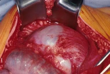
 Leiomyoma