Esophageal Perforation
Iatrogenic Disease
Esophagus
Esophageal Fistula
Esophageal Achalasia
Abscess
Tympanic Membrane Perforation
Uterine Perforation
Postoperative Complications
Fishes
Peptic Ulcer Perforation
Pleural empyema: An unusual presentation of esophageal perforation. (1/124)
A 67-year-old patient presented with pleural empyema as the sole manifestation of thoracic esophageal perforation, 2 weeks after accidental fish bone ingestion. Nonspecific chest pain and general deterioration, unusual presenting symptoms in themselves, accounted for the extreme delay in the diagnosis. The empyema was treated surgically, and the esophageal perforation conservatively. Despite the poor prognostic factors, the patient recovered completely after 50 days in hospital. (+info)Airway injury during anesthesia: a closed claims analysis. (2/124)
BACKGROUND: Airway injury during general anesthesia is a significant source of morbidity for patients and a source of liability for anesthesiologists. To identify recurrent patterns of injury, the authors analyzed claims for airway injury in the American Society of Anesthesiologists (ASA) Closed Claims Project database. METHODS: The ASA Closed Claims database is a standardized collection of case summaries derived from professional liability insurance companies closed claims files. All claims for airway injury were reviewed in depth and were compared to other claims during general anesthesia. RESULTS: Approximately 6% (266) of 4,460 claims in the database were for airway injury. The most frequent sites of injury were the larynx (33%), pharynx (19%), and esophagus (18%). Injuries to the esophagus and trachea were more frequently associated with difficult intubation. Injuries to temporomandibular joint and the larynx were more frequently associated with nondifficult intubation. Injuries to the esophagus were more severe and resulted in a higher payment to the plaintiff than claims for other sites of airway injury. Difficult intubation (odds ratio = 4.53, 95% confidence interval [CI] = 2.36, 8.71), age older than 60 yr (odds ratio = 2.97, 95% CI = 1.51, 5.87), and female gender (odds ratio = 2.43, 95% CI = 1.09, 5.42) were associated with claims for pharyngoesophageal perforation. Early signs of perforation, e.g., pneumothorax and subcutaneous emphysema, were present in only 51% of perforation claims, whereas late sequelae, e.g., retropharyngeal abscess and mediastinitis, occurred in 65%. CONCLUSION: Patients in whom tracheal intubation has been difficult should be observed for and told to watch for the development of symptoms and signs of retropharyngeal abscess, mediastinitis, or both. (+info)The accuracy of gastric insufflation in testing for gastroesophageal perforations during laparoscopic Nissen fundoplication. (3/124)
BACKGROUND: Laparoscopic Nissen fundoplication is an effective technique for the symptomatic relief of the manifestations of gastroesophageal reflux disorder but is associated with a 0.8-1% rate of gastroesophageal perforation. Early detection and repair of these injuries is critical to patient outcome, but occult injuries occur and may be missed. Gastric insufflation technique evaluates the integrity of the gastroesophageal wall after laparoscopic Nissen fundoplication. Gastric insufflation technique involves occlusion of the proximal stomach with a noncrushing bowel clamp while insufflating the submerged gastroesophageal junction. We conducted an animal study to assess the utility of gastric insufflation technique. METHODS: Five pigs (mean weight, 40.4 kg) underwent testing of laparoscopic gastric insufflation technique. In four animals, laparoscopic Nissen fundoplication was performed and then gastroesophageal junction injuries were created (3-5 mm distraction-type wall injuries). Non-crushing bowel clamps provided occlusion of the pylorus and then the proximal stomach during gastroesophageal insufflation. The gastroesophageal junction was then submerged. In the fifth animal, gastric insufflation technique was repeated while calibrated injuries were created to determine the smallest detectable injury. An injury was considered detectable if rising air bubbles were noted from the submerged gastroesophageal structures. Maximal luminal pressures needed to detect injuries were recorded with an in-line manometer. RESULTS: In all animals, 5-7 mm injuries of the gastroesophageal junction were easily detected using gastric insufflation technique when the proximal stomach was occluded. When the pylorus alone was occluded, detection of gastroesophageal injuries was inconsistent. Small injuries (<3 mm) of the esophagus were difficult to visualize with pyloric occlusion alone but were consistently detectable with proximal stomach occlusion at pressures less than 20 mm Hg. When the pylorus alone was occluded, the smallest detectable stomach perforation was a 16-gauge needle puncture while applying maximal gastric pressure (40-60 mm Hg) and a 2.5 mm linear injury when generating lower pressures (20 mm Hg). CONCLUSION: Proximal stomach occlusion and insufflation appears to effectively detect esophageal injuries of likely clinical importance (>2.5 mm). Pyloric occlusion and insufflation reliably evaluates the anterior stomach for injury. Gastric insufflation technique is a useful method for detecting gastroesophageal injury after laparoscopic Nissen fundoplication. (+info)Spontaneous oesophageal perforation due to mediastinal tuberculous lymphadenitis - atypical presentation of tuberculosis. (4/124)
Spontaneous non-traumatic oesophageal perforation secondary to bursting of a mediastinal tuberculous abscess into the oesophagus is rare. The diagnosis is delayed, as perforation remains localised due to mediastinal lymph nodes. Patient can be effectively managed by paraoesophageal drainage of the mediastinal abscess and oesophageal diversion. (+info)Oesophageal perforation following perioperative transoesophageal echocardiography. (5/124)
Transoesophageal echocardiography (TOE) is being used more often by cardiothoracic anaesthetists for the perioperative management of cardiac problems. Reports of iatrogenic oesophageal perforation by instrumentation of the oesophagus are increasing. Although TOE is considered safe, it may be more risky during surgery, because the probe is passed and manipulated in an anaesthetized patient. It may be in place for several hours so the risk of mucosal pressure and thermal damage is increased. Patients on cardiopulmonary bypass are also fully anticoagulated. We describe a case of oesophageal perforation following insertion of the TOE probe in a patient with gross cardiomegaly. Oesophageal distortion by cardiac enlargement may increase the risk of oesophageal perforation. Difficulty in passage of the TOE probe should be regarded with suspicion and withdrawal should be contemplated because the symptoms of oesophageal perforation are often delayed and non-specific. Delay in investigation, diagnosis and treatment will increase morbidity and mortality. (+info)Esophageal perforation associated with profound shock successfully managed with hemodynamic assistance using percutaneous cardiopulmonary support. (6/124)
A 51-year-old man was admitted to our hospital with complaints of severe chest pain, nausea, and vomiting. These symptoms had progressed rapidly and he was in shock. It was necessary to make a correct diagnosis as early as possible. However, the hemodynamic condition of the patient deteriorated rapidly before a definitive diagnosis could be established in spite of conventional therapies. Under hemodynamic assistance with percutaneous cardiopulmonary support (PCPS), a final diagnosis of esophageal perforation was made by esophagography. Our report illustrates a new application of PCPS for highly selected cases of noncardiogenic shock as a "bridge" until an accurate diagnosis is made and a specific treatment is applied. (+info)Boerhaave syndrome: report of a case treated non-operatively. (7/124)
An unique case of Boerhaave's Syndrome is presented in which the patient survived without any surgical treatment. We believe that this was due to non-contamination of the mediastinal and pleural cavities as shown by serial contrast roentgenograms of the esophagus. (+info)Esophageal perforation in a sword swallower. (8/124)
We present the case of a 59-year-old man who sustained an esophageal perforation as a result of sword swallowing. An esophagogram established the diagnosis, and surgical repair was attempted. However, 19 days later, a persistent leak and deterioration of the patient's condition necessitated a transhiatal esophagectomy with a left cervical esophagogastrostomy. The patient recovered and has resumed his daily activities at the circus, with the exception of sword swallowing. This case report presents an unusual mechanism for a potentially lethal injury. Our search of the English-language medical literature revealed no other report of esophageal perforation resulting from sword swallowing. Management of such an injury is often difficult, and a favorable outcome is dependent on prompt diagnosis and treatment. (+info)Esophageal perforation is a medical condition that refers to a hole or tear in the esophagus, which is the muscular tube that connects the throat to the stomach. This condition can occur as a result of various factors such as trauma, forceful vomiting (Boerhaave's syndrome), swallowing sharp objects, or complications from medical procedures like endoscopy.
Esophageal perforation is a serious medical emergency that requires immediate attention and treatment. If left untreated, it can lead to severe complications such as mediastinitis (inflammation of the tissue surrounding the heart), sepsis, and even death. Treatment typically involves surgical repair of the perforation, antibiotics to prevent infection, and supportive care to manage any associated symptoms or complications.
Intestinal perforation is a medical condition that refers to a hole or tear in the lining of the intestine. This can occur anywhere along the gastrointestinal tract, including the small intestine, large intestine (colon), or stomach. Intestinal perforation allows the contents of the intestines, such as digestive enzymes and bacteria, to leak into the abdominal cavity, which can lead to a serious inflammatory response known as peritonitis.
Intestinal perforation can be caused by various factors, including:
* Mechanical trauma (e.g., gunshot wounds, stab wounds)
* Inflammatory bowel disease (e.g., Crohn's disease, ulcerative colitis)
* Diverticulitis
* Appendicitis
* Intestinal obstruction
* Infections (e.g., typhoid fever, tuberculosis)
* Certain medications (e.g., nonsteroidal anti-inflammatory drugs, corticosteroids)
* Radiation therapy
* Ischemic bowel disease (lack of blood flow to the intestines)
Symptoms of intestinal perforation may include sudden abdominal pain, nausea, vomiting, fever, and decreased bowel movements. Treatment typically involves surgery to repair the perforation and remove any damaged tissue. Antibiotics are also administered to prevent infection. In severe cases, a temporary or permanent colostomy or ileostomy may be necessary.
Iatrogenic disease refers to any condition or illness that is caused, directly or indirectly, by medical treatment or intervention. This can include adverse reactions to medications, infections acquired during hospitalization, complications from surgical procedures, or injuries caused by medical equipment. It's important to note that iatrogenic diseases are unintended and often preventable with proper care and precautions.
The esophagus is the muscular tube that connects the throat (pharynx) to the stomach. It is located in the midline of the neck and chest, passing through the diaphragm to enter the abdomen and join the stomach. The main function of the esophagus is to transport food and liquids from the mouth to the stomach for digestion.
The esophagus has a few distinct parts: the upper esophageal sphincter (a ring of muscle that separates the esophagus from the throat), the middle esophagus, and the lower esophageal sphincter (another ring of muscle that separates the esophagus from the stomach). The lower esophageal sphincter relaxes to allow food and liquids to enter the stomach and then contracts to prevent stomach contents from flowing back into the esophagus.
The walls of the esophagus are made up of several layers, including mucosa (a moist tissue that lines the inside of the tube), submucosa (a layer of connective tissue), muscle (both voluntary and involuntary types), and adventitia (an outer layer of connective tissue).
Common conditions affecting the esophagus include gastroesophageal reflux disease (GERD), Barrett's esophagus, esophageal cancer, esophageal strictures, and eosinophilic esophagitis.
Mediastinal diseases refer to a group of conditions that affect the mediastinum, which is the area in the chest separating the lungs and containing various vital structures such as the heart, esophagus, trachea, thymus gland, lymph nodes, blood vessels, and nerves. These diseases can be benign or malignant (cancerous) and may cause symptoms due to compression or invasion of surrounding tissues. Examples of mediastinal diseases include:
1. Mediastinal tumors: Abnormal growths in the mediastinum, which can be benign or malignant. Common types include thymomas, germ cell tumors, lymphomas, and neurogenic tumors.
2. Mediastinitis: Inflammation of the mediastinal tissues, often caused by infections, trauma, or complications from medical procedures.
3. Enlarged lymph nodes: Abnormal swelling of the lymph nodes in the mediastinum can be a sign of various conditions, including infections, cancer, and autoimmune disorders.
4. Mediastinal cysts: Fluid-filled sacs that develop in the mediastinum, which are usually benign but may cause symptoms due to compression or infection.
5. Aneurysms or dissections of the aorta: Abnormal weakening or tearing of the aortic wall within the mediastinum, which can lead to life-threatening complications if not treated promptly.
6. Esophageal diseases: Conditions affecting the esophagus, such as tumors, strictures, or motility disorders, may present with symptoms related to the mediastinum.
7. Thyroid disorders: Enlargement of the thyroid gland (goiter) can extend into the mediastinum and cause compression symptoms.
8. Hematomas or effusions: Accumulation of blood (hematoma) or fluid (effusion) in the mediastinal space due to trauma, surgery, or other underlying conditions.
Early diagnosis and appropriate treatment are crucial for managing mediastinal diseases and improving patient outcomes.
"Foreign bodies" refer to any object or substance that is not normally present in a particular location within the body. These can range from relatively harmless items such as splinters or pieces of food in the skin or gastrointestinal tract, to more serious objects like bullets or sharp instruments that can cause significant damage and infection.
Foreign bodies can enter the body through various routes, including ingestion, inhalation, injection, or penetrating trauma. The location of the foreign body will determine the potential for harm and the necessary treatment. Some foreign bodies may pass through the body without causing harm, while others may require medical intervention such as removal or surgical extraction.
It is important to seek medical attention if a foreign body is suspected, as untreated foreign bodies can lead to complications such as infection, inflammation, and tissue damage.
Mediastinitis is a medical condition that refers to the inflammation of the mediastinum, which is the area in the chest that separates the lungs and contains various vital structures such as the heart, esophagus, trachea, thymus gland, and major blood vessels. Mediastinitis can be caused by bacterial or fungal infections, trauma, or complications from medical procedures such as esophageal surgery or heart catheterization.
The symptoms of mediastinitis may include chest pain, fever, difficulty swallowing, shortness of breath, cough, and neck stiffness. The diagnosis is typically made through imaging tests such as X-rays, CT scans, or MRI scans, and confirmed with laboratory tests that identify the causative organism. Treatment usually involves antibiotics or antifungal medications to eliminate the infection, along with supportive care such as pain management, fluids, and nutrition. In severe cases, surgery may be necessary to drain infected fluid or remove damaged tissue.
Dilation, also known as dilatation, refers to the process of expanding or enlarging a body passage or cavity. In medical terms, it typically refers to the widening of a bodily opening or hollow organ, allowing for increased flow or access. This can occur naturally, such as during childbirth when the cervix dilates to allow for the passage of a baby, or it can be induced through medical procedures or interventions.
For example, dilation of the pupils is a natural response to darkness or certain medications, while dilation of blood vessels is a common side effect of some drugs and can also occur in response to changes in temperature or emotional state. Dilation of the stomach or intestines may be necessary for medical procedures such as endoscopies or surgeries.
It's important to note that dilation can also refer to the abnormal enlargement of a body part, such as dilated cardiomyopathy, which refers to an enlarged and weakened heart muscle.
Esophagoscopy is a medical procedure that involves the visual examination of the esophagus, which is the tube that connects the throat to the stomach. This procedure is typically carried out using an esophagogastroduodenoscope (EGD), a flexible tube with a camera and light on the end.
During the procedure, the EGD is inserted through the mouth and down the throat into the esophagus, allowing the medical professional to examine its lining for any abnormalities such as inflammation, ulcers, or tumors. The procedure may also involve taking tissue samples (biopsies) for further examination and testing.
Esophagoscopy is commonly used to diagnose and monitor conditions such as gastroesophageal reflux disease (GERD), Barrett's esophagus, esophageal cancer, and other disorders affecting the esophagus. It may also be used to treat certain conditions, such as removing polyps or foreign objects from the esophagus.
An esophageal fistula is an abnormal connection or passage between the esophagus (the tube that carries food and liquids from the throat to the stomach) and another organ, such as the trachea (windpipe) or the skin. This condition can result from complications of certain medical conditions, including cancer, prolonged infection, or injury to the esophagus.
Esophageal fistulas can cause a variety of symptoms, including difficulty swallowing, coughing, chest pain, and fever. They can also lead to serious complications, such as pneumonia or sepsis, if left untreated. Treatment for an esophageal fistula typically involves surgical repair of the abnormal connection, along with management of any underlying conditions that may have contributed to its development.
Esophageal stenosis is a medical condition characterized by the narrowing or constriction of the esophagus, which is the muscular tube that connects the throat to the stomach. This narrowing can make it difficult to swallow food and liquids, leading to symptoms such as dysphagia (difficulty swallowing), pain or discomfort while swallowing, regurgitation, and weight loss.
Esophageal stenosis can be caused by a variety of factors, including:
1. Scarring or fibrosis due to prolonged acid reflux or gastroesophageal reflux disease (GERD)
2. Radiation therapy for cancer treatment
3. Ingestion of corrosive substances
4. Eosinophilic esophagitis, an allergic condition that affects the esophagus
5. Esophageal tumors or cancers
6. Surgical complications
Depending on the underlying cause and severity of the stenosis, treatment options may include medications to manage symptoms, dilation procedures to widen the narrowed area, or surgery to remove the affected portion of the esophagus. It is important to seek medical attention if you experience any difficulty swallowing or other symptoms related to esophageal stenosis.
Esophageal achalasia is a rare disorder of the esophagus, the tube that carries food from the mouth to the stomach. In this condition, the muscles at the lower end of the esophagus fail to relax properly during swallowing, making it difficult for food and liquids to pass into the stomach. This results in symptoms such as difficulty swallowing (dysphagia), regurgitation of food, chest pain, and weight loss. The cause of esophageal achalasia is not fully understood, but it is believed to be related to damage to the nerves that control the muscles of the esophagus. Treatment options include medications to relax the lower esophageal sphincter, botulinum toxin injections, and surgical procedures such as laparoscopic Heller myotomy or peroral endoscopic myotomy (POEM).
An abscess is a localized collection of pus caused by an infection. It is typically characterized by inflammation, redness, warmth, pain, and swelling in the affected area. Abscesses can form in various parts of the body, including the skin, teeth, lungs, brain, and abdominal organs. They are usually treated with antibiotics to eliminate the infection and may require drainage if they are large or located in a critical area. If left untreated, an abscess can lead to serious complications such as sepsis or organ failure.
Drainage, in medical terms, refers to the removal of excess fluid or accumulated collections of fluids from various body parts or spaces. This is typically accomplished through the use of medical devices such as catheters, tubes, or drains. The purpose of drainage can be to prevent the buildup of fluids that may cause discomfort, infection, or other complications, or to treat existing collections of fluid such as abscesses, hematomas, or pleural effusions. Drainage may also be used as a diagnostic tool to analyze the type and composition of the fluid being removed.
Tympanic membrane perforation, also known as a ruptured eardrum, is a tear or hole in the tympanic membrane, which separates the outer ear canal and the middle ear. The tympanic membrane plays a crucial role in hearing by transmitting sound vibrations from the outer ear to the inner ear. A perforation can result from various causes such as infection, trauma, pressure changes, or explosive blasts, leading to symptoms like hearing loss, tinnitus, vertigo, and ear discharge. The extent and location of the perforation determine the severity of the symptoms and the course of treatment, which may include observation, antibiotics, or surgical repair.
Uterine perforation is a medical condition that refers to the piercing or puncturing of the uterine wall. This can occur during various medical procedures such as dilatation and curettage (D&C), insertion of an intrauterine device (IUD), or during childbirth. It can also be caused by trauma or infection. Uterine perforation can lead to serious complications, such as bleeding, infection, and damage to surrounding organs. If left untreated, it can be life-threatening. Symptoms of uterine perforation may include severe abdominal pain, heavy vaginal bleeding, fever, and signs of shock. Immediate medical attention is required for proper diagnosis and treatment.
Postoperative complications refer to any unfavorable condition or event that occurs during the recovery period after a surgical procedure. These complications can vary in severity and may include, but are not limited to:
1. Infection: This can occur at the site of the incision or inside the body, such as pneumonia or urinary tract infection.
2. Bleeding: Excessive bleeding (hemorrhage) can lead to a drop in blood pressure and may require further surgical intervention.
3. Blood clots: These can form in the deep veins of the legs (deep vein thrombosis) and can potentially travel to the lungs (pulmonary embolism).
4. Wound dehiscence: This is when the surgical wound opens up, which can lead to infection and further complications.
5. Pulmonary issues: These include atelectasis (collapsed lung), pneumonia, or respiratory failure.
6. Cardiovascular problems: These include abnormal heart rhythms (arrhythmias), heart attack, or stroke.
7. Renal failure: This can occur due to various reasons such as dehydration, blood loss, or the use of certain medications.
8. Pain management issues: Inadequate pain control can lead to increased stress, anxiety, and decreased mobility.
9. Nausea and vomiting: These can be caused by anesthesia, opioid pain medication, or other factors.
10. Delirium: This is a state of confusion and disorientation that can occur in the elderly or those with certain medical conditions.
Prompt identification and management of these complications are crucial to ensure the best possible outcome for the patient.
I believe there may be a misunderstanding in your question. The term "fishes" is not typically used in a medical context. "Fish" or "fishes" refers to any aquatic organism belonging to the taxonomic class Actinopterygii (bony fish), Chondrichthyes (sharks and rays), or Agnatha (jawless fish).
However, if you are referring to a condition related to fish or consuming fish, there is a medical issue called scombroid fish poisoning. It's a foodborne illness caused by eating spoiled or improperly stored fish from the Scombridae family, which includes tuna, mackerel, and bonito, among others. The bacteria present in these fish can produce histamine, which can cause symptoms like skin flushing, headache, diarrhea, and itchy rash. But again, this is not related to the term "fishes" itself but rather a condition associated with consuming certain types of fish.
Peptic ulcer perforation is a serious and sightful gastrointestinal complication characterized by the penetration or erosion of an acid-peptic ulcer through the full thickness of the stomach or duodenal wall, resulting in spillage of gastric or duodenal contents into the peritoneal cavity. This leads to chemical irritation and/or bacterial infection of the abdominal cavity, causing symptoms such as sudden severe abdominal pain, tenderness, rigidity, and potentially life-threatening sepsis if not promptly diagnosed and treated with surgical intervention, antibiotics, and supportive care.
Treatment outcome is a term used to describe the result or effect of medical treatment on a patient's health status. It can be measured in various ways, such as through symptoms improvement, disease remission, reduced disability, improved quality of life, or survival rates. The treatment outcome helps healthcare providers evaluate the effectiveness of a particular treatment plan and make informed decisions about future care. It is also used in clinical research to compare the efficacy of different treatments and improve patient care.
Esophagus23
- An esophageal perforation is a hole in the esophagus. (medlineplus.gov)
- A perforation in the uppermost (neck region) part of the esophagus may heal by itself if you do not eat or drink for a period of time. (medlineplus.gov)
- Surgery is often needed to repair a perforation in the middle or bottom portions of the esophagus. (medlineplus.gov)
- With an esophageal perforation, an injury creates a hole in the esophagus. (medicinelearners.com)
- Perforation of the esophagus can lead to inflammation of the mediastinum. (medicinelearners.com)
- In the case of an esophageal perforation, all layers of the wall of the esophagus are damaged in such a way that a perforation occurs. (medicinelearners.com)
- An esophageal foreign body is a swallowed object that cannot be transported to the stomach due to its size, but gets stuck in the esophagus. (medicinelearners.com)
- In addition to esophageal foreign bodies, severe reflux diseases can also lead to perforation of the esophagus. (medicinelearners.com)
- Spontaneous perforation of the esophagus is also known as Boerhaave's syndrome. (medicinelearners.com)
- It is a perforation of the esophagus. (medicinelearners.com)
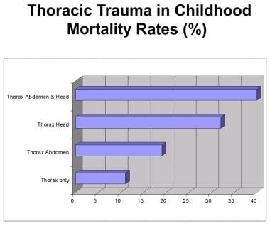
- Of all the perforations of the alimentary tract, perforations of the esophagus are considered the most dire and life-threatening. (johnshopkins.edu)
- However, we have learned that esophageal perforations in children are: (i) more often iatrogenic, (ii) more likely to occur within the cervical esophagus, and (iii) not generally associated with an underlying malignant disease process. (johnshopkins.edu)
- Cases of iatrogenic perforation of the esophagus occurring during a 3-year period were identified from the hospital endoscopy database. (elsevierpure.com)
- Available at https://nieuws.kuleuven.be/en/content/2014/mysterious-esophagus-disease-is-autoimmune-after-all . (medscape.com)
- It is characterized anatomically by a congenital obstruction of the esophagus with interruption of the continuity of the esophageal wall. (wikipedia.org)
- On plain X-ray, a feeding tube will not be seen pass through the esophagus and remain coiled in the upper oesophageal pouch. (wikipedia.org)
- The diagnostic criteria of EE include esophageal and/or upper gastrointestinal tract symptoms accompanied by ≥15 intraepithelial eosinophils/high power field (HPF) in 1 or more biopsy specimens without pathologic gastroesophageal reflux disease (GERD), as shown by normal pH monitoring of the distal esophagus or the lack of response to high-dose proton pump inhibitor (PPI) medication [ 2 ]. (hindawi.com)
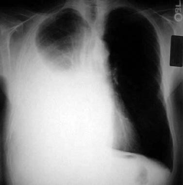
- A CT scan of the abdomen and pelvis was performed and this revealed "pneumomediastinum with air around the distal esophagus, suggestive of an esophageal perforation and a left-sided pneumothorax. (empr.com)
- There is a small risk of bleeding or tearing a hole in the esophagus (called a perforation ) with this procedure, which could require surgery or other treatments to fix. (cancer.org)
- Abnormalities of the esophagus which delay esophageal emptying such as stricture or achalasia. (almishkat.pk)
- Anatomy of lymphatic drainage of the esophagus and lymph node metastasis of thoracic esophageal cancer. (myesr.org)
- 13] Japan Esophageal Society : Japanese Classification of Esophageal Cancer, 11th Edition: part I. Esophagus 14:1-36 2017. (myesr.org)
- 18] Saito H, Sato T & Miyazaki M : Extramural lymphatic drainage from the thoracic esophagus based on minute cadaveric dissections: fundamentals for the sentinel node navigation surgery for the thoracic esophageal cancers. (myesr.org)
Mediastinitis4
- Among them, esophageal perforation is associated with severe morbidity, including dysphagia, malnutrition, and infection with the potential development of mediastinitis. (nih.gov)
- However, the most dangerous complication of an esophageal perforation is mediastinitis. (medicinelearners.com)
- Acute mediastinitis usually results from esophageal perforation or occurs after median sternotomy. (msdmanuals.com)
- The possibility of infectious mediastinitis raised the concern for a micro-perforation post-endoscopy given the agitation of the patient during the procedure, but it was judged less likely by the infectious disease specialists. (acc.org)
Thoracic esophageal perforations2
Endoscopic10
- As the incidence of esophageal perforation increases with the advancement of invasive endoscopic procedures, early recognition of clinical features and implementation of effective treatment are essential for a favorable clinical outcome with minimal morbidity and mortality. (nih.gov)
- Perforation was suspected after 10 of 492 endoscopic dilatation procedures done in patients with obstructing esophageal malignancies. (elsevierpure.com)
- 1. Cervical esophageal perforations at the time of endoscopic ultrasound: a prospective evaluation of frequency, outcomes, and patient management. (nih.gov)
- 5. Repair of an EUS--induced duodenal perforation with endoscopic clips. (nih.gov)
- 13. Endoscopic negative pressure therapy (ENPT) in head and neck surgery: first experiences in treatment of postoperative salivary fistulas and cervical esophageal perforations. (nih.gov)
- Current guidelines recommend performing endoscopic biopsy to obtain four to six tissue samples from the esophageal mucosa to confirm diagnosis. (mayoclinic.org)
- Given that dietary therapy actually prevents esophageal inflammation, is associated with lower costs and has fewer known side effects than currently available pharmacologic and endoscopic interventions, this approach is gaining gradual acceptance as a treatment for EoE in both children and adults. (mayoclinic.org)
- Achalasia is a rare oesophageal motility disorder and is classically treated by endoscopic pneumatic dilation (PD) or laparoscopic Heller's myotomy (LHM) combined with an antireflux procedure. (bmj.com)
- Endoscopic ultrasonography in the staging of esophageal carcinoma after preoperative radiotherapy and chemotherapy. (myesr.org)
- Assessment of the Diagnostic Performance of Endoscopic Ultrasonography After Conventional Endoscopy for the Evaluation of Esophageal Squamous Cell Carcinoma Invasion Depth. (myesr.org)
Diagnosis6
- Controversies over the diagnosis and management of esophageal perforation remain, and debate still exists over the optimal therapeutic approach. (nih.gov)
- Background: Recognition of the importance of early diagnosis and aggressive, definitive surgical intervention has brought about a dramatic decline in mortality related to distal esophageal perforation. (usuhs.edu)
- Diagnosis of esophageal motility disorders: Esophageal pressure topography vs. conventional line tracing. (medscape.com)
- The upper neck pouch sign is another sign that helps in the antenatal diagnosis of esophageal atresia and it may be detected soon after birth as the affected infant will be unable to swallow its own saliva. (wikipedia.org)
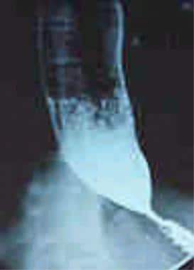
- While contrast esophagography may be the most reliable way to identify esophageal perforation, for some patients this may not be an option, so contrast-enhanced chest CT may help with diagnosis. (empr.com)
- Anatomopathologic examination of the esophageal biopsy showed no signs of invasion, leading to the diagnosis of mucocutaneous fungal infection. (cdc.gov)
Boerhaave1
- Boerhaave syndrome, or spontaneous esophageal rupture, is a rare condition that "occurs as the result of barotrauma secondary to forceful retching and vomiting against a closed glottis. (empr.com)
Achalasia3
- The invasion of the esophageal neural plexus by the tumor can cause nonrelaxation of the LES, thus mimicking achalasia. (medscape.com)
- Pneumatic dilatation for achalasia carries a significant and recognized risk of esophageal perforation. (medscape.com)
- Importance of preoperative and postoperative pH monitoring in patients with esophageal achalasia. (medscape.com)
Esophagitis2
- Certain pills or esophageal ulcers (eg, in patients with AIDS and esophagitis) can contribute. (msdmanuals.com)
- Gastrointestinal: Common: Esophagitis, esophageal erosions, esophageal ulcers. (almishkat.pk)
Gastro-esophage1
- Ideally, the tip should be at least 10 cm beyond the gastro-esophageal junction 1 . (radiopaedia.org)
Squamous cell carc1
- Pattern of lymph node metastases of esophageal squamous cell carcinoma based on the anatomical lymphatic drainage system: Node metastasis in esophageal cancer. (myesr.org)
Complication1
- A case report of esophageal perforation: Complication of nasogastric tube placement. (radiopaedia.org)
Cervical esophageal4
- 2. Cervical esophageal perforation during EUS: a national survey. (nih.gov)
- Cervical esophageal cancer: A population-based study. (myesr.org)
- Surgical management of cervical esophageal carcinoma with larynx preservation and reconstruction. (myesr.org)
- Cervical esophageal cancer: a gap in cancer knowledge. (myesr.org)
Iatrogenic3
- An iatrogenic esophageal perforation is usually caused by endoscopy. (medicinelearners.com)
- The management of patients with iatrogenic perforation of esophageal cancers is controversial. (elsevierpure.com)
- Immediate placement of a coated self-expanding metal stent is an effective treatment for iatrogenic perforation of an obstructing esophageal malignancy. (elsevierpure.com)
Lead to perforation1
- Such trauma can lead to perforation, periesophageal abscesses, and fibrosis. (nih.gov)
Ulcers1
- Esophageal erosions, ulcers, and necrosis are frequently the result of gavage accidents. (nih.gov)
Obstruction1
- Esophageal obstruction by a tangled nasogastric tube. (radiopaedia.org)
Rupture1
- For this patient, ghost pepper ingestion led to forceful retching and vomiting resulting in esophageal rupture that could have easily been confused with discomfort after a large spicy meal. (empr.com)
Gastric Perforation1
- Anatomopathologic examination of the gastric perforation showed necrosis and inflammation. (cdc.gov)
Endoscopy2
- Under certain circumstances, however, the perforation site can be overlooked during endoscopy due to superimposed blood. (medicinelearners.com)
- A procedure may help find the area of the perforation, such as an upper endoscopy (EGD) or a colonoscopy. (medlineplus.gov)
Diseases1
- During a recent presentation at Mayo Clinic's Esophageal Diseases Course in Phoenix, Arizona, Mayo Clinic gastroenterologist Jeffrey A. Alexander, M.D. , explained current research findings and clinical experience related to the use of elimination diets in the treatment of EoE. (mayoclinic.org)
Surgical5
- Although there remains a clear role for surgical therapy in selected children with esophageal perforations, the management paradigm has clearly shifted towards less invasive treatment modalities as the first line of therapy in children who are otherwise clinically stable. (johnshopkins.edu)
- Most studies recommend surgical intervention for patients with esophageal perforation and thoracic abscess. (amjcaserep.com)
- A surgical consultation must be obtained as soon as a perforation is identified. (medscape.com)
- In some of these so-called long gap cases, though, an advanced surgical treatment developed by John Foker, MD, may be utilized to elongate and then join the short esophageal segments. (wikipedia.org)
- require emergency surgical exploration of the mediastinum with primary repair of the esophageal tear and drainage of the pleural space and mediastinum. (msdmanuals.com)
Inflammation5
- This leads to chronic inflammation and, in the worst case, to perforation. (medicinelearners.com)
- Patients with esophageal perforation become acutely ill within hours, with severe chest pain and dyspnea due to mediastinal inflammation. (msdmanuals.com)
- Treatment of EoE is directed at reducing inflammation and reversing esophageal fibrosis that develops as a result of long-term inflammatory activity. (mayoclinic.org)
- Esophageal histologic sampling is required repeatedly during the food reintroduction period, as it is the only way to determine which foods are triggering the inflammation. (mayoclinic.org)
- According to the latest consensus EE represents a chronic immune/antigen-mediated esophageal disease characterized clinically by symptoms related to esophageal dysfunction and histologically by eosinophil-predominant inflammation [ 1 ]. (hindawi.com)
Mucosa1
- Eosinophilic infiltration of the esophageal mucosa is responsible for esophageal symptoms which can range from mild to debilitating dysphagia and food impaction, when untreated. (hindawi.com)
Secondary3
- This study comprised all patients (age, 41-73 years) undergoing free tissue transfer for the repair of chronic esophageal perforation secondary to anterior cervical discectomy and fusion at an academic tertiary care center. (nih.gov)
- The secondary outcomes included the need for re-treatment, lower oesophageal sphincter pressure, oesophageal emptying and the rate of complications. (bmj.com)
- 5. Hutchinson R, Ahmed AR, Menzies D. A case of intramural oesophageal dissection secondary to nasogastric tube insertion. (radiopaedia.org)
Gastrointestinal1
- NSAIDs, including celecoxib, cause an increased risk of serious gastrointestinal adverse events including bleeding, ulceration, and perforation of the stomach or intestines, which can be fatal. (nih.gov)
Symptoms2
- Patients present with progressive dysphagia , weight loss , chronic worsening gastro-esophageal reflux and hoarseness , cough, vocal cord paralysis , or other signs and symptoms of mediastinal invasion. (radiopaedia.org)
- Several types of treatment can be used to help prevent or relieve symptoms of esophageal cancer. (cancer.org)
Incidence1
- Incidence and distribution of distant metastases from newly diagnosed esophageal carcinoma. (myesr.org)
Oesophagus1
- Put simply, esophageal perforation happens when your oesophagus is torn. (edcasworldwide.com)
Occurs2
- People will often have a few days of pain before the intestinal perforation occurs. (medlineplus.gov)
- Treatment is much simpler and safer when it is started before the perforation occurs. (medlineplus.gov)
Dilatation2
- Therefore, an informed consent emphasizing this risk of perforation must be obtained from patients prior to the dilatation. (medscape.com)
- After the dilatation, administer a small amount of water-soluble contrast material to evaluate for perforation. (medscape.com)
Patients10
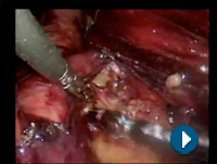
- Post operative esophageal mucosal leaks, can be treated using minimal invasive techniques in selected patients. (sages.org)
- however, further clinical evidence is required to determine its suitability for other patients with esophageal perforation complicated by mediastinal abscess. (amjcaserep.com)
- We reviewed the management of perforated esophageal malignancies at a single institution with a large volume of patients with esophageal cancer. (elsevierpure.com)
- Patients with a small perforation without any evidence of infection or communication with the pleural or peritoneal cavities may receive conservative therapy with broad-spectrum antibiotics and close observation in the hospital. (medscape.com)
- Herbella FA, Colleoni R, Bot L, Vicentine FP, Patti MG. High-resolution manometry findings in patients after sclerotherapy for esophageal varices. (medscape.com)
- Classifying esophageal motility by pressure topography characteristics: a study of 400 patients and 75 controls. (medscape.com)
- This notion is supported by the fact that there are numerous reports of patients with multiple oesophageal rings with intraepithelial eosinophils that had been ascribed to acid reflux, but who did not respond to standard acid suppression therapy. (hindawi.com)
- Two patients (40%) underwent emergent surgery which included partial excision of atrial wall, closure with bovine pericardial patch and closure of esophageal lesion. (jafib.com)
- The objective of this study was to investigate whether fasting in the month of Ramadan had any effect on the peptic ulcer frequency and peptic ulcer perforation in Muslim patients presenting to the Accident and Emergency Department of a large hospital in the United Arab Emirates. (who.int)
- We conducted a retrospective descriptive study of peptic ulcer perforation among patients presenting to the Accident and Emergency Department of Al Ain Hospital in the United Arab Emirates who were then treated at the Departments of Medicine and Surgery. (who.int)
Peptic1
- Peptic ulcer perforation occurred more frequently after Ramadan but the difference was not significant. (who.int)
Uncommon1
- Esophageal carcinoma is relatively uncommon. (radiopaedia.org)
Surgery4
- Surgery will depend on the location and size of the perforation. (medlineplus.gov)
- Despite significant advances in modern surgery and intensive care medicine, esophageal perforation continues to present a diagnostic and therapeutic challenge. (nih.gov)
- Esophageal perforation is an extremely life-threatening medical emergency that requires immediate surgery. (medicinelearners.com)
- Esophageal Perforation Following Anterior Cervical Spine Surgery: Case Report and Review of the Literature. (duke.edu)
Mediastinum1
- Skin emphysema and fluid levels in the mediastinum also indicate perforation. (medicinelearners.com)
Abscess2
- You may also have a chest CT scan to look for an abscess in the chest or esophageal cancer. (medlineplus.gov)
- However, it rarely causes esophageal perforation and mediastinal abscess. (amjcaserep.com)
Reflux1
- Identifying response to acid suppressive therapy in functional dyspepsia using a random starting day trial�is gastro-oesophageal reflux necessary The usefulness of a structured questionnaire within the evaluation of symptomatic gastroesophageal reflux disease. (dnahelix.com)
Strictures1
- Household bleaches (3 to 6% sodium hypochlorite) usually cause esophageal irritation, but rarely cause strictures or serious injury such as perforation. (cdc.gov)
Less invasive1
- however, less invasive approaches to esophageal perforation continue to evolve. (nih.gov)
Severe1
- However, the outcome will depend on how severe the perforation is, and for how long it was present before treatment. (medlineplus.gov)
Occur2
- Esophageal perforations may also occur, increasing the risk of cancer. (groupeproxim.ca)
- Anatomy of right recurrent nerve node: why does early metastasis of esophageal cancer occur in it? (myesr.org)
Mucosal1
- Five oesophageal perforations occurred during PD (5%) while 12 mucosal tears (11%) occurred during LHM. (bmj.com)
Complicate1
- may complicate esophagoscopy or insertion of a Sengstaken-Blakemore or Minnesota tube (for esophageal variceal bleeding). (msdmanuals.com)
Cancer4
- In addition, there are certain regions where individuals are at particularly high risk of developing esophageal cancer, e.g. (radiopaedia.org)
- The most accurate imaging modality for the T staging of esophageal cancer. (radiopaedia.org)
- People with esophageal cancer often have already lost weight before the cancer was found. (cancer.org)
- Some people with esophageal cancer may need to have a feeding tube, usually called a jejunostomy tube (or J-tube), put in place before treatment. (cancer.org)
Injuries1
- This review will attempt to summarize the pathogenesis and diagnostic evaluation of esophageal injuries, and highlight the evolving therapeutic options for the management of esophageal perforation. (nih.gov)
Stricture1
- Left untreated, this disorder is also associated with esophageal remodeling and stricture formation. (mayoclinic.org)
Management3
- Lambright E. Management of esophageal perforation. (medlineplus.gov)
- Herein we examine our experiences in the management of esophageal perforation with microvascular free tissue transfer. (nih.gov)
- 11. ERCP-related perforations: risk factors and management. (nih.gov)
Treatment2
- This video demonstrates that laparoscopic techniques can be safely used in the treatment of post operative esophageal perforations. (sages.org)
- The optimal treatment in cases of long gap esophageal atresia remains controversial. (wikipedia.org)
Common2
- The most common cause of an esophageal perforation is injury during a medical procedure. (medlineplus.gov)
- The most common ingested foreign objects that cause perforation include fish bones, dentures, and peach stones. (medicinelearners.com)