Eye Infections
Eye Infections, Bacterial
Conjunctivitis, Inclusion
Conjunctivitis, Bacterial
Eye
Conjunctivitis
Eye Infections, Parasitic
Keratitis, Dendritic
Eye Infections, Viral
Ribotyping
Eye Infections, Fungal
Eye Injuries
Dry Eye Syndromes
Eye Enucleation
Visual Acuity
Ocular Physiological Phenomena
Compound Eye, Arthropod
Eye Protective Devices
Retina
Ophthalmic Solutions
Fixation, Ocular
Posterior Eye Segment
Glaucoma
Vitreous Body
Sclera
Axial Length, Eye
Anterior Chamber
Myopia
Aqueous Humor
Iris
Ciliary Body
Lens, Crystalline
Visual Fields
Aetiological study of the presumed ocular histoplasmosis syndrome in the Netherlands. (1/235)
AIM: To investigate whether presumed ocular histoplasmosis syndrome in the Netherlands is caused by Histoplasma capsulatum and whether other risk factors might play a role in the pathogenesis of this syndrome. METHODS: 23 patients were clinically diagnosed as having presumed ocular histoplasmosis syndrome based on the following criteria: peripapillary atrophy, punched out lesions, a macular disciform lesion or scar in one eye without vitritis. As controls, 66 sex and age matched healthy volunteers were used. Serum samples from both patients and controls were tested for the presence of antibodies against H capsulatum, Toxoplasma gondii, Toxocara canis et cati, Ascaris sp, and for the presence of antigens of Cryptococcus neoformans. Serum samples were also tested for the presence of autoantibodies against retinal or choroidal proteins. To investigate other risk factors, patients and controls were asked to fill in a health and travel related questionnaire. Ten patients with ocular toxoplasmosis were used as a disease control group. RESULTS: None of the patients with presumed ocular histoplasmosis syndrome or controls had circulating antibodies directed against H capsulatum. No risk factors could be identified and no indications for autoimmunity and no evidence for the role of the other infectious agents could be demonstrated. CONCLUSIONS: In a Dutch group of patients fulfilling the criteria of a disease currently named presumed ocular histoplasmosis syndrome, no risk factors or relation with the fungus H capsulatum could be detected. (+info)Contact lens-induced infection--a new model of Candida albicans keratitis. (2/235)
PURPOSE: A model of experimental keratomycosis was established that mimics human disease in which the only fungi present are those that are actively growing within the cornea. METHODS: Dutch-belted rabbits received a subconjunctival injection of triamcinolone acetonide to one eye. One day later the epithelium was removed from the central cornea and a standardized inoculum of Candida albicans blastoconidia was placed on the corneal surface and covered with a contact lens. The lids were closed with a lateral tarsorrhaphy. After 24 hours, the lid sutures and contact lens were removed. Five days later the animals were killed, and their corneas were subjected to separate isolate recovery and histology studies. A group of similarly infected rabbits without corticosteroid injection served as controls. RESULTS: Both groups developed invasive corneal disease. Although isolate recovery was not significantly different from corticosteroid-treated rabbits compared with controls, fungal biomass was increased. Hyphal invasion was limited to the anterior cornea in control eyes, but penetrated deep stroma in most of the corticosteroid-treated rabbits. CONCLUSIONS: Invasive corneal disease can be established with a surface inoculum. Corticosteroid administration increased corneal penetration of hyphae. Quantitative isolate recovery is not a reliable measure of the fungal load within the cornea. (+info)A study of mycotic keratitis in Mumbai. (3/235)
A total of 1010 clinically suspected cases of mycotic keratitis were studied from 1988 to 1996 for evidence of fungal infection and for identification of the aetiologic agents of keratitis in Mumbai. Of these 367 cases were reported positive by microscopy and culture. Seventy nine percent of the cases were between the ages 21 and 50 years. Male patients were more often affected than females. Eighty eight percent of patients were farmers or construction workers and 89.92% of cases gave a definite history of antecedent corneal trauma. A single fungal isolate was obtained in 307 cases and multiple isolates in 20 cases. Mixed isolates of bacteria and fungi were grown in 40 cases. The predominant isolate was Aspergillus species in 219 cases, followed by Candida species (36), Fusarium species (33) and Penicillium species (34). Filamentous fungal isolates from 22 cases remained unidentified. Mycotic keratitis should be suspected in every patient with a corneal lesion and should be ruled out before commencing steroids and antiboitics. (+info)Microbial decontamination of human donor eyes with povidone-iodine: penetration, toxicity, and effectiveness. (4/235)
BACKGROUND/AIMS: Povidone-iodine (PVP -I) is applied for microbial decontamination of human eyes donated for transplantation. Concentrations and immersion times vary greatly. The effectiveness and toxicity of PVP-I were assessed for different decontamination protocols. METHODS: Human donor eyes and corneas were immersed in different concentrations (5-100 mg/ml) of PVP-I for different times (2-30 minutes). The penetration of iodine into the corneal tissue was assessed by x ray microanalysis. Microbial contamination was determined by taking cultures of the limbal areas and storage solutions and by incubation of the corneoscleral buttons in antibiotic-free culture medium. Cytotoxicity of PVP-I for corneal fibroblasts in culture was assessed using the MTT assay. RESULTS: Depending on concentration and immersion time iodine was found to penetrate into the epithelium, Bowman's layer, and stroma in amounts equivalent to 2-40 mg/ml PVP-I. The MTT assay demonstrated that 2.5 mg/ml PVP-I caused total damage to fibroblasts in vitro. Rinsing eyes with tap water and subsequent immersion in PVP-I reduced the rate of contamination from 82 out of 106 to 69 out of 106 and 37 out of 106, respectively. Antibiotics in the storage medium further reduced contamination from about 40% to 3%. Microbial contamination was not reduced by increasing the concentration and immersion times beyond 5 mg/ml PVP-I for 2 minutes. CONCLUSION: Immersion of human donor eyes in 5 mg/ml PVP-I solution for 2 minutes significantly reduces microbial contamination of donor corneas without relevant penetration of iodine into the corneal layers. Higher PVP-I concentrations and longer immersion times do not further reduce contamination, whereas the amount of iodine penetrating the corneal layers is elevated above the level cytotoxic for corneal fibroblasts. In view of this, concentrations above 5 mg/ml of PVP-I and immersion periods over 2 minutes are not recommended for reduction of the contamination rate of donor eyes. (+info)Fungal corneal ulcers of onion harvesters in southern Taiwan. (5/235)
Fungal corneal ulcers related to agriculture has been reported throughout the world, especially in tropical areas. Most of them were sporadic and had histories of ocular trauma or use of topical corticosteroids and topical antibiotics. Five onion harvesters had fungal corneal ulcers during the same harvest period in Southern Taiwan. The authors think that this is the first report of a group occurrence relating to agricultural workers. Although all of the patients improved after medical and surgical management, their vision was greatly decreased. It is suggested that the tropical climate, the harvest procedure, the characteristic monsoon, and lack of eye protection were involved. Therefore, the importance of the eye protection, hygiene education, and improving medical care to reduce the occurrence of fungal corneal ulcer in agriculture workers must be emphasised. (+info)Mycotic keratitis due to Curvularia senegalensis and in vitro antifungal susceptibilities of Curvularia spp. (6/235)
A case of mycotic keratitis due to Curvularia senegalensis is reported. This case represents the third known reported infection caused by this rare species. Fungal hyphae were detected in corneal scrapings, and repeated cultures were positive for this fungi. The patient was presumed cured after a corneal transplant and treatment with itraconazole, but the infection recurred and the patient is waiting for a keratoplasty. The in vitro antifungal susceptibilities of the case strain and another 24 strains belonging to seven species of Curvularia were tested for six antifungal agents. With the exception of flucytosine, and occasionally fluconazole, the other drugs assayed (amphotericin B, miconazole, itraconazole, and ketoconazole) were highly effective in vitro. (+info)Differences in virulence between two Candida albicans strains in experimental keratitis. (7/235)
PURPOSE: To study the differences in disease caused by two wild-type strains of Candida albicans in a model of contact lens-facilitated keratitis in rabbits. METHODS: Two strains, SC5314 and VE175, were examined. Standardized inocula were placed on the debrided corneal surface of one eye in Dutch belted rabbits and covered with a contact lens. A temporary tarsorrhaphy was opened after 24 hours with removal of the contact lens. Six days later, corneas were photographed and animals killed. Corneas were bisected with one half for quantitative isolate recovery and the other for stromal penetration by hyphae. RESULTS: Strain SC5314 was significantly more virulent. The mean hyphal penetration into the cornea was 24.4% +/- 8.5% of the corneal thickness, and in three of six corneas hyphae penetrated through the entire cornea. In contrast, for VE175, the mean hyphal penetration was 2.6% +/- 1.2%. The difference between these two strains was statistically significant (P = 0.0297). Hyphae did not penetrate into the deep layers of the cornea in any of the six rabbits infected with VE175. The grading of clinical disease was consistent with histology, in that strain SC5314 caused more severe infection than VE175 and the difference was statistically significant (P = 0.0048). There was no difference in isolate recovery. CONCLUSIONS: Wild-type strains of C. albicans can differ significantly in virulence as measured by depth of fungal invasion into corneas and clinical evaluation of infection. Further characterization of the intrinsic genetic differences between such strains may help identify factors responsible for fungal virulence. (+info)PCR-RFLP-mediated detection and speciation of bacterial species causing endophthalmitis. (8/235)
PURPOSE: To determine the usefulness of polymerase chain reaction-restriction fragment length polymorphism (PCR-RFLP) in the identification and speciation of bacteria causing endophthalmitis. METHODS: PCR-RFLP was performed on 53 strains of 14 bacterial species (eight Gram positive and five Gram negative) collected from both keratitis and endophthalmitis patients. Two pairs of oligonucleotide primers based on the 16S rDNA gene were used to PCR-amplify 1.2- and 1.0-kb fragments of bacterial genomic DNA. RFLPs within the PCR product were used to speciate the organisms. RESULTS: The sensitivity of the nested PCR amplification reaction was one organism. All bacteria tested could be identified and speciated using RFLP analysis except for Escherichia coli and Serratia marcescens, which could not be interdifferentiated using RFLP. Molecular analysis of two vitreous samples from two eyes with typical signs of bacterial endophthalmitis confirmed the presence of E. coli in the vitreous from a culture-positive case with E. coli endophthalmitis and revealed the presence of Staphylococcus epidermidis in the vitreous of a culture-negative case. CONCLUSIONS: It is expected that this technique will provide a useful laboratory tool for future microbiologic diagnosis of patients presenting with endophthalmitis, especially for those eyes that prove culture negative. (+info)Eye infections, also known as ocular infections, are conditions characterized by the invasion and multiplication of pathogenic microorganisms in any part of the eye or its surrounding structures. These infections can affect various parts of the eye, including the conjunctiva (conjunctivitis), cornea (keratitis), eyelid (blepharitis), or the internal structures of the eye (endophthalmitis, uveitis). The symptoms may include redness, pain, discharge, itching, blurred vision, and sensitivity to light. The cause can be bacterial, viral, fungal, or parasitic, and the treatment typically involves antibiotics, antivirals, or antifungals, depending on the underlying cause.
Bacterial eye infections, also known as bacterial conjunctivitis or bacterial keratitis, are caused by the invasion of bacteria into the eye. The most common types of bacteria that cause these infections include Staphylococcus aureus, Streptococcus pneumoniae, and Haemophilus influenzae.
Bacterial conjunctivitis is an inflammation of the conjunctiva, the thin membrane that covers the white part of the eye and the inner surface of the eyelids. Symptoms include redness, swelling, pain, discharge, and a gritty feeling in the eye. Bacterial keratitis is an infection of the cornea, the clear front part of the eye. Symptoms include severe pain, sensitivity to light, tearing, and decreased vision.
Bacterial eye infections are typically treated with antibiotic eye drops or ointments. It is important to seek medical attention promptly if you suspect a bacterial eye infection, as untreated infections can lead to serious complications such as corneal ulcers and vision loss. Preventive measures include good hygiene practices, such as washing your hands frequently and avoiding touching or rubbing your eyes.
Inclusion conjunctivitis is a type of bacterial conjunctivitis (inflammation of the conjunctiva) that is caused by specific types of bacteria, most commonly Chlamydia trachomatis. It is also known as trachoma, which is a leading infectious cause of blindness worldwide. The infection leads to the formation of small, inclusion-containing intracytoplasmic inclusions in the conjunctival epithelial cells, hence the name "inclusion conjunctivitis."
The symptoms of inclusion conjunctivitis include redness, irritation, and discharge from the eyes. It can also cause swelling of the lymph nodes near the ears. In severe cases, it can lead to scarring and damage to the cornea, potentially resulting in vision loss. The infection is typically spread through direct contact with eye or nose discharge from an infected person, and it can also be sexually transmitted.
Treatment for inclusion conjunctivitis usually involves antibiotics, such as azithromycin or doxycycline, to eliminate the bacteria causing the infection. It is important to complete the full course of treatment to ensure that the infection is fully cleared and to prevent recurrence. In addition, good hygiene practices, such as frequent handwashing and avoiding sharing personal items like towels and washcloths, can help prevent the spread of the infection.
"Hemolysin factors" is not a standard medical term, and it seems there might be some confusion in the question. However, I can provide information on hemolysins, which are substances that cause lysis (rupture) of red blood cells, resulting in the release of their contents into the surrounding fluid.
Hemolysins can be produced by various sources, such as:
1. Bacterial hemolysins: Some bacteria produce hemolysins as a virulence factor to aid in infecting the host. These hemolysins can be classified into two main types: exotoxins (secreted by the bacterium) and endotoxins (integral components of the bacterial cell membrane). Examples include streptolysin O and streptolysin S from Streptococcus pyogenes, hemolysin from Escherichia coli, and α-toxin from Staphylococcus aureus.
2. Complement system: The complement system is a part of the immune response that can cause hemolysis through the membrane attack complex (MAC). This complex forms pores in the red blood cell membrane, leading to lysis.
3. Autoimmune disorders: In some autoimmune diseases, such as autoimmune hemolytic anemia, the body produces antibodies against its own red blood cells, causing complement-mediated hemolysis.
4. Medicines and chemicals: Certain medications or chemicals can cause hemolysis as a side effect. These include some antibiotics (e.g., cephalosporins), chemotherapeutic agents, and snake venoms.
If you meant to ask about something else related to "hemolysin factors," please provide more context so I can give a more accurate answer.
Keratoconjunctivitis is a medical term that refers to the inflammation of both the cornea (the clear, outer layer at the front of the eye) and the conjunctiva (the mucous membrane that covers the inner surface of the eyelids and the white part of the eye).
The condition can cause symptoms such as redness, pain, sensitivity to light, watery eyes, and a gritty or burning sensation in the eyes. Keratoconjunctivitis can be caused by various factors, including viral or bacterial infections, allergies, or environmental irritants like dust, smoke, or chemical fumes.
Treatment for keratoconjunctivitis depends on the underlying cause of the condition and may include medications such as antibiotics, antivirals, or anti-inflammatory agents to reduce inflammation and relieve symptoms. In some cases, artificial tears or lubricants may also be recommended to help keep the eyes moist and comfortable.
Bacterial conjunctivitis is a type of conjunctivitis (inflammation of the conjunctiva) that is caused by bacterial infection. The most common bacteria responsible for this condition are Staphylococcus aureus, Streptococcus pneumoniae, and Haemophilus influenzae.
The symptoms of bacterial conjunctivitis include redness, swelling, and pain in the eye, along with a thick, sticky discharge that can cause the eyelids to stick together, especially upon waking up. Other symptoms may include tearing, itching, and sensitivity to light. Bacterial conjunctivitis is highly contagious and can spread easily through contact with infected individuals or contaminated objects such as towels, handkerchiefs, or makeup.
Treatment for bacterial conjunctivitis typically involves the use of antibiotic eye drops or ointments to eliminate the infection. In some cases, oral antibiotics may also be prescribed. It is important to seek medical attention if you suspect that you have bacterial conjunctivitis, as untreated infections can lead to serious complications such as corneal ulcers and vision loss.
The eye is the organ of sight, primarily responsible for detecting and focusing on visual stimuli. It is a complex structure composed of various parts that work together to enable vision. Here are some of the main components of the eye:
1. Cornea: The clear front part of the eye that refracts light entering the eye and protects the eye from harmful particles and microorganisms.
2. Iris: The colored part of the eye that controls the amount of light reaching the retina by adjusting the size of the pupil.
3. Pupil: The opening in the center of the iris that allows light to enter the eye.
4. Lens: A biconvex structure located behind the iris that further refracts light and focuses it onto the retina.
5. Retina: A layer of light-sensitive cells (rods and cones) at the back of the eye that convert light into electrical signals, which are then transmitted to the brain via the optic nerve.
6. Optic Nerve: The nerve that carries visual information from the retina to the brain.
7. Vitreous: A clear, gel-like substance that fills the space between the lens and the retina, providing structural support to the eye.
8. Conjunctiva: A thin, transparent membrane that covers the front of the eye and the inner surface of the eyelids.
9. Extraocular Muscles: Six muscles that control the movement of the eye, allowing for proper alignment and focus.
The eye is a remarkable organ that allows us to perceive and interact with our surroundings. Various medical specialties, such as ophthalmology and optometry, are dedicated to the diagnosis, treatment, and management of various eye conditions and diseases.
Conjunctivitis is an inflammation or infection of the conjunctiva, a thin, clear membrane that covers the inner surface of the eyelids and the outer surface of the eye. The condition can cause redness, itching, burning, tearing, discomfort, and a gritty feeling in the eyes. It can also result in a discharge that can be clear, yellow, or greenish.
Conjunctivitis can have various causes, including bacterial or viral infections, allergies, irritants (such as smoke, chlorine, or contact lens solutions), and underlying medical conditions (like dry eye or autoimmune disorders). Treatment depends on the cause of the condition but may include antibiotics, antihistamines, anti-inflammatory medications, or warm compresses.
It is essential to maintain good hygiene practices, like washing hands frequently and avoiding touching or rubbing the eyes, to prevent spreading conjunctivitis to others. If you suspect you have conjunctivitis, it's recommended that you consult an eye care professional for a proper diagnosis and treatment plan.
Keratitis is a medical condition that refers to inflammation of the cornea, which is the clear, dome-shaped surface at the front of the eye. The cornea plays an essential role in focusing vision, and any damage or infection can cause significant visual impairment. Keratitis can result from various causes, including bacterial, viral, fungal, or parasitic infections, as well as trauma, allergies, or underlying medical conditions such as dry eye syndrome. Symptoms of keratitis may include redness, pain, tearing, sensitivity to light, blurred vision, and a feeling of something foreign in the eye. Treatment for keratitis depends on the underlying cause but typically includes antibiotics, antivirals, or anti-fungal medications, as well as measures to alleviate symptoms and promote healing.
Parasitic eye infections are conditions characterized by the invasion and infestation of the eye or its surrounding structures by parasites. These can be protozoans, helminths, or ectoparasites. Examples of such infections include Acanthamoeba keratitis, which is caused by a free-living amoeba found in water and soil; Toxoplasmosis, which is caused by the protozoan Toxoplasma gondii; Loiasis, which is caused by the parasitic filarial worm Loa loa; and Demodicosis, which is caused by the mite Demodex folliculorum. Symptoms can vary depending on the type of parasite but often include redness, pain, discharge, and vision changes. Treatment typically involves antiparasitic medications and sometimes surgery to remove the parasites or damaged tissue. Prevention measures include good hygiene practices and avoiding contact with contaminated water or soil.
Dendritic keratitis is a specific form of keratitis, which is inflammation of the cornea. The term "dendritic" refers to the characteristic appearance of the lesion on the cornea, which resembles a branching tree or a dendrite.
Dendritic keratitis is most commonly caused by herpes simplex virus type 1 (HSV-1) infection, although other infectious and non-infectious etiologies can also produce similar lesions. The condition is characterized by the presence of a branching, dendrite-like ulcer on the corneal epithelium, often accompanied by symptoms such as eye pain, redness, photophobia (sensitivity to light), and tearing.
Treatment for dendritic keratitis typically involves antiviral medications to manage the underlying HSV-1 infection, as well as measures to promote corneal healing and reduce discomfort. It is essential to seek prompt medical attention if you suspect dendritic keratitis, as untreated or improperly managed cases can lead to serious complications, including corneal scarring, vision loss, and potential blindness.
Viral eye infections are caused by viruses that invade different parts of the eye, leading to inflammation and irritation. Some common types of viral eye infections include conjunctivitis (pink eye), keratitis, and dendritic ulcers. These infections can cause symptoms such as redness, watering, soreness, sensitivity to light, and discharge. In some cases, viral eye infections can also lead to complications like corneal scarring and vision loss if left untreated. They are often highly contagious and can spread through contact with contaminated surfaces or respiratory droplets. Antiviral medications may be used to treat certain types of viral eye infections, but in many cases, the infection will resolve on its own over time. Preventive measures such as good hygiene and avoiding touching the eyes can help reduce the risk of viral eye infections.
'Chlamydia trachomatis' is a species of bacterium that is the causative agent of several infectious diseases in humans. It is an obligate intracellular pathogen, meaning it can only survive and reproduce inside host cells. The bacteria are transmitted through sexual contact, and can cause a range of genital tract infections, including urethritis, cervicitis, pelvic inflammatory disease, and epididymitis. In women, chlamydial infection can also lead to serious complications such as ectopic pregnancy and infertility.
In addition to genital infections, 'Chlamydia trachomatis' is also responsible for two other diseases: trachoma and lymphogranuloma venereum (LGV). Trachoma is a leading cause of preventable blindness worldwide, affecting mostly children in developing countries. It is spread through contact with contaminated hands, clothing, or eye secretions. LGV is a sexually transmitted infection that can cause inflammation of the lymph nodes, rectum, and genitals.
'Chlamydia trachomatis' infections are often asymptomatic, making them difficult to diagnose and treat. However, they can be detected through laboratory tests such as nucleic acid amplification tests (NAATs) or culture. Treatment typically involves antibiotics such as azithromycin or doxycycline. Prevention measures include safe sex practices, regular screening for STIs, and good hygiene.
Ribotyping is a molecular technique used in microbiology to identify and differentiate bacterial strains based on their specific PCR-amplified ribosomal RNA (rRNA) genes. This method involves the use of specific DNA probes or primers to target conserved regions of the rRNA operon, followed by hybridization or sequencing to analyze the resulting patterns. These patterns, known as "ribotypes," are unique to different bacterial species and strains, making ribotyping a valuable tool in epidemiological studies, outbreak investigations, and taxonomic classification of bacteria.
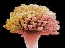
Fungal eye infections, also known as fungal keratitis or ocular fungal infections, are caused by the invasion of fungi into the eye. The most common types of fungi that cause these infections include Fusarium, Aspergillus, and Candida. These infections can affect any part of the eye, including the cornea, conjunctiva, sclera, and vitreous humor.
Fungal eye infections often present with symptoms such as redness, pain, sensitivity to light, tearing, blurred vision, and discharge. In severe cases, they can lead to corneal ulcers, perforation of the eye, and even blindness if left untreated. Risk factors for fungal eye infections include trauma to the eye, contact lens wear, immunosuppression, and pre-existing eye conditions such as dry eye or previous eye surgery.
Diagnosis of fungal eye infections typically involves a thorough eye examination, including visual acuity testing, slit lamp examination, and sometimes corneal scrapings for microbiological culture and sensitivity testing. Treatment usually involves topical antifungal medications, such as natamycin or amphotericin B, and in some cases may require oral or intravenous antifungal therapy. In severe cases, surgical intervention may be necessary to remove infected tissue or repair any damage caused by the infection.
Eye diseases are a range of conditions that affect the eye or visual system, causing damage to vision and, in some cases, leading to blindness. These diseases can be categorized into various types, including:
1. Refractive errors: These include myopia (nearsightedness), hyperopia (farsightedness), astigmatism, and presbyopia, which affect the way light is focused on the retina and can usually be corrected with glasses or contact lenses.
2. Cataracts: A clouding of the lens inside the eye that leads to blurry vision, glare, and decreased contrast sensitivity. Cataract surgery is the most common treatment for this condition.
3. Glaucoma: A group of diseases characterized by increased pressure in the eye, leading to damage to the optic nerve and potential blindness if left untreated. Treatment includes medications, laser therapy, or surgery.
4. Age-related macular degeneration (AMD): A progressive condition that affects the central part of the retina called the macula, causing blurry vision and, in advanced stages, loss of central vision. Treatment may include anti-VEGF injections, laser therapy, or nutritional supplements.
5. Diabetic retinopathy: A complication of diabetes that affects the blood vessels in the retina, leading to bleeding, leakage, and potential blindness if left untreated. Treatment includes laser therapy, anti-VEGF injections, or surgery.
6. Retinal detachment: A separation of the retina from its underlying tissue, which can lead to vision loss if not treated promptly with surgery.
7. Amblyopia (lazy eye): A condition where one eye does not develop normal vision, often due to a misalignment or refractive error in childhood. Treatment includes correcting the underlying problem and encouraging the use of the weaker eye through patching or other methods.
8. Strabismus (crossed eyes): A misalignment of the eyes that can lead to amblyopia if not treated promptly with surgery, glasses, or other methods.
9. Corneal diseases: Conditions that affect the transparent outer layer of the eye, such as keratoconus, Fuchs' dystrophy, and infectious keratitis, which can lead to vision loss if not treated promptly.
10. Uveitis: Inflammation of the middle layer of the eye, which can cause vision loss if not treated promptly with anti-inflammatory medications or surgery.
Eye movements, also known as ocular motility, refer to the voluntary or involuntary motion of the eyes that allows for visual exploration of our environment. There are several types of eye movements, including:
1. Saccades: rapid, ballistic movements that quickly shift the gaze from one point to another.
2. Pursuits: smooth, slow movements that allow the eyes to follow a moving object.
3. Vergences: coordinated movements of both eyes in opposite directions, usually in response to a three-dimensional stimulus.
4. Vestibulo-ocular reflex (VOR): automatic eye movements that help stabilize the gaze during head movement.
5. Optokinetic nystagmus (OKN): rhythmic eye movements that occur in response to large moving visual patterns, such as when looking out of a moving vehicle.
Abnormalities in eye movements can indicate neurological or ophthalmological disorders and are often assessed during clinical examinations.
Eye injuries refer to any damage or trauma caused to the eye or its surrounding structures. These injuries can vary in severity and may include:
1. Corneal abrasions: A scratch or scrape on the clear surface of the eye (cornea).
2. Chemical burns: Occurs when chemicals come into contact with the eye, causing damage to the cornea and other structures.
3. Eyelid lacerations: Cuts or tears to the eyelid.
4. Subconjunctival hemorrhage: Bleeding under the conjunctiva, the clear membrane that covers the white part of the eye.
5. Hyphema: Accumulation of blood in the anterior chamber of the eye, which is the space between the cornea and iris.
6. Orbital fractures: Breaks in the bones surrounding the eye.
7. Retinal detachment: Separation of the retina from its underlying tissue, which can lead to vision loss if not treated promptly.
8. Traumatic uveitis: Inflammation of the uvea, the middle layer of the eye, caused by trauma.
9. Optic nerve damage: Damage to the optic nerve, which transmits visual information from the eye to the brain.
Eye injuries can result from a variety of causes, including accidents, sports-related injuries, violence, and chemical exposure. It is important to seek medical attention promptly for any suspected eye injury to prevent further damage and potential vision loss.
Dry eye syndrome, also known as keratoconjunctivitis sicca, is a condition characterized by insufficient lubrication and moisture of the eyes. This occurs when the tears produced by the eyes are not sufficient in quantity or quality to keep the eyes moist and comfortable. The medical definition of dry eye syndromes includes the following symptoms:
1. A gritty or sandy sensation in the eyes
2. Burning or stinging sensations
3. Redness and irritation
4. Blurred vision that improves with blinking
5. Light sensitivity
6. A feeling of something foreign in the eye
7. Stringy mucus in or around the eyes
8. Difficulty wearing contact lenses
9. Watery eyes, which may seem contradictory but can be a response to dryness
10. Eye fatigue and discomfort after prolonged screen time or reading
The causes of dry eye syndromes can include aging, hormonal changes, certain medical conditions (such as diabetes, rheumatoid arthritis, lupus, Sjogren's syndrome), medications (antihistamines, decongestants, antidepressants, birth control pills), environmental factors (dry air, wind, smoke, dust), and prolonged screen time or reading.
Treatment for dry eye syndromes depends on the severity of the condition and its underlying causes. It may include artificial tears, lifestyle changes, prescription medications, and in some cases, surgical procedures to improve tear production or drainage.
Eye abnormalities refer to any structural or functional anomalies that affect the eye or its surrounding tissues. These abnormalities can be present at birth (congenital) or acquired later in life due to various factors such as injury, disease, or aging. Some examples of eye abnormalities include:
1. Strabismus: Also known as crossed eyes, strabismus is a condition where the eyes are misaligned and point in different directions.
2. Nystagmus: This is an involuntary movement of the eyes that can be horizontal, vertical, or rotatory.
3. Cataracts: A cataract is a clouding of the lens inside the eye that can cause vision loss.
4. Glaucoma: This is a group of eye conditions that damage the optic nerve and can lead to vision loss.
5. Retinal disorders: These include conditions such as retinal detachment, macular degeneration, and diabetic retinopathy.
6. Corneal abnormalities: These include conditions such as keratoconus, corneal ulcers, and Fuchs' dystrophy.
7. Orbital abnormalities: These include conditions such as orbital tumors, thyroid eye disease, and Graves' ophthalmopathy.
8. Ptosis: This is a condition where the upper eyelid droops over the eye.
9. Color blindness: A condition where a person has difficulty distinguishing between certain colors.
10. Microphthalmia: A condition where one or both eyes are abnormally small.
These are just a few examples of eye abnormalities, and there are many others that can affect the eye and its functioning. If you suspect that you have an eye abnormality, it is important to consult with an ophthalmologist for proper diagnosis and treatment.
Eye burns typically refer to injuries or damage to the eyes caused by exposure to harmful substances, extreme temperatures, or radiation. This can result in a variety of symptoms, including redness, pain, tearing, swelling, and blurred vision.
Chemical eye burns can occur when the eyes come into contact with strong acids, alkalis, or other irritants. These substances can cause damage to the cornea, conjunctiva, and other structures of the eye. The severity of the burn will depend on the type and concentration of the chemical, as well as the length of time it was in contact with the eye.
Thermal eye burns can result from exposure to hot or cold temperatures, such as steam, flames, or extreme cold. These types of burns can cause damage to the surface of the eye and may require medical attention to prevent further complications.
Radiation eye burns can occur after exposure to high levels of ultraviolet (UV) light, such as from welding torches, sun lamps, or tanning beds. Prolonged exposure to these sources can cause damage to the cornea and other structures of the eye, leading to symptoms like pain, redness, and sensitivity to light.
If you experience symptoms of an eye burn, it is important to seek medical attention as soon as possible. Treatment may include flushing the eyes with water or saline solution, administering medication to relieve pain and inflammation, or in severe cases, surgery to repair damaged tissue.
Eye enucleation is a surgical procedure that involves the removal of the entire eyeball, leaving the eye muscles, eyelids, and orbital structures intact. This procedure is typically performed to treat severe eye conditions or injuries, such as uncontrollable pain, blindness, cancer, or trauma. After the eyeball is removed, an implant may be placed in the socket to help maintain its shape and appearance. The optic nerve and other surrounding tissues are cut during the enucleation procedure, which means that vision cannot be restored in the affected eye. However, the remaining eye structures can still function normally, allowing for regular blinking, tear production, and eyelid movement.
Eye color is a characteristic determined by variations in a person's genes. The color of the eyes depends on the amount and type of pigment called melanin found in the eye's iris.
There are three main types of eye colors: brown, blue, and green. Brown eyes have the most melanin, while blue eyes have the least. Green eyes have a moderate amount of melanin combined with a golden tint that reflects light to give them their unique color.
Eye color is a polygenic trait, which means it is influenced by multiple genes. The two main genes responsible for eye color are OCA2 and HERC2, both located on chromosome 15. These genes control the production, transport, and storage of melanin in the iris.
It's important to note that eye color can change during infancy and early childhood due to the development of melanin in the iris. Additionally, some medications or medical conditions may also cause changes in eye color over time.
An Eye Bank is an organization that collects, stores, and distributes donated human eyes for corneal transplantation and other ocular medical research purposes. The eye bank's primary function is to ensure the quality of the donated tissue and make it available for those in need of sight-restoring procedures.
The cornea, the clear front part of the eye, can be surgically transplanted from a deceased donor to a recipient with corneal damage or disease, thereby improving or restoring their vision. The eye bank's role includes obtaining consent for donation, retrieving the eyes from the donor, evaluating the tissue for suitability, preserving it properly, and then allocating it to surgeons for transplantation.
Eye banks follow strict medical guidelines and adhere to ethical standards to ensure the safety and quality of the donated tissues. The process involves screening potential donors for infectious diseases and other conditions that may affect the quality or safety of the cornea. Once deemed suitable, the corneas are carefully removed, preserved in specific solutions, and stored until they are needed for transplantation.
In addition to corneal transplants, eye banks also support research and education in ophthalmology by providing human eye tissues for various studies aimed at advancing our understanding of eye diseases and developing new treatments.
Visual acuity is a measure of the sharpness or clarity of vision. It is usually tested by reading an eye chart from a specific distance, such as 20 feet (6 meters). The standard eye chart used for this purpose is called the Snellen chart, which contains rows of letters that decrease in size as you read down the chart.
Visual acuity is typically expressed as a fraction, with the numerator representing the testing distance and the denominator indicating the smallest line of type that can be read clearly. For example, if a person can read the line on the eye chart that corresponds to a visual acuity of 20/20, it means they have normal vision at 20 feet. If their visual acuity is 20/40, it means they must be as close as 20 feet to see what someone with normal vision can see at 40 feet.
It's important to note that visual acuity is just one aspect of overall vision and does not necessarily reflect other important factors such as peripheral vision, depth perception, color vision, or contrast sensitivity.
Eye neoplasms, also known as ocular tumors or eye cancer, refer to abnormal growths of tissue in the eye. These growths can be benign (non-cancerous) or malignant (cancerous). Eye neoplasms can develop in various parts of the eye, including the eyelid, conjunctiva, cornea, iris, ciliary body, choroid, retina, and optic nerve.
Benign eye neoplasms are typically slow-growing and do not spread to other parts of the body. They may cause symptoms such as vision changes, eye pain, or a noticeable mass in the eye. Treatment options for benign eye neoplasms include monitoring, surgical removal, or radiation therapy.
Malignant eye neoplasms, on the other hand, can grow and spread rapidly to other parts of the body. They may cause symptoms such as vision changes, eye pain, floaters, or flashes of light. Treatment options for malignant eye neoplasms depend on the type and stage of cancer but may include surgery, radiation therapy, chemotherapy, or a combination of these treatments.
It is important to note that early detection and treatment of eye neoplasms can improve outcomes and prevent complications. Regular eye exams with an ophthalmologist are recommended for early detection and prevention of eye diseases, including eye neoplasms.
"Ocular physiological phenomena" is not a standardized medical term with a specific definition. However, I can provide some examples of ocular physiological phenomena, which refer to various normal functions and processes that occur in the eye. Here are a few examples:
1. Accommodation: The ability of the eye to change optical power to maintain a clear image or focus on an object as its distance varies. This is primarily achieved by changing the curvature of the lens through the action of the ciliary muscles.
2. Pupillary reflex: The automatic adjustment of the pupil's size in response to changes in light intensity. In bright light, the pupil constricts (miosis), while in dim light, it dilates (mydriasis). This reflex helps regulate the amount of light that enters the eye.
3. Tear production: The continuous secretion of tears by the lacrimal glands to keep the eyes moist and protected from dust, microorganisms, and other foreign particles.
4. Extraocular muscle function: The coordinated movement of the six extraocular muscles that control eyeball rotation and enable various gaze directions.
5. Color vision: The ability to perceive and distinguish different colors based on the sensitivity of photoreceptor cells (cones) in the retina to specific wavelengths of light.
6. Dark adaptation: The process by which the eyes adjust to low-light conditions, improving visual sensitivity primarily through changes in the rod photoreceptors' sensitivity and pupil dilation.
7. Light adaptation: The ability of the eye to adjust to different levels of illumination, mainly through alterations in pupil size and photoreceptor cell response.
These are just a few examples of ocular physiological phenomena. There are many more processes and functions that occur within the eye, contributing to our visual perception and overall eye health.
A compound eye is a characteristic type of eye found in arthropods, including insects, crustaceans, and some extinct fossil groups. Each eye is composed of numerous individual photoreceptor units called ommatidia, which function together to provide a wide field of vision and excellent motion detection capabilities.
In an arthropod compound eye, each ommatidium contains a group of visual cells (called retinula cells) surrounding a central rhabdomere, which is the light-sensitive structure that converts light into electrical signals. The number of ommatidia in a compound eye can vary greatly between species and even within different regions of an individual's eye, ranging from just a few to tens of thousands.
Compound eyes offer several advantages for arthropods:
1. Wide Field of Vision: Compound eyes provide a panoramic view of the environment, allowing arthropods to detect predators, prey, or mates from various directions simultaneously.
2. Motion Detection: The apposition-type compound eye (one type of compound eye structure) is particularly adept at detecting motion due to the neural processing of signals between adjacent ommatidia. This allows arthropods to respond quickly to potential threats or opportunities.
3. Light Adaptation: Compound eyes can adapt to different light conditions, allowing arthropods to function effectively in both bright daylight and dimly lit environments. Some species have specialized regions within their compound eyes that are optimized for specific light conditions, such as the dorsal rim area in insects, which is sensitive to polarized skylight.
4. UV Sensitivity: Many arthropods can detect ultraviolet (UV) light due to the presence of photopigments within their ommatidia that absorb UV wavelengths. This ability allows them to perceive patterns and cues in their environment that are invisible to humans, such as floral guides in bees or mate-recognition signals in certain insects.
Despite their limitations in terms of resolution and image quality compared to vertebrate eyes, compound eyes have evolved to serve the unique needs and ecological roles of arthropods effectively.
Eye protective devices are specialized equipment designed to protect the eyes from various hazards and injuries. They include items such as safety glasses, goggles, face shields, welding helmets, and full-face respirators. These devices are engineered to provide a barrier between the eyes and potential dangers like chemical splashes, impact particles, radiation, and other environmental hazards.
Safety glasses are designed to protect against flying debris, dust, and other airborne particles. They typically have side shields to prevent objects from entering the eye from the sides. Goggles offer a higher level of protection than safety glasses as they form a protective seal around the eyes, preventing liquids and fine particles from reaching the eyes.
Face shields and welding helmets are used in industrial settings to protect against radiation, sparks, and molten metal during welding or cutting operations. Full-face respirators are used in environments with harmful airborne particles or gases, providing protection for both the eyes and the respiratory system.
It is essential to choose the appropriate eye protective device based on the specific hazard present to ensure adequate protection.
The retina is the innermost, light-sensitive layer of tissue in the eye of many vertebrates and some cephalopods. It receives light that has been focused by the cornea and lens, converts it into neural signals, and sends these to the brain via the optic nerve. The retina contains several types of photoreceptor cells including rods (which handle vision in low light) and cones (which are active in bright light and are capable of color vision).
In medical terms, any pathological changes or diseases affecting the retinal structure and function can lead to visual impairment or blindness. Examples include age-related macular degeneration, diabetic retinopathy, retinal detachment, and retinitis pigmentosa among others.
Intraocular pressure (IOP) is the fluid pressure within the eye, specifically within the anterior chamber, which is the space between the cornea and the iris. It is measured in millimeters of mercury (mmHg). The aqueous humor, a clear fluid that fills the anterior chamber, is constantly produced and drained, maintaining a balance that determines the IOP. Normal IOP ranges from 10-21 mmHg, with average values around 15-16 mmHg. Elevated IOP is a key risk factor for glaucoma, a group of eye conditions that can lead to optic nerve damage and vision loss if not treated promptly and effectively. Regular monitoring of IOP is essential in diagnosing and managing glaucoma and other ocular health issues.
Penetrating eye injuries are a type of ocular trauma where a foreign object or substance pierces the outer layers of the eye and damages the internal structures. This can result in serious harm to various parts of the eye, such as the cornea, iris, lens, or retina, and may potentially cause vision loss or blindness if not promptly treated.
The severity of a penetrating eye injury depends on several factors, including the type and size of the object that caused the injury, the location of the wound, and the extent of damage to the internal structures. Common causes of penetrating eye injuries include sharp objects, such as metal shards or glass fragments, projectiles, such as pellets or bullets, and explosive materials.
Symptoms of a penetrating eye injury may include pain, redness, sensitivity to light, blurred vision, floaters, or the presence of a foreign body in the eye. If you suspect that you have sustained a penetrating eye injury, it is essential to seek immediate medical attention from an ophthalmologist or other healthcare professional with experience in treating eye trauma.
Treatment for penetrating eye injuries may include removing any foreign objects or substances from the eye, repairing damaged tissues, and administering medications to prevent infection and reduce inflammation. In some cases, surgery may be necessary to repair the injury and restore vision. Preventing eye injuries is crucial, and appropriate protective eyewear should be worn when engaging in activities that pose a risk of eye trauma.
Ophthalmic solutions are sterile, single-use or multi-dose preparations in a liquid form that are intended for topical administration to the eye. These solutions can contain various types of medications, such as antibiotics, anti-inflammatory agents, antihistamines, or lubricants, which are used to treat or prevent ocular diseases and conditions.
The pH and osmolarity of ophthalmic solutions are carefully controlled to match the physiological environment of the eye and minimize any potential discomfort or irritation. The solutions may be packaged in various forms, including drops, sprays, or irrigations, depending on the intended use and administration route.
It is important to follow the instructions for use provided by a healthcare professional when administering ophthalmic solutions, as improper use can lead to eye injury or reduced effectiveness of the medication.
Foreign bodies in the eye refer to any object or particle that is not normally present in the eye and becomes lodged in it. These foreign bodies can range from small particles like sand or dust to larger objects such as metal shavings or glass. They can cause irritation, pain, redness, watering, and even vision loss if they are not removed promptly and properly.
The symptoms of an eye foreign body may include:
* A feeling that something is in the eye
* Pain or discomfort in the eye
* Redness or inflammation of the eye
* Watering or tearing of the eye
* Sensitivity to light
* Blurred vision or difficulty seeing
If you suspect that you have a foreign body in your eye, it is important to seek medical attention immediately. An eye care professional can examine your eye and determine the best course of treatment to remove the foreign body and prevent any further damage to your eye.
Eye movement measurements, also known as oculometry, refer to the measurement and analysis of eye movements. This can include assessing the direction, speed, range, and patterns of eye movement. These measurements are often used in research and clinical settings to understand various aspects of vision, perception, and cognition. They can be used to diagnose and monitor conditions that affect eye movement, such as strabismus (crossed eyes), amblyopia (lazy eye), or neurological disorders. Additionally, eye movement measurements are also used in areas such as human-computer interaction, marketing research, and virtual reality to understand how individuals interact with their environment.
Ocular fixation is a term used in ophthalmology and optometry to refer to the ability of the eyes to maintain steady gaze or visual focus on an object. It involves the coordinated movement of the extraocular muscles that control eye movements, allowing for clear and stable vision.
In medical terminology, fixation specifically refers to the state in which the eyes are aligned and focused on a single point in space. This is important for maintaining visual perception and preventing blurring or double vision. Ocular fixation can be affected by various factors such as muscle weakness, nerve damage, or visual processing disorders.
Assessment of ocular fixation is often used in eye examinations to evaluate visual acuity, eye alignment, and muscle function. Abnormalities in fixation may indicate the presence of underlying eye conditions or developmental delays that require further investigation and treatment.
The posterior segment of the eye refers to the back portion of the interior of the eye, including the vitreous, retina, choroid, and optic nerve. This region is responsible for processing visual information and transmitting it to the brain. The retina contains photoreceptor cells that convert light into electrical signals, which are then sent through the optic nerve to the brain for interpretation as images. Disorders of the posterior eye segment can lead to vision loss or blindness.
Glaucoma is a group of eye conditions that damage the optic nerve, often caused by an abnormally high pressure in the eye (intraocular pressure). This damage can lead to permanent vision loss or even blindness if left untreated. The most common type is open-angle glaucoma, which has no warning signs and progresses slowly. Angle-closure glaucoma, on the other hand, can cause sudden eye pain, redness, nausea, and vomiting, as well as rapid vision loss. Other less common types of glaucoma also exist. While there is no cure for glaucoma, early detection and treatment can help slow or prevent further vision loss.
The vitreous body, also known simply as the vitreous, is the clear, gel-like substance that fills the space between the lens and the retina in the eye. It is composed mainly of water, but also contains collagen fibers, hyaluronic acid, and other proteins. The vitreous helps to maintain the shape of the eye and provides a transparent medium for light to pass through to reach the retina. With age, the vitreous can become more liquefied and may eventually separate from the retina, leading to symptoms such as floaters or flashes of light.
The sclera is the tough, white, fibrous outer coating of the eye in humans and other vertebrates, covering about five sixths of the eyeball's surface. It provides protection for the delicate inner structures of the eye and maintains its shape. The sclera is composed mainly of collagen and elastic fiber, making it strong and resilient. Its name comes from the Greek word "skleros," which means hard.
Axial length, in the context of the eye, refers to the measurement of the distance between the front and back portions of the eye, specifically from the cornea (the clear front "window" of the eye) to the retina (the light-sensitive tissue at the back of the eye). This measurement is typically expressed in millimeters (mm).
The axial length of the eye is an important factor in determining the overall refractive power of the eye and can play a role in the development of various eye conditions, such as myopia (nearsightedness) or hyperopia (farsightedness). Changes in axial length, particularly elongation, are often associated with an increased risk of developing myopia. Regular monitoring of axial length can help eye care professionals track changes in the eye and manage these conditions more effectively.
The anterior chamber is the front portion of the eye, located between the cornea (the clear front "window" of the eye) and the iris (the colored part of the eye). It is filled with a clear fluid called aqueous humor that provides nutrients to the structures inside the eye and helps maintain its shape. The anterior chamber plays an important role in maintaining the overall health and function of the eye.
Myopia, also known as nearsightedness, is a common refractive error of the eye. It occurs when the eye is either too long or the cornea (the clear front part of the eye) is too curved. As a result, light rays focus in front of the retina instead of directly on it, causing distant objects to appear blurry while close objects remain clear.
Myopia typically develops during childhood and can progress gradually or rapidly until early adulthood. It can be corrected with glasses, contact lenses, or refractive surgery such as LASIK. Regular eye examinations are essential for people with myopia to monitor any changes in their prescription and ensure proper correction.
While myopia is generally not a serious condition, high levels of nearsightedness can increase the risk of certain eye diseases, including cataracts, glaucoma, retinal detachment, and myopic degeneration. Therefore, it's crucial to manage myopia effectively and maintain regular follow-ups with an eye care professional.
Aqueous humor is a clear, watery fluid that fills the anterior and posterior chambers of the eye. It is produced by the ciliary processes in the posterior chamber and circulates through the pupil into the anterior chamber, where it provides nutrients to the cornea and lens, maintains intraocular pressure, and helps to shape the eye. The aqueous humor then drains out of the eye through the trabecular meshwork and into the canal of Schlemm, eventually reaching the venous system.
In medical terms, the iris refers to the colored portion of the eye that surrounds the pupil. It is a circular structure composed of thin, contractile muscle fibers (radial and circumferential) arranged in a regular pattern. These muscles are controlled by the autonomic nervous system and can adjust the size of the pupil in response to changes in light intensity or emotional arousal. By constricting or dilating the iris, the amount of light entering the eye can be regulated, which helps maintain optimal visual acuity under various lighting conditions.
The color of the iris is determined by the concentration and distribution of melanin pigments within the iris stroma. The iris also contains blood vessels, nerves, and connective tissue that support its structure and function. Anatomically, the iris is continuous with the ciliary body and the choroid, forming part of the uveal tract in the eye.
The ciliary body is a part of the eye's internal structure that is located between the choroid and the iris. It is composed of muscle tissue and is responsible for adjusting the shape of the lens through a process called accommodation, which allows the eye to focus on objects at varying distances. Additionally, the ciliary body produces aqueous humor, the clear fluid that fills the anterior chamber of the eye and helps to nourish the eye's internal structures. The ciliary body is also responsible for maintaining the shape and position of the lens within the eye.
In medical terms, "tears" are a clear, salty liquid that is produced by the tear glands (lacrimal glands) in our eyes. They serve to keep the eyes moist, protect against dust and other foreign particles, and help to provide clear vision by maintaining a smooth surface on the front of the eye. Tears consist of water, oil, and mucus, which help to prevent evaporation and ensure that the tears spread evenly across the surface of the eye. Emotional or reflexive responses, such as crying or yawning, can also stimulate the production of tears.
The crystalline lens is a biconvex transparent structure in the eye that helps to refract (bend) light rays and focus them onto the retina. It is located behind the iris and pupil and is suspended by small fibers called zonules that connect it to the ciliary body. The lens can change its shape to accommodate and focus on objects at different distances, a process known as accommodation. With age, the lens may become cloudy or opaque, leading to cataracts.
Visual fields refer to the total area in which objects can be seen while keeping the eyes focused on a central point. It is the entire area that can be observed using peripheral (side) vision while the eye gazes at a fixed point. A visual field test is used to detect blind spots or gaps (scotomas) in a person's vision, which could indicate various medical conditions such as glaucoma, retinal damage, optic nerve disease, brain tumors, or strokes. The test measures both the central and peripheral vision and maps the entire area that can be seen when focusing on a single point.
In the context of medical terminology, I believe you may be referring to "pursuit" as it relates to neurological tests. A smooth pursuit is a type of eye movement in which the eyes smoothly and slowly follow a moving object. It requires coordination between the extraocular muscles, vestibular system, and visual system. If there are issues with any of these systems, smooth pursuit can be affected, leading to abnormalities such as jerky or saccadic movements.
Therefore, "smooth pursuit" is a medical term used to describe the normal, coordinated movement of the eyes that allows for the tracking of moving objects in a smooth and continuous manner.
 ReNu
ReNu Fungal Eye Infection Statistics | Types of Diseases | Fungal Diseases | CDC
 Fungal Keratitis: Background, Pathophysiology, Epidemiology
 Fungal Infections | Fungi | Fungus | MedlinePlus
 Fungal Eye Infections: Types, Symptoms, Treatment, and Surgery
 What You Can Do About Diabetes-Related Fungal Infections - NaturalNews.com
 Prednisone (Oral Route) - Mayo Clinic
 Biomicroscopic and histopathologic considerations regarding the feasibility of surgical excision of subfoveal neovascular...
 U.S. Fifth Circuit Affirms Dismissal of Claim for Disability Benefits where Loss of Eye from Fungal Infection not 'Accident'...
 Overview of Fungal Infections - Infections - MSD Manual Consumer Version
 PW and Other Law Firms to Represent Against Bausch & Lomb - Parker Waichman LLP
 Refractive Eye Surgery: Helping Patients Make Informed Decisions About LASIK | AAFP
 Gemmily: Package Insert - Drugs.com
 Mycology Update 2017 | PPT
 India's New Epidemic: 'Black Fungus' Patients Lose Eyes, Nose, Jaw To Infection | IBTimes
 Sjogren's syndrome: Symptoms, diet, and treatment
 Flavopiridol Protects Against Fungal Keratitis by Alleviating Inflammation Through the Promotion of Autophagy | Research Square
 Fungal Infection Treatment: Folk Medicine
 Ticlast (Azelastine Hydrochloride and Fluticasone Propionate): Uses, Dosage, Side Effects, Interactions, Warning
 Skin Crest Clinic Dermatology Podcasts
 THE 'VIRTUAL' ~ MEDICAL PATHOLOGY, FORENSICS & VIROLOGY CENTER SECTION III - Martindale Center
 Sneezing and Watery Eyes in Cats