Fasciitis
Fasciitis, Necrotizing
Fasciitis, Plantar
Debridement
Fascia
Heel Spur
Streptococcus pyogenes
Fournier Gangrene
Cellulitis
High-Energy Shock Waves
Periodontal Abscess
Fatal Outcome
Vibrio vulnificus
Leg Dermatoses
Medicine, East Asian Traditional
Morganella morganii
Soft Tissue Infections
Shock, Septic
An overview of shock wave therapy in musculoskeletal disorders. (1/50)
Shock waves are high-energy acoustic waves generated under water with high voltage explosion and vaporization. Shock wave in urology (lithotripsy) is primarily used to disintegrate urolithiasis, whereas shock wave in orthopedics (orthotripsy) is not used to disintegrate tissues, rather to induce neovascularization, improve blood supply and tissue regeneration. The application of shock wave therapy in certain musculoskeletal disorders has been around for approximately 15 years, and the success rate in non-union of long bone fracture, calcifying tendonitis of the shoulder, lateral epicondylitis of the elbow and proximal plantar fasciitis ranged from 65% to 91%. The complications are low and negligible. Recently, shock wave therapy was extended to treat other conditions including avascular necrosis of femoral head, patellar tendonitis (jumper's knee), osteochondritis dessicans and non-calcifying tendonitis of the shoulder. Shock wave therapy is a novel therapeutic modality without the need of surgery and surgical risks as well as surgical pain. It is convenient and cost-effective. The exact mechanism of shock wave therapy remains unknown. Based on the results of animal studies in our laboratory, it appears that the mechanism of shock waves first stimulates the early expression of angiogenesis-related growth factors including eNOS (endothelial nitric oxide synthase), VEGF (vessel endothelial growth factor) and PCNA (proliferating cell nuclear antigen), then induces the ingrowth of neovascularization that improves blood supply and increases cell proliferation and eventual tissue regeneration to repair tendon or bone tissues. The rise of angiogenic markers occurred in as early as one week and only lasted for approximately 8 weeks, whereas the neovascularization was first noted in 4 weeks and persisted for 12 weeks or longer along with cell proliferation. These findings support the clinical observation that the effect of shock wave therapy appears to be dose-dependent and symptom improvement with time. Additional information including the cellular and molecular changes after shock wave therapy are needed for further clarification on the mechanism of shock wave therapy in musculoskeletal system. (+info)Extracorporeal shock wave therapy for plantar fasciitis: randomised controlled multicentre trial. (2/50)
OBJECTIVE: To determine the effectiveness of extracorporeal shock wave therapy compared with placebo in the treatment of chronic plantar fasciitis. DESIGN: Randomised, blinded, multicentre trial with parallel group design. SETTING: Nine hospitals and one outpatient clinic in Germany. PARTICIPANTS: 272 patients with chronic plantar fasciitis recalcitrant to conservative therapy for at least six months: 135 patients were allocated extracorporeal shock wave therapy and 137 were allocated placebo. MAIN OUTCOME MEASURES: Primary end point was the success rate 12 weeks after intervention based on the Roles and Maudsley score. Secondary end points encompassed subjective pain ratings and walking ability up to a year after the last intervention. RESULTS: The primary end point could be assessed in 94% (n=256) of patients. The success rate 12 weeks after intervention was 34% (n=43) in the extracorporeal shock wave therapy group and 30% (n=39) in the placebo group (95% confidence interval - 8.0% to 15.1%). No difference was found in the secondary end points. Few side effects were reported. CONCLUSIONS: Extracorporeal shock wave therapy is ineffective in the treatment of chronic plantar fasciitis. (+info)Diagnostic and therapeutic injection of the ankle and foot. (3/50)
Joint and soft tissue injection of the ankle and foot region is a useful diagnostic and therapeutic tool for the family physician. This article reviews the injection procedure for the plantar fascia, ankle joint, tarsal tunnel, interdigital space, and first metatarsophalangeal joint. Indications for plantar fascia injection include degeneration secondary to repetitive use and traumatic injuries that are unresponsive to conservative treatment. Diagnostic aspiration or therapeutic injection of the ankle or first metatarsophalangeal joints can be performed for management of advanced osteoarthritis, rheumatoid arthritis, and other inflammatory arthritides such as gout, or synovitis or an arthrosis such as "turf toe." Persistent pain and disability resulting from tarsal tunnel syndrome, an analog of carpal tunnel syndrome of the wrist respond to local injection therapy. A painful interdigital space, such as that occurring in patients with Morton's neuroma, is commonly relieved with corticosteroid injection. The proper technique, choice and quantity of pharmaceuticals, and appropriate follow-up are essential for effective outcomes. (+info)Diagnosing heel pain in adults. (4/50)
Heel pain is a common condition in adults that may cause significant discomfort and disability. A variety of soft tissue, osseous, and systemic disorders can cause heel pain. Narrowing the differential diagnosis begins with a history and physical examination of the lower extremity to pinpoint the anatomic origin of the heel pain. The most common cause of heel pain in adults is plantar fasciitis. Patients with plantar fasciitis report increased heel pain with their first steps in the morning or when they stand up after prolonged sitting. Tenderness at the calcaneal tuberosity usually is apparent on examination and is increased with passive dorsiflexion of the toes. Tendonitis also may cause heel pain. Achilles tendonitis is associated with posterior heel pain. Bursae adjacent to the Achilles tendon insertion may become inflamed and cause pain. Calcaneal stress fractures are more likely to occur in athletes who participate in sports that require running and jumping. Patients with plantar heel pain accompanied by tingling, burning, or numbness may have tarsal tunnel syndrome. Heel pad atrophy may present with diffuse plantar heel pain, especially in patients who are older and obese. Less common causes of heel pain, which should be considered when symptoms are prolonged or unexplained, include osteomyelitis, bony abnormalities (such as calcaneal stress fracture), or tumor. Heel pain rarely is a presenting symptom in patients with systemic illnesses, but the latter may be a factor in persons with bilateral heel pain, pain in other joints, or known inflammatory arthritis conditions. (+info)The relationship between the flexible flatfoot and plantar fasciitis: ultrasonographic evaluation. (5/50)
BACKGROUND: The purpose of this study was to investigate the relationship between flexible flatfoot and plantar fasciitis. METHODS: Twenty-three subjects with flexible flatfoot and 23 subjects with normal arched feet were enrolled. Footprint analysis was used to evaluate the foot conditions in both groups to calculate the individual arch index. We compared the sonographic images of plantar fascia in the flexible flatfoot group with the normal arch group using high-frequency ultrasound. RESULTS: The analysis results indicated that the thickening of the plantar fascia in the flexible flatfoot group was significantly different from the normal arch group. In the flexible flatfoot group, 10 of 23 patients (43.4%) had plantar fasciitis, but only two subjects (8.7%) in the normal arch group had plantar fasciitis. CONCLUSIONS: There was a higher incidence of plantar fasciitis in the flexible flatfoot group than the normal arch control group in this study. (+info)Advantages of color B-mode imaging with contrast optimization in sonography of low-contrast musculoskeletal lesions and structures in the foot and ankle. (6/50)
OBJECTIVE: The purpose of this study was to evaluate the utility of color B-mode imaging with contrast optimization in evaluating low-contrast lesions of the foot and ankle (Morton neuromas and plantar fasciitis). METHODS: The sonographic examinations of 49 consecutive patients with a diagnosis of plantar fasciitis or Morton neuroma imaged with both conventional gray scale imaging and color B-mode imaging with contrast optimization (Photopic; Siemens Medical Solutions, Mountain View, CA) were reviewed. In every patient, matched pairs of images obtained with conventional gray and color maps (Photopic) were acquired and stored as DICOM (Digital Imaging and Communications in Medicine) images on a sonographic workstation. Each image was assessed independently by 2 musculoskeletal radiologists trained in musculoskeletal sonography for overall image contrast (lesion to background), conspicuity of regional tissue boundaries, visualization of deep tissue boundaries, and how well the internal characteristics of the structure were visualized. RESULTS: Three-way analysis of variance showed that Photopic imaging resulted in statistically significantly improved overall image contrast, definition of regional soft tissue boundaries, including deep soft tissue boundaries, and depiction of the internal characteristics of the structure being examined. CONCLUSIONS: Color imaging with contrast optimization improves overall image contrast and better defines deep soft tissue boundaries and the internal morphologic characteristics of Morton neuromas and the plantar fascia compared with conventional gray scale imaging. (+info)Plantar fasciitis: diagnosis and therapeutic considerations. (7/50)
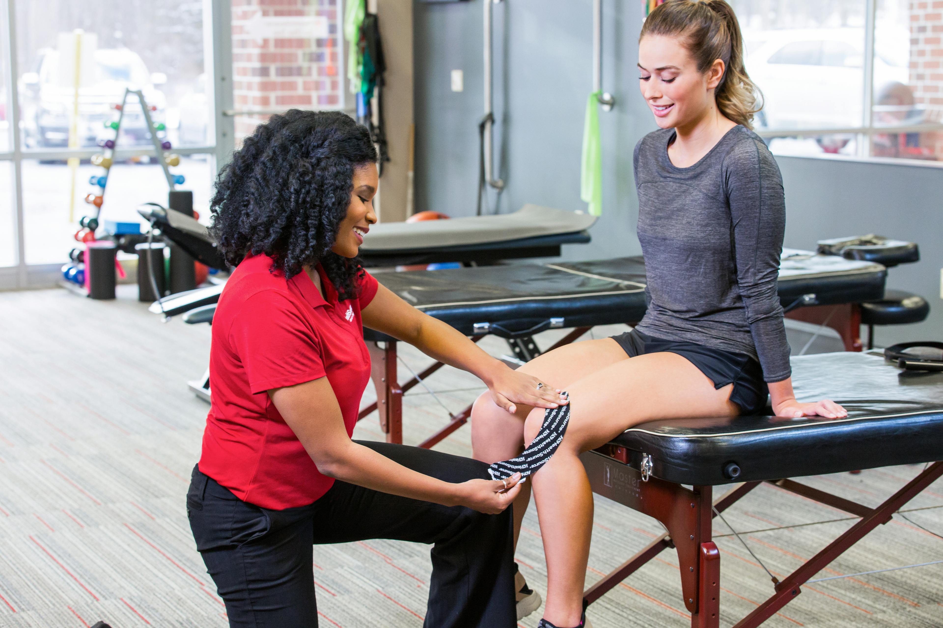
Plantar fasciitis is the most common cause of inferior heel pain. The pain and discomfort associated with this condition can have a dramatic impact on physical mobility. The etiology of this condition is not clearly understood and is probably multi-factorial in nature. Weight gain, occupation-related activity, anatomical variations, poor biomechanics, overexertion, and inadequate footwear are contributing factors. Although plantar fasciitis is generally regarded as a self-limited condition, it can take months to years to resolve, presenting a challenge for clinicians. Many treatment options are available that demonstrate variable levels of efficacy. Conservative therapies include rest and avoidance of potentially aggravating activities, stretching and strengthening exercises, orthotics, arch supports, and night splinting. Other considerations include use of anti-inflammatory agents, ultrasonic shockwave therapy, and, in the most extreme cases, surgery. This article reviews plantar fasciitis, presents the most effective treatment options currently available, and proposes nutritional considerations that may be beneficial in the management of this condition. (+info)Clinical utility of sonography in diagnosing plantar fasciitis. (8/50)
OBJECTIVE: The purpose of this study was to investigate the efficacy of sonography in the detection of plantar fasciitis (PF) compared with magnetic resonance imaging (MRI) findings in subjects with inferior heel pain. METHODS: Seventy-seven patients with unilateral (n = 9) and bilateral (n = 68) heel pain were studied. Seventy-seven age- and sex-matched asymptomatic subjects served as a control group. Magnetic resonance imaging was used to establish a diagnosis of PF with sagittal T1-weighted, T2-weighted, and short tau inversion recovery sequences. The sonographic appearances of PF were compared with MRI findings. Plantar fascia and heel pad thickness were also measured on both imaging modalities. RESULTS: Compared with MRI, sonography showed 80% sensitivity and 88.5% specificity in assessing PF. A strong correlation was found between plantar fascia and fat pad thickness measurements done by sonography (P < .001; r = 0.854) and MRI (P < .001; r = 0.798). Compared with the asymptomatic volunteers, patients with PF had significant increases in plantar fascia and heel pad thicknesses, weight, and body mass index (P = .0001). Heel pad thickness was also significantly increased with pain duration (P = .021). CONCLUSIONS: Although MRI is the modality of choice in the morphologic assessment of different plantar fascia lesions, sonography can also serve as an effective tool and may substitute MRI in the diagnosis of PF. (+info)Fasciitis is a medical condition characterized by inflammation or irritation of the fascia, which are the bands of connective tissue that surround muscles, tendons, and bones in the body. The most common type of fasciitis is plantar fasciitis, which affects the fascia on the bottom of the foot and can cause heel pain. Other types of fasciitis include:
* Achilles tendonitis or Achilles tendinopathy, which affects the fascia that connects the calf muscle to the heel bone
* Shin splints, which affect the fascia that covers the front of the lower leg
* Necrotizing fasciitis, a rare and serious bacterial infection that can cause extensive tissue damage and is potentially life-threatening.
The symptoms of fasciitis may include pain, stiffness, or tenderness in the affected area, especially after prolonged periods of rest or physical activity. Treatment for fasciitis typically involves rest, ice, compression, and elevation (RICE) of the affected area, as well as physical therapy exercises to stretch and strengthen the fascia and surrounding muscles. In some cases, medication or surgery may be necessary to relieve symptoms and promote healing.
Necrotizing fasciitis is a serious bacterial infection that affects the fascia, which is the tissue that surrounds muscles, nerves, and blood vessels. The infection can also spread to the muscle and skin. It is often caused by a combination of different types of bacteria, including group A Streptococcus and Staphylococcus aureus.
The infection causes extensive tissue damage and necrosis (death) of the fascia and surrounding tissues. It can progress rapidly and can be fatal if not treated promptly with aggressive surgical debridement (removal of dead tissue) and antibiotics.
Symptoms of necrotizing fasciitis include severe pain, swelling, redness, and warmth in the affected area; fever; chills; and general weakness. It is important to seek medical attention immediately if these symptoms occur, as early diagnosis and treatment can significantly improve outcomes.
Plantar fasciitis is a medical condition that involves inflammation of the plantar fascia, which is a thick band of tissue that runs along the bottom of your foot, connecting your heel bone to your toes. This tissue supports the arch of your foot and absorbs shock when you walk or run.
Plantar fasciitis is often caused by repetitive stress or overuse, leading to small tears and inflammation in the fascia. People who have high arches or flat feet, those who spend a lot of time on their feet, and athletes who engage in activities that put repeated stress on the heel and attached tissue, such as runners, are at a higher risk of developing plantar fasciitis.
Symptoms of plantar fasciitis include pain and stiffness in the heel or bottom of the foot, especially when taking the first few steps after getting out of bed or after prolonged periods of sitting or standing. The pain may worsen over time if left untreated, making it difficult to walk, climb stairs, or participate in physical activities.
Treatment for plantar fasciitis typically includes rest, ice, compression, and elevation (RICE) therapy, as well as physical therapy exercises to stretch and strengthen the foot and lower leg muscles. In some cases, medication, orthotics, or even surgery may be necessary to alleviate severe pain and inflammation.
Debridement is a medical procedure that involves the removal of dead, damaged, or infected tissue to improve the healing process or prevent further infection. This can be done through various methods such as surgical debridement (removal of tissue using scalpel or scissors), mechanical debridement (use of wound irrigation or high-pressure water jet), autolytic debridement (using the body's own enzymes to break down and reabsorb dead tissue), and enzymatic debridement (application of topical enzymes to dissolve necrotic tissue). The goal of debridement is to promote healthy tissue growth, reduce the risk of infection, and improve overall wound healing.
A fascia is a band or sheet of connective tissue, primarily collagen, that covers, connects, and separates muscles, organs, and other structures in the body. It provides support and stability, allows for smooth movement between structures, and has the ability to transmit forces throughout the body. Fascia is found throughout the body, and there are several layers of it, including superficial fascia, deep fascia, and visceral fascia. Injury, inflammation, or strain to the fascia can cause pain and restriction of movement.
A heel spur, also known as a calcaneal spur, is a bony growth or projection that develops on the underside of the heel bone (calcaneus). It typically occurs where the plantar fascia, a band of tissue that supports the arch of the foot, attaches to the heel bone.
Heel spurs are often caused by repetitive stress and strain on the foot, particularly in people who have plantar fasciitis, an inflammation of the plantar fascia. Over time, this tension can cause the body to lay down new bone tissue, leading to the formation of a spur.
Heel spurs themselves are not necessarily painful, but they can cause pain and discomfort if they rub against shoes or if they irritate surrounding tissues. Treatment for heel spurs typically involves addressing the underlying causes of the condition, such as plantar fasciitis, through measures such as rest, ice, stretching exercises, physical therapy, and orthotics. In some cases, surgery may be necessary to remove the spur.
In medical terms, "heel" generally refers to the posterior and largest part of the foot, specifically the calcaneus bone. The heel is the first part of the foot to make contact with the ground during walking or running, and it plays a crucial role in supporting the body's weight and absorbing shock during movement.
The term "heel" can also be used to describe a structure or device that is attached to the back of a shoe or boot to provide additional height, support, or protection to the wearer's heel. These types of heels are often worn for fashion purposes or to compensate for differences in leg length.
Streptococcus pyogenes is a Gram-positive, beta-hemolytic streptococcus bacterium that causes various suppurative (pus-forming) and nonsuppurative infections in humans. It is also known as group A Streptococcus (GAS) due to its ability to produce the M protein, which confers type-specific antigenicity and allows for serological classification into more than 200 distinct Lancefield groups.
S. pyogenes is responsible for a wide range of clinical manifestations, including pharyngitis (strep throat), impetigo, cellulitis, erysipelas, scarlet fever, rheumatic fever, and acute poststreptococcal glomerulonephritis. In rare cases, it can lead to invasive diseases such as necrotizing fasciitis (flesh-eating disease) and streptococcal toxic shock syndrome (STSS).
The bacterium is typically transmitted through respiratory droplets or direct contact with infected skin lesions. Effective prevention strategies include good hygiene practices, such as frequent handwashing and avoiding sharing personal items, as well as prompt recognition and treatment of infections to prevent spread.
Streptococcal infections are a type of infection caused by group A Streptococcus bacteria (Streptococcus pyogenes). These bacteria can cause a variety of illnesses, ranging from mild skin infections to serious and potentially life-threatening conditions such as sepsis, pneumonia, and necrotizing fasciitis (flesh-eating disease).
Some common types of streptococcal infections include:
* Streptococcal pharyngitis (strep throat) - an infection of the throat and tonsils that can cause sore throat, fever, and swollen lymph nodes.
* Impetigo - a highly contagious skin infection that causes sores or blisters on the skin.
* Cellulitis - a bacterial infection of the deeper layers of the skin and underlying tissue that can cause redness, swelling, pain, and warmth in the affected area.
* Scarlet fever - a streptococcal infection that causes a bright red rash on the body, high fever, and sore throat.
* Necrotizing fasciitis - a rare but serious bacterial infection that can cause tissue death and destruction of the muscles and fascia (the tissue that covers the muscles).
Treatment for streptococcal infections typically involves antibiotics to kill the bacteria causing the infection. It is important to seek medical attention if you suspect a streptococcal infection, as prompt treatment can help prevent serious complications.
Foot diseases refer to various medical conditions that affect the foot, including its structures such as the bones, joints, muscles, tendons, ligaments, blood vessels, and nerves. These conditions can cause symptoms like pain, swelling, numbness, difficulty walking, and skin changes. Examples of foot diseases include:
1. Plantar fasciitis: inflammation of the band of tissue that connects the heel bone to the toes.
2. Bunions: a bony bump that forms on the joint at the base of the big toe.
3. Hammertoe: a deformity in which the toe is bent at the middle joint, resembling a hammer.
4. Diabetic foot: a group of conditions that can occur in people with diabetes, including nerve damage, poor circulation, and increased risk of infection.
5. Athlete's foot: a fungal infection that affects the skin between the toes and on the soles of the feet.
6. Ingrown toenails: a condition where the corner or side of a toenail grows into the flesh of the toe.
7. Gout: a type of arthritis that causes sudden, severe attacks of pain, swelling, redness, and tenderness in the joints, often starting with the big toe.
8. Foot ulcers: open sores or wounds that can occur on the feet, especially in people with diabetes or poor circulation.
9. Morton's neuroma: a thickening of the tissue around a nerve between the toes, causing pain and numbness.
10. Osteoarthritis: wear and tear of the joints, leading to pain, stiffness, and reduced mobility.
Foot diseases can affect people of all ages and backgrounds, and some may be prevented or managed with proper foot care, hygiene, and appropriate medical treatment.
Fournier gangrene is a type of necrotizing fasciitis, which is a severe soft tissue infection that involves the fascia (the layer of connective tissue covering the muscle). Fournier gangrene specifically affects the genital region and can spread to the abdominal wall or thighs. It's characterized by rapid progression, extensive tissue damage, and a high mortality rate if not treated promptly with surgical debridement (removal of dead tissue) and antibiotics. The infection typically involves multiple types of bacteria, both aerobic and anaerobic, and can arise from various sources such as urinary tract infections, anal abscesses, or trauma to the genital area.
Cellulitis is a medical condition characterized by an infection and inflammation of the deeper layers of the skin (dermis and subcutaneous tissue) and surrounding soft tissues. It's typically caused by bacteria, most commonly group A Streptococcus and Staphylococcus aureus.
The affected area often becomes red, swollen, warm, and painful, and may be accompanied by systemic symptoms such as fever, chills, and fatigue. Cellulitis can spread rapidly and potentially become life-threatening if left untreated, so it's important to seek medical attention promptly if you suspect you have this condition. Treatment typically involves antibiotics, rest, elevation of the affected limb (if applicable), and pain management.
High-energy shock waves are intense, short pulses of mechanical energy that can be used in medical treatments. They are created by rapidly accelerating and decelerating a substance, such as gas or liquid, to produce a compression wave that travels through a medium. When this compression wave encounters a boundary between tissues with different acoustic impedances, it reflects back and creates a shock wave with high-energy peaks.
In medical terms, high-energy shock waves are often used in the treatment of various conditions, such as kidney stones (lithotripsy), musculoskeletal disorders (extracorporeal shock wave therapy or ESWT), and wound healing. The high-energy peaks of the shock waves can cause cavitation, tissue fracture, and other biological effects that can help break up kidney stones, stimulate tissue regeneration, and improve blood flow to promote healing.
It is important to note that while high-energy shock waves have therapeutic benefits, they can also cause harm if not used properly. Therefore, it is essential to receive treatment from a qualified medical professional who has experience in administering shock wave therapy.
Vibrio infections are a group of bacterial illnesses caused by various species of the Vibrio genus, which are gram-negative, comma-shaped bacteria. These bacteria naturally inhabit warm marine and brackish waters and can be found in higher concentrations during warmer months. The most common types of Vibrio infections are:
1. Vibrio vulnificus: This species is responsible for causing severe wound infections and primary septicemia, often following the consumption of raw or undercooked seafood or exposure of open wounds to contaminated seawater. People with weakened immune systems, liver disease, or iron overload disorders are at higher risk of developing severe complications from Vibrio vulnificus infections.
2. Vibrio parahaemolyticus: This species is the leading cause of seafood-associated bacterial gastroenteritis worldwide. Infection typically occurs after consuming raw or undercooked shellfish, particularly oysters. Symptoms include watery diarrhea, abdominal cramps, nausea, vomiting, fever, and headache.
3. Vibrio cholerae: This species is the causative agent of cholera, a severe diarrheal disease that can lead to rapid dehydration and even death if left untreated. Cholera is typically transmitted through contaminated food or water and is more common in areas with poor sanitation and hygiene practices.
4. Vibrio alginolyticus: This species can cause wound infections and ear infections (otitis externa) following exposure to contaminated seawater. It is less commonly associated with gastroenteritis than Vibrio parahaemolyticus.
Prevention measures for Vibrio infections include cooking seafood thoroughly, avoiding cross-contamination of raw and cooked seafood, practicing good hygiene, and covering wounds when exposed to seawater. People with weakened immune systems should avoid consuming raw or undercooked seafood and take extra precautions when handling or swimming in seawater.
A periodontal abscess is a localized collection of pus in the tissues surrounding and supporting the teeth, caused by an infection. It's typically characterized by symptoms such as pain, swelling, redness, and sometimes drainage of pus from the affected area. The infection usually arises from dental plaque that accumulates on the teeth and gums, leading to periodontal disease. If left untreated, a periodontal abscess can result in tissue destruction, bone loss, and even tooth loss. Treatment typically involves draining the abscess, removing any infected tissue, and providing oral hygiene instruction to prevent future infections. In some cases, antibiotics may also be prescribed to help clear up the infection.
A fatal outcome is a term used in medical context to describe a situation where a disease, injury, or illness results in the death of an individual. It is the most severe and unfortunate possible outcome of any medical condition, and is often used as a measure of the severity and prognosis of various diseases and injuries. In clinical trials and research, fatal outcome may be used as an endpoint to evaluate the effectiveness and safety of different treatments or interventions.
"Vibrio vulnificus" is a gram-negative, comma-shaped bacterium that is commonly found in warm coastal waters. It can cause severe human illness in individuals who consume contaminated seafood or have open wounds that come into contact with seawater. The resulting infections can lead to septicemia and necrotizing fasciitis, which can be life-threatening if not promptly treated with antibiotics and medical attention.
People with weakened immune systems, liver disease, or iron overload disorders are at higher risk of developing severe illness from Vibrio vulnificus infections. It is important for individuals who fall into these high-risk categories to take precautions when handling raw seafood or swimming in warm coastal waters.
Leg dermatoses is a general term that refers to various skin conditions affecting the legs. This can include a wide range of inflammatory, infectious, or degenerative diseases that cause symptoms such as redness, itching, scaling, blistering, or pigmentation changes on the leg skin. Examples of specific leg dermatoses include stasis dermatitis, venous eczema, contact dermatitis, lichen planus, psoriasis, and cellulitis among others. Accurate diagnosis usually requires a thorough examination and sometimes a biopsy to determine the specific type of dermatosis and appropriate treatment.
East Asian traditional medicine (ETAM) refers to the traditional medical systems that have been practiced in China, Japan, Korea, and other countries in this region for centuries. The most well-known forms of ETAM are Traditional Chinese Medicine (TCM), Kampo (Japanese traditional medicine), and Korean traditional medicine (KTM).
TCM is a comprehensive medical system that includes acupuncture, moxibustion, herbal medicine, dietary therapy, tuina (Chinese massage), and qigong (breathing exercises) among its modalities. TCM is based on the concept of balancing the flow of qi (vital energy) through a system of channels or meridians in the body.
Kampo is a Japanese adaptation of Chinese medicine that emphasizes the use of herbal formulas to treat illness and maintain health. Kampo practitioners often prescribe individualized herbal formulas based on the patient's unique pattern of symptoms, which are determined through careful diagnosis and examination.
KTM is a traditional Korean medical system that combines elements of Chinese and Japanese medicine with indigenous Korean practices. KTM includes acupuncture, moxibustion, herbal medicine, cupping, and various forms of manual therapy.
While ETAM has been practiced for centuries and has a rich cultural heritage, it is important to note that its safety and efficacy have not always been rigorously studied using modern scientific methods. As such, it is essential to consult with a qualified healthcare provider before pursuing any form of traditional medicine.
In medical terms, the "neck" is defined as the portion of the body that extends from the skull/head to the thorax or chest region. It contains 7 cervical vertebrae, muscles, nerves, blood vessels, lymphatic vessels, and glands (such as the thyroid gland). The neck is responsible for supporting the head, allowing its movement in various directions, and housing vital structures that enable functions like respiration and circulation.
Eosinophilia is a medical condition characterized by an abnormally high concentration of eosinophils in the circulating blood. Eosinophils are a type of white blood cell that play an important role in the immune system, particularly in fighting off parasitic infections and regulating allergic reactions. However, when their numbers become excessively high, they can contribute to tissue damage and inflammation.
Eosinophilia is typically defined as a count of more than 500 eosinophils per microliter of blood. Mild eosinophilia (up to 1,500 cells/μL) may not cause any symptoms and may be discovered during routine blood tests. However, higher levels of eosinophilia can lead to various symptoms such as coughing, wheezing, skin rashes, and organ damage, depending on the underlying cause.
The causes of eosinophilia are varied and can include allergic reactions, parasitic infections, autoimmune disorders, certain medications, and some types of cancer. Accurate diagnosis and treatment of eosinophilia require identification and management of the underlying cause.
"Morganella morganii" is a species of gram-negative, facultatively anaerobic, rod-shaped bacteria that is commonly found in the environment, including in soil, water, and associated with various animals. In humans, it can be part of the normal gut flora but can also cause infections, particularly in immunocompromised individuals or following surgical procedures. It is known to cause a variety of infections, such as urinary tract infections, wound infections, pneumonia, and bacteremia (bloodstream infection). The bacteria can produce a number of virulence factors, including enzymes that help it evade the host's immune system and cause tissue damage. It is resistant to many antibiotics, which can make treatment challenging.
Soft tissue infections are medical conditions that involve infection of the soft tissues of the body, which include the skin, muscles, fascia (the connective tissue that surrounds muscles), and tendons. These infections can be caused by various types of bacteria, viruses, fungi, or parasites.
Soft tissue infections can range from mild to severe, depending on the type of organism causing the infection, the extent of tissue involvement, and the patient's overall health status. Some common types of soft tissue infections include:
1. Cellulitis: This is a bacterial infection that affects the skin and underlying tissues. It typically presents as a red, swollen, warm, and painful area on the skin, often accompanied by fever and chills.
2. Abscess: An abscess is a localized collection of pus in the soft tissues, caused by an infection. It can appear as a swollen, tender, and warm lump under the skin, which may be filled with pus.
3. Necrotizing fasciitis: This is a rare but severe soft tissue infection that involves the rapid destruction of fascia and surrounding tissues. It is often caused by a mixture of bacteria and can progress rapidly, leading to shock, organ failure, and even death if not treated promptly.
4. Myositis: This is an inflammation of the muscle tissue, which can be caused by a bacterial or viral infection. Symptoms may include muscle pain, swelling, weakness, and fever.
5. Erysipelas: This is a superficial skin infection that affects the upper layers of the skin and the lymphatic vessels. It typically presents as a raised, red, and painful rash with clear borders.
Treatment for soft tissue infections depends on the type and severity of the infection but may include antibiotics, drainage of pus or abscesses, and surgery in severe cases. Preventive measures such as good hygiene, wound care, and prompt treatment of injuries can help reduce the risk of developing soft tissue infections.
Septic shock is a serious condition that occurs as a complication of an infection that has spread throughout the body. It's characterized by a severe drop in blood pressure and abnormalities in cellular metabolism, which can lead to organ failure and death if not promptly treated.
In septic shock, the immune system overreacts to an infection, releasing an overwhelming amount of inflammatory chemicals into the bloodstream. This leads to widespread inflammation, blood vessel dilation, and leaky blood vessels, which can cause fluid to leak out of the blood vessels and into surrounding tissues. As a result, the heart may not be able to pump enough blood to vital organs, leading to organ failure.
Septic shock is often caused by bacterial infections, but it can also be caused by fungal or viral infections. It's most commonly seen in people with weakened immune systems, such as those who have recently undergone surgery, have chronic medical conditions, or are taking medications that suppress the immune system.
Prompt diagnosis and treatment of septic shock is critical to prevent long-term complications and improve outcomes. Treatment typically involves aggressive antibiotic therapy, intravenous fluids, vasopressors to maintain blood pressure, and supportive care in an intensive care unit (ICU).
An encyclopedia is a comprehensive reference work containing articles on various topics, usually arranged in alphabetical order. In the context of medicine, a medical encyclopedia is a collection of articles that provide information about a wide range of medical topics, including diseases and conditions, treatments, tests, procedures, and anatomy and physiology. Medical encyclopedias may be published in print or electronic formats and are often used as a starting point for researching medical topics. They can provide reliable and accurate information on medical subjects, making them useful resources for healthcare professionals, students, and patients alike. Some well-known examples of medical encyclopedias include the Merck Manual and the Stedman's Medical Dictionary.
 Plantar fasciitis
 The pathomechanics of plantar fasciitis
 Plantar Fasciitis: Practice Essentials, Anatomy, Pathophysiology
 Plantar Fasciitis | University Hospitals
 Yogasanas For Plantar Fasciitis
 Plantar Fasciitis and Bone Spurs - OrthoInfo - AAOS
 Plantar Fasciitis | Dr. Gabe Mirkin on Health
 Running with Plantar Fasciitis: When to Stop, How to Heal, and More
 Plantar fasciitis by Revere - WalkingCo
 Heel Spurs and Plantar Fasciitis
 Member Case Study: Plantar Fasciitis
 How to Treat Plantar Fasciitis | Plantar Fasciitis Treatment
 Acu-Life 24/7 Plantar Fasciitis Night Splint and Day Arch Brace | Walgreens
 Corrective Exercise to Relieve Plantar Fasciitis | #MuscleMonday
 Twins' Correa exits early after aggravating plantar fasciitis | theScore.com
 Plantar Fasciitis
 Plantar Fasciitis Socks - Compression Socks with Arch Support
 What Is plantar fasciitis? | 2017-01-23 | ISHN
 Tis the Season for Plantar Fasciitis
 The Plantar Fasciitis Insoles - Hammacher Schlemmer
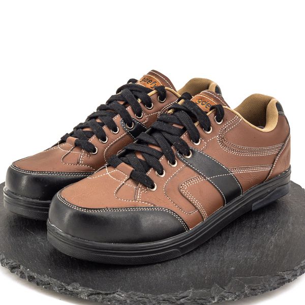
 Women's Shoes for Plantar Fasciitis | Aetrex Comfort Shoes | Aetrex
 Making the Rounds: Plantar fasciitis | Parkview Health
 Plantar Fasciitis Compression Sleeves - Heel Pain | Zensah