Aseptic or avascular necrosis of the femoral head. The major types are idiopathic (primary), as a complication of fractures or dislocations, and LEGG-CALVE-PERTHES DISEASE.
The hemispheric articular surface at the upper extremity of the thigh bone. (Stedman, 26th ed)
The longest and largest bone of the skeleton, it is situated between the hip and the knee.
The upper part of the human body, or the front or upper part of the body of an animal, typically separated from the rest of the body by a neck, and containing the brain, mouth, and sense organs.
The pathological process occurring in cells that are dying from irreparable injuries. It is caused by the progressive, uncontrolled action of degradative ENZYMES, leading to MITOCHONDRIAL SWELLING, nuclear flocculation, and cell lysis. It is distinct it from APOPTOSIS, which is a normal, regulated cellular process.
Serum glycoprotein produced by activated MACROPHAGES and other mammalian MONONUCLEAR LEUKOCYTES. It has necrotizing activity against tumor cell lines and increases ability to reject tumor transplants. Also known as TNF-alpha, it is only 30% homologous to TNF-beta (LYMPHOTOXIN), but they share TNF RECEPTORS.

The prevalence and clinicopathological appearance of extension of osteonecrosis in the femoral head. (1/547)

In about 50% of cases, osteonecrosis of the femoral head is known to occupy more than one site. There is controversy as to whether a single focus may increase in size. We have reviewed 606 consecutive femoral heads which had been surgically removed for osteonecrosis. Extension of osteonecrosis was observed in only two (0.3%) and was confirmed histopathologically by the enlargement of the necrotic segment beyond the repair zone formed for the primary necrosis into the adjacent, previously uninvolved bone. In both cases, the necrotic regions were wedge-shaped and occupied over 80% of the femoral head. It appears that an increase in size is extremely rare and that osteonecrosis is due to a single event. Our findings may be of value in assessing the use of joint-salvage procedures for osteonecrosis of the femoral head.  (+info)

Decrease in the mesenchymal stem-cell pool in the proximal femur in corticosteroid-induced osteonecrosis. (2/547)

We have evaluated bone-marrow activity in the proximal femur of patients with corticosteroid-induced osteonecrosis and compared it with that of patients with osteonecrosis related to sickle-cell disease and with a control group without osteonecrosis. Bone marrow was obtained by puncture of the femoral head outside the area of necrosis and in the intertrochanteric region. The activity of stromal cells was assessed by culturing fibroblast colony-forming units (FCFUs). We found a decrease in the number of FCFUs outside the area of osteonecrosis in the upper end of the femur of patients with corticosteroid-induced osteonecrosis compared with the other groups. We suggest that glucocorticosteroids may also have an adverse effect on bone by decreasing the number of progenitors. The possible relevance of this finding to osteonecrosis is discussed.  (+info)

Slipped capital femoral epiphysis after septic arthritis of the hip in an adolescent: report of a case. (3/547)

Septic arthritis of the hip must be managed promptly to avoid the serious complications associated with the condition. In the case reported here, the diagnosis was delayed and was complicated by a slipped capital femoral epiphysis. The patient, an adolescent boy previously in good health, presented with a 2-week history of hip pain and systemic illness. Septic arthritis was diagnosed and was managed by incision and drainage and antibiotic therapy. Two weeks later he presented with a subcutaneous abscess and a slipped capital femoral epiphysis, which was pinned in situ. There was a 2.5-cm leg-length discrepancy. Avascular necrosis of the femoral head subsequently developed leaving the boy with a permanent disability.  (+info)

Cyclosporin A mono-therapy in nephrotic syndrome with contra-indication of steroid therapy. (4/547)

We describe three cases of nephrotic syndrome with a contra-indication for steroid therapy successfully treated with cyclosporin A (CsA). A 21-year-old man with focal segmental glomerulosclerosis (FSGS) complicated by necrosis of the femoral head, and a 34-year-old woman and a 48-year-old man with minimal change disease (MCD) complicated by psychogenic reaction and diabetes mellitus, respectively, were given CsA at initial dosages of 3.8-5.0 mg/kg/day and immediately remitted completely. However, two of these patients suffered relapses when CsA was tapered. They are currently maintained in complete or partial remission on CsA at dosages of 3.2-4.7 mg/kg/day. These findings suggest that CsA mono-therapy may be useful in nephrotic syndrome patients contra-indicated for steroid therapy.  (+info)

Increased level of apolipoprotein B/apolipoprotein A1 ratio as a potential risk for osteonecrosis. (5/547)

OBJECTIVE: This study was performed to investigate whether a high ratio of apolipoprotein B to apolipoprotein A1 (apo B/apo A1 ratio) is significantly associated with the risk of developing non-traumatic osteonecrosis of the femoral head (ON). METHODS: Fifty consecutive non-traumatic ON cases were compared with 50 age and sex matched controls, using both univariate and stepwise discriminant analyses, regarding the factors of corticosteroid, alcohol, cigarettes, cholesterol, triglyceride, and apo B/apo A1 ratio. To eliminate the possibility that ON or osteoarthritic change itself can increase the apo B/apo A1 ratio, a further 32 consecutive cases comprising nine traumatic ON and 23 osteoarthritis (OA) patients were analysed using Scheffe's test. RESULTS: There was a significant association between a high apo B/apo A1 ratio and the development of non-traumatic ON with both univariate (p=0.0001) and stepwise discriminant analyses (partial r(2)=0.1239, p=0.0004). The apo B/apo A1 ratio in the non-traumatic ON group was significantly higher than that in the traumatic ON (p<0.01), control (p<0.001), or the OA groups (p<0.001). CONCLUSION: A high apo B/apo A1 ratio is significantly associated with the risk of developing ON. This ratio may be useful for assessing the potential risk of developing osteonecrosis.  (+info)

Treatment of osteonecrosis of the femoral head by free vascularized fibular grafting: an analysis of surgical outcome and patient health status. (6/547)

OBJECTIVE: To evaluate the limb-specific outcome and general health status of patients with osteonecrosis of the femoral head treated with vascularized fibular grafting. DESIGN: A retrospective review. SETTING: A single tertiary care centre. PATIENTS: Fifty-five consecutive patients with osteonecrosis of the femoral head who underwent fibular grafting (8 bilaterally). INTERVENTION: Vascularized fibular grafting. OUTCOME MEASURES: Limb-specific scores (Harris Hip Score, St. Michael's Hospital Hip Score), general health status (Nottingham Health Profile, SF-36 health status survey) and radiographic outcome measures (Steinberg stage). RESULTS: Patients were young (mean age 34 years, range from 18 to 52 years) and 80% had advanced osteonecrosis (Steinberg stages IV and V). Fifty-nine hips were followed up for an average of 50 months (range from 24 to 117 months) after vascularized fibular grafting. Sixteen hips (27%) were converted to total hip arthroplasty (THA). To date, 73% of hips treated with vascularized fibular grafting have required no further surgery. Preoperative and postoperative Harris Hip Scores were 57.3 and 83.6 respectively (p < 0.001). As measured by patient-oriented health status questionnaires (SF-36, Nottingham Health Profile) and compared with population controls, patients had normal mental health scores and only slight decreases in physical component scores. CONCLUSIONS: Free vascularized fibular grafting for osteonecrosis of the femoral head provides satisfactory pain relief, functional improvement and general health status and halts the progression of symptomatic disease.  (+info)

Diagnostic criteria for non-traumatic osteonecrosis of the femoral head. A multicentre study. (7/547)

Six major and seven minor diagnostic criteria have been developed by the Japanese Investigation Committee for osteonecrosis of the femoral head (ONFH). We have carried out a multicentre study to clarify these. We studied prospectively 277 hips in 222 patients, from six hospitals, who had ONFH and other hip pathology and from whom histological material was available. We identified five criteria with high specificity: 1) collapse of the femoral head without narrowing of the joint space or acetabular abnormality on radiographs, including the crescent sign; 2) demarcating sclerosis in the femoral head without narrowing or acetabular abnormality; 3) a 'cold-in-hot' appearance on the bone scan; 4) a low-intensity band on T1-weighted images (band pattern); and 5) evidence of trabecular and marrow necrosis on histological examination. With any combination of two of these criteria, the sensitivity and specificity of the diagnosis were 91% and 99%, respectively.  (+info)

Femoral head necrosis treated with vascularized iliac crest graft. (8/547)

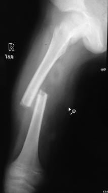
We reviewed 24 hips with avascular necrosis of the femoral head in 24 patients treated with vascularized iliac bone grafts 12 years after operation. In 7 patients the necrosis was classified as Ficat Stage II and in 17 patients as Stage III. Eight patients showed poor results. In 6 hips with fair results, moderate progression of the necrosis was noted at 3 to 8 years postoperatively. In 5 hips showing good results, slow progression with incipient signs of arthrosis were noted 8 years after surgery. In the remaining 5 patients with excellent results, no evidence of progression was noted 9 to 14 years postoperatively. The method described is recommended for treatment in the Ficat Stage II and early Stage III, when necrosis does not yet involve the complete femoral head.  (+info)

Femoral head necrosis, also known as avascular necrosis of the femoral head, is a medical condition that results from the interruption of blood flow to the femoral head, which is the rounded end of the thigh bone that fits into the hip joint. This lack of blood supply can cause the bone tissue to die, leading to the collapse of the femoral head and eventually resulting in hip joint damage or arthritis.

The condition can be caused by a variety of factors, including trauma, alcohol abuse, corticosteroid use, radiation therapy, and certain medical conditions such as sickle cell disease and lupus. Symptoms may include pain in the hip or groin, limited range of motion, and difficulty walking. Treatment options depend on the severity and progression of the necrosis and may include medication, physical therapy, or surgical intervention.

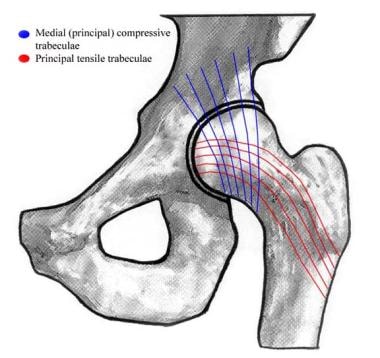
The femoral head is the rounded, ball-like top portion of the femur (thigh bone) that fits into the hip socket (acetabulum) to form the hip joint. It has a smooth, articular cartilage surface that allows for smooth and stable articulation with the pelvis. The femoral head is connected to the femoral neck, which is a narrower section of bone that angles downward and leads into the shaft of the femur. Together, the femoral head and neck provide stability and range of motion to the hip joint.

The femur is the medical term for the thigh bone, which is the longest and strongest bone in the human body. It connects the hip bone to the knee joint and plays a crucial role in supporting the weight of the body and allowing movement during activities such as walking, running, and jumping. The femur is composed of a rounded head, a long shaft, and two condyles at the lower end that articulate with the tibia and patella to form the knee joint.

In medical terms, the "head" is the uppermost part of the human body that contains the brain, skull, face, eyes, nose, mouth, and ears. It is connected to the rest of the body by the neck and is responsible for many vital functions such as sight, hearing, smell, taste, touch, and thought processing. The head also plays a crucial role in maintaining balance, speech, and eating.

Necrosis is the premature death of cells or tissues due to damage or injury, such as from infection, trauma, infarction (lack of blood supply), or toxic substances. It's a pathological process that results in the uncontrolled and passive degradation of cellular components, ultimately leading to the release of intracellular contents into the extracellular space. This can cause local inflammation and may lead to further tissue damage if not treated promptly.

There are different types of necrosis, including coagulative, liquefactive, caseous, fat, fibrinoid, and gangrenous necrosis, each with distinct histological features depending on the underlying cause and the affected tissues or organs.

Tumor Necrosis Factor-alpha (TNF-α) is a cytokine, a type of small signaling protein involved in immune response and inflammation. It is primarily produced by activated macrophages, although other cell types such as T-cells, natural killer cells, and mast cells can also produce it.

TNF-α plays a crucial role in the body's defense against infection and tissue injury by mediating inflammatory responses, activating immune cells, and inducing apoptosis (programmed cell death) in certain types of cells. It does this by binding to its receptors, TNFR1 and TNFR2, which are found on the surface of many cell types.

In addition to its role in the immune response, TNF-α has been implicated in the pathogenesis of several diseases, including autoimmune disorders such as rheumatoid arthritis, inflammatory bowel disease, and psoriasis, as well as cancer, where it can promote tumor growth and metastasis.

Therapeutic agents that target TNF-α, such as infliximab, adalimumab, and etanercept, have been developed to treat these conditions. However, these drugs can also increase the risk of infections and other side effects, so their use must be carefully monitored.

Quadratus femoris use in the Meyer's muscle pedicle grafting to prevent avascular necrosis of femur head. It originates on the ... From there, it passes laterally to its insertion on the posterior side of the head of the femur: the quadrate tubercle on the ... Right femur. Posterior surface. Structures surrounding right hip-joint. Nerves of the right lower extremity Posterior view. ... Since the muscle works to laterally rotate and adduct the femur, actions involving the lower body can strain the muscle. In ...
... about 10,000 to 20,000 people develop avascular necrosis of the head of the femur in the US each year.[citation needed] Cases ... Avascular necrosis most commonly affects the ends of long bones, such as the femur. Other common sites include the humerus ( ... Radiography of avascular necrosis of left femoral head. Man of 45 years with AIDS. Nuclear magnetic resonance of avascular ... Jacobs MA, Loeb PE, Hungerford DS (August 1989). "Core decompression of the distal femur for avascular necrosis of the knee" ( ...
... collapse and fracture of the neck of the femur. The recommended treatment is surgical removal of the head of the femur, but ... LCP disease is an avascular necrosis of the femoral head in small-breed dogs, usually those weighing up to 25 lbs. LCP disease ... Perthes disease is a childhood hip disorder initiated by a disruption of blood flow to the head of the femur. Due to the lack ... X-rays are necessary to make the diagnosis and show increased opacity and focal lysis in the head of the femur, and later in ...
The head of the femur is relevant to orthopedic surgery because it can undergo avascular necrosis and consequent ... The femoral head (femur head or head of the femur) is the highest part of the thigh bone (femur). It is supported by the ... Femur head Hip joint. Lateral view. Femur head Mechlenburg, I.; Nyengaard, J.R.; Gelineck, J.; Soballe, K. (April 2007). " ... The fovea capitis is a small, concave depression within the head of the femur that serves as an attachment point for the ...
During the trial, her doctor testified that she had been taking the drug to treat avascular necrosis at the head of her femur. ... Sunwoo, Carla (November 20, 2012). "Park Si-yeon heading to Hollywood". Korea JoongAng Daily. Archived from the original on ...
The heads of the humeri and femora with many fossils show necrosis of the bone tissue, caused by a too rapid ascent after deep ... With early forms, the amphicoelous or amphiplat neck vertebrae bore double-headed neck ribs; later forms had single-headed ribs ... Their heads and teeth were very large, suited to grab and rip apart large animals. Their morphology allowed for a high swimming ... The humerus in the upper arm, and the femur in the upper leg, had become large flat bones, expanded at their outer ends. The ...
... to the head of the femur. The blood supply to the femoral head and neck is enhanced by the artery of the ligamentum teres ... In adults it does not provide enough blood supply to prevent avascular necrosis in upper femur fractures. The obturator artery ... it helps to supply the head and neck of the femur on its own. The articular branch is usually patent until roughly 15 years of ... ramifies in the fat at the bottom of the acetabulum and sends a twig along the ligament of head of femur (ligamentum teres) ...
... femur head necrosis MeSH C05.116.852.175.570 - Legg-Perthes disease MeSH C05.116.900.307 - intervertebral disc displacement ...
... femur head necrosis MeSH C23.550.727.045 - anaplasia MeSH C23.550.727.152 - cell transformation, neoplastic MeSH C23.550. ... dental pulp necrosis MeSH C23.550.717.365 - fat necrosis MeSH C23.550.717.427 - gangrene MeSH C23.550.717.489 - infarction MeSH ...
The heads of the humeri and femora of many fossils show necrosis of the bone tissue, caused by nitrogen bubble formation due to ... They may stand on their tail to hold their head above water. Sirenians exhibit pachyostosis, a condition in which the ribs and ... Surface swimming causes wave-making drag, and the animal must hold its head up in the air while breathing, causing more drag. ...
Legg-Calvé-Perthes syndrome, also called Avascular Necrosis of the Femoral Head, is where the ball section of the femur in the ... The head should be of moderate width at the ears, narrowing to the eyes, and slightly flat between the ears. There should be a ... Small V-shaped ears of moderate thickness are carried forward on the head.[citation needed] When the dog is alert, the tip of ... Other differences in the Parson Russell can include a longer head and larger chest as well as overall a larger body size. The ...
... which becomes important to avoid avascular necrosis of the head of the femur when the blood supply from the medial and lateral ... The joint space between the femoral head and the superior acetabulum is normally between 2 and 7 mm. The head of the femur is ... This artery is not present in everyone but can become the only blood supply to the bone in the head of the femur when the neck ... The hip joint is a ball and socket synovial joint formed by the articulation of the rounded head of the femur and the cup-like ...
Zhang LD, Kang JF, Xue HL (July 1990). "Distribution of lesions in the head and neck of the humerus and the femur in dysbaric ... soft tissue and bony necrosis); Skin grafts and flaps (compromised); Thermal burns. There is no reliable evidence to support ... There is some evidence that HBOT is effective for late radiation tissue injury of bone and soft tissues of the head and neck. ... Some people with radiation injuries of the head, neck or bowel show an improvement in quality of life. Importantly, no such ...
... also known as Perthes disease or avascular necrosis of the femoral head, is characterized by a deformity of the head of the ... dysplasia is an inherited disease in dogs that is characterized by abnormal development of the acetabulum and head of the femur ... The disease in dogs is usually nodular skin lesions of the head and trunk. Aspergillosis* is a fungal disease that in dogs is ... It is a common result of head trauma in dogs. Most commonly it occurs in brachycephalic (short nosed) breeds. Horner's syndrome ...
... of the lesions were in the humeral head (shoulder), 16% in the femoral head (hip), 40% in the lower end of the femur (lower ... Dysbaric osteonecrosis or DON is a form of avascular necrosis where there is death of a portion of the bone that is thought to ... Zhang, LD; Kang, JF; Xue, HL (July 1990). "Distribution of lesions in the head and neck of the humerus and the femur in ... This process takes place over months to years and eventually causes disabling arthritis, particularly of the femoral head (hip ...
1990). "Distribution of lesions in the head and neck of the humerus and the femur in dysbaric osteonecrosis". Undersea Biomed. ... McFarlane RM, Wermuth RE (May 1966). "The use of hyperbaric oxygen to prevent necrosis in experimental pedicle flaps and ... "Pathophysiology and natural history of avascular necrosis of bone". Joint Bone Spine. 73 (5): 500-7. doi:10.1016/j.jbspin. ... Soft Tissue and Bony Necrosis) Skin Grafts & Flaps (Compromised) Sudden Sensorineural Hearing Loss Thermal Burns Medical ...
Vanhoenacker F. "Femoral head-Avascular necrosis, femoral head". Radiology Intelligent Assistant. doi:10.5832/m216-1-119-0. ... What most clinicians observe is that the head of the femur seems to almost have a system of its own somewhat separate from the ... There are many theories as to how the head of the femur dies unlike why it is happening. The main mechanism is through external ... Although Chandler's Disease is specific to the head of the femur there are many other diseases that are analogous to the ...
Avascular necrosis of the femoral head occurs frequently (20%) in intracapsular hip fractures, because the blood supply is ... Traction is contraindicated in femoral neck fractures due to it affecting blood flow to the head of the femur. The latest ... An axial load along the shaft of the femur results in compressive stress. Bending load at the neck of the femur causes tensile ... Femoral head fractures are a rare kind of hip fracture that may also be the result of a fall but are more commonly caused by ...
Avascular necrosis of the femoral head (Legg-Calvé-Perthes disease) typically occurs in children aged 4-8, and is also more ... diminution of soft tissue planes around the hip joint or slight demineralisation of the proximal femur. The main reason for ... One such previously suspected complication was coxa magna, which is an overgrowth of the femoral head and broadening of the ... whether continuous high intra-articular pressure in transient synovitis could cause avascular necrosis of the femoral head ( ...
... which results in slippage of the overlying end of the femur (metaphysis). Normally, the head of the femur, called the capital, ... another cause of avascular necrosis of the femoral head, seen in younger children than SCFE Hip dysplasia Drehmann sign - ... Therefore the head of the femur is usually pinned 'as is'. A small incision is made in the outer side of the upper thigh and ... The appearance of the head of the femur in relation to the shaft likens that of a "melting ice cream cone", visible with ...
Damage to the artery following a femoral neck fracture may lead to avascular necrosis (ischemic) of the femoral neck/head. The ... Branches of the medial circumflex femoral artery supplying the head and neck of the femur are often torn in femoral neck ... It winds around the medial side of the femur to pass along the posterior aspect of the femur. It first passes between the ... It delivers most of the arterial supply to the femoral head and femoral neck via branches - the posterior retinacular arteries ...
Another example of a pressure epiphysis is the head of the humerus which is part of the shoulder complex. condyles of femur and ... Pathologies of the epiphysis include avascular necrosis and osteochondritis dissecans (OCD). OCD involves the subchondral bone ... the head of the femur, part of the hip joint complex). Pressure epiphyses assist in transmitting the weight of the human body ... Femur: Longest bone in the human body. Located in the thigh region, between the hip and the knee. Fibula: One of two bones in ...
... in some mammals embedded in the tendon of the lateral head of the gastrocnemius muscle behind the lateral condyle of the femur ... Sesamoid bones generally have a very limited blood supply, rendering them prone to avascular necrosis (bone death from lack of ...
Bone tumors were located in the femurs, mandible, maxilla, tibias, or in one case each the hard palate and sacrum. In other ... In a minority of cases, the tumor tissues have scattered mast cells, sites of numerous neutrophils, and areas of necrosis (i.e ... Overall, 51.5% of their tumors were in the head and neck areas (most commonly the tongue, followed by the larynx, gums, ... One study suggested that tumor cells with high rates of proliferation, tumors containing areas of necrosis, tumor sizes >10 cm ...
eds.). Histophatology of the nontraumatic necrosis of the femoral head. Neville BW, Damm DD, Allen CA, Bouquot JE (2002). Oral ... in the replicative capacity of osteoblastic cells in the proximal femur of patients with osteonecrosis of the femoral head". J ... "Necrosis of the lower jaw in makers of Lucifer matches". Am J Dent Science. 1 (series 3): 96-7. 1867. "The History of ... Noel HR (1868). "A lecture on caries and necrosis of bone". Am J Dent Science. 1 (series 3): 425, 482. Barrett WC (1898). Oral ...
The femur head is flat and irregular. People develop osteoarthritis at an early age. Hip dysplasia is considered to be a ... Cases of femoral nerve palsy and avascular necrosis of the femoral head have been reported with the use of the Pavlik harness, ... Two forms of femoral dysplasia are coxa vara, in which the femur head grows at too narrow an angle to the shaft, and coxa valga ... For adults it describes hips showing abnormal femur head or acetabular x-rays. Some sources prefer the term "hip dysplasia" ...
From proximal view, the head of the humerus is concave, which is a sign of a substantial cap of cartilage at the head of ... Avascular necrosis, also known as bone necrosis, is associated with decompression syndrome (DCS). It is caused by expose rapid ... The femur of Pistosaurus is longer than its humerus. Its anterior margin is almost straight whereas the posterior margin is ... The head also resembled that of a plesiosaur, but with the primitive palate of a nothosaur, and numerous, sharp teeth ideal for ...
... resection of the femur three times; performing lithotomy twenty-one times; femoral eighteen times; and removal of the entire ... of which the surgeon general placed him at the head. He was appointed surgeon U.S. volunteers, and was one of the medical ... lower jaw for phosphor-necrosis twice. There have been published reports of other cases of interest treated by him. He was ...
He is best known for pioneering "the use of ivory hip prostheses to replace ununited fractures of the neck of the femur," and ... After his death, she abruptly resigned from her post as head of the Department of Medicine, and served as a consultant at the ... and avascular necrosis. His designs were based on a Thomson hemiarthroplasty, but with major modifications to the stem, and ... He is best known for pioneering the use of "ivory hip prostheses to replace ununited fractures of the neck of the femur," and ...
Less severe trauma might contuse the bone to cause an area of necrosis which might then separate. In some cases, the absence of ... Sagittal MRI: Linear low T1 signal at the articular surfaces of the lateral aspects of the medial condyle of the femur confirms ... Less frequent locations include the patella, vertebrae, the femoral head, and the glenoid of the scapula. The oldest case of ... To diagnose osteochondritis dissecans, an X-ray, CT scan or MRI scan can be performed to show necrosis of subchondral bone, ...
... of femoral head is a safe and effective procedure and has better outcome than bone marrow for early stage of avascular necrosis ... Implantation of autologous bone marrow stem cells in avascular necrosis ... Conclusions: Implantation of autologous bone marrow stem cells in avascular necrosis of femoral head is a safe and effective ... Materials and methods: Forty patients (60 hips) with stage I, II or III (ARCO system) osteonecrosis of femoral head were ...
Osteonecrosis is a disease characterized by a derangement of osseous circulation that leads to necrosis of osseous tissue. ... Aseptic necrosis of the femur head. Pathogenesis: the theory of circulation]. Acta Orthop Belg. 1981 Mar-Apr. 47(2):198-9. [ ... Ficat RP, Arlet J. Necrosis of the femoral head. In: Hungerford DS, ed. Ischemia and Necrosis of Bone. Baltimore: Williams and ... Idiopathic bone necrosis of the femoral head. Early diagnosis and treatment. J Bone Joint Surg Br. 1985 Jan. 67(1):3-9. [QxMD ...
... of the femoral head is a pathologic process that results from interruption of blood supply to the bone. AVN of the hip is ... common pathway of traumatic or nontraumatic factors that compromise the already precarious circulation of the femoral head. ... Arlet J, Ficat P. [Non-traumatic avascular femur head necrosis. New methods of examination and new concepts] [Polish]. Chir ... encoded search term (Femoral Head Avascular Necrosis) and Femoral Head Avascular Necrosis What to Read Next on Medscape ...
Quadratus femoris use in the Meyers muscle pedicle grafting to prevent avascular necrosis of femur head. It originates on the ... From there, it passes laterally to its insertion on the posterior side of the head of the femur: the quadrate tubercle on the ... Right femur. Posterior surface. Structures surrounding right hip-joint. Nerves of the right lower extremity Posterior view. ... Since the muscle works to laterally rotate and adduct the femur, actions involving the lower body can strain the muscle. In ...
... and trochanteric fractures of the proximal femur with head involvement, unmanageable by other techniques. 5. Revision ... Indications For Use: 1 . Non-inflammatory degenerative joint disease including osteoarthritis and avascular necrosis. 2. ...
Ikeuchi M, Komatsu M, Tani T. Giant bone island of femur with femoral head necrosis: a case report. Arch Orthop Trauma Surg. ... They can be found at any osseous site, with the pelvis and long bones (especially the proximal femur) most commonly involved. ... Forty-one (89%) patients had 124 enostoses (2-15 mm). The most common sites of occurrence were the proximal femur (34%), the ... Giant bone island of femur. Case report, literature review, and its distinction from low grade osteosarcoma. Skeletal Radiol. ...
A hip fracture is a break in the upper portion of the femur (thighbone). Most hip fractures occur in elderly patients whose ... Even if the fracture does heal, a condition called avascular necrosis may develop in the head of the femur. This causes damage ... The ball is the head of the femur, which is the upper part of the thighbone. The socket is called the acetabulum. The ... The blood supply to the head of the femur comes through a structure called the posterior capsule. The posterior capsule is ...
... , Z. ZHAO* ... Anterior Hip (Femur) Dislocation. In: StatPearls Treasure Island. StatPearls Publishing; 2020.. *Gangji V, Hauzeur JP. Cellular ... Femoral head necrosis (FHN) is a mobility disorder, associated with hip joint[1,2]. According to an estimate, 70 % of the ... Femoral head necrosis is a mobility disorder and till now no satisfactory solution has been reported. The principal ...
avascular or aseptic or ischemic or ischaemic or femur head or femoral head or bone or bones) adj2 (necrosis or necroses)).tw. ... Avascular necrosis (AVN) is also known as osteonecrosis, aseptic necrosis, and ischemic necrosis.1,2 AVN is a sequela of ... Imaging in avascular necrosis of the femoral head. 2011 May 25 [cited 2011 Apr 12]. In: Medscape Reference [Internet]. New York ... Explode a subject heading. *. Before a word, indicates that the marked subject heading is a primary topic; or, after a word, a ...
... avascular necrosis of the femoral head, nonunion of previous fractures of the femur; congenital hip dysplasia, protrusio ...
In this condition, the head of the femur loses some of its blood supply and dies. Hip fractures and some childhood hip ... Other degenerative hip diseases leading to hip replacement surgery include avascular necrosis. ... Doctors cover the femur or thighbone with a metal covering and place plastic on the tibia, so the metal and plastic implants ...
We also performed electromagnetic (EM) computer simulations of virtual prototypes of bone cancer, femur fracture, and diabetic ... It shows the head, neck, and leg of the femur. The exploitation of electric field indicates the feasibility of a subsequent ... often resulting in avascular necrosis of the femoral head. EM simulations of femoral neck fractures were presented as examples ... Human head thickness for an adult male is around 19.4 cm, so to irradiate the center of a human head the exposure should have a ...
... and aseptic necrosis of the femur head (n = 1).. A malignancy was diagnosed within 2 years after onset of myositis in five ... and head flexors and extensors, a sum score was calculated, resulting in a maximum score of 130 for normal muscle strength.27 ...
Aseptic necrosis of the hip is the death of bone tissue in the head of the thigh bone (femur) due to poor blood supply. ... Aseptic necrosis of the hip is caused by any event or health problem that blocks blood supply to the head of the femur. ... Aseptic Necrosis of the Hip. (Osteonecrosis of the Hip; Avascular Necrosis of the Hip; Ischemic Necrosis of the Hip; ... Legg-Calvé-Perthes disease is a type of aseptic necrosis in children. It affects the growth plate at the upper end of the thigh ...
... in the replicative capacity of osteoblastic cells in the proximal femur of patients with osteonecrosis of the femoral head. The ... Non-traumatic avascular necrosis of the femoral head. JBJS. 1995 Mar 1;77(3):459-74. [Pubmed] [Google Scholar] ... 1 There are three basic categories of cause for avascular necrosis. Approximately 10% of Avascular necrosis of the talus is ... Avascular necrosis occurs when the blood supply to the talus is compromised and leads to ischemic bone death. About 60% of the ...
diagnostic imaging; disease management; femur head necrosis; osteonecrosis Citazione Manenti, G., Altobelli, S., Pugliese, L., ... The role of imaging in diagnosis and management of femoral head avascular necrosis. Manenti G.;Altobelli S.;Pugliese L.; ... The role of imaging in diagnosis and management of femoral head avascular necrosis. CLINICAL CASES IN MINERAL AND BONE ... The role of imaging in diagnosis and management of femoral head avascular necrosis. CLINICAL CASES IN MINERAL AND BONE ...
Like Legg-Calvé-Perthes disease, avascular necrosis of the femoral head causes the upper ends of the thigh bones (femurs) to ... Mutations in the COL2A1 gene can sometimes result in a condition known as avascular necrosis of the femoral head, which is ... Type II collagen and avascular necrosis of the femoral head. N Engl J Med. 2005 Jun 2;352(22):2268-70. doi: 10.1056/NEJMp058072 ... avascular necrosis of the femoral head, or Legg-Calve-Perthes disease in a single family, consequent upon a p.Gly1170Ser ...
Femur Head Necrosis. *Foot Deformities. *Foot Fracture. *Foot Injuries. *Fragility Fractures. *Ganglion Cyst ...
... the femur where the circulation to the head is cut off, the head of the femur undergoes avascular necrosis, changes similar to ... causing collapse of the head of the femur. Over time, the head of the femur heals and remodels in the collapsed position, ... If the head of the femur cannot be contained in the acetabulum, and the head flattens, prognosis tends to be less favorable. ... Do X-rays of the hips show partial or total head involvement? If the whole head of the femur is involved, the prognosis is less ...
Avascular Necrosis of Head of Femur - Stage of Arrival in Hospital and Management Options January 10, 2019 admin 0 ...
Anahtar Kelimeler : femoral head; dislocation, epiphysis; femur head necrosis Copyright © 2023 - TOTBİD Dergisi ... In case of failed treatment, SCFE has devastating results like avascular necrosis, chondrolysis, and femoroacetabular ...
... with dislocation of the right femoral head with necrosis, and extensive soft tissue and muscular abscesses around the proximal ... femur and into the right pelvis and lower abdominal retroperitoneum. Synovial fluid culture grew Hib. The patient underwent 3 ...
Femur Head Necrosis 31% * Femoral Neck Fractures 13% * Pain 12% 60 Scopus citations ... Effect of Length Stability on Complications in the Treatment of Preadolescent Diaphyseal Femur Fractures Treated With Elastic ...
... nailing of the femur in this age group is a concern because of the possibility of avascular necrosis of the femoral head. This ... Blood supply to the proximal femur has been reserved with no signs of avascular necrosis. The ALFNs lateral entry point makes ... There was missing bone from the femur. Open but rapidly closing growth plates. ... as well as nonunions and malunions of the femur (Fig 1). The ALFN can also be used to stabilize corrective osteotomies in bone ...
Osteonecrosis is a disease characterized by a derangement of osseous circulation that leads to necrosis of osseous tissue. ... Aseptic necrosis of the femur head. Pathogenesis: the theory of circulation]. Acta Orthop Belg. 1981 Mar-Apr. 47(2):198-9. [ ... Ficat RP, Arlet J. Necrosis of the femoral head. In: Hungerford DS, ed. Ischemia and Necrosis of Bone. Baltimore: Williams and ... Idiopathic bone necrosis of the femoral head. Early diagnosis and treatment. J Bone Joint Surg Br. 1985 Jan. 67(1):3-9. [QxMD ...
Avascular Necrosis of Head of Femur Knee Joint. *Rheumatoid Arthritis. *Osteoarthritis. Causes Of Joint Problems ... Tumours of head and neck. • Rheumatoid Arthritis. • Congenital Cervical Rib. Rare Causes Include. • Congenital spinal canal ...
Femur Head Necrosis Medicine & Life Sciences 19% * Therapeutics Medicine & Life Sciences 13% ... N2 - The clinical and radiographic outcomes of 25 patients with avascular necrosis (AVN) of the femoral head complicating ... AB - The clinical and radiographic outcomes of 25 patients with avascular necrosis (AVN) of the femoral head complicating ... The clinical and radiographic outcomes of 25 patients with avascular necrosis (AVN) of the femoral head complicating ...

No FAQ available that match "femur head necrosis"