Fibrin
Fibrin Tissue Adhesive
Fibrin Fibrinogen Degradation Products
Fibrinogen
Tissue Adhesives
Fibrinopeptide B
Blood Coagulation
Batroxobin
Fibrinopeptide A
Factor XIII
Fibrinolysin
Plasminogen
Fibrinogens, Abnormal
Factor XIIIa
Ancrod
alpha-2-Antiplasmin
Tissue Plasminogen Activator
Plasminogen Activators
Gels
Thrombin Time
Aminocaproic Acid
Disseminated Intravascular Coagulation
Fibrin Modulating Agents
Urokinase-Type Plasminogen Activator
Blood Platelets
Hemostatics
Thromboplastin
Nephelometry and Turbidimetry
Hemostasis
Antifibrinolytic Agents
Blood Coagulation Disorders
Transglutaminases
Antithrombin III
Microscopy, Electron, Scanning
Pit and Fissure Sealants
Marketing
Social Marketing
Brachial Plexus
Brachial Plexus Neuropathies
Sciatic Nerve
Myocardial Reperfusion Injury
Disease Models, Animal
National Heart, Lung, and Blood Institute (U.S.)
Reperfusion Injury
Peptides
Myocardium
Myocardial Ischemia
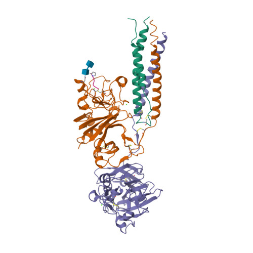
Comparison of the fibrin-binding activities in the N- and C-termini of fibronectin. (1/2092)
Fibronectin (Fn) binds to fibrin in clots by covalent and non-covalent interactions. The N- and C-termini of Fn each contain one non-covalent fibrin-binding site, which are composed of type 1 (F1) structural repeats. We have previously localized the N-terminal site to the fourth and fifth F1 repeats (4F1.5F1). In the current studies, using proteolytic and recombinant proteins representing both the N- and C-terminal fibrin-binding regions, we localized and characterized the C-terminal fibrin-binding site, compared the relative fibrin-binding activities of both sites and determined the contribution of each site to the fibrin-binding activity of intact Fn. By fibrin-affinity chromatography, a protein composed of the 10F1 repeat through to the C-terminus of Fn (10F1-COOH), expressed in COS-1 cells, and 10F1-12F1, produced in Saccharomyces cerevisiae, displayed fibrin-binding activity. However, since 10F1 and 10F1.11F1 were not active, the presence of 12F1 is required for fibrin binding. A proteolytic fragment of 14.4 kDa, beginning 14 residues N-terminal to 10F1, was isolated from the fibrin-affinity matrix. Radio-iodinated 14.4 kDa fibrin-binding peptide/protein (FBP) demonstrated a dose-dependent and saturable binding to fibrin-coated wells that was both competitively inhibited and reversed by unlabelled 14.4 kDa FBP. Comparison of the fibrin-binding affinities of proteolytic FBPs from the N-terminus (25.9 kDa FBP), the C-terminus (14.4 kDa) and intact Fn by ELISA yielded estimated Kd values of 216, 18 and 2.1 nM, respectively. The higher fibrin-binding affinity of the N-terminus was substantiated by the ability of both a recombinant 4F1.5F1 and a monoclonal antibody (mAb) to this site to maximally inhibit biotinylated Fn binding to fibrin by 80%, and by blocking the 90% inhibitory activity of a polyclonal anti-Fn, by absorption with the 25.9 kDa FBP. We propose that whereas the N-terminal site appears to contribute to most of the binding activity of native Fn to fibrin, the specific binding of the C-terminal site may strengthen this interaction. (+info)Exosites 1 and 2 are essential for protection of fibrin-bound thrombin from heparin-catalyzed inhibition by antithrombin and heparin cofactor II. (2/2092)
Assembly of ternary thrombin-heparin-fibrin complexes, formed when fibrin binds to exosite 1 on thrombin and fibrin-bound heparin binds to exosite 2, produces a 58- and 247-fold reduction in the heparin-catalyzed rate of thrombin inhibition by antithrombin and heparin cofactor II, respectively. The greater reduction for heparin cofactor II reflects its requirement for access to exosite 1 during the inhibitory process. Protection from inhibition by antithrombin and heparin cofactor II requires ligation of both exosites 1 and 2 because minimal protection is seen when exosite 1 variants (gamma-thrombin and thrombin Quick 1) or an exosite 2 variant (Arg93 --> Ala, Arg97 --> Ala, and Arg101 --> Ala thrombin) is substituted for thrombin. Likewise, the rate of thrombin inhibition by the heparin-independent inhibitor, alpha1-antitrypsin Met358 --> Arg, is decreased less than 2-fold in the presence of soluble fibrin and heparin. In contrast, thrombin is protected from inhibition by a covalent antithrombin-heparin complex, suggesting that access of heparin to exosite 2 of thrombin is hampered when ternary complex formation occurs. These results reveal the importance of exosites 1 and 2 of thrombin in assembly of the ternary complex and the subsequent protection of thrombin from inhibition by heparin-catalyzed inhibitors. (+info)Isolation of SMTP-3, 4, 5 and -6, novel analogs of staplabin, and their effects on plasminogen activation and fibrinolysis. (3/2092)
Four novel triprenyl phenol metabolites, designated SMTP-3, -4, -5, and -6, have been isolated from cultures of Stachybotrys microspora IFO 30018 by solvent extraction and successive chromatographic fractionation using silica gel and silica ODS columns. A combination of spectroscopic analyses showed that SMTP-3, -4, -5, and -6 are staplabin analogs, containing a serine, a phenylalanine, a leucine or a tryptophan moiety in respective molecules in place of the N-carboxybutyl portion of the staplabin molecule. SMTP-4, -5, and -6 were active at 0.15 to 0.3 mM in enhancing urokinase-catalyzed plasminogen activation and plasminogen binding to fibrin, as well as plasminogen- and urokinase-mediated fibrinolysis. On the other hand, the concentration of staplabin required to exert such effects was 0.4 to 0.6 mM, and SMTP-3 was inactive at concentrations up to 0.45 mM. (+info)Blood-borne tissue factor: another view of thrombosis. (4/2092)
Arterial thrombosis is considered to arise from the interaction of tissue factor (TF) in the vascular wall with platelets and coagulation factors in circulating blood. According to this paradigm, coagulation is initiated after a vessel is damaged and blood is exposed to vessel-wall TF. We have examined thrombus formation on pig arterial media (which contains no stainable TF) and on collagen-coated glass slides (which are devoid of TF) exposed to flowing native human blood. In both systems the thrombi that formed during a 5-min perfusion stained intensely for TF, much of which was not associated with cells. Antibodies against TF caused approximately 70% reduction in the amount of thrombus formed on the pig arterial media and also reduced thrombi on the collagen-coated glass slides. TF deposited on the slides was active, as there was abundant fibrin in the thrombi. Factor VIIai, a potent inhibitor of TF, essentially abolished fibrin production and markedly reduced the mass of the thrombi. Immunoelectron microscopy revealed TF-positive membrane vesicles that we frequently observed in large clusters near the surface of platelets. TF, measured by factor Xa formation, was extracted from whole blood and plasma of healthy subjects. By using immunostaining, TF-containing neutrophils and monocytes were identified in peripheral blood; our data raise the possibility that leukocytes are the main source of blood TF. We suggest that blood-borne TF is inherently thrombogenic and may be involved in thrombus propagation at the site of vascular injury. (+info)Use of high-intensity focused ultrasound to control bleeding. (5/2092)
OBJECTIVE: High-intensity focused ultrasound (HIFU) has been shown to be effective in controlling hemorrhage from punctures in blood vessels. The objective of the current study was to investigate the capability of HIFU to stop bleeding after a more severe type of vascular injury, namely longitudinal incisions of arteries and veins. METHODS: The superficial femoral arteries, common femoral arteries, carotid arteries, and jugular veins of four anesthetized pigs were exposed surgically. A longitudinal incision, 2 to 8 mm in length, was produced in the vessel. HIFU treatment was applied within 5 seconds of the onset of the bleeding. The HIFU probe consisted of a high-power, 3.5-MHz, piezoelectric transducer with an ellipsoidal focal spot that was 1 mm in cross section and 9 mm in axial dimension. The entire incision area was scanned with the HIFU beam at a rate of 15 to 25 times/second and a linear displacement of 5 to 10 mm. A total of 76 incisions and HIFU treatments were performed. RESULTS: Control of bleeding (major hemosatsis) was achieved in all 76 treatments, with complete hemostasis achieved in 69 treatments (91%). The average treatment times of major and complete hemostasis were 17 and 25 seconds, respectively. After the treatment, 74% of the vessels in which complete hemostasis was achieved were patent with distal blood flow and 26% were occluded. The HIFU-treated vessels showed a consistent coagulation of the adventitia surrounding the vessels, with a remarkably localized injury to the vessel wall. Extensive fibrin deposition at the treatment site was observed. CONCLUSION: HIFU may provide a useful method of achieving hemostasis for arteries and veins in a variety of clinical applications. (+info)Chronic protein undernutrition and an acute inflammatory stimulus elicit different protein kinetic responses in plasma but not in muscle of piglets. (6/2092)
The changes in protein metabolism of severe childhood malnutrition are generally perceived as a metabolic adaptation to chronic protein undernutrition. However, severe malnutrition is invariably accompanied by infections which also have profound effects on protein metabolism. This study aimed to distinguish the effect of protein undernutrition from that of an inflammatory stimulus on muscle and plasma protein synthesis rates. Two groups of five piglets consumed diets containing either 23% or 3% protein for 4 wk. They then were infused intravenously with 2H3-leucine before and 48 h after subcutaneous injections of turpentine to measure the fractional synthesis rates (FSR) of muscle protein and both the FSR and the absolute synthesis rates (ASR) of albumin and fibrinogen. Prior to turpentine injection, compared to control piglets, protein-deficient piglets had significantly lower muscle FSR and plasma concentrations of both albumin and fibrinogen, although only albumin had lower FSR and ASR. Turpentine injection decreased muscle FSR but increased the FSR, ASR and plasma concentrations of both albumin and fibrinogen in control piglets. In protein-deficient piglets, the inflammatory stress caused a further decrease in muscle protein FSR and in plasma albumin concentration despite marked increases in albumin FSR and ASR. Fibrinogen FSR, ASR and plasma concentration were increased. We conclude that protein undernutrition and inflammation elicit the same kinetic response in muscle protein but different kinetic responses in plasma proteins. Furthermore, whereas protein deficiency reduces the plasma albumin pool via a reduction in albumin synthesis, inflammation reduces it through a stimulation of catabolism and/or loss from the intravascular space. (+info)Differential regulation of beta1 integrins by chemoattractants regulates neutrophil migration through fibrin. (7/2092)
Chemoattractants differ in their capacity to stimulate neutrophils to adhere to and to migrate through matrices containing fibrin. Formyl methionyl leucyl phenylalanine (fMLP) stimulates neutrophils to adhere closely to, but not to migrate into, fibrin gels. Leukotriene B4 (LTB4) stimulates neutrophils to adhere loosely to and to migrate through fibrin gels. We report that alpha5beta1 integrins regulate the different migratory behaviors on fibrin gels of neutrophils in response to these chemoattractants. fMLP, but not LTB4, activated neutrophil beta1 integrins, as measured by binding of mAb 15/7 to an activation epitope on the beta1 integrins. Antibodies or peptides that block alpha5beta1 integrins prevented fMLP-stimulated neutrophils from forming zones of close apposition on fibrin and reversed fMLP's inhibitory effect on neutrophil chemotaxis through fibrin. In contrast, neither peptides nor antibodies that block beta1 integrins affected the capacity of LTB4-stimulated neutrophils to form zones of loose apposition or to migrate through fibrin gels. These results suggest that chemoattractants generate at least two different messages that direct neutrophils, and perhaps other leukocytes, to accumulate at specific anatomic sites: a general message that induces neutrophils to crawl and a specific message that prepares neutrophils to stop when they contact appropriate matrix proteins for activated beta1 integrins. (+info)Malfunction of Bjork-Shiley valve prosthesis in tricuspid position. (8/2092)
Eight months after triple valve replacement with Bjork-Shiley tilting disc valves a patient developed symptoms and signs suggesting malfunction of the prosthesis in the tricuspid position. This was confirmed by echocardiography and angiocardiography, and at operation thedisc of the prosthesis was found to be stuck half-open by fibrin and clot. A further 11 patients with the same tupe of prosthesis in the triscupid position were then studied by phonocardiography and echocardiography. In one of these the prosthesis was found to be stuck and this was confirmed by angiocardiography and surgery. These 2 cases are reported in detail and the findings in the other 10 are discussed. The implications of this high incidence of malfunction of the Bjork-Shiley prosthesis in the tricuspid position are considered. Echocardiography appears to be essential in the follow-up of such patients. (+info)Fibrin is defined as a protein that is formed from fibrinogen during the clotting of blood. It plays an essential role in the formation of blood clots, also known as a clotting or coagulation cascade. When an injury occurs and bleeding starts, fibrin threads form a net-like structure that entraps platelets and red blood cells to create a stable clot, preventing further loss of blood.
The process of forming fibrin from fibrinogen is initiated by thrombin, another protein involved in the coagulation cascade. Thrombin cleaves fibrinogen into fibrin monomers, which then polymerize to form long strands of fibrin. These strands cross-link with each other through a process catalyzed by factor XIIIa, forming a stable clot that protects the wound and promotes healing.
It is important to note that abnormalities in fibrin formation or breakdown can lead to bleeding disorders or thrombotic conditions, respectively. Proper regulation of fibrin production and degradation is crucial for maintaining healthy hemostasis and preventing excessive clotting or bleeding.
A fibrin tissue adhesive is a type of surgical glue that is used to approximate and secure together cut or wounded tissues in the body during surgical procedures. It is made from fibrin, a protein involved in blood clotting, and is often combined with other substances like thrombin and calcium chloride to promote clot formation and enhance adhesion.
Fibrin tissue adhesives work by mimicking the body's natural clotting process. When applied to the wound site, the fibrinogen component of the adhesive is converted into fibrin by the thrombin component, creating a stable fibrin clot that holds the edges of the wound together. This helps to promote healing and reduce the risk of complications such as bleeding or infection.
Fibrin tissue adhesives are commonly used in various surgical procedures, including dermatologic, ophthalmic, orthopedic, and neurologic surgeries. They offer several advantages over traditional suturing methods, such as reduced operation time, less trauma to the tissues, and improved cosmetic outcomes. However, they may not be suitable for all types of wounds or surgical sites, and their use should be determined by a qualified healthcare professional based on individual patient needs and circumstances.
Fibrin(ogen) degradation products (FDPs) are a group of proteins that result from the breakdown of fibrinogen and fibrin, which are key components of blood clots. This process occurs during the normal physiological process of fibrinolysis, where clots are dissolved to maintain blood flow.
FDPs can be measured in the blood as a marker for the activation of the coagulation and fibrinolytic systems. Elevated levels of FDPs may indicate the presence of a disorder that causes abnormal clotting or bleeding, such as disseminated intravascular coagulation (DIC), deep vein thrombosis (DVT), pulmonary embolism (PE), or certain types of cancer.
It is important to note that FDPs are not specific to any particular disorder and their measurement should be interpreted in conjunction with other clinical and laboratory findings.
Fibrinogen is a soluble protein present in plasma, synthesized by the liver. It plays an essential role in blood coagulation. When an injury occurs, fibrinogen gets converted into insoluble fibrin by the action of thrombin, forming a fibrin clot that helps to stop bleeding from the injured site. Therefore, fibrinogen is crucial for hemostasis, which is the process of stopping bleeding and starting the healing process after an injury.
Fibrinolysis is the natural process in the body that leads to the dissolution of blood clots. It is a vital part of hemostasis, the process that regulates bleeding and wound healing. Fibrinolysis occurs when plasminogen activators convert plasminogen to plasmin, an enzyme that breaks down fibrin, the insoluble protein mesh that forms the structure of a blood clot. This process helps to prevent excessive clotting and maintains the fluidity of the blood. In medical settings, fibrinolysis can also refer to the therapeutic use of drugs that stimulate this process to dissolve unwanted or harmful blood clots, such as those that cause deep vein thrombosis or pulmonary embolism.
Tissue adhesives, also known as surgical glues or tissue sealants, are medical devices used to approximate and hold together tissues or wounds in place of traditional sutures or staples. They work by creating a bond between the tissue surfaces, helping to promote healing and reduce the risk of infection. Tissue adhesives can be synthetic or biologically derived and are often used in various surgical procedures, including ophthalmic, dermatological, and pediatric surgeries. Some common types of tissue adhesives include cyanoacrylate-based glues, fibrin sealants, and collagen-based sealants.
Fibrinopeptide B is a small protein molecule that is cleaved and released from the larger fibrinogen protein during the blood clotting process, also known as coagulation. Fibrinogen is converted to fibrin by the action of thrombin, an enzyme that activates the coagulation cascade. Thrombin cuts specific peptide bonds in fibrinogen, releasing fibrinopeptides A and B from the resulting fibrin monomers.
The release of fibrinopeptide B is a critical step in the formation of a stable blood clot because it allows for the exposure of binding sites on the fibrin molecules that facilitate their polymerization into an insoluble network, trapping platelets and other components to form a clot. The measurement of fibrinopeptide B levels can be used as a marker for thrombin activity and fibrin formation in various clinical settings, such as monitoring the effectiveness of anticoagulant therapy or diagnosing conditions associated with abnormal blood clotting.
Blood coagulation, also known as blood clotting, is a complex process that occurs in the body to prevent excessive bleeding when a blood vessel is damaged. This process involves several different proteins and chemical reactions that ultimately lead to the formation of a clot.
The coagulation cascade is initiated when blood comes into contact with tissue factor, which is exposed after damage to the blood vessel wall. This triggers a series of enzymatic reactions that activate clotting factors, leading to the formation of a fibrin clot. Fibrin is a protein that forms a mesh-like structure that traps platelets and red blood cells to form a stable clot.
Once the bleeding has stopped, the coagulation process is regulated and inhibited to prevent excessive clotting. The fibrinolytic system degrades the clot over time, allowing for the restoration of normal blood flow.
Abnormalities in the blood coagulation process can lead to bleeding disorders or thrombotic disorders such as deep vein thrombosis and pulmonary embolism.
Batroxobin is a serine protease enzyme that is isolated from the venom of Bothrops atrox, also known as the South American fer-de-lance snake. It has thrombin-like activity and can induce fibrinogen to form fibrin, which is an important step in blood clotting. Batroxobin is used medically as a defibrinating agent to treat conditions such as snake envenomation, cerebral infarction, and arterial thrombosis. It may also be used for research purposes to study hemostasis and coagulation.
Fibrinopeptide A is a small protein molecule that is cleaved and released from the larger fibrinogen protein during the blood clotting process. Specifically, it is removed by the enzyme thrombin as part of the conversion of fibrinogen to fibrin, which is the main structural component of a blood clot. The measurement of Fibrinopeptide A in the blood can be used as a marker for ongoing thrombin activation and fibrin formation, which are key events in coagulation and hemostasis. Increased levels of Fibrinopeptide A may indicate abnormal or excessive blood clotting, such as in disseminated intravascular coagulation (DIC) or deep vein thrombosis (DVT).
Factor XIII, also known as fibrin stabilizing factor, is a protein involved in the clotting process of blood. It is a transglutaminase enzyme that cross-links fibrin molecules to form a stable clot. Factor XIII becomes activated during the coagulation cascade, and its activity helps strengthen the clot and protect it from premature degradation by proteolytic enzymes. A deficiency in Factor XIII can lead to a bleeding disorder characterized by prolonged bleeding after injury or surgery.
Fibrinolysin is defined as a proteolytic enzyme that dissolves or breaks down fibrin, a protein involved in the clotting of blood. This enzyme is produced by certain cells, such as endothelial cells that line the interior surface of blood vessels, and is an important component of the body's natural mechanism for preventing excessive blood clotting and maintaining blood flow.
Fibrinolysin works by cleaving specific bonds in the fibrin molecule, converting it into soluble degradation products that can be safely removed from the body. This process is known as fibrinolysis, and it helps to maintain the balance between clotting and bleeding in the body.
In medical contexts, fibrinolysin may be used as a therapeutic agent to dissolve blood clots that have formed in the blood vessels, such as those that can occur in deep vein thrombosis or pulmonary embolism. It is often administered in combination with other medications that help to enhance its activity and specificity for fibrin.
Plasminogen is a glycoprotein that is present in human plasma, and it is the inactive precursor of the enzyme plasmin. Plasmin is a serine protease that plays a crucial role in the dissolution of blood clots by degrading fibrin, one of the major components of a blood clot.
Plasminogen can be activated to form plasmin through the action of various activators, such as tissue plasminogen activator (tPA) and urokinase-type plasminogen activator (uPA). Once activated, plasmin can break down fibrin and other proteins, helping to prevent excessive clotting and promoting the normal turnover of extracellular matrix components.
Abnormalities in plasminogen activation have been implicated in various diseases, including thrombosis, fibrosis, and cancer. Therefore, understanding the regulation and function of plasminogen is important for developing therapies to treat these conditions.
Abnormal fibrinogen refers to any variation in the structure, function, or concentration of fibrinogen proteins outside of their normal physiological range. Fibrinogen is a soluble glycoprotein complex produced by the liver that plays a crucial role in blood coagulation. It is composed of three pairs of nonidentical polypeptide chains (Aα, Bβ, and γ) and is converted into fibrin by thrombin during the coagulation cascade.
Abnormalities in fibrinogen can be quantitative or qualitative and may result from genetic mutations, acquired conditions, or medications. Examples of abnormal fibrinogens include:
1. Hypofibrinogenemia: A decrease in the concentration of fibrinogen below the normal range (200-400 mg/dL). This can be caused by genetic defects, liver disease, or consumption during disseminated intravascular coagulation (DIC).
2. Afibrinogenemia: A rare autosomal recessive disorder characterized by the complete absence of fibrinogen due to mutations in the genes encoding its subunits. This condition results in a severe bleeding diathesis.
3. Dysfibrinogenemia: A qualitative defect in fibrinogen structure or function caused by genetic mutations affecting the assembly, configuration, or stability of the fibrinogen complex. These abnormalities can lead to impaired clot formation, increased fibrinolysis, or both, resulting in a bleeding diathesis or thrombotic tendency.
4. Dysproteinemias: Abnormal fibrinogens may also be observed in various dysproteinemias, such as dysglobulinemias and paraproteinemias, where monoclonal immunoglobulins produced by plasma cell dyscrasias can interfere with fibrinogen function.
5. Medication-induced abnormalities: Certain medications, like fibrinolytic agents (e.g., tissue plasminogen activator), can lower fibrinogen levels or impair its function by promoting premature fibrin degradation.
In summary, various genetic and acquired conditions can lead to the production of abnormal fibrinogens with altered structure, stability, or function. These defects may result in bleeding diatheses, thrombotic tendencies, or both, depending on the specific nature of the abnormality.
Factor XIIIa is a protein involved in the blood clotting process. It is a activated form of Factor XIII, which is a protransglutaminase enzyme that plays a role in stabilizing blood clots. Factor XIIIa cross-links fibrin molecules in the clot to form a more stable and insoluble clot. This action helps prevent further bleeding from the site of injury.
Factor XIIIa is formed when thrombin, another protein involved in blood clotting, cleaves and activates Factor XIII. Once activated, Factor XIIIa catalyzes the formation of covalent bonds between fibrin molecules, creating a mesh-like structure that strengthens the clot.
Deficiencies or dysfunctions in Factor XIIIa can lead to bleeding disorders, including factor XIII deficiency, which is a rare but serious condition characterized by prolonged bleeding and an increased risk of spontaneous hemorrhage.
Thrombin is a serine protease enzyme that plays a crucial role in the coagulation cascade, which is a complex series of biochemical reactions that leads to the formation of a blood clot (thrombus) to prevent excessive bleeding during an injury. Thrombin is formed from its precursor protein, prothrombin, through a process called activation, which involves cleavage by another enzyme called factor Xa.
Once activated, thrombin converts fibrinogen, a soluble plasma protein, into fibrin, an insoluble protein that forms the structural framework of a blood clot. Thrombin also activates other components of the coagulation cascade, such as factor XIII, which crosslinks and stabilizes the fibrin network, and platelets, which contribute to the formation and growth of the clot.
Thrombin has several regulatory mechanisms that control its activity, including feedback inhibition by antithrombin III, a plasma protein that inactivates thrombin and other serine proteases, and tissue factor pathway inhibitor (TFPI), which inhibits the activation of factor Xa, thereby preventing further thrombin formation.
Overall, thrombin is an essential enzyme in hemostasis, the process that maintains the balance between bleeding and clotting in the body. However, excessive or uncontrolled thrombin activity can lead to pathological conditions such as thrombosis, atherosclerosis, and disseminated intravascular coagulation (DIC).
Ancrod is a thrombin-like enzyme that is derived from the venom of the Malayan pit viper (Calloselasma rhodostoma). It has been used in clinical settings as an anticoagulant and for the treatment of cerebral thrombosis, although its use is not widespread due to the availability of other effective treatments and potential side effects.
Ancrod works by selectively cleaving fibrinogen, a protein involved in blood clotting, into fibrin degradation products. This action reduces the formation of blood clots and increases the bleeding time, making it useful as an anticoagulant. However, ancrod also has potential side effects such as bleeding complications, allergic reactions, and anaphylaxis, which limit its use in clinical practice.
It is important to note that the use of ancrod and other snake venom-derived enzymes for medical purposes should only be done under the supervision of a qualified healthcare professional, and with careful monitoring of potential side effects.
Alpha-2-antiplasmin (α2AP) is a protein found in the blood plasma that inhibits fibrinolysis, the process by which blood clots are broken down. It does this by irreversibly binding to and inhibiting plasmin, an enzyme that degrades fibrin clots.
Alpha-2-antiplasmin is one of the most important regulators of fibrinolysis, helping to maintain a balance between clot formation and breakdown. Deficiencies or dysfunction in alpha-2-antiplasmin can lead to an increased risk of bleeding due to uncontrolled plasmin activity.
Tissue Plasminogen Activator (tPA) is a thrombolytic enzyme, which means it dissolves blood clots. It is naturally produced by the endothelial cells that line the interior surface of blood vessels. tPA activates plasminogen, a zymogen, to convert it into plasmin, a protease that breaks down fibrin, the structural protein in blood clots. This enzyme is used medically as a thrombolytic drug under various brand names, such as Activase and Alteplase, to treat conditions like acute ischemic stroke, pulmonary embolism, and deep vein thrombosis by dissolving the clots and restoring blood flow.
Afibrinogenemia is a rare genetic disorder characterized by the complete absence or severely decreased levels of fibrinogen, a protein involved in blood clotting. This condition leads to an increased risk of excessive bleeding due to the inability to form proper blood clots. It is caused by mutations in the genes that provide instructions for making the three chains (Aα, Bβ, and γ) that make up the fibrinogen protein. Inheritance is autosomal recessive, meaning an individual must inherit two copies of the defective gene, one from each parent, to have the condition.
Thrombosis is the formation of a blood clot (thrombus) inside a blood vessel, obstructing the flow of blood through the circulatory system. When a clot forms in an artery, it can cut off the supply of oxygen and nutrients to the tissues served by that artery, leading to damage or tissue death. If a thrombus forms in the heart, it can cause a heart attack. If a thrombus breaks off and travels through the bloodstream, it can lodge in a smaller vessel, causing blockage and potentially leading to damage in the organ that the vessel supplies. This is known as an embolism.
Thrombosis can occur due to various factors such as injury to the blood vessel wall, abnormalities in blood flow, or changes in the composition of the blood. Certain medical conditions, medications, and lifestyle factors can increase the risk of thrombosis. Treatment typically involves anticoagulant or thrombolytic therapy to dissolve or prevent further growth of the clot, as well as addressing any underlying causes.
Plasminogen activators are a group of enzymes that play a crucial role in the body's fibrinolytic system, which is responsible for breaking down and removing blood clots. These enzymes activate plasminogen, a zymogen (inactive precursor) found in circulation, converting it into plasmin - a protease that degrades fibrin, the insoluble protein mesh that forms the structural basis of a blood clot.
There are two main types of plasminogen activators:
1. Tissue Plasminogen Activator (tPA): This is a serine protease primarily produced by endothelial cells lining blood vessels. tPA has a higher affinity for fibrin-bound plasminogen and is therefore more specific in activating plasmin at the site of a clot, helping to localize fibrinolysis and minimize bleeding risks.
2. Urokinase Plasminogen Activator (uPA): This is another serine protease found in various tissues and body fluids, including urine. uPA can be produced by different cell types, such as macrophages and fibroblasts. Unlike tPA, uPA does not have a strong preference for fibrin-bound plasminogen and can activate plasminogen in a more general manner, which might contribute to its role in processes like tissue remodeling and cancer progression.
Plasminogen activators are essential for maintaining vascular homeostasis by ensuring the proper removal of blood clots and preventing excessive fibrin accumulation. They have also been implicated in various pathological conditions, including thrombosis, hemorrhage, and tumor metastasis.
In medical terms, "gels" are semi-solid colloidal systems in which a solid phase is dispersed in a liquid medium. They have a viscous consistency and can be described as a cross between a solid and a liquid. The solid particles, called the gel network, absorb and swell with the liquid component, creating a system that has properties of both solids and liquids.
Gels are widely used in medical applications such as wound dressings, drug delivery systems, and tissue engineering due to their unique properties. They can provide a moist environment for wounds to heal, control the release of drugs over time, and mimic the mechanical properties of natural tissues.
Thrombin time (TT) is a medical laboratory test that measures the time it takes for a clot to form after thrombin, an enzyme that converts fibrinogen to fibrin in the final step of the coagulation cascade, is added to a plasma sample. This test is used to evaluate the efficiency of the conversion of fibrinogen to fibrin and can be used to detect the presence of abnormalities in the coagulation system, such as the presence of heparin or dysfibrinogenemia. Increased thrombin time may indicate the presence of a systemic anticoagulant or a deficiency in fibrinogen.
Aminocaproic acid is an antifibrinolytic medication, which means it helps to prevent the breakdown of blood clots. It works by blocking plasmin, an enzyme in your body that dissolves blood clots.
This drug is used for the treatment of bleeding conditions due to various causes, such as:
1. Excessive menstrual bleeding (menorrhagia)
2. Bleeding after tooth extraction or surgery
3. Hematuria (blood in urine) due to certain medical procedures or conditions like kidney stones
4. Intracranial hemorrhage (bleeding inside the skull)
5. Hereditary angioedema, a genetic disorder that causes swelling of various parts of the body
Aminocaproic acid is available in oral and injectable forms. Common side effects include nausea, vomiting, diarrhea, and headache. Serious side effects are rare but may include allergic reactions, seizures, or vision changes. It's essential to use this medication under the supervision of a healthcare professional, as improper usage might lead to blood clots, stroke, or other severe complications.
Disseminated Intravascular Coagulation (DIC) is a complex medical condition characterized by the abnormal activation of the coagulation cascade, leading to the formation of blood clots in small blood vessels throughout the body. This process can result in the consumption of clotting factors and platelets, which can then lead to bleeding complications. DIC can be caused by a variety of underlying conditions, including sepsis, trauma, cancer, and obstetric emergencies.
The term "disseminated" refers to the widespread nature of the clotting activation, while "intravascular" indicates that the clotting is occurring within the blood vessels. The condition can manifest as both bleeding and clotting complications, which can make it challenging to diagnose and manage.
The diagnosis of DIC typically involves laboratory tests that evaluate coagulation factors, platelet count, fibrin degradation products, and other markers of coagulation activation. Treatment is focused on addressing the underlying cause of the condition while also managing any bleeding or clotting complications that may arise.
Fibrin modulating agents are pharmaceutical substances that interact with fibrin, a protein involved in the blood clotting process. These agents can either stabilize or destabilize the fibrin structure, thereby affecting the formation, growth, or dissolution of blood clots. They have various clinical applications, such as in the management of thrombosis (abnormal blood clotting) and hemorrhage (excessive bleeding). Examples of fibrin modulating agents include:
1. Fibrin glue: A combination of fibrinogen and thrombin used to promote wound healing by enhancing clot formation.
2. Fibrinolytic agents: Medications that dissolve blood clots, such as alteplase (tPA), reteplase, and tenecteplase.
3. Fibrinogen inhibitors: Substances that interfere with fibrinogen's ability to form a clot, like eptifibatide and tirofiban.
4. Antifibrinolytic agents: Medications that prevent the breakdown of blood clots, such as tranexamic acid and aminocaproic acid.
Urokinase-type plasminogen activator (uPA) is a serine protease enzyme that plays a crucial role in the degradation of the extracellular matrix and cell migration. It catalyzes the conversion of plasminogen to plasmin, which then breaks down various proteins in the extracellular matrix, leading to tissue remodeling and repair.
uPA is synthesized as a single-chain molecule, pro-uPA, which is activated by cleavage into two chains, forming the mature and active enzyme. uPA binds to its specific receptor, uPAR, on the cell surface, where it exerts its proteolytic activity.
Abnormal regulation of uPA and uPAR has been implicated in various pathological conditions, including cancer, where they contribute to tumor invasion and metastasis. Therefore, uPA is a potential target for therapeutic intervention in cancer and other diseases associated with excessive extracellular matrix degradation.
Blood coagulation tests, also known as coagulation studies or clotting tests, are a series of medical tests used to evaluate the blood's ability to clot. These tests measure the functioning of various clotting factors and regulatory proteins involved in the coagulation cascade, which is a complex process that leads to the formation of a blood clot to prevent excessive bleeding.
The most commonly performed coagulation tests include:
1. Prothrombin Time (PT): Measures the time it takes for a sample of plasma to clot after the addition of calcium and tissue factor, which activates the extrinsic pathway of coagulation. The PT is reported in seconds and can be converted to an International Normalized Ratio (INR) to monitor anticoagulant therapy.
2. Activated Partial Thromboplastin Time (aPTT): Measures the time it takes for a sample of plasma to clot after the addition of calcium, phospholipid, and a contact activator, which activates the intrinsic pathway of coagulation. The aPTT is reported in seconds and is used to monitor heparin therapy.
3. Thrombin Time (TT): Measures the time it takes for a sample of plasma to clot after the addition of thrombin, which directly converts fibrinogen to fibrin. The TT is reported in seconds and can be used to detect the presence of fibrin degradation products or abnormalities in fibrinogen function.
4. Fibrinogen Level: Measures the amount of fibrinogen, a protein involved in clot formation, present in the blood. The level is reported in grams per liter (g/L) and can be used to assess bleeding risk or the effectiveness of fibrinogen replacement therapy.
5. D-dimer Level: Measures the amount of D-dimer, a protein fragment produced during the breakdown of a blood clot, present in the blood. The level is reported in micrograms per milliliter (µg/mL) and can be used to diagnose or exclude venous thromboembolism (VTE), such as deep vein thrombosis (DVT) or pulmonary embolism (PE).
These tests are important for the diagnosis, management, and monitoring of various bleeding and clotting disorders. They can help identify the underlying cause of abnormal bleeding or clotting, guide appropriate treatment decisions, and monitor the effectiveness of therapy. It is essential to interpret these test results in conjunction with a patient's clinical presentation and medical history.
Blood platelets, also known as thrombocytes, are small, colorless cell fragments in our blood that play an essential role in normal blood clotting. They are formed in the bone marrow from large cells called megakaryocytes and circulate in the blood in an inactive state until they are needed to help stop bleeding. When a blood vessel is damaged, platelets become activated and change shape, releasing chemicals that attract more platelets to the site of injury. These activated platelets then stick together to form a plug, or clot, that seals the wound and prevents further blood loss. In addition to their role in clotting, platelets also help to promote healing by releasing growth factors that stimulate the growth of new tissue.
Hemostatics are substances or agents that promote bleeding cessation or prevent the spread of bleeding. They can act in various ways, such as by stimulating the body's natural clotting mechanisms, constricting blood vessels to reduce blood flow, or forming a physical barrier to block the bleeding site.
Hemostatics are often used in medical settings to manage wounds, injuries, and surgical procedures. They can be applied directly to the wound as a powder, paste, or gauze, or they can be administered systemically through intravenous injection. Examples of hemostatic agents include fibrin sealants, collagen-based products, thrombin, and oxidized regenerated cellulose.
It's important to note that while hemostatics can be effective in controlling bleeding, they should be used with caution and only under the guidance of a healthcare professional. Inappropriate use or overuse of hemostatic agents can lead to complications such as excessive clotting, thrombosis, or tissue damage.
Thromboplastin is a substance that activates the coagulation cascade, leading to the formation of a clot (thrombus). It's primarily found in damaged or injured tissues and blood vessels, as well as in platelets (thrombocytes). There are two types of thromboplastin:
1. Extrinsic thromboplastin (also known as tissue factor): This is a transmembrane glycoprotein that is primarily found in subendothelial cells and released upon injury to the blood vessels. It initiates the extrinsic pathway of coagulation by binding to and activating Factor VII, ultimately leading to the formation of thrombin and fibrin clots.
2. Intrinsic thromboplastin (also known as plasma thromboplastin or factor III): This term is used less frequently and refers to a labile phospholipid component present in platelet membranes, which plays a role in the intrinsic pathway of coagulation.
In clinical settings, the term "thromboplastin" often refers to reagents used in laboratory tests like the prothrombin time (PT) and activated partial thromboplastin time (aPTT). These reagents contain a source of tissue factor and calcium ions to initiate and monitor the coagulation process.
Nephelometry and turbidimetry are methods used in clinical laboratories to measure the amount of particles, such as proteins or cells, present in a liquid sample. The main difference between these two techniques lies in how they detect and quantify the particles.
1. Nephelometry: This is a laboratory method that measures the amount of light scattered by suspended particles in a liquid medium at a 90-degree angle to the path of the incident light. When light passes through a sample containing particles, some of the light is absorbed, while some is scattered in various directions. In nephelometry, a light beam is shone into the sample, and a detector measures the intensity of the scattered light at a right angle to the light source. The more particles present in the sample, the higher the intensity of scattered light, which correlates with the concentration of particles in the sample. Nephelometry is often used to measure the levels of immunoglobulins, complement components, and other proteins in serum or plasma.
2. Turbidimetry: This is another laboratory method that measures the amount of light blocked or absorbed by suspended particles in a liquid medium. In turbidimetry, a light beam is shone through the sample, and the intensity of the transmitted light is measured. The more particles present in the sample, the more light is absorbed or scattered, resulting in lower transmitted light intensity. Turbidimetric measurements are typically reported as percent transmittance, which is the ratio of the intensity of transmitted light to that of the incident light expressed as a percentage. Turbidimetry can be used to measure various substances, such as proteins, cells, and crystals, in body fluids like urine, serum, or plasma.
In summary, nephelometry measures the amount of scattered light at a 90-degree angle, while turbidimetry quantifies the reduction in transmitted light intensity due to particle presence. Both methods are useful for determining the concentration of particles in liquid samples and are commonly used in clinical laboratories for diagnostic purposes.
Hemostasis is the physiological process that occurs to stop bleeding (bleeding control) when a blood vessel is damaged. This involves the interaction of platelets, vasoconstriction, and blood clotting factors leading to the formation of a clot. The ultimate goal of hemostasis is to maintain the integrity of the vascular system while preventing excessive blood loss.
Antifibrinolytic agents are a class of medications that inhibit the breakdown of blood clots. They work by blocking the action of enzymes called plasminogen activators, which convert plasminogen to plasmin, the main enzyme responsible for breaking down fibrin, a protein that forms the framework of a blood clot.
By preventing the conversion of plasminogen to plasmin, antifibrinolytic agents help to stabilize existing blood clots and prevent their premature dissolution. These medications are often used in clinical settings where excessive bleeding is a concern, such as during or after surgery, childbirth, or trauma.
Examples of antifibrinolytic agents include tranexamic acid, aminocaproic acid, and epsilon-aminocaproic acid. While these medications can be effective in reducing bleeding, they also carry the risk of thromboembolic events, such as deep vein thrombosis or pulmonary embolism, due to their pro-coagulant effects. Therefore, they should be used with caution and only under the close supervision of a healthcare provider.
Blood coagulation disorders, also known as bleeding disorders or clotting disorders, refer to a group of medical conditions that affect the body's ability to form blood clots properly. Normally, when a blood vessel is injured, the body's coagulation system works to form a clot to stop the bleeding and promote healing.
In blood coagulation disorders, there can be either an increased tendency to bleed due to problems with the formation of clots (hemorrhagic disorder), or an increased tendency for clots to form inappropriately even without injury, leading to blockages in the blood vessels (thrombotic disorder).
Examples of hemorrhagic disorders include:
1. Hemophilia - a genetic disorder that affects the ability to form clots due to deficiencies in clotting factors VIII or IX.
2. Von Willebrand disease - another genetic disorder caused by a deficiency or abnormality of the von Willebrand factor, which helps platelets stick together to form a clot.
3. Liver diseases - can lead to decreased production of coagulation factors, increasing the risk of bleeding.
4. Disseminated intravascular coagulation (DIC) - a serious condition where clotting and bleeding occur simultaneously due to widespread activation of the coagulation system.
Examples of thrombotic disorders include:
1. Factor V Leiden mutation - a genetic disorder that increases the risk of inappropriate blood clot formation.
2. Antithrombin III deficiency - a genetic disorder that impairs the body's ability to break down clots, increasing the risk of thrombosis.
3. Protein C or S deficiencies - genetic disorders that lead to an increased risk of thrombosis due to impaired regulation of the coagulation system.
4. Antiphospholipid syndrome (APS) - an autoimmune disorder where the body produces antibodies against its own clotting factors, increasing the risk of thrombosis.
Treatment for blood coagulation disorders depends on the specific diagnosis and may include medications to manage bleeding or prevent clots, as well as lifestyle changes and monitoring to reduce the risk of complications.
Transglutaminases are a family of enzymes that catalyze the post-translational modification of proteins by forming isopeptide bonds between the carboxamide group of peptide-bound glutamine residues and the ε-amino group of lysine residues. This process is known as transamidation or cross-linking. Transglutaminases play important roles in various biological processes, including cell signaling, differentiation, apoptosis, and tissue repair. There are several types of transglutaminases, such as tissue transglutaminase (TG2), factor XIII, and blood coagulation factor XIIIA. Abnormal activity or expression of these enzymes has been implicated in various diseases, such as celiac disease, neurodegenerative disorders, and cancer.
Antithrombin III is a protein that inhibits the formation of blood clots (thrombi) in the body. It does this by inactivating several enzymes involved in coagulation, including thrombin and factor Xa. Antithrombin III is produced naturally by the liver and is also available as a medication for the prevention and treatment of thromboembolic disorders, such as deep vein thrombosis and pulmonary embolism. It works by binding to and neutralizing excess clotting factors in the bloodstream, thereby reducing the risk of clot formation.
Scanning electron microscopy (SEM) is a type of electron microscopy that uses a focused beam of electrons to scan the surface of a sample and produce a high-resolution image. In SEM, a beam of electrons is scanned across the surface of a specimen, and secondary electrons are emitted from the sample due to interactions between the electrons and the atoms in the sample. These secondary electrons are then detected by a detector and used to create an image of the sample's surface topography. SEM can provide detailed images of the surface of a wide range of materials, including metals, polymers, ceramics, and biological samples. It is commonly used in materials science, biology, and electronics for the examination and analysis of surfaces at the micro- and nanoscale.
Pit and fissure sealants are a preventive dental treatment that involves the application of a thin, plastic coating to the chewing surfaces of teeth, usually the molars and premolars. The goal of this treatment is to protect the pits and fissures, which are the grooves and depressions on the chewing surfaces of teeth, from decay.
The sealant material flows into the pits and fissures, creating a smooth, protective barrier that prevents food and bacteria from becoming trapped in these areas and causing cavities. The procedure is typically quick, painless, and non-invasive, and can be performed during a routine dental checkup. Sealants are most commonly recommended for children and adolescents, but they may also be appropriate for adults who are at high risk of tooth decay.
I am not aware of a specific medical definition for the term "marketing." In general, marketing refers to the activities involved in promoting, selling, and distributing a product or service. This can include market research, product development, advertising, public relations, sales, and customer service. Marketing is used in many industries, including healthcare, to connect with and engage customers, build brand awareness, and increase sales.
I'm sorry for any confusion, but "Social Marketing" is not a term typically used in the field of medicine. Instead, it is a marketing strategy that uses commercial marketing techniques and principles to influence the behavior of target audiences in order to improve their personal well-being and that of their communities. This approach has been applied to various public health and healthcare initiatives, such as promoting healthy lifestyles, preventing substance abuse, and increasing vaccination rates.
However, if you're looking for a medical definition related to social aspects or interactions, there might be some confusion. In that case, I would need more context to provide an accurate definition. Could you please clarify your question?
The brachial plexus is a network of nerves that originates from the spinal cord in the neck region and supplies motor and sensory innervation to the upper limb. It is formed by the ventral rami (branches) of the lower four cervical nerves (C5-C8) and the first thoracic nerve (T1). In some cases, contributions from C4 and T2 may also be included.
The brachial plexus nerves exit the intervertebral foramen, pass through the neck, and travel down the upper chest before branching out to form major peripheral nerves of the upper limb. These include the axillary, radial, musculocutaneous, median, and ulnar nerves, which further innervate specific muscles and sensory areas in the arm, forearm, and hand.
Damage to the brachial plexus can result in various neurological deficits, such as weakness or paralysis of the upper limb, numbness, or loss of sensation in the affected area, depending on the severity and location of the injury.
Brachial plexus neuropathies refer to a group of conditions that affect the brachial plexus, which is a network of nerves that originates from the spinal cord in the neck and travels down the arm. These nerves are responsible for providing motor and sensory function to the shoulder, arm, and hand.
Brachial plexus neuropathies can occur due to various reasons, including trauma, compression, inflammation, or tumors. The condition can cause symptoms such as pain, numbness, weakness, or paralysis in the affected arm and hand.
The specific medical definition of brachial plexus neuropathies is:
"A group of conditions that affect the brachial plexus, characterized by damage to the nerves that results in motor and/or sensory impairment of the upper limb. The condition can be congenital or acquired, with causes including trauma, compression, inflammation, or tumors."
The sciatic nerve is the largest and longest nerve in the human body, running from the lower back through the buttocks and down the legs to the feet. It is formed by the union of the ventral rami (branches) of the L4 to S3 spinal nerves. The sciatic nerve provides motor and sensory innervation to various muscles and skin areas in the lower limbs, including the hamstrings, calf muscles, and the sole of the foot. Sciatic nerve disorders or injuries can result in symptoms such as pain, numbness, tingling, or weakness in the lower back, hips, legs, and feet, known as sciatica.
Myocardial reperfusion injury is a pathological process that occurs when blood flow is restored to the heart muscle (myocardium) after a period of ischemia or reduced oxygen supply, such as during a myocardial infarction (heart attack). The restoration of blood flow, although necessary to salvage the dying tissue, can itself cause further damage to the heart muscle. This paradoxical phenomenon is known as myocardial reperfusion injury.
The mechanisms behind myocardial reperfusion injury are complex and involve several processes, including:
1. Oxidative stress: The sudden influx of oxygen into the previously ischemic tissue leads to an overproduction of reactive oxygen species (ROS), which can damage cellular structures, such as proteins, lipids, and DNA.
2. Calcium overload: During reperfusion, there is an increase in calcium influx into the cardiomyocytes (heart muscle cells). This elevated intracellular calcium level can disrupt normal cellular functions, leading to further damage.
3. Inflammation: Reperfusion triggers an immune response, with the recruitment of inflammatory cells, such as neutrophils and monocytes, to the site of injury. These cells release cytokines and other mediators that can exacerbate tissue damage.
4. Mitochondrial dysfunction: The restoration of blood flow can cause mitochondria, the powerhouses of the cell, to malfunction, leading to the release of pro-apoptotic factors and contributing to cell death.
5. Vasoconstriction and microvascular obstruction: During reperfusion, there may be vasoconstriction of the small blood vessels (microvasculature) in the heart, which can further limit blood flow and contribute to tissue damage.
Myocardial reperfusion injury is a significant concern because it can negate some of the benefits of early reperfusion therapy, such as thrombolysis or primary percutaneous coronary intervention (PCI), used to treat acute myocardial infarction. Strategies to minimize myocardial reperfusion injury are an area of active research and include pharmacological interventions, ischemic preconditioning, and remote ischemic conditioning.
Animal disease models are specialized animals, typically rodents such as mice or rats, that have been genetically engineered or exposed to certain conditions to develop symptoms and physiological changes similar to those seen in human diseases. These models are used in medical research to study the pathophysiology of diseases, identify potential therapeutic targets, test drug efficacy and safety, and understand disease mechanisms.
The genetic modifications can include knockout or knock-in mutations, transgenic expression of specific genes, or RNA interference techniques. The animals may also be exposed to environmental factors such as chemicals, radiation, or infectious agents to induce the disease state.
Examples of animal disease models include:
1. Mouse models of cancer: Genetically engineered mice that develop various types of tumors, allowing researchers to study cancer initiation, progression, and metastasis.
2. Alzheimer's disease models: Transgenic mice expressing mutant human genes associated with Alzheimer's disease, which exhibit amyloid plaque formation and cognitive decline.
3. Diabetes models: Obese and diabetic mouse strains like the NOD (non-obese diabetic) or db/db mice, used to study the development of type 1 and type 2 diabetes, respectively.
4. Cardiovascular disease models: Atherosclerosis-prone mice, such as ApoE-deficient or LDLR-deficient mice, that develop plaque buildup in their arteries when fed a high-fat diet.
5. Inflammatory bowel disease models: Mice with genetic mutations affecting intestinal barrier function and immune response, such as IL-10 knockout or SAMP1/YitFc mice, which develop colitis.
Animal disease models are essential tools in preclinical research, but it is important to recognize their limitations. Differences between species can affect the translatability of results from animal studies to human patients. Therefore, researchers must carefully consider the choice of model and interpret findings cautiously when applying them to human diseases.
Reperfusion injury is a complex pathophysiological process that occurs when blood flow is restored to previously ischemic tissues, leading to further tissue damage. This phenomenon can occur in various clinical settings such as myocardial infarction (heart attack), stroke, or peripheral artery disease after an intervention aimed at restoring perfusion.
The restoration of blood flow leads to the generation of reactive oxygen species (ROS) and inflammatory mediators, which can cause oxidative stress, cellular damage, and activation of the immune system. This results in a cascade of events that may lead to microvascular dysfunction, capillary leakage, and tissue edema, further exacerbating the injury.
Reperfusion injury is an important consideration in the management of ischemic events, as interventions aimed at restoring blood flow must be carefully balanced with potential harm from reperfusion injury. Strategies to mitigate reperfusion injury include ischemic preconditioning (exposing the tissue to short periods of ischemia before a prolonged ischemic event), ischemic postconditioning (applying brief periods of ischemia and reperfusion after restoring blood flow), remote ischemic preconditioning (ischemia applied to a distant organ or tissue to protect the target organ), and pharmacological interventions that scavenge ROS, reduce inflammation, or improve microvascular function.
Peptides are short chains of amino acid residues linked by covalent bonds, known as peptide bonds. They are formed when two or more amino acids are joined together through a condensation reaction, which results in the elimination of a water molecule and the formation of an amide bond between the carboxyl group of one amino acid and the amino group of another.
Peptides can vary in length from two to about fifty amino acids, and they are often classified based on their size. For example, dipeptides contain two amino acids, tripeptides contain three, and so on. Oligopeptides typically contain up to ten amino acids, while polypeptides can contain dozens or even hundreds of amino acids.
Peptides play many important roles in the body, including serving as hormones, neurotransmitters, enzymes, and antibiotics. They are also used in medical research and therapeutic applications, such as drug delivery and tissue engineering.
The myocardium is the middle layer of the heart wall, composed of specialized cardiac muscle cells that are responsible for pumping blood throughout the body. It forms the thickest part of the heart wall and is divided into two sections: the left ventricle, which pumps oxygenated blood to the rest of the body, and the right ventricle, which pumps deoxygenated blood to the lungs.
The myocardium contains several types of cells, including cardiac muscle fibers, connective tissue, nerves, and blood vessels. The muscle fibers are arranged in a highly organized pattern that allows them to contract in a coordinated manner, generating the force necessary to pump blood through the heart and circulatory system.
Damage to the myocardium can occur due to various factors such as ischemia (reduced blood flow), infection, inflammation, or genetic disorders. This damage can lead to several cardiac conditions, including heart failure, arrhythmias, and cardiomyopathy.
Myocardial ischemia is a condition in which the blood supply to the heart muscle (myocardium) is reduced or blocked, leading to insufficient oxygen delivery and potential damage to the heart tissue. This reduction in blood flow typically results from the buildup of fatty deposits, called plaques, in the coronary arteries that supply the heart with oxygen-rich blood. The plaques can rupture or become unstable, causing the formation of blood clots that obstruct the artery and limit blood flow.
Myocardial ischemia may manifest as chest pain (angina pectoris), shortness of breath, fatigue, or irregular heartbeats (arrhythmias). In severe cases, it can lead to myocardial infarction (heart attack) if the oxygen supply is significantly reduced or cut off completely, causing permanent damage or death of the heart muscle. Early diagnosis and treatment of myocardial ischemia are crucial for preventing further complications and improving patient outcomes.
 Fibrin
Fibrin glue
Fibrin scaffold
Fibrin monomer
Fibrin ring granuloma
Platelet-rich fibrin
Fibrin degradation product
Platelet-rich fibrin matrix
Massive perivillous fibrin deposition
Fibrin-associated diffuse large B-cell lymphoma
Peter Reed Morrison
Fibrinopeptide
Fibrinogen beta chain
Fibrinogen alpha chain
Coagulation
Streptokinase
Thrombus perviousness
Villitis of unknown etiology
Glued intraocular lens
Factor XIII
Solitary rectal ulcer syndrome
Pilonidal disease
Hementin
Thrombin
Orthognathic surgery
Annexin
William D. Spotnitz
Dysfibrinogenemia
Infectious mononucleosis
Fibrinolysis syndrome
Fibrin - Wikipedia
 fibrin
 Fibrin degradation products blood test: MedlinePlus Medical Encyclopedia
 fibrin sealant topical | Cigna
 Fibrin Formation & Degradation Hemostasis Reagents - HORIBA
 Efficacy Studies to Support Marketing of Fibrin Sealant Products Manufactured for Commercial Use | FDA
 IgG subclass distribution of the rheumatoid arthritis-specific autoantibodies to citrullinated fibrin
 Platelet-Rich Fibrin May Lead to Better Outcomes with Fat Grafting | ASPS
 Long-Standing Motor and Sensory Recovery following Acute Fibrin Sealant Based Neonatal Sciatic Nerve Repair
 fibrin split products | Taber's Medical Dictionary
 How to treat the presence of fibrin in dialysate fluid?
 Fibrin in the kidney in myelomatosis. | Journal of Clinical Pathology
 PRF - Platelet Rich Fibrin In Glendale, CA - New Look Skin Center
 Fibrin glue instillation under skin flaps to prevent seroma-related morbidity following breast and axillary surgery | Cochrane
 Altmetric - Treatment of venous ulcers with fibrin sealant derived from snake venom
 Effect of autologous lyophilized platelet‑rich fibrin on the reconstruction of osteochondral defects in rabbits
 Clinical trial of Fibrin sealant in burning: The NCT00157131 study
Soluble High-Molecular-Weight E Fragments in the Plasmin-Induced Degradation Products of Cross-Linked Human Fibrin | Clinical...
Medical Science Monitor | Evaluating the Efficacy of Platelet-Rich Fibrin Matrix versus Subepithelial Connective Tissue Grafts...
 THE ART, MANNER, METHOD, PROCESS AND SYSTEM FOR THE FABRICATION OF ELECTROSPUN NANOFIBROUS FIBRIN SCAFFOLD FOR TISSUE...
 Platelet-rich fibrin in combination with mandibular or maxillary non-vascularized bone graft: a systematic review
![Histonet] fibrin](data:image/png;base64,iVBORw0KGgoAAAANSUhEUgAAABAAAAAQCAMAAAAoLQ9TAAAAqFBMVEX////7+/vk6e/N0NPQ09b19fimpb2oqcGvscenrMKYoLmMmLOPn7eTqL2wvMuyu8nGy9Xb3ePo6eyEgqt6fLCqr9DGytm9wMm1ucOzyuK6y9+4xdmwuc+uscSxsb6MjKqSl77R1eGGi7GRlK6ouNbZ3+mhqct8haOet9WarM2BkK54jbKBnLpxeJ+0tsjAx9llfad9eptubpxoj7uAoMi9wtuAhrXL0OMmdt5PAAAAwklEQVQYlYWP23qCMBCEJwlpsSeTtVksohDkIIhQ26Lv/2YNd73r3O3MP998C/wr/fAYr56eX17f1sZSMMTm3XGy3X6kuyzeHw5AXjgvACKrkn25AY6cVbVY2oJMUwCtot1JLrdMO46BXoHSswZkd5JUAoMKU3zB2E8C9BmMKNA2v34lgaJvYMUk5GgjHi20/wHmSdaTDZRnS1UF1POcmmWV2JgiJLq5Oa+UiixnTbkkx2vetv0wdK64ub9v1ve71/gFEAsQHcbdJjcAAAAASUVORK5CYII=) Histonet] fibrin
Histonet] fibrin
Management of limbal stem cell deficiency by amnion-assisted conjunctival epithelial redirection using vacuum-dried amniotic...
 Platelet Rich Fibrin Services| Versailles Dental Clinic
 Human Fibrin(ogen) Degradation Products, Mouse Monoclonal Antibody | BioVendor R&D
![Fibronectin Matrix Formation is a Prerequisite for Colonization of Kidney Tumor Cells in Fibrin [Abstract]](data:image/png;base64,iVBORw0KGgoAAAANSUhEUgAAABAAAAAQCAMAAAAoLQ9TAAAAsVBMVEUCAwCezuqw1+7D4PLv9vvb5Oxzu+B/wOOOx+afrrhqtt7U5/ZrlqxRcYFXjKgJDg34+/zH2eYmKy2lyd/I3ed3m6++2OWQp7F0ortgkapAYXExSVSFoq4eMDdsr9IZJirDyMyttro2Oz27y9fl8Paqw9S20eNicHgaHh2GmqZDUlk4RUotNzuRsMGVwttkgpJAWWQpPkZdoMIPGRtKfph6maVUZ25YhZxygIZCSUrb4OJtH3QgAAAAt0lEQVQYlVXNyRKCMBBF0RcQSYQQCI5RQFEZFEXF+f8/TGK5wLPoxa2ubvi9n+V3+lgxFjiOw1jqBIytN5CQ2yyXBVKzFCgtMCyMXRGG+8owgMMQE4yOqAkhFRkVqMdtcF1MKaUu5VycMiwwU4ht7TxvEh08Ba+vCRUh1MGDGrQuzeCKmw6xEHG7cI36F9z1DVshefBkfrJtIfUXSnmC5hxF07qq8BSka4xXbnQNITOzy0LP+vP+AAF+D9hWDWgVAAAAAElFTkSuQmCC) Fibronectin Matrix Formation is a Prerequisite for Colonization of Kidney Tumor Cells in Fibrin [Abstract]
Fibronectin Matrix Formation is a Prerequisite for Colonization of Kidney Tumor Cells in Fibrin [Abstract]
 The conversion of fibrinogen to fibrin at the surface of curliated E. coli bacteria leads to the generation of proinflammatory...
Histonet] Trichrome staining and fibrin
 The effects of the fibrin-derived peptide Bbeta(15-42) in acute and chronic rodent models of myocardial ischemia-reperfusion<...
 SARS-CoV-2 spike protein S1 induces fibrin(ogen) resistant to fibrinolysis: Implications for microclot formation in COVID-19 -...
Clot17
- The polymerized fibrin, together with platelets, forms a hemostatic plug or clot over a wound site. (wikipedia.org)
- The cross-linked fibrin forms a mesh atop the platelet plug that completes the clot. (wikipedia.org)
- Fibrin sealant is made of two substances from human plasma that work together to help your blood clot. (cigna.com)
- Also, it has a customizable formulation that can be adjusted to increase or decrease fibrin clot formation and stability. (hindawi.com)
- Lastly, success has been reported with recombinant tissue-type plasminogen activator or tPA like Alteplase when used to treat catheter obstruction by fibrin clot. (ndtv.com)
- Fibrin glue (FG) instillation under skin flaps after surgery produces a 'fibrin clot', sealing leaky lymph vessels, which leads to reduced seroma formation and related comorbidities. (cochrane.org)
- Fibrin glue (FG) combines fibrinogen and thrombin, under the presence of factor XIII and calcium chloride, and produces a 'fibrin clot' as would occur through the natural clotting cascade. (cochrane.org)
- Increased FDPs are seen in primary fibrinolysis as well as during fibrin clot breakdown. (biovendor.com)
- Mimicking the coagulation process, FG forms an insoluble fibrin clot which appears as a white, elastic mass which firmly adheres to tissue and which can be used to achieve haemostasis or seal tissue sealing ( 2 , 3 ). (iiarjournals.org)
- 1-4 Hemostasis is achieved with EVARREST Fibrin Sealant Patch when the formed fibrin clot integrates with the patch component and adheres to the wound surface, thus also providing a physical barrier to bleeding. (jnjmedtech.com)
- The main structural component of the blood clot is fibrin, a fibrous network that forms within the blood clot, thereby increasing its mechanical rigidity. (maastrichtuniversity.nl)
- Here, we describe a technique for placing a fibrin clot within a meniscus tear site repaired with an inside-out technique. (drrobertlaprademd.com)
- A passing suture through the tear aids to shuttle the fibrin clot into the tear site and is then secured within the tear by tying down the repair sutures around the clot. (drrobertlaprademd.com)
- Plasmin lyses clots by breaking down the fibrinogen and fibrin contained in a clot. (medscape.com)
- The history of thrombolytic therapy began in 1933, when it was discovered that filtrates of broth cultures of certain streptococcal strains (beta-hemolytic streptococci) could dissolve a fibrin clot. (medscape.com)
- The ability of these substances to catalyze the conversion of plasminogen to plasmin is affected only slightly by the presence or absence of local fibrin clot. (medscape.com)
- Additionally, studies have shown that in comparison to nonconditioned teeth, acid conditioned tooth roots presented a greater tendency to maintain fibrin clot, exposing collagen fibrils and increasing the levels of proteoglycans 8 . (bvsalud.org)
Rich Fibrin34
- A "second-generation" platelet concentrate called platelet-rich fibrin (PRF) might enhance the outcomes of fat grafting for plastic surgery procedures, reports an experimental study in the June issue of Plastic and Reconstructive Surgery ®, the official medical journal of the American Society of Plastic Surgeons (ASPS). (plasticsurgery.org)
- Platelet-rich fibrin is a newer biomaterial with several potential advantages, including simpler preparation and no need for external additives. (plasticsurgery.org)
- Platelet-rich fibrin is also easier to produce, which may avoid some of the variations in PRP preparations and effectiveness reported in previous studies. (plasticsurgery.org)
- PRF-Platelet Rich Fibrin Therapy is the Next Generation of PRP-Platelet Rich Plasma Therapy . (newlookskincenter.com)
- Can Platelet Rich Fibrin Be Combined with Other Fillers? (newlookskincenter.com)
- The present study aimed to investigate the effect of lyophilized platelet‑rich fibrin (L‑PRF) on the repair of osteochondral defects in rabbits. (spandidos-publications.com)
- Platelet-rich fibrin (PRF), commonly known as a second-generation PC, was shown to have a high capacity to improve wound healing and tissue repair owing to the gradual release of growth factors during its slow degradation along with its intrinsic fibrin scaffolding, which offers a unique three-dimensional (3-D) microstructure for promoting proliferation and differentiation of recruited cells ( 12 , 13 ). (spandidos-publications.com)
- Platelet-rich fibrin is a second-generation platelet concentrate, showing a substantial regenerative property with minimal to no inflammatory reactions upon application in defects [10]. (termedia.pl)
- Case reports have shown significant results of adding platelet-rich fibrin to bone grafting procedures for bone regeneration in sinus augmentation, cleft reconstruction, and fistula management [11, 12]. (termedia.pl)
- Platelet Rich Fibrin a better and faster healing. (versaillesdentalclinic.com)
- Platelet Rich Fibrin, or PRF, is a concentration of platelets and fibrin in a small amount of plasma. (versaillesdentalclinic.com)
- The use of Platelet Rich Fibrin for a higher success rate of the Dental bone graft. (versaillesdentalclinic.com)
- Platelet Rich Fibrin (PRF) is an autologous concentration of platelets and fibrin that can be used as a scaffold to support tissue regeneration. (versaillesdentalclinic.com)
- There are a multitude of benefits to using Platelet Rich Fibrin (PRF) in Dentistry. (versaillesdentalclinic.com)
- Benefits of using platelet rich fibrin to promote a better dental implant / bone graft healing. (versaillesdentalclinic.com)
- Platelet Rich Fibrin, or PRF, is a fibrin matrix derived from centrifuged whole blood. (versaillesdentalclinic.com)
- The uniqueness of the Vivostat ® system is a novel patented biotechnological process that enables reliable and reproducible preparation of autologous Fibrin Sealant or Platelet Rich Fibrin (PRF ® ) without using cryoprecipitation and without the need for a separate thrombin component. (vivostat.com)
- Platelet concentrates have been used in medicine for over 20 years now, but the last 5 years have witnessed an explosion in research on platelet-rich fibrin (PRF) because of its ability to promote healing of both bone and soft tissue. (quintessence-publishing.com)
- As Dr Robert E. Marx writes, "Understanding Platelet-Rich Fibrin is a book for this decade that transcends all specialties of dentistry and many of medicine," so it's definitely one you'll want to read. (quintessence-publishing.com)
- Ageless Aesthetics uses the latest generation of Smart Blood Concentrates to generate Platelet-Rich Fibrin (PRF) which is a complex Fibrin Matrix rich in Platelets, Leukocytes, and Mesenchymal stem cells. (agelessaestheticsrn.com)
- At Abbotsford Oral Surgery and Dental Implant Centre, we utilize platelet-rich fibrin, or PRF, in many types of bone and tissue grafting for dental implants. (abbotsfordoralsurgery.com)
- If you are interested in adding platelet-rich fibrin to your treatment to shorten your overall healing time, please contact us for a consultation with Dr. Esmail, where he will discuss your options and determine a plan for optimal healing. (abbotsfordoralsurgery.com)
- Platelet-rich fibrin (L-PRF) is derived from the concentration of the patient's blood platelets. (aboutfacesurgicalarts.com)
- When is platelet-rich fibrin used? (aboutfacesurgicalarts.com)
- How is platelet-rich fibrin prepared? (aboutfacesurgicalarts.com)
- Preparing platelet-rich fibrin is an easy and gentle process. (aboutfacesurgicalarts.com)
- If you're looking for an easy and non-invasive way of getting rid of wrinkles and acne scars or improving your sexual wellbeing, platelet-rich fibrin could be right for you. (restorecenterforaesthetics.com)
- EZPRF produces Platelet-Rich Fibrin from your own whole blood sample. (restorecenterforaesthetics.com)
- How Does Platelet Rich Fibrin Work? (restorecenterforaesthetics.com)
- Simultaneous sinus floor elevation and implant placement using leukocyte- and platelet-rich fibrin as a sole graft material. (bvsalud.org)
- This study aimed to assess the outcome of simultaneous SFE and implant placement, using leukocyte- and platelet-rich fibrin (LPRF) as a sole graft material. (bvsalud.org)
- Impact of the combined application of Advanced-Platelet Rich Fibrin (A-PRF) and low-level laser therapy (LLLT) on healing, discomfort and quality of life in patients undergoing palatal subepithelial connective tissue graft removal. (who.int)
- After the initial clinical periodontal examination and preparation of the patients, they will be divided into two groups: Group 1: Platelet rich fibrin (PRF) + low level laser teraphy (LLLT) - 15 patients will undergo root coverage surgery with subepithelial connective tissue graft (SCTG), where this graft will be removed from the palate region and this donor area will receive platelet rich fibrin (PRF) membranes to help heal the area. (who.int)
- Group 2: Platelet rich fibrin (PRF) - 15 patients will undergo root coverage surgery with subepithelial connective tissue graft (SCTG), where this graft will be removed from the palate region and this donor area will receive two platelet rich fibrin (PRF) membranes to help heal the area. (who.int)
Sealant28
- What is the most important information I should know about fibrin sealant topical? (cigna.com)
- You should not be treated with fibrin sealant topical if you are allergic to cows or products made from cows. (cigna.com)
- Fibrin sealant topical is used to help control bleeding during surgery when other ways to close a wound or incision (such as stitches, bands, or heat) cannot be used. (cigna.com)
- Fibrin sealant topical may also be used to prevent leaks from a wound left in stomach tissue after a colostomy is removed. (cigna.com)
- Fibrin sealant topical is sometimes used to help skin tissues stick together during skin graft procedures or cosmetic surgery. (cigna.com)
- Fibrin sealant topical may also be used for purposes not listed in this medication guide. (cigna.com)
- What should I discuss with my healthcare provider before I receive fibrin sealant topical? (cigna.com)
- It is not known whether fibrin sealant topical will harm an unborn baby. (cigna.com)
- It is not known whether fibrin sealant topical passes into breast milk or if it could harm a nursing baby. (cigna.com)
- How should I use fibrin sealant topical? (cigna.com)
- Fibrin sealant topical can be sprayed or dripped onto the affected skin or surgical area. (cigna.com)
- Fibrin sealant topical is made from human plasma (part of the blood) which may contain viruses and other infectious agents. (cigna.com)
- Some people develop symptoms of a viral infection after being treated with fibrin sealant topical. (cigna.com)
- Because you will receive fibrin sealant topical in a clinical setting, you are not likely to miss a dose. (cigna.com)
- What should I avoid after being treated with fibrin sealant topical? (cigna.com)
- What are the possible side effects of fibrin sealant topical? (cigna.com)
- Fibrin sealant is sometimes applied with a spray device that uses gas pressure. (cigna.com)
- This may be more likely when high pressure settings are improperly used by a healthcare provider during the spray application of fibrin sealant. (cigna.com)
- Although manufacturers and clinicians in the United States have been actively engaged in the development and testing of fibrin sealants, only one fibrin sealant product has been licensed in this country. (fda.gov)
- Therefore, this study evaluated neuroprotection and regeneration after neonatal peripheral nerve coaptation with fibrin sealant. (hindawi.com)
- AX+CFS: SNA followed by end-to-end coaptation with commercial fibrin sealant. (hindawi.com)
- Overall, the present data suggest that acute repair of neonatal peripheral nerves with fibrin sealant results in neuroprotection and regeneration of motor and sensory axons. (hindawi.com)
- Fibrin sealant has been applied in neurosurgery for decades, being effective and biocompatible, with no side effects to the nervous system microenvironment [ 21 ]. (hindawi.com)
- Currently, the source of commercial fibrin sealant is the human blood. (hindawi.com)
- In this regard, much effort by the Center for the Study of Venoms and Venomous Animals (CEVAP) has been made to produce a new fibrin sealant that uses a serine protease from a Brazilian snake ( Crotalus durissus terrificus ) with thrombin-like activity. (hindawi.com)
- This study aimed to investigate whether fibrin glue (FG) (a fibrin sealant containing human factor XII and fibrinogen) has a positive effect on the healing and the integrity of the ileoileal anastomosis in rats. (iiarjournals.org)
- FG is a fibrin sealant containing human factor XII with human fibrinogen. (iiarjournals.org)
- EVARREST ™ Fibrin Sealant Patch is a sterile, bio-absorbable combination product, comprised of two biological components (human plasma-derived fibrinogen and thrombin) embedded in a flexible composite patch component. (jnjmedtech.com)
Cross-linked fibrin2
- When splitting this cross-linked fibrin, characteristic products called D-dimers are released. (horiba.com)
- 2. Besides the known D dimer and E fragments solubilized during the lysis of cross-linked fibrin, other fragments have been observed on sodium dodecyl sulphate-polyacrylamide gel electrophoresis which have a molecular weight of about 135 000. (portlandpress.com)
Thrombin9
- These platelets have thrombin receptors on their surfaces that bind serum thrombin molecules, which in turn convert soluble fibrinogen in the serum into fibrin at the wound site. (wikipedia.org)
- Fibrin is formed after thrombin cleavage of fibrinopeptide A (FPA) from fibrinogen Aalpha-chains, thus initiating fibrin polymerization. (wikipedia.org)
- It is converted to fibrin by thrombin (an enzyme) in the final stage of the blood clotting process. (daviddarling.info)
- During plasma coagulation, soluble fibrin is generated by the influence of thrombin on fibrinogen. (horiba.com)
- This document pertains to commercially-produced fibrin sealants composed of purified, virus-inactivated/removed human fibrinogen and human or bovine thrombin, with or without added components such as virus-inactivated/removed human factor XIII and/or aprotinin. (fda.gov)
- In addition, we found that inhibition of fibrin formation with the thrombin inhibitor hirudin in nude mice in vivo significantly reduced the metastatic outgrowth of kidney tumor cells. (jcancer.org)
- Numerous studies have shown that fibrin-bound thrombin (IIa) is protected from inhibition by antithrombin (AT) + heparin (H) due to the formation of a ternary fibrin.IIa.H complex. (mcmaster.ca)
- Fibrinogen is activated to fibrin by the newly activated thrombin. (medscape.com)
- This test method involves measuring the rate of fibrinogen to fibrin conversion in diluted sample under the influence of excess thrombin. (cdc.gov)
Glue4
- D-dimer Fibrin glue Fibrin scaffold Fibrinolysis Kehrel BE (2003). (wikipedia.org)
- Among several advantages, the nonhuman fibrin glue hampers transmission of hepatitis and HIV/AIDS [ 23 - 27 ]. (hindawi.com)
- Conclusion: Fibrin glue has a positive effect on ileoileal anastomoses. (iiarjournals.org)
- However, only few studies are using fibrin glue (FG) for small bowel anastomoses and the results are inconclusive. (iiarjournals.org)
Fibrinolysis5
- Plasmin acts directly to break down fibrin filaments and dissolve clots in a process by fibrinolysis . (daviddarling.info)
- D-Dimers is degradation products of fibrin, biomarker for activation of plasma coagulation and/or fibrinolysis. (horiba.com)
- Fibrinolysis is the enzymatic breakdown of fibrin in blood clots. (biovendor.com)
- Under normal conditions, the fibrinolytic process is localized on the fibrin clots because alpha2-antiplasmin and the plasminogen activator inhibitors prevent fibrinolysis from spreading. (biovendor.com)
- Fibrinolytic enzymes enhance the normal process of fibrinolysis (fibrin dissolving). (arthurandrew.com)
Autologous2
- After standardization of the technique, fibrin-cultured autologous epithelial sheets were transplanted onto the corneal surface of 5 patients with limbal stem cell deficiency due to chemical burns. (arvojournals.org)
- After transplantation of autologous fibrin-cultured limbal cells, the patients' corneas showed complete epithelialization and a decrease in corneal scarring and vascularization by 2 weeks following surgery. (arvojournals.org)
Ogen1
- From a previously validated highly sensitive and specific enzyme-linked immunosorbent assay (ELISA) onto in vitro deiminated human fibrinogen - antihuman fibrin(ogen) autoantibodies (AhFibA)-ELISA - we derived and calibrated four ELISAs, using monoclonal antibodies to each of the four IgG subclasses, to determine the proportions of AhFibA subclasses in the sera. (nih.gov)
Insoluble protein3
- Fibrin forms long strands of tough insoluble protein that are bound to the platelets. (wikipedia.org)
- Fibrin is an insoluble protein that is produced in response to bleeding and forms the key component of blood clotting . (shytobuy.uk)
- Fibrinogen, the protein of the blood plasma, is converted into the insoluble protein fibrin during the clotting process. (britannica.com)
Advanced Fibrin Defense1
- So take on the day with confidence and give a natural boost to your overall well-being with Neprinol AFD (Advanced Fibrin Defense) Capsules today! (shytobuy.uk)
Clots8
- Fibrin degradation products (FDPs) are the substances left behind when clots dissolve in the blood. (medlineplus.gov)
- Fibrin has a major function in hemostasis as one of the primary components of blood clots. (biovendor.com)
- The compound is a fibrin-binding agent designed to detect blood clots using MRI. (salesandmarketingnetwork.com)
- After injection the agent localizes in fibrin, a constituent of all blood clots, allowing visualization using MRI. (salesandmarketingnetwork.com)
- If fibrin undigested proteins and other unwanted particles are not removed from your blood, it can trap red blood cells and form unnecessary blood clots. (shytobuy.uk)
- Fibrin clots have been used to augment healing for meniscus tears located in avascular regions due to their potential to promote healing and ability to be easily prepared at a low cost. (drrobertlaprademd.com)
- Placement of fibrin clots within a meniscus repair can be technically challenging. (drrobertlaprademd.com)
- These fibrin threads form a mesh to clump platelets because they are cross-linked to form blood clots. (medscape.com)
Platelets and fibrin1
- The centrifuge rapidly spins your blood and isolates the platelets and fibrin from the rest of your blood. (aboutfacesurgicalarts.com)
Fibrinogen is converted1
- As a result of this interaction, fibrinogen is converted to fibrin and fibrinogen-derived peptides, termed fibrinopeptides, are generated. (lu.se)
Plasmin4
- In addition to the coagulation system, blood contains a fibrinolytic (fibrin break-down) system, the end-product of which is an enzyme called plasmin , formed from a precursor called plasminogen . (daviddarling.info)
- 1. The factor XIII-mediated cross-linked α chains in fibrin have no effect on the nature of the fragments released during the solubilization of fibrin by plasmin. (portlandpress.com)
- Plasmin cuts the fibrin mesh at various places, leading to the production of circulating fragments that are cleared by other proteases. (biovendor.com)
- Fibrin-bound plasminogen will be converted by thrombolytic drugs to plasmin, the rate-limiting step in thrombolysis. (medscape.com)
Protein5
- Fibrin (also called Factor Ia) is a fibrous, non-globular protein involved in the clotting of blood. (wikipedia.org)
- Fibrin is an insoluble, fibrous protein that is essential to blood clotting (Figure 1). (daviddarling.info)
- Developed in the blood from a soluble protein, fibrinogen , fibrin is laid down at the site of a wound in the form of a mesh, which then dries and hardens so that the bleeding stops. (daviddarling.info)
- Bio-Fibrin® , being made of protein and sugar-dissolving enzymes, is partially neutralized by protein or sugar in the stomach. (royaltyhealth.com)
- As a result, you will get more value out of Bio-Fibrin® if taken without protein and sugar. (royaltyhealth.com)
Scaffold1
- Once removed from its natural environment, plasma coagulates and a fibrin scaffold starts to form. (restorecenterforaesthetics.com)
Matrix6
- Fibrinogen alpha (FGA), fibrinogen beta (FGB), and fibrinogen gamma (FGG) polymerize to form an insoluble fibrin matrix. (biovendor.com)
- Knowles LM, Gurski LA, Maranchie JK, Pilch J. Fibronectin Matrix Formation is a Prerequisite for Colonization of Kidney Tumor Cells in Fibrin. (jcancer.org)
- Colony formation was significantly more efficient in tumor cells embedded in fibrin compared to matrigel and this effect correlates with the capacity of tumor cells to assemble a fibronectin matrix and generate stress fibers. (jcancer.org)
- Inhibition of stress fiber formation with the Rho kinase inhibitor Y-27632, in turn, reduced fibronectin matrix assembly and colony formation in fibrin suggesting that spreading is a critical mechanism for the outgrowth of metastatic kidney tumor cells. (jcancer.org)
- Fibrin is then formed into the fibrin matrix. (medscape.com)
- Plasminogen gathers in the fibrin matrix. (medscape.com)
Coagulation cascade1
- Excessive generation of fibrin due to activation of the coagulation cascade leads to thrombosis, the blockage of a vessel by an agglutination of red blood cells, platelets, polymerized fibrin and other components. (wikipedia.org)
Fibrinolytic2
- Fibrovera also provides fibrinolytic (fibrin-eating) enzymes and co-factors that are specifically selected to promote normal fibrin metabolism. (arthurandrew.com)
- The proprietary fibrinolytic enzyme blend in Fibrovera has been added to support healthy fibrin levels. (arthurandrew.com)
Lysis1
- Ineffective generation or premature lysis of fibrin increases the likelihood of a hemorrhage. (wikipedia.org)
Formation3
- Here we show that fibrin promotes colony formation in primary kidney tumor cells from patients with kidney metastasis. (jcancer.org)
- Interestingly, stress fiber formation in fibrin was a specific function of metastatic kidney tumor cells while non-metastatic cells remained round. (jcancer.org)
- Thus, ATH is a potent inhibitor of fibrin-bound IIa, likely due to the formation of fibrin.ATH-IIa as opposed to fibrin.IIa.H ternary complexes. (mcmaster.ca)
FDPs1
- These broken fibrin fragments are called fibrin degradation products (FDPs). (medscape.com)
Strands1
- Though adding heparin (500U/L) is a usual intervention to deal with the presence of fibrin strands in the dialysate fluid, it is advisable to use heparin prophylactically, that is even in the absence of fibrin in the fluid. (ndtv.com)
Covalent2
- Whereas it is known that the fibrin D dimer fragment contains the cross-linked γ chain residues of the originating fibrin, the presumed covalent cross-linking of the D-E fragments has not been proved. (portlandpress.com)
- We investigated factors affecting the inhibition of fibrin.IIa by a covalent complex of AT and H (ATH). (mcmaster.ca)
Fibers2
- The model describes the fibrin network as a network of fibers that become thicker in time. (maastrichtuniversity.nl)
- These properties are necessary to increase fibrin network linkage to root collagen fibers, resulting in a better periodontal regenerative procedure 4,6 . (bvsalud.org)
Monomer1
- The rate of IIa reaction with ATH was decreased 2-3-fold by fibrin monomer as compared to 57-fold for AT + heparin with high AT affinity. (mcmaster.ca)
Hemorrhage1
- Fibrin deposition is also associated with infection, where it protects against IFNG-mediated hemorrhage. (biovendor.com)
Inhibition1
- However, in the presence of fibrin, the addition of 40-fold excess H decreased the rate of ATH inhibition of IIa by 1 order of magnitude. (mcmaster.ca)
Blood8
- Red blood cells trapped by fibrin. (daviddarling.info)
- This treatment performed by our team at New Look Skin Center , requires a very specific centrifuge system, which uses a slower, longer spinning process to separate the growth factors and fibrin from the red blood cells. (newlookskincenter.com)
- Fibrin was originally thought to be essential for platelet aggregation, based on in vitro studies using anticoagulated blood. (biovendor.com)
- This potent formula is designed to manage fibrin levels in blood and support normal flow. (shytobuy.uk)
- Production of fibrin in excess amounts results in fibrin buildup that makes your blood thick and sticky, thereby affecting the normal flow. (shytobuy.uk)
- The powerful enzymes in the Neprinol formula target excess fibrin buildup and other impurities in your blood to help it flow freely. (shytobuy.uk)
- A high viscosity (excessive thick blood) can be caused due to the presence of excess fibrin or other undigested proteins in the blood. (shytobuy.uk)
- It contains all the different substances, such as platelets, white blood cells, and Mesenchymal stem cells, held together by a fibrin scaffolding. (restorecenterforaesthetics.com)
Proteins1
- In the rheumatoid synovium, deiminated ('citrullinated') forms of fibrin are the major targets of IgG autoantibodies to citrullinated proteins (ACPA), the most specific serological markers of rheumatoid arthritis (RA). (nih.gov)
20211
- 2021. https://www.tabers.com/tabersonline/view/Tabers-Dictionary/759970/all/fibrin_split_products. (tabers.com)
Epithelial2
- Outgrowth of epithelial cells from all explants, regardless of donor age, was seen after 3-4 days on fibrin and reached confluence in 14-21 days. (arvojournals.org)
- Fibrin gels, therefore, represent a standardized and efficient substrate for adhesion, proliferation, and maintenance of limbal epithelial cells for successful clinical application. (arvojournals.org)
Plasma1
- Platelet Rich Plasma / Fibrin is injected strategically into areas of the face to stimulate collagen, tighten the skin, improve skin texture, and reduce the appearance of lines and wrinkles. (agelessaestheticsrn.com)
Absence2
- The relative difference in the effect of fibrin on the ATH reaction with RA-IIa compared to that for reactions of AT + H with RA-IIa is consistent with the fact that, in the absence of fibrin, the rate of the ATH reaction with RA-IIa relative to IIa was much less reduced (8-fold) compared to the corresponding reactions of AT + H (decreased 306 fold). (mcmaster.ca)
- Similarly, the addition of excess H in the absence of fibrin gave only a small decrease in rate of ATH + IIa reaction. (mcmaster.ca)
Membrane1
- A simplified chair-side procedure results in the production of a fibrin membrane that is capable of stimulating the release of many important growth factors that contribute to healing and tissue regeneration throughout the body. (aboutfacesurgicalarts.com)
Capsules3
- Neprinol capsules aim to offer fibrin control to support normal circulation, which is quintessential for all your bodily functions. (shytobuy.uk)
- Bio-Fibrin (150 capsules) FREE Shipping! (royaltyhealth.com)
- Be the first to review "Bio-Fibrin (150 capsules) FREE Shipping! (royaltyhealth.com)
Fragment1
- The image at the left is a crystal structure of the double-d fragment from human fibrin with two bound ligands. (wikipedia.org)
Excessive1
- The phenomenon of estrogen dominance has been widely discussed with its relationship to excessive fibrin. (arthurandrew.com)
Complexes2
- Fibrin Degradation Products (FDP) that occur are very heterogeneous and include products derived from fibrin, soluble complexes, degradation products from fibrinogen, and from nonstabilized fibrin. (biovendor.com)
- Finally, electrophoresis of fibrin +/- ((125)I-)IIa +/- ((125)I-)ATH on native and denaturing gels showed that ATH forms ATH-IIa complexes that remain bound to fibrin through the ATH component. (mcmaster.ca)
Clinical1
- This document outlines the agency's current position with regard to clinical data used to support licensure of safe and effective commercially-produced fibrin sealants in the United States. (fda.gov)
Adhesive1
- Overall, our results indicate that adhesive interactions with fibrin play an important role for the progression of renal cell carcinoma and that inhibiting these interactions could be a promising strategy for treatment and prevention of kidney cancer metastasis. (jcancer.org)
Stem cell2
- Fibrin-cultured cells retained characteristics similar to those of the limbal stem cell compartment in situ. (arvojournals.org)
- We decided to use the three-dimensional culture of Schwann cells differentiated from endometrial stem cell in fibrin gel. (scirp.org)
Presence5
- How to treat the presence of fibrin in dialysate fluid? (ndtv.com)
- Home » Frequently asked Questions on Health » How to treat the presence of fibrin in dialysate fluid? (ndtv.com)
- The presence of fibrin in dialysate fluid may indicate poor dialysate outflow, resulting in drainage volume being less than the inflow volume. (ndtv.com)
- Furthermore, although the reaction of AT + H with a IIa mutant with decreased H binding (RA-IIa) was inhibited 2-3-fold in the presence of fibrin, reaction rates of ATH + RA-IIa were not reduced by fibrin. (mcmaster.ca)
- A detailed examination of needle biopsy material revealed the presence of capillary adhesions to Bowman's capsule and fibrin in the glomerular mesangial deposits, which was suggestive of a progressive autoimmune disease. (cdc.gov)

![Fibronectin Matrix Formation is a Prerequisite for Colonization of Kidney Tumor Cells in Fibrin [Abstract]](https://jcancer.org/ms/images/openaccess.jpg)