Fibroma, Ossifying
Fibroma, Desmoplastic
Desmoplastic Small Round Cell Tumor
Abdominal Neoplasms
Ganglioglioma
Jaw Neoplasms
Ameloblastoma
Fibrous Dysplasia of Bone
Sarcoma, Small Cell
Cerebellar Neoplasms
Medulloblastoma
Poxviridae
Gingival Neoplasms
Cementoma
Collagen Type XII
Chondroma
Thecoma
Radiography, Panoramic
Neuroectodermal Tumors, Primitive
Leporipoxvirus
RNA-Binding Protein EWS
Hutchinson's Melanotic Freckle
Neoplasms, Adnexal and Skin Appendage
WT1 Proteins
Sarcoma, Ewing
Basal Cell Nevus Syndrome
Chondroblastoma
Osteoma
Carcinoma, Small Cell
Palatal Neoplasms
Paraparesis, Spastic
Pancreatic Stellate Cells
Rhabdomyoma
Immunohistochemistry
Rhabdomyosarcoma, Embryonal
Melanoma
Maxillary Sinus Neoplasms
Heart Neoplasms
Chimerin Proteins
Dental Cementum
Myxoma virus
Neoplasms, Connective Tissue
Soft Tissue Neoplasms
Facial Neoplasms
Supratentorial Neoplasms
Bicuspid
Oncogene Proteins, Fusion
Tomography, X-Ray Computed
Mohs Surgery
Femoral Neoplasms
Gingival Diseases
Pancreatic Neoplasms
Chromium Isotopes
Astrocytoma
Tumor Markers, Biological
Ethmoid Sinus
Carcinoma, Pancreatic Ductal
Myofibroblasts
Heterogeneous-Nuclear Ribonucleoproteins
Biopsy
Stromal Cells
Rhabdomyosarcoma, Alveolar
Jaw Cysts
Odontoma
Magnetic Resonance Imaging
Fibroblasts
Neuroectodermal Tumors, Primitive, Peripheral
Desmin
Frontal Bone
S100 Proteins
Collagen
Tumor Virus Infections
Granuloma, Giant Cell
Chromosomes, Human, Pair 22
Curettage
Mesothelioma
Neoplasm Recurrence, Local
Togo
Rhabdomyosarcoma
Radiotherapy for desmoplastic fibroma of bone: a case report. (1/16)
Desmoplastic fibroma is a rare benign tumour of bone. Diagnosis is not easy and is often made by excluding other tumours. Histopathological diagnosis of this tumour is also sometimes not easy. The treatment modalities for this tumour are non-uniform and often controversial. In the present case surgical options were left aside because the patient did not consent to surgery, so radiotherapy was used, with success at 3-year follow-up. This case is presented here along with a review of relevant literature. (+info)Collagenous fibroma (desmoplastic fibroblastoma). (2/16)
A Collagenous Fibroma (Desmoplastic Fibroblastoma) is a rare, benign, slowly growing, fibroblastic, soft tissue lesion. Here, the case of a 28-year-old woman, who presented with a 1-year history of a slowly growing painless mass in the right anterior aspect of her neck, is described. This type of tumor was first described by Evans in 1995, and named as a Desmoplastic fibroblastoma but was renamed, by Nielsen in 1996 as a Collagenous Fibroma. This type of tumor is frequently reported in men with a mean age at occurance of 50 years. Clinically, a Collagenous fibroma presents as a firm, well-circumscribed subcutaneous, or intramuscular, painless mass of long duration. They are mostly located in the neck and extremities. The tumors range in size from 1 to 20 cm and predominantly occurs within the subcutaneous tissue, but fascial and skeletal muscle involvement is common. The treatment of a Collagenous Fibroma is a total surgical excision. No tumor recurrence has been reported the literature during the follow-up period and no tumor recurrence was observed in our case at the 1-year follow-up. (+info)Desmoplastic fibroma-like tumor of maxillofacial region associated with tuberous sclerosis. (3/16)
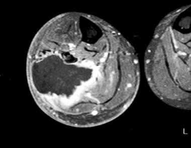
Desmoplastic fibroma is a rare primary tumor of bone that histologically and biologically mimics the extra-abdominal desmoid tumor of soft tissue. It usually presents in patients during the first three decades of life and often involves the mandible or long bones of the skeleton. Its clinical behavior is characterized by a locally aggressive, infiltrating, and destructing course, often with invasion of surrounding tissues but without metastasis. We present herein the clinicopathological features of a desmoplastic fibroma-like tumor involving the left maxillofacial region in a 14-year-old Hispanic boy with tuberous sclerosis. (+info)Desmoplastic fibroma of the cervical spine. (4/16)
There have been only a few cases of desmoplastic fibroma of the spine in the literature and only one of them was purely located on the cervical spine. We report a new patient with the diagnosis of desmoplastic fibroma of the fourth cervical spine. The patient had the complaints of left arm and neck pain. After his radiological evaluation, a mass lesion was found on the left lamina of the fourth cervical spine. Surgical treatment was performed, and the histopathological examination revealed the diagnosis of desmoplastic fibroma. Patients with desmoplastic fibroma of the cervical spine may present with the arm and neck pain mimicking cervical disc disease. Higher index of suspicion by the clinicians must be practiced to make the appropriate diagnosis. Successful surgical outcome may be achieved in these patients. (+info)Case report of intra-osseous fibroma: a study on odontogenic and desmoplastic fibromas with a review of the literature. (5/16)
Intra-osseous fibromas of the jaw are classified by origin. Intra-osseous odontogenic fibromas have odontogenic epithelia, while desmoplastic fibromas do not. However, it is often difficult to determine the odontogenic origin for central fibromas. Three subjects with a diagnosis of intra-osseous fibroma were examined. Case 1 was a 35-year-old man found to have a panoramic radiograph from the right premolar to the mandibular ramus in the mandible that exhibited multilocular radiolucency. Within the radiolucency, small-radioopaque bodies were observed. Case 2 was a 13-year-old female, in whom a panoramic radiograph from the left premolar to the molar in the mandible showed multilocular radiolucency. Case 3 was a 51-year-old female who exhibited a heart-shaped radiolucency in the panoramic radiograph of the left first molar area in the mandible. We also reviewed the literature for previously reported cases of intra-osseous odontogenic and desmoplastic fibroma. In 64 cases of intra-osseous odontogenic fibroma and 68 cases of desmoplastic fibroma we extracted data on age, sex, location, and radiographic findings. Based on the analysis of the reported literature cases, re-evaluation of the patients in our study revealed that case 1 could be classified as desmoplastic fibroma, while cases 2 and 3 were intra-osseous odontogenic fibromas. (+info)Desmoplastic fibroma of the frontal bone. (6/16)
Desmoplastic fibroma is a benign but locally aggressive tumor arising usually from the mandible, pelvis and long bones with a potential for recurrence. We report a case of desmoplastic fibroma of the frontal bone in a young male. (+info)Desmoplastic fibroma of the mandible--review of the literature and presentation of a rare case. (7/16)
(+info)Desmoplastic fibroblastoma (collagenous fibroma): a case identified in the buccal mucosa. (8/16)
(+info)A fibroma is a benign (non-cancerous) tumor that consists primarily of fibrous or connective tissue. It can occur in various parts of the body, including the skin, mouth, and internal organs. The term "fibroma" is often used to describe any benign fibrous growth, but there are specific types of fibromas such as dermatofibroma (found in the skin), oral fibroma (found in the mouth), and benign fibrous histiocytoma (found in soft tissues).
It's important to note that while fibromas are generally harmless, they can cause discomfort or problems depending on their size and location. If a fibroma is causing issues or there's concern about its growth or malignancy, it should be evaluated by a healthcare professional for potential removal or further assessment.
A fibroma, ossifying is a benign (non-cancerous) tumor that typically develops in the periodontal ligament, which is the tissue that connects the tooth to the jawbone. This type of fibroma is characterized by the formation of bone-like tissue within the tumor. It usually appears as a firm, slow-growing nodule or mass that can cause pain or discomfort, particularly when biting down on the affected tooth.
The exact cause of ossifying fibromas is not well understood, but they are thought to arise from an overgrowth of cells in the periodontal ligament. They are more common in women than men and typically occur in people between the ages of 20 and 40. Treatment usually involves surgical removal of the tumor, along with any affected tissue or teeth. In some cases, recurrence may occur, so regular follow-up appointments with a dental professional are recommended.
Desmoplastic fibroma is a very rare benign (non-cancerous) tumor of the connective tissue. It typically develops in the bones, but can also occur in soft tissues. The tumor is characterized by the overgrowth of collagen-producing cells (fibroblasts), leading to the formation of a firm, fibrous mass. Desmoplastic fibromas are slow-growing and typically do not spread to other parts of the body (metastasize). However, they can cause significant damage to the affected bone or tissue as they grow, potentially leading to fractures or deformities. Treatment usually involves surgical removal of the tumor.
A Desmoplastic Small Round Cell Tumor (DSRCT) is a rare and aggressive type of cancer that primarily affects young adults, typically between the ages of 10 to 30. It is characterized by the presence of small round tumor cells that are surrounded by a dense fibrous or desmoplastic stroma.
DSRCTs usually originate in the abdominal cavity, particularly in the peritoneum, which is the membrane that lines the abdominal wall and covers the organs within it. However, they can also occur in other parts of the body such as the lungs, mediastinum, and pelvis.
The tumor cells in DSRCTs typically have a specific chromosomal abnormality known as the t(11;22)(p13;q12) translocation, which results in the fusion of two genes - EWSR1 and WT1. This genetic alteration is thought to contribute to the development and progression of DSRCTs.
DSRCTs are highly aggressive tumors that tend to spread rapidly throughout the body, making them difficult to treat. Treatment options typically include a combination of surgery, chemotherapy, and radiation therapy, although the prognosis for patients with DSRCTs is generally poor, with a five-year survival rate of less than 15%.
Abdominal neoplasms refer to abnormal growths or tumors in the abdomen that can be benign (non-cancerous) or malignant (cancerous). These growths can occur in any of the organs within the abdominal cavity, including the stomach, small intestine, large intestine, liver, pancreas, spleen, and kidneys.
Abdominal neoplasms can cause various symptoms depending on their size, location, and type. Some common symptoms include abdominal pain or discomfort, bloating, changes in bowel habits, unexplained weight loss, fatigue, and fever. In some cases, abdominal neoplasms may not cause any symptoms until they have grown quite large or spread to other parts of the body.
The diagnosis of abdominal neoplasms typically involves a combination of physical exam, medical history, imaging studies such as CT scans or MRIs, and sometimes biopsy to confirm the type of tumor. Treatment options depend on the type, stage, and location of the neoplasm but may include surgery, radiation therapy, chemotherapy, targeted therapy, or a combination of these approaches.
Mandibular neoplasms refer to abnormal growths or tumors that develop in the mandible, which is the lower jawbone. These growths can be benign (non-cancerous) or malignant (cancerous). Benign neoplasms are typically slow-growing and rarely spread to other parts of the body, while malignant neoplasms can invade surrounding tissues and may metastasize (spread) to distant sites.
Mandibular neoplasms can have various causes, including genetic mutations, exposure to certain chemicals or radiation, and infection with certain viruses. The symptoms of mandibular neoplasms may include swelling or pain in the jaw, difficulty chewing or speaking, numbness in the lower lip or chin, loose teeth, and/or a lump or mass in the mouth or neck.
The diagnosis of mandibular neoplasms typically involves a thorough clinical examination, imaging studies such as X-rays, CT scans, or MRI scans, and sometimes a biopsy to confirm the type and extent of the tumor. Treatment options depend on the type, stage, and location of the neoplasm, and may include surgery, radiation therapy, chemotherapy, or a combination of these approaches. Regular follow-up care is essential to monitor for recurrence or metastasis.
Ganglioglioma is a rare, typically slow-growing tumor that occurs in the brain or spinal cord. It is composed of both neuronal (ganglion cell) and glial elements. These tumors most commonly occur in the temporal lobe of the brain and are usually found in children and young adults.
Gangliogliomas can be benign or malignant, with the majority being low-grade (benign). Symptoms vary depending on the location of the tumor but may include seizures, headaches, changes in behavior or cognition, and motor weakness or paralysis. Treatment typically involves surgical removal of the tumor, and in some cases, radiation therapy or chemotherapy may be recommended.
It's important to note that while I strive to provide accurate information, my responses should not be used as a substitute for professional medical advice, diagnosis, or treatment. Always consult with a qualified healthcare provider for any medical concerns.
Maxillary neoplasms refer to abnormal growths or tumors in the maxilla, which is the upper jaw bone. These growths can be benign (non-cancerous) or malignant (cancerous). Benign neoplasms are slow-growing and do not spread to other parts of the body, while malignant neoplasms can invade surrounding tissues and spread to distant sites.
Maxillary neoplasms can cause various symptoms such as swelling, pain, numbness, loose teeth, or difficulty in chewing or swallowing. They may also cause nasal congestion, nosebleeds, or visual changes if they affect the eye or orbit. The diagnosis of maxillary neoplasms usually involves a combination of clinical examination, imaging studies such as CT or MRI scans, and biopsy to determine the type and extent of the tumor.
Treatment options for maxillary neoplasms depend on several factors, including the type, size, location, and stage of the tumor, as well as the patient's overall health and preferences. Treatment may include surgery, radiation therapy, chemotherapy, or a combination of these modalities. Regular follow-up care is essential to monitor for recurrence or metastasis and ensure optimal outcomes.
Jaw neoplasms refer to abnormal growths or tumors in the jawbone (mandible) or maxilla (upper jaw). These growths can be benign (non-cancerous) or malignant (cancerous). Benign neoplasms are not considered life-threatening, but they can still cause problems by invading nearby tissues and causing damage. Malignant neoplasms, on the other hand, can spread to other parts of the body and can be life-threatening if not treated promptly and effectively.
Jaw neoplasms can present with various symptoms such as swelling, pain, loose teeth, numbness or tingling in the lips or tongue, difficulty chewing or swallowing, and jaw stiffness or limited movement. The diagnosis of jaw neoplasms typically involves a thorough clinical examination, imaging studies such as X-rays, CT scans, or MRI, and sometimes a biopsy to determine the type and extent of the tumor.
Treatment options for jaw neoplasms depend on several factors, including the type, size, location, and stage of the tumor, as well as the patient's overall health and medical history. Treatment may involve surgery, radiation therapy, chemotherapy, or a combination of these modalities. Regular follow-up care is essential to monitor for recurrence or metastasis (spread) of the neoplasm.
Ameloblastoma is a slow-growing, non-cancerous tumor that develops in the jawbone, typically in the lower jaw. It originates from the cells that form the enamel (the hard, outer surface of the teeth). This tumor can cause swelling, pain, and displacement or loosening of teeth. In some cases, it may also lead to fractures of the jawbone.
There are different types of ameloblastomas, including solid or multicystic, unicystic, and peripheral ameloblastoma. Treatment usually involves surgical removal of the tumor, with careful monitoring to ensure that it does not recur. In rare cases, more aggressive treatment may be necessary if the tumor is large or has invaded surrounding tissues.
It's important to note that while ameloblastomas are generally benign, they can still cause significant morbidity and should be treated promptly by an oral and maxillofacial surgeon or other qualified healthcare professional.
Fibrous Dysplasia of Bone is a rare, benign bone disorder that is characterized by the replacement of normal bone tissue with fibrous (scar-like) and immature bone tissue. This results in weakened bones that are prone to fractures, deformities, and pain. The condition can affect any bone in the body but most commonly involves the long bones of the legs, arms, and skull. It can occur as an isolated finding or as part of a genetic disorder called McCune-Albright syndrome. The exact cause of fibrous dysplasia is not fully understood, but it is believed to result from a genetic mutation that occurs during early bone development. There is no cure for fibrous dysplasia, and treatment typically focuses on managing symptoms and preventing complications.
Odontogenic tumors are a group of neoplasms that originate from the dental tissues or their remnants, including the odontogenic epithelium, ectomesenchyme, and/or their derivatives. These tumors can be benign or malignant and may affect the jaw bones and surrounding structures. They can cause various symptoms, such as swelling, pain, loosening of teeth, and altered bite. The classification of odontogenic tumors includes a wide range of entities with different biological behaviors, clinical features, and treatment approaches. Accurate diagnosis is essential for proper management and prognosis.
Small cell sarcoma is a very rare and aggressive type of cancer that affects the connective tissues in the body, such as muscles, tendons, bones, cartilage, and fat. It is called "small cell" because the cancer cells are small and appear round or oval in shape, with scant cytoplasm and finely granular chromatin.
Small cell sarcoma typically occurs in adults between the ages of 40 and 70, and it can develop in any part of the body. However, it is most commonly found in the extremities, trunk, and retroperitoneum. The exact cause of small cell sarcoma is not known, but it is thought to be associated with genetic mutations that occur during a person's lifetime.
Small cell sarcoma can be difficult to diagnose because it often does not cause any symptoms until it has advanced to an aggressive stage. When symptoms do occur, they may include pain, swelling, or a lump in the affected area. Diagnosis typically involves a biopsy of the tumor tissue, followed by imaging tests such as CT scans, MRI scans, or PET scans to determine the extent of the cancer.
Treatment for small cell sarcoma usually involves surgery to remove the tumor, followed by radiation therapy and/or chemotherapy to kill any remaining cancer cells. However, because small cell sarcoma is so rare and aggressive, treatment options may be limited, and the prognosis is often poor. Clinical trials of new treatments are also an option for some patients.
Cerebellar neoplasms refer to abnormal growths or tumors that develop in the cerebellum, which is the part of the brain responsible for coordinating muscle movements and maintaining balance. These tumors can be benign (non-cancerous) or malignant (cancerous), and they can arise from various types of cells within the cerebellum.
The most common type of cerebellar neoplasm is a medulloblastoma, which arises from primitive nerve cells in the cerebellum. Other types of cerebellar neoplasms include astrocytomas, ependymomas, and brain stem gliomas. Symptoms of cerebellar neoplasms may include headaches, vomiting, unsteady gait, coordination problems, and visual disturbances. Treatment options depend on the type, size, and location of the tumor, as well as the patient's overall health and age. Treatment may involve surgery, radiation therapy, chemotherapy, or a combination of these approaches.
Medulloblastoma is a type of malignant brain tumor that originates in the cerebellum, which is the part of the brain located at the back of the skull and controls coordination and balance. It is one of the most common types of pediatric brain tumors, although it can also occur in adults.
Medulloblastomas are typically made up of small, round cancer cells that grow quickly and can spread to other parts of the central nervous system, such as the spinal cord. They are usually treated with a combination of surgery, radiation therapy, and chemotherapy. The exact cause of medulloblastoma is not known, but it is thought to be related to genetic mutations or abnormalities that occur during development.
Poxviridae is a family of large, complex, double-stranded DNA viruses that includes many significant pathogens affecting humans and animals. The most well-known member of this family is the Variola virus, which causes smallpox in humans, a highly contagious and deadly disease that has been eradicated through global vaccination efforts. Other important human pathogens in this family include the Monkeypox virus, which can cause a smallpox-like illness, and the Molluscum contagiosum virus, which causes benign skin tumors.
Poxviruses have a unique ability to replicate in the cytoplasm of host cells, rather than in the nucleus like many other DNA viruses. They also have a complex structure, with a large, brick-shaped virion that contains a lateral body, a core, and an outer envelope. The genome of poxviruses is relatively large, ranging from 130 to 375 kilobases in length, and encodes many genes involved in viral replication, host immune evasion, and modulation of host cell processes.
Poxviridae is further divided into two subfamilies: Chordopoxvirinae, which includes viruses that infect vertebrates, and Entomopoxvirinae, which includes viruses that infect insects. The Chordopoxvirinae subfamily is divided into several genera, including Orthopoxvirus (which includes Variola, Monkeypox, and Vaccinia viruses), Parapoxvirus (which includes Orf virus and Bovine papular stomatitis virus), and Yatapoxvirus (which includes Yaba monkey tumor virus and Tanapox virus).
Overall, Poxviridae is a diverse family of viruses that pose significant public health and agricultural threats, and continue to be the subject of ongoing research and development efforts aimed at understanding their biology and developing new vaccines and therapies.
Gingival neoplasms refer to abnormal growths or tumors that occur in the gingiva, which are the part of the gums that surround the teeth. These growths can be benign (non-cancerous) or malignant (cancerous). Benign neoplasms include conditions such as fibromas, papillomas, and hemangiomas, while malignant neoplasms are typically squamous cell carcinomas.
Gingival neoplasms can present with a variety of symptoms, including swelling, bleeding, pain, and loose teeth. They may also cause difficulty with chewing, speaking, or swallowing. The exact cause of these neoplasms is not always known, but risk factors include tobacco use, alcohol consumption, poor oral hygiene, and certain viral infections.
Diagnosis of gingival neoplasms typically involves a thorough clinical examination, including a dental exam and biopsy. Treatment options depend on the type and stage of the neoplasm, but may include surgery, radiation therapy, chemotherapy, or a combination of these approaches. Regular dental check-ups and good oral hygiene practices can help to detect gingival neoplasms at an early stage and improve treatment outcomes.
Cementoma is a benign (non-cancerous) tumor that primarily affects the jaw bones, particularly the lower jaw (mandible). It is characterized by the growth of abnormal cementum-like tissue within the bone. Cementum is a hard tissue that covers the roots of teeth and helps anchor them to the jawbone.
There are different types of cementomas, including:
1. Periapical cemental dysplasia (PCD): This type of cementoma usually affects the anterior region of the lower jaw and is often associated with non-vital teeth. It typically presents as a small, radiopaque (dark) area on an X-ray.
2. Florid cemento-osseous dysplasia (FCOD): FCOD is a more widespread form of cementoma that affects multiple areas of the jawbones. It primarily affects middle-aged women and can cause significant bone remodeling, leading to radiopaque lesions on X-rays.
3. Gigantiform cementoma: This rare, aggressive type of cementoma typically affects children and adolescents. It can cause rapid bone growth and expansion, resulting in facial deformities and functional impairments.
4. Ossifying fibroma: Although not strictly a cementoma, ossifying fibroma shares some similarities with these tumors. It is characterized by the formation of both bone and cementum-like tissue within the lesion.
Treatment for cementomas depends on their size, location, and growth rate. Small, asymptomatic lesions may not require treatment, while larger or symptomatic ones might need surgical removal to prevent complications such as tooth displacement, infection, or pathological fractures. Regular follow-ups with dental X-rays are essential to monitor the progression of these lesions.
Collagen type XII is a type of collagen that is found in the extracellular matrix of various tissues, including tendons, ligaments, and skin. It is a fibril-associated collagen that is closely associated with collagens type I and III. Collagen type XII has been shown to play a role in regulating the organization and diameter of collagen fibrils. Mutations in the gene for collagen type XII have been associated with certain types of muscular dystrophy and Bethlem myopathy, which are genetic disorders that affect muscle strength and tone. Additionally, it has been suggested to play a role in the development of osteoarthritis.
A chondroma is a benign, slow-growing tumor that develops in the cartilage. Cartilage is a type of connective tissue found in various parts of the body, including the joints, ribcage, and nose. Chondromas are most commonly found in the hands and feet.
Chondromas are typically small, measuring less than 2 centimeters in diameter, and they usually do not cause any symptoms. However, if a chondroma grows large enough to press on nearby nerves or blood vessels, it may cause pain, numbness, or weakness in the affected area.
Chondromas are usually diagnosed through imaging tests such as X-rays, CT scans, or MRI scans. If a chondroma is suspected based on these tests, a biopsy may be performed to confirm the diagnosis and rule out other types of tumors.
Treatment for chondromas typically involves surgical removal of the tumor. In most cases, this can be done using minimally invasive techniques that allow for quicker recovery times. After surgery, patients will need to follow up with their healthcare provider to ensure that the tumor has been completely removed and to monitor for any signs of recurrence.
A thecoma is a type of ovarian sex cord-stromal tumor, which are rare tumors that develop from the supporting cells (stromal cells) or the cells that produce hormones (sex cord cells) in the ovary. These tumors account for about 2% of all ovarian tumors.
Thecomas specifically arise from stromal cells that produce estrogen and other sex hormones. They are typically slow-growing and may not cause any symptoms, or they may cause symptoms related to hormonal imbalances such as irregular menstrual periods, vaginal bleeding, or postmenopausal bleeding. In some cases, thecomas can also grow large enough to cause abdominal discomfort or bloating.
Most thecomas are benign (non-cancerous), but a small percentage of them can be malignant (cancerous) and may spread to other parts of the body. Treatment for thecomas typically involves surgical removal of the tumor, and in some cases, hormonal therapy or chemotherapy may also be recommended.
Panoramic radiography is a specialized type of dental X-ray imaging that captures a panoramic view of the entire mouth, including the teeth, upper and lower jaws, and surrounding structures. It uses a special machine that rotates around the head, capturing images as it moves. This technique provides a two-dimensional image that is helpful in diagnosing and planning treatment for various dental conditions such as impacted teeth, bone abnormalities, and jaw disorders.
The panoramic radiograph can also be used to assess the development and positioning of wisdom teeth, detect cysts or tumors in the jaws, and evaluate the effects of trauma or injury to the mouth. It is a valuable tool for dental professionals as it allows them to see a comprehensive view of the oral structures, which may not be visible with traditional X-ray techniques.
It's important to note that while panoramic radiography provides valuable information, it should be used in conjunction with other diagnostic tools and clinical examinations to ensure accurate diagnosis and treatment planning.
Neuroectodermal tumors, primitive (PNETs) are a group of highly malignant and aggressive neoplasms that arise from neuroectodermal cells, which are the precursors to the nervous system during embryonic development. These tumors can occur anywhere in the body but are most commonly found in the central nervous system, particularly in the brain and spinal cord.
PNETs are characterized by small, round, blue cells that have a high degree of cellularity and mitotic activity. They are composed of undifferentiated or poorly differentiated cells that can differentiate along various neural lineages, including neuronal, glial, and epithelial. This feature makes their diagnosis challenging, as they can resemble other small round blue cell tumors, such as lymphomas, rhabdomyosarcomas, and Ewing sarcoma.
Immunohistochemical staining and molecular genetic testing are often required to confirm the diagnosis of PNETs. These tests typically reveal the expression of neural markers, such as NSE, Synaptophysin, and CD99, and the presence of specific chromosomal abnormalities, such as the EWS-FLI1 fusion gene in Ewing sarcoma.
PNETs are aggressive tumors with a poor prognosis, and their treatment typically involves a multimodal approach that includes surgery, radiation therapy, and chemotherapy. Despite these treatments, the five-year survival rate for patients with PNETs is less than 30%.
Bone neoplasms are abnormal growths or tumors that develop in the bone. They can be benign (non-cancerous) or malignant (cancerous). Benign bone neoplasms do not spread to other parts of the body and are rarely a threat to life, although they may cause problems if they grow large enough to press on surrounding tissues or cause fractures. Malignant bone neoplasms, on the other hand, can invade and destroy nearby tissue and may spread (metastasize) to other parts of the body.
There are many different types of bone neoplasms, including:
1. Osteochondroma - a benign tumor that develops from cartilage and bone
2. Enchondroma - a benign tumor that forms in the cartilage that lines the inside of the bones
3. Chondrosarcoma - a malignant tumor that develops from cartilage
4. Osteosarcoma - a malignant tumor that develops from bone cells
5. Ewing sarcoma - a malignant tumor that develops in the bones or soft tissues around the bones
6. Giant cell tumor of bone - a benign or occasionally malignant tumor that develops from bone tissue
7. Fibrosarcoma - a malignant tumor that develops from fibrous tissue in the bone
The symptoms of bone neoplasms vary depending on the type, size, and location of the tumor. They may include pain, swelling, stiffness, fractures, or limited mobility. Treatment options depend on the type and stage of the tumor but may include surgery, radiation therapy, chemotherapy, or a combination of these treatments.
Leporipoxvirus is a genus of viruses in the Poxviridae family, which includes double-stranded DNA viruses. This genus primarily consists of pathogens that infect rabbits and hares. Two well-known examples of Leporipoxviruses are myxoma virus and rabbit (hare) fibroma virus.
1. Myxoma Virus: It is the causative agent of myxomatosis, a often fatal disease in European rabbits (Oryctolagus cuniculus). The virus is transmitted through insect vectors, primarily mosquitoes and fleas. Infected rabbits develop skin lesions, swelling around the eyes and genitals, and eventually die due to internal organ failure.
2. Rabbit (Hare) Fibroma Virus: This Leporipoxvirus causes benign tumors called fibromas in rabbits and hares. The tumors typically develop on the skin or mucous membranes but can also occur internally. While these growths are not fatal, they can cause significant stress and discomfort for affected animals.
It is important to note that Leporipoxviruses do not pose a direct threat to humans as they primarily infect rabbits and hares. However, researchers study these viruses due to their potential applications in cancer therapy and vaccine development.
Oncogenic viruses are a type of viruses that have the ability to cause cancer in host cells. They do this by integrating their genetic material into the DNA of the infected host cell, which can lead to the disruption of normal cellular functions and the activation of oncogenes (genes that have the potential to cause cancer). This can result in uncontrolled cell growth and division, ultimately leading to the formation of tumors. Examples of oncogenic viruses include human papillomavirus (HPV), hepatitis B virus (HBV), and human T-cell leukemia virus type 1 (HTLV-1). It is important to note that only a small proportion of viral infections lead to cancer, and the majority of cancers are not caused by viruses.
Ewing Sarcoma (EWS) RNA-Binding Protein, also known as EWSR1, is a protein that plays a role in gene expression by binding to RNA. It is a member of the FET family of proteins, which also includes FUS and TAF15. These proteins are involved in various cellular processes such as transcription, splicing, and translation.
Mutations in the EWSR1 gene have been associated with several types of cancer, most notably Ewing sarcoma, a rare tumor that typically affects children and adolescents. In Ewing sarcoma, a fusion protein is formed when EWSR1 combines with another protein, most commonly ETS translocation variant 1 (ETV1), FLI1, ERG or FEV. This fusion protein can lead to abnormal gene expression and tumor formation.
EWSR1 has also been found to be involved in other types of cancer such as acute myeloid leukemia, clear cell sarcoma, desmoplastic small round cell tumors and liposarcomas.
It's important to note that while EWSR1 is a RNA-binding protein, it can also bind to DNA in certain contexts, such as when it forms a fusion protein with an ETS transcription factor in Ewing sarcoma.
Hutchinson's melanotic freckle, also known as Hutchinson's melanotic macule or naevus, is a type of pigmented lesion that can be a precursor to malignant melanoma, a serious form of skin cancer. It is typically characterized by the presence of darkly pigmented, irregularly shaped patches on the skin, often found on the face or neck.
The lesions are usually brown or black in color and may have an uneven border or surface. They can vary in size from a few millimeters to several centimeters in diameter. Hutchinson's melanotic freckles are typically larger, darker, and more irregularly shaped than common freckles.
These lesions are named after Sir Jonathan Hutchinson, an English surgeon and pathologist who first described them in the late 19th century. It is important to note that while Hutchinson's melanotic freckles can be a sign of increased risk for developing melanoma, not all such lesions will become cancerous. However, any changes in size, shape, or color of these lesions should be evaluated by a healthcare professional as soon as possible.
Neoplasms, adnexal and skin appendage refer to abnormal growths or tumors that develop in the sweat glands, hair follicles, or other structures associated with the skin. These growths can be benign (non-cancerous) or malignant (cancerous), and they can occur anywhere on the body.
Adnexal neoplasms are tumors that arise from the sweat glands or hair follicles, including the sebaceous glands, eccrine glands, and apocrine glands. These tumors can range in size and severity, and they may cause symptoms such as pain, itching, or changes in the appearance of the skin.
Skin appendage neoplasms are similar to adnexal neoplasms, but they specifically refer to tumors that arise from structures such as hair follicles, nails, and sweat glands. Examples of skin appendage neoplasms include pilomatricomas (tumors of the hair follicle), trichilemmomas (tumors of the outer root sheath of the hair follicle), and sebaceous adenomas (tumors of the sebaceous glands).
It is important to note that while many adnexal and skin appendage neoplasms are benign, some can be malignant and may require aggressive treatment. If you notice any unusual growths or changes in your skin, it is important to consult with a healthcare professional for further evaluation and care.
Wilms' Tumor 1 (WT1) proteins are a group of transcription factors that play crucial roles in the development of the human body, particularly in the formation of the urinary and reproductive systems. The WT1 gene encodes these proteins, and mutations in this gene have been associated with several diseases, most notably Wilms' tumor, a type of kidney cancer in children.
WT1 proteins contain four domains: an N-terminal transcriptional activation domain, a zinc finger domain that binds to DNA, a nuclear localization signal, and a C-terminal transcriptional repression domain. These proteins regulate the expression of various target genes involved in cell growth, differentiation, and apoptosis (programmed cell death).
Abnormalities in WT1 protein function or expression have been linked to several developmental disorders, including Denys-Drash syndrome, Frasier syndrome, and Wilms' tumor. These conditions are characterized by genitourinary abnormalities, such as kidney dysplasia, ambiguous genitalia, and an increased risk of developing Wilms' tumor.
Ewing sarcoma is a type of cancer that originates in bones or the soft tissues surrounding them, such as muscles and tendons. It primarily affects children and adolescents, although it can occur in adults as well. The disease is characterized by small, round tumor cells that typically grow quickly and are prone to metastasize (spread) to other parts of the body, most commonly the lungs, bones, and bone marrow.
Ewing sarcoma is caused by a genetic abnormality, specifically a chromosomal translocation that results in the fusion of two genes, EWSR1 and FLI1. This gene fusion leads to the formation of an abnormal protein that disrupts normal cell growth and division processes, ultimately resulting in cancer.
Symptoms of Ewing sarcoma can vary depending on the location and size of the tumor but may include pain or swelling in the affected area, fever, fatigue, and weight loss. Diagnosis typically involves imaging studies such as X-rays, CT scans, or MRI scans to locate the tumor, followed by a biopsy to confirm the presence of cancer cells. Treatment may involve surgery, radiation therapy, chemotherapy, or a combination of these approaches, depending on the stage and location of the disease.
Basal Cell Nevus Syndrome (BCNS), also known as Gorlin-Goltz Syndrome, is a rare genetic disorder that is characterized by the development of multiple basal cell carcinomas (BCCs), which are skin cancer tumors that arise from the basal cells in the outermost layer of the skin.
The syndrome is caused by mutations in the PTCH1 gene, which regulates the hedgehog signaling pathway involved in embryonic development and tissue growth regulation. The condition is inherited in an autosomal dominant manner, meaning that a child has a 50% chance of inheriting the mutated gene from an affected parent.
Individuals with BCNS typically develop hundreds to thousands of BCCs over their lifetime, often beginning in childhood or adolescence. They may also have other benign and malignant tumors, such as medulloblastomas (brain tumors), fibromas, and rhabdomyosarcomas.
Additional features of BCNS can include:
1. Facial abnormalities, such as a broad nasal bridge, widely spaced eyes, and pits or depressions on the palms and soles.
2. Skeletal abnormalities, such as spine deformities, rib anomalies, and jaw cysts.
3. Developmental delays and intellectual disabilities in some cases.
4. Increased risk of other cancers, including breast, ovarian, and lung cancer.
Early detection and management of BCCs and other tumors are crucial for individuals with BCNS to prevent complications and improve their quality of life. Regular dermatological examinations, sun protection measures, and surgical removal of tumors are common treatment approaches.
Chondroblastoma is a rare, benign (non-cancerous) bone tumor that typically develops in the epiphysis, which is the rounded end of a long bone near a joint. It primarily affects children and adolescents, with around 90% of cases occurring before the age of 20.
The tumor arises from chondroblasts, cells responsible for producing cartilage during bone growth. Chondroblastoma is usually slow-growing and typically causes localized pain, swelling, or tenderness in the affected area. In some cases, it may weaken the bone and lead to fractures.
Treatment generally involves surgical removal of the tumor, followed by curettage (scraping) of the surrounding bone tissue and replacement with bone grafts or substitutes. Recurrence is possible but rare, and long-term prognosis is usually favorable.
Skin neoplasms refer to abnormal growths or tumors in the skin that can be benign (non-cancerous) or malignant (cancerous). They result from uncontrolled multiplication of skin cells, which can form various types of lesions. These growths may appear as lumps, bumps, sores, patches, or discolored areas on the skin.
Benign skin neoplasms include conditions such as moles, warts, and seborrheic keratoses, while malignant skin neoplasms are primarily classified into melanoma, squamous cell carcinoma, and basal cell carcinoma. These three types of cancerous skin growths are collectively known as non-melanoma skin cancers (NMSCs). Melanoma is the most aggressive and dangerous form of skin cancer, while NMSCs tend to be less invasive but more common.
It's essential to monitor any changes in existing skin lesions or the appearance of new growths and consult a healthcare professional for proper evaluation and treatment if needed.
Osteoma is a benign (noncancerous) tumor that is made up of mature bone tissue. It usually grows slowly over a period of years and is most commonly found in the skull or jaw, although it can occur in other bones of the body as well. Osteomas are typically small, but they can grow to be several centimeters in size. They may cause symptoms if they press on nearby tissues or structures, such as nerves or blood vessels. In some cases, osteomas may not cause any symptoms and may only be discovered during routine imaging studies. Treatment for osteoma is typically not necessary unless it is causing problems or growing rapidly. If treatment is needed, it may involve surgical removal of the tumor.
Carcinoma, small cell is a type of lung cancer that typically starts in the bronchi (the airways that lead to the lungs). It is called "small cell" because the cancer cells are small and appear round or oval in shape. This type of lung cancer is also sometimes referred to as "oat cell carcinoma" due to the distinctive appearance of the cells, which can resemble oats when viewed under a microscope.
Small cell carcinoma is a particularly aggressive form of lung cancer that tends to spread quickly to other parts of the body. It is strongly associated with smoking and is less common than non-small cell lung cancer (NSCLC), which accounts for about 85% of all lung cancers.
Like other types of lung cancer, small cell carcinoma may not cause any symptoms in its early stages. However, as the tumor grows and spreads, it can cause a variety of symptoms, including coughing, chest pain, shortness of breath, hoarseness, and weight loss. Treatment for small cell carcinoma typically involves a combination of chemotherapy, radiation therapy, and sometimes surgery.
Palatal neoplasms refer to abnormal growths or tumors that occur on the palate, which is the roof of the mouth. These growths can be benign (non-cancerous) or malignant (cancerous). Benign neoplasms are typically slower growing and less likely to spread, while malignant neoplasms are more aggressive and can invade nearby tissues and organs.
Palatal neoplasms can have various causes, including genetic factors, environmental exposures, and viral infections. They may present with symptoms such as mouth pain, difficulty swallowing, swelling or lumps in the mouth, bleeding, or numbness in the mouth or face.
The diagnosis of palatal neoplasms typically involves a thorough clinical examination, imaging studies, and sometimes biopsy to determine the type and extent of the growth. Treatment options depend on the type, size, location, and stage of the neoplasm but may include surgery, radiation therapy, chemotherapy, or a combination of these approaches. Regular follow-up care is essential to monitor for recurrence or spread of the neoplasm.
Paraparesis, spastic type, is a medical term used to describe a condition characterized by partial weakness or loss of voluntary movement in the lower extremities (legs). The term "paraparesis" comes from Greek words "para" meaning beside or beyond, and "paresis" meaning loosening or relaxation.
In spastic paraparesis, the muscle tone is increased, causing stiffness and resistance to movement, particularly during quick or forceful movements. This increased muscle tone, also known as spasticity, results from an upper motor neuron lesion in the brain or spinal cord that affects the corticospinal tract, which carries signals from the brain to the muscles.
Spastic paraparesis can be caused by various conditions, including spinal cord injuries, multiple sclerosis, hereditary spastic paraplegia, and stroke, among others. The severity of symptoms may vary widely, ranging from mild weakness to complete paralysis. Treatment options for spastic paraparesis depend on the underlying cause and may include physical therapy, medications, surgery, or a combination of these approaches.
Peritoneal neoplasms refer to tumors or cancerous growths that develop in the peritoneum, which is the thin, transparent membrane that lines the inner wall of the abdomen and covers the organs within it. These neoplasms can be benign (non-cancerous) or malignant (cancerous). Malignant peritoneal neoplasms are often associated with advanced stages of gastrointestinal, ovarian, or uterine cancers and can spread (metastasize) to other parts of the abdomen.
Peritoneal neoplasms can cause various symptoms such as abdominal pain, bloating, nausea, vomiting, loss of appetite, and weight loss. Diagnosis typically involves imaging tests like CT scans or MRIs, followed by a biopsy to confirm the presence of cancerous cells. Treatment options may include surgery, chemotherapy, radiation therapy, or a combination of these approaches, depending on the type, stage, and location of the neoplasm.
Pancreatic stellate cells (PSCs) are adult, tissue-specific mesenchymal cells that are found in the exocrine portion of the pancreas. They are star-shaped and are located in the periacinar area, where they normally remain quiescent. However, in response to injury or inflammation, such as in chronic pancreatitis or pancreatic cancer, PSCs become activated and transform into a myofibroblast-like phenotype.
Activated PSCs play a key role in the pathogenesis of pancreatic fibrosis, which is characterized by an excessive accumulation of extracellular matrix (ECM) proteins, such as collagen and fibronectin. This process can lead to the destruction of the normal pancreatic architecture and function. Activated PSCs also produce various growth factors and cytokines that promote the growth and survival of pancreatic cancer cells, contributing to the aggressive behavior of this disease.
Overall, PSCs play a critical role in the development and progression of pancreatic diseases, making them an important target for therapeutic intervention.
Rhabdomyoma is a rare, benign tumor that arises from the striated muscle tissue, which is the type of muscle that enables movement and action in the body. These tumors most commonly occur in the heart (cardiac rhabdomyomas) or in the head and neck region (extracardiac rhabdomyomas). Cardiac rhabdomyomas are often associated with genetic disorders such as tuberous sclerosis complex, while extracardiac rhabdomyomas can be found in various locations like the skin, tongue, or skeletal muscles.
Cardiac rhabdomyomas typically appear in infancy or early childhood and may not cause any symptoms. However, they can potentially lead to complications such as heart rhythm abnormalities, obstruction of blood flow, or heart failure. Extracardiac rhabdomyomas are usually slow-growing and asymptomatic but can cause issues depending on their size and location. Surgical removal may be necessary if the tumor interferes with vital functions or causes discomfort.
It is essential to note that while rhabdomyomas are generally benign, they can undergo malignant transformation in rare cases, leading to a more aggressive form called rhabdomyosarcoma. Regular follow-ups and monitoring are crucial for early detection and management of any changes in the tumor's behavior.
Immunohistochemistry (IHC) is a technique used in pathology and laboratory medicine to identify specific proteins or antigens in tissue sections. It combines the principles of immunology and histology to detect the presence and location of these target molecules within cells and tissues. This technique utilizes antibodies that are specific to the protein or antigen of interest, which are then tagged with a detection system such as a chromogen or fluorophore. The stained tissue sections can be examined under a microscope, allowing for the visualization and analysis of the distribution and expression patterns of the target molecule in the context of the tissue architecture. Immunohistochemistry is widely used in diagnostic pathology to help identify various diseases, including cancer, infectious diseases, and immune-mediated disorders.
Rhabdomyosarcoma, embryonal is a type of soft tissue sarcoma, which is a cancer that develops in the body's connective tissues, such as muscles, tendons, ligaments, and cartilage. Specifically, embryonal rhabdomyosarcoma is a subtype of rhabdomyosarcoma that arises from cells that are in the process of becoming muscle cells. This type of cancer typically affects children, with most cases diagnosed before the age of 10.
Embryonal rhabdomyosarcoma can develop in various parts of the body, including the head and neck, genitourinary tract (reproductive and urinary organs), and extremities. The tumors are often aggressive and fast-growing, but they can be treated with a combination of surgery, radiation therapy, and chemotherapy.
The medical definition of embryonal rhabdomyosarcoma is: "A malignant neoplasm composed of small, round to avoid cells with hyperchromatic nuclei and scant cytoplasm, often arranged in a loose, fascicular pattern. It arises from primitive muscle cells and typically affects children and adolescents. The tumor can develop in various parts of the body, including the head and neck, genitourinary tract, and extremities."
Melanoma is defined as a type of cancer that develops from the pigment-containing cells known as melanocytes. It typically occurs in the skin but can rarely occur in other parts of the body, including the eyes and internal organs. Melanoma is characterized by the uncontrolled growth and multiplication of melanocytes, which can form malignant tumors that invade and destroy surrounding tissue.
Melanoma is often caused by exposure to ultraviolet (UV) radiation from the sun or tanning beds, but it can also occur in areas of the body not exposed to the sun. It is more likely to develop in people with fair skin, light hair, and blue or green eyes, but it can affect anyone, regardless of their skin type.
Melanoma can be treated effectively if detected early, but if left untreated, it can spread to other parts of the body and become life-threatening. Treatment options for melanoma include surgery, radiation therapy, chemotherapy, immunotherapy, and targeted therapy, depending on the stage and location of the cancer. Regular skin examinations and self-checks are recommended to detect any changes or abnormalities in moles or other pigmented lesions that may indicate melanoma.
Maxillary sinus neoplasms refer to abnormal growths or tumors that develop in the maxillary sinuses, which are located in the upper part of your cheekbones, below your eyes. These growths can be benign (non-cancerous) or malignant (cancerous).
Benign neoplasms may include conditions such as an osteoma (a benign bone tumor), a papilloma (a benign growth of the lining of the sinus), or a fibrous dysplasia (a condition where bone is replaced by fibrous tissue).
Malignant neoplasms, on the other hand, can be primary (originating in the maxillary sinuses) or secondary (spreading to the maxillary sinuses from another site in the body). Common types of malignant tumors that arise in the maxillary sinus include squamous cell carcinoma, adenocarcinoma, and mucoepidermoid carcinoma.
Symptoms of maxillary sinus neoplasms may include nasal congestion, nosebleeds, facial pain or numbness, vision changes, and difficulty swallowing or speaking. Treatment options depend on the type, size, and location of the tumor but may include surgery, radiation therapy, chemotherapy, or a combination of these approaches.
Heart neoplasms are abnormal growths or tumors that develop within the heart tissue. They can be benign (noncancerous) or malignant (cancerous). Benign tumors, such as myxomas and rhabdomyomas, are typically slower growing and less likely to spread, but they can still cause serious complications if they obstruct blood flow or damage heart valves. Malignant tumors, such as angiosarcomas and rhabdomyosarcomas, are fast-growing and have a higher risk of spreading to other parts of the body. Symptoms of heart neoplasms can include shortness of breath, chest pain, fatigue, and irregular heart rhythms. Treatment options depend on the type, size, and location of the tumor, and may include surgery, radiation therapy, or chemotherapy.
Chimerin proteins are a group of intracellular signaling proteins that contain a protein kinase C (PKC) phosphorylation site and a GTPase-activating protein (GAP) domain, which regulates Rho GTPases. These proteins play important roles in various cellular processes such as neurite outgrowth, axon guidance, and synaptic plasticity. They are named "chimerin" because they contain domains derived from two different proteins: the N-terminal region is similar to that of a neuronal protein called semaphorin 4D, while the C-terminal region contains the GAP domain found in Ras GTPase-activating proteins. There are several isoforms of chimerin proteins, including Chimerin1 (Chn1), Chimerin2 (Chn2), and Chimerin3 (Chn3), which differ in their tissue distribution and subcellular localization.
Dental cementum is a type of hard connective tissue that covers the root of a tooth. It is primarily composed of calcium salts and collagen fibers, and it serves to attach the periodontal ligaments (the fibers that help secure the tooth in its socket) to the tooth's root. Cementum also helps protect the root of the tooth and contributes to the maintenance of tooth stability. It continues to grow and deposit new layers throughout an individual's life, which can be seen as incremental lines called "cementum annulations."
Myxoma virus (MYXV) is a member of the Poxviridae family, specifically in the Leporipoxvirus genus. It is a double-stranded DNA virus that naturally infects European rabbits (Oryctolagus cuniculus) and causes a fatal disease called myxomatosis. The virus is transmitted through insect vectors such as mosquitoes and fleas, and it replicates in the cytoplasm of infected cells.
Myxoma virus has been studied extensively as a model organism for viral pathogenesis and host-pathogen interactions. It has also been explored as a potential oncolytic virus for cancer therapy due to its ability to selectively infect and kill certain types of cancer cells while leaving normal cells unharmed. However, it is important to note that the use of Myxoma virus in humans is still experimental and requires further research and development before it can be considered safe and effective for therapeutic purposes.
Neoplasms of connective tissue are abnormal growths or tumors that develop from the cells that form the body's supportive framework, including bones, cartilage, tendons, ligaments, and other connective tissues. These neoplasms can be benign (non-cancerous) or malignant (cancerous), and they can cause various symptoms depending on their location and size.
There are several types of connective tissue neoplasms, including:
1. Fibroma: A benign tumor that arises from fibrous connective tissue.
2. Fibrosarcoma: A malignant tumor that develops from fibrous connective tissue.
3. Lipoma: A benign tumor that arises from fat cells.
4. Liposarcoma: A malignant tumor that develops from fat cells.
5. Chondroma: A benign tumor that arises from cartilage.
6. Chondrosarcoma: A malignant tumor that develops from cartilage.
7. Osteoma: A benign tumor that arises from bone.
8. Osteosarcoma: A malignant tumor that develops from bone.
9. Giant cell tumors: Benign or malignant tumors that contain many giant cells, which are large, multinucleated cells.
10. Synovial sarcoma: A malignant tumor that arises from the synovial tissue that lines joints and tendons.
Connective tissue neoplasms can cause various symptoms depending on their location and size. For example, a benign lipoma may cause a painless lump under the skin, while a malignant osteosarcoma may cause bone pain, swelling, and fractures. Treatment options for connective tissue neoplasms include surgery, radiation therapy, chemotherapy, or a combination of these approaches.
Soft tissue neoplasms refer to abnormal growths or tumors that develop in the soft tissues of the body. Soft tissues include muscles, tendons, ligaments, fascia, nerves, blood vessels, fat, and synovial membranes (the thin layer of cells that line joints and tendons). Neoplasms can be benign (non-cancerous) or malignant (cancerous), and their behavior and potential for spread depend on the specific type of neoplasm.
Benign soft tissue neoplasms are typically slow-growing, well-circumscribed, and rarely spread to other parts of the body. They can often be removed surgically with a low risk of recurrence. Examples of benign soft tissue neoplasms include lipomas (fat tumors), schwannomas (nerve sheath tumors), and hemangiomas (blood vessel tumors).
Malignant soft tissue neoplasms, on the other hand, can grow rapidly, invade surrounding tissues, and may metastasize (spread) to distant parts of the body. They are often more difficult to treat than benign neoplasms and require a multidisciplinary approach, including surgery, radiation therapy, and chemotherapy. Examples of malignant soft tissue neoplasms include sarcomas, such as rhabdomyosarcoma (arising from skeletal muscle), leiomyosarcoma (arising from smooth muscle), and angiosarcoma (arising from blood vessels).
It is important to note that soft tissue neoplasms can occur in any part of the body, and their diagnosis and treatment require a thorough evaluation by a healthcare professional with expertise in this area.
Facial neoplasms refer to abnormal growths or tumors that develop in the tissues of the face. These growths can be benign (non-cancerous) or malignant (cancerous). Facial neoplasms can occur in any of the facial structures, including the skin, muscles, bones, nerves, and glands.
Benign facial neoplasms are typically slow-growing and do not spread to other parts of the body. Examples include papillomas, hemangiomas, and neurofibromas. While these tumors are usually harmless, they can cause cosmetic concerns or interfere with normal facial function.
Malignant facial neoplasms, on the other hand, can be aggressive and invasive. They can spread to other parts of the face, as well as to distant sites in the body. Common types of malignant facial neoplasms include basal cell carcinoma, squamous cell carcinoma, and melanoma.
Treatment for facial neoplasms depends on several factors, including the type, size, location, and stage of the tumor. Treatment options may include surgery, radiation therapy, chemotherapy, or a combination of these approaches. It is important to seek medical attention promptly if you notice any unusual growths or changes in the skin or tissues of your face.
Supratentorial neoplasms refer to tumors that originate in the region of the brain located above the tentorium cerebelli, which is a dual layer of dura mater (the protective outer covering of the brain) that separates the cerebrum from the cerebellum. This area includes the cerebral hemispheres, basal ganglia, thalamus, hypothalamus, and pineal gland. Supratentorial neoplasms can be benign or malignant and may arise from various cell types such as neurons, glial cells, meninges, or blood vessels. They can cause a variety of neurological symptoms depending on their size, location, and rate of growth.
A bicuspid valve, also known as a mitral valve in the heart, is a heart valve that has two leaflets or cusps. It lies between the left atrium and the left ventricle and helps to regulate blood flow between these two chambers of the heart. In a healthy heart, the bicuspid valve opens to allow blood to flow from the left atrium into the left ventricle and closes tightly to prevent blood from flowing back into the left atrium during contraction of the ventricle.
A congenital heart defect known as a bicuspid aortic valve occurs when the aortic valve, which normally has three leaflets or cusps, only has two. This can lead to narrowing of the valve (aortic stenosis) or leakage of the valve (aortic regurgitation), which can cause symptoms and may require medical treatment.
An oncogene protein fusion is a result of a genetic alteration in which parts of two different genes combine to create a hybrid gene that can contribute to the development of cancer. This fusion can lead to the production of an abnormal protein that promotes uncontrolled cell growth and division, ultimately resulting in a malignant tumor. Oncogene protein fusions are often caused by chromosomal rearrangements such as translocations, inversions, or deletions and are commonly found in various types of cancer, including leukemia and sarcoma. These genetic alterations can serve as potential targets for cancer diagnosis and therapy.
X-ray computed tomography (CT or CAT scan) is a medical imaging method that uses computer-processed combinations of many X-ray images taken from different angles to produce cross-sectional (tomographic) images (virtual "slices") of the body. These cross-sectional images can then be used to display detailed internal views of organs, bones, and soft tissues in the body.
The term "computed tomography" is used instead of "CT scan" or "CAT scan" because the machines take a series of X-ray measurements from different angles around the body and then use a computer to process these data to create detailed images of internal structures within the body.
CT scanning is a noninvasive, painless medical test that helps physicians diagnose and treat medical conditions. CT imaging provides detailed information about many types of tissue including lung, bone, soft tissue and blood vessels. CT examinations can be performed on every part of the body for a variety of reasons including diagnosis, surgical planning, and monitoring of therapeutic responses.
In computed tomography (CT), an X-ray source and detector rotate around the patient, measuring the X-ray attenuation at many different angles. A computer uses this data to construct a cross-sectional image by the process of reconstruction. This technique is called "tomography". The term "computed" refers to the use of a computer to reconstruct the images.
CT has become an important tool in medical imaging and diagnosis, allowing radiologists and other physicians to view detailed internal images of the body. It can help identify many different medical conditions including cancer, heart disease, lung nodules, liver tumors, and internal injuries from trauma. CT is also commonly used for guiding biopsies and other minimally invasive procedures.
In summary, X-ray computed tomography (CT or CAT scan) is a medical imaging technique that uses computer-processed combinations of many X-ray images taken from different angles to produce cross-sectional images of the body. It provides detailed internal views of organs, bones, and soft tissues in the body, allowing physicians to diagnose and treat medical conditions.
Mohs surgery, also known as Mohs micrographic surgery, is a precise surgical technique used to treat common types of skin cancer. It's primarily used for basal cell carcinomas and squamous cell carcinomas that have recurred, are large, aggressive, or in critical areas where preservation of healthy tissue is important, such as the face.
The procedure involves removing the visible tumor along with a thin layer of surrounding tissue. This layer is then processed and examined under a microscope while the patient waits. If cancer cells are found in the margin of the removed tissue, another layer of tissue is taken from that specific area and examined. This process continues until no cancer cells are found in the margins, ensuring complete removal of the tumor while minimizing the removal of healthy tissue.
The main advantage of Mohs surgery is its ability to accurately assess the depth and extent of the cancer, leading to high cure rates and improved cosmetic outcomes. However, it's a specialized procedure that requires extensive training and should be performed by a fellowship-trained Mohs surgeon.
Femoral neoplasms refer to abnormal growths or tumors that develop in the femur, which is the long thigh bone in the human body. These neoplasms can be benign (non-cancerous) or malignant (cancerous). Benign femoral neoplasms are slow-growing and rarely spread to other parts of the body, while malignant neoplasms are aggressive and can invade nearby tissues and organs, as well as metastasize (spread) to distant sites.
There are various types of femoral neoplasms, including osteochondromas, enchondromas, chondrosarcomas, osteosarcomas, and Ewing sarcomas, among others. The specific type of neoplasm is determined by the cell type from which it arises and its behavior.
Symptoms of femoral neoplasms may include pain, swelling, stiffness, or weakness in the thigh, as well as a palpable mass or limited mobility. Diagnosis typically involves imaging studies such as X-rays, CT scans, or MRI, as well as biopsy to determine the type and grade of the tumor. Treatment options may include surgery, radiation therapy, chemotherapy, or a combination of these approaches, depending on the type, size, location, and stage of the neoplasm.
Gingival diseases are infections or inflammations that affect the gingiva, which is the part of the gum around the base of the teeth. These diseases can be caused by bacteria found in dental plaque and can lead to symptoms such as redness, swelling, bleeding, and receding gums. If left untreated, gingival diseases can progress to periodontal disease, a more serious condition that can result in tooth loss. Common types of gingival diseases include gingivitis and periodontitis.
Pancreatic neoplasms refer to abnormal growths in the pancreas that can be benign or malignant. The pancreas is a gland located behind the stomach that produces hormones and digestive enzymes. Pancreatic neoplasms can interfere with the normal functioning of the pancreas, leading to various health complications.
Benign pancreatic neoplasms are non-cancerous growths that do not spread to other parts of the body. They are usually removed through surgery to prevent any potential complications, such as blocking the bile duct or causing pain.
Malignant pancreatic neoplasms, also known as pancreatic cancer, are cancerous growths that can invade and destroy surrounding tissues and organs. They can also spread (metastasize) to other parts of the body, such as the liver, lungs, or bones. Pancreatic cancer is often aggressive and difficult to treat, with a poor prognosis.
There are several types of pancreatic neoplasms, including adenocarcinomas, neuroendocrine tumors, solid pseudopapillary neoplasms, and cystic neoplasms. The specific type of neoplasm is determined through various diagnostic tests, such as imaging studies, biopsies, and blood tests. Treatment options depend on the type, stage, and location of the neoplasm, as well as the patient's overall health and preferences.
Skull neoplasms refer to abnormal growths or tumors that develop within the skull. These growths can be benign (non-cancerous) or malignant (cancerous). They can originate from various types of cells, such as bone cells, nerve cells, or soft tissues. Skull neoplasms can cause various symptoms depending on their size and location, including headaches, seizures, vision problems, hearing loss, and neurological deficits. Treatment options include surgery, radiation therapy, and chemotherapy. It is important to note that a neoplasm in the skull can also refer to metastatic cancer, which has spread from another part of the body to the skull.
Chromium isotopes are different forms of the chemical element Chromium (Cr), which have different numbers of neutrons in their atomic nuclei. This results in each isotope having a different atomic mass, although they all have the same number of protons (24) and therefore share the same chemical properties.
The most common and stable chromium isotopes are Chromium-52 (Cr-52), Chromium-53 (Cr-53), Chromium-54 (Cr-54), and Chromium-56 (Cr-56). The other less abundant isotopes of Chromium, such as Chromium-50 (Cr-50) and Chromium-51 (Cr-51), are radioactive and undergo decay to become stable isotopes.
Chromium is an essential trace element for human health, playing a role in the metabolism of carbohydrates, lipids, and proteins. It is also used in various industrial applications, such as in the production of stainless steel and other alloys.
Astrocytoma is a type of brain tumor that arises from astrocytes, which are star-shaped glial cells in the brain. These tumors can occur in various parts of the brain and can have different grades of malignancy, ranging from low-grade (I or II) to high-grade (III or IV). Low-grade astrocytomas tend to grow slowly and may not cause any symptoms for a long time, while high-grade astrocytomas are more aggressive and can grow quickly, causing neurological problems.
Symptoms of astrocytoma depend on the location and size of the tumor but may include headaches, seizures, weakness or numbness in the limbs, difficulty speaking or swallowing, changes in vision or behavior, and memory loss. Treatment options for astrocytomas include surgery, radiation therapy, chemotherapy, or a combination of these approaches. The prognosis for astrocytoma varies widely depending on the grade and location of the tumor, as well as the age and overall health of the patient.
Tumor markers are substances that can be found in the body and their presence can indicate the presence of certain types of cancer or other conditions. Biological tumor markers refer to those substances that are produced by cancer cells or by other cells in response to cancer or certain benign (non-cancerous) conditions. These markers can be found in various bodily fluids such as blood, urine, or tissue samples.
Examples of biological tumor markers include:
1. Proteins: Some tumor markers are proteins that are produced by cancer cells or by other cells in response to the presence of cancer. For example, prostate-specific antigen (PSA) is a protein produced by normal prostate cells and in higher amounts by prostate cancer cells.
2. Genetic material: Tumor markers can also include genetic material such as DNA, RNA, or microRNA that are shed by cancer cells into bodily fluids. For example, circulating tumor DNA (ctDNA) is genetic material from cancer cells that can be found in the bloodstream.
3. Metabolites: Tumor markers can also include metabolic products produced by cancer cells or by other cells in response to cancer. For example, lactate dehydrogenase (LDH) is an enzyme that is released into the bloodstream when cancer cells break down glucose for energy.
It's important to note that tumor markers are not specific to cancer and can be elevated in non-cancerous conditions as well. Therefore, they should not be used alone to diagnose cancer but rather as a tool in conjunction with other diagnostic tests and clinical evaluations.
The ethmoid sinuses are a pair of air-filled spaces located in the ethmoid bone, which is a part of the skull that forms the upper portion of the nasal cavity and the inner eye socket. These sinuses are divided into anterior and posterior groups and are present in adults, but not at birth. They continue to grow and develop until early adulthood.
The ethmoid sinuses are lined with mucous membrane, which helps to warm, humidify, and filter the air we breathe. They are surrounded by a network of blood vessels and nerves, making them susceptible to inflammation and infection. Inflammation of the ethmoid sinuses can lead to conditions such as sinusitis, which can cause symptoms such as nasal congestion, headache, and facial pain.
Pancreatic ductal carcinoma (PDC) is a specific type of cancer that forms in the ducts that carry digestive enzymes out of the pancreas. It's the most common form of exocrine pancreatic cancer, making up about 90% of all cases.
The symptoms of PDC are often vague and can include abdominal pain, jaundice (yellowing of the skin and eyes), unexplained weight loss, and changes in bowel movements. These symptoms can be similar to those caused by other less serious conditions, which can make diagnosis difficult.
Pancreatic ductal carcinoma is often aggressive and difficult to treat. The prognosis for PDC is generally poor, with a five-year survival rate of only about 9%. Treatment options may include surgery, chemotherapy, radiation therapy, or a combination of these approaches. However, because PDC is often not detected until it has advanced, treatment is frequently focused on palliative care to relieve symptoms and improve quality of life.
Myofibroblasts are specialized cells that are present in various tissues throughout the body. They play a crucial role in wound healing and tissue repair, but they can also contribute to the development of fibrosis or scarring when their activation and proliferation persist beyond the normal healing process. Here is a medical definition of myofibroblasts:
Medical Definition of Myofibroblasts:
Myofibroblasts are modified fibroblasts that exhibit features of both smooth muscle cells and fibroblasts, including the expression of alpha-smooth muscle actin stress fibers. They are involved in the contraction of wounds, tissue remodeling, and the production of extracellular matrix components such as collagen, elastin, and fibronectin. Myofibroblasts can differentiate from various cell types, including resident fibroblasts, epithelial cells (epithelial-mesenchymal transition), endothelial cells (endothelial-mesenchymal transition), and circulating fibrocytes. Persistent activation of myofibroblasts can lead to excessive scarring and fibrosis in various organs, such as the lungs, liver, kidneys, and heart.
Heterogeneous Nuclear Ribonucleoproteins (hnRNPs) are a type of nuclear protein complex associated with nascent RNA transcripts in the nucleus of eukaryotic cells. They play crucial roles in various aspects of RNA metabolism, including processing, transport, stability, and translation.
The term "heterogeneous" refers to the diverse range of proteins that make up these complexes, while "nuclear" indicates their location within the nucleus. The hnRNPs are composed of a core protein component and associated RNA molecules, primarily heterogeneous nuclear RNAs (hnRNAs) or pre-messenger RNAs (pre-mRNAs).
There are over 20 different hnRNP proteins identified so far, each with distinct functions and structures. Some of the well-known hnRNPs include hnRNP A1, hnRNP C, and hnRNP U. These proteins contain several domains that facilitate RNA binding, protein-protein interactions, and post-translational modifications.
The primary function of hnRNPs is to regulate gene expression at the post-transcriptional level by interacting with RNA molecules. They participate in splicing, 3' end processing, export, localization, stability, and translation of mRNAs. Dysregulation of hnRNP function has been implicated in various human diseases, including neurological disorders and cancer.
A biopsy is a medical procedure in which a small sample of tissue is taken from the body to be examined under a microscope for the presence of disease. This can help doctors diagnose and monitor various medical conditions, such as cancer, infections, or autoimmune disorders. The type of biopsy performed will depend on the location and nature of the suspected condition. Some common types of biopsies include:
1. Incisional biopsy: In this procedure, a surgeon removes a piece of tissue from an abnormal area using a scalpel or other surgical instrument. This type of biopsy is often used when the lesion is too large to be removed entirely during the initial biopsy.
2. Excisional biopsy: An excisional biopsy involves removing the entire abnormal area, along with a margin of healthy tissue surrounding it. This technique is typically employed for smaller lesions or when cancer is suspected.
3. Needle biopsy: A needle biopsy uses a thin, hollow needle to extract cells or fluid from the body. There are two main types of needle biopsies: fine-needle aspiration (FNA) and core needle biopsy. FNA extracts loose cells, while a core needle biopsy removes a small piece of tissue.
4. Punch biopsy: In a punch biopsy, a round, sharp tool is used to remove a small cylindrical sample of skin tissue. This type of biopsy is often used for evaluating rashes or other skin abnormalities.
5. Shave biopsy: During a shave biopsy, a thin slice of tissue is removed from the surface of the skin using a sharp razor-like instrument. This technique is typically used for superficial lesions or growths on the skin.
After the biopsy sample has been collected, it is sent to a laboratory where a pathologist will examine the tissue under a microscope and provide a diagnosis based on their findings. The results of the biopsy can help guide further treatment decisions and determine the best course of action for managing the patient's condition.
Paranasal sinus neoplasms refer to abnormal growths or tumors that develop within the paranasal sinuses, which are air-filled cavities located inside the skull near the nasal cavity. These tumors can be benign (noncancerous) or malignant (cancerous), and they can arise from various types of tissue within the sinuses, such as the lining of the sinuses (mucosa), bone, or other soft tissues.
Paranasal sinus neoplasms can cause a variety of symptoms, including nasal congestion, nosebleeds, facial pain or numbness, and visual disturbances. The diagnosis of these tumors typically involves a combination of imaging studies (such as CT or MRI scans) and biopsy to determine the type and extent of the tumor. Treatment options may include surgery, radiation therapy, chemotherapy, or a combination of these approaches, depending on the specific type and stage of the neoplasm.
Stromal cells, also known as stromal/stroma cells, are a type of cell found in various tissues and organs throughout the body. They are often referred to as the "connective tissue" or "supporting framework" of an organ because they play a crucial role in maintaining the structure and function of the tissue. Stromal cells include fibroblasts, adipocytes (fat cells), and various types of progenitor/stem cells. They produce and maintain the extracellular matrix, which is the non-cellular component of tissues that provides structural support and biochemical cues for other cells. Stromal cells also interact with immune cells and participate in the regulation of the immune response. In some contexts, "stromal cells" can also refer to cells found in the microenvironment of tumors, which can influence cancer growth and progression.
Neoplasm invasiveness is a term used in pathology and oncology to describe the aggressive behavior of cancer cells as they invade surrounding tissues and organs. This process involves the loss of cell-to-cell adhesion, increased motility and migration, and the ability of cancer cells to degrade the extracellular matrix (ECM) through the production of enzymes such as matrix metalloproteinases (MMPs).
Invasive neoplasms are cancers that have spread beyond the original site where they first developed and have infiltrated adjacent tissues or structures. This is in contrast to non-invasive or in situ neoplasms, which are confined to the epithelial layer where they originated and have not yet invaded the underlying basement membrane.
The invasiveness of a neoplasm is an important prognostic factor in cancer diagnosis and treatment, as it can indicate the likelihood of metastasis and the potential effectiveness of various therapies. In general, more invasive cancers are associated with worse outcomes and require more aggressive treatment approaches.
Alveolar Rhabdomyosarcoma (ARMS) is a type of soft tissue sarcoma, which is a rare cancer that affects the muscles and connective tissues. ARMS is characterized by the presence of specific genetic alterations involving the PAX3 or PAX7 genes, which are fused with the FOXO1 gene. These genetic changes lead to the formation of abnormal proteins that promote uncontrolled cell growth and division, resulting in the development of tumors.
ARMS typically affects children and adolescents, although it can occur in adults as well. The most common sites for ARMS include the extremities, trunk, head, and neck. The alveolar subtype is named for its histological resemblance to lung tissue, with tumors forming small, thin-walled cavities or spaces that look like the air sacs (alveoli) in the lungs.
ARMS tends to be more aggressive than other types of rhabdomyosarcoma and has a higher risk of metastasis (spreading to other parts of the body). Treatment usually involves a combination of surgery, radiation therapy, and chemotherapy. The prognosis for ARMS depends on several factors, including the patient's age, the size and location of the tumor, and the extent of spread at the time of diagnosis.
A jaw cyst is a pathological cavity filled with fluid or semi-fluid material, which forms within the jaw bones. They are typically classified as odontogenic (developing from tooth-forming tissues) or non-odontogenic (developing from other tissues). The most common types of odontogenic jaw cysts include dentigerous cysts (formed around the crown of an unerupted tooth) and follicular cysts (formed from the inflammation of a developing tooth's tissue). Non-odontogenic cysts, such as nasopalatine duct cysts and keratocystic odontogenic tumors, can also occur in the jaw bones. Jaw cysts may cause symptoms like swelling, pain, or displacement of teeth, but some may not present any symptoms until they grow large enough to be detected on a radiographic examination. Treatment typically involves surgical removal of the cyst and, if necessary, reconstruction of the affected bone.
Chondromatosis is a medical condition characterized by the abnormal formation of multiple cartilaginous nodules or masses within a joint or soft tissue. It is often seen in synovial joints, where the synovial membrane that lines the joint cavity produces these cartilage nodules.
There are two types of chondromatosis: primary and secondary. Primary chondromatosis, also known as synovial chondromatosis, is a rare condition where the cartilaginous nodules develop spontaneously within the synovium. Over time, these nodules may calcify or ossify, turning into bone-like structures. Secondary chondromatosis occurs as a result of degenerative joint disease, trauma, or other underlying conditions that cause cartilage to break off and float freely in the synovial fluid, eventually forming nodules.
Symptoms of chondromatosis may include joint pain, swelling, stiffness, and limited range of motion. In some cases, the condition may lead to osteoarthritis or other joint complications if left untreated. Treatment typically involves surgical removal of the cartilaginous nodules, followed by management of any underlying conditions that may have contributed to the development of chondromatosis.
Odontoma is a type of odontogenic tumor, which means it arises from the tissues that form teeth. It is considered a benign or non-cancerous tumor and is typically slow-growing. Odontomas are usually asymptomatic and are often discovered on routine dental X-rays or during procedures such as wisdom tooth removal.
Odontomas can be classified into two types: complex and compound. Complex odontomas are composed of a haphazard mixture of dental tissue, including enamel, dentin, and cementum, while compound odontomas contain small tooth-like structures called denticles.
These tumors typically occur in the posterior region of the jaw, and while they are usually asymptomatic, some patients may experience symptoms such as swelling, pain, or displacement of teeth. Treatment for odontomas typically involves surgical removal of the tumor.
Medical Definition:
Magnetic Resonance Imaging (MRI) is a non-invasive diagnostic imaging technique that uses a strong magnetic field and radio waves to create detailed cross-sectional or three-dimensional images of the internal structures of the body. The patient lies within a large, cylindrical magnet, and the scanner detects changes in the direction of the magnetic field caused by protons in the body. These changes are then converted into detailed images that help medical professionals to diagnose and monitor various medical conditions, such as tumors, injuries, or diseases affecting the brain, spinal cord, heart, blood vessels, joints, and other internal organs. MRI does not use radiation like computed tomography (CT) scans.
Fibroblasts are specialized cells that play a critical role in the body's immune response and wound healing process. They are responsible for producing and maintaining the extracellular matrix (ECM), which is the non-cellular component present within all tissues and organs, providing structural support and biochemical signals for surrounding cells.
Fibroblasts produce various ECM proteins such as collagens, elastin, fibronectin, and laminins, forming a complex network of fibers that give tissues their strength and flexibility. They also help in the regulation of tissue homeostasis by controlling the turnover of ECM components through the process of remodeling.
In response to injury or infection, fibroblasts become activated and start to proliferate rapidly, migrating towards the site of damage. Here, they participate in the inflammatory response, releasing cytokines and chemokines that attract immune cells to the area. Additionally, they deposit new ECM components to help repair the damaged tissue and restore its functionality.
Dysregulation of fibroblast activity has been implicated in several pathological conditions, including fibrosis (excessive scarring), cancer (where they can contribute to tumor growth and progression), and autoimmune diseases (such as rheumatoid arthritis).
Neuroectodermal tumors, primitive, peripheral (PNET) are a group of rare and aggressive malignancies that primarily affect children and young adults. These tumors arise from the primitive neuroectodermal cells, which are the precursors to the nervous system. PNETs can occur in various locations throughout the body, but when they occur outside the central nervous system (CNS), they are referred to as peripheral PNETs (pPNETs).
Peripheral PNETs are similar to Ewing sarcoma, another type of small, round blue cell tumor that arises from primitive neuroectodermal cells. In fact, some researchers consider pPNETs and Ewing sarcomas to be part of the same disease spectrum, known as the Ewing family of tumors (EFT).
Peripheral PNETs can occur in any part of the body, but they most commonly affect the bones and soft tissues of the trunk, extremities, and head and neck region. The symptoms of pPNET depend on the location and size of the tumor, but they may include pain, swelling, decreased mobility, and systemic symptoms such as fever and weight loss.
The diagnosis of pPNET typically involves a combination of imaging studies (such as MRI or CT scans), biopsy, and molecular testing. The treatment usually involves a multimodal approach that includes surgery, chemotherapy, and radiation therapy. Despite aggressive treatment, the prognosis for patients with pPNET remains poor, with a five-year survival rate of approximately 30%.
Desmin is a type of intermediate filament protein that is primarily found in the cardiac and skeletal muscle cells, as well as in some types of smooth muscle cells. It is an important component of the cytoskeleton, which provides structural support to the cell and helps maintain its shape. Desmin plays a crucial role in maintaining the integrity of the sarcomere, which is the basic contractile unit of the muscle fiber. Mutations in the desmin gene can lead to various forms of muscular dystrophy and other inherited muscle disorders.
The frontal bone is the bone that forms the forehead and the upper part of the eye sockets (orbits) in the skull. It is a single, flat bone that has a prominent ridge in the middle called the superior sagittal sinus, which contains venous blood. The frontal bone articulates with several other bones, including the parietal bones at the sides and back, the nasal bones in the center of the face, and the zygomatic (cheek) bones at the lower sides of the orbits.
S100 proteins are a family of calcium-binding proteins that are involved in the regulation of various cellular processes, including cell growth and differentiation, intracellular signaling, and inflammation. They are found in high concentrations in certain types of cells, such as nerve cells (neurons), glial cells (supporting cells in the nervous system), and skin cells (keratinocytes).
The S100 protein family consists of more than 20 members, which are divided into several subfamilies based on their structural similarities. Some of the well-known members of this family include S100A1, S100B, S100 calcium-binding protein A8 (S100A8), and S100 calcium-binding protein A9 (S100A9).
Abnormal expression or regulation of S100 proteins has been implicated in various pathological conditions, such as neurodegenerative diseases, cancer, and inflammatory disorders. For example, increased levels of S100B have been found in the brains of patients with Alzheimer's disease, while overexpression of S100A8 and S100A9 has been associated with the development and progression of certain types of cancer.
Therefore, understanding the functions and regulation of S100 proteins is important for developing new diagnostic and therapeutic strategies for various diseases.
A myoma, also known as a leiomyoma or fibroid, is a benign (noncancerous) tumor that originates from the smooth muscle cells in the wall of a visceral organ. The term "myoma" is often used to describe these growths when they occur in the uterus, where they are typically referred to as uterine fibroids. Uterine fibroids can vary in size, shape, and location within the uterine wall. They are quite common, especially among women of reproductive age, and may not always cause symptoms. However, in some cases, they can lead to issues such as heavy menstrual bleeding, pelvic pain, or infertility. Myomas can also occur in other organs, like the gastrointestinal tract, but they are most frequently found in the uterus.
Collagen is the most abundant protein in the human body, and it is a major component of connective tissues such as tendons, ligaments, skin, and bones. Collagen provides structure and strength to these tissues and helps them to withstand stretching and tension. It is made up of long chains of amino acids, primarily glycine, proline, and hydroxyproline, which are arranged in a triple helix structure. There are at least 16 different types of collagen found in the body, each with slightly different structures and functions. Collagen is important for maintaining the integrity and health of tissues throughout the body, and it has been studied for its potential therapeutic uses in various medical conditions.
A tumor virus infection is a condition in which a person's cells become cancerous or transformed due to the integration and disruption of normal cellular functions by a viral pathogen. These viruses are also known as oncoviruses, and they can cause tumors or cancer by altering the host cell's genetic material, promoting uncontrolled cell growth and division, evading immune surveillance, and inhibiting apoptosis (programmed cell death).
Examples of tumor viruses include:
1. DNA tumor viruses: These are double-stranded DNA viruses that can cause cancer in humans. Examples include human papillomavirus (HPV), hepatitis B virus (HBV), and Merkel cell polyomavirus (MCV).
2. RNA tumor viruses: Also known as retroviruses, these single-stranded RNA viruses can cause cancer in humans. Examples include human T-cell leukemia virus type 1 (HTLV-1) and human immunodeficiency virus (HIV).
Tumor virus infections are responsible for approximately 15-20% of all cancer cases worldwide, making them a significant public health concern. Prevention strategies, such as vaccination against HPV and HBV, have been shown to reduce the incidence of associated cancers.
A giant cell granuloma is a type of non-cancerous (benign) lesion characterized by the presence of large collections of immune cells called macrophages, which have fused together to form multinucleated giant cells. These lesions can occur in various tissues throughout the body but are most commonly found in the oral cavity and jawbone.
Giant cell granulomas can be further classified into two types: central (or bone) giant cell granuloma and peripheral giant cell granuloma. Central giant cell granulomas arise from the bone, while peripheral giant cell granulomas occur in the soft tissues of the gingiva or mouth lining.
Central giant cell granulomas are more aggressive than peripheral ones and can cause significant bone destruction if left untreated. They typically affect younger individuals, with a higher prevalence in females than males. The exact cause of central giant cell granulomas is not well understood but may be associated with local trauma, hormonal imbalances, or genetic factors.
Peripheral giant cell granulomas are less aggressive and usually present as painless, slow-growing nodules on the gums. They typically affect adults, with a higher prevalence in females than males. Peripheral giant cell granulomas may be associated with local irritants such as plaque, calculus, or dental restorations.
Treatment for giant cell granulomas depends on their size, location, and aggressiveness. Surgical excision is the most common treatment approach, but other options such as curettage, corticosteroid injections, or medication therapy may also be considered. Regular follow-up appointments with a healthcare provider are essential to monitor for recurrence.
Human chromosome pair 22 consists of two rod-shaped structures present in the nucleus of each cell in the human body. Each chromosome is made up of DNA tightly coiled around histone proteins, forming a complex structure called a chromatin.
Chromosome pair 22 is one of the 22 autosomal pairs of human chromosomes, meaning they are not sex chromosomes (X or Y). Chromosome 22 is the second smallest human chromosome, with each arm of the chromosome designated as p and q. The short arm is labeled "p," and the long arm is labeled "q."
Chromosome 22 contains several genes that are associated with various genetic disorders, including DiGeorge syndrome, velocardiofacial syndrome, and cat-eye syndrome, which result from deletions or duplications of specific regions on the chromosome. Additionally, chromosome 22 is the location of the NRXN1 gene, which has been associated with an increased risk for autism spectrum disorder (ASD) and schizophrenia when deleted or disrupted.
Understanding the genetic makeup of human chromosome pair 22 can provide valuable insights into human genetics, evolution, and disease susceptibility, as well as inform medical diagnoses, treatments, and research.
Curettage is a medical procedure that involves scraping or removing tissue from the lining of an organ or body cavity, typically performed using a curette, which is a long, thin surgical instrument with a looped or sharp end. In gynecology, curettage is often used to remove tissue from the uterus during a procedure called dilation and curettage (D&C) to diagnose or treat abnormal uterine bleeding, or to remove residual placental or fetal tissue following a miscarriage or abortion. Curettage may also be used in other medical specialties to remove damaged or diseased tissue from areas such as the nose, throat, or skin.
Mesothelioma is a rare and aggressive form of cancer that develops in the mesothelial cells, which are the thin layers of tissue that cover many of the internal organs. The most common site for mesothelioma to occur is in the pleura, the membrane that surrounds the lungs. This type is called pleural mesothelioma. Other types include peritoneal mesothelioma (which occurs in the lining of the abdominal cavity) and pericardial mesothelioma (which occurs in the lining around the heart).
Mesothelioma is almost always caused by exposure to asbestos, a group of naturally occurring minerals that were widely used in construction, insulation, and other industries because of their heat resistance and insulating properties. When asbestos fibers are inhaled or ingested, they can become lodged in the mesothelium, leading to inflammation, scarring, and eventually cancerous changes in the cells.
The symptoms of mesothelioma can take many years to develop after exposure to asbestos, and they may include chest pain, coughing, shortness of breath, fatigue, and weight loss. Treatment options for mesothelioma depend on the stage and location of the cancer, but may include surgery, radiation therapy, chemotherapy, or a combination of these approaches. Unfortunately, the prognosis for mesothelioma is often poor, with a median survival time of around 12-18 months after diagnosis.
Local neoplasm recurrence is the return or regrowth of a tumor in the same location where it was originally removed or treated. This means that cancer cells have survived the initial treatment and started to grow again in the same area. It's essential to monitor and detect any local recurrence as early as possible, as it can affect the prognosis and may require additional treatment.
I'm sorry for any confusion, but "Togo" is not a medical term. It is the name of a country in West Africa, known officially as the Togolese Republic. If you have any questions about medical terminology or health-related topics, please don't hesitate to ask!
Rhabdomyosarcoma is a type of cancer that develops in the body's soft tissues, specifically in the muscle cells. It is a rare and aggressive form of sarcoma, which is a broader category of cancers that affect the connective tissues such as muscles, tendons, cartilages, bones, blood vessels, and fatty tissues.
Rhabdomyosarcomas can occur in various parts of the body, including the head, neck, arms, legs, trunk, and genitourinary system. They are more common in children than adults, with most cases diagnosed before the age of 18. The exact cause of rhabdomyosarcoma is not known, but genetic factors and exposure to radiation or certain chemicals may increase the risk.
There are several subtypes of rhabdomyosarcoma, including embryonal, alveolar, pleomorphic, and spindle cell/sclerosing. The type and stage of the cancer determine the treatment options, which may include surgery, radiation therapy, chemotherapy, or a combination of these approaches. Early diagnosis and prompt treatment are crucial for improving the prognosis and long-term survival rates.
A tooth is classified as "unerupted" when it has not yet penetrated through the gums and entered the oral cavity. This can apply to both primary (baby) teeth and permanent (adult) teeth. The reasons for a tooth's failure to erupt can vary, including crowding of teeth, lack of sufficient space, or anatomical barriers such as bone or soft tissue. In some cases, unerupted teeth may need to be monitored or treated, depending on the specific situation and any symptoms experienced by the individual.
 Desmoplastic fibroma
Alan L. Schiller
Fibroblastic and myofibroblastic tumors
Edmontosaurus
Bactrosaurus
Brachylophosaurus
Fibrocartilaginous mesenchymoma of bone
Desmoplastic melanoma
Collagenous fibroma
Gilmoreosaurus
Giant-cell fibroma
Hadrosaurus
Primary tumors of the heart
List of MeSH codes (C04)
Hadrosauridae
International Classification of Diseases for Oncology
List of pathologists
List of skin conditions
Ameloblastoma
Desmoplastic fibroma - Wikipedia
 Pathology Outlines - Desmoplastic fibroma
 Desmoplastic fibroma or bone desmoid tumor: two cases - The Desmoid Tumor Research Foundation
Imaging findings in desmoplastic fibroma of bone: Distinctive T2 characteristics - Fingerprint - Mayo Clinic
 Fibrous Cortical Defect and Nonossifying Fibroma Imaging: Practice Essentials, Radiography, Computed Tomography
 Myles L. Pensak, MD,FACS
Desmoplastic Fibroblastoma: A Case of a Rapidly Growing Mass in the Foot in: Journal of the American Podiatric Medical...
Rivista Italiana di Chirurgia Maxillo-Facciale 2001 August;12(2) - Minerva Medica - Journals
 Ovary | College of American Pathologists
Fibrous Dysplasia Pathology: Overview, Epidemiology, Clinical Features
 Ewing's Sarcoma - Pathology - Orthobullets
 Lump Under Ear Lobe Behind Jaw Bone | 6 Causes Of This Lump
 BVS Paraguay
 Diagnostic Imaging: Musculoskeletal Non-Traumatic Disease, 3rd Edition - 9780323834735
 Vimentin Monoclonal Antibody (V9), Biotin (MA5-11880)
Vimentin Monoclonal Antibody (V9) (MA5-11883)
Department of Oral Pathology, Manipal College of Dental Sciences, Manipal - Research output - Manipal Academy of Higher...
Code System Concept
Home - Atlas of Musculoskeletal Oncology - OrthopaedicsOne
Imaging in Fibrous Cortical Defect and Nonossifying Fibroma: Overview, Radiography, Computed Tomography
 SPEMD | REVISTA CIENTIFICA
YUHSpace: Browsing DSpace
 Case Details, Rosai's Collection of Surgical Pathology Seminars
Mammary and femoral hydatid cysts<...
 Plantar Fibromatosis : Wheeless' Textbook of Orthopaedics
Orthopedic Surgery for Fibrous Dysplasia: Background, Problem, Epidemiology
 Revision history of "Bone" - Libre Pathology
Fibroblastoma1
- Desmoplastic fibroblastoma of the thigh. (japmaonline.org)
Mandible1
- Desmoplastic fibroma of the mandible. (minervamedica.it)
Fibrous3
- In medicine, a desmoplastic fibroma is a benign, but locally aggressive, fibrous and rare tumor of the bone, affecting children and young adults, potentially resulting in cortical bone destruction. (wikipedia.org)
- The terms fibroxanthoma , nonossifying fibroma (NOF), fibrous cortical defect (FCD), and benign fibrous histiocytoma have all been used interchangeably in the radiology literature (see the images below). (medscape.com)
- Moreover, the fibrous nature of the tumor has given rise to various other names, such as pleural fibroma, submesothelial fibroma, and localized fibrous tumor. (medscape.com)
Benign4
- Desmoplastic fibroma is a disease from connective tissues that become a benign tumor. (painbalance.org)
- Central odontogenic fibroma (COF) is an extremely rare benign tumor that accounts for 0.1% of all odontogenic tumors. (pleasefireme.com)
- Peripheral odontogenic fibroma (formerly known as fibromatous and ossifying epulis) is a benign, often slow-growing tumor that arises from periodontal structures (gums, ligaments, and bone). (pleasefireme.com)
- BENIGN ODONTOGENIC TUMORS, EPITHELIAL Ameloblastomas were classified as solid/multicystic, extraosseous/peripheral, desmoplastic, and unicystic types in the 2005 classification. (pleasefireme.com)
Mandibular2
- Se presenta un caso de fibroma desmoplásico en una niña de 3 años de edad con historia de aumento de volumen de 6 meses de evolución en zona de hueso mandibular del lado derecho. (bvsalud.org)
- This is the case of a desmoplastic fibroma affecting the jaw of a 3 year old girl with history of volume increase of 6 month-evolution in the right mandibular bone. (bvsalud.org)
Osteosarcoma1
- Some cases have been described, in which an osteosarcoma has arisen from a desmoplastic fibroma. (wikipedia.org)
Malignant2
- They are composed of irregular nests of malignant cells with stromal invasion, often associated with a desmoplastic reaction. (cap.org)
- The term fibroma is inappropriate because the neoplasm exhibits defined histologic features that differ from those of fibromas and may express malignant behavior. (medscape.com)
Tumor1
- Treatments for desmoplastic fibroma are primarily surgery to remove the tumor. (painbalance.org)
Ossifying2
- Ossifying fibromas can cause noticeable swelling along with a soft lump on your jawbone on one side. (agenziafunebreemanuelegiudice.it)
- She was diagnosed as non ossifying fibroma of the right femur with secondary valgus deformity of the right knee, and was treated in our hospital. (bvsalud.org)
Lump2
- Fibroma symptoms are jaw pain and a lump in the upper neck area. (painbalance.org)
- This is a case report of 25-years-old female, presenting with lump in left breast mimicking fibroadenoma and lump in right thigh mimicking fibroma. (aku.edu)
Rare2
- BACKGROUND: Nonossifying fibroma is common in children and adolescents, and nonossifying fibroma with genu valgum is rare in the clinic. (bvsalud.org)
- CONCLUSION: Nonossifying fibroma with genu valgum is rare in the clinic. (bvsalud.org)
Round cell tumor3
- Desmoplastic small-round-cell tumor (DSRCT) is a type of soft tissue sarcoma , a cancerous soft tissue tumor that typically occurs as masses in the abdomen . (mdwiki.org)
- Display of small round blue cells characteristic of desmoplastic small-round-cell tumor. (mdwiki.org)
- Desmoplastic small round cell tumor is a rare sarcoma of teens and young adults. (cancer.org)
Chondromyxoid2
- They can be classified based on consistency into hard or soft fibroma, based on histological characteristics into desmoplastic, chondromyxoid, ossifying, non-ossifying fibroma. (edu.ng)
- Cartilage-forming tumors: Chondromas and chondromyxoid fibromas are rare. (medscape.com)
Ameloblastic fibroma1
- Sometimes, ameloblastic fibroma is confused with ameloblastoma. (facty.com)
Ovarian3
- Cardiac and ovarian fibromas occur in approximately 2% and 20% of individuals respectively. (nih.gov)
- preservation of ovarian tissue whenever ovarian fibromas require surgical treatment. (nih.gov)
- INTERVENTIONS: After her admission to the hospital, we performed pelviscopic removal of left ovarian fibroma. (bvsalud.org)
Connective1
- Connective tissue tumors: Desmoplastic fibroma is very rare in the skull (in the literature, only case reports exist). (medscape.com)
Granuloma1
- Histiocytic tumors: Giant cell granuloma, nonossifying fibroma, and xanthoma are very rare in the skull. (medscape.com)
Tumors1
- Desmoplastic ameloblastomas infiltrate the tooth-bearing region and the alveolar, the bony ridge of the upper teeth, causing small nests of tumors. (facty.com)
Ameloblastomas4
- Expression of alphal(IV)/alpha2(IV) and alpha5(IV)/alpha6(IV) chains was stronger in desmoplastic than in ordinary ameloblastomas. (elsevierpure.com)
- A histopathologic study of 116 ameloblastomas with special reference to the desmoplastic variant. (i-scholar.in)
- Fibroma typically occur in younger adults between the ages of 15 and 25, while most ameloblastomas happen during later years. (facty.com)
- Ameloblastic fibromas are less invasive, and they're usually smaller than ameloblastomas, millimeters rather than centimeters or larger. (facty.com)
Cardiac4
- The co-occurrence of medulloblastoma and cardiac fibroma in Gorlin-Goltz syndrome is extremely rare. (bvsalud.org)
- The present article discusses a patient diagnosed with Gorlin-Goltz syndrome and concurrent medulloblastoma and cardiac fibroma. (bvsalud.org)
- The histopathological sections were diagnostic of desmoplastic nodular medulloblastoma, WHO grade 4 and cardiac fibroma. (bvsalud.org)
- CONCLUSION: Co-occurrence of medulloblastoma and cardiac fibroma is extremely rare and poses a management dilemma. (bvsalud.org)
Bone1
- 13. Desmoplastic fibroma of bone. (nih.gov)
Tissue1
- They can also be classified based on tissue of origin or location in the body, it can also be classified into superficial or deep fibroma. (edu.ng)
Type1
- Central odontogenic fibroma, WHO* type. (i-scholar.in)
Cases1
- Some cases have been described, in which an osteosarcoma has arisen from a desmoplastic fibroma. (wikipedia.org)