Foot
Hand Dermatoses
Facial Dermatoses
Skin Diseases
Skin Diseases, Vesiculobullous
Linear IgA Bullous Dermatosis
Sweet Syndrome
Dermatitis, Occupational
Diabetic Foot
Leg Dermatoses
Scalp Dermatoses
Foot Deformities, Acquired
Foot Ulcer
Erythema
Pyoderma Gangrenosum
Hyperpigmentation
Foot Deformities
Neurodermatitis
Dapsone
Foot Bones
Pruritus
Pigmentation Disorders
Purpura
Hypopigmentation
Acantholysis
Eczema
Lichen Planus
Foot Joints
Dermatitis, Exfoliative
Foot Deformities, Congenital
Skin Care
Dermatitis Herpetiformis
Skin Diseases, Papulosquamous
Keratolytic Agents
Patch Tests
Scabies
Skin
Dermatitis, Allergic Contact
Paraneoplastic Syndromes
Chemexfoliation
Parapsoriasis
Hair Preparations
Acitretin
Agricultural Workers' Diseases
Hand, Foot and Mouth Disease
Fluorescent Antibody Technique, Direct
Dermatitis, Atopic
Vulvar Lichen Sclerosus
Ultraviolet Therapy
Skin Diseases, Genetic
Rosacea
Acrodermatitis
Pemphigoid, Bullous
Acanthosis Nigricans
Pemphigoid Gestationis
Dermatitis, Contact
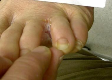
Double blind, randomised study of continuous terbinafine compared with intermittent itraconazole in treatment of toenail onychomycosis. The LION Study Group. (1/176)
OBJECTIVE: To compare the efficacy and safety of continuous terbinafine with intermittent itraconazole in the treatment of toenail onychomycosis. DESIGN: Prospective, randomised, double blind, double dummy, multicentre, parallel group study lasting 72 weeks. SETTING: 35 centres in six European countries. SUBJECTS: 496 patients aged 18 to 75 years with a clinical and mycological diagnosis of dermatophyte onychomycosis of the toenail. INTERVENTIONS: Study patients were randomly divided into four parallel groups to receive either terbinafine 250 mg a day for 12 or 16 weeks (groups T12 and T16) or itraconazole 400 mg a day for 1 week in every 4 weeks for 12 or 16 weeks (groups I3 and I4). MAIN OUTCOME MEASURES: Assessment of primary efficacy at week 72 was mycological cure, defined as negative results on microscopy and culture of samples from the target toenail. RESULTS: At week 72 the mycological cure rates were 75.7% (81/107) in the T12 group and 80. 8% (80/99) in the T16 group compared with 38.3% (41/107) in the I3 group and 49.1 % (53/108) in the I4 group. All comparisons (T12 v I3, T12 v I4, T16 v I3, T16 v I4) showed significantly higher cure rates in the terbinafine groups (all P<0.0001). Also, all secondary clinical outcome measures were significantly in favour of terbinafine at week 72. There were no differences in the number or type of adverse events recorded in the terbinafine or itraconazole groups. CONCLUSION: Continuous terbinafine is significantly more effective than intermittent itraconazole in the treatment of patients with toenail onychomycosis. (+info)N-terminal deletion in a desmosomal cadherin causes the autosomal dominant skin disease striate palmoplantar keratoderma. (2/176)
The N-terminal extracellular domain of the cadherins, calcium-dependent cell adhesion molecules, has been shown by X-ray crystallography to be involved in two types of interaction: lateral strand dimers and adhesive dimers. Here we describe the first human mutation in a cadherin present in desmosome cell junctions that removes a portion of this highly conserved first extracellular domain. The mutation, in the DSG1 gene coding for a desmoglein (Dsg1), results in the deletion of the first and much of the second beta-strand of the first cadherin repeat and part of the first Ca2+-binding site, and would be expected to compromise strand dimer formation. It causes a dominantly inherited skin disease, striate palmoplantar keratoderma (SPPK), mapping to chromosome 18q12.1, in which affected individuals have marked hyperkeratotic bands on the palms and soles. In a three generation Dutch family with SPPK, we have found a G-->A transition in the 3" splice acceptor site of intron 2 of the DSG1 gene which segregated with the disease phenotype. This causes aberrant splicing of exon 2 to exon 4, which are in-frame, with the consequent removal of exon 3 encoding part of the prosequence, the mature protein cleavage site and part of the first extracellular domain. This mutation emphasizes the importance of this part of the molecule for cadherin function, and of the Dsg1 protein and hence desmosomes in epidermal function. (+info)Treatment of toenail onychomycosis with 2% butenafine and 5% Melaleuca alternifolia (tea tree) oil in cream. (3/176)
The prevalence of onychomycosis, a superficial fungal infection that destroys the entire nail unit, is rising, with no satisfactory cure. The objective of this randomized, double-blind, placebo-controlled study was to examine the clinical efficacy and tolerability of 2% butenafine hydrochloride and 5% Melaleuca alternifolia oil incorporated in a cream to manage toenail onychomycosis in a cohort. Sixty outpatients (39 M, 21 F) aged 18-80 years (mean 29.6) with 6-36 months duration of disease were randomized to two groups (40 and 20), active and placebo. After 16 weeks, 80% of patients using medicated cream were cured, as opposed to none in the placebo group. Four patients in the active treatment group experienced subjective mild inflammation without discontinuing treatment. During follow-up, no relapse occurred in cured patients and no improvement was seen in medication-resistant and placebo participants. (+info)Systematic review of topical treatments for fungal infections of the skin and nails of the feet. (4/176)
OBJECTIVE: To identify and synthesise the evidence for efficacy and cost effectiveness of topical treatments for superficial fungal infections of the skin and nails of the feet. DESIGN: Systematic review. INTERVENTIONS: Topical treatments for superficial fungal infections. MAIN OUTCOME MEASURES: Cure confirmed by culture and microscopy for skin and by culture for nails in patients with clinically diagnosed fungal infections. RESULTS: Of 126 trials identified in 121 papers, 72 (57.1%) met the inclusion criteria. Placebo controlled trials yielded pooled relative risks of failure to cure skin infections: allylamines (0.30, 95% confidence interval 0.24 to 0.38); azoles (0.54, 0.42 to 0.68); undecenoic acid (0.28, 0. 11 to 0.74); and tolnaftate (0.46, 0.17 to 1.22). Although meta-analysis of 11 trials comparing allylamines and azoles showed a relative risk of failure to cure of 0.88 (0.78 to 0.99) in favour of allylamines, there was evidence of language bias. Seven reports in English favoured allylamines (0.79, 0.69 to 0.91), but four reports in foreign languages showed no difference between the two drugs (1. 01, 0.90 to 1.13). Neither trial of nail infections showed significant differences between alternative topical treatments. CONCLUSIONS: Allylamines, azoles, and undecenoic acid were efficacious in placebo controlled trials. There are sufficient comparative trials to judge relative efficacy only between allylamines and azoles. Allylamines cure slightly more infections than azoles but are much more expensive than azoles. The most cost effective strategy is first to treat with azoles or undecenoic acid and to use allylamines only if that fails. (+info)Onychomycosis caused by Scytalidium dimidiatum. Report of two cases. Review of the taxonomy of the synanamorph and anamorph forms of this coelomycete. (5/176)
The authors report two cases of onychomycosis in the dystrophic form, one of them involving an HIV-positive patient, provoked by Scytalidium dimidiatum, previously called Scytalidium lignicola. The subject is reviewed from the taxonomic viewpoint, considering the anamorph Hendersonula toruloidea as a synonym of Nattrassia mangiferae, and having Scytalidium dimidiatum as the major synanamorph. According to many mycologists, Scytalidium hyalinum may be a separate species or a hyaline mutant of Scytalidium dimidiatum. Scytalidium lignicola Pesante 1957 was considered to be the type-species of the genus by ELLIS (1971)13 and later to be a "conidial state" of Hendersonula toruloidea by the same author, today known as Nattrassia mangiferae. The microorganism lives only on the roots of certain plants (mainly Platanus and Pinus). It produces pycnidia and is not considered to be a pathogen, although it is considered as a possible emerging agent capable of provoking opportunistic fungal lesions. The importance of this topic as one of the most outstanding in fungal taxonomy, so likely to be modified over time, as well as its interest in the field of dermatologic mycology, are emphasized. (+info)Synthesis of viral DNA and late capsid protein L1 in parabasal spinous cell layers of naturally occurring benign warts infected with human papillomavirus type 1. (6/176)
We investigated human papillomavirus type 1 (HPV1)-specific transcription, viral DNA replication, and viral protein expression in naturally occurring benign tumors by in situ hybridization, 5-bromodeoxyuridine (BrdU) incorporation, and immunohistochemistry and obtained results different from other HPV-infected benign tumors characterized so far. Moderate amounts of transcripts with a putative coding potential for E6/E7, E1, and E2 were demonstrated from the first subrabasal cell layer throughout the stratum spinosum and granulosum. In addition very large amounts of E4 and L1 transcripts were present in the same epithelial layers. This finding was substantiated by the demonstration of L1 and E4 protein already in the bottom-most spinous cell layer. Furthermore massive amplification of the viral DNA as measured by BrdU incorporation and different methods of in situ hybridization took place in the lowest 5 to 10 suprabasal cell layers. These findings are in contrast to the assumption that late gene expression and viral DNA synthesis are restricted to the more differentiated cell layers of the epithelium and point to differences in the regulation of the vegetative life cycle between different papillomavirus types. (+info)Mycobacterium thermoresistible recovered from a cutaneous lesion in an otherwise healthy individual. (7/176)
This is the first report of coinfection by Mycobacterium thermoresistible and Mycobacterium fortuitum and only the fifth case of human infection by M. thermoresistible reported in the world literature. (+info)White grain mycetoma caused by a Cylindrocarpon sp. in India. (8/176)
We describe a case of white grain eumycetoma of the foot of an Indian male caused by a slow-growing, poorly sporulating fungus that does not match any known agent of this infection. Histologic examination of a biopsy tissue specimen showed oval, lobular, white granules composed of hyaline, septate hyphae, and thick-walled chlamydospores. Culture of granules from a draining sinus yielded compact, very-slow-growing, poorly sporulating colonies producing a strong reddish brown pigment that diffused into the medium. The fungus was identified as a Cylindrocarpon sp. based on the development of rare cylindrical conidia borne from solitary phialides lacking collarettes, in addition to chlamydospores formed singly or in short chains. (+info)Foot dermatoses refer to various skin conditions that affect the feet. These can include inflammatory conditions like eczema and psoriasis, infectious diseases such as athlete's foot (tinea pedis), fungal infections, bacterial infections, viral infections (like plantar warts caused by HPV), and autoimmune blistering disorders. Additionally, contact dermatitis from irritants or allergens can also affect the feet. Proper diagnosis is essential to determine the best course of treatment for each specific condition.
In medical terms, the foot is the part of the lower limb that is distal to the leg and below the ankle, extending from the tarsus to the toes. It is primarily responsible for supporting body weight and facilitating movement through push-off during walking or running. The foot is a complex structure made up of 26 bones, 33 joints, and numerous muscles, tendons, ligaments, and nerves that work together to provide stability, balance, and flexibility. It can be divided into three main parts: the hindfoot, which contains the talus and calcaneus (heel) bones; the midfoot, which includes the navicular, cuboid, and cuneiform bones; and the forefoot, which consists of the metatarsals and phalanges that form the toes.
Hand dermatoses is a general term used to describe various inflammatory skin conditions that affect the hands. These conditions can cause symptoms such as redness, swelling, itching, blistering, scaling, and cracking of the skin on the hands. Common examples of hand dermatoses include:
1. Irritant contact dermatitis: A reaction that occurs when the skin comes into contact with irritants such as chemicals, soaps, or detergents.
2. Allergic contact dermatitis: A reaction that occurs when the skin comes into contact with allergens, such as nickel, rubber, or poison ivy.
3. Atopic dermatitis (eczema): A chronic skin condition characterized by dry, itchy, and inflamed skin.
4. Psoriasis: A chronic skin condition characterized by red, scaly patches that can occur anywhere on the body, including the hands.
5. Dyshidrotic eczema: A type of eczema that causes small blisters to form on the sides of the fingers, palms, and soles of the feet.
6. Lichen planus: An inflammatory skin condition that can cause purple or white patches to form on the hands and other parts of the body.
7. Scabies: A contagious skin condition caused by mites that burrow into the skin and lay eggs, causing intense itching and a rash.
Treatment for hand dermatoses depends on the specific diagnosis and may include topical creams or ointments, oral medications, phototherapy, or avoidance of triggers.
Facial dermatoses refer to various skin conditions that affect the face. These can include a wide range of disorders, such as:
1. Acne vulgaris: A common skin condition characterized by the formation of comedones (blackheads and whiteheads) and inflammatory papules, pustules, and nodules. It primarily affects the face, neck, chest, and back.
2. Rosacea: A chronic skin condition that causes redness, flushing, and visible blood vessels on the face, along with bumps or pimples and sometimes eye irritation.
3. Seborrheic dermatitis: A common inflammatory skin disorder that causes a red, itchy, and flaky rash, often on the scalp, face, and eyebrows. It can also affect other oily areas of the body, like the sides of the nose and behind the ears.
4. Atopic dermatitis (eczema): A chronic inflammatory skin condition that causes red, itchy, and scaly patches on the skin. While it can occur anywhere on the body, it frequently affects the face, especially in infants and young children.
5. Psoriasis: An autoimmune disorder that results in thick, scaly, silvery, or red patches on the skin. It can affect any part of the body, including the face.
6. Contact dermatitis: A skin reaction caused by direct contact with an allergen or irritant, resulting in redness, itching, and inflammation. The face can be affected when allergens or irritants come into contact with the skin through cosmetics, skincare products, or other substances.
7. Lupus erythematosus: An autoimmune disorder that can cause a butterfly-shaped rash on the cheeks and nose, along with other symptoms like joint pain, fatigue, and photosensitivity.
8. Perioral dermatitis: A inflammatory skin condition that causes redness, small bumps, and dryness around the mouth, often mistaken for acne. It can also affect the skin around the nose and eyes.
9. Vitiligo: An autoimmune disorder that results in the loss of pigmentation in patches of skin, which can occur on the face and other parts of the body.
10. Tinea faciei: A fungal infection that affects the facial skin, causing red, scaly, or itchy patches. It is also known as ringworm of the face.
These are just a few examples of skin conditions that can affect the face. If you experience any unusual symptoms or changes in your skin, it's essential to consult a dermatologist for proper diagnosis and treatment.
Skin diseases, also known as dermatological conditions, refer to any medical condition that affects the skin, which is the largest organ of the human body. These diseases can affect the skin's function, appearance, or overall health. They can be caused by various factors, including genetics, infections, allergies, environmental factors, and aging.
Skin diseases can present in many different forms, such as rashes, blisters, sores, discolorations, growths, or changes in texture. Some common examples of skin diseases include acne, eczema, psoriasis, dermatitis, fungal infections, viral infections, bacterial infections, and skin cancer.
The symptoms and severity of skin diseases can vary widely depending on the specific condition and individual factors. Some skin diseases are mild and can be treated with over-the-counter medications or topical creams, while others may require more intensive treatments such as prescription medications, light therapy, or even surgery.
It is important to seek medical attention if you experience any unusual or persistent changes in your skin, as some skin diseases can be serious or indicative of other underlying health conditions. A dermatologist is a medical doctor who specializes in the diagnosis and treatment of skin diseases.
Vesiculobullous skin diseases are a group of disorders characterized by the formation of blisters (vesicles) and bullae (larger blisters) on the skin. These blisters form when there is a separation between the epidermis (outer layer of the skin) and the dermis (layer beneath the epidermis) due to damage in the area where they join, known as the dermo-epidermal junction.
There are several types of vesiculobullous diseases, each with its own specific causes and symptoms. Some of the most common types include:
1. Pemphigus vulgaris: an autoimmune disorder where the immune system mistakenly attacks proteins that help to hold the skin together, causing blisters to form.
2. Bullous pemphigoid: another autoimmune disorder, but in this case, the immune system attacks a different set of proteins, leading to large blisters and inflammation.
3. Dermatitis herpetiformis: a skin condition associated with celiac disease, where gluten ingestion triggers an immune response that leads to the formation of itchy blisters.
4. Pemphigoid gestationis: a rare autoimmune disorder that occurs during pregnancy and causes blisters on the abdomen and other parts of the body.
5. Epidermolysis bullosa: a group of inherited disorders where there is a fragile skin structure, leading to blistering and wound formation after minor trauma or friction.
Treatment for vesiculobullous diseases depends on the specific diagnosis and may include topical or systemic medications, such as corticosteroids, immunosuppressants, or antibiotics, as well as wound care and prevention of infection.
Linear IgA Bullous Dermatosis (LABD) is an autoimmune blistering disorder characterized by the production of autoantibodies against the 97-kDa component of the basement membrane zone, leading to the formation of tense blisters and erosions. It can occur in both children and adults, with different subtypes and clinical presentations.
In LABD, there is a linear deposition of IgA along the basement membrane zone on direct immunofluorescence (DIF) studies, which helps to distinguish it from other autoimmune blistering disorders like bullous pemphigoid or pemphigus vulgaris.
The condition can be idiopathic or associated with medications, infections, or underlying medical conditions such as inflammatory bowel disease or hematologic malignancies. Treatment typically involves systemic corticosteroids and other immunosuppressive agents to control the blister formation and prevent complications.
Sweet syndrome, also known as acute febrile neutrophilic dermatosis, is a skin condition characterized by the rapid onset of painful, red, and swollen skin lesions. The lesions are often accompanied by fever and elevated white blood cell count, particularly an increase in neutrophils.
The medical definition of Sweet syndrome includes the following criteria:
1. Abrupt onset of painful, erythematous (red), and edematous (swollen) papules, plaques, or nodules.
2. Fever greater than 38°C (100.4°F).
3. Leukocytosis with a predominance of neutrophils in the peripheral blood.
4. Histopathological evidence of a dense dermal infiltrate of neutrophils without evidence of vasculitis.
5. Rapid response to systemic corticosteroids.
Sweet syndrome can be associated with various medical conditions, such as infections, malignancies, and inflammatory diseases, or it can occur without an identifiable underlying cause (idiopathic).
Occupational dermatitis is a specific type of contact dermatitis that results from exposure to certain substances or conditions in the workplace. It can be caused by direct contact with chemicals, irritants, or allergens present in the work environment. This condition typically affects the skin on the hands and forearms but can also involve other areas of the body, depending on the nature of the exposure.
There are two main types of occupational dermatitis:
1. Irritant contact dermatitis (ICD): This type occurs when the skin comes into direct contact with an irritating substance, leading to redness, swelling, itching, and sometimes blistering. Common irritants include solvents, detergents, oils, and other industrial chemicals.
2. Allergic contact dermatitis (ACD): This type is a result of an allergic reaction to a specific substance. The immune system identifies the allergen as harmful and mounts a response, causing skin inflammation. Common allergens include latex, metals (such as nickel), and certain plants (like poison ivy).
Prevention measures for occupational dermatitis include using appropriate personal protective equipment (PPE) like gloves, masks, and aprons, as well as practicing good hygiene, such as washing hands regularly and avoiding touching the face with contaminated hands. If you suspect you have developed occupational dermatitis, consult a healthcare professional for proper diagnosis and treatment.
Foot diseases refer to various medical conditions that affect the foot, including its structures such as the bones, joints, muscles, tendons, ligaments, blood vessels, and nerves. These conditions can cause symptoms like pain, swelling, numbness, difficulty walking, and skin changes. Examples of foot diseases include:
1. Plantar fasciitis: inflammation of the band of tissue that connects the heel bone to the toes.
2. Bunions: a bony bump that forms on the joint at the base of the big toe.
3. Hammertoe: a deformity in which the toe is bent at the middle joint, resembling a hammer.
4. Diabetic foot: a group of conditions that can occur in people with diabetes, including nerve damage, poor circulation, and increased risk of infection.
5. Athlete's foot: a fungal infection that affects the skin between the toes and on the soles of the feet.
6. Ingrown toenails: a condition where the corner or side of a toenail grows into the flesh of the toe.
7. Gout: a type of arthritis that causes sudden, severe attacks of pain, swelling, redness, and tenderness in the joints, often starting with the big toe.
8. Foot ulcers: open sores or wounds that can occur on the feet, especially in people with diabetes or poor circulation.
9. Morton's neuroma: a thickening of the tissue around a nerve between the toes, causing pain and numbness.
10. Osteoarthritis: wear and tear of the joints, leading to pain, stiffness, and reduced mobility.
Foot diseases can affect people of all ages and backgrounds, and some may be prevented or managed with proper foot care, hygiene, and appropriate medical treatment.
The term "diabetic foot" refers to a condition that affects the feet of people with diabetes, particularly when the disease is not well-controlled. It is characterized by a combination of nerve damage (neuropathy) and poor circulation (peripheral artery disease) in the feet and lower legs.
Neuropathy can cause numbness, tingling, or pain in the feet, making it difficult for people with diabetes to feel injuries, cuts, blisters, or other foot problems. Poor circulation makes it harder for wounds to heal and increases the risk of infection.
Diabetic foot ulcers are a common complication of diabetic neuropathy and can lead to serious infections, hospitalization, and even amputation if not treated promptly and effectively. Preventive care, including regular foot exams, proper footwear, and good blood glucose control, is essential for people with diabetes to prevent or manage diabetic foot problems.
Leg dermatoses is a general term that refers to various skin conditions affecting the legs. This can include a wide range of inflammatory, infectious, or degenerative diseases that cause symptoms such as redness, itching, scaling, blistering, or pigmentation changes on the leg skin. Examples of specific leg dermatoses include stasis dermatitis, venous eczema, contact dermatitis, lichen planus, psoriasis, and cellulitis among others. Accurate diagnosis usually requires a thorough examination and sometimes a biopsy to determine the specific type of dermatosis and appropriate treatment.
Scalp dermatoses refer to various skin conditions that affect the scalp. These can include inflammatory conditions such as seborrheic dermatitis (dandruff, cradle cap), psoriasis, atopic dermatitis (eczema), and lichen planus; infectious processes like bacterial folliculitis, tinea capitis (ringworm of the scalp), and viral infections; as well as autoimmune conditions such as alopecia areata. Symptoms can range from mild scaling and itching to severe redness, pain, and hair loss. The specific diagnosis and treatment of scalp dermatoses depend on the underlying cause.
Acquired foot deformities refer to structural abnormalities of the foot that develop after birth, as opposed to congenital foot deformities which are present at birth. These deformities can result from various factors such as trauma, injury, infection, neurological conditions, or complications from a medical condition like diabetes or arthritis.
Examples of acquired foot deformities include:
1. Hammertoe - A deformity where the toe bends downward at the middle joint, resembling a hammer.
2. Claw toe - A more severe form of hammertoe where the toe also curls under, forming a claw-like shape.
3. Mallet toe - A condition where the end joint of a toe is bent downward, causing it to resemble a mallet.
4. Bunions - A bony bump that forms on the inside of the foot at the big toe joint, often causing pain and difficulty wearing shoes.
5. Tailor's bunion (bunionette) - A similar condition to a bunion, but it occurs on the outside of the foot near the little toe joint.
6. Charcot foot - A severe deformity that can occur in people with diabetes or other neurological conditions, characterized by the collapse and dislocation of joints in the foot.
7. Cavus foot - A condition where the arch of the foot is excessively high, causing instability and increasing the risk of ankle injuries.
8. Flatfoot (pes planus) - A deformity where the arch of the foot collapses, leading to pain and difficulty walking.
9. Pronation deformities - Abnormal rotation or tilting of the foot, often causing instability and increasing the risk of injury.
Treatment for acquired foot deformities varies depending on the severity and underlying cause but may include orthotics, physical therapy, medication, or surgery.
A foot ulcer is a wound or sore on the foot that occurs most commonly in people with diabetes, but can also affect other individuals with poor circulation or nerve damage. These ulcers can be challenging to heal and are prone to infection, making it essential for individuals with foot ulcers to seek medical attention promptly.
Foot ulcers typically develop due to prolonged pressure on bony prominences of the foot, leading to breakdown of the skin and underlying tissues. The development of foot ulcers can be attributed to several factors, including:
1. Neuropathy (nerve damage): This condition causes a loss of sensation in the feet, making it difficult for individuals to feel pain or discomfort associated with pressure points, leading to the formation of ulcers.
2. Peripheral artery disease (PAD): Reduced blood flow to the lower extremities can impair wound healing and make the body more susceptible to infection.
3. Deformities: Structural foot abnormalities, such as bunions or hammertoes, can cause increased pressure on specific areas of the foot, increasing the risk of ulcer formation.
4. Poorly fitting shoes: Shoes that are too tight, narrow, or ill-fitting can create friction and pressure points, contributing to the development of foot ulcers.
5. Trauma: Injuries or trauma to the feet can lead to the formation of ulcers, particularly in individuals with neuropathy who may not feel the initial pain associated with the injury.
6. Foot care neglect: Failure to inspect and care for the feet regularly can result in undetected wounds or sores that progress into ulcers.
Foot ulcers are classified based on their depth, severity, and extent of tissue involvement. Proper assessment, treatment, and prevention strategies are crucial in managing foot ulcers and minimizing the risk of complications such as infection, gangrene, and amputation.
Erythema is a term used in medicine to describe redness of the skin, which occurs as a result of increased blood flow in the superficial capillaries. This redness can be caused by various factors such as inflammation, infection, trauma, or exposure to heat, cold, or ultraviolet radiation. In some cases, erythema may also be accompanied by other symptoms such as swelling, warmth, pain, or itching. It is a common finding in many medical conditions and can vary in severity from mild to severe.
Keratosis, in general, refers to a skin condition characterized by the abnormal growth or development of keratin, a protein that forms part of the outer layer of the skin (epidermis). There are several types of keratosis, including:
1. Seborrheic Keratosis: benign, often pigmented, rough, and scaly growths that can appear anywhere on the body. They tend to increase in number with age.
2. Actinic Keratosis: rough, scaly patches or spots on the skin that are caused by long-term exposure to sunlight or artificial UV light. These have the potential to develop into squamous cell carcinoma, a type of skin cancer.
3. Solar Keratosis: another term for actinic keratosis, as it is primarily caused by sun damage.
4. Keratosis Pilaris: a common condition where small, rough bumps appear on the skin, often on the arms, thighs, or cheeks. These are caused by excess keratin blocking hair follicles.
5. Follicular Keratosis: a disorder characterized by the formation of horny plugs within the hair follicles, leading to rough, sandpaper-like bumps on the skin.
6. Intraepidermal Keratosis: a term used to describe the abnormal accumulation of keratin in the epidermis, which can lead to various skin conditions.
It's important to consult with a healthcare professional or dermatologist for proper diagnosis and treatment if you suspect having any form of keratosis.
Pyoderma gangrenosum is a rare, inflammatory skin condition that typically begins as a small pustule or blister, which then rapidly progresses to form painful ulcers with a characteristic violaceous (bluish-purple) undermined border. The etiology of pyoderma gangrenosum is not entirely clear, but it's often associated with an underlying systemic disease, such as inflammatory bowel disease, rheumatoid arthritis, or hematologic disorders.
The pathophysiology of pyoderma gangrenosum involves a dysregulated immune response and neutrophil-mediated tissue damage. Diagnosis is often based on the clinical presentation and exclusion of other conditions with similar lesions. Treatment typically includes systemic immunosuppressive therapy, such as corticosteroids, cyclosporine, or biologic agents, along with local wound care to promote healing and prevent infection.
It's important to note that pyoderma gangrenosum can be a challenging condition to manage, and a multidisciplinary approach involving dermatologists, internists, and surgeons may be necessary for optimal care.
Foot injuries refer to any damage or trauma caused to the various structures of the foot, including the bones, muscles, tendons, ligaments, blood vessels, and nerves. These injuries can result from various causes such as accidents, sports activities, falls, or repetitive stress. Common types of foot injuries include fractures, sprains, strains, contusions, dislocations, and overuse injuries like plantar fasciitis or Achilles tendonitis. Symptoms may vary depending on the type and severity of the injury but often include pain, swelling, bruising, difficulty walking, and reduced range of motion. Proper diagnosis and treatment are crucial to ensure optimal healing and prevent long-term complications.
Vulvar diseases refer to a range of medical conditions that affect the vulva, which is the external female genital area including the mons pubis, labia majora and minora, clitoris, and the vaginal opening. These conditions can cause various symptoms such as itching, burning, pain, soreness, irritation, or abnormal growths or lesions. Some common vulvar diseases include:
1. Vulvitis: inflammation of the vulva that can be caused by infection, allergies, or irritants.
2. Lichen sclerosus: a chronic skin condition that causes thin, white patches on the vulva.
3. Lichen planus: an inflammatory condition that affects the skin and mucous membranes, including the vulva.
4. Vulvar cancer: a rare type of cancer that develops in the tissues of the vulva.
5. Genital warts: caused by human papillomavirus (HPV) infection, these are small growths or bumps on the vulva.
6. Pudendal neuralgia: a nerve condition that causes pain in the vulvar area.
7. Vestibulodynia: pain or discomfort in the vestibule, the area surrounding the vaginal opening.
It is important to consult a healthcare professional if experiencing any symptoms related to vulvar diseases for proper diagnosis and treatment.
Folliculitis is a medical condition characterized by inflammation of one or more hair follicles, typically appearing as small red bumps or pimples that surround the affected follicle. It can occur anywhere on the body where hair grows, but it's most common in areas exposed to friction, heat, and tight clothing such as the neck, back, legs, arms, and buttocks.
Folliculitis can be caused by various factors, including bacterial or fungal infections, irritation from shaving or waxing, ingrown hairs, and exposure to chemicals or sweat. The severity of folliculitis ranges from mild cases that resolve on their own within a few days to severe cases that may require medical treatment.
Treatment for folliculitis depends on the underlying cause. For bacterial infections, antibiotics may be prescribed, while antifungal medications are used for fungal infections. In some cases, topical treatments such as creams or gels may be sufficient to treat mild folliculitis, while more severe cases may require oral medication or other medical interventions.
Hyperpigmentation is a medical term that refers to the darkening of skin areas due to an increase in melanin, the pigment that provides color to our skin. This condition can affect people of all races and ethnicities, but it's more noticeable in those with lighter skin tones.
Hyperpigmentation can be caused by various factors, including excessive sun exposure, hormonal changes (such as during pregnancy), inflammation, certain medications, and underlying medical conditions like Addison's disease or hemochromatosis. It can also result from skin injuries, such as cuts, burns, or acne, which leave dark spots known as post-inflammatory hyperpigmentation.
There are several types of hyperpigmentation, including:
1. Melasma: This is a common form of hyperpigmentation that typically appears as symmetrical, blotchy patches on the face, particularly the forehead, cheeks, and upper lip. It's often triggered by hormonal changes, such as those experienced during pregnancy or while taking birth control pills.
2. Solar lentigos (age spots or liver spots): These are small, darkened areas of skin that appear due to prolonged sun exposure over time. They typically occur on the face, hands, arms, and decolletage.
3. Post-inflammatory hyperpigmentation: This type of hyperpigmentation occurs when an injury or inflammation heals, leaving behind a darkened area of skin. It's more common in people with darker skin tones.
Treatment for hyperpigmentation depends on the underlying cause and may include topical creams, chemical peels, laser therapy, or microdermabrasion. Preventing further sun damage is crucial to managing hyperpigmentation, so wearing sunscreen with a high SPF and protective clothing is recommended.
Foot deformities refer to abnormal changes in the structure and/or alignment of the bones, joints, muscles, ligaments, or tendons in the foot, leading to a deviation from the normal shape and function of the foot. These deformities can occur in various parts of the foot, such as the toes, arch, heel, or ankle, and can result in pain, difficulty walking, and reduced mobility. Some common examples of foot deformities include:
1. Hammertoes: A deformity where the toe bends downward at the middle joint, resembling a hammer.
2. Mallet toes: A condition where the end joint of the toe is bent downward, creating a mallet-like shape.
3. Claw toes: A combination of both hammertoes and mallet toes, causing all three joints in the toe to bend abnormally.
4. Bunions: A bony bump that forms on the inside of the foot at the base of the big toe, caused by the misalignment of the big toe joint.
5. Tailor's bunion (bunionette): A similar condition to a bunion but occurring on the outside of the foot, at the base of the little toe.
6. Flat feet (pes planus): A condition where the arch of the foot collapses, causing the entire sole of the foot to come into contact with the ground when standing or walking.
7. High arches (pes cavus): An excessively high arch that doesn't provide enough shock absorption and can lead to pain and instability.
8. Cavus foot: A condition characterized by a very high arch and tight heel cord, often leading to an imbalance in the foot structure and increased risk of ankle injuries.
9. Haglund's deformity: A bony enlargement on the back of the heel, which can cause pain and irritation when wearing shoes.
10. Charcot foot: A severe deformity that occurs due to nerve damage in the foot, leading to weakened bones, joint dislocations, and foot collapse.
Foot deformities can be congenital (present at birth) or acquired (develop later in life) due to various factors such as injury, illness, poor footwear, or abnormal biomechanics. Proper diagnosis, treatment, and management are essential for maintaining foot health and preventing further complications.
Neurodermatitis, also known as lichen simplex chronicus, is a skin condition characterized by chronic itching and scratching of the skin. It typically affects areas that are easy to reach and can be triggered by stress, anxiety, or other underlying skin conditions such as eczema or psoriasis. The constant scratching leads to thickening and darkening of the skin, which can cause discomfort and distress. Treatment usually involves a combination of topical medications, lifestyle changes, and behavioral modifications to reduce scratching and alleviate symptoms.
Dapsone is a medication that belongs to a class of drugs called sulfones. It is primarily used to treat bacterial skin infections such as leprosy and dermatitis herpetiformis (a skin condition associated with coeliac disease). Dapsone works by killing the bacteria responsible for these infections.
In addition, dapsone has anti-inflammatory properties and is sometimes used off-label to manage inflammatory conditions such as vasculitis, bullous pemphigoid, and chronic urticaria. It is available in oral tablet form and topical cream or gel form.
Like all medications, dapsone can cause side effects, which may include nausea, loss of appetite, and headache. More serious side effects, such as methemoglobinemia (a blood disorder that affects the body's ability to transport oxygen), peripheral neuropathy (nerve damage that causes pain, numbness, or weakness in the hands and feet), and liver damage, can occur but are less common.
It is important for patients taking dapsone to be monitored by a healthcare provider to ensure safe and effective use of the medication.
Dermatitis is a general term that describes inflammation of the skin. It is often characterized by redness, swelling, itching, and tenderness. There are many different types of dermatitis, including atopic dermatitis (eczema), contact dermatitis, seborrheic dermatitis, and nummular dermatitis.
Atopic dermatitis is a chronic skin condition that often affects people with a family history of allergies, such as asthma or hay fever. It typically causes dry, scaly patches on the skin that can be extremely itchy.
Contact dermatitis occurs when the skin comes into contact with an irritant or allergen, such as poison ivy or certain chemicals. This type of dermatitis can cause redness, swelling, and blistering.
Seborrheic dermatitis is a common condition that causes a red, itchy rash, often on the scalp, face, or other areas of the body where oil glands are located. It is thought to be related to an overproduction of oil by the skin's sebaceous glands.
Nummular dermatitis is a type of eczema that causes round, coin-shaped patches of dry, scaly skin. It is more common in older adults and often occurs during the winter months.
Treatment for dermatitis depends on the underlying cause and severity of the condition. In some cases, over-the-counter creams or lotions may be sufficient to relieve symptoms. Prescription medications, such as corticosteroids or immunosuppressants, may be necessary in more severe cases. Avoiding triggers and irritants can also help prevent flare-ups of dermatitis.
'Foot bones,' also known as the tarsal and metatarsal bones, are the 26 bones that make up the foot in humans. The foot is divided into three parts: the hindfoot, midfoot, and forefoot.
The hindfoot contains two bones: the talus, which connects to the leg bone (tibia), and the calcaneus (heel bone). These bones form the ankle joint and heel.
The midfoot is made up of five irregularly shaped bones called the navicular, cuboid, and three cuneiform bones. These bones help form the arch of the foot and connect the hindfoot to the forefoot.
The forefoot contains the metatarsals (five long bones) and the phalanges (14 small bones). The metatarsals connect the midfoot to the toes, while the phalanges make up the toes themselves.
These bones work together to provide stability, support, and movement for the foot, allowing us to walk, run, and jump.
Pruritus is a medical term derived from Latin, in which "prurire" means "to itch." It refers to an unpleasant sensation on the skin that provokes the desire or reflex to scratch. This can be caused by various factors, such as skin conditions (e.g., dryness, eczema, psoriasis), systemic diseases (e.g., liver disease, kidney failure), nerve disorders, psychological conditions, or reactions to certain medications.
Pruritus can significantly affect a person's quality of life, leading to sleep disturbances, anxiety, and depression. Proper identification and management of the underlying cause are essential for effective treatment.
Pigmentation disorders are conditions that affect the production or distribution of melanin, the pigment responsible for the color of skin, hair, and eyes. These disorders can cause changes in the color of the skin, resulting in areas that are darker (hyperpigmentation) or lighter (hypopigmentation) than normal. Examples of pigmentation disorders include melasma, age spots, albinism, and vitiligo. The causes, symptoms, and treatments for these conditions can vary widely, so it is important to consult a healthcare provider for an accurate diagnosis and treatment plan.
Purpura is a medical term that refers to the appearance of purple-colored spots on the skin or mucous membranes, caused by bleeding underneath the skin due to various factors such as blood clotting disorders, vasculitis (inflammation of the blood vessels), severe thrombocytopenia (low platelet count), or use of certain medications. These spots can vary in size and shape, ranging from small pinpoint hemorrhages (petechiae) to larger, irregularly shaped patches (ecchymoses). The bleeding is usually not caused by trauma or injury to the area. It's important to consult a healthcare professional if you notice any unexplained purpuric spots on your skin or mucous membranes, as they can indicate an underlying medical condition that requires further evaluation and treatment.
Hypopigmentation is a medical term that refers to a condition where there is a decrease in the amount of pigment (melanin) in the skin, resulting in lighter patches or spots on the skin. This can occur due to various reasons such as skin injuries, certain skin disorders like vitiligo, fungal infections, burns, or as a side effect of some medical treatments like chemotherapy or radiation therapy. It is different from albinism, which is a genetic condition where the body is unable to produce melanin at all.
Acantholysis is a medical term that refers to the separation of the cells in the upper layer of the skin (the epidermis), specifically between the pickle cell layer (stratum spinosum) and the granular cell layer (stratum granulosum). This separation results in the formation of distinct, round, or oval cells called acantholytic cells, which are typically seen in certain skin conditions.
Acantholysis is a characteristic feature of several skin disorders, including:
1. Pemphigus vulgaris: A rare autoimmune blistering disorder where the immune system produces antibodies against desmoglein-1 and -3 proteins, leading to acantholysis and formation of flaccid blisters.
2. Pemphigus foliaceus: Another autoimmune blistering disorder that specifically targets desmoglein-1 protein, causing superficial blisters and erosions on the skin.
3. Hailey-Hailey disease (familial benign chronic pemphigus): An autosomal dominant genetic disorder affecting ATP2C1 gene, leading to defective calcium transport and abnormal keratinocyte adhesion, resulting in acantholysis and recurrent skin eruptions.
4. Darier's disease (keratosis follicularis): An autosomal dominant genetic disorder affecting ATP2A2 gene, causing dysfunction of calcium transport and abnormal keratinocyte adhesion, resulting in acantholysis and characteristic papular or keratotic skin lesions.
5. Grover's disease (transient acantholytic dermatosis): An acquired skin disorder of unknown cause, characterized by the development of pruritic, red, and scaly papules and vesicles due to acantholysis.
The presence of acantholysis in these conditions can be confirmed through histopathological examination of skin biopsies.
Eczema is a medical condition characterized by inflammation of the skin, which leads to symptoms such as redness, itching, scaling, and blistering. It is often used to describe atopic dermatitis, a chronic relapsing form of eczema, although there are several other types of eczema with different causes and characteristics.
Atopic dermatitis is believed to be caused by a combination of genetic and environmental factors, and it often affects people with a family history of allergic conditions such as asthma or hay fever. The condition typically begins in infancy or childhood and can persist into adulthood, although it may improve over time.
Eczema can affect any part of the body, but it is most commonly found on the hands, feet, behind the knees, inside the elbows, and on the face. The rash of eczema is often accompanied by dry, scaly skin, and people with the condition may experience periods of flare-ups and remissions.
Treatment for eczema typically involves a combination of moisturizers to keep the skin hydrated, topical corticosteroids to reduce inflammation, and antihistamines to relieve itching. In severe cases, systemic immunosuppressive drugs may be necessary. It is also important for people with eczema to avoid triggers that can worsen their symptoms, such as harsh soaps, scratchy fabrics, and stress.
Pyoderma is a term used in medicine to describe a bacterial skin infection. It's derived from two Greek words: "pyon" meaning pus and "derma" meaning skin.
The infection can result in inflammation, often characterized by redness, swelling, warmth, and pain. Pus-filled blisters or boils may also form, which can rupture and crust over as the infection progresses.
Pyoderma can occur in people of all ages but is particularly common in children. The causative bacteria are often Staphylococcus aureus or Streptococcus pyogenes. The condition can be superficial, affecting only the top layer of the skin (epidermis), or it can be deeper, involving the dermis and/or subcutaneous tissue.
Treatment typically involves antibiotics, either topical or oral, depending on the severity and extent of the infection. In some cases, drainage of pus-filled abscesses may be necessary. Preventive measures such as good hygiene and keeping skin clean and dry can help reduce the risk of pyoderma.
Lichen Planus is a chronic, autoimmune skin condition that can also affect the mucous membranes inside the mouth, genitals, and eyes. It is characterized by the appearance of purplish, flat-topped bumps or lesions on the skin, which may be itchy. The exact cause of Lichen Planus is unknown, but it is believed to occur when the immune system mistakenly attacks cells in the skin or mucous membranes. Certain medications, viral infections, and genetic factors may increase the risk of developing this condition. Treatment typically focuses on managing symptoms and may include topical corticosteroids, oral medications, or light therapy.
"Foot joints" is a general term that refers to the various articulations or connections between the bones in the foot. There are several joints in the foot, including:
1. The ankle joint (tibiotalar joint): This is the joint between the tibia and fibula bones of the lower leg and the talus bone of the foot.
2. The subtalar joint (talocalcaneal joint): This is the joint between the talus bone and the calcaneus (heel) bone.
3. The calcaneocuboid joint: This is the joint between the calcaneus bone and the cuboid bone, which is one of the bones in the midfoot.
4. The tarsometatarsal joints (Lisfranc joint): These are the joints that connect the tarsal bones in the midfoot to the metatarsal bones in the forefoot.
5. The metatarsophalangeal joints: These are the joints between the metatarsal bones and the phalanges (toes) in the forefoot.
6. The interphalangeal joints: These are the joints between the phalanges within each toe.
Each of these foot joints plays a specific role in supporting the foot, absorbing shock, and allowing for movement and flexibility during walking and other activities.
Exfoliative dermatitis is a severe form of widespread inflammation of the skin (dermatitis), characterized by widespread scaling and redness, leading to the shedding of large sheets of skin. It can be caused by various factors such as drug reactions, underlying medical conditions (like lymphoma or leukemia), or extensive eczema. Treatment typically involves identifying and removing the cause, along with supportive care, such as moisturizers and medications to control inflammation and itching. In severe cases, hospitalization may be necessary for close monitoring and management of fluid and electrolyte balance.
Skin diseases of viral origin are conditions that affect the skin caused by viral infections. These infections can lead to various symptoms such as rashes, blisters, papules, and skin lesions. Some common examples of viral skin diseases include:
1. Herpes Simplex Virus (HSV) infection: This causes cold sores or genital herpes, which are characterized by small, painful blisters on the skin.
2. Varicella-zoster virus (VZV) infection: This causes chickenpox and shingles, which are characterized by itchy, fluid-filled blisters on the skin.
3. Human Papillomavirus (HPV) infection: This causes warts, which are small, rough growths on the skin.
4. Molluscum contagiosum: This is a viral infection that causes small, raised, and pearly white bumps on the skin.
5. Measles: This is a highly contagious viral disease characterized by fever, cough, runny nose, and a rash that spreads all over the body.
6. Rubella: Also known as German measles, this viral infection causes a red rash on the face and neck that spreads to the rest of the body.
Viral skin diseases can be spread through direct contact with an infected person or contaminated objects, such as towels or bedding. Some viral skin diseases can be prevented through vaccination, while others can be treated with antiviral medications or other therapies.
Congenital foot deformities refer to abnormal structural changes in the foot that are present at birth. These deformities can vary from mild to severe and may affect the shape, position, or function of one or both feet. Common examples include clubfoot (talipes equinovarus), congenital vertical talus, and cavus foot. Congenital foot deformities can be caused by genetic factors, environmental influences during fetal development, or a combination of both. Treatment options may include stretching, casting, surgery, or a combination of these approaches, depending on the severity and type of the deformity.
Skin care, in a medical context, refers to the practice of maintaining healthy skin through various hygienic, cosmetic, and therapeutic measures. This can include:
1. Cleansing: Using appropriate cleansers to remove dirt, sweat, and other impurities without stripping the skin of its natural oils.
2. Moisturizing: Applying creams or lotions to keep the skin hydrated and prevent dryness.
3. Sun Protection: Using sunscreens, hats, and protective clothing to shield the skin from harmful ultraviolet (UV) rays which can cause sunburn, premature aging, and skin cancer.
4. Skin Care Products: Using over-the-counter or prescription products to manage specific skin conditions like acne, eczema, psoriasis, or rosacea.
5. Regular Check-ups: Regularly examining the skin for any changes, growths, or abnormalities that may indicate a skin condition or disease.
6. Lifestyle Factors: Maintaining a healthy lifestyle, including a balanced diet, regular exercise, adequate sleep, and avoiding habits like smoking and excessive alcohol consumption, which can negatively impact skin health.
It's important to note that while some general skincare advice applies to most people, individual skincare needs can vary greatly depending on factors like age, skin type (oily, dry, combination, sensitive), and specific skin conditions or concerns. Therefore, it's often beneficial to seek personalized advice from a dermatologist or other healthcare provider.
Dermatitis herpetiformis (DH) is a chronic, autoimmune blistering skin disorder that is characterized by the presence of symmetrical, pruritic (itchy), papulo-vesicular (papules and small fluid-filled blisters) eruptions on the extensor surfaces of the body, such as the elbows, knees, buttocks, and shoulders. It is often associated with gluten sensitivity or celiac disease, a condition that causes an abnormal immune response to gluten, a protein found in wheat, barley, and rye.
The exact cause of DH is not fully understood, but it is believed to result from the interaction between genetic, environmental, and immunological factors. The disorder is characterized by the presence of IgA antibodies in the skin, which trigger an immune response that leads to the formation of the characteristic rash.
DH is typically treated with a gluten-free diet, which can help to control the symptoms and prevent complications such as malabsorption and nutritional deficiencies. Medications such as dapsone may also be used to control the itching and blistering associated with the disorder. In some cases, topical corticosteroids or other anti-inflammatory medications may be prescribed to help manage symptoms.
It is important to note that DH is a chronic condition that requires ongoing management and monitoring. People with DH should work closely with their healthcare provider to develop an appropriate treatment plan and monitor their progress over time.
Mite infestations refer to the presence and multiplication of mites, which are tiny arthropods belonging to the class Arachnida, on or inside a host's body. This can occur in various sites such as the skin, lungs, or gastrointestinal tract, depending on the specific mite species.
Skin infestations by mites, also known as dermatophilosis or mange, are common and may cause conditions like scabies (caused by Sarcoptes scabiei) or demodecosis (caused by Demodex spp.). These conditions can lead to symptoms such as itching, rash, and skin lesions.
Lung infestations by mites, although rare, can occur in people who work in close contact with mites, such as farmers or laboratory workers. This condition is called "mite lung" or "farmer's lung," which is often caused by exposure to high levels of dust containing mite feces and dead mites.
Gastrointestinal infestations by mites can occur in animals but are extremely rare in humans. The most common example is the intestinal roundworm, which belongs to the phylum Nematoda rather than Arachnida.
It's important to note that mite infestations can be treated with appropriate medical interventions and prevention measures.
Papulosquamous skin diseases are a group of chronic inflammatory disorders of the skin characterized by the development of papules (small, solid, often conical bump) and scales. These diseases include psoriasis, lichen planus, and seborrheic dermatitis among others. The skin lesions in these conditions are often red, scaly, and may be pruritic (itchy). They can vary in severity and distribution, and can have a significant impact on a person's quality of life. The exact cause of these diseases is not fully understood, but they are believed to involve an abnormal immune response and genetic factors. Treatment typically involves a combination of topical therapies, phototherapy, and systemic medications.
Keratolytic agents are substances that cause the softening and sloughing off of excess keratin, the protein that makes up the outermost layer of the skin (stratum corneum). These agents help to break down and remove dead skin cells, increase moisture retention, and promote the growth of new skin cells. They are commonly used in the treatment of various dermatological conditions such as psoriasis, eczema, warts, calluses, and ichthyosis. Examples of keratolytic agents include salicylic acid, urea, lactic acid, and retinoic acid.
A patch test is a method used in clinical dermatology to identify whether a specific substance causes allergic inflammation of the skin (contact dermatitis). It involves applying small amounts of potential allergens to patches, which are then placed on the skin and left for a set period of time, usually 48 hours. The skin is then examined for signs of an allergic reaction such as redness, swelling or blistering. This helps in identifying the specific substances that an individual may be allergic to, enabling appropriate avoidance measures and treatment.
Scabies is a contagious skin condition caused by the infestation of the human itch mite (Sarcoptes scabiei var. hominis). The female mite burrows into the upper layer of the skin, where it lays its eggs and causes an intensely pruritic (itchy) rash. The rash is often accompanied by small red bumps and blisters, typically found in areas such as the hands, wrists, elbows, armpits, waistline, genitals, and buttocks. Scabies is transmitted through direct skin-to-skin contact with an infected individual or through sharing of contaminated items like bedding or clothing. It can affect people of all ages, races, and socioeconomic backgrounds, but it is particularly common in crowded living conditions, nursing homes, and child care facilities. Treatment usually involves topical medications or oral drugs that kill the mites and their eggs, as well as thorough cleaning and laundering of bedding, clothing, and towels to prevent reinfestation.
In medical terms, the skin is the largest organ of the human body. It consists of two main layers: the epidermis (outer layer) and dermis (inner layer), as well as accessory structures like hair follicles, sweat glands, and oil glands. The skin plays a crucial role in protecting us from external factors such as bacteria, viruses, and environmental hazards, while also regulating body temperature and enabling the sense of touch.
Allergic contact dermatitis is a type of inflammatory skin reaction that occurs when the skin comes into contact with a substance (allergen) that the immune system recognizes as foreign and triggers an allergic response. This condition is characterized by redness, itching, swelling, blistering, and cracking of the skin, which usually develops within 24-48 hours after exposure to the allergen. Common allergens include metals (such as nickel), rubber, medications, fragrances, and cosmetics. It is important to note that a person must first be sensitized to the allergen before developing an allergic response upon subsequent exposures.
Dermatologic agents are medications, chemicals, or other substances that are applied to the skin (dermis) for therapeutic or cosmetic purposes. They can be used to treat various skin conditions such as acne, eczema, psoriasis, fungal infections, and wounds. Dermatologic agents include topical corticosteroids, antibiotics, antifungals, retinoids, benzoyl peroxide, salicylic acid, and many others. They can come in various forms such as creams, ointments, gels, lotions, solutions, and patches. It is important to follow the instructions for use carefully to ensure safety and effectiveness.
Paraneoplastic syndromes refer to a group of rare disorders that are caused by an abnormal immune system response to a cancerous (malignant) tumor. These syndromes are characterized by symptoms or signs that do not result directly from the growth of the tumor itself, but rather from substances produced by the tumor or the body's immune system in response to the tumor.
Paraneoplastic syndromes can affect various organs and systems in the body, including the nervous system, endocrine system, skin, and joints. Examples of paraneoplastic syndromes include Lambert-Eaton myasthenic syndrome (LEMS), which affects nerve function and causes muscle weakness; cerebellar degeneration, which can cause difficulty with coordination and balance; and dermatomyositis, which is an inflammatory condition that affects the skin and muscles.
Paraneoplastic syndromes can occur in association with a variety of different types of cancer, including lung cancer, breast cancer, ovarian cancer, and lymphoma. Treatment typically involves addressing the underlying cancer, as well as managing the symptoms of the paraneoplastic syndrome.
Chemexfoliation is a medical term that refers to the use of chemical agents to exfoliate or remove the outer layers of the skin. It is also known as chempeel, derma peeling, or chemabrasion. This procedure is commonly used in dermatology and cosmetic surgery to improve the appearance of the skin, reduce fine lines and wrinkles, treat acne, uneven pigmentation, and sun damage.
During a chemexfoliation procedure, a chemical solution is applied to the skin, which causes the outer layers to blister and eventually peel off. The type of chemical agent used depends on the individual's skin type and the desired outcome. Commonly used chemicals include alpha-hydroxy acids (AHAs), beta-hydroxy acids (BHAs), trichloroacetic acid (TCA), and phenol.
After the procedure, the skin may be red, swollen, and sensitive for several days. It is important to avoid sun exposure and use a broad-spectrum sunscreen to protect the new skin. Multiple treatments may be necessary to achieve the desired results. Chemexfoliation should only be performed by a qualified healthcare professional in a controlled medical setting.
Parapsoriasis is a term used to describe two uncommon, chronic, and relatively benign inflammatory skin conditions. These are small plaque parapsoriasis (SPP) and large plaque parapsoriasis (LPP), also known as retiform or digitate dermatosis of Köbner.
Small plaque parapsoriasis is characterized by scaly, thin, pink to red patches or plaques, usually less than 3-5 cm in diameter. The lesions are often asymptomatic or mildly pruritic and can be found on the trunk and proximal extremities.
Large plaque parapsoriasis presents as larger, irregularly shaped, scaly patches or thin plaques, typically greater than 5 cm in diameter. The lesions are often asymptomatic but may occasionally be pruritic. LPP is considered a precursor to a rare cutaneous T-cell lymphoma called mycosis fungoides, especially when the lesions become thicker or more numerous over time.
It's important to note that these conditions can sometimes be challenging to diagnose and may require a skin biopsy for accurate diagnosis. Dermatologists and pathologists should carefully evaluate the clinical presentation, histopathological features, and any potential progression to ensure appropriate management.
Hair preparations refer to cosmetic or grooming products that are specifically formulated to be applied to the hair or scalp for various purposes such as cleansing, conditioning, styling, coloring, or promoting hair growth. These preparations can come in different forms, including shampoos, conditioners, hair masks, serums, gels, mousses, sprays, and dyes. They may contain a wide range of ingredients, such as detergents, moisturizers, proteins, vitamins, minerals, and other nutrients that can help improve the health, appearance, and manageability of the hair. Some hair preparations may also contain medications or natural extracts that have therapeutic properties for treating specific hair or scalp conditions, such as dandruff, dryness, oiliness, thinning, or hair loss.
Acitretin is a synthetic form of retinoic acid, which is a type of vitamin A. It is used to treat severe psoriasis and other skin conditions. Acitretin works by slowing down the rapid growth of skin cells that cause the symptoms of psoriasis. It comes in the form of a capsule and is taken orally.
Common side effects of acitretin include dryness of the skin, lips, and mouth, itching, peeling, redness, or stickiness of the palms and soles, hair loss, and changes in nail growth. Less common but more serious side effects can include liver damage, increased levels of lipids in the blood, and birth defects if taken during pregnancy.
It is important to note that acitretin can cause birth defects, so women who are pregnant or planning to become pregnant should not take this medication. Additionally, because acitretin can remain in the body for a long time, it is recommended that women of childbearing age use effective contraception while taking this medication and for at least three years after stopping it.
"Agricultural Workers' Diseases" is a term used to describe a variety of health conditions and illnesses that are associated with agricultural work. These can include both acute and chronic conditions, and can be caused by a range of factors including exposure to chemicals, dusts, allergens, physical injuries, and biological agents such as bacteria and viruses.
Some common examples of Agricultural Workers' Diseases include:
1. Pesticide poisoning: This can occur when agricultural workers are exposed to high levels of pesticides or other chemicals used in farming. Symptoms can range from mild skin irritation to severe neurological damage, depending on the type and amount of chemical exposure.
2. Respiratory diseases: Agricultural workers can be exposed to a variety of dusts and allergens that can cause respiratory problems such as asthma, bronchitis, and farmer's lung. These conditions are often caused by prolonged exposure to moldy hay, grain dust, or other organic materials.
3. Musculoskeletal injuries: Agricultural workers are at risk of developing musculoskeletal injuries due to the physical demands of their job. This can include back pain, repetitive strain injuries, and sprains and strains from lifting heavy objects.
4. Zoonotic diseases: Agricultural workers who come into contact with animals are at risk of contracting zoonotic diseases, which are illnesses that can be transmitted between animals and humans. Examples include Q fever, brucellosis, and leptospirosis.
5. Heat-related illnesses: Agricultural workers who work outside in hot weather are at risk of heat-related illnesses such as heat exhaustion and heat stroke.
Prevention of Agricultural Workers' Diseases involves a combination of engineering controls, personal protective equipment, and training to help workers understand the risks associated with their job and how to minimize exposure to hazards.
Hand, foot, and mouth disease (HFMD) is a mild, contagious viral infection common in infants and children but can sometimes occur in adults. The disease is often caused by coxsackievirus A16 or enterovirus 71.
The name "hand, foot and mouth" comes from the fact that blister-like sores usually appear in the mouth (and occasionally on the buttocks and legs) along with a rash on the hands and feet. The disease is not related to foot-and-mouth disease (also called hoof-and-mouth disease), which affects cattle, sheep, and swine.
HFMD is spread through close personal contact, such as hugging and kissing, or through the air when an infected person coughs or sneezes. It can also be spread by touching objects and surfaces that have the virus on them and then touching the face. People with HFMD are most contagious during the first week of their illness but can still be contagious for weeks after symptoms go away.
There is no specific treatment for HFMD, and it usually resolves on its own within 7-10 days. However, over-the-counter pain relievers and fever reducers may help alleviate symptoms. It's important to encourage good hygiene practices, such as handwashing and covering the mouth and nose when coughing or sneezing, to prevent the spread of HFMD.
The Fluorescent Antibody Technique (FAT), Direct is a type of immunofluorescence assay used in laboratory diagnostic tests. It is a method for identifying and locating specific antigens in cells or tissues by using fluorescent-labeled antibodies that directly bind to the target antigen.
In this technique, a sample (such as a tissue section or cell smear) is prepared and then treated with a fluorescently labeled primary antibody that specifically binds to the antigen of interest. After washing away unbound antibodies, the sample is examined under a fluorescence microscope. If the antigen is present in the sample, it will be visible as distinct areas of fluorescence, allowing for the direct visualization and localization of the antigen within the cells or tissues.
Direct FAT is commonly used in diagnostic laboratories to identify and diagnose various infectious diseases, including bacterial, viral, and fungal infections. It can also be used to detect specific proteins or antigens in research and clinical settings.
Atopic dermatitis is a chronic, inflammatory skin condition that is commonly known as eczema. It is characterized by dry, itchy, and scaly patches on the skin that can become red, swollen, and cracked over time. The condition often affects the skin on the face, hands, feet, and behind the knees, and it can be triggered or worsened by exposure to certain allergens, irritants, stress, or changes in temperature and humidity. Atopic dermatitis is more common in people with a family history of allergies, such as asthma or hay fever, and it often begins in infancy or early childhood. The exact cause of atopic dermatitis is not fully understood, but it is thought to involve a combination of genetic and environmental factors that affect the immune system and the skin's ability to maintain a healthy barrier function.
Vulvitis is a medical condition that refers to the inflammation of the vulva, which is the external female genital area including the mons pubis, labia majora and minora, clitoris, and the external openings of the urethra and vagina. The inflammation can result from various factors such as infection, allergies, irritants, or skin conditions. Symptoms may include redness, swelling, itching, burning, and pain in the affected area. Treatment for vulvitis depends on the underlying cause and may involve medication, lifestyle changes, or avoidance of irritants.
Vulvar Lichen Sclerosus (VLS) is a chronic inflammatory skin condition that affects the genital skin, particularly the vulva in women. It is characterized by thin, white, crinkly skin that can be patchy or involve the entire vulvar area. The skin may become fragile and tear easily, leading to pain, itching (pruritus), discomfort, and soreness. In some cases, VLS can cause scarring and narrowing of the vaginal opening, which can make sexual intercourse painful.
The exact cause of Vulvar Lichen Sclerosus is not known, but it may be associated with hormonal imbalances, genetics, or an autoimmune response. While there is no cure for VLS, various treatments can help manage the symptoms and prevent complications. Topical corticosteroids are often used to reduce inflammation and relieve itching. Regular follow-ups with a healthcare provider are essential to monitor the condition and adjust treatment as necessary.
Ultraviolet (UV) therapy, also known as phototherapy, is a medical treatment that uses ultraviolet light to treat various skin conditions. The UV light can be delivered through natural sunlight or artificial sources, such as specialized lamps or lasers.
In medical settings, controlled doses of UV light are used to target specific areas of the skin. The most common type of UV therapy is narrowband UVB (NB-UVB) phototherapy, which uses a specific wavelength of UVB light to treat conditions such as psoriasis, eczema, vitiligo, and dermatitis.
The goal of UV therapy is to reduce inflammation, slow skin cell growth, and improve the overall appearance of the skin. It is important to note that while UV therapy can be effective in treating certain skin conditions, it also carries risks such as skin aging and an increased risk of skin cancer. Therefore, it should only be administered under the supervision of a qualified healthcare professional.
Genetic skin diseases are a group of disorders caused by mutations or alterations in the genetic material (DNA), which can be inherited from one or both parents. These mutations affect the structure, function, or development of the skin and can lead to various conditions with different symptoms, severity, and prognosis.
Some examples of genetic skin diseases include:
1. Epidermolysis Bullosa (EB): A group of disorders characterized by fragile skin and mucous membranes that blister and tear easily, leading to painful sores and wounds. There are several types of EB, each caused by mutations in different genes involved in anchoring the epidermis to the dermis.
2. Ichthyosis: A family of genetic disorders characterized by dry, thickened, scaly, or rough skin. The severity and symptoms can vary widely, depending on the specific type and underlying genetic cause.
3. Neurofibromatosis: A group of conditions caused by mutations in the NF1 gene, which regulates cell growth and division. The most common types, NF1 and NF2, are characterized by the development of benign tumors called neurofibromas on the skin and nerves, as well as other symptoms affecting various organs and systems.
4. Tuberous Sclerosis Complex (TSC): A genetic disorder caused by mutations in the TSC1 or TSC2 genes, which control cell growth and division. TSC is characterized by the development of benign tumors in multiple organs, including the skin, brain, heart, kidneys, and lungs.
5. Xeroderma Pigmentosum (XP): A rare genetic disorder caused by mutations in genes responsible for repairing DNA damage from ultraviolet (UV) radiation. People with XP are extremely sensitive to sunlight and have a high risk of developing skin cancer and other complications.
6. Incontinentia Pigmenti (IP): A genetic disorder that affects the development and growth of skin, hair, nails, teeth, and eyes. IP is caused by mutations in the IKBKG gene and primarily affects females.
7. Darier's Disease: An inherited skin disorder characterized by greasy, crusted, keratotic papules and plaques, usually located on the trunk, scalp, and seborrheic areas of the body. Darier's disease is caused by mutations in the ATP2A2 gene.
These are just a few examples of genetic skin disorders. There are many more, each with its unique set of symptoms, causes, and treatments. If you or someone you know has a genetic skin disorder, it is essential to consult with a dermatologist or other healthcare professional for proper diagnosis and treatment.
Medical professionals define "flatfoot" or "pes planus" as a postural deformity in which the arch of the foot collapses, leading to the entire sole of the foot coming into complete or near-complete contact with the ground. This condition can be classified as flexible (the arch reappears when the foot is not bearing weight) or rigid (the arch does not reappear). Flatfoot can result from various factors such as genetics, injury, aging, or certain medical conditions like rheumatoid arthritis and cerebral palsy. In some cases, flatfoot may not cause any symptoms or problems; however, in other instances, it can lead to pain, discomfort, or difficulty walking. Treatment options for flatfoot depend on the severity of the condition and associated symptoms and may include physical therapy, orthotics, bracing, or surgery.
Rosacea is a chronic skin condition primarily characterized by persistent redness, inflammation, and visible blood vessels on the face, particularly the nose, cheeks, forehead, and chin. It can also cause small, red, pus-filled bumps. Rosacea typically affects adults between 30 and 50 years old, with fair skin types being more susceptible. The exact cause of rosacea is unknown, but it's believed to be a combination of genetic and environmental factors, including abnormal facial blood vessels, immune system issues, and certain triggers (such as sun exposure, emotional stress, hot or cold weather, heavy exercise, alcohol consumption, spicy foods, and certain skin care products). There is no cure for rosacea, but various treatments can help control its symptoms and improve the appearance of the skin. These may include topical medications, oral antibiotics, laser therapy, and lifestyle modifications to avoid triggers.
Acrodermatitis is a term that describes several inflammatory skin conditions characterized by redness, swelling, and blistering that typically affect the hands, feet, and face. The medical definition of acrodermatitis includes various subtypes, each with its own specific causes and symptoms. Here are some of the most common forms of acrodermatitis:
1. Acrodermatitis enteropathica: A rare inherited disorder caused by a deficiency in zinc absorption. Symptoms include redness, swelling, blistering, and crusting around the mouth, eyes, and genitals, as well as the hands and feet.
2. Acrodermatitis continua of Hallopeau: A rare chronic inflammatory skin condition that affects the fingertips and toes. Symptoms include redness, swelling, blistering, and crusting, which can lead to nail deformities and loss.
3. Gianotti-Crosti syndrome (acrodermatitis papulosa): A viral exanthem that typically affects children between 6 months and 15 years of age. Symptoms include red, raised bumps on the face, buttocks, and extremities, which can be itchy and painful.
4. Pustular acrodermatitis: A rare skin condition characterized by pustules (pus-filled bumps) that form on the palms of the hands and soles of the feet. Symptoms may also include fever, chills, and fatigue.
5. Infantile acrodermatitis: A rare inflammatory skin disorder that affects infants and young children. Symptoms include redness, swelling, and blistering around the mouth, eyes, and genitals, as well as the hands and feet.
Treatment for acrodermatitis depends on the underlying cause of the condition. In some cases, topical creams or ointments may be sufficient to manage symptoms, while in others, systemic treatments such as antibiotics or immunosuppressive drugs may be necessary.
According to the American Academy of Ophthalmology and the National Organization for Rare Disorders, bullous pemphigoid is an autoimmune blistering disorder characterized by the formation of large, fluid-filled blisters (bullae) on the skin and mucous membranes. This condition primarily affects older adults, with most cases occurring in individuals over 60 years of age.
In bullous pemphigoid, the immune system mistakenly produces antibodies against proteins called BP230 and BP180, which are found in the basement membrane zone – a layer that separates the epidermis (outer skin layer) from the dermis (inner skin layer). This autoimmune response leads to the formation of blisters, causing significant discomfort and potential complications if left untreated.
The symptoms of bullous pemphigoid typically include:
1. Large, fluid-filled blisters on the skin, often appearing on the trunk, arms, or legs. These blisters may be itchy or painful.
2. Blisters that rupture easily, leading to raw, open sores.
3. Mucous membrane involvement, such as blisters in the mouth, nose, eyes, or genital area.
4. Skin redness and irritation.
5. Fluid-filled bumps (papules) or pus-filled bumps (pustules).
6. Scarring and skin discoloration after blisters heal.
Treatment for bullous pemphigoid usually involves a combination of medications to control the immune response, reduce inflammation, and promote healing. These may include corticosteroids, immunosuppressants, or other targeted therapies. In some cases, antibiotics may also be prescribed to help manage secondary infections that can occur due to blister formation.
It is essential to consult with a healthcare professional for an accurate diagnosis and treatment plan if you suspect you have bullous pemphigoid or are experiencing related symptoms.
A blister is a small fluid-filled bubble that forms on the skin due to friction, burns, or contact with certain chemicals or irritants. Blisters are typically filled with a clear fluid called serum, which is a component of blood. They can also be filled with blood (known as blood blisters) if the blister is caused by a more severe injury.
Blisters act as a natural protective barrier for the underlying skin and tissues, preventing infection and promoting healing. It's generally recommended to leave blisters intact and avoid breaking them, as doing so can increase the risk of infection and delay healing. If a blister is particularly large or painful, medical attention may be necessary to prevent complications.
Acanthosis nigricans is a medical condition characterized by the darkening and thickening of the skin in certain areas of the body. These areas typically include the back of the neck, armpits, groin, and skin folds. The skin becomes velvety to touch, and may have a "dirty" appearance.
The condition is often associated with insulin resistance, which can be a sign of type 2 diabetes or prediabetes. It can also be linked to obesity, hormonal imbalances, certain medications, and some rare genetic syndromes.
In addition to the changes in skin color and texture, people with acanthosis nigricans may also experience itching, odor, or discomfort in the affected areas. Treatment typically involves addressing the underlying cause of the condition, such as managing diabetes or losing weight. Topical treatments may also be used to improve the appearance of the skin.
Pemphigoid Gestationis (PG) is a rare autoimmune blistering disorder that occurs during pregnancy or after delivery. It is also known as herpes gestationis, although it has no relation to the herpes virus.
In PG, the immune system produces antibodies against proteins in the basement membrane zone of the skin and mucous membranes, leading to formation of fluid-filled blisters (vesicles) and bullae.
The onset of symptoms typically occurs during the second or third trimester of pregnancy, although they can appear earlier or even after delivery. The lesions usually start around the umbilicus (belly button) and then spread to other parts of the body, such as the abdomen, arms, and legs.
PG is a serious condition that requires prompt medical attention and treatment to prevent complications for both the mother and the fetus. Treatment may include topical or systemic corticosteroids, antihistamines, and immunosuppressive drugs. The condition usually resolves after delivery, but it can recur in subsequent pregnancies.
Contact dermatitis is a type of inflammation of the skin that occurs when it comes into contact with a substance that the individual has developed an allergic reaction to or that causes irritation. It can be divided into two main types: allergic contact dermatitis and irritant contact dermatitis.
Allergic contact dermatitis is caused by an immune system response to a substance, known as an allergen, which the individual has become sensitized to. When the skin comes into contact with this allergen, it triggers an immune reaction that results in inflammation and characteristic symptoms such as redness, swelling, itching, and blistering. Common allergens include metals (such as nickel), rubber, medications, fragrances, and cosmetics.
Irritant contact dermatitis, on the other hand, is caused by direct damage to the skin from a substance that is inherently irritating or corrosive. This can occur after exposure to strong acids, alkalis, solvents, or even prolonged exposure to milder irritants like water or soap. Symptoms of irritant contact dermatitis include redness, pain, burning, and dryness at the site of contact.
The treatment for contact dermatitis typically involves avoiding further exposure to the allergen or irritant, as well as managing symptoms with topical corticosteroids, antihistamines, or other medications as needed. In some cases, patch testing may be performed to identify specific allergens that are causing the reaction.
Pemphigus is a group of rare, autoimmune blistering diseases that affect the skin and mucous membranes. In these conditions, the immune system mistakenly produces antibodies against desmoglein proteins, which are crucial for maintaining cell-to-cell adhesion in the epidermis (outermost layer of the skin). This results in the loss of keratinocyte cohesion and formation of flaccid blisters filled with serous fluid.
There are several types of pemphigus, including:
1. Pemphigus vulgaris - The most common form, primarily affecting middle-aged to older adults, with widespread erosions and flaccid blisters on the skin and mucous membranes (e.g., mouth, nose, genitals).
2. Pemphigus foliaceus - A more superficial form, mainly involving the skin, causing crusted erosions and scaly lesions without mucosal involvement. It is more prevalent in older individuals and in certain geographical regions like the Middle East.
3. Paraneoplastic pemphigus - A rare type associated with underlying neoplasms (cancers), such as lymphomas or carcinomas, characterized by severe widespread blistering of both skin and mucous membranes, along with antibodies against additional antigens besides desmogleins.
4. IgA pemphigus - A less common form characterized by localized or generalized erosions and blisters, with IgA autoantibodies targeting the basement membrane zone.
Treatment for pemphigus typically involves high-dose systemic corticosteroids, often in combination with immunosuppressive agents (e.g., azathioprine, mycophenolate mofetil, rituximab) to control the disease activity and prevent complications. Regular follow-ups with dermatologists and oral specialists are essential for monitoring treatment response and managing potential side effects.
 Two feet-one hand syndrome
Juvenile plantar dermatosis
Athlete's foot
Piezogenic papules
Pruritic urticarial papules and plaques of pregnancy
Schamberg disease
List of MeSH codes (C17)
Lichen planus
List of skin conditions
List of infectious sheep and goat diseases
Eumycetoma
List of ICD-9 codes 680-709: diseases of the skin and subcutaneous tissue
Nigra
Edema
Blister
West Highland White Terrier
Itch
List of diseases (A)
Palmoplantar keratoderma
Skin infection
Topical steroid
Chilblains
Piramal Group
List of syndromes
Cydnidae
Nakajo syndrome
Chronic recurrent multifocal osteomyelitis
List of topics characterized as pseudoscience
Short bowel syndrome
Fungal infection
Two feet-one hand syndrome - Wikipedia
 Work-Related Dermatoses of the Feet in Professional Dancers: A Pilot Study. | Med Probl Perform Art;38(1): 16-22, 2023 Mar. ...
 Scaly Rashes of the Feet: Could It Be Fungal?
![ICD-10 Code for Febrile neutrophilic dermatosis [Sweet]- L98.2- Codify by AAPC](data:image/png;base64,iVBORw0KGgoAAAANSUhEUgAAABAAAAAQCAYAAAAf8/9hAAACHElEQVQ4jX2TPUjVURjGf885f78ixECairIgDCShHFokKmxqyeg61KIVNIVDS5Bwb0JBbdIHpXCHiOpvRLQEJQ1NfQwNIThIaQ6Vk0oFeu85b8O96V8v9G7nfZ/nOc/7vueIjZGOtmEaQPSAbQI54BfSKywWyZ3/moVrjZh6tHgV0yWgHvhA8H2UyqJRL4AOYAXjBmrOk8uFNYE09bA4DjqxKmjcAX1GlLB4GOl05uJn0JwjlwtJ5bxUALWCTax5UxcoRTEBdayroVZsKQ8MiUfFnfgwRdAu6sIWohoBSOpnACgHV+HEfUQnXPkPVvcFwgzBtyf40A80ABDdOLC3QiyVkfWAW8biBVAbit2Ymwa6gQZ86E+AYzWbQB8h9uF8dci+gMkT40OwLRlgTwK2PbuMCj8MYn6EGOcxM1AX0FmtTmeQOxxoAxsI/jdYC8YnzL3H23HgQa1TMwd8q8l7u0hQH9gPsEBwd3Hudq2Am00wJhAHq97HkG3F3EuSeAZTDtGEWZHIAug+ZvOrHVt8LdLRNtAUYgizBQDqkpRSeEdT2M/m7yv83NaOs3ZUbTeqBdkIwe2pJJ6MXUNczngbrNh3R5AlGJ3AgfWDVp5TZwvVR9I8BDzPlG+CDuEYRdyC0Au8XSNbyuTcMPz/M4FZAecMsytAAiwjXWdybph8Pq4X+BeP7+3GuwFMRzGe4lTGrBezNyQqcvLcbBb+FxNszTinW2jUAAAAAElFTkSuQmCC) ICD-10 Code for Febrile neutrophilic dermatosis [Sweet]- L98.2- Codify by AAPC
ICD-10 Code for Febrile neutrophilic dermatosis [Sweet]- L98.2- Codify by AAPC
 Dermatoses among paddy field workers - A descriptive, cross-sectional pilot study - Indian Journal of Dermatology, Venereology...
 Advanced Search Results - Public Health Image Library(PHIL)
 Eczema Types and Causes - Healthy Skin Care
 Sarnol-HC - Side Effects, Uses, Dosage, Overdose, Pregnancy, Alcohol | RxWiki
Mantadil - Side Effects, Uses, Dosage, Overdose, Pregnancy, Alcohol | RxWiki
Corticaine - Side Effects, Uses, Dosage, Overdose, Pregnancy, Alcohol | RxWiki
Anamantle HC - Side Effects, Uses, Dosage, Overdose, Pregnancy, Alcohol | RxWiki
Paper Tape Prevents Foot Blisters: A Randomized Prevention T... : Clinical Journal of Sport Medicine
 Table of Contents - June 20, 1983, 50 (2) | Cleveland Clinic Journal of Medicine
784292007 - Acute vesicular eczema of foot - SNOMED CT
 SciELO - Brazil - Pyodermitis Pyodermitis
 The Weekly Roundup: August 21-25
Athlete's foot - Wikipedia
 Why Does the Skin on My Dog's Nose and Feet Feel so Tough? (Hyperkeratosis)
 Postinflammatory Hyperpigmentation: Practice Essentials, Pathophysiology, Etiology
 VetContact = Veterinary Medicine Information Service: News, Presentations, Reports, Classifieds, Events, Products
 Risk Factors and Comorbidities for Onychomycosis Implications for Treatment with Topical Therapy | JCAD | The Journal of...
 Sever disease Archives - Keep Kids Healthy
 Common Kinds of Skin Disease.pdf
 Sociological Factors Impact the Rising Prevalence of Skin Cancer | PracticeUpdate
List of all cheats | Mods, Cheat, Updated
 Severe Acute Malnutrition among Hospitalized Children, Aden, Yemen
 Indian Journal of Dermatology
 Dotinurad by Fortress Biotech for Chronic Kidney Disease (Chronic Renal Failure): Likelihood of Approval
 Ovid - General Dermatology: An Atlas of Investigation and Diagnosis | Wolters Kluwer
Juvenile3
- Juvenile plantar dermatosis is a condition that affects mainly pre-adolescents and causes chronic dry skin on the bottom of the feet. (drmartinsloan.com)
- Cracked soles may indicate medical problems such as atopic dermatitis, hypothyroidism, juvenile plantar dermatosis, psoriasis or palmoplantar keratoderma. (healthfully.com)
- Juvenile plantar dermatosis , also known as atopic winter feet or forefoot dermatitis, is a condition that causes chronic dry skin on the feet. (georgiafootankle.com)
Eczema2
- The more common examples of skin diseases are acne, athlete's foot, and eczema. (edocr.com)
- Allergic inflammatory dermatoses (eczema, dermatitis, diaper dermatitis and intertrigo). (drugfieldpharma.com)
Athlete's22
- Two feet-one hand syndrome (TFOHS), is a long-term fungal condition where athlete's foot or fungal toe nail infections in both feet is associated with tinea manuum in one hand. (wikipedia.org)
- Risk factors are the same as for athlete's foot and include excessive sweating and weakened immune system. (wikipedia.org)
- Athlete's foot is the most common fungal disease, with possibly more than 50% of the population affected at some time. (wikipedia.org)
- For the athletic footwear company, see The Athlete's Foot . (wikipedia.org)
- Athlete's foot , known medically as tinea pedis , is a common skin infection of the feet caused by a fungus . (wikipedia.org)
- [6] Athlete's foot fungus may infect any part of the foot, but most often grows between the toes. (wikipedia.org)
- A severe case of athlete's foot. (wikipedia.org)
- Athlete's foot is caused by a number of different funguses , [3] including species of Trichophyton , Epidermophyton , and Microsporum . (wikipedia.org)
- Athlete's foot was first medically described in 1908. (wikipedia.org)
- [9] Globally, athlete's foot affects about 15% of the population. (wikipedia.org)
- Athlete's foot occurs most often between the toes (interdigital), with the space between the fourth and fifth digits (the little toe and the fore toe) most commonly affected. (wikipedia.org)
- An acute ulcerative variant of interdigital athlete's foot caused by T. mentagrophytes is characterized by pain, maceration of the skin, erosions and fissuring of the skin, crusting, and an odor due to secondary bacterial infection. (wikipedia.org)
- Plantar athlete's foot (moccasin foot) is also caused by T. rubrum which typically causes asymptomatic, slightly erythematous plaques (areas of redness of the skin) to form on the plantar surface (sole) of the foot that are often covered by fine, powdery hyperkeratotic scales . (wikipedia.org)
- The vesiculobullous type of athlete's foot is less common and is usually caused by T. mentagrophytes and is characterized by a sudden outbreak of itchy blisters and vesicles on an erythematous base, [7] usually appearing on the sole of the foot. (wikipedia.org)
- From athlete's foot to toe walking, your pediatrician can help evaluate your child's foot problems. (keepkidshealthy.com)
- athlete's foot - common in older teens, who can have dry, scaling skin on their feet that itches or burns, especially between their toes, athlete's foot (tinea pedis) is less common in younger children who are more likely to have JPD. (keepkidshealthy.com)
- Athlete's foot can usually be treated with an over-the-counter antifungal medication. (keepkidshealthy.com)
- fungal nail infections - in addition to athlete's foot, kids can also get a fungal infection in their toe nails (onychomycosis). (keepkidshealthy.com)
- Athlete's foot, also known as Tinea Pedis and refers to the disease and not the organism, is another kind of skin disease, one that is a parasitic fungal infection that occurs in the epidermis of the human foot and is more common in males than in females. (edocr.com)
- Many dermatophytes or fungi can cause athlete's foot, one of which is the fungi species tinea curis or jock itch. (edocr.com)
- It is a marketing partner for various branded drugs indicated for the treatment of radiation dermatitis, severe acne, athlete's foot, inflammatory and pruritic manifestations of corticosteroid-responsive dermatoses, and other skin infections. (pharmaceutical-technology.com)
- Tinea pedis (athlete's foot), tinea cruris (jock itch), tinea corporis (ringworm), tinea versicolor and cutaneous candidiasis. (drugfieldpharma.com)
Lesions4
- All professional dancers were affected by skin lesions of the feet at T0. (bvsalud.org)
- The distribution of the hypermelanotic lesions depends on the location of the original inflammatory dermatosis. (medscape.com)
- DOI: 10.1099/ dermatosis caused by the penetration 2 elevated, ulcerative lesions on the vir.0.82858-0 of larvae, mostly of a dog or cat hook- lateral side of the right foot, and from 3. (cdc.gov)
- Rocca FF, Pereyra E. Phlyctenar lesions in the feet of diabetic patients. (medscape.com)
Papulosa nigra2
- Dermatosis Papulosa Nigra, also known as 'DPN', is a genetically mediated condition resulting in the development of small benign pigmented growths, typically around the eyes. (lasersurgery.com)
- Can Dermatosis Papulosa Nigra be removed? (lasersurgery.com)
Inflammatory dermatoses1
- Betamethasone dipropionate topical is for inflammatory dermatoses responsive to steroids. (medscape.com)
Psoriasis1
- Psoriasis connected with arthropathica disabilities, chronic pruritic dermatoses and scleroderma. (lazne-lednice.cz)
Atopic dermatitis1
- Neosalus (Exeltis) can be helpful in managing various types of dermatoses such as atopic dermatitis and allergic contact dermatitis, notes Exeltis. (absolutefootandankleclinic.com)
Lichenoid1
- [ 2 ] Typically, postinflammatory hyperpigmentation is most severe in patients whose basal cell layer of the epidermis is disrupted such as lichenoid dermatoses or lupus erythematosus. (medscape.com)
Acute1
- aka "moccasin foot"), acute ulcerative, [11] and vesiculobullous. (wikipedia.org)
Rheumatoid Arthritis2
- Can Obesity Worsen Rheumatoid Arthritis of the Feet? (drmartinsloan.com)
- Rheumatoid arthritis (RA), an autoimmune condition that causes painful inflammation and swelling of the joints, often affects the feet and ankles, and can worsen due to obesity. (drmartinsloan.com)
Dermatitis1
- Dermatosis is often thought of as one and the same as dermatitis, which refers to skin inflammation. (edocr.com)
Inflammation1
- Poor blood circulation in the feet and legs are sometimes caused by inflammation in the blood vessels, known as vasculitis. (drmartinsloan.com)
Contagious1
- Most dermatoses are not contagious but inorder to fully understand and treat them, adequate research and study must be done. (edocr.com)
Ankle8
- Remember the RICE protocal (Rest, Ice, Compression, and Elevation) and an age appropriate dose of a nonsteroidal anti-inflammatory drug, like ibuprofen or naproxen, if your child has a mild sprain, and see your pediatrician if your child sprains their ankle and has severe pain, can't bear weight on their foot, or isn't getting better. (keepkidshealthy.com)
- While it can be normal, if it is leading to foot, ankle, knee, or back pain, then your child likely needs arch supports for his shoes (pronation insoles) or custom made orthotics. (keepkidshealthy.com)
- If you have any concerns with poor circulation in your feet contact Dr. Martin Sloan of Lake Pointe Foot & Ankle . (drmartinsloan.com)
- Our doctor will treat your foot and ankle needs. (drmartinsloan.com)
- We offer the newest diagnostic and treatment technologies for all your foot and ankle needs. (drmartinsloan.com)
- The recommendations of the American Academy of Foot and Ankle Surgeons assert that you should lubricate dry skin before deep problems develop 1 . (healthfully.com)
- Check to see if they have Clubfoot (condition that affects your child's foot and ankle, twisting the heel and toes inward) which is one of the most common nonmajor birth defects. (georgiafootankle.com)
- After the initiation of prednisone therapy in early 1988, the dermatosis of his feet spread to involve the dorsum of his right foot, ankle, and lower leg and his left forearm. (jamanetwork.com)
Chronic2
- Especially during chronic dermatoses. (lazne-lednice.cz)
- Incidence of bullosis diabeticorum--a controversial cause of chronic foot ulceration. (medscape.com)
Scaly1
- Scaly Rashes of the Feet: Could It Be Fungal? (skintherapyletter.com)
Soles8
- What Can I Do for the Cracked Soles of My Feet? (healthfully.com)
- While dry skin can seem rather innocuous, cracked soles of the feet can often itch, burn and even bleed, making you vulnerable to infection. (healthfully.com)
- Fortunately, medical experts have several recommendations for treating dry and cracked soles of the feet, particularly the areas most prone to cracking, the heels and the areas between the toes. (healthfully.com)
- Cracked soles of the feet can be symptomatic of various conditions, some mild and others more serious, according to the New Zealand Dermatological Society 2 . (healthfully.com)
- CNN Health's guide to dry skin includes several lifestyle modifications and home remedies for treating skin conditions such as cracked soles of the feet. (healthfully.com)
- Look for key ingredients in your moisturizer that can help treat dry, cracked skin on the soles of your feet. (healthfully.com)
- Consult your doctor or a specialist regarding cracked or dry skin on the soles of your feet that does not respond to self-treatment, or that causes you pain. (healthfully.com)
- The skin on the weight-bearing parts of the soles of the feet are typically involved. (georgiafootankle.com)
Infections1
- Using intermittent antifungal treatment in susceptible people, and avoiding scratching the feet or picking at fungal toenail infections are further preventative measures. (wikipedia.org)
Symptoms2
- Often the feet are affected for several years before symptoms of a diffuse scaling rash on the palm of one hand appear, which is when most affected people then seek medical help. (wikipedia.org)
- There are other systematic diseases that manifest their symptoms through the skin, including measles and rubella, among others, but such diseases are not classified as dermatoses. (edocr.com)
Severe5
- This skin disease usually affects the spaces between the toes, but can also appear in extensive patterns on the bottom and sides of the foot in severe cases. (edocr.com)
- Kwashiorkor presents with edema, usually seen in the feet, lower legs and arms where in severe cases it may also be seen in the upper limbs and face. (scirp.org)
- Because poor circulation can lead to more severe foot complications, it is recommended that you consult with a podiatrist for a proper diagnosis and advice on treatment options. (drmartinsloan.com)
- Corns on the feet are generally small, hardened areas of skin that can cause severe pain and discomfort. (drmartinsloan.com)
- The use of specialized footwear is recommended for high-risk patients with diabetes including those with severe neuropathy, foot deformities or history of amputation. (medscape.com)
Absent2
- If a history of preceding inflammatory dermatosis is unclear or absent, skin biopsy is warranted to exclude other underlying causes of hyperpigmentation. (medscape.com)
- Flat feet is a condition in which the arch, the curved middle part of the bottom of the foot, is absent or has collapsed. (drmartinsloan.com)
Disease4
- Click here to know more Common Kinds of Skin Disease By Naisarasri Sri Skin disease, which is generically known as dermatosis in the medical world, can cover all parts of the human body. (edocr.com)
- Poor circulation can lead to a number of foot complications including neuropathy and peripheral artery disease. (drmartinsloan.com)
- Poor blood circulation in the feet and legs is can be caused by peripheral artery disease (PAD), which is the result of a buildup of plaque in the arteries. (drmartinsloan.com)
- For diabetic patients with smoking history or who have prior history of lower extremity complications, loss of protective sensations, structural abnormalities or peripheral artery disease should be referred to foot care specialist for ongoing preventive care and lifelong surveillance. (medscape.com)
Examination2
- Signs in a hand therefore require an examination of feet. (wikipedia.org)
- Foot examination should include inspection of the skin, assessment of foot deformities, neurologic assessment, and vascular assessment including pulses in the legs and feet. (medscape.com)
Rigid flat feet2
- On the other hand, some rigid flat feet, which is more rare, and might require treatment. (keepkidshealthy.com)
- Do I Have Flexible or Rigid Flat Feet? (drmartinsloan.com)
Prevention1
- Prevention is focused on hygiene measures such as keeping feet dry and applying antiseptic powder. (wikipedia.org)
Toes2
- Typically, the hand that is used to scratch the infected feet or toes is the one that contracts tinea manuum. (wikipedia.org)
- Given the fact that toes are positioned in front of the feet, they typically sustain the brunt of such trauma. (georgiafootankle.com)
Friction on the feet1
- This problem may happen from repetitive movements causing friction on the feet. (georgiafootankle.com)
Conditions2
- Hyperkeratosis of foot pads and nasal surfaces can also be associated with other medical conditions. (pethealthnetwork.com)
- Foot problems can also complicate conditions for people with diabetes. (healthfully.com)
Toenails1
- Cutaneous involvement consisted of a long history of a scaling dermatosis of the planta and dystrophic, thickened toenails. (jamanetwork.com)
Frequent2
- Hyperkeratosis (96.1%), onychomycosis (27.5%), and subungual hematoma (11.8%) were the most frequent dermatoses of the feet of professional dancers. (bvsalud.org)
- The single lesion on the foot was the most frequent site. (unboundmedicine.com)
Shock1
- There needs to be enough healthy, thick skin to help absorb shock, and protect us while we're on our feet. (botanicadayspa.com)
Extremity1
- Provider documented and assessed an ulcer of the right foot, PVD and the HPI has Atherosclerosis of extremity with ulceration. (aapc.com)
Botox1
- Botox - Azzalure - we now offer the most popular cosmetic procedure in the UK including muscle relaxation to forehead, frown lines and crow's feet or laughter lines. (uk.com)
Common7
- This article will deal with the diagnosis and treatment of common eruptions on the feet. (skintherapyletter.com)
- Relapses are very common with any type of tinea infection of the feet, so intermittent maintenance using topical antifungals should be considered after clearance has been achieved. (skintherapyletter.com)
- Friction foot blisters are one of the most common injuries encountered in hikers, runners, and endurance athletes. (lww.com)
- [3] The next most common area is the bottom of the foot. (wikipedia.org)
- You might joke around that your baby has stinky feet, but it isn't funny when your teen takes off their shoes and everyone leaves the room… Learn what's causing it and about other common foot problems.problems? (keepkidshealthy.com)
- A broken toe is a common foot injury, and can be caused for various reasons. (georgiafootankle.com)
- Although most people try to avoid foot trauma such as banging, stubbing, or dropping heavy objects on their feet, the unfortunate fact is that it is a common occurrence. (georgiafootankle.com)
Refers2
- Plantar" here refers to the sole of the foot. (wikipedia.org)
- Dermatosis is a term that refers to the diseases of the integral system. (drlinex.com)
Baby's1
- in-toeing - unless a baby's foot is rigid (a sign of club foot), in-toeing is usually normal and doesn't require treatment. (keepkidshealthy.com)
Itches1
- If the dry skin on your feet itches, apply a cold compress. (healthfully.com)
Corns1
- Simply wearing open-heeled shoes, exercising frequently, walking barefoot or placing excess pressure on your feet can result in developing corns and calluses that ultimately create cracked foot skin. (healthfully.com)
Itchy1
- It is characterized by itchy bubbles containing fluid under the skin's surface that arise on the hands and feet of the patient. (mylustre.com)
Blister3
- Paper tape was applied to a randomly selected foot before the race, either to participants' blister-prone areas or randomly selected location if there was no blister history, with untaped areas of the same foot used as the control. (lww.com)
- Development of a blister anywhere on the study foot. (lww.com)
- blisters - if your kids are active, or if they are relatively inactive, but you end up on a long walk on a vacation, they will likely end up with a friction blister on their feet at some point. (keepkidshealthy.com)
Diabetes2
- Those who are living with diabetes may be more likely to experience poor circulation in their feet. (drmartinsloan.com)
- General preventive foot selfcare education should be given to all patients with diabetes. (medscape.com)
Ulcers2
- Feet inspected at every visit for patients with evidence of sensory loss or prior ulceration or amputation and comprehensive foot evaluation for others at least annually to identify risk factors for ulcers and amputations. (medscape.com)
- A multidisciplinary approach is required for management of foot ulcers and high-risk feet (eg, dialysis patients and those with Charcot foot, prior ulcers, or amputation). (medscape.com)
Blisters3
- To determine whether paper tape prevents foot blisters in multistage ultramarathon runners. (lww.com)
- Paper tape was found to prevent both the incidence and frequency of foot blisters in runners. (lww.com)
- If you have any open cuts, broken skin or blisters on your feet it's important to make sure they heal well and are kept infection-free. (botanicadayspa.com)
Pilot Study1
- Work-Related Dermatoses of the Feet in Professional Dancers: A Pilot Study. (bvsalud.org)
Penetration1
- The saran wrap will promote the penetration of the gel into the foot to help break down rough calluses and dry, cracked skin while promoting smoother and softer feet. (botanicadayspa.com)
Creams1
- Aside from a Urea gel, opt for creams that are made for feet and have either lactic acid or salicylic acid. (botanicadayspa.com)