Fractures, Malunited
Radius Fractures
Fracture Healing
Fracture Fixation, Internal
Hip Fractures
Dental Occlusion, Traumatic
Fracture Fixation
Osteoporotic Fractures
Fractures, Spontaneous
Fractures, Stress
Femoral Neck Fractures
Fracture Fixation, Intramedullary
Rib Fractures
Skull Fractures
Fractures, Compression
Osteoporosis
Bone Plates
Bone Nails
Orbital Fractures
Colles' Fracture
Bony Callus
Periprosthetic Fractures
Tarsal Tunnel Syndrome
Tibial Nerve
Electrodiagnosis
Carpal Tunnel Syndrome
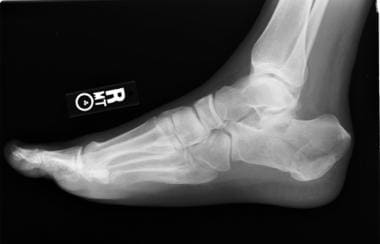
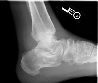
Foot
Surgical treatment of nonunited fractures of the odontoid process, with special reference to occipitocervical fusion for unreducible atlantoaxial subluxation or instability. (1/112)
Fifty-seven consecutive patients treated surgically for nonunited fractures of the odontoid process were reviewed. All patients presented late, exhibiting neurological deficits subsequent to nonunion. Delay in presentation was between 6 and 120 months (mean 32 months) after the original injury, due to missed diagnosis or inappropriate management. Seven patients who were reduced in traction underwent a Gallie atlantoaxial fusion. In the remaining 50 patients who were unreducible, an occipitocervical arthrodesis was performed. They were followed up for a minimum of 2 years, except one who died from postoperative respiratory failure. All patients obtained a solid bony union, including two in whom nonunion occurred following atlantoaxial fusion, and occipitocervical fusion was added as a rescue. Thirty-eight patients achieved excellent neurological recovery, nine still had some disability, five retained their neurological deficits and two reported a deterioration. In two patients, a recurrence in a traumatic episode was experienced long after a resolution. Our findings demonstrate that occipitocervical arthrodesis is preferable for unreducible subluxation or instability of atlantoaxial articulation in nonunion of odontoid fractures. (+info)The treatment of femoral shaft fractures in children: a systematic overview and critical appraisal of the literature. (2/112)
OBJECTIVE: Through a critical systematic overview of the literature on the treatment of pediatric femoral shaft fractures to determine if any method of treatment can be recommended over others. DATA SOURCES: A MEDLINE search was performed for all cohort and randomized clinical trials for the years 1966 to 1996. STUDY SELECTION: Of 1217 identified articles, 15 cohort studies (where 2 or more treatments were compared in the same study) reported the treatment of children with femoral fractures. DATA EXTRACTION: Information was abstracted and articles rated for quality blind to author, institution and journal. DATA SYNTHESIS: Children having early application of a hip spica cast had an average hospital stay of 11 days (range from 5 to 29 days), average charges of $5784 (range from $590 to $11,800), average rates of limb-length discrepancy (greater than 2 cm) of 3% (range from 0 to 25%), angulatory malunion rates (greater than 10 degrees) of 8% (range from 0 to 19%), and rotational malunion rates (greater than 10 degrees) of 13% (range from 0 to 5%). The costs and malunion rates of early application of a hip spica cast were lower than for traction. Internal fixation (including intramedullary nails) had low angulatory malunion rates compared with early application of a hip spica cast but higher over-lengthening rates (greater than 2 cm) of 25% (range from 5% to 100%) and mean rotational malunion rates (greater than 10 degrees) of 25% (range from 11% to 32%). CONCLUSION: Early application of a hip spica cast had lower costs and malunion rates than traction. (+info)The role of physiotherapy and clinical predictors of outcome after fracture of the distal radius. (3/112)
The capacity for physiotherapy to improve the outcome after fracture of the distal radius is unproven. We carried out a randomised controlled trial on 96 patients, comparing conventional physiotherapy with a regime of home exercises. The function of the upper limb was assessed at the time of removal of the plaster cast and at three and six months after injury. Factors which may predict poor outcome in these patients were sought. Grip strength and hand function did not significantly differ between the two groups. Flexion and extension of the wrist were the only movements to improve with physiotherapy at six months (p = 0.001). Predictors of poor functional outcome were malunion and impaired function before the fracture. These patients presented with pain, decreased rotation of the forearm and low functional scores at six weeks. Our study has shown that home exercises are adequate rehabilitation after uncomplicated fracture of the distal radius, and routine referral for a course of physiotherapy should be discouraged. The role of physiotherapy in patients at high risk of a poor outcome requires further investigation. (+info)The Sauve-Kapandji procedure for post-traumatic disorders of the distal radio-ulnar joint. (4/112)
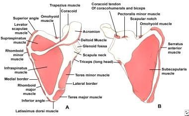
We present the results of a retrospective series of 41 Sauve-Kapandji procedures carried out for complications of fractures of the distal radius. All the operations were undertaken by one surgeon with a mean follow-up of 32 months. A total of 37 patients was available for clinical review. The indications for surgery were pain on the ulnar side of the wrist and decreased rotation of the forearm. Intraperiosteal and extraperiosteal techniques were used for resection of the ulna, with no difference in outcome. Patients were assessed for pain, rotation of the forearm and complications. A Mayo Modified Wrist Score was used. Pain was improved in 25 of the 37 patients, and unchanged in ten. Rotation of the forearm returned to within 7 degrees of the uninjured side. The results are discussed in relation to the presence of preoperative malunion of the distal radius, age and the functional outcome. Age is not a contraindication for this procedure. (+info)Heterotopic ossification following internal fixation or arthroplasty for displaced femoral neck fractures: a prospective randomized study. (5/112)
One hundred hips in 99 patients of 75 years or older, with a displaced femoral neck fracture, were studied for heterotopic ossification (HO). The patients were randomized to either internal fixation or total hip arthroplasty (THA). In the THA group HO was found in 32 of 45 hips compared with 1 of 39 in the internal fixation group (P < 0.0012). The frequency of HO after THA corresponds well with findings in other studies on patients receiving THA for osteoarthrosis. In cervical fractures the surgical procedure of total hip replacement seems to be a prerequisite for HO, indicating that the procedure itself is more important than the patient's age and the diagnosis. Severe symptoms due to HO were found in only one patient. HO following THA for a femoral neck fracture is of little clinical importance and prophylaxis is unnecessary. (+info)Complications of fracture treatment by traditional bonesetters in southwest Nigeria. (6/112)
BACKGROUND: Traditional bonesetters (TBS) practice widely in Nigeria. OBJECTIVE: Our aim was to evaluate the types of complications seen in patients previously treated by TBS and to assess factors that may predispose to the complications. METHODS: We carried out a prospective non-randomized controlled study in a general hospital in southwest Nigeria. All patients brought into the hospital over the 10-month study period with fractures who had been treated previously by a TBS and, as a control, all patients brought directly to and treated by us were studied. Each patient was assessed and prescribed the most appropriate treatment for their fracture: reduction, immobilization (operatively and otherwise) and physiotherapy. Malunion, non-union, delayed union, gangrene, stiffness of joints and loss of joint motion, Volkman's ischaemic contracture and tetanus were all investigated. RESULTS: Over half of the patients in the TBS subgroup had malunion, and a quarter had non-union. Only one out of the 36 (2.8%) had no complaints and was satisfied with the outcome of treatment of his fractures by the TBS. In the orthodox subgroup, there were seven complications as a result of treatment of a total of 49 bones (14%). Most of the complications involved the loss of joint motion. CONCLUSIONS: There were no statistically significant associations between the complications recorded and the ages of the patients, types of bone fractured or the duration of treatment in patients who were in the TBS subgroup. The introduction of a health insurance scheme in Nigeria may make it easier for individuals and families to be able to afford proper fracture treatment in hospitals. (+info)Recurrent supracondylar humerus fracture following prior malunion. (7/112)
In this report, two patients sustained a recurrent supracondylar humerus fracture following malunion of a previous supracondylar humerus fracture. The patients were treated for their first fracture at 5 and 6 years of age, respectively. One underwent open reduction with percutaneous pinning, and the other was treated with closed reduction with casting. Both patients healed in a moderate degree of extension after the first fracture. Two years later, both sustained a second fracture of the supracondylar humerus. Both had closed reduction with percutaneous pinning and went on to heal uneventfully. We speculate that ensuing post-traumatic extension deformity may accentuate a child's tendency for elbow hyperextension. Extension malunion may place the child at increased risk for a second fracture via similar mechanisms of injury. (+info)The effect of rotational malunion of the radius and the ulna on supination and pronation. (8/112)
We have assessed the influence of isolated and combined rotational malunion of the radius and ulna on the rotation of the forearm. Osteotomies were made in both the radius and the ulna at the mid-diaphyseal level of five cadaver forearms and stabilised with intramedullary metal implants. Malunion about the axis of the respective forearm bone was produced at intervals of 10 degrees. The ranges of pronation and supination were recorded by a potentiometer under computer control. We examined rotational malunions of 10 degrees to 80 degrees of either the radius or ulna alone and combined rotational malunions of 20 degrees to 60 degrees of both the radius and ulna. Malunion of the ulna in supination had little effect on rotation of the forearm. Malunion of either the radius or of the ulna in pronation gave a moderate reduction of rotation of the forearm. By contrast, malunion of the radius in supination markedly reduced rotation of the forearm, especially with malunion greater than 60 degrees. Combined rotational malunion produced contrasting results. A combination of rotational malunion of the radius and ulna in the same direction had an effect similar to that of an isolated malunion of the radius. A combination in the opposite direction gave the largest limitation of the range of movement. Clinically, rotational malunion may be isolated or part of a complex angular/rotational deformity and rotational malunion may lead to marked impairment of rotation of the forearm. A reproducible method for assessing rotational malunion is therefore needed. (+info)Malunited fractures refer to a type of fracture where the bones do not heal in their proper alignment or position. This can occur due to various reasons such as inadequate reduction of the fracture fragments during initial treatment, improper casting or immobilization, or failure of the patient to follow proper immobilization instructions. Malunited fractures can result in deformity, limited range of motion, and decreased functionality of the affected limb. Additional treatments such as surgery may be required to correct the malunion and restore normal function.
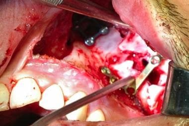
A jaw fracture, also known as a mandibular fracture, is a break in the lower jawbone. It can occur at any point along the bone, from the condyle (the rounded end that articulates with the skull) to the symphysis (the area where the two halves of the jaw meet in the front).
Jaw fractures are typically caused by trauma, such as a direct blow to the face during sports injuries, traffic accidents, or physical assaults. They can also result from falls, particularly in older adults with osteoporosis.
Symptoms of jaw fractures may include pain, swelling, bruising, difficulty speaking, chewing, or opening the mouth wide, and malocclusion (the teeth do not fit together properly when biting down). In some cases, there may be visible deformity or mobility in the jaw.
Diagnosis of jaw fractures typically involves a thorough physical examination, dental X-rays, CT scans, or other imaging studies to assess the location and severity of the fracture. Treatment may involve immobilization with wires or braces, pain management, antibiotics to prevent infection, and in some cases, surgery to realign and stabilize the bone fragments.
A bone fracture is a medical condition in which there is a partial or complete break in the continuity of a bone due to external or internal forces. Fractures can occur in any bone in the body and can vary in severity from a small crack to a shattered bone. The symptoms of a bone fracture typically include pain, swelling, bruising, deformity, and difficulty moving the affected limb. Treatment for a bone fracture may involve immobilization with a cast or splint, surgery to realign and stabilize the bone, or medication to manage pain and prevent infection. The specific treatment approach will depend on the location, type, and severity of the fracture.
Osteotomy is a surgical procedure in which a bone is cut to shorten, lengthen, or change its alignment. It is often performed to correct deformities or to realign bones that have been damaged by trauma or disease. The bone may be cut straight across (transverse osteotomy) or at an angle (oblique osteotomy). After the bone is cut, it can be realigned and held in place with pins, plates, or screws until it heals. This procedure is commonly performed on bones in the leg, such as the femur or tibia, but can also be done on other bones in the body.
A radius fracture is a break in the bone that runs from the wrist to the elbow, located on the thumb side of the forearm. Radius fractures can occur as a result of a fall, direct blow to the forearm, or a high-energy collision such as a car accident. There are various types of radius fractures, including:
1. Distal radius fracture: A break at the end of the radius bone, near the wrist joint, which is the most common type of radius fracture.
2. Radial shaft fracture: A break in the middle portion of the radius bone.
3. Radial head and neck fractures: Breaks in the upper part of the radius bone, near the elbow joint.
4. Comminuted fracture: A complex radius fracture where the bone is broken into multiple pieces.
5. Open (compound) fracture: A radius fracture with a wound or laceration in the skin, allowing for communication between the outside environment and the fractured bone.
6. Intra-articular fracture: A radius fracture that extends into the wrist joint or elbow joint.
7. Torus (buckle) fracture: A stable fracture where one side of the bone is compressed, causing it to buckle or bend, but not break completely through.
Symptoms of a radius fracture may include pain, swelling, tenderness, bruising, deformity, limited mobility, and in some cases, numbness or tingling in the fingers. Treatment options depend on the type and severity of the fracture but can range from casting to surgical intervention with implant fixation.
Fracture healing is the natural process by which a broken bone repairs itself. When a fracture occurs, the body responds by initiating a series of biological and cellular events aimed at restoring the structural integrity of the bone. This process involves the formation of a hematoma (a collection of blood) around the fracture site, followed by the activation of inflammatory cells that help to clean up debris and prepare the area for repair.
Over time, specialized cells called osteoblasts begin to lay down new bone matrix, or osteoid, along the edges of the broken bone ends. This osteoid eventually hardens into new bone tissue, forming a bridge between the fracture fragments. As this process continues, the callus (a mass of newly formed bone and connective tissue) gradually becomes stronger and more compact, eventually remodeling itself into a solid, unbroken bone.
The entire process of fracture healing can take several weeks to several months, depending on factors such as the severity of the injury, the patient's age and overall health, and the location of the fracture. In some cases, medical intervention may be necessary to help promote healing or ensure proper alignment of the bone fragments. This may include the use of casts, braces, or surgical implants such as plates, screws, or rods.
Fracture fixation, internal, is a surgical procedure where a fractured bone is fixed using metal devices such as plates, screws, or rods that are implanted inside the body. This technique helps to maintain the alignment and stability of the broken bone while it heals. The implants may be temporarily or permanently left inside the body, depending on the nature and severity of the fracture. Internal fixation allows for early mobilization and rehabilitation, which can result in a faster recovery and improved functional outcome.
Mandibular injuries refer to damages or traumas that affect the mandible, which is the lower part of the jawbone. These injuries can result from various causes, such as road accidents, physical assaults, sports-related impacts, or falls. Mandibular injuries may include fractures, dislocations, soft tissue damage, or dental injuries.
Symptoms of mandibular injuries might include pain, swelling, bruising, difficulty speaking, chewing, or opening the mouth wide, and in some cases, visible deformity or misalignment of the jaw. Depending on the severity and type of injury, treatment options may range from conservative management with pain control and soft diet to surgical intervention for fracture reduction and fixation. Immediate medical attention is crucial to ensure proper diagnosis, appropriate treatment, and prevention of potential complications.
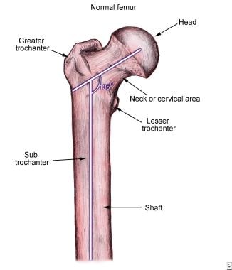
A hip fracture is a medical condition referring to a break in the upper part of the femur (thigh) bone, which forms the hip joint. The majority of hip fractures occur due to falls or direct trauma to the area. They are more common in older adults, particularly those with osteoporosis, a condition that weakens bones and makes them more prone to breaking. Hip fractures can significantly impact mobility and quality of life, often requiring surgical intervention and rehabilitation.
Dental occlusion, traumatic is a term used to describe an abnormal bite or contact between the upper and lower teeth that results in trauma or injury to the oral structures. This can occur when there is a discrepancy in the alignment of the teeth or jaws, such as an overbite, underbite, or crossbite, which causes excessive force or pressure on certain teeth or tissues.
Traumatic dental occlusion can result in various dental and oral health issues, including tooth wear, fractures, mobility of teeth, gum recession, and temporomandibular joint (TMJ) disorders. It is important to diagnose and treat traumatic dental occlusion early to prevent further damage and alleviate any discomfort or pain. Treatment options may include orthodontic treatment, adjustment of the bite, restoration of damaged teeth, or a combination of these approaches.
A femoral fracture is a medical term that refers to a break in the thigh bone, which is the longest and strongest bone in the human body. The femur extends from the hip joint to the knee joint and is responsible for supporting the weight of the upper body and allowing movement of the lower extremity. Femoral fractures can occur due to various reasons such as high-energy trauma, low-energy trauma in individuals with weak bones (osteoporosis), or as a result of a direct blow to the thigh.
Femoral fractures can be classified into different types based on their location, pattern, and severity. Some common types of femoral fractures include:
1. Transverse fracture: A break that occurs straight across the bone.
2. Oblique fracture: A break that occurs at an angle across the bone.
3. Spiral fracture: A break that occurs in a helical pattern around the bone.
4. Comminuted fracture: A break that results in multiple fragments of the bone.
5. Open or compound fracture: A break in which the bone pierces through the skin.
6. Closed or simple fracture: A break in which the bone does not pierce through the skin.
Femoral fractures can cause severe pain, swelling, bruising, and difficulty walking or bearing weight on the affected leg. Diagnosis typically involves a physical examination, medical history, and imaging tests such as X-rays or CT scans. Treatment may involve surgical intervention, including the use of metal rods, plates, or screws to stabilize the bone, followed by rehabilitation and physical therapy to restore mobility and strength.
A spinal fracture, also known as a vertebral compression fracture, is a break in one or more bones (vertebrae) of the spine. This type of fracture often occurs due to weakened bones caused by osteoporosis, but it can also result from trauma such as a car accident or a fall.
In a spinal fracture, the front part of the vertebra collapses, causing the height of the vertebra to decrease, while the back part of the vertebra remains intact. This results in a wedge-shaped deformity of the vertebra. Multiple fractures can lead to a hunched forward posture known as kyphosis or dowager's hump.
Spinal fractures can cause pain, numbness, tingling, or weakness in the back, legs, or arms, depending on the location and severity of the fracture. In some cases, spinal cord compression may occur, leading to more severe symptoms such as paralysis or loss of bladder and bowel control.
A comminuted fracture is a type of bone break where the bone is shattered into three or more pieces. This type of fracture typically occurs after high-energy trauma, such as a car accident or a fall from a great height. Commminuted fractures can also occur in bones that are weakened by conditions like osteoporosis or cancer. Because of the severity and complexity of comminuted fractures, they often require extensive treatment, which may include surgery to realign and stabilize the bone fragments using metal screws, plates, or rods.
Fracture fixation is a surgical procedure in orthopedic trauma surgery where a fractured bone is stabilized using various devices and techniques to promote proper healing and alignment. The goal of fracture fixation is to maintain the broken bone ends in correct anatomical position and length, allowing for adequate stability during the healing process.
There are two main types of fracture fixation:
1. Internal fixation: In this method, metal implants like plates, screws, or intramedullary rods are inserted directly into the bone to hold the fragments in place. These implants can be either removed or left in the body once healing is complete, depending on the type and location of the fracture.
2. External fixation: This technique involves placing pins or screws through the skin and into the bone above and below the fracture site. These pins are then connected to an external frame that maintains alignment and stability. External fixators are typically used when there is significant soft tissue damage, infection, or when internal fixation is not possible due to the complexity of the fracture.
The choice between internal and external fixation depends on various factors such as the type and location of the fracture, patient's age and overall health, surgeon's preference, and potential complications. Both methods aim to provide a stable environment for bone healing while minimizing the risk of malunion, nonunion, or deformity.
Osteoporotic fractures are breaks or cracks in bones that occur as a result of osteoporosis, a condition characterized by weak and brittle bones. Osteoporosis causes bones to lose density and strength, making them more susceptible to fractures, even from minor injuries or falls.
The most common types of osteoporotic fractures are:
1. Hip fractures: These occur when the upper part of the thigh bone (femur) breaks, often due to a fall. Hip fractures can be serious and may require surgery and hospitalization.
2. Vertebral compression fractures: These occur when the bones in the spine (vertebrae) collapse, causing height loss, back pain, and deformity. They are often caused by everyday activities, such as bending or lifting.
3. Wrist fractures: These occur when the bones in the wrist break, often due to a fall. Wrist fractures are common in older adults with osteoporosis.
4. Other fractures: Osteoporotic fractures can also occur in other bones, such as the pelvis, ribs, and humerus (upper arm bone).
Prevention is key in managing osteoporosis and reducing the risk of osteoporotic fractures. This includes getting enough calcium and vitamin D, engaging in regular weight-bearing exercise, avoiding smoking and excessive alcohol consumption, and taking medications as prescribed by a healthcare provider.
Spontaneous fractures are bone breaks that occur without any identifiable trauma or injury. They are typically caused by underlying medical conditions that weaken the bones, making them more susceptible to breaking under normal stress or weight. The most common cause of spontaneous fractures is osteoporosis, a condition characterized by weak and brittle bones. Other potential causes include various bone diseases, certain cancers, long-term use of corticosteroids, and genetic disorders affecting bone strength.
It's important to note that while the term "spontaneous" implies that the fracture occurred without any apparent cause, it is usually the result of an underlying medical condition. Therefore, if you experience a spontaneous fracture, seeking medical attention is crucial to diagnose and manage the underlying cause to prevent future fractures and related complications.
Stress fractures are defined as small cracks or severe bruising in bones that occur from repetitive stress or overuse. They most commonly occur in weight-bearing bones, such as the legs and feet, but can also occur in the arms, hips, and back. Stress fractures differ from regular fractures because they typically do not result from a single, traumatic event. Instead, they are caused by repeated stress on the bone that results in microscopic damage over time. Athletes, military personnel, and individuals who engage in high-impact activities or have weak bones (osteoporosis) are at increased risk of developing stress fractures. Symptoms may include pain, swelling, tenderness, and difficulty walking or bearing weight on the affected bone.
A femoral neck fracture is a type of hip fracture that occurs in the narrow, vertical section of bone just below the ball of the femur (thigh bone) that connects to the hip socket. This area is called the femoral neck. Femoral neck fractures can be categorized into different types based on their location and the direction of the fractured bone.
These fractures are typically caused by high-energy trauma, such as car accidents or falls from significant heights, in younger individuals. However, in older adults, particularly those with osteoporosis, femoral neck fractures can also result from low-energy trauma, like a simple fall from standing height.
Femoral neck fractures are often serious and require prompt medical attention. Treatment usually involves surgery to realign and stabilize the broken bone fragments, followed by rehabilitation to help regain mobility and strength. Potential complications of femoral neck fractures include avascular necrosis (loss of blood flow to the femoral head), nonunion or malunion (improper healing), and osteoarthritis in the hip joint.
An ulna fracture is a break in the ulna bone, which is one of the two long bones in the forearm. The ulna is located on the pinky finger side of the forearm and functions to support the elbow joint and assist in rotation and movement of the forearm. Ulna fractures can occur at various points along the bone, including the shaft, near the wrist, or at the elbow end of the bone. Symptoms may include pain, swelling, bruising, tenderness, deformity, limited mobility, and in some cases, numbness or tingling in the fingers. Treatment typically involves immobilization with a cast or splint, followed by rehabilitation exercises to restore strength and range of motion. In severe cases, surgery may be required to realign and stabilize the fractured bone.
Intramedullary fracture fixation is a surgical technique used to stabilize and align bone fractures. In this procedure, a metal rod or nail is inserted into the marrow cavity (intramedullary canal) of the affected bone, spanning the length of the fracture. The rod is then secured to the bone using screws or other fixation devices on either side of the fracture. This provides stability and helps maintain proper alignment during the healing process.
The benefits of intramedullary fixation include:
1. Load sharing: The intramedullary rod shares some of the load bearing capacity with the bone, which can help reduce stress on the healing bone.
2. Minimal soft tissue dissection: Since the implant is inserted through the medullary canal, there is less disruption to the surrounding muscles, tendons, and ligaments compared to other fixation methods.
3. Biomechanical stability: Intramedullary fixation provides rotational and bending stiffness, which helps maintain proper alignment of the fracture fragments during healing.
4. Early mobilization: Patients with intramedullary fixation can often begin weight bearing and rehabilitation exercises earlier than those with other types of fixation, leading to faster recovery times.
Common indications for intramedullary fracture fixation include long bone fractures in the femur, tibia, humerus, and fibula, as well as certain pelvic and spinal fractures. However, the choice of fixation method depends on various factors such as patient age, fracture pattern, location, and associated injuries.
Rib fractures are breaks or cracks in the bones that make up the rib cage, which is the protective structure around the lungs and heart. Rib fractures can result from direct trauma to the chest, such as from a fall, motor vehicle accident, or physical assault. They can also occur from indirect forces, such as during coughing fits in people with weakened bones (osteoporosis).
Rib fractures are painful and can make breathing difficult, particularly when taking deep breaths or coughing. In some cases, rib fractures may lead to complications like punctured lungs (pneumothorax) or collapsed lungs (atelectasis), especially if multiple ribs are broken in several places.
It is essential to seek medical attention for suspected rib fractures, as proper diagnosis and management can help prevent further complications and promote healing. Treatment typically involves pain management, breathing exercises, and, in some cases, immobilization or surgery.
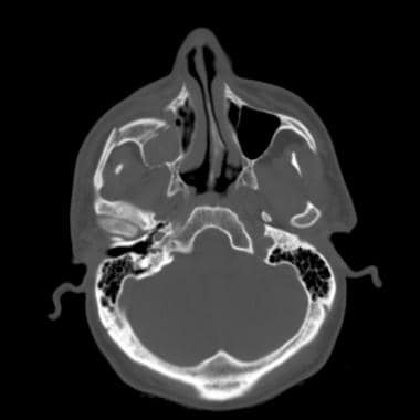
A skull fracture is a break in one or more of the bones that form the skull. It can occur from a direct blow to the head, penetrating injuries like gunshot wounds, or from strong rotational forces during an accident. There are several types of skull fractures, including:
1. Linear Skull Fracture: This is the most common type, where there's a simple break in the bone without any splintering, depression, or displacement. It often doesn't require treatment unless it's near a sensitive area like an eye or ear.
2. Depressed Skull Fracture: In this type, a piece of the skull is pushed inward toward the brain. Surgery may be needed to relieve pressure on the brain and repair the fracture.
3. Diastatic Skull Fracture: This occurs along the suture lines (the fibrous joints between the skull bones) that haven't fused yet, often seen in infants and young children.
4. Basilar Skull Fracture: This involves fractures at the base of the skull. It can be serious due to potential injury to the cranial nerves and blood vessels located in this area.
5. Comminuted Skull Fracture: In this severe type, the bone is shattered into many pieces. These fractures usually require extensive surgical repair.
Symptoms of a skull fracture can include pain, swelling, bruising, bleeding (if there's an open wound), and in some cases, clear fluid draining from the ears or nose (cerebrospinal fluid leak). Severe fractures may cause brain injury, leading to symptoms like confusion, loss of consciousness, seizures, or neurological deficits. Immediate medical attention is necessary for any suspected skull fracture.
A mandibular fracture is a break or crack in the lower jaw (mandible) bone. It can occur at any point along the mandible, but common sites include the condyle (the rounded end near the ear), the angle (the curved part of the jaw), and the symphysis (the area where the two halves of the jaw meet in the front). Mandibular fractures are typically caused by trauma, such as a direct blow to the face or a fall. Symptoms may include pain, swelling, bruising, difficulty chewing or speaking, and malocclusion (misalignment) of the teeth. Treatment usually involves immobilization with wires or screws to allow the bone to heal properly.
A tooth fracture is a dental health condition characterized by a break or crack in the tooth structure. It can occur in different parts of the tooth, including the crown (the visible part), root, or filling. Tooth fractures can result from various factors such as trauma, biting or chewing on hard objects, grinding or clenching teeth, and having large, old amalgam fillings that weaken the tooth structure over time. Depending on the severity and location of the fracture, it may cause pain, sensitivity, or affect the tooth's functionality and appearance. Treatment options for tooth fractures vary from simple bonding to root canal treatment or even extraction in severe cases. Regular dental check-ups are essential for early detection and management of tooth fractures.
A compression fracture is a type of bone fracture that occurs when there is a collapse of a vertebra in the spine. This type of fracture is most commonly seen in the thoracic and lumbar regions of the spine. Compression fractures are often caused by weakened bones due to osteoporosis, but they can also result from trauma or tumors that weaken the bone.
In a compression fracture, the front part (anterior) of the vertebra collapses, while the back part (posterior) remains intact, causing the height of the vertebra to decrease. This can lead to pain, deformity, and decreased mobility. In severe cases, multiple compression fractures can result in a condition called kyphosis, which is an abnormal curvature of the spine that leads to a hunchback appearance.
Compression fractures are typically diagnosed through imaging tests such as X-rays, CT scans, or MRI scans. Treatment may include pain medication, bracing, physical therapy, or in some cases, surgery. Preventive measures such as maintaining a healthy diet, getting regular exercise, and taking medications to prevent or treat osteoporosis can help reduce the risk of compression fractures.
An intra-articular fracture is a type of fracture that involves the joint surface or articular cartilage of a bone. These types of fractures can occur in any joint, but they are most commonly seen in the weight-bearing joints such as the knee, ankle, and wrist.
Intra-articular fractures can be caused by high-energy trauma, such as motor vehicle accidents or falls from significant heights, or by low-energy trauma, such as a simple fall in older adults with osteoporosis.
These types of fractures are often complex and may involve displacement or depression of the joint surface, which can increase the risk of developing post-traumatic arthritis. Therefore, prompt diagnosis and appropriate treatment are essential to ensure optimal outcomes and minimize long-term complications. Treatment options for intra-articular fractures may include surgical fixation with plates, screws, or pins, as well as joint replacement in some cases.
Osteoporosis is a systemic skeletal disease characterized by low bone mass, deterioration of bone tissue, and disruption of bone architecture, leading to increased risk of fractures, particularly in the spine, wrist, and hip. It mainly affects older people, especially postmenopausal women, due to hormonal changes that reduce bone density. Osteoporosis can also be caused by certain medications, medical conditions, or lifestyle factors such as smoking, alcohol abuse, and a lack of calcium and vitamin D in the diet. The diagnosis is often made using bone mineral density testing, and treatment may include medication to slow bone loss, promote bone formation, and prevent fractures.
Bone plates are medical devices used in orthopedic surgery to stabilize and hold together fractured or broken bones during the healing process. They are typically made of surgical-grade stainless steel, titanium, or other biocompatible materials. The plate is shaped to fit the contour of the bone and is held in place with screws that are inserted through the plate and into the bone on either side of the fracture. This provides stability and alignment to the broken bones, allowing them to heal properly. Bone plates can be used to treat a variety of fractures, including those that are complex or unstable. After healing is complete, the bone plate may be left in place or removed, depending on the individual's needs and the surgeon's recommendation.
I believe you are referring to "bone pins" or "bone nails" rather than "bone nails." These terms are used in the medical field to describe surgical implants made of metal or biocompatible materials that are used to stabilize and hold together fractured bones during the healing process. They can also be used in spinal fusion surgery to provide stability and promote bone growth between vertebrae.
Bone pins or nails typically have a threaded or smooth shaft, with a small diameter that allows them to be inserted into the medullary canal of long bones such as the femur or tibia. They may also have a head or eyelet on one end that allows for attachment to external fixation devices or other surgical instruments.
The use of bone pins and nails has revolutionized orthopedic surgery, allowing for faster healing times, improved stability, and better functional outcomes for patients with fractures or spinal deformities.
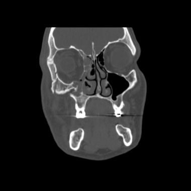
Orbital fractures refer to breaks in the bones that make up the eye socket, also known as the orbit. These bones include the maxilla, zygoma, frontal bone, and palatine bone. Orbital fractures can occur due to trauma, such as a blunt force injury or a penetrating wound.
There are several types of orbital fractures, including:
1. Blowout fracture: This occurs when the thin bone of the orbital floor is broken, often due to a direct blow to the eye. The force of the impact can cause the eyeball to move backward, breaking the bone and sometimes trapping the muscle that moves the eye (the inferior rectus).
2. Blow-in fracture: This type of fracture involves the breakage of the orbital roof, which is the bone that forms the upper boundary of the orbit. It typically occurs due to high-impact trauma, such as a car accident or a fall from a significant height.
3. Direct fracture: A direct fracture happens when there is a break in one or more of the bones that form the walls of the orbit. This type of fracture can result from a variety of traumas, including motor vehicle accidents, sports injuries, and assaults.
4. Indirect fracture: An indirect fracture occurs when the force of an injury is transmitted to the orbit through tissues surrounding it, causing the bone to break. The most common type of indirect orbital fracture is a blowout fracture.
Orbital fractures can cause various symptoms, including pain, swelling, bruising, and double vision. In some cases, the fracture may also lead to enophthalmos (sinking of the eye into the orbit) or telecanthus (increased distance between the inner corners of the eyes). Imaging tests, such as CT scans, are often used to diagnose orbital fractures and determine the best course of treatment. Treatment may include observation, pain management, and in some cases, surgery to repair the fracture and restore normal function.
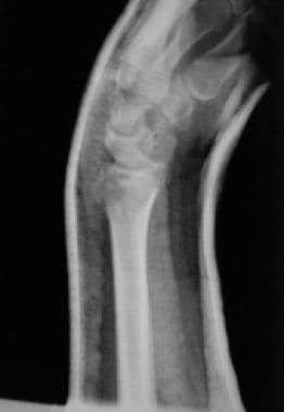
A Colles' fracture is a specific type of fracture in the distal end of the radius bone in the forearm, which is the larger of the two bones in the lower arm. This type of fracture occurs when the wrist is forcefully bent backward (dorsiflexion), often as a result of falling onto an outstretched hand.
In a Colles' fracture, the distal end of the radius bone breaks and is displaced downward and angulated backward, resulting in a characteristic "dinner fork" deformity. This type of fracture is more common in older individuals, particularly women with osteoporosis, but can also occur in younger people as a result of high-energy trauma.
Colles' fractures are typically treated with immobilization using a cast or splint to hold the bones in proper alignment while they heal. In some cases, surgery may be necessary to realign and stabilize the fracture, particularly if there is significant displacement or instability of the bone fragments.
Bony callus is a medical term that refers to the specialized tissue that forms in response to a bone fracture. It is a crucial part of the natural healing process, as it helps to stabilize and protect the broken bone while it mends.
When a bone is fractured, the body responds by initiating an inflammatory response, which triggers the production of various cells and signaling molecules that promote healing. As part of this process, specialized cells called osteoblasts begin to produce new bone tissue at the site of the fracture. This tissue is initially soft and pliable, allowing it to bridge the gap between the broken ends of the bone.
Over time, this soft callus gradually hardens and calcifies, forming a bony callus that helps to stabilize the fracture and provide additional support as the bone heals. The bony callus is typically composed of a mixture of woven bone (which is less organized than normal bone) and more structured lamellar bone (which is similar in structure to normal bone).
As the bone continues to heal, the bony callus may be gradually remodeled and reshaped by osteoclasts, which are specialized cells that break down and remove excess or unwanted bone tissue. This process helps to restore the bone's original shape and strength, allowing it to function normally again.
It is worth noting that excessive bony callus formation can sometimes lead to complications, such as stiffness, pain, or decreased range of motion in the affected limb. In some cases, surgical intervention may be necessary to remove or reduce the size of the bony callus and promote proper healing.
Periprosthetic fractures are defined as fractures that occur in close proximity to a prosthetic joint, such as those found in total hip or knee replacements. These types of fractures typically occur as a result of low-energy trauma, and can be caused by a variety of factors including osteoporosis, bone weakness, or loosening of the prosthetic implant.
Periprosthetic fractures are classified based on the location of the fracture in relation to the prosthesis, as well as the stability of the implant. Treatment options for periprosthetic fractures may include non-surgical management, such as immobilization with a brace or cast, or surgical intervention, such as open reduction and internal fixation (ORIF) or revision arthroplasty.
The management of periprosthetic fractures can be complex and requires careful consideration of various factors, including the patient's age, overall health status, bone quality, and functional needs. As such, these types of fractures are typically managed by orthopedic surgeons with experience in joint replacement surgery and fracture care.
Tarsal Tunnel Syndrome (TTS) is a compressive neuropathy of the tibial nerve as it passes through the tarsal tunnel, a fibro-osseous canal formed by the medial malleolus and the talus bones on the inner ankle. The tibial nerve and its branches provide sensory innervation to the sole of the foot and motor function to several muscles in the lower leg and foot.
In TTS, increased pressure or compression within the tarsal tunnel leads to entrapment of the tibial nerve or its branches, resulting in pain, numbness, tingling, or burning sensations along the distribution of the affected nerves. Common causes include space-occupying lesions (e.g., ganglion cysts, varicosities), trauma, tenosynovitis, or systemic conditions like diabetes and rheumatoid arthritis.
Diagnosis typically involves a thorough clinical examination, including the patient's history, physical examination, and specialized tests such as nerve conduction studies and electromyography (EMG). Treatment options may include conservative measures like immobilization, orthotics, nonsteroidal anti-inflammatory drugs (NSAIDs), or corticosteroid injections. In severe cases or when conservative treatments fail, surgical decompression of the tarsal tunnel might be necessary to alleviate symptoms and prevent further nerve damage.
The Tibial nerve is a major branch of the sciatic nerve that originates in the lower back and runs through the buttock and leg. It provides motor (nerve impulses that control muscle movement) and sensory (nerve impulses that convey information about touch, temperature, and pain) innervation to several muscles and skin regions in the lower limb.
More specifically, the Tibial nerve supplies the following structures:
1. Motor Innervation: The Tibial nerve provides motor innervation to the muscles in the back of the leg (posterior compartment), including the calf muscles (gastrocnemius and soleus) and the small muscles in the foot (intrinsic muscles). These muscles are responsible for plantarflexion (pointing the foot downward) and inversion (turning the foot inward) of the foot.
2. Sensory Innervation: The Tibial nerve provides sensory innervation to the skin on the sole of the foot, as well as the heel and some parts of the lower leg.
The Tibial nerve travels down the leg, passing behind the knee and through the calf, where it eventually joins with the common fibular (peroneal) nerve to form the tibial-fibular trunk. This trunk then divides into several smaller nerves that innervate the foot's intrinsic muscles and skin.
Damage or injury to the Tibial nerve can result in various symptoms, such as weakness or paralysis of the calf and foot muscles, numbness or tingling sensations in the sole of the foot, and difficulty walking or standing on tiptoes.
Electrodiagnosis, also known as electromyography (EMG), is a medical diagnostic procedure that evaluates the health and function of muscles and nerves. It measures the electrical activity of skeletal muscles at rest and during contraction, as well as the conduction of electrical signals along nerves.
The test involves inserting a thin needle electrode into the muscle to record its electrical activity. The physician will ask the patient to contract and relax the muscle while the electrical activity is recorded. The resulting data can help diagnose various neuromuscular disorders, such as nerve damage or muscle diseases, by identifying abnormalities in the electrical signals.
Electrodiagnosis can be used to diagnose conditions such as carpal tunnel syndrome, peripheral neuropathy, muscular dystrophy, and amyotrophic lateral sclerosis (ALS), among others. It is a valuable tool in the diagnosis and management of neuromuscular disorders, helping physicians to develop appropriate treatment plans for their patients.
Carpal Tunnel Syndrome (CTS) is a common peripheral nerve disorder that affects the median nerve, which runs from the forearm into the hand through a narrow tunnel-like structure in the wrist called the carpal tunnel. The condition is caused by compression or pinching of the median nerve as it passes through this tunnel, leading to various symptoms such as numbness, tingling, and weakness in the hand and fingers.
The median nerve provides sensation to the thumb, index finger, middle finger, and half of the ring finger. It also controls some small muscles in the hand that allow for fine motor movements. When the median nerve is compressed or damaged due to CTS, it can result in a range of symptoms including:
1. Numbness, tingling, or burning sensations in the fingers (especially the thumb, index finger, middle finger, and half of the ring finger)
2. Pain or discomfort in the hand, wrist, or forearm
3. Weakness in the hand, leading to difficulty gripping objects or making a fist
4. A sensation of swelling or inflammation in the fingers, even if there is no visible swelling present
5. Nighttime symptoms that may disrupt sleep patterns
The exact cause of Carpal Tunnel Syndrome can vary from person to person, but some common risk factors include:
1. Repetitive hand and wrist motions (such as typing, writing, or using tools)
2. Prolonged exposure to vibrations (from machinery or power tools)
3. Wrist trauma or fractures
4. Pregnancy and hormonal changes
5. Certain medical conditions like diabetes, rheumatoid arthritis, and thyroid disorders
6. Obesity
7. Smoking
Diagnosis of Carpal Tunnel Syndrome typically involves a physical examination, medical history review, and sometimes specialized tests like nerve conduction studies or electromyography to confirm the diagnosis and assess the severity of the condition. Treatment options may include splinting, medication, corticosteroid injections, and in severe cases, surgery to relieve pressure on the median nerve.
In medical terms, toes are the digits located at the end of the foot. Humans typically have five toes on each foot, consisting of the big toe (hallux), second toe, third toe, fourth toe, and little toe (fifth toe). The bones of the toes are called phalanges, with the exception of the big toe, which has a different bone structure and is composed of a proximal phalanx, distal phalanx, and sometimes a sesamoid bone.
Toes play an essential role in maintaining balance and assisting in locomotion by helping to push off the ground during walking or running. They also contribute to the overall stability and posture of the body. Various medical conditions can affect toes, such as ingrown toenails, bunions, hammertoes, and neuromas, which may require specific treatments or interventions to alleviate pain, restore function, or improve appearance.
In medical terms, the foot is the part of the lower limb that is distal to the leg and below the ankle, extending from the tarsus to the toes. It is primarily responsible for supporting body weight and facilitating movement through push-off during walking or running. The foot is a complex structure made up of 26 bones, 33 joints, and numerous muscles, tendons, ligaments, and nerves that work together to provide stability, balance, and flexibility. It can be divided into three main parts: the hindfoot, which contains the talus and calcaneus (heel) bones; the midfoot, which includes the navicular, cuboid, and cuneiform bones; and the forefoot, which consists of the metatarsals and phalanges that form the toes.
Medical professionals define "flatfoot" or "pes planus" as a postural deformity in which the arch of the foot collapses, leading to the entire sole of the foot coming into complete or near-complete contact with the ground. This condition can be classified as flexible (the arch reappears when the foot is not bearing weight) or rigid (the arch does not reappear). Flatfoot can result from various factors such as genetics, injury, aging, or certain medical conditions like rheumatoid arthritis and cerebral palsy. In some cases, flatfoot may not cause any symptoms or problems; however, in other instances, it can lead to pain, discomfort, or difficulty walking. Treatment options for flatfoot depend on the severity of the condition and associated symptoms and may include physical therapy, orthotics, bracing, or surgery.
 Complications of distal radius fracture fixation
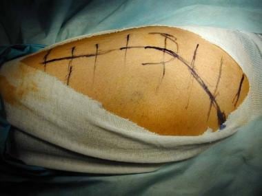
 View of Evaluation of management of malunited supracondylar fracture of humerus by lateral closing wedge osteotomy
 Zygomaticomaxillary Complex Fractures: Practice Essentials, Problem, Epidemiology
 Tarsal tunnel syndrome - Wikipedia
 Filip Stockmans's research works | KU Leuven, Leuven (ku leuven) and other places
 Fracture malunion | Radiology Reference Article | Radiopaedia.org
 Can Total Wrist Arthroplasty Be an Option for Treatment of Highly Comminuted Distal Radius Fracture in Selected Patients?...
 Three-dimensional postoperative accuracy of extra-articular forearm osteotomies using CT-scan based patient-specific surgical...
 Centers of Excellence<...
 Thieme E-Journals - Journal of Wrist Surgery / Abstract
 Surgical Neurology International
 Nonreamed locking intramedullary nailing for open fractures of the tibia.
 BVS Brasil
 Reverse total shoulder arthroplasty for complex proximal humeral fractures in the elderly: How to improve outcomes and avoid...
 Publikationen über Medizinische Analysen mit Simi Systemen
 Orthopedic Surgery Update Orthopedic Surgery - 医疗专业人员 - 妙佑医疗国际
 Fronto orbital complex fracture surgery recovery time
 DeCS
DeCS Relationship between stability of external fixator device and healing process of pilon fracture: Biomechanics perspective |...
 Search results | Anatomia Collection: anatomical plates 1522-1867
 Bradley Evanoff - Research output
- Research Profiles at Washington University School of Medicine
Bradley Evanoff - Research output
- Research Profiles at Washington University School of Medicine Thoracic Outlet Compression Syndromes (TOS) - Neurologic Disorders - MSD Manual Professional Edition
 Pediatric Both Bone Forearm Fractures : Wheeless' Textbook of Orthopaedics
 Government Medical College, Bhavnagar