Fractures, Ununited
Tibial Fractures
Fracture Fixation, Internal
Fracture Healing
Hip Fractures
Fracture Fixation
Osteoporotic Fractures
Radius Fractures
Fractures, Spontaneous
Fractures, Stress
Femoral Neck Fractures
Fracture Fixation, Intramedullary
Rib Fractures
Skull Fractures
Fractures, Compression
Osteoporosis
Bone Plates
Bone Nails
Orbital Fractures
Colles' Fracture
Bony Callus
Periprosthetic Fractures
Bone Density
New Hampshire
World War II
Encyclopedias as Topic
Electric Power Supplies
Nonunion of tibial stress fractures in patients with deformed arthritic knees. Treatment using modular total knee arthroplasty. (1/349)
In two years we treated four women with ununited stress fractures of their proximal tibial diaphyses. They all had arthritis and valgus deformity. The stress fractures had been treated elsewhere by non-operative means in three patients and by open reduction and internal fixation in one, but had failed to unite. After treatment with a modular total knee prosthesis with a long tibial stem extension, all the fractures united. A modular total knee prosthesis is suitable for the rare and difficult problem of ununited tibial stress fractures in patients with deformed arthritic knees since it corrects the deformity and the adverse biomechanics at the fracture site, stabilises the fracture and treats the arthritis. (+info)Expression of the gene encoding the matrix gla protein by mature osteoblasts in human fracture non-unions. (2/349)
BACKGROUND: Osteoblast phenotypic abnormality, namely the expression of collagen type III, has been shown previously in fracture non-union woven bone. AIMS: To investigate osteoblasts from fracture non-unions for evidence of gene expression of non-collagenous bone matrix proteins that have been implicated in mineralisation, namely matrix gla protein (MGP), osteonectin, osteopontin, and osteocalcin. MGP is a consistent component of bone matrix, but there are no reports of osteoblasts in the skeleton expressing the gene for MGP, and the site of synthesis of skeletal MGP (perhaps the liver) has yet to be determined. METHODS: Biopsies from normally healing human fractures and non-unions were examined by means of in situ hybridisation, using 35S labelled probes and autoradiography to disclose levels of gene expression. RESULTS: In normally healing fractures, mature osteoblasts on woven bone were negative for MGP mRNA, but positive for osteonectin, osteopontin, and osteocalcin mRNA molecules. In non-unions, osteoblasts displayed a novel phenotype: they were positive for MGP mRNA, in addition to osteonectin, osteopontin, and osteocalcin mRNA molecules. CONCLUSIONS: Mature osteoblasts in slowly healing fractures have an unusual phenotype: they express the gene encoding MGP, which indicates that control of osteoblast gene expression in non-unions is likely to be abnormal. This might be of importance in the pathogenesis of non-uniting human fractures, and is of current interest given the emerging status of MGP as an inhibitor of mineralisation. (+info)The relationship between the site of nonunion of the scaphoid and scaphoid nonunion advanced collapse (SNAC). (3/349)
We studied retrospectively the radiographs of 33 patients with late symptoms after scaphoid nonunion in an attempt to relate the incidence of scaphoid nonunion advanced collapse (SNAC) to the level of the original fracture. We found differing patterns for nonunion at the proximal, middle and distal thirds. The mean intervals between fracture and complaint were 20.9, 6.7 and 12.6 years and obvious degenerative changes occurred in 85.7%, 40.0% and 33.3%, for the six proximal-, eight middle- and two distal-third nonunions, respectively. Nonunion at the proximal and middle thirds showed the first degenerative changes at the radioscaphoid joint, and this was followed by narrowing of the scaphocapitate and then the lunocapitate joints. In our two nonunions of the distal third degenerative changes were seen only at the lunocapitate joint. Most patients with SNAC and nonunion of the middle or distal third showed dorsal intercalated instability; few patients with nonunion of the proximal third developed this deformity. We discuss the initial management of nonunion of the scaphoid at different levels in the light of our findings, and make recommendations. (+info)Ipsilateral vascularised fibular transport for massive defects of the tibia. (4/349)
The ipsilateral and contralateral fibulae have been used as a vascularised bone graft for loss of tibial bone usually by methods which have involved specialised microvascular techniques to preserve or re-establish the blood supply. We have developed a method of tibialisation of the fibula using the Ilizarov fixator system, ipsilateral vascularised fibular transport (IVFT), and have used it in five patients with massive loss of tibial bone after treatment of an open fracture, infected nonunion or chronic osteomyelitis. All had successful transport, proximal and distal union, and hypertrophy of the graft without fracture. One developed a squamous-cell carcinoma which ultimately required amputation of the limb. The advantage of IVFT is that the fibular segment retains its vascularity without the need for microvascular dissection or anastomoses. Superiosteal formation of new bone occurs if the tibial periosteal bed is retained. Other procedures such as corticotomy and lengthening can be carried out concurrently. (+info)Trochanteric non-union in revision total hip arthroplasty: does it matter? (5/349)
The aims of this study were to assess whether trochanteric non-union is an important factor in revision total hip arthroplasty in terms of postoperative morbidity. We studied prospectively 97 consecutive patients undergoing revision total hip arthroplasty in the years 1992-1996. All operations were performed by one surgeon through a Charnley trans-trochanteric approach. The patients were followed-up over a period of 1-4 years and at 12 months postsurgery were assessed using a modified scoring system devised by D'Aubigne. Anatomical union of the greater trochanter was assessed by an anterior-posterior pelvic radiograph at 12 months to decide if the greater trochanter was united in the correct anatomical position. The trochanteric non-union rate was 18.5% (18 out of 97 patients). There was no significant difference between the patients in terms of pain, function and satisfaction scores at one year between those with trochanteric union and those without. This study suggests that trochanteric non-union post revision total hip arthroplasty is not a cause of increased morbidity. (+info)Complications of Marchetti locked nailing for humeral shaft fractures. (6/349)
In this retrospective study 50 humeral fractures (36 acute, 6 pathological fractures and 8 non-unions) were treated by retrograde locked bundled Marchetti nailing. No intraoperative complications occurred. Postoperative complications included 7 non-unions (4/36 acute fractures and 3/8 delayed union), and 2 intraarticular penetrations of the secondary nails. However, at the subsequent removal of the implant 5 supracondylar fractures occurred. (+info)Treatment of nonunion around the olecranon fossa of the humerus by intramedullary locked nailing. (7/349)
Nonunion of fractures of the olecranon fossa of the humerus presents a difficult surgical problem. The distal fragment is usually small and osteoporotic and stable fixation is not easy to achieve. We describe a modification of the technique of locked nailing by which the distal aspect of the nail is placed in the subchondral region of the trochlea. Good results were obtained in seven out of eight patients with this technique. (+info)Fractures of the distal radius treated by internal fixation and early function. A prospective study of 73 consecutive patients. (8/349)
Stable fixation of fractures of the distal radius can be achieved by using two 2.0 mm titanium plates placed on the radial and intermediate columns angled 50 degrees to 70 degrees apart. We describe our results with this method in a prospective series of 74 fractures (58 severely comminuted) in 73 consecutive patients. Early postoperative mobilisation was possible in all except four wrists. All of the 73 patients, except two with other injuries, returned to work and daily activities with no limitations. The anatomical results were excellent or good in 72 patients and fair in one. Our discussion includes details of important technical considerations based on an analysis of the specific complications which were seen early in the series. (+info)Ununited fracture is a medical term used to describe a fractured bone that has failed to heal properly. This condition is also known as a nonunion fracture. In a normal healing process, the broken ends of the bone will grow together, or "unite," over time as new bone tissue forms. However, in some cases, the bones may not reconnect due to various reasons such as infection, poor blood supply, excessive motion at the fracture site, or inadequate stabilization of the fracture.
Ununited fractures can cause significant pain, swelling, and deformity in the affected area. They may also lead to a decreased range of motion, weakness, and instability in the joint near the fracture. Treatment for ununited fractures typically involves surgical intervention to promote bone healing, such as bone grafting or internal fixation with screws or plates. In some cases, electrical stimulation or ultrasound therapy may also be used to help promote bone growth and healing.
A tibial fracture is a medical term that refers to a break in the shin bone, which is called the tibia. The tibia is the larger of the two bones in the lower leg and is responsible for supporting much of your body weight. Tibial fractures can occur in various ways, such as from high-energy trauma like car accidents or falls, or from low-energy trauma in individuals with weakened bones due to osteoporosis or other medical conditions.
Tibial fractures can be classified into different types based on the location, pattern, and severity of the break. Some common types of tibial fractures include:
1. Transverse fracture: A straight break that goes across the bone.
2. Oblique fracture: A diagonal break that slopes across the bone.
3. Spiral fracture: A break that spirals around the bone, often caused by twisting or rotational forces.
4. Comminuted fracture: A break where the bone is shattered into multiple pieces.
5. Open fracture: A break in which the bone pierces through the skin, increasing the risk of infection.
6. Closed fracture: A break in which the bone does not pierce through the skin.
Tibial fractures can cause symptoms such as pain, swelling, bruising, deformity, and difficulty walking or bearing weight on the affected leg. Treatment for tibial fractures may include immobilization with a cast or brace, surgery to realign and stabilize the bone with plates, screws, or rods, and rehabilitation to restore strength, mobility, and function to the injured limb.
Fracture fixation, internal, is a surgical procedure where a fractured bone is fixed using metal devices such as plates, screws, or rods that are implanted inside the body. This technique helps to maintain the alignment and stability of the broken bone while it heals. The implants may be temporarily or permanently left inside the body, depending on the nature and severity of the fracture. Internal fixation allows for early mobilization and rehabilitation, which can result in a faster recovery and improved functional outcome.
A bone fracture is a medical condition in which there is a partial or complete break in the continuity of a bone due to external or internal forces. Fractures can occur in any bone in the body and can vary in severity from a small crack to a shattered bone. The symptoms of a bone fracture typically include pain, swelling, bruising, deformity, and difficulty moving the affected limb. Treatment for a bone fracture may involve immobilization with a cast or splint, surgery to realign and stabilize the bone, or medication to manage pain and prevent infection. The specific treatment approach will depend on the location, type, and severity of the fracture.
Fracture healing is the natural process by which a broken bone repairs itself. When a fracture occurs, the body responds by initiating a series of biological and cellular events aimed at restoring the structural integrity of the bone. This process involves the formation of a hematoma (a collection of blood) around the fracture site, followed by the activation of inflammatory cells that help to clean up debris and prepare the area for repair.
Over time, specialized cells called osteoblasts begin to lay down new bone matrix, or osteoid, along the edges of the broken bone ends. This osteoid eventually hardens into new bone tissue, forming a bridge between the fracture fragments. As this process continues, the callus (a mass of newly formed bone and connective tissue) gradually becomes stronger and more compact, eventually remodeling itself into a solid, unbroken bone.
The entire process of fracture healing can take several weeks to several months, depending on factors such as the severity of the injury, the patient's age and overall health, and the location of the fracture. In some cases, medical intervention may be necessary to help promote healing or ensure proper alignment of the bone fragments. This may include the use of casts, braces, or surgical implants such as plates, screws, or rods.
A hip fracture is a medical condition referring to a break in the upper part of the femur (thigh) bone, which forms the hip joint. The majority of hip fractures occur due to falls or direct trauma to the area. They are more common in older adults, particularly those with osteoporosis, a condition that weakens bones and makes them more prone to breaking. Hip fractures can significantly impact mobility and quality of life, often requiring surgical intervention and rehabilitation.
A femoral fracture is a medical term that refers to a break in the thigh bone, which is the longest and strongest bone in the human body. The femur extends from the hip joint to the knee joint and is responsible for supporting the weight of the upper body and allowing movement of the lower extremity. Femoral fractures can occur due to various reasons such as high-energy trauma, low-energy trauma in individuals with weak bones (osteoporosis), or as a result of a direct blow to the thigh.
Femoral fractures can be classified into different types based on their location, pattern, and severity. Some common types of femoral fractures include:
1. Transverse fracture: A break that occurs straight across the bone.
2. Oblique fracture: A break that occurs at an angle across the bone.
3. Spiral fracture: A break that occurs in a helical pattern around the bone.
4. Comminuted fracture: A break that results in multiple fragments of the bone.
5. Open or compound fracture: A break in which the bone pierces through the skin.
6. Closed or simple fracture: A break in which the bone does not pierce through the skin.
Femoral fractures can cause severe pain, swelling, bruising, and difficulty walking or bearing weight on the affected leg. Diagnosis typically involves a physical examination, medical history, and imaging tests such as X-rays or CT scans. Treatment may involve surgical intervention, including the use of metal rods, plates, or screws to stabilize the bone, followed by rehabilitation and physical therapy to restore mobility and strength.
A spinal fracture, also known as a vertebral compression fracture, is a break in one or more bones (vertebrae) of the spine. This type of fracture often occurs due to weakened bones caused by osteoporosis, but it can also result from trauma such as a car accident or a fall.
In a spinal fracture, the front part of the vertebra collapses, causing the height of the vertebra to decrease, while the back part of the vertebra remains intact. This results in a wedge-shaped deformity of the vertebra. Multiple fractures can lead to a hunched forward posture known as kyphosis or dowager's hump.
Spinal fractures can cause pain, numbness, tingling, or weakness in the back, legs, or arms, depending on the location and severity of the fracture. In some cases, spinal cord compression may occur, leading to more severe symptoms such as paralysis or loss of bladder and bowel control.
A comminuted fracture is a type of bone break where the bone is shattered into three or more pieces. This type of fracture typically occurs after high-energy trauma, such as a car accident or a fall from a great height. Commminuted fractures can also occur in bones that are weakened by conditions like osteoporosis or cancer. Because of the severity and complexity of comminuted fractures, they often require extensive treatment, which may include surgery to realign and stabilize the bone fragments using metal screws, plates, or rods.
Fracture fixation is a surgical procedure in orthopedic trauma surgery where a fractured bone is stabilized using various devices and techniques to promote proper healing and alignment. The goal of fracture fixation is to maintain the broken bone ends in correct anatomical position and length, allowing for adequate stability during the healing process.
There are two main types of fracture fixation:
1. Internal fixation: In this method, metal implants like plates, screws, or intramedullary rods are inserted directly into the bone to hold the fragments in place. These implants can be either removed or left in the body once healing is complete, depending on the type and location of the fracture.
2. External fixation: This technique involves placing pins or screws through the skin and into the bone above and below the fracture site. These pins are then connected to an external frame that maintains alignment and stability. External fixators are typically used when there is significant soft tissue damage, infection, or when internal fixation is not possible due to the complexity of the fracture.
The choice between internal and external fixation depends on various factors such as the type and location of the fracture, patient's age and overall health, surgeon's preference, and potential complications. Both methods aim to provide a stable environment for bone healing while minimizing the risk of malunion, nonunion, or deformity.
Osteoporotic fractures are breaks or cracks in bones that occur as a result of osteoporosis, a condition characterized by weak and brittle bones. Osteoporosis causes bones to lose density and strength, making them more susceptible to fractures, even from minor injuries or falls.
The most common types of osteoporotic fractures are:
1. Hip fractures: These occur when the upper part of the thigh bone (femur) breaks, often due to a fall. Hip fractures can be serious and may require surgery and hospitalization.
2. Vertebral compression fractures: These occur when the bones in the spine (vertebrae) collapse, causing height loss, back pain, and deformity. They are often caused by everyday activities, such as bending or lifting.
3. Wrist fractures: These occur when the bones in the wrist break, often due to a fall. Wrist fractures are common in older adults with osteoporosis.
4. Other fractures: Osteoporotic fractures can also occur in other bones, such as the pelvis, ribs, and humerus (upper arm bone).
Prevention is key in managing osteoporosis and reducing the risk of osteoporotic fractures. This includes getting enough calcium and vitamin D, engaging in regular weight-bearing exercise, avoiding smoking and excessive alcohol consumption, and taking medications as prescribed by a healthcare provider.
A radius fracture is a break in the bone that runs from the wrist to the elbow, located on the thumb side of the forearm. Radius fractures can occur as a result of a fall, direct blow to the forearm, or a high-energy collision such as a car accident. There are various types of radius fractures, including:
1. Distal radius fracture: A break at the end of the radius bone, near the wrist joint, which is the most common type of radius fracture.
2. Radial shaft fracture: A break in the middle portion of the radius bone.
3. Radial head and neck fractures: Breaks in the upper part of the radius bone, near the elbow joint.
4. Comminuted fracture: A complex radius fracture where the bone is broken into multiple pieces.
5. Open (compound) fracture: A radius fracture with a wound or laceration in the skin, allowing for communication between the outside environment and the fractured bone.
6. Intra-articular fracture: A radius fracture that extends into the wrist joint or elbow joint.
7. Torus (buckle) fracture: A stable fracture where one side of the bone is compressed, causing it to buckle or bend, but not break completely through.
Symptoms of a radius fracture may include pain, swelling, tenderness, bruising, deformity, limited mobility, and in some cases, numbness or tingling in the fingers. Treatment options depend on the type and severity of the fracture but can range from casting to surgical intervention with implant fixation.
Spontaneous fractures are bone breaks that occur without any identifiable trauma or injury. They are typically caused by underlying medical conditions that weaken the bones, making them more susceptible to breaking under normal stress or weight. The most common cause of spontaneous fractures is osteoporosis, a condition characterized by weak and brittle bones. Other potential causes include various bone diseases, certain cancers, long-term use of corticosteroids, and genetic disorders affecting bone strength.
It's important to note that while the term "spontaneous" implies that the fracture occurred without any apparent cause, it is usually the result of an underlying medical condition. Therefore, if you experience a spontaneous fracture, seeking medical attention is crucial to diagnose and manage the underlying cause to prevent future fractures and related complications.
Stress fractures are defined as small cracks or severe bruising in bones that occur from repetitive stress or overuse. They most commonly occur in weight-bearing bones, such as the legs and feet, but can also occur in the arms, hips, and back. Stress fractures differ from regular fractures because they typically do not result from a single, traumatic event. Instead, they are caused by repeated stress on the bone that results in microscopic damage over time. Athletes, military personnel, and individuals who engage in high-impact activities or have weak bones (osteoporosis) are at increased risk of developing stress fractures. Symptoms may include pain, swelling, tenderness, and difficulty walking or bearing weight on the affected bone.
A femoral neck fracture is a type of hip fracture that occurs in the narrow, vertical section of bone just below the ball of the femur (thigh bone) that connects to the hip socket. This area is called the femoral neck. Femoral neck fractures can be categorized into different types based on their location and the direction of the fractured bone.
These fractures are typically caused by high-energy trauma, such as car accidents or falls from significant heights, in younger individuals. However, in older adults, particularly those with osteoporosis, femoral neck fractures can also result from low-energy trauma, like a simple fall from standing height.
Femoral neck fractures are often serious and require prompt medical attention. Treatment usually involves surgery to realign and stabilize the broken bone fragments, followed by rehabilitation to help regain mobility and strength. Potential complications of femoral neck fractures include avascular necrosis (loss of blood flow to the femoral head), nonunion or malunion (improper healing), and osteoarthritis in the hip joint.
An ulna fracture is a break in the ulna bone, which is one of the two long bones in the forearm. The ulna is located on the pinky finger side of the forearm and functions to support the elbow joint and assist in rotation and movement of the forearm. Ulna fractures can occur at various points along the bone, including the shaft, near the wrist, or at the elbow end of the bone. Symptoms may include pain, swelling, bruising, tenderness, deformity, limited mobility, and in some cases, numbness or tingling in the fingers. Treatment typically involves immobilization with a cast or splint, followed by rehabilitation exercises to restore strength and range of motion. In severe cases, surgery may be required to realign and stabilize the fractured bone.
Intramedullary fracture fixation is a surgical technique used to stabilize and align bone fractures. In this procedure, a metal rod or nail is inserted into the marrow cavity (intramedullary canal) of the affected bone, spanning the length of the fracture. The rod is then secured to the bone using screws or other fixation devices on either side of the fracture. This provides stability and helps maintain proper alignment during the healing process.
The benefits of intramedullary fixation include:
1. Load sharing: The intramedullary rod shares some of the load bearing capacity with the bone, which can help reduce stress on the healing bone.
2. Minimal soft tissue dissection: Since the implant is inserted through the medullary canal, there is less disruption to the surrounding muscles, tendons, and ligaments compared to other fixation methods.
3. Biomechanical stability: Intramedullary fixation provides rotational and bending stiffness, which helps maintain proper alignment of the fracture fragments during healing.
4. Early mobilization: Patients with intramedullary fixation can often begin weight bearing and rehabilitation exercises earlier than those with other types of fixation, leading to faster recovery times.
Common indications for intramedullary fracture fixation include long bone fractures in the femur, tibia, humerus, and fibula, as well as certain pelvic and spinal fractures. However, the choice of fixation method depends on various factors such as patient age, fracture pattern, location, and associated injuries.
Rib fractures are breaks or cracks in the bones that make up the rib cage, which is the protective structure around the lungs and heart. Rib fractures can result from direct trauma to the chest, such as from a fall, motor vehicle accident, or physical assault. They can also occur from indirect forces, such as during coughing fits in people with weakened bones (osteoporosis).
Rib fractures are painful and can make breathing difficult, particularly when taking deep breaths or coughing. In some cases, rib fractures may lead to complications like punctured lungs (pneumothorax) or collapsed lungs (atelectasis), especially if multiple ribs are broken in several places.
It is essential to seek medical attention for suspected rib fractures, as proper diagnosis and management can help prevent further complications and promote healing. Treatment typically involves pain management, breathing exercises, and, in some cases, immobilization or surgery.
A skull fracture is a break in one or more of the bones that form the skull. It can occur from a direct blow to the head, penetrating injuries like gunshot wounds, or from strong rotational forces during an accident. There are several types of skull fractures, including:
1. Linear Skull Fracture: This is the most common type, where there's a simple break in the bone without any splintering, depression, or displacement. It often doesn't require treatment unless it's near a sensitive area like an eye or ear.
2. Depressed Skull Fracture: In this type, a piece of the skull is pushed inward toward the brain. Surgery may be needed to relieve pressure on the brain and repair the fracture.
3. Diastatic Skull Fracture: This occurs along the suture lines (the fibrous joints between the skull bones) that haven't fused yet, often seen in infants and young children.
4. Basilar Skull Fracture: This involves fractures at the base of the skull. It can be serious due to potential injury to the cranial nerves and blood vessels located in this area.
5. Comminuted Skull Fracture: In this severe type, the bone is shattered into many pieces. These fractures usually require extensive surgical repair.
Symptoms of a skull fracture can include pain, swelling, bruising, bleeding (if there's an open wound), and in some cases, clear fluid draining from the ears or nose (cerebrospinal fluid leak). Severe fractures may cause brain injury, leading to symptoms like confusion, loss of consciousness, seizures, or neurological deficits. Immediate medical attention is necessary for any suspected skull fracture.
A mandibular fracture is a break or crack in the lower jaw (mandible) bone. It can occur at any point along the mandible, but common sites include the condyle (the rounded end near the ear), the angle (the curved part of the jaw), and the symphysis (the area where the two halves of the jaw meet in the front). Mandibular fractures are typically caused by trauma, such as a direct blow to the face or a fall. Symptoms may include pain, swelling, bruising, difficulty chewing or speaking, and malocclusion (misalignment) of the teeth. Treatment usually involves immobilization with wires or screws to allow the bone to heal properly.
A tooth fracture is a dental health condition characterized by a break or crack in the tooth structure. It can occur in different parts of the tooth, including the crown (the visible part), root, or filling. Tooth fractures can result from various factors such as trauma, biting or chewing on hard objects, grinding or clenching teeth, and having large, old amalgam fillings that weaken the tooth structure over time. Depending on the severity and location of the fracture, it may cause pain, sensitivity, or affect the tooth's functionality and appearance. Treatment options for tooth fractures vary from simple bonding to root canal treatment or even extraction in severe cases. Regular dental check-ups are essential for early detection and management of tooth fractures.
A compression fracture is a type of bone fracture that occurs when there is a collapse of a vertebra in the spine. This type of fracture is most commonly seen in the thoracic and lumbar regions of the spine. Compression fractures are often caused by weakened bones due to osteoporosis, but they can also result from trauma or tumors that weaken the bone.
In a compression fracture, the front part (anterior) of the vertebra collapses, while the back part (posterior) remains intact, causing the height of the vertebra to decrease. This can lead to pain, deformity, and decreased mobility. In severe cases, multiple compression fractures can result in a condition called kyphosis, which is an abnormal curvature of the spine that leads to a hunchback appearance.
Compression fractures are typically diagnosed through imaging tests such as X-rays, CT scans, or MRI scans. Treatment may include pain medication, bracing, physical therapy, or in some cases, surgery. Preventive measures such as maintaining a healthy diet, getting regular exercise, and taking medications to prevent or treat osteoporosis can help reduce the risk of compression fractures.
An intra-articular fracture is a type of fracture that involves the joint surface or articular cartilage of a bone. These types of fractures can occur in any joint, but they are most commonly seen in the weight-bearing joints such as the knee, ankle, and wrist.
Intra-articular fractures can be caused by high-energy trauma, such as motor vehicle accidents or falls from significant heights, or by low-energy trauma, such as a simple fall in older adults with osteoporosis.
These types of fractures are often complex and may involve displacement or depression of the joint surface, which can increase the risk of developing post-traumatic arthritis. Therefore, prompt diagnosis and appropriate treatment are essential to ensure optimal outcomes and minimize long-term complications. Treatment options for intra-articular fractures may include surgical fixation with plates, screws, or pins, as well as joint replacement in some cases.
Osteoporosis is a systemic skeletal disease characterized by low bone mass, deterioration of bone tissue, and disruption of bone architecture, leading to increased risk of fractures, particularly in the spine, wrist, and hip. It mainly affects older people, especially postmenopausal women, due to hormonal changes that reduce bone density. Osteoporosis can also be caused by certain medications, medical conditions, or lifestyle factors such as smoking, alcohol abuse, and a lack of calcium and vitamin D in the diet. The diagnosis is often made using bone mineral density testing, and treatment may include medication to slow bone loss, promote bone formation, and prevent fractures.
Bone plates are medical devices used in orthopedic surgery to stabilize and hold together fractured or broken bones during the healing process. They are typically made of surgical-grade stainless steel, titanium, or other biocompatible materials. The plate is shaped to fit the contour of the bone and is held in place with screws that are inserted through the plate and into the bone on either side of the fracture. This provides stability and alignment to the broken bones, allowing them to heal properly. Bone plates can be used to treat a variety of fractures, including those that are complex or unstable. After healing is complete, the bone plate may be left in place or removed, depending on the individual's needs and the surgeon's recommendation.
I believe you are referring to "bone pins" or "bone nails" rather than "bone nails." These terms are used in the medical field to describe surgical implants made of metal or biocompatible materials that are used to stabilize and hold together fractured bones during the healing process. They can also be used in spinal fusion surgery to provide stability and promote bone growth between vertebrae.
Bone pins or nails typically have a threaded or smooth shaft, with a small diameter that allows them to be inserted into the medullary canal of long bones such as the femur or tibia. They may also have a head or eyelet on one end that allows for attachment to external fixation devices or other surgical instruments.
The use of bone pins and nails has revolutionized orthopedic surgery, allowing for faster healing times, improved stability, and better functional outcomes for patients with fractures or spinal deformities.
Orbital fractures refer to breaks in the bones that make up the eye socket, also known as the orbit. These bones include the maxilla, zygoma, frontal bone, and palatine bone. Orbital fractures can occur due to trauma, such as a blunt force injury or a penetrating wound.
There are several types of orbital fractures, including:
1. Blowout fracture: This occurs when the thin bone of the orbital floor is broken, often due to a direct blow to the eye. The force of the impact can cause the eyeball to move backward, breaking the bone and sometimes trapping the muscle that moves the eye (the inferior rectus).
2. Blow-in fracture: This type of fracture involves the breakage of the orbital roof, which is the bone that forms the upper boundary of the orbit. It typically occurs due to high-impact trauma, such as a car accident or a fall from a significant height.
3. Direct fracture: A direct fracture happens when there is a break in one or more of the bones that form the walls of the orbit. This type of fracture can result from a variety of traumas, including motor vehicle accidents, sports injuries, and assaults.
4. Indirect fracture: An indirect fracture occurs when the force of an injury is transmitted to the orbit through tissues surrounding it, causing the bone to break. The most common type of indirect orbital fracture is a blowout fracture.
Orbital fractures can cause various symptoms, including pain, swelling, bruising, and double vision. In some cases, the fracture may also lead to enophthalmos (sinking of the eye into the orbit) or telecanthus (increased distance between the inner corners of the eyes). Imaging tests, such as CT scans, are often used to diagnose orbital fractures and determine the best course of treatment. Treatment may include observation, pain management, and in some cases, surgery to repair the fracture and restore normal function.
A Colles' fracture is a specific type of fracture in the distal end of the radius bone in the forearm, which is the larger of the two bones in the lower arm. This type of fracture occurs when the wrist is forcefully bent backward (dorsiflexion), often as a result of falling onto an outstretched hand.
In a Colles' fracture, the distal end of the radius bone breaks and is displaced downward and angulated backward, resulting in a characteristic "dinner fork" deformity. This type of fracture is more common in older individuals, particularly women with osteoporosis, but can also occur in younger people as a result of high-energy trauma.
Colles' fractures are typically treated with immobilization using a cast or splint to hold the bones in proper alignment while they heal. In some cases, surgery may be necessary to realign and stabilize the fracture, particularly if there is significant displacement or instability of the bone fragments.
Bony callus is a medical term that refers to the specialized tissue that forms in response to a bone fracture. It is a crucial part of the natural healing process, as it helps to stabilize and protect the broken bone while it mends.
When a bone is fractured, the body responds by initiating an inflammatory response, which triggers the production of various cells and signaling molecules that promote healing. As part of this process, specialized cells called osteoblasts begin to produce new bone tissue at the site of the fracture. This tissue is initially soft and pliable, allowing it to bridge the gap between the broken ends of the bone.
Over time, this soft callus gradually hardens and calcifies, forming a bony callus that helps to stabilize the fracture and provide additional support as the bone heals. The bony callus is typically composed of a mixture of woven bone (which is less organized than normal bone) and more structured lamellar bone (which is similar in structure to normal bone).
As the bone continues to heal, the bony callus may be gradually remodeled and reshaped by osteoclasts, which are specialized cells that break down and remove excess or unwanted bone tissue. This process helps to restore the bone's original shape and strength, allowing it to function normally again.
It is worth noting that excessive bony callus formation can sometimes lead to complications, such as stiffness, pain, or decreased range of motion in the affected limb. In some cases, surgical intervention may be necessary to remove or reduce the size of the bony callus and promote proper healing.
Periprosthetic fractures are defined as fractures that occur in close proximity to a prosthetic joint, such as those found in total hip or knee replacements. These types of fractures typically occur as a result of low-energy trauma, and can be caused by a variety of factors including osteoporosis, bone weakness, or loosening of the prosthetic implant.
Periprosthetic fractures are classified based on the location of the fracture in relation to the prosthesis, as well as the stability of the implant. Treatment options for periprosthetic fractures may include non-surgical management, such as immobilization with a brace or cast, or surgical intervention, such as open reduction and internal fixation (ORIF) or revision arthroplasty.
The management of periprosthetic fractures can be complex and requires careful consideration of various factors, including the patient's age, overall health status, bone quality, and functional needs. As such, these types of fractures are typically managed by orthopedic surgeons with experience in joint replacement surgery and fracture care.
Bone density refers to the amount of bone mineral content (usually measured in grams) in a given volume of bone (usually measured in cubic centimeters). It is often used as an indicator of bone strength and fracture risk. Bone density is typically measured using dual-energy X-ray absorptiometry (DXA) scans, which provide a T-score that compares the patient's bone density to that of a young adult reference population. A T-score of -1 or above is considered normal, while a T-score between -1 and -2.5 indicates osteopenia (low bone mass), and a T-score below -2.5 indicates osteoporosis (porous bones). Regular exercise, adequate calcium and vitamin D intake, and medication (if necessary) can help maintain or improve bone density and prevent fractures.
I'm sorry for any confusion, but "New York" is not a medical term or concept. New York refers to a state in the United States, as well as its largest city. If you have any questions about medical terminology or concepts, I would be happy to help with those!
I'm sorry for any confusion, but "New Hampshire" is not a medical term or concept. It is one of the 50 states in the United States of America, located in the New England region. If you have any questions related to medical topics, I would be happy to try and help answer those for you!
I must clarify that there is no medical definition for "World War II." World War II (1939-1945) was a major global conflict involving many of the world's nations, including all of the great powers, organized into two opposing military alliances: the Allies and the Axis. It was marked by significant events, such as the Holocaust, and had profound social, economic, and political consequences. The medical field did play a crucial role during this time, with advancements in battlefield medicine, military medicine, and the treatment of injuries and diseases on a large scale. However, there is no specific medical definition or concept associated with World War II itself.
An encyclopedia is a comprehensive reference work containing articles on various topics, usually arranged in alphabetical order. In the context of medicine, a medical encyclopedia is a collection of articles that provide information about a wide range of medical topics, including diseases and conditions, treatments, tests, procedures, and anatomy and physiology. Medical encyclopedias may be published in print or electronic formats and are often used as a starting point for researching medical topics. They can provide reliable and accurate information on medical subjects, making them useful resources for healthcare professionals, students, and patients alike. Some well-known examples of medical encyclopedias include the Merck Manual and the Stedman's Medical Dictionary.
Veterans hospitals, also known as Veterans Administration (VA) hospitals, are healthcare facilities provided by the US Department of Veterans Affairs. These hospitals offer comprehensive medical care, including inpatient and outpatient services, to eligible veterans. The services offered include surgery, mental health counseling, rehabilitation, long-term care, and other specialized treatments. The mission of veterans hospitals is to provide high-quality healthcare to those who have served in the US military.
Electric power supplies are devices that convert electrical energy from a source into a form suitable for powering various types of equipment or devices. They can include a wide range of products such as batteries, generators, transformers, and rectifiers. The main function of an electric power supply is to maintain a stable voltage and current to the load, despite variations in the input voltage or changes in the load's electrical characteristics.
In medical terminology, electric power supplies are used in various medical devices such as diagnostic equipment, therapeutic machines, and monitoring systems. They provide a reliable source of power to these devices, ensuring their proper functioning and enabling accurate measurements and treatments. In some cases, medical power supplies may also include features such as uninterruptible power supply (UPS) systems or emergency power-off functions to ensure patient safety in the event of a power failure or other electrical issues.
"Military medicine" is a specific branch of medical practice that deals with the diagnosis, treatment, and prevention of diseases and injuries in military populations. It encompasses the provision of healthcare services to military personnel, both in peacetime and during times of conflict or emergency situations. This may include providing care in combat zones, managing mass casualties, delivering preventive medicine programs, conducting medical research, and providing medical support during peacekeeping missions and humanitarian assistance efforts. Military medicine also places a strong emphasis on the development and use of specialized equipment, techniques, and protocols to ensure the best possible medical care for military personnel in challenging environments.
 Robert O. Becker
Dulcie Mary Pillers
Mary S. Sherman
West Africa
Carl Gussenbauer
San Baw
Erasmus Darwin Hudson
Myint Myint Khin (writer)
List of Perelman School of Medicine at the University of Pennsylvania alumni
Sternum
List of MeSH codes (C21)
Violence against women in Mexico
1990s
Robert O. Becker - Wikipedia
 Complications of distal radius fracture fixation
 Provincial Medical and Surgical Journal: s1-5 (111) | The BMJ
 The effects of low-intensity pulsed ultrasound and pulsed electromagnetic fields bone growth stimulation in acute fractures: a...
 Tibial Nonunions: Practice Essentials, Pathophysiology, Etiology
Human Laboratory and Clinical Evidence of Effects of Electromagnetic Fields | Assessment of the Possible Health Effects of...
Ulnar-Sided Wrist Pain: Background, Wrist Anatomy, Kinematics, Pathomechanics, Clinical Presentation
 Silicone Substrate with Collagen and Carbon Nanotubes Exposed to Pulsed Current for MSC Osteodifferentiation
 GSD Health Problems - German Shepherd Health Problems
 James Gamble - Stanford Medicine Children's Health
 Nonreamed locking intramedullary nailing for open fractures of the tibia.
 The Railway Surgeon - Google B ger
 Search | Journeyman Pictures
 DeCS
DeCS
 Conversion Total Hip Replacement | Sant Parmanand Hospital
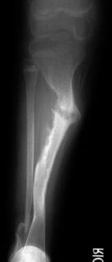
NONUNION TIIBIA WITH ARTHRITIS KNEE | Sant Parmanand Hospital
 Idaho Humane Society - ValPons
 Top Hip Surgeons
HuGE Navigator|Genopedia|PHGKB
 Walking simulator Lokomat | Eternity Life Tourism
 سلولهای تکهستهای به دست آمده از مغز استخوان خودی ممکن است بر درمان جوشناخوردگی استخوانهای بلند تأثیر مثبت داشته باشند
 Collections: Medicine in the Americas, 1610-1920 / Languages: English - Digital Collections - National Library of Medicine...
Formats: Text / Languages: English / Genre: Advertisements - Digital Collections - National Library of Medicine Search Results
Giant Cell-Rich Osteosarcoma (GCRO) of the Spine on T6 and T7 Vertebral Mass Case Report Literature Review
 Orthopedics suggested topics for Master Degree thesis - Dr.Saroj Health Blog
 Obturator Dislocation of the Hip with Associated Femoral Head Impaction and Medial Wall Fracture of the Acetabulum | Journal of...
Multiple Cantilever K-wiring Technique for Severely Comminuted Articular Fragments in Neglected Distal Humerus Fracture with...
 Bhaskar Bhowal (Cast Immobilisation): Influence Statistics Netherlands
 Research - lifemat
Fixation15
- [ 8 ] The presence of a fracture gap after fixation, open fractures, and transverse fracture type were also associated with nonunion. (medscape.com)
- Pathological fractures in the proximal part of the femur are treated by Zickel-nail fixation. (sarose.com.np)
- The adjunctive use of methylmethacrylate in fixation of pathological fractures. (sarose.com.np)
- Intramedullary fixation of pathological fractures and lesions of the subtrochanteric region of the femur. (sarose.com.np)
- Current concepts of external fixation of fractures. (sarose.com.np)
- Compression plate fixation and the effect of different types of internal fixation on fracture healing. (sarose.com.np)
- Severely comminuted fractures of distal humerus are challenging to treat and multiple cantilever K-wiring can be used as a preferable alternative for fixation. (jocr.co.in)
- Here, we present an unusual case of a 5-week-old unreduced anterior dislocation of the elbow joint with medial epicondyle and lateral condyle humerus fracture in a 30-year-old male patient and describe a unique technique for fixation of comminuted articular fragments. (jocr.co.in)
- Here, we report the long-term outcome of a young man treated with open reduction and internal fixation (ORIF) of the medial wall fracture, a valgus osteotomy of the femur and a restock of the impaction with autologous bone graft. (jocr.co.in)
- BACKGROUND: With the proliferation of different fixation screws, there is an increasing trend to recommend early internal fixation of the broken scaphoid even if the fracture is not displaced. (keyopinionleaders.com)
- The benefits and risks of early fixation of scaphoid fractures have not been established. (keyopinionleaders.com)
- He sustained a closed fracture nine years ago due to a fall from a height and was treated with internal fixation at a local hospital. (biomedcentral.com)
- Treatment of ununited fractures by onlay bone grafts without screws or tie fixation and without breaking down of fibrous union. (ijoro.org)
- Cannulated screw fixation of refractory olecranon stress fractures with and without associated injuries allows a return to baseball. (musc.edu)
- Anterior approach for operative fixation of coronoid fractures in complex elbow instability. (musc.edu)
FEMUR4
- Pre-op x-ray showed ununited fracture neck of femur with failed osteosynthesis. (sphdelhi.org)
- Pritchett, J.W.: Supracondylar Fractures of the Femur. (tophipsurgeons.com)
- Complete fractures of the femur in Paget's disease of bone. (sarose.com.np)
- The aim of this study was to evaluate a standardized method of treatment of femoral nonunion of the isthmal femur excluding non-united metaphyseal fractures. (biomedcentral.com)
Nonunion20
- The subcutaneous position of the tibia results in a greater incidence of open fractures and provides less soft-tissue coverage, factors that produce a higher incidence of nonunion and infected nonunion. (medscape.com)
- Although appropriate and prompt treatment is needed to treat tibial injuries successfully, the incidence of a nonunion is more closely related to the fracture characteristics than to subsequent treatment. (medscape.com)
- Treatment of a tibial nonunion depends on fracture classification, location of the nonunion, lower-extremity alignment, fracture stability, presence of infection, soft-tissue injury (including nerve deficits), and patient characteristics and possible concomitant injuries. (medscape.com)
- Infected nonunions should be treated in an attempt to sterilize the nonunion site, but stability of the fracture site should not be sacrificed. (medscape.com)
- The term nonunion refers to a fracture that will not unite without additional surgical or nonsurgical intervention (usually by 6-9 months). (medscape.com)
- The development of a tibial nonunion is related most often to the type and degree of injury, but several additional factors may predispose a patient to a tibial nonunion, such as the degree of fracture comminution and bone loss, whether the fracture is open, and the degree of soft-tissue injury. (medscape.com)
- for open fractures greater than 5 cm, the likelihood of delayed healing or nonunion was 5.7 times greater than that for closed fractures. (medscape.com)
- In an observational study involving 200 patients who experienced tibial fractures, Fong et al found that nonunion was more likely to occur in fractures with less than 25% cortical continuity. (medscape.com)
- The highest risk of nonunion occurred in cases involving an open fracture in conjunction with a fracture gap. (medscape.com)
- Nonunion is a serious complication following long-bone fracture that is known as a therapeutic challenge for surgeons and is associated with significant morbidity. (ijos.ir)
- Nonsteroidal anti-inflammatory drugs' impact on nonunion and infection rates in long-bone fractures. (ucsf.edu)
- Nonunion humeral shaft fractures often lead to pain, prolonged disability leading to reoperation, long-term absence from work, and impaired quality of life. (biomedcentral.com)
- Several factors have been reported to be associated with failure, including initial open fracture, low-grade infections, associated injury, the type of nonunion, and the location of nonunion. (jtraumainj.org)
- Several reasons for these diverging results have been suggested including technical improvements in implant design, and the introduction of interlocking screws leading to intramedullary treatment of complex fractures and thus, to more tenacious, complex nonunion [ 11 ],[ 14 ]. (biomedcentral.com)
- A prospective case series was performed, and all patients were included in this study who had been treated with intramedullary nailing of a femoral shaft fracture and had developed femoral shaft nonunion. (biomedcentral.com)
- Various modalities of treatment are available for the management of delayed and nonunion of long bone fractures. (ijoro.org)
- 15 patients with delayed and nonunion of long bone fractures were studied between January 2013 to January 2015 and were followed up for a period of 1 year. (ijoro.org)
- Percutaneous autologous bone marrow injection is a minimally invasive, safe and cost effective option in the management of delayed and nonunion of long bone fractures and gives good functional results. (ijoro.org)
- Delayed union and nonunion of fractures. (ijoro.org)
- This prospective, longitudinal study documents the baseline functional abilities of 40 consecutive patients with nonunion of a fracture in the lower limb. (ox.ac.uk)
Anconeal process1
- There are three different types of elbow dysplasia: UAP (ununited anconeal process), FCP (fractured coronoid process), and OCD (osteochondrosis). (total-german-shepherd.com)
Distal Radius Fractures2
- Volar locking plates are an increasingly popular treatment for distal radius fractures. (keyopinionleaders.com)
- A systematic review and meta-analysis of the pronator quadratus repair following volar plating of distal radius fractures. (harvard.edu)
Tibia3
- Scholars@Duke publication: Nonreamed locking intramedullary nailing for open fractures of the tibia. (duke.edu)
- A 60 year old gentleman presented with ununited fracture of leg bone (tibia). (sphdelhi.org)
- But this patient also had arthritis of the knee joint, therefore we planned for Total Knee Replacement with long stem in tibia to bypass the fracture site. (sphdelhi.org)
Scaphoid8
- van der Molen AB, Groothoff JW, Visser GJ, Robinson PH, Eisma WH (1999) Time off work due to scaphoid fractures and other carpal injuries in The Netherlands in the period 1990 to 1993. (springer.com)
- Should acute scaphoid fractures be fixed? (keyopinionleaders.com)
- These were investigated in eighty-eight patients who were of working age with clearly defined minimally displaced or undisplaced bicortical fractures of the waist of the scaphoid. (keyopinionleaders.com)
- The clinical negligence team at Penningtons Manches LLP has recently settled a claim against Darent Valley Hospital for negligently failing to diagnose and treat a fractured scaphoid. (penningtonslaw.com)
- Fractures of the scaphoid are commonly associated with falls of the type our client described. (penningtonslaw.com)
- As a result, the possibility of a fractured scaphoid was overlooked. (penningtonslaw.com)
- While a scaphoid fracture cannot necessarily be confirmed by examination or X-Ray on the date of injury, if such a fracture is suspected, a patient's wrist should be immobilised and re-assessed within the next 10-14 days at which point any fracture should be identifiable on radiology. (penningtonslaw.com)
- We thoroughly investigated the case and sought independent medical expert evidence on the standard of care our client received in A&E. Our expert confirmed that the hospital had failed in its duty to our client and had not properly considered and managed the possibility of a scaphoid fracture and that the fracture should have been diagnosed at a much earlier stage. (penningtonslaw.com)
Injuries5
- One of the biggest changes I have enjoyed in the field has been the development of minimally invasive surgery to fix athletic injuries and fractures. (stanfordchildrens.org)
- Although hamate fractures are increasing in incidence secondary to the popularity of sports activities involving racquets, bats, and clubs, these injuries remain relatively rare. (medscape.com)
- Used most commonly in the field of orthopedics, PEMF is a reparative technique for treatment of non-union fractures from sports or accident related injuries. (ghpcounselingservices.com)
- Jefferson Fx (named after British neurosurgeon who defined it) (unstable but neurologically intact Fx) 7% of all C/S injuries. (elpasochiropractorblog.com)
- Osteology of the coronoid process with clinical correlation to coronoid fractures in terrible triad injuries. (musc.edu)
Operative4
- PEMF and LIPUS significantly shorten time to radiological union for acute fractures undergoing non-operative treatment and acute fractures of the upper limb. (springer.com)
- Deformity following un-united and mal-united adult clavicle fractures is being increasingly recognised as the indications for operative treatment of displaced fractures of the clavicle. (orthopaper.com)
- The results were compareble with other published studies, and the pre contured clavicle plate represents one of the best operative management of these fractures. (orthopaper.com)
- More than a quarter million patients with severe ununited fractures have benefited worldwide from this surgically non-invasive treatment, without risk, discomfort, or the high costs of operative repair. (ghpcounselingservices.com)
Supracondylar Fractures1
- Remodeling of the humerus after supracondylar fractures in childhood. (sarose.com.np)
Tibial8
- Walker NA, Denegar CR, Preische J (2007) Low-intensity pulsed ultrasound and pulsed electromagnetic field in the treatment of tibial fractures: a systematic review. (springer.com)
- Heckman JD, Ryaby JP, McCabe J, Frey JJ, Kilcoyne RF (1994) Acceleration of tibial fracture-healing by non-invasive, low-intensity pulsed ultrasound. (springer.com)
- Tibial diaphyseal fractures that do not show enough bridging callus to achieve clinical stability by 16 weeks are considered to be delayed union fractures. (medscape.com)
- The use of nonreamed interlocking tibial nails in the management of open fractures of the tibial shaft has gained wide acceptance. (duke.edu)
- This technique has been reported to have reproducible good results with a low incidence of complications in Type I, Type II, and Type IIIA open tibial shaft fractures. (duke.edu)
- The treatment of 72 open fractures of the tibial shaft with nonreamed interlocking intramedullary nailing is detailed. (duke.edu)
- There were 27 Type I, 22 Type II, 11 Type IIIA, and 12 Type IIIB open tibial shaft fractures. (duke.edu)
- The use of nonreamed locking intramedullary nailing in Types I, II, IIIA, and IIIB open fractures of the tibial shaft is supported. (duke.edu)
Long bone2
- Bonafede M, Espindle D, Bower AG (2013) The direct and indirect costs of long bone fractures in a working age US population. (springer.com)
- Non Steroidal Anti-Inflammatory Medications Impact On Non-Union and Infection Rates in Long Bone Fractures. (ucsf.edu)
Diaphyseal fractures2
- Furthermore, we found significant results that suggest that the use of PEMF or LIPUS in acute diaphyseal fractures may accelerate the time to clinical union. (springer.com)
- Furthermore, PEMF or LIPUS bone growth stimulation accelerates the time to clinical union for acute diaphyseal fractures. (springer.com)
Elbow5
- Anterior dislocation of the elbow is comparatively less frequent and is often associated with fractures of the distal humerus. (jocr.co.in)
- We present a 5-week-old neglected anterior dislocation of the right elbow joint with lateral condyle and medial epicondyle humerus fracture and a unique cantilever K-wiring technique used for its treatment. (jocr.co.in)
- Radiographs revealed lateral condyle and medial epicondyle humerus fracture and an unreduced anterior dislocation of the right elbow joint. (jocr.co.in)
- Neglected fracture-dislocation of the elbow is challenging and is further complicated by comminuted fragments with loss of bone stock. (jocr.co.in)
- Elbow dislocation is a serious injury requiring immediate surgical intervention, especially when neglected and associated with fractures [1] . (jocr.co.in)
Shaft5
- Healing problems were twice as great for distal shaft fractures and fractures with a postoperative diastasis. (medscape.com)
- Pritchett, J.W.: Delayed Union of Humeral Shaft Fractures Treated by Closed Flexible Intramedullary Nailing. (tophipsurgeons.com)
- The management of these non-united femoral shaft fractures or delayed union still represents a challenge for the treating surgeon. (biomedcentral.com)
- The purpose of this study is to assess the clinical and radiological outcome of a pre-contoured clavicle plate in the treatment of acute, displaced, mid-shaft clavicle fractures. (orthopaper.com)
- This study presents the results of 26 patients treated with an anatomical congruent clavicle plate for acute displaced, mid-shaft fractures of the clavicle at a tertiary referral centre. (orthopaper.com)
Forearm Fractures1
- Clinical and radiographic comparison of single-sugar-tong splint to long-arm cast immobilization for pediatric forearm fractures. (musc.edu)
Bone fracture1
- Biophysical stimulation methods [ 9 ] such as pulsed electric currents and electromagnetic fields have been clinically shown to significantly improve bone fracture repair-even the healing of nonunions, which is an advanced stage of fracture where healing is ceased by the body [ 14 - 16 ]. (hindawi.com)
Carpal2
- Estimates suggest hamate fractures constitute 2% of all carpal fractures. (medscape.com)
- Hamate fractures account for 2% of all carpal fractures. (medscape.com)
Dislocations3
- With the increasing burden of COVID-19 on the hospitals, cases of conservatively managed fractures and dislocations with adverse outcomes have become more prevalent. (jocr.co.in)
- In 12% of the patients, the dislocation is combined with a femoral head fracture (complex dislocations) [1, 2] which could result in severe complications such as avascular necrosis (AVN) and subsequent early secondary osteoarthritis. (jocr.co.in)
- In general, the treatment of complex hip dislocations depends on the associated fracture (e.g., femoral head fracture, femoral neck fracture, and acetabular fracture). (jocr.co.in)
Femoral neck1
- Stress fractures of the femoral neck. (sarose.com.np)
Acute5
- The aim of this systematic review and meta-analysis was to evaluate the best currently available evidence from randomized controlled trials comparing pulsed electromagnetic fields (PEMF) or low-intensity pulsed ultrasound (LIPUS) bone growth stimulation with placebo for acute fractures. (springer.com)
- We performed a systematic literature search of the medical literature from 1980 to 2013 for randomized clinical trials concerning acute fractures in adults treated with PEMF or LIPUS. (springer.com)
- Current evidence from randomized trials is insufficient to conclude a benefit of PEMF or LIPUS bone growth stimulation in reducing the incidence of nonunions when used for treatment in acute fractures. (springer.com)
- However, our systematic review and meta-analysis suggest that PEMF or LIPUS can be beneficial in the treatment of acute fractures regarding time to radiological and clinical union. (springer.com)
- Griffin XL, Costello I, Costa ML (2008) The role of low intensity pulsed ultrasound therapy in the management of acute fractures: a systematic review. (springer.com)
Occur5
- A fracture in which union fails to occur, the ends of the bone becoming rounded and eburnated, and a false joint occurs. (bvsalud.org)
- Indentation fractures of the femoral head have been reported to occur in 35%-55% of patients after traumatic obturator dislocation [3, 4, 5]. (jocr.co.in)
- Type I fractures involving the hook of the hamate are the most common and can occur via several different mechanisms. (medscape.com)
- These usually occur in the nondominant hand and account for approximately one third of hamate fractures. (medscape.com)
- [ 4 ] Most commonly, these fractures occur with a punch-press injury or dorsopalmar compression of the wrist between heavy weights. (medscape.com)
Pathological1
- CT was combined like consider pathological compression fracture of the T6 vertebral body with spinal compression, T6, and T7 bone mass destruction. (fortunejournals.com)
Fibula1
- Fatigue fractures of the fibula. (sarose.com.np)
Patient's1
- For autologous human adipose tissue-derived MSCs, enhanced differentiation into osteoblastic phenotype can be advantageous and may be used with matrix biomaterials for regeneration of the patient's own damaged bones due to fractures, osteoporosis, and deformities. (hindawi.com)
Instability1
- Ulnar Styloid Base Fractures Cause Distal Radioulnar Joint Instability in a Cadaveric Model. (harvard.edu)
Transverse1
- Note Jefferson Fx with pillar and transverse foramina fx requiring posterior occipital-cervical fusion (below right image). (elpasochiropractorblog.com)
Complication1
- The fracture united in all cases with no complication and high patient satisfaction on the Disability of the Arm and Shoulder (DASH) score and Pain Visual analogue score (VAS). (orthopaper.com)
Posterior2
- Morphological characteristics of the posterior malleolar fragment according to ankle fracture patterns: a computed tomography-based study. (harvard.edu)
- Mechanism: C1 compression (e.g., diving into shallow waters) causing burst Fx-classically 4-parts of the anterior and posterior arch of C1. (elpasochiropractorblog.com)
Risk of bone2
- After therapy, the bones become stronger, which reduces the risk of bone loss and fractures. (elclinics.com)
- Vitamin D receptor genotype and the risk of bone fractures in women. (musc.edu)
Stress2
- A stress fracture in children. (sarose.com.np)
- Guha AR, Marynissen H. Stress fracture of the hook of the hamate. (medscape.com)
Olecranon1
- the right scapula and an II° open comminuted fracture of the right olecranon. (jocr.co.in)
Radiological2
- With regard to time to radiological union, we found heterogeneous results that significantly favoured PEMF or LIPUS bone growth stimulation only in non-operatively treated fractures or fractures of the upper limb. (springer.com)
- Radiological diagnosis of fractures. (sarose.com.np)
Dislocation3
- A 22-year-old truck driver involved in a ski accident sustained an obturator dislocation of the right hip associated with a femoral head impaction in the weight-bearing zone and a medial wall fracture of the acetabulum. (jocr.co.in)
- An obturator hip dislocation with a femoral head and a medial wall acetabular fracture is a very rare combination. (jocr.co.in)
- He sustained an obturator dislocation of the right hip associated with a severe femoral head impaction fracture in the weight-bearing zone and a medial wall fracture of the acetabulum (Fig. 1, 2, 3). (jocr.co.in)
Healing5
- Fracture healing is a dynamic, progressive process, and intervention is warranted within 3-5 months after injury if monthly radiographic studies do not show progression of fracture healing. (medscape.com)
- Typically, the term delayed union is used for a fracture that has not united within a period that would typically be considered adequate for bone healing. (medscape.com)
- Bones contain a matrix of collagen proteins, which are piezoelectric materials that accumulate small electrical charges when subjected to mechanical stresses, leading to stimulation and deposition of intracellular calcium during fracture healing [ 13 ]. (hindawi.com)
- The biology of fracture healing in long bones. (sarose.com.np)
- Basic concepts regarding fracture healing and the current options and future directions in managing bone fractures. (ijoro.org)
Injury1
- Imaging: initial x-radiography then CT that helps to delineate another injury such as facet/pedicle Fx further. (elpasochiropractorblog.com)
Screws1
- He had sustained fracture of the left hip 6 month ago which was fixed with screws. (sphdelhi.org)