Ganglion Cysts
Synovial Cyst
Cysts
Bone Cysts
Posterior Cruciate Ligament
Nerve Compression Syndromes
Shoulder Pain
Ganglia
Anterior Cruciate Ligament
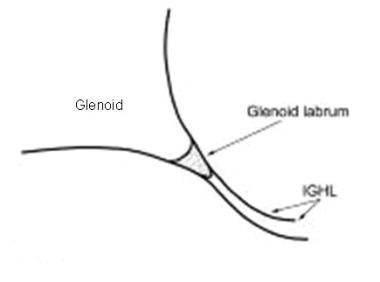
Shoulder Joint
Retinal Ganglion Cells
Suction
Ganglia, Spinal
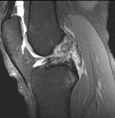
Anterior cruciate ligament ganglion: case report. (1/66)
CONTEXT: A ganglion is a cystic formation close to joints or tendinous sheaths, frequently found in the wrist, foot or knee. Intra-articular ganglia of the knee are rare, and most of them are located in the anterior cruciate ligament. The clinical picture for these ganglia comprises pain and movement restrictions in the knee, causing significant impairment to the patient. Symptoms are non-specific, and anterior cruciate ligament ganglia are usually diagnosed through magnetic resonance imaging or arthroscopy. Not all ganglia diagnosed through magnetic resonance imaging need to undergo surgical treatment: only those that cause clinical signs and symptoms do. Surgical results are considered good or excellent in the vast majority of cases. CASE REPORT: A 29-year-old male presented with pain in the left knee during a marathon race. Physical examination revealed limitation in the maximum range of knee extension and pain in the posterior aspect of the left knee. Radiographs of the left knee were normal, but magnetic resonance imaging revealed a multi-lobed cystic structure adjacent to the anterior cruciate ligament, which resembled a ganglion cyst. The mass was removed through arthroscopy, and pathological examination revealed a synovial cyst. Patient recovery was excellent, and he resumed his usual training routine five months later. (+info)A locus on mouse chromosome 6 that determines resistance to herpes simplex virus also influences reactivation, while an unlinked locus augments resistance of female mice. (2/66)
During studies to determine a role for tumor necrosis factor (TNF) in herpes simplex virus type 1 (HSV-1) infection using TNF receptor null mutant mice, we discovered a genetic locus, closely linked to the TNF p55 receptor (Tnfrsf1a) gene on mouse chromosome 6 (c6), that determines resistance or susceptibility to HSV-1. We named this locus the herpes resistance locus, Hrl, and showed that it also mediates resistance to HSV-2. Hrl has at least two alleles, Hrl(r), expressed by resistant strains like C57BL/6 (B6), and Hrl(s), expressed by susceptible strains like 129S6 (129) and BALB/c. Although Hrl is inherited as an autosomal dominant gene, resistance to HSV-1 is strongly sex biased such that female mice are significantly more resistant than male mice. Analysis of backcrosses between resistant B6 and susceptible 129 mice revealed that a second locus, tentatively named the sex modifier locus, Sml, functions to augment resistance of female mice. Besides determining resistance, Hrl is one of several genes involved in the control of HSV-1 replication in the eye and ganglion. Remarkably, Hrl also affects reactivation of HSV-1, possibly by interaction with some unknown gene(s). We showed that Hrl is distinct from Cmv1, the gene that determines resistance to murine cytomegalovirus, which is encoded in the major NK cell complex just distal of p55 on c6. Hrl has been mapped to a roughly 5-centimorgan interval on c6, and current efforts are focused on obtaining a high-resolution map for Hrl. (+info)A case of extensor digitorum brevis manus. (3/66)
The extensor digitorum brevis manus (EDBM), a relatively rare anomalous muscle on the dorsal hand, may be misdiagnosed as a ganglion, a synovial nodule or cyst, or a soft-tissue tumor. MRI scans can help to distinguish EDBM from tumors. EDBM should be included in the differential diagnosis of soft tissue masses on the dorsal aspect of the hand. (+info)Leiomyoma of the hand mimicking a pearl ganglion. (4/66)
Leiomyomas rarely occur in the hand. To our knowledge, there have been no reports of a leiomyoma of the hand mimicking a pearl ganglion in the English literature. We report such a case with a leiomyoma of the right third finger in a 59-year-old woman. The tumor was excised together with the underlying sheet of tissue. The pathology revealed that the tumor was linked to the underlying structure of a vascular wall by a stalk of tumor tissue. This report serves to remind clinicians to include leiomyoma in the differential diagnosis when encountering a 'ganglion-like lesion'. Also, this report demonstrates the link between a leiomyoma and its underlying origin. (+info)The role of Pax2 in mouse inner ear development. (5/66)
The paired box transcription factor, Pax2, is important for cochlear development in the mouse inner ear. Two mutant alleles of Pax2, a knockout and a frameshift mutation (Pax21Neu), show either agenesis or severe malformation of the cochlea, respectively. In humans, mutations in the PAX2 gene cause renal coloboma syndrome that is characterized by kidney abnormalities, optic nerve colobomas and mild sensorineural deafness. To better understand the role of Pax2 in inner ear development, we examined the inner ear phenotype in the Pax2 knockout mice using paint-fill and gene expression analyses. We show that Pax2-/- ears often lack a distinct saccule, and the endolymphatic duct and common crus are invariably fused. However, a rudimentary cochlea is always present in all Pax2 knockout inner ears. Cochlear outgrowth in the mutants is arrested at an early stage due to apoptosis of cells that normally express Pax2 in the cochlear anlage. Lack of Pax2 affects tissue specification within the cochlear duct, particularly regions between the sensory tissue and the stria vascularis. Because the cochlear phenotypes observed in Pax2 mutants are more severe than those observed in mice lacking Otx1 and Otx2, we postulate that Pax2 plays a key role in regulating the differential growth within the cochlear duct and thus, its proper outgrowth and coiling. (+info)Unusual localization of multiple myxoid (mucous) cysts of toes. (6/66)
Myxoid cysts of fingers and toes are observed frequently on the lateral or dorsal aspects of the distal digits. They are usually solitary nodules. Both subungual localization and multiplicity are quite rare. We present a 74-year-old woman with digital subungual mucous cysts located on all toes. (+info)Drosophila Grainyhead specifies late programmes of neural proliferation by regulating the mitotic activity and Hox-dependent apoptosis of neuroblasts. (7/66)
The Drosophila central nervous system is generated by stem-cell-like progenitors called neuroblasts. Early in development, neuroblasts switch through a temporal series of transcription factors modulating neuronal fate according to the time of birth. At later stages, it is known that neuroblasts switch on expression of Grainyhead (Grh) and maintain it through many subsequent divisions. We report that the function of this conserved transcription factor is to specify the regionalised patterns of neurogenesis that are characteristic of postembryonic stages. In the thorax, Grh prolongs neural proliferation by maintaining a mitotically active neuroblast. In the abdomen, Grh terminates neural proliferation by regulating the competence of neuroblasts to undergo apoptosis in response to Abdominal-A expression. This study shows how a factor specific to late-stage neural progenitors can regulate the time at which neural proliferation stops, and identifies mechanisms linking it to the Hox axial patterning system. (+info)Ganglion cyst of the anterior cruciate ligament: a case report. (8/66)
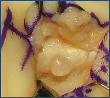
A ganglion is a cystic swelling that usually arises close to tendons or joints. Its occurrence inside a joint is rare, and its diagnosis is usually incidental during magnetic resonance imaging or arthroscopy. It may be painful or asymptomatic. Some patients may have a trauma history. Ganglia may mimic intra-articular lesions like tears of the anterior cruciate ligament or meniscus. Magnetic resonance imaging is the investigation of choice for diagnosis. Ganglia commonly arise from the anterior cruciate ligament, but can also arise from other structures such as the posterior cruciate ligament or meniscus. Ganglia are typically treated by arthroscopic excision and debridement. We report a case of ganglion cyst of the anterior cruciate ligament in a 16-year-old man. (+info)A ganglion cyst is a type of fluid-filled sac that commonly develops on the back of the wrist, hands, or fingers. These cysts usually contain a clear, jelly-like material and are connected to a joint or tendon sheath. The exact cause of ganglion cysts is unknown, but they may form as a result of repetitive trauma or degeneration of the joint tissue.
Ganglion cysts can vary in size from small (pea-sized) to large (golf ball-sized). They are usually painless, but if they press on a nerve, they can cause tingling, numbness, or discomfort. In some cases, ganglion cysts may resolve on their own without treatment, while others may require medical intervention such as aspiration (draining the fluid) or surgical removal.
A Synovial Cyst is a type of benign cyst that typically develops in the synovium, which is the membrane that lines and lubricates joint capsules. These cysts are filled with synovial fluid, which is the same lubricating fluid found inside joints. They usually form as a result of degenerative changes, trauma, or underlying joint diseases such as osteoarthritis.
Synovial cysts commonly occur in the spine (particularly in the facet joints), but they can also develop in other areas of the body, including the knees, hips, and hands. While synovial cysts are generally not harmful, they may cause discomfort or pain if they press on nearby nerves or restrict movement in the affected joint. Treatment options for synovial cysts range from conservative measures like physical therapy and pain management to surgical intervention in severe cases.
A cyst is a closed sac, having a distinct membrane and division between the sac and its surrounding tissue, that contains fluid, air, or semisolid material. Cysts can occur in various parts of the body, including the skin, internal organs, and bones. They can be caused by various factors, such as infection, genetic predisposition, or blockage of a duct or gland. Some cysts may cause symptoms, such as pain or discomfort, while others may not cause any symptoms at all. Treatment for cysts depends on the type and location of the cyst, as well as whether it is causing any problems. Some cysts may go away on their own, while others may need to be drained or removed through a surgical procedure.
A bone cyst is a fluid-filled sac that develops within a bone. It can be classified as either simple (unicameral) or aneurysmal. Simple bone cysts are more common in children and adolescents, and they typically affect the long bones of the arms or legs. These cysts are usually asymptomatic unless they become large enough to weaken the bone and cause a fracture. Aneurysmal bone cysts, on the other hand, can occur at any age and can affect any bone, but they are most common in the leg bones and spine. They are characterized by rapidly growing blood-filled sacs that can cause pain, swelling, and fractures.
Both types of bone cysts may be treated with observation, medication, or surgery depending on their size, location, and symptoms. It is important to note that while these cysts can be benign, they should still be evaluated and monitored by a healthcare professional to ensure proper treatment and prevention of complications.
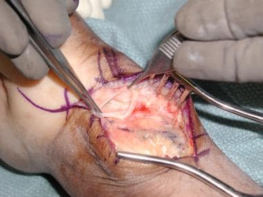
Arthroscopy is a minimally invasive surgical procedure where an orthopedic surgeon uses an arthroscope (a thin tube with a light and camera on the end) to diagnose and treat problems inside a joint. The surgeon makes a small incision, inserts the arthroscope into the joint, and then uses the attached camera to view the inside of the joint on a monitor. They can then insert other small instruments through additional incisions to repair or remove damaged tissue.
Arthroscopy is most commonly used for joints such as the knee, shoulder, hip, ankle, and wrist. It offers several advantages over traditional open surgery, including smaller incisions, less pain and bleeding, faster recovery time, and reduced risk of infection. The procedure can be used to diagnose and treat a wide range of conditions, including torn ligaments or cartilage, inflamed synovial tissue, loose bone or cartilage fragments, and joint damage caused by arthritis.
The Posterior Cruciate Ligament (PCL) is one of the major ligaments in the knee, providing stability to the joint. It is a strong band of tissue located in the back of the knee, connecting the thighbone (femur) to the shinbone (tibia). The PCL limits the backward motion of the tibia relative to the femur and provides resistance to forces that tend to push the tibia backwards. It also assists in maintaining the overall alignment and function of the knee joint during various movements and activities. Injuries to the PCL are less common compared to injuries to the Anterior Cruciate Ligament (ACL) but can still occur due to high-energy trauma, such as motor vehicle accidents or sports incidents involving direct impact to the front of the knee.
Nerve compression syndromes refer to a group of conditions characterized by the pressure or irritation of a peripheral nerve, causing various symptoms such as pain, numbness, tingling, and weakness in the affected area. This compression can occur due to several reasons, including injury, repetitive motion, bone spurs, tumors, or swelling. Common examples of nerve compression syndromes include carpal tunnel syndrome, cubital tunnel syndrome, radial nerve compression, and ulnar nerve entrapment at the wrist or elbow. Treatment options may include physical therapy, splinting, medications, injections, or surgery, depending on the severity and underlying cause of the condition.
Shoulder pain is a condition characterized by discomfort or hurt in the shoulder joint, muscles, tendons, ligaments, or surrounding structures. The shoulder is one of the most mobile joints in the body, and this mobility makes it prone to injury and pain. Shoulder pain can result from various causes, including overuse, trauma, degenerative conditions, or referred pain from other areas of the body.
The shoulder joint is a ball-and-socket joint made up of three bones: the humerus (upper arm bone), scapula (shoulder blade), and clavicle (collarbone). The rotator cuff, a group of four muscles that surround and stabilize the shoulder joint, can also be a source of pain if it becomes inflamed or torn.
Shoulder pain can range from mild to severe, and it may be accompanied by stiffness, swelling, bruising, weakness, numbness, tingling, or reduced mobility in the affected arm. The pain may worsen with movement, lifting objects, or performing certain activities, such as reaching overhead or behind the back.
Medical evaluation is necessary to determine the underlying cause of shoulder pain and develop an appropriate treatment plan. Treatment options may include rest, physical therapy, medication, injections, or surgery, depending on the severity and nature of the condition.
A ganglion is a cluster of neuron cell bodies in the peripheral nervous system. Ganglia are typically associated with nerves and serve as sites for sensory processing, integration, and relay of information between the periphery and the central nervous system (CNS). The two main types of ganglia are sensory ganglia, which contain pseudounipolar neurons that transmit sensory information to the CNS, and autonomic ganglia, which contain multipolar neurons that control involuntary physiological functions.
Examples of sensory ganglia include dorsal root ganglia (DRG), which are associated with spinal nerves, and cranial nerve ganglia, such as the trigeminal ganglion. Autonomic ganglia can be further divided into sympathetic and parasympathetic ganglia, which regulate different aspects of the autonomic nervous system.
It's worth noting that in anatomy, "ganglion" refers to a group of nerve cell bodies, while in clinical contexts, "ganglion" is often used to describe a specific type of cystic structure that forms near joints or tendons, typically in the wrist or foot. These ganglia are not related to the peripheral nervous system's ganglia but rather are fluid-filled sacs that may cause discomfort or pain due to their size or location.
Cyst fluid refers to the fluid accumulated within a cyst, which is a closed sac-like or capsular structure, typically filled with liquid or semi-solid material. Cysts can develop in various parts of the body for different reasons, and the composition of cyst fluid may vary depending on the type of cyst and its location.
In some cases, cyst fluid might contain proteins, sugars, hormones, or even cells from the surrounding tissue. Infected cysts may have pus-like fluid, while cancerous or precancerous cysts might contain abnormal cells or tumor markers. The analysis of cyst fluid can help medical professionals diagnose and manage various medical conditions, including infections, inflammatory diseases, genetic disorders, and cancers.
It is important to note that the term 'cyst fluid' generally refers to the liquid content within a cyst, but the specific composition and appearance of this fluid may vary significantly depending on the underlying cause and type of cyst.
The Anterior Cruciate Ligament (ACL) is a major stabilizing ligament in the knee. It is one of the four strong bands of tissue that connect the bones of the knee joint together. The ACL runs diagonally through the middle of the knee and helps to control the back and forth motion of the knee, as well as provide stability to the knee joint. Injuries to the ACL often occur during sports or physical activities that involve sudden stops, changes in direction, or awkward landings.
The shoulder joint, also known as the glenohumeral joint, is the most mobile joint in the human body. It is a ball and socket synovial joint that connects the head of the humerus (upper arm bone) to the glenoid cavity of the scapula (shoulder blade). The shoulder joint allows for a wide range of movements including flexion, extension, abduction, adduction, internal rotation, and external rotation. It is surrounded by a group of muscles and tendons known as the rotator cuff that provide stability and enable smooth movement of the joint.
Retinal Ganglion Cells (RGCs) are a type of neuron located in the innermost layer of the retina, the light-sensitive tissue at the back of the eye. These cells receive visual information from photoreceptors (rods and cones) via intermediate cells called bipolar cells. RGCs then send this visual information through their long axons to form the optic nerve, which transmits the signals to the brain for processing and interpretation as vision.
There are several types of RGCs, each with distinct morphological and functional characteristics. Some RGCs are specialized in detecting specific features of the visual scene, such as motion, contrast, color, or brightness. The diversity of RGCs allows for a rich and complex representation of the visual world in the brain.
Damage to RGCs can lead to various visual impairments, including loss of vision, reduced visual acuity, and altered visual fields. Conditions associated with RGC damage or degeneration include glaucoma, optic neuritis, ischemic optic neuropathy, and some inherited retinal diseases.
In medical terms, suction refers to the process of creating and maintaining a partial vacuum in order to remove fluids or gases from a body cavity or wound. This is typically accomplished using specialized medical equipment such as a suction machine, which uses a pump to create the vacuum, and a variety of different suction tips or catheters that can be inserted into the area being treated.
Suction is used in a wide range of medical procedures and treatments, including wound care, surgical procedures, respiratory therapy, and diagnostic tests. It can help to remove excess fluids such as blood or pus from a wound, clear secretions from the airways during mechanical ventilation, or provide a means of visualizing internal structures during endoscopic procedures.
It is important to use proper technique when performing suctioning, as excessive or improperly applied suction can cause tissue damage or bleeding. Medical professionals are trained in the safe and effective use of suction equipment and techniques to minimize risks and ensure optimal patient outcomes.
Spinal ganglia, also known as dorsal root ganglia, are clusters of nerve cell bodies located in the peripheral nervous system. They are situated along the length of the spinal cord and are responsible for transmitting sensory information from the body to the brain. Each spinal ganglion contains numerous neurons, or nerve cells, with long processes called axons that extend into the periphery and innervate various tissues and organs. The cell bodies within the spinal ganglia receive sensory input from these axons and transmit this information to the central nervous system via the dorsal roots of the spinal nerves. This allows the brain to interpret and respond to a wide range of sensory stimuli, including touch, temperature, pain, and proprioception (the sense of the position and movement of one's body).
An ovarian cyst is a sac or pouch filled with fluid that forms on the ovary. Ovarian cysts are quite common in women during their childbearing years, and they often cause no symptoms. In most cases, ovarian cysts disappear without treatment over a few months. However, larger or persistent cysts may require medical intervention, including surgical removal.
There are various types of ovarian cysts, such as functional cysts (follicular and corpus luteum cysts), which develop during the menstrual cycle due to hormonal changes, and non-functional cysts (dermoid cysts, endometriomas, and cystadenomas), which can form due to different causes.
While many ovarian cysts are benign, some may have malignant potential or indicate an underlying medical condition like polycystic ovary syndrome (PCOS). Regular gynecological check-ups, including pelvic examinations and ultrasounds, can help detect and monitor ovarian cysts.
 Ganglion cyst