Gastritis, Atrophic
Helicobacter pylori
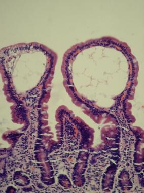
Gastric Mucosa
Helicobacter Infections
Pyloric Antrum
Gastritis, Hypertrophic
Metaplasia
Stomach
Pepsinogen A
Bile Reflux
Stomach Ulcer
Gastrins
Peptic Ulcer
Pepsinogen C
Helicobacter felis
Helicobacter heilmannii
Parietal Cells, Gastric
Chronic Disease
Duodenitis
Anemia, Pernicious
Helicobacter
Biopsy
Urease
Pepsinogens
Postgastrectomy Syndromes
Gastric Juice
Precancerous Conditions
Endoscopy, Gastrointestinal
Campylobacter
Atrophy
Acinonyx
Endoscopy
Anti-Ulcer Agents
Amoxicillin
Autoimmune Diseases
Omeprazole
Duodenogastric Reflux
Gastroenterostomy
Intrinsic Factor
Esophagitis
Gastrointestinal Diseases
Gastroesophageal Reflux
Giant hypertrophic gastritis and acute hepatitis associated with cytomegalovirus infection. (1/34)
A 38-year-old man developed prominent hypoproteinemia after acute elevation of serum transaminase levels. Giant hypertrophy of the gastric mucosa, a short serum albumin half-life, and the absence of massive hepatocyte necrosis established the diagnosis of protein-losing gastropathy. The hypoproteinemia, gastric fold hypertrophy and hepatitis remitted spontaneously within 4 months. A high antibody titer against cytomegalovirus suggested an association between the viral infection and the patient's disease. (+info)Menetrier's disease in a child. (2/34)
A case of Menetrier's disease in a 3 year old child presenting with subtotal pyloric stenosis and fatal outcome due to postoperative complications is reported. It is emphasized that although radiographic and gastroscopic studies are helpful, a full-thickness mucosal biopsy is essential for the diagnosis of Menetrier's disease. (+info)Remission of severe anemia persisting for over 20 years after eradication of Helicobacter pylori in cases of Menetrier's disease and atrophic gastritis: Helicobacter pylori as a pathogenic factor in iron-deficiency anemia. (3/34)
A man with a 20-year history of recurrent iron-deficiency anemia complicated by Helicobacter pylori-positive Menetrier's disease was observed over a 10-year clinical course, during which time he was successfully treated for the anemia and a gastric Helicobacter pylori (H. pylori) infection through eradication. Considering the satisfactory therapeutic results in this case, we performed eradication therapy on another H. pylori-positive atrophic gastritis case with a 24-year history of iron-deficiency anemia of unknown etiology, and again, complete remission was obtained. The clinical evidence from these two cases suggests that the gastric H. pylori infection was deeply involved in the pathogeneses of the iron-deficiency anemia. We believe that these case reports will provide useful information on H. pylon-involved pathology in the fields of hematology and gastroenterology. (+info)Hypertrophic gastropathy resembling Menetrier's disease in transgenic mice overexpressing transforming growth factor alpha in the stomach. (4/34)
Transforming growth factor alpha (TGF alpha) is thought to participate in the normal and pathologic processes of numerous tissues, including the gastric mucosa. To explore its role in vivo, transgenic mice were generated overexpressing TGF alpha in the stomach. TGF alpha induced dramatic structural and functional lesions of the glandular stomach that were similar to Menetrier's disease in humans. Transgenic mice developed severe adenomatous hyperplasia that resulted in a striking nodular thickening or hypertrophy of the gastric mucosa. Secretions obtained from affected stomachs contained no detectable gastric acid, suggesting that parietal cell function had been greatly impaired. These findings demonstrate that overproduction of TGF alpha can stimulate cellular proliferation, suppress acid secretion, and perturb organogenesis of the stomach of transgenic mice. Moreover, TGF alpha may contribute to the pathogenesis of related human hypertrophic gastropathies, such as Menetrier's disease. (+info)Menetrier's disease in korea: report of two cases and review of cases in a gastric cancer prevalent region. (5/34)
Menetrier's disease is a rare disease of the stomach generally described as hypertrophic gastropathy associated with hypoproteinemia. Gastric resection is still the most definitive treatment for the disease, but the appropriate extent of resection has not been determined. One of the major factors that would determine the extent of gastric resection in Menetrier's disease is its malignant potential. We present two recent cases of Menetrier's disease treated in our institution and review cases of the disease reported in Korea where the incidence of gastric cancer is one of the highest in the world. (+info)Cytomegalovirus-associated discrete gastrointestinal masses in macaques infected with the simian immunodeficiency virus. (6/34)
Cytomegalovirus (CMV)-associated gastrointestinal masses have been reported in human acquired immune deficiency syndrome patients. This is the first report on CMV-associated gastrointestinal masses in simian immunodeficiency virus (SIV)-infected macaques. Two SIV-infected macaques presented at necropsy with multiple nodular or umbilicated masses within the gastrointestinal tract. In one animal, the masses were located throughout the gastrointestinal tract, whereas in the other, the masses were restricted to the proximal small intestine. Grossly, the masses were indistinguishable from those caused by neoplastic conditions such as lymphoma and, histologically, were composed of hyperplastic glandular tissue, dense neutrophilic infiltrates within the lamina propria, and multifocal proprial hemorrhage. Frequent cytomegalic cells with basophilic intranuclear inclusions were found in affected regions. Immunohistochemistry for CMV demonstrated frequent immunopositive cells within affected areas. Furthermore, immunohistochemistry for the proliferation marker Ki-67 demonstrated increased proliferation in hyperplastic glands and crypts. CMV should be considered a cause of discrete mass lesions in the gastrointestinal tract of SIV-infected macaques. (+info)Interferon gamma induction of gastric mucous neck cell hypertrophy. (7/34)
Chronic inflammation of the gastric epithelium is believed to induce mucosal changes that can eventually develop into gastric cancer. In gastrin-deficient (G-/-) mice exhibiting chronic inflammation in the hypochlorhydric stomach, we documented a prominent fundic mucous cell lineage sharing morphological similarity with preneoplastic changes reported in Helicobacter-infected mice. To study the identity and origin of this cell lineage, we screened for different gastric mucosal cell markers. The clusters of large, foamy cells stained for trefoil factor 2 (TFF2/SP), MUC6 and the lectin Griffonia Simplicifolia II (GSII), but not for the intestine-specific transcription factor Cdx2, suggested that they arise from gastric mucous neck cells. Ki67-labeled GSII-positive neck cells in Helicobacter felis-infected, but not G-/- stomachs, suggested that mucous neck cell proliferation accounted for expansion of this compartment in the H. felis model of gastritis, but not the G-/- model. Using RNase protection assays and quantitative PCR, we found that interferon gamma (IFNgamma) was the most abundant proinflammatory cytokine in the G-/- stomach. We also found that this Th1 cytokine can increase the abundance of mucous neck cells, since its infusion into mice recapitulated the appearance of these cells as observed in both G-/- and H. felis-infected mice. Using the human gastric cell line NCI-N87, we showed that IFNgamma induces the secretion of mucus and expression of MUC6, TFF2 and pepsinogen II, but not of pepsinogen I and intrinsic factor. In conclusion, our results demonstrate that inflammation, specifically the proinflammatory cytokine IFNgamma, induced expansion of the fundic mucous neck cell compartment, which likely represents both increased mucus production and cell number. (+info)CCR7 deficiency causes ectopic lymphoid neogenesis and disturbed mucosal tissue integrity. (8/34)
Homeostatic trafficking of lymphocytes through extralymphoid tissues has been recently observed, and a potential role in immune surveillance and the establishment of peripheral tolerance are considered. However, the mechanisms regulating lymphocyte recirculation through peripheral tissues under noninflammatory conditions are not well understood. Here, we demonstrate that the chemokine receptor CCR7 controls not only lymphocyte trafficking to and within secondary lymphoid organs but also homeostatic migration of T and B lymphocytes through nonlymphoid tissues. Lack of CCR7 results in a massive accumulation of lymphocytes in epithelial tissues. In particular, the gastrointestinal mucosal tissue of CCR7-/- mice is highly permissive for the formation of lymphoid aggregates, which develop into ectopic follicular structures with major topologic characteristics of lymph nodes. Flow cytometry analysis of CD4+ T cells derived from ectopic follicles revealed that CD44hiCD62Llo effector memory T cells predominate in the gastric lymphoid aggregates. In aged mice, lack of CCR7 induced age-dependent histomorphologic changes in the stomach with profound cystic hyperplasia and an increased rate of mucosal proliferation resembling Menetrier disease. Thus, CCR7 regulates the cellular organization of visceral tissue by governing life-long recirculation of naive and memory lymphocytes under homeostatic conditions. (+info)Gastritis is a medical condition characterized by inflammation of the lining of the stomach. It can be caused by various factors, including bacterial infections (such as Helicobacter pylori), regular use of nonsteroidal anti-inflammatory drugs (NSAIDs), excessive alcohol consumption, and stress.
Gastritis can present with a range of symptoms, such as abdominal pain or discomfort, nausea, vomiting, loss of appetite, and bloating. In some cases, gastritis may not cause any noticeable symptoms. Depending on the severity and duration of inflammation, gastritis can lead to complications like stomach ulcers or even stomach cancer if left untreated.
There are two main types of gastritis: acute and chronic. Acute gastritis develops suddenly and may last for a short period, while chronic gastritis persists over time, often leading to atrophy of the stomach lining. Diagnosis typically involves endoscopy and tissue biopsy to assess the extent of inflammation and rule out other potential causes of symptoms. Treatment options depend on the underlying cause but may include antibiotics, proton pump inhibitors, or lifestyle modifications.
Atrophic gastritis is a condition characterized by the inflammation and atrophy (wasting away) of the stomach lining, specifically the mucous membrane called the gastric mucosa. This process involves the loss of glandular cells in the stomach, which can result in decreased acid production and potential vitamin B12 deficiency due to reduced intrinsic factor production. Atrophic gastritis can be caused by various factors, including autoimmune disorders, chronic bacterial infection (usually with Helicobacter pylori), and the use of certain medications such as proton pump inhibitors. It can increase the risk of developing stomach cancer, so regular monitoring is often recommended.
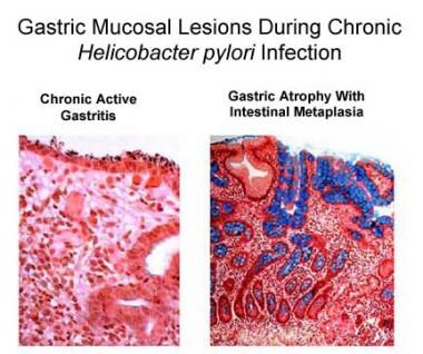
Helicobacter pylori (H. pylori) is a gram-negative, microaerophilic bacterium that colonizes the stomach of approximately 50% of the global population. It is closely associated with gastritis and peptic ulcer disease, and is implicated in the pathogenesis of gastric adenocarcinoma and mucosa-associated lymphoid tissue (MALT) lymphoma. H. pylori infection is usually acquired in childhood and can persist for life if not treated. The bacterium's spiral shape and flagella allow it to penetrate the mucus layer and adhere to the gastric epithelium, where it releases virulence factors that cause inflammation and tissue damage. Diagnosis of H. pylori infection can be made through various tests, including urea breath test, stool antigen test, or histological examination of a gastric biopsy. Treatment typically involves a combination of antibiotics and proton pump inhibitors to eradicate the bacteria and promote healing of the stomach lining.
Gastric mucosa refers to the innermost lining of the stomach, which is in contact with the gastric lumen. It is a specialized mucous membrane that consists of epithelial cells, lamina propria, and a thin layer of smooth muscle. The surface epithelium is primarily made up of mucus-secreting cells (goblet cells) and parietal cells, which secrete hydrochloric acid and intrinsic factor, and chief cells, which produce pepsinogen.
The gastric mucosa has several important functions, including protection against self-digestion by the stomach's own digestive enzymes and hydrochloric acid. The mucus layer secreted by the epithelial cells forms a physical barrier that prevents the acidic contents of the stomach from damaging the underlying tissues. Additionally, the bicarbonate ions secreted by the surface epithelial cells help neutralize the acidity in the immediate vicinity of the mucosa.
The gastric mucosa is also responsible for the initial digestion of food through the action of hydrochloric acid and pepsin, an enzyme that breaks down proteins into smaller peptides. The intrinsic factor secreted by parietal cells plays a crucial role in the absorption of vitamin B12 in the small intestine.
The gastric mucosa is constantly exposed to potential damage from various factors, including acid, pepsin, and other digestive enzymes, as well as mechanical stress due to muscle contractions during digestion. To maintain its integrity, the gastric mucosa has a remarkable capacity for self-repair and regeneration. However, chronic exposure to noxious stimuli or certain medical conditions can lead to inflammation, erosions, ulcers, or even cancer of the gastric mucosa.
Helicobacter infections are caused by the bacterium Helicobacter pylori (H. pylori), which colonizes the stomach lining and is associated with various gastrointestinal diseases. The infection can lead to chronic active gastritis, peptic ulcers, gastric mucosa-associated lymphoid tissue (MALT) lymphoma, and gastric cancer.
The spiral-shaped H. pylori bacteria are able to survive in the harsh acidic environment of the stomach by producing urease, an enzyme that neutralizes gastric acid in their immediate vicinity. This allows them to adhere to and colonize the epithelial lining of the stomach, where they can cause inflammation (gastritis) and disrupt the normal functioning of the stomach.
Transmission of H. pylori typically occurs through oral-oral or fecal-oral routes, and infection is more common in developing countries and in populations with lower socioeconomic status. The diagnosis of Helicobacter infections can be confirmed through various tests, including urea breath tests, stool antigen tests, or gastric biopsy with histology and culture. Treatment usually involves a combination of antibiotics and proton pump inhibitors to eradicate the bacteria and reduce stomach acidity.
The pyloric antrum is the distal part of the stomach, which is the last portion that precedes the pylorus and the beginning of the duodenum. It is a thickened, muscular area responsible for grinding and mixing food with gastric juices during digestion. The pyloric antrum also helps regulate the passage of chyme (partially digested food) into the small intestine through the pyloric sphincter, which controls the opening and closing of the pylorus. This region is crucial in the gastrointestinal tract's motor functions and overall digestive process.
Hypertrophic gastritis is a relatively uncommon condition characterized by thickened folds in the stomach lining (gastric mucosa) due to an increase in the number of cells and/or the size of the cells. This chronic inflammatory condition can lead to atrophy of the glands, intestinal metaplasia, and an increased risk of developing gastric cancer. It is often associated with autoimmune disorders, such as Hashimoto's thyroiditis, pernicious anemia, or type A atrophic gastritis.
The condition can be asymptomatic or may present with symptoms like abdominal pain, nausea, vomiting, bloating, and loss of appetite. Diagnosis typically involves endoscopy with biopsy to assess the extent of inflammation and cellular changes in the stomach lining. Treatment usually includes proton pump inhibitors to reduce acid secretion, as well as addressing any underlying conditions that may be contributing to the development or progression of hypertrophic gastritis.
Metaplasia is a term used in pathology to describe the replacement of one differentiated cell type with another differentiated cell type within a tissue or organ. It is an adaptive response of epithelial cells to chronic irritation, inflammation, or injury and can be reversible if the damaging stimulus is removed. Metaplastic changes are often associated with an increased risk of cancer development in the affected area.
For example, in the case of gastroesophageal reflux disease (GERD), chronic exposure to stomach acid can lead to metaplasia of the esophageal squamous epithelium into columnar epithelium, a condition known as Barrett's esophagus. This metaplastic change is associated with an increased risk of developing esophageal adenocarcinoma.
In anatomical terms, the stomach is a muscular, J-shaped organ located in the upper left portion of the abdomen. It is part of the gastrointestinal tract and plays a crucial role in digestion. The stomach's primary functions include storing food, mixing it with digestive enzymes and hydrochloric acid to break down proteins, and slowly emptying the partially digested food into the small intestine for further absorption of nutrients.
The stomach is divided into several regions, including the cardia (the area nearest the esophagus), the fundus (the upper portion on the left side), the body (the main central part), and the pylorus (the narrowed region leading to the small intestine). The inner lining of the stomach, called the mucosa, is protected by a layer of mucus that prevents the digestive juices from damaging the stomach tissue itself.
In medical contexts, various conditions can affect the stomach, such as gastritis (inflammation of the stomach lining), peptic ulcers (sores in the stomach or duodenum), gastroesophageal reflux disease (GERD), and stomach cancer. Symptoms related to the stomach may include abdominal pain, bloating, nausea, vomiting, heartburn, and difficulty swallowing.
Gastroscopy is a medical procedure that involves the insertion of a gastroscope, which is a thin, flexible tube with a camera and light on the end, through the mouth and into the digestive tract. The gastroscope allows the doctor to visually examine the lining of the esophagus, stomach, and duodenum (the first part of the small intestine) for any abnormalities such as inflammation, ulcers, or tumors.
The procedure is usually performed under sedation to minimize discomfort, and it typically takes only a few minutes to complete. Gastroscopy can help diagnose various conditions, including gastroesophageal reflux disease (GERD), gastritis, stomach ulcers, and Barrett's esophagus. It can also be used to take tissue samples for biopsy or to treat certain conditions, such as bleeding or the removal of polyps.
Pepsinogen A is the inactive precursor form of the enzyme pepsin, which is produced in the stomach chief cells. Once exposed to acidic environment in the stomach, pepsinogen A is converted into its active form, pepsin. Pepsin plays a crucial role in digestion by breaking down proteins into smaller peptides. An elevated level of pepsinogen A in the blood may indicate damage to the stomach lining, such as that seen in gastritis or gastric cancer.
Stomach neoplasms refer to abnormal growths in the stomach that can be benign or malignant. They include a wide range of conditions such as:
1. Gastric adenomas: These are benign tumors that develop from glandular cells in the stomach lining.
2. Gastrointestinal stromal tumors (GISTs): These are rare tumors that can be found in the stomach and other parts of the digestive tract. They originate from the stem cells in the wall of the digestive tract.
3. Leiomyomas: These are benign tumors that develop from smooth muscle cells in the stomach wall.
4. Lipomas: These are benign tumors that develop from fat cells in the stomach wall.
5. Neuroendocrine tumors (NETs): These are tumors that develop from the neuroendocrine cells in the stomach lining. They can be benign or malignant.
6. Gastric carcinomas: These are malignant tumors that develop from the glandular cells in the stomach lining. They are the most common type of stomach neoplasm and include adenocarcinomas, signet ring cell carcinomas, and others.
7. Lymphomas: These are malignant tumors that develop from the immune cells in the stomach wall.
Stomach neoplasms can cause various symptoms such as abdominal pain, nausea, vomiting, weight loss, and difficulty swallowing. The diagnosis of stomach neoplasms usually involves a combination of imaging tests, endoscopy, and biopsy. Treatment options depend on the type and stage of the neoplasm and may include surgery, chemotherapy, radiation therapy, or targeted therapy.
Achlorhydria is a medical condition characterized by the absence or near-absence of hydrochloric acid in the stomach. Hydrochloric acid is a digestive fluid that helps to break down food, particularly proteins, and also creates an acidic environment that prevents harmful bacteria from growing in the stomach.
Achlorhydria can be caused by various factors, including certain medications, autoimmune disorders, aging, or surgical removal of the stomach. Symptoms of achlorhydria may include indigestion, bloating, abdominal pain, and malabsorption of nutrients. If left untreated, it can lead to complications such as anemia, vitamin B12 deficiency, and increased risk of gastrointestinal infections.
It is important to note that achlorhydria can be diagnosed through various tests, including a gastric acid analysis or a pH test. Treatment for achlorhydria may involve supplementing with hydrochloric acid or other digestive enzymes, modifying the diet, and addressing any underlying conditions.
Bile reflux is a condition in which bile flows backward from the small intestine into the stomach and sometimes into the esophagus, causing symptoms such as heartburn, nausea, vomiting a greenish-yellow fluid (bile), and abdominal pain. Bile is a digestive fluid produced by the liver that helps to break down fats in the small intestine. Normally, a muscle called the sphincter of Oddi prevents bile from flowing backward into the stomach. However, if this muscle becomes weak or damaged, bile reflux can occur.
Bile reflux is different from gastroesophageal reflux disease (GERD), which occurs when stomach acid flows backward into the esophagus. Although both conditions can cause similar symptoms, such as heartburn and regurgitation, they require different treatments. Bile reflux can increase the risk of complications such as inflammation of the stomach lining (gastritis), ulcers, and cancer of the esophagus. If left untreated, bile reflux can lead to serious health problems, so it is important to seek medical attention if you experience symptoms.
A stomach ulcer, also known as a gastric ulcer, is a sore that forms in the lining of the stomach. It's caused by a breakdown in the mucous layer that protects the stomach from digestive juices, allowing acid to come into contact with the stomach lining and cause an ulcer. The most common causes are bacterial infection (usually by Helicobacter pylori) and long-term use of nonsteroidal anti-inflammatory drugs (NSAIDs). Stomach ulcers may cause symptoms such as abdominal pain, bloating, heartburn, and nausea. If left untreated, they can lead to more serious complications like internal bleeding, perforation, or obstruction.
Gastrins are a group of hormones that are produced by G cells in the stomach lining. These hormones play an essential role in regulating gastric acid secretion and motor functions of the gastrointestinal tract. The most well-known gastrin is known as "gastrin-17," which is released into the bloodstream and stimulates the release of hydrochloric acid from parietal cells in the stomach lining.
Gastrins are stored in secretory granules within G cells, and their release is triggered by several factors, including the presence of food in the stomach, gastrin-releasing peptide (GRP), and vagus nerve stimulation. Once released, gastrins bind to specific receptors on parietal cells, leading to an increase in intracellular calcium levels and the activation of enzymes that promote hydrochloric acid secretion.
Abnormalities in gastrin production can lead to several gastrointestinal disorders, including gastrinomas (tumors that produce excessive amounts of gastrin), which can cause severe gastric acid hypersecretion and ulcers. Conversely, a deficiency in gastrin production can result in hypochlorhydria (low stomach acid levels) and impaired digestion.
A peptic ulcer is a sore or erosion in the lining of your stomach and the first part of your small intestine (duodenum). The most common causes of peptic ulcers are bacterial infection and long-term use of nonsteroidal anti-inflammatory drugs (NSAIDs) such as aspirin, ibuprofen, or naproxen.
The symptoms of a peptic ulcer include abdominal pain, often in the upper middle part of your abdomen, which can be dull, sharp, or burning and may come and go for several days or weeks. Other symptoms can include bloating, burping, heartburn, nausea, vomiting, loss of appetite, and weight loss. Severe ulcers can cause bleeding in the digestive tract, which can lead to anemia, black stools, or vomit that looks like coffee grounds.
If left untreated, peptic ulcers can result in serious complications such as perforation (a hole through the wall of the stomach or duodenum), obstruction (blockage of the digestive tract), and bleeding. Treatment for peptic ulcers typically involves medications to reduce acid production, neutralize stomach acid, and kill the bacteria causing the infection. In severe cases, surgery may be required.
Pepsinogen C is not typically referred to as a medical term. However, pepsinogens are proenzymes, or inactive forms, of the enzyme pepsin, which plays a crucial role in digesting proteins in the stomach. Pepsinogen C is one of the three types of pepsinogens (A, C, and F) found in the gastric mucosa.
Pepsinogen C is produced mainly by the chief cells in the fundic region of the stomach. Its primary function is to protect the gastric mucosa from self-digestion by remaining in an inactive state until it is converted into pepsin upon exposure to hydrochloric acid in the stomach.
While pepsinogen C has been studied in relation to gastric diseases, such as atrophic gastritis and gastric cancer, it is not commonly used as a clinical marker or diagnostic tool compared to pepsinogen I and pepsinogen II.
Stomach diseases refer to a range of conditions that affect the stomach, a muscular sac located in the upper part of the abdomen and is responsible for storing and digesting food. These diseases can cause various symptoms such as abdominal pain, nausea, vomiting, heartburn, indigestion, loss of appetite, and bloating. Some common stomach diseases include:
1. Gastritis: Inflammation of the stomach lining that can cause pain, irritation, and ulcers.
2. Gastroesophageal reflux disease (GERD): A condition where stomach acid flows back into the esophagus, causing heartburn and damage to the esophageal lining.
3. Peptic ulcers: Open sores that develop on the lining of the stomach or duodenum, often caused by bacterial infections or long-term use of nonsteroidal anti-inflammatory drugs (NSAIDs).
4. Stomach cancer: Abnormal growth of cancerous cells in the stomach, which can spread to other parts of the body if left untreated.
5. Gastroparesis: A condition where the stomach muscles are weakened or paralyzed, leading to difficulty digesting food and emptying the stomach.
6. Functional dyspepsia: A chronic disorder characterized by symptoms such as pain, bloating, and fullness in the upper abdomen, without any identifiable cause.
7. Eosinophilic esophagitis: A condition where eosinophils, a type of white blood cell, accumulate in the esophagus, causing inflammation and difficulty swallowing.
8. Stomal stenosis: Narrowing of the opening between the stomach and small intestine, often caused by scar tissue or surgical complications.
9. Hiatal hernia: A condition where a portion of the stomach protrudes through the diaphragm into the chest cavity, causing symptoms such as heartburn and difficulty swallowing.
These are just a few examples of stomach diseases, and there are many other conditions that can affect the stomach. Proper diagnosis and treatment are essential for managing these conditions and preventing complications.
A duodenal ulcer is a type of peptic ulcer that develops in the lining of the first part of the small intestine, called the duodenum. It is characterized by a break in the mucosal layer of the duodinal wall, leading to tissue damage and inflammation. Duodenal ulcers are often caused by an imbalance between digestive acid and mucus production, which can be exacerbated by factors such as bacterial infection (commonly with Helicobacter pylori), nonsteroidal anti-inflammatory drug use, smoking, and stress. Symptoms may include gnawing or burning abdominal pain, often occurring a few hours after meals or during the night, bloating, nausea, vomiting, loss of appetite, and weight loss. Complications can be severe, including bleeding, perforation, and obstruction of the duodenum. Diagnosis typically involves endoscopy, and treatment may include antibiotics (if H. pylori infection is present), acid-suppressing medications, lifestyle modifications, and potentially surgery in severe cases.
"Helicobacter felis" is a gram-negative, spiral-shaped bacterium that colonizes the stomachs of cats and other animals. It is closely related to "Helicobacter pylori," which is a well-known cause of gastritis, peptic ulcers, and gastric cancer in humans. "Helicobacter felis" has been associated with similar gastrointestinal diseases in cats and has been occasionally found in human stomachs, although its role in human pathogenesis is not as clearly established as that of "Helicobacter pylori."
Helicobacter heilmannii (previously known as Gastrospirillum hominis) is a gram-negative, spiral-shaped bacterium that can be found in the stomach and is associated with gastritis and peptic ulcer disease. It is one of several species of Helicobacter that can infect the stomach, along with H. pylori, which is a more common cause of these conditions. The infection by H. heilmannii is less common and its transmission routes are not well understood, but it is believed to be associated with close contact with animals, particularly dogs and cats. Its identification and diagnosis can be challenging due to difficulties in culturing the bacterium and detecting it in gastric biopsies.
Parietal cells, also known as oxyntic cells, are a type of cell found in the gastric glands of the stomach lining. They play a crucial role in digestion by releasing hydrochloric acid and intrinsic factor into the stomach lumen. Hydrochloric acid is essential for breaking down food particles and creating an acidic environment that kills most bacteria, while intrinsic factor is necessary for the absorption of vitamin B12 in the small intestine. Parietal cells are stimulated by histamine, acetylcholine, and gastrin to release their secretory products.
A chronic disease is a long-term medical condition that often progresses slowly over a period of years and requires ongoing management and care. These diseases are typically not fully curable, but symptoms can be managed to improve quality of life. Common chronic diseases include heart disease, stroke, cancer, diabetes, arthritis, and COPD (chronic obstructive pulmonary disease). They are often associated with advanced age, although they can also affect children and younger adults. Chronic diseases can have significant impacts on individuals' physical, emotional, and social well-being, as well as on healthcare systems and society at large.
Duodenitis is a medical condition characterized by inflammation of the duodenum, which is the first part of the small intestine that receives chyme (partially digested food) from the stomach. The inflammation can cause symptoms such as abdominal pain, nausea, vomiting, and loss of appetite.
Duodenitis can be caused by various factors, including bacterial infections (such as Helicobacter pylori), regular use of nonsteroidal anti-inflammatory drugs (NSAIDs), excessive alcohol consumption, and autoimmune disorders like Crohn's disease. In some cases, the cause may remain unidentified, leading to a diagnosis of "non-specific duodenitis."
Treatment for duodenitis typically involves addressing the underlying cause, such as eradicating H. pylori infection or discontinuing NSAID use. Acid-suppressing medications and antacids may also be prescribed to alleviate symptoms and promote healing of the duodenal lining. In severe cases, endoscopic procedures or surgery might be necessary to manage complications like bleeding, perforation, or obstruction.
Pernicious anemia is a specific type of vitamin B12 deficiency anemia that is caused by a lack of intrinsic factor, a protein made in the stomach that is needed to absorb vitamin B12. The absence of intrinsic factor leads to poor absorption of vitamin B12 from food and results in its deficiency.
Vitamin B12 is essential for the production of healthy red blood cells, which carry oxygen throughout the body. Without enough vitamin B12, the body cannot produce enough red blood cells, leading to anemia. Pernicious anemia typically develops slowly over several years and can cause symptoms such as fatigue, weakness, pale skin, shortness of breath, and a decreased appetite.
Pernicious anemia is an autoimmune disorder, which means that the body's immune system mistakenly attacks healthy cells in the stomach lining, leading to a loss of intrinsic factor production. It is more common in older adults, particularly those over 60 years old, and can also be associated with other autoimmune disorders such as type 1 diabetes, Hashimoto's thyroiditis, and Addison's disease.
Treatment for pernicious anemia typically involves vitamin B12 replacement therapy, either through oral supplements or injections of the vitamin. In some cases, dietary changes may also be recommended to ensure adequate intake of vitamin B12-rich foods such as meat, fish, poultry, and dairy products.
Dyspepsia is a medical term that refers to discomfort or pain in the upper abdomen, often accompanied by symptoms such as bloating, nausea, belching, and early satiety (feeling full quickly after starting to eat). It is also commonly known as indigestion. Dyspepsia can have many possible causes, including gastroesophageal reflux disease (GERD), peptic ulcers, gastritis, and functional dyspepsia (a condition in which there is no obvious structural or biochemical explanation for the symptoms). Treatment for dyspepsia depends on the underlying cause.
"Helicobacter" is a genus of gram-negative, spiral-shaped bacteria that are commonly found in the stomach. The most well-known species is "Helicobacter pylori," which is known to cause various gastrointestinal diseases, such as gastritis, peptic ulcers, and gastric cancer. These bacteria are able to survive in the harsh acidic environment of the stomach by producing urease, an enzyme that neutralizes stomach acid. Infection with "Helicobacter pylori" is usually acquired in childhood and can persist for life if not treated.
A biopsy is a medical procedure in which a small sample of tissue is taken from the body to be examined under a microscope for the presence of disease. This can help doctors diagnose and monitor various medical conditions, such as cancer, infections, or autoimmune disorders. The type of biopsy performed will depend on the location and nature of the suspected condition. Some common types of biopsies include:
1. Incisional biopsy: In this procedure, a surgeon removes a piece of tissue from an abnormal area using a scalpel or other surgical instrument. This type of biopsy is often used when the lesion is too large to be removed entirely during the initial biopsy.
2. Excisional biopsy: An excisional biopsy involves removing the entire abnormal area, along with a margin of healthy tissue surrounding it. This technique is typically employed for smaller lesions or when cancer is suspected.
3. Needle biopsy: A needle biopsy uses a thin, hollow needle to extract cells or fluid from the body. There are two main types of needle biopsies: fine-needle aspiration (FNA) and core needle biopsy. FNA extracts loose cells, while a core needle biopsy removes a small piece of tissue.
4. Punch biopsy: In a punch biopsy, a round, sharp tool is used to remove a small cylindrical sample of skin tissue. This type of biopsy is often used for evaluating rashes or other skin abnormalities.
5. Shave biopsy: During a shave biopsy, a thin slice of tissue is removed from the surface of the skin using a sharp razor-like instrument. This technique is typically used for superficial lesions or growths on the skin.
After the biopsy sample has been collected, it is sent to a laboratory where a pathologist will examine the tissue under a microscope and provide a diagnosis based on their findings. The results of the biopsy can help guide further treatment decisions and determine the best course of action for managing the patient's condition.
Urease is an enzyme that catalyzes the hydrolysis of urea into ammonia and carbon dioxide. It is found in various organisms, including bacteria, fungi, and plants. In medicine, urease is often associated with certain bacterial infections, such as those caused by Helicobacter pylori, which can produce large amounts of this enzyme. The presence of urease in these infections can lead to increased ammonia production, contributing to the development of gastritis and peptic ulcers.
Pepsinogens are inactive precursor forms of the enzyme pepsin, which is produced in the stomach. They are composed of two types: Pepsinogen I (or gastric intrinsic factor) and Pepsinogen II. When exposed to acid in the stomach, these pepsinogens get converted into their active form, pepsin, which helps digest proteins in food. Measurement of pepsinogens in blood can be used as a diagnostic marker for certain stomach conditions, such as atrophic gastritis and gastric cancer.
Postgastrectomy syndromes refer to a group of clinical manifestations that can occur as complications or sequelae following a gastrectomy, which is the surgical removal of all or part of the stomach. These syndromes are relatively common and can have a significant impact on the patient's quality of life.
There are several types of postgastrectomy syndromes, including:
1. Dumping syndrome: This occurs when the remaining portion of the stomach is unable to adequately regulate the passage of food into the small intestine, leading to symptoms such as nausea, vomiting, abdominal cramps, diarrhea, dizziness, and sweating.
2. Gastroparesis: This is a condition where the stomach is unable to empty properly due to decreased motility, leading to symptoms such as bloating, nausea, vomiting, and early satiety.
3. Nutritional deficiencies: Following gastrectomy, there can be malabsorption of certain nutrients, including vitamin B12, iron, calcium, and folate, leading to anemia, osteoporosis, and other health problems.
4. Afferent loop syndrome: This is a rare complication that occurs when the afferent loop, which carries digestive enzymes from the pancreas and bile from the liver to the small intestine, becomes obstructed or narrowed, leading to symptoms such as abdominal pain, nausea, vomiting, and jaundice.
5. Alkaline reflux gastritis: This occurs when the alkaline contents of the small intestine reflux into the remnant stomach, causing inflammation and ulceration.
6. Bile reflux: This is a condition where bile from the small intestine flows back into the stomach, leading to symptoms such as abdominal pain, nausea, vomiting, and heartburn.
Treatment of postgastrectomy syndromes depends on the specific type and severity of the syndrome, and may include dietary modifications, medication, or surgical intervention.
Gastric juice is a digestive fluid that is produced in the stomach. It is composed of several enzymes, including pepsin, which helps to break down proteins, and gastric amylase, which begins the digestion of carbohydrates. Gastric juice also contains hydrochloric acid, which creates a low pH environment in the stomach that is necessary for the activation of pepsin and the digestion of food. Additionally, gastric juice contains mucus, which helps to protect the lining of the stomach from the damaging effects of the hydrochloric acid. The production of gastric juice is controlled by hormones and the autonomic nervous system.
A precancerous condition, also known as a premalignant condition, is a state of abnormal cellular growth and development that has a higher-than-normal potential to progress into cancer. These conditions are characterized by the presence of certain anomalies in the cells, such as dysplasia (abnormal changes in cell shape or size), which can indicate an increased risk for malignant transformation.
It is important to note that not all precancerous conditions will eventually develop into cancer, and some may even regress on their own. However, individuals with precancerous conditions are often at a higher risk of developing cancer compared to the general population. Regular monitoring and appropriate medical interventions, if necessary, can help manage this risk and potentially prevent or detect cancer at an early stage when it is more treatable.
Examples of precancerous conditions include:
1. Dysplasia in the cervix (cervical intraepithelial neoplasia or CIN)
2. Atypical ductal hyperplasia or lobular hyperplasia in the breast
3. Actinic keratosis on the skin
4. Leukoplakia in the mouth
5. Barrett's esophagus in the digestive tract
Regular medical check-ups, screenings, and lifestyle modifications are crucial for individuals with precancerous conditions to monitor their health and reduce the risk of cancer development.
Gastrointestinal endoscopy is a medical procedure that allows direct visualization of the inner lining of the digestive tract, which includes the esophagus, stomach, small intestine, large intestine (colon), and sometimes the upper part of the small intestine (duodenum). This procedure is performed using an endoscope, a long, thin, flexible tube with a light and camera at its tip. The endoscope is inserted through the mouth for upper endoscopy or through the rectum for lower endoscopy (colonoscopy), and the images captured by the camera are transmitted to a monitor for the physician to view.
Gastrointestinal endoscopy can help diagnose various conditions, such as inflammation, ulcers, tumors, polyps, or bleeding in the digestive tract. It can also be used for therapeutic purposes, such as removing polyps, taking tissue samples (biopsies), treating bleeding, and performing other interventions to manage certain digestive diseases.
There are different types of gastrointestinal endoscopy procedures, including:
1. Upper Endoscopy (Esophagogastroduodenoscopy or EGD): This procedure examines the esophagus, stomach, and duodenum.
2. Colonoscopy: This procedure examines the colon and rectum.
3. Sigmoidoscopy: A limited examination of the lower part of the colon (sigmoid colon) using a shorter endoscope.
4. Enteroscopy: An examination of the small intestine, which can be performed using various techniques, such as push enteroscopy, single-balloon enteroscopy, or double-balloon enteroscopy.
5. Capsule Endoscopy: A procedure that involves swallowing a small capsule containing a camera, which captures images of the digestive tract as it passes through.
Gastrointestinal endoscopy is generally considered safe when performed by experienced medical professionals. However, like any medical procedure, there are potential risks and complications, such as bleeding, infection, perforation, or adverse reactions to sedatives used during the procedure. Patients should discuss these risks with their healthcare provider before undergoing gastrointestinal endoscopy.
'Campylobacter' is a genus of gram-negative, spiral-shaped bacteria that are commonly found in the intestinal tracts of animals, including birds and mammals. These bacteria are a leading cause of bacterial foodborne illness worldwide, with Campylobacter jejuni being the most frequently identified species associated with human infection.
Campylobacter infection, also known as campylobacteriosis, typically causes symptoms such as diarrhea (often bloody), abdominal cramps, fever, and vomiting. The infection is usually acquired through the consumption of contaminated food or water, particularly undercooked poultry, raw milk, and contaminated produce. It can also be transmitted through contact with infected animals or their feces.
While most cases of campylobacteriosis are self-limiting and resolve within a week without specific treatment, severe or prolonged infections may require antibiotic therapy. In rare cases, Campylobacter infection can lead to serious complications such as bacteremia (bacterial bloodstream infection), meningitis, or Guillain-Barré syndrome, a neurological disorder that can cause muscle weakness and paralysis.
Preventive measures include proper food handling and cooking techniques, thorough handwashing, and avoiding cross-contamination between raw and cooked foods.
Atrophy is a medical term that refers to the decrease in size and wasting of an organ or tissue due to the disappearance of cells, shrinkage of cells, or decreased number of cells. This process can be caused by various factors such as disuse, aging, degeneration, injury, or disease.
For example, if a muscle is immobilized for an extended period, it may undergo atrophy due to lack of use. Similarly, certain medical conditions like diabetes, cancer, and heart failure can lead to the wasting away of various tissues and organs in the body.
Atrophy can also occur as a result of natural aging processes, leading to decreased muscle mass and strength in older adults. In general, atrophy is characterized by a decrease in the volume or weight of an organ or tissue, which can have significant impacts on its function and overall health.
"Acinonyx" is a genus name that refers to a single species of big cat, the cheetah. The correct medical definition of "Acinonyx" is:
* Acinonyx jubatus: a large, slender wild cat that is known for its incredible speed and unique adaptations for running. It is the fastest land animal, capable of reaching speeds up to 60-70 miles per hour. The cheetah's body is built for speed, with long legs, a flexible spine, and a non-retractable claw that provides traction while running.
The cheetah's habitat ranges from the savannas of Africa to the deserts of Iran. It primarily hunts medium-sized ungulates, such as gazelles and wildebeest. The cheetah's population has been declining due to habitat loss, human-wildlife conflict, and illegal wildlife trade. Conservation efforts are underway to protect this iconic species and its habitat.
Endoscopy is a medical procedure that involves the use of an endoscope, which is a flexible tube with a light and camera at the end, to examine the interior of a body cavity or organ. The endoscope is inserted through a natural opening in the body, such as the mouth or anus, or through a small incision. The images captured by the camera are transmitted to a monitor, allowing the physician to visualize the internal structures and detect any abnormalities, such as inflammation, ulcers, or tumors. Endoscopy can also be used for diagnostic purposes, such as taking tissue samples for biopsy, or for therapeutic purposes, such as removing polyps or performing minimally invasive surgeries.
Anti-ulcer agents are a class of medications that are used to treat and prevent ulcers in the gastrointestinal tract. These medications work by reducing the production of stomach acid, neutralizing stomach acid, or protecting the lining of the stomach and duodenum from damage caused by stomach acid.
There are several types of anti-ulcer agents, including:
1. Proton pump inhibitors (PPIs): These medications block the action of proton pumps in the stomach, which are responsible for producing stomach acid. PPIs include drugs such as omeprazole, lansoprazole, and pantoprazole.
2. H-2 receptor antagonists: These medications block the action of histamine on the H-2 receptors in the stomach, reducing the production of stomach acid. Examples include ranitidine, famotidine, and cimetidine.
3. Antacids: These medications neutralize stomach acid and provide quick relief from symptoms such as heartburn and indigestion. Common antacids include calcium carbonate, magnesium hydroxide, and aluminum hydroxide.
4. Protective agents: These medications form a barrier between the stomach lining and stomach acid, protecting the lining from damage. Examples include sucralfate and misoprostol.
Anti-ulcer agents are used to treat conditions such as gastroesophageal reflux disease (GERD), peptic ulcers, and Zollinger-Ellison syndrome. It is important to take these medications as directed by a healthcare provider, as they can have side effects and interactions with other medications.
Bacterial antigens are substances found on the surface or produced by bacteria that can stimulate an immune response in a host organism. These antigens can be proteins, polysaccharides, teichoic acids, lipopolysaccharides, or other molecules that are recognized as foreign by the host's immune system.
When a bacterial antigen is encountered by the host's immune system, it triggers a series of responses aimed at eliminating the bacteria and preventing infection. The host's immune system recognizes the antigen as foreign through the use of specialized receptors called pattern recognition receptors (PRRs), which are found on various immune cells such as macrophages, dendritic cells, and neutrophils.
Once a bacterial antigen is recognized by the host's immune system, it can stimulate both the innate and adaptive immune responses. The innate immune response involves the activation of inflammatory pathways, the recruitment of immune cells to the site of infection, and the production of antimicrobial peptides.
The adaptive immune response, on the other hand, involves the activation of T cells and B cells, which are specific to the bacterial antigen. These cells can recognize and remember the antigen, allowing for a more rapid and effective response upon subsequent exposures.
Bacterial antigens are important in the development of vaccines, as they can be used to stimulate an immune response without causing disease. By identifying specific bacterial antigens that are associated with virulence or pathogenicity, researchers can develop vaccines that target these antigens and provide protection against infection.
Gastric acid, also known as stomach acid, is a digestive fluid produced in the stomach. It's primarily composed of hydrochloric acid (HCl), potassium chloride (KCl), and sodium chloride (NaCl). The pH of gastric acid is typically between 1.5 and 3.5, making it a strong acid that helps to break down food by denaturing proteins and activating digestive enzymes.
The production of gastric acid is regulated by the enteric nervous system and several hormones. The primary function of gastric acid is to initiate protein digestion, activate pepsinogen into the active enzyme pepsin, and kill most ingested microorganisms. However, an excess or deficiency in gastric acid secretion can lead to various gastrointestinal disorders such as gastritis, ulcers, and gastroesophageal reflux disease (GERD).
Amoxicillin is a type of antibiotic known as a penicillin. It works by interfering with the ability of bacteria to form cell walls, which is necessary for their growth and survival. By disrupting this process, amoxicillin can kill bacteria and help to clear up infections.
Amoxicillin is used to treat a variety of bacterial infections, including respiratory tract infections, ear infections, skin infections, and urinary tract infections. It is available as a tablet, capsule, chewable tablet, or liquid suspension, and is typically taken two to three times a day.
Like all antibiotics, amoxicillin should be used only under the direction of a healthcare provider, and it is important to take the full course of treatment as prescribed, even if symptoms improve before the medication is finished. Misuse of antibiotics can lead to the development of drug-resistant bacteria, which can make infections more difficult to treat in the future.
Autoimmune diseases are a group of disorders in which the immune system, which normally protects the body from foreign invaders like bacteria and viruses, mistakenly attacks the body's own cells and tissues. This results in inflammation and damage to various organs and tissues in the body.
In autoimmune diseases, the body produces autoantibodies that target its own proteins or cell receptors, leading to their destruction or malfunction. The exact cause of autoimmune diseases is not fully understood, but it is believed that a combination of genetic and environmental factors contribute to their development.
There are over 80 different types of autoimmune diseases, including rheumatoid arthritis, lupus, multiple sclerosis, type 1 diabetes, Hashimoto's thyroiditis, Graves' disease, psoriasis, and inflammatory bowel disease. Symptoms can vary widely depending on the specific autoimmune disease and the organs or tissues affected. Treatment typically involves managing symptoms and suppressing the immune system to prevent further damage.
Omeprazole is defined as a proton pump inhibitor (PPI) used in the treatment of gastroesophageal reflux disease (GERD), gastric ulcers, and other conditions where reducing stomach acid is desired. It works by blocking the action of the proton pumps in the stomach, which are responsible for producing stomach acid. By inhibiting these pumps, omeprazole reduces the amount of acid produced in the stomach, providing relief from symptoms such as heartburn and pain caused by excess stomach acid.
It is available in various forms, including tablets, capsules, and oral suspension, and is typically taken once or twice a day, depending on the condition being treated. As with any medication, omeprazole should be used under the guidance of a healthcare professional, and its potential side effects and interactions with other medications should be carefully considered before use.
Duodenogastric reflux (DGR) is a medical condition in which the contents of the duodenum, the first part of the small intestine, flow backward into the stomach. This occurs when the pyloric sphincter, a muscle that separates the stomach and duodenum, fails to function properly, allowing the reflux of duodenal juice into the stomach.
Duodenogastric refluxate typically contains bile acids, digestive enzymes, and other stomach-irritating substances. Chronic DGR can lead to gastritis (inflammation of the stomach lining), ulcers, and other gastrointestinal complications. Symptoms may include abdominal pain, bloating, nausea, vomiting, heartburn, and indigestion. Treatment usually involves medications that reduce acid production or neutralize stomach acid, as well as lifestyle modifications to minimize reflux triggers.
The gastric fundus is the upper, rounded portion of the stomach that lies above the level of the cardiac orifice and extends up to the left dome-shaped part of the diaphragm. It is the part of the stomach where food and liquids are first stored after entering through the esophagus. The gastric fundus contains parietal cells, which secrete hydrochloric acid, and chief cells, which produce pepsinogen, a precursor to the digestive enzyme pepsin. It is also the site where the hormone ghrelin is produced, which stimulates appetite.
Bacterial antibodies are a type of antibodies produced by the immune system in response to an infection caused by bacteria. These antibodies are proteins that recognize and bind to specific antigens on the surface of the bacterial cells, marking them for destruction by other immune cells. Bacterial antibodies can be classified into several types based on their structure and function, including IgG, IgM, IgA, and IgE. They play a crucial role in the body's defense against bacterial infections and provide immunity to future infections with the same bacteria.
Endoscopy of the digestive system, also known as gastrointestinal (GI) endoscopy, is a medical procedure that allows healthcare professionals to visually examine the inside lining of the digestive tract using a flexible tube with a light and camera attached to it, called an endoscope. This procedure can help diagnose and treat various conditions affecting the digestive system, including gastroesophageal reflux disease (GERD), ulcers, inflammatory bowel disease (IBD), and cancer.
There are several types of endoscopy procedures that focus on different parts of the digestive tract:
1. Esophagogastroduodenoscopy (EGD): This procedure examines the esophagus, stomach, and duodenum (the first part of the small intestine). It is often used to investigate symptoms such as difficulty swallowing, abdominal pain, or bleeding in the upper GI tract.
2. Colonoscopy: This procedure explores the large intestine (colon) and rectum. It is commonly performed to screen for colon cancer, as well as to diagnose and treat conditions like inflammatory bowel disease, diverticulosis, or polyps.
3. Sigmoidoscopy: Similar to a colonoscopy, this procedure examines the lower part of the colon (sigmoid colon) and rectum. It is often used as a screening tool for colon cancer and to investigate symptoms like rectal bleeding or changes in bowel habits.
4. Upper GI endoscopy: This procedure focuses on the esophagus, stomach, and duodenum, using a thin, flexible tube with a light and camera attached to it. It is used to diagnose and treat conditions such as GERD, ulcers, and difficulty swallowing.
5. Capsule endoscopy: This procedure involves swallowing a small capsule containing a camera that captures images of the digestive tract as it passes through. It can help diagnose conditions in the small intestine that may be difficult to reach with traditional endoscopes.
Endoscopy is typically performed under sedation or anesthesia to ensure patient comfort during the procedure. The images captured by the endoscope are displayed on a monitor, allowing the healthcare provider to assess the condition of the digestive tract and make informed treatment decisions.
Gastroenterostomy is a surgical procedure that creates an anastomosis (a connection or junction) between the stomach and the small intestine, usually between the stomach's lesser curvature and the jejunum (the second part of the small intestine). This procedure is often performed to bypass a diseased or obstructed portion of the gastrointestinal tract, such as in the case of gastric ulcers, tumors, or other conditions that prevent normal digestion and absorption.
There are different types of gastroenterostomy procedures, including:
1. Billroth I (or "gastroduodenostomy"): The stomach is connected directly to the duodenum (the first part of the small intestine).
2. Billroth II (or "gastrojejunostomy"): The stomach is connected to the jejunum, bypassing the duodenum.
3. Roux-en-Y gastrojejunostomy: A more complex procedure in which a portion of the jejunum is separated and reconnected further down the small intestine, creating a Y-shaped configuration. This type of gastroenterostomy is often used in bariatric surgery for weight loss.
The choice of gastroenterostomy technique depends on the specific medical condition being treated and the patient's overall health status.
The Intrinsic Factor is a glycoprotein secreted by the parietal cells in the stomach lining. It plays an essential role in the absorption of vitamin B12 (cobalamin) in the small intestine. After binding with vitamin B12, the intrinsic factor-vitamin B12 complex moves through the digestive tract and gets absorbed in the ileum region of the small intestine. Deficiency in Intrinsic Factor can lead to Vitamin B12 deficiency disorders like pernicious anemia.
Gastric acidity determination is a medical test used to measure the amount of acid in the stomach. This test is often performed to diagnose or monitor conditions such as gastritis, gastroesophageal reflux disease (GERD), and Zollinger-Ellison syndrome. The test involves measuring the pH level of the stomach contents using a thin, flexible tube called a catheter that is passed through the nose and down into the stomach. In some cases, a small sample of stomach fluid may also be collected for further testing.
The normal range for gastric acidity is typically considered to be a pH level below 4. A higher pH level may indicate that the stomach is producing too little acid, while a lower pH level may suggest that it is producing too much. Based on the results of the test, healthcare providers can develop an appropriate treatment plan for the underlying condition causing abnormal gastric acidity.
Bismuth is a heavy, brittle, white metallic element (symbol: Bi; atomic number: 83) that is found in various minerals and is used in several industrial, medical, and household products. In medicine, bismuth compounds are commonly used as antidiarrheal and anti-ulcer agents due to their antibacterial properties. They can be found in medications like Pepto-Bismol and Kaopectate. It's important to note that bismuth itself is not used medically, but its compounds have medical applications.
Bacterial proteins are a type of protein that are produced by bacteria as part of their structural or functional components. These proteins can be involved in various cellular processes, such as metabolism, DNA replication, transcription, and translation. They can also play a role in bacterial pathogenesis, helping the bacteria to evade the host's immune system, acquire nutrients, and multiply within the host.
Bacterial proteins can be classified into different categories based on their function, such as:
1. Enzymes: Proteins that catalyze chemical reactions in the bacterial cell.
2. Structural proteins: Proteins that provide structural support and maintain the shape of the bacterial cell.
3. Signaling proteins: Proteins that help bacteria to communicate with each other and coordinate their behavior.
4. Transport proteins: Proteins that facilitate the movement of molecules across the bacterial cell membrane.
5. Toxins: Proteins that are produced by pathogenic bacteria to damage host cells and promote infection.
6. Surface proteins: Proteins that are located on the surface of the bacterial cell and interact with the environment or host cells.
Understanding the structure and function of bacterial proteins is important for developing new antibiotics, vaccines, and other therapeutic strategies to combat bacterial infections.
Thymectomy is a surgical procedure that involves the removal of the thymus gland. The thymus gland is a part of the immune system located in the upper chest, behind the sternum (breastbone), and above the heart. It is responsible for producing white blood cells called T-lymphocytes, which help fight infections.
Thymectomy is often performed as a treatment option for patients with certain medical conditions, such as:
* Myasthenia gravis: an autoimmune disorder that causes muscle weakness and fatigue. In some cases, the thymus gland may contain abnormal cells that contribute to the development of myasthenia gravis. Removing the thymus gland can help improve symptoms in some patients with this condition.
* Thymomas: tumors that develop in the thymus gland. While most thymomas are benign (non-cancerous), some can be malignant (cancerous) and may require surgical removal.
* Myasthenic syndrome: a group of disorders characterized by muscle weakness and fatigue, similar to myasthenia gravis. In some cases, the thymus gland may be abnormal and contribute to the development of these conditions. Removing the thymus gland can help improve symptoms in some patients.
Thymectomy can be performed using various surgical approaches, including open surgery (through a large incision in the chest), video-assisted thoracoscopic surgery (VATS, using small incisions and a camera to guide the procedure), or robotic-assisted surgery (using a robot to perform the procedure through small incisions). The choice of surgical approach depends on several factors, including the size and location of the thymus gland, the patient's overall health, and the surgeon's expertise.
Campylobacter infections are illnesses caused by the bacterium *Campylobacter jejuni* or other species of the genus *Campylobacter*. These bacteria are commonly found in the intestines of animals, particularly birds, and can be transmitted to humans through contaminated food, water, or contact with infected animals.
The most common symptom of Campylobacter infection is diarrhea, which can range from mild to severe and may be bloody. Other symptoms may include abdominal cramps, fever, nausea, and vomiting. The illness usually lasts about a week, but in some cases, it can lead to serious complications such as bacteremia (bacteria in the bloodstream), meningitis, or Guillain-Barré syndrome, a neurological disorder that can cause muscle weakness and paralysis.
Campylobacter infections are typically treated with antibiotics, but in mild cases, they may resolve on their own without treatment. Prevention measures include cooking meat thoroughly, washing hands and surfaces that come into contact with raw meat, avoiding unpasteurized dairy products and untreated water, and handling pets, particularly birds and reptiles, with care.
Esophagitis is a medical condition characterized by inflammation and irritation of the esophageal lining, which is the muscular tube that connects the throat to the stomach. This inflammation can cause symptoms such as difficulty swallowing, chest pain, heartburn, and acid reflux.
Esophagitis can be caused by various factors, including gastroesophageal reflux disease (GERD), infection, allergies, medications, and chronic vomiting. Prolonged exposure to stomach acid can also cause esophagitis, leading to a condition called reflux esophagitis.
If left untreated, esophagitis can lead to complications such as strictures, ulcers, and Barrett's esophagus, which is a precancerous condition that increases the risk of developing esophageal cancer. Treatment for esophagitis typically involves addressing the underlying cause, managing symptoms, and protecting the esophageal lining to promote healing.
Antacids are a type of medication that is used to neutralize stomach acid and provide rapid relief from symptoms such as heartburn, indigestion, and stomach discomfort. They work by chemically reacting with the stomach acid to reduce its acidity. Antacids may contain one or more active ingredients, including aluminum hydroxide, calcium carbonate, magnesium hydroxide, and sodium bicarbonate.
Antacids are available over-the-counter in various forms, such as tablets, chewable tablets, liquids, and powders. They can provide quick relief from acid reflux and related symptoms; however, they may not be effective for treating the underlying cause of these symptoms. Therefore, if you experience frequent or severe symptoms, it is recommended to consult a healthcare professional for further evaluation and treatment.
Lymphocytosis is a medical term that refers to an abnormal increase in the number of lymphocytes (a type of white blood cell) in the peripheral blood. A normal lymphocyte count ranges from 1,000 to 4,800 cells per microliter (μL) of blood in adults. Lymphocytosis is typically defined as a lymphocyte count greater than 4,800 cells/μL in adults or higher than age-specific normal values in children.
There are various causes of lymphocytosis, including viral infections (such as mononucleosis), bacterial infections, tuberculosis, fungal infections, parasitic infections, autoimmune disorders, allergies, and certain cancers like chronic lymphocytic leukemia or lymphoma. It is essential to investigate the underlying cause of lymphocytosis through a thorough clinical evaluation, medical history, physical examination, and appropriate diagnostic tests, such as blood tests, imaging studies, or biopsies.
It's important to note that an isolated episode of mild lymphocytosis is often not clinically significant and may resolve on its own without any specific treatment. However, persistent or severe lymphocytosis requires further evaluation and management based on the underlying cause.
Gastrointestinal diseases refer to a group of conditions that affect the gastrointestinal (GI) tract, which includes the organs from the mouth to the anus, responsible for food digestion, absorption, and elimination of waste. These diseases can affect any part of the GI tract, causing various symptoms such as abdominal pain, bloating, diarrhea, constipation, nausea, vomiting, and weight loss.
Common gastrointestinal diseases include:
1. Gastroesophageal reflux disease (GERD) - a condition where stomach acid flows back into the esophagus, causing heartburn and other symptoms.
2. Peptic ulcers - sores that develop in the lining of the stomach or duodenum, often caused by bacterial infection or long-term use of nonsteroidal anti-inflammatory drugs (NSAIDs).
3. Inflammatory bowel disease (IBD) - a group of chronic inflammatory conditions of the intestine, including Crohn's disease and ulcerative colitis.
4. Irritable bowel syndrome (IBS) - a functional gastrointestinal disorder characterized by abdominal pain, bloating, and altered bowel habits.
5. Celiac disease - an autoimmune disorder where the ingestion of gluten leads to damage in the small intestine.
6. Diverticular disease - a condition that affects the colon, causing diverticula (small pouches) to form and potentially become inflamed or infected.
7. Constipation - a common gastrointestinal symptom characterized by infrequent bowel movements, hard stools, and difficulty passing stools.
8. Diarrhea - a common gastrointestinal symptom characterized by loose, watery stools and frequent bowel movements.
9. Food intolerances and allergies - adverse reactions to specific foods or food components that can cause various gastrointestinal symptoms.
10. Gastrointestinal infections - caused by bacteria, viruses, parasites, or fungi that can lead to a range of symptoms, including diarrhea, vomiting, and abdominal pain.
Gastroesophageal reflux (GER) is the retrograde movement of stomach contents into the esophagus, which can cause discomfort and symptoms. It occurs when the lower esophageal sphincter (a ring of muscle between the esophagus and stomach) relaxes inappropriately, allowing the acidic or non-acidic gastric contents to flow back into the esophagus.
Gastroesophageal reflux becomes gastroesophageal reflux disease (GERD) when it is more severe, persistent, and/or results in complications such as esophagitis, strictures, or Barrett's esophagus. Common symptoms of GERD include heartburn, regurgitation, chest pain, difficulty swallowing, and chronic cough or hoarseness.
Gastrin-secreting cells, also known as G cells, are endocrine cells that produce and release the hormone gastrin. These cells are primarily located in the antrum of the stomach, but they can also be found in the duodenum and pancreas. Gastrin stimulates the release of hydrochloric acid from parietal cells in the stomach, which aids in digestion by breaking down proteins and activating other digestive enzymes.
Gastrin secretion is regulated by several factors, including the presence of food in the stomach, particularly protein-rich foods, and the hormone gastric inhibitory peptide (GIP), which is released from the small intestine in response to nutrient absorption. Gastrin also plays a role in regulating gastric motility and mucosal growth.
Abnormalities in gastrin-secreting cells can lead to various gastrointestinal disorders, such as Zollinger-Ellison syndrome, which is characterized by excessive gastrin production and severe gastric acid hypersecretion, leading to peptic ulcers and other digestive complications.
 Gastric folds
 38 CFR Appendix C to Part 4 - Appendix C to Part 4-Alphabetical Index of Disabilities | Electronic Code of Federal Regulations ...
 Pediatric Hypertrophic Pyloric Stenosis: Practice Essentials, Background, Pathophysiology
 Glendale Animal Hospital - Veterinarian in Glendale, AZ USA :: Lhasa Apso Glendale Animal Hospital - Veterinarian in Glendale,...
 Difficult canine vomiting cases (Proceedings)
 Namespace
 Baby’s Pregnancy Calendar
 Clinical pharmacy in Gastroenterology | PPT
 Clinical Trials by Condition: G
 Cep135 Mouse Gene Details | centrosomal protein 135 | International Mouse Phenotyping Consortium
 Sugano K., Tack J., Kuipers E.J., Graham D.Y., El-Omar E.M., Miura S., Haruma K., Asaka M., Uemura N., Malfertheiner P. Kyoto...
 Gastritis And VA Disability Benefits - The Veterans Law Office
 Acute Gastritis In Dogs - Inside Out Dog Training
![PRIME PubMed | [Chronic inflammation of the stomach]](data:image/png;base64,iVBORw0KGgoAAAANSUhEUgAAABAAAAAQCAMAAAAoLQ9TAAAAjVBMVEVHcEyABSSMBCaMBCaMBCaeFTaoGDqjAyqAACGaBCqMBCaMBCbCAC+MAyWlASipFzvEAzKOABP9/PySASTAACaCAAObAAKdACDZvMGpAA/AnqTRcX+pV2WoZnGze4TSrrS+ABTSnKXbyMzk1tqYITiIJTi+jZXqvMPx6+2wDzandXyZT1qrK0XIU2fUi5eD4+TyAAAADHRSTlMARNP5/NP6+UjRVlXxulOyAAAAwUlEQVQYlTWPR5bCMBAFG3hgm7GE1MrJORHvf7yxx0ztqv6mGyA/X25fLuccIL8zVn5h7J5D8a9SbqmAbBf5DmGbsi1I+al9GoZ4axoKtGxCF7UL1naI8xqa2WvXdklwhX6xoGfv3kvSlOp2UbWFiDqgoNToulcCR5jwhdEZU/WVGvonB9EjflprH4pP3cgJUDek9FT81dYPRQiBjJUmVpOvRsVXP/2dbgznXJCNIxzo+pwRYnfyA3AoMkp3Ox2v8AuqgBQxU3E0TgAAAABJRU5ErkJggg==) PRIME PubMed | [Chronic inflammation of the stomach]
PRIME PubMed | [Chronic inflammation of the stomach] Our Honey | Bee Natural Honey
 Búsqueda | BVS Bolivia
 PDF) Pathology of the Gastrointestinal Tract by Fatima Carneiro | Booksdo.com
 Binge Eating Disorder (Eating Binges): Symptoms, Diagnosis and Treatment - Symptoma
 Cutaneous and Mucosal Lichen Planus: A Comprehensive Review of Clinical Subtypes, Risk Factors, Diagnosis, and Prognosis
 胃炎 - 香港腸胃醫學診斷
 Case studies | Doctor's Medical Opinion
 Gastritis and gastropathy: More than meets the eye | Nel | Continuing Medical Education
 Diagnostic Gastrointestinal Endoscopy - WSAVA2008 - VIN