Hair Cells, Auditory
Hair
Hair Cells, Auditory, Inner
Hair Cells, Auditory, Outer
Hair Cells, Vestibular
Saccule and Utricle
Cochlea
Hair Follicle
Organ of Corti
Ear, Inner
Stereocilia
Hair Cells, Ampulla
Mechanotransduction, Cellular
Lateral Line System
Hearing
Spiral Ganglion
Evoked Potentials, Auditory, Brain Stem
Hair Preparations
Transcription Factor Brn-3C
Acoustic Maculae
Vestibule, Labyrinth
Hearing Loss
Rana catesbeiana
Neomycin
Cochlear Diseases
Stria Vascularis
Hearing Loss, Noise-Induced
Basilar Membrane
Cochlear Nerve
Tectorial Membrane
Turtles
Cochlear Microphonic Potentials
Cilia
Endolymph
Cochlear Duct
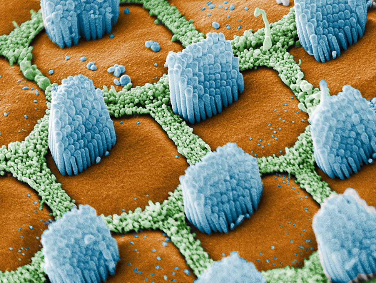
Microscopy, Electron, Scanning
Mechanoreceptors
Otoacoustic Emissions, Spontaneous
Gentamicins
Chinchilla
Guinea Pigs
Zebrafish
Pyridinium Compounds
Hearing Loss, Sensorineural
Otolithic Membrane
Semicircular Canals
Chickens
Membrane Potentials
Gerbillinae
Organ Culture Techniques
Vestibulocochlear Nerve
Basic Helix-Loop-Helix Transcription Factors
Batrachoidiformes
Presbycusis
Patch-Clamp Techniques
Dihydrostreptomycin Sulfate
Aminoglycosides
Vestibular Nerve
Gene Expression Regulation, Developmental
Anion Transport Proteins
Electrophysiology
Cell Differentiation
Efferent Pathways
Scalp
Calbindin 2
Molecular Motor Proteins
Calcium
Auditory Pathways
Myosins
Mice, Knockout
Sound
Plant Roots
Ear
Kanamycin
Usher Syndromes
Immunohistochemistry
Cell Death
S100 Calcium Binding Protein G
KCNQ Potassium Channels
Xanthenes
Models, Biological
Epithelium
Mice, Inbred C57BL
Evoked Potentials, Auditory
Synapses
Perilymph
Mice, Inbred CBA
Cell Transdifferentiation
Labyrinth Diseases
Zebrafish Proteins
Receptors, Notch
Large-Conductance Calcium-Activated Potassium Channels
Mutation
Microscopy, Confocal
Potassium Channels, Calcium-Activated
Mice, Transgenic
Amino Acid Transport Systems, Acidic
Hearing Disorders
Plasma Membrane Calcium-Transporting ATPases
Scala Tympani
Action Potentials
In Situ Hybridization
Ganglia, Sensory
Sensory Receptor Cells
Cell Polarity
Afferent Pathways
Hearing Loss, Central
Tympanic Membrane
Signal Transduction
Cell Count
Quaternary Ammonium Compounds
Ion Channel Gating
Membrane Proteins
PAX2 Transcription Factor
Cadherins
Stem Cells
Keratins, Hair-Specific
Audiometry, Evoked Response
Green Fluorescent Proteins
Olivary Nucleus
SOXB1 Transcription Factors
Phenotype
Microscopy, Electron
Exocytosis
Ear, Middle
Cell Membrane
Streptomycin
Phalloidine
Synaptic Vesicles
Nerve Tissue Proteins
Calcium Channels
Rana pipiens
4-Aminopyridine
Calcium Channels, L-Type
Lizards
Mollusca
Morphogenesis
Chelating Agents
Vestibular Diseases
Labyrinth Supporting Cells
Large-Conductance Calcium-Activated Potassium Channel beta Subunits
Adaptation, Physiological
Columbidae
Molecular Sequence Data
Temporal Bone
Larva
Epidermis
Chick Embryo
Plant Epidermis
Protein Synthesis Inhibitors
Dose-Response Relationship, Drug
Cells, Cultured
Synaptic Transmission
Bromodeoxyuridine
Dyneins
Potassium Channels
Sebaceous Glands
Immunocytochemical and morphological evidence for intracellular self-repair as an important contributor to mammalian hair cell recovery. (1/1112)
Although recent studies have provided evidence for hair cell regeneration in mammalian inner ears, the mechanism underlying this regenerative process is still under debate. Here we report immunocytochemical, histological, electron microscopic, and autoradiographic evidence that, in cultured postnatal rat utricles, a substantial number of hair cells can survive gentamicin insult even their stereocilia are lost. These partially damaged hair cells can survive for a prolonged time and regrow the stereocilia. Although the number of stereocilia-bearing hair cells increases over time after gentamicin insult, hair cell and supporting cell numbers remain essentially unchanged. Tritiated thymidine autoradiography and bromodeoxyuridine immunocytochemistry of the cultures demonstrate that cell proliferation in the sensory epithelium is very limited and is far below the number of recovered hair cells. Furthermore, terminal deoxynucleotidyl transferase-mediated biotinylated UTP nick end labeling analysis indicates that gentamicin-induced apoptosis in the sensory epithelium occurs mainly during a 2 d treatment period, and additional cell death is minimal 2-11 d after treatment. Considered together, intracellular repair of partially damaged hair cells can be an important contributor to spontaneous hair cell recovery in mammalian inner ears. (+info)Synapses involving auditory nerve fibers in primate cochlea. (2/1112)
The anatomical mechanisms for processing auditory signals are extremely complex and incompletely understood, despite major advances already made with the use of electron microscopy. A major enigma, for example, is the presence in the mammalian cochlea of a double hair cell receptor system. A renewed attempt to discover evidence of synaptic coupling between the two systems in the primate cochlea, postulated from physiological studies, has failed. However, in the outer spiral bundle the narrow and rigid clefts seen between pairs of presumptive afferent fibers suggest the possibility of dendro-dendritic interaction confined to the outer hair cell system. The clustering of afferent processes within folds of supporting cells subjacent to outer hair cells is in contrast to the lack of such close associations in the inner hair cell region. The difference reinforces the suggestion of functional interaction of some sort between the outer hair cell afferent nerve processes. (+info)Gene disruption of p27(Kip1) allows cell proliferation in the postnatal and adult organ of corti. (3/1112)
Hearing loss is most often the result of hair-cell degeneration due to genetic abnormalities or ototoxic and traumatic insults. In the postembryonic and adult mammalian auditory sensory epithelium, the organ of Corti, no hair-cell regeneration has ever been observed. However, nonmammalian hair-cell epithelia are capable of regenerating sensory hair cells as a consequence of nonsensory supporting-cell proliferation. The supporting cells of the organ of Corti are highly specialized, terminally differentiated cell types that apparently are incapable of proliferation. At the molecular level terminally differentiated cells have been shown to express high levels of cell-cycle inhibitors, in particular, cyclin-dependent kinase inhibitors [Parker, S. B., et al. (1995) Science 267, 1024-1027], which are thought to be responsible for preventing these cells from reentering the cell cycle. Here we report that the cyclin-dependent kinase inhibitor p27(Kip1) is selectively expressed in the supporting-cell population of the organ of Corti. Effects of p27(Kip1)-gene disruption include ongoing cell proliferation in postnatal and adult mouse organ of Corti at time points well after mitosis normally has ceased during embryonic development. This suggests that release from p27(Kip1)-induced cell-cycle arrest is sufficient to allow supporting-cell proliferation to occur. This finding may provide an important pathway for inducing hair-cell regeneration in the mammalian hearing organ. (+info)2E4 (kaptin): a novel actin-associated protein from human blood platelets found in lamellipodia and the tips of the stereocilia of the inner ear. (4/1112)
Platelet activation, crucial for hemostasis, requires actin polymerization, yet the molecular mechanisms by which localized actin polymerization is mediated are not clear. Here we report the characterization of a novel actin-binding protein, 2E4, originally isolated from human blood platelets and likely to be involved in the actin rearrangements occurring during activation. 2E4 binds to filamentous (F)-actin by F-actin affinity chromatography and is eluted from F-actin affinity columns and extracted from cells with ATP. Its presence at the leading edge of platelets spread on glass and in the lamellipodia of motile fibroblasts suggests a role in actin dynamics. Using localization to obtain clues about function, we stained the sensory epithelium of the embryonic inner ear to determine whether 2E4 is at the barbed end of actin filaments during their elongation. Indeed, 2E4 was present at the tips of the elongating stereocilium. 2E4 is novel by DNA sequence and has no identifiable structural motifs. Its unusual amino acid sequence, its ATP-sensitive actin association and its location at sites of actin polymerization in cells suggest 2E4 plays a unique role in the actin rearrangements that accompany platelet activation and stereocilia formation. (+info)Electrical response properties of avian lagena type II hair cells: a model system for vestibular filtering. (5/1112)
Data presented represent the first electrical recordings from avian lagena type II hair cells. The perforated-patch variant of the whole cell recording technique was used to investigate how the macroscopic currents shaped the voltage response of the hair cells. Voltage-clamp data separated cells into two broad classes on the basis of differences in activation rates, rates and degree of inactivation, and pharmacological sensitivity. Current-clamp recordings revealed low-quality membrane voltage oscillations (Qc < 1) during pulse current injections. Oscillation frequency correlated with activation rate of the macroscopic currents. The quality of membrane oscillations (Qc) varied linearly with frequency for cells with little inactivation. For cells with rapid inactivation, no relationship was found between Qc and frequency. Rapid inactivation may serve to extend the bandwidth of vestibular hair cells. The frequency measured from voltage responses to pulsed currents may reflect the corner frequency of the cell. The filtering properties of avian lagena hair cells are like those found in all other vestibular end organs, suggesting that the electrical membrane properties of these cells are not responsible for specializing them to a particular stimulus modality. (+info)Functional expression of exogenous proteins in mammalian sensory hair cells infected with adenoviral vectors. (6/1112)
To understand the function of specific proteins in sensory hair cells, it is necessary to add or inactivate those proteins in a system where their physiological effects can be studied. Unfortunately, the usefulness of heterologous expression systems for the study of many hair cell proteins is limited by the inherent difficulty of reconstituting the hair cell's exquisite cytoarchitecture. Expression of exogenous proteins within hair cells themselves may provide an alternative approach. Because recombinant viruses were efficient vectors for gene delivery in other systems, we screened three viral vectors for their ability to express exogenous genes in hair cells of organotypic cultures from mouse auditory and vestibular organs. We observed no expression of the genes for beta-galactosidase or green fluorescent protein (GFP) with either herpes simplex virus or adeno-associated virus. On the other hand, we found robust expression of GFP in hair cells exposed to a recombinant, replication-deficient adenovirus that carried the gene for GFP driven by a cytomegalovirus promoter. Titers of 4 x 10(7) pfu/ml were sufficient for expression in 50% of the approximately 1,000 hair cells in the utricular epithelium; < 1% of the nonhair cells in the epithelium were GFP positive. Expression of GFP was evident as early as 12 h postinfection, was maximal at 4 days, and continued for at least 10 days. Over the first 36 h there was no evidence of toxicity. We recorded normal voltage-dependent and transduction currents from infected cells identified by GFP fluorescence. At longer times hair bundle integrity was compromised despite a cell body that appeared healthy. To assess the ability of adenovirus-mediated gene transfer to alter hair cell function we introduced the gene for the ion channel Kir2.1. We used an adenovirus vector encoding Kir2.1 fused to GFP under the control of an ecdysone promoter. Unlike the diffuse distribution within the cell body we observed with GFP, the ion channel-GFP fusion showed a pattern of fluorescence that was restricted to the cell membrane and a few extranuclear punctate regions. Patch-clamp recordings confirmed the expression of an inward rectifier with a conductance of 43 nS, over an order of magnitude larger than the endogenous inward rectifier. The zero-current potential in infected cells was shifted by -17 mV. These results demonstrate an efficient method for gene transfer into both vestibular and auditory hair cells in culture, which can be used to study the effects of gene products on hair cell function. (+info)The supporting-cell antigen: a receptor-like protein tyrosine phosphatase expressed in the sensory epithelia of the avian inner ear. (7/1112)
After noise- or drug-induced hair-cell loss, the sensory epithelia of the avian inner ear can regenerate new hair cells. Few molecular markers are available for the supporting-cell precursors of the hair cells that regenerate, and little is known about the signaling mechanisms underlying this regenerative response. Hybridoma methodology was used to obtain a monoclonal antibody (mAb) that stains the apical surface of supporting cells in the sensory epithelia of the inner ear. The mAb recognizes the supporting-cell antigen (SCA), a protein that is also found on the apical surfaces of retinal Muller cells, renal tubule cells, and intestinal brush border cells. Expression screening and molecular cloning reveal that the SCA is a novel receptor-like protein tyrosine phosphatase (RPTP), sharing similarity with human density-enhanced phosphatase, an RPTP thought to have a role in the density-dependent arrest of cell growth. In response to hair-cell damage induced by noise in vivo or hair-cell loss caused by ototoxic drug treatment in vitro, some supporting cells show a dramatic decrease in SCA expression levels on their apical surface. This decrease occurs before supporting cells are known to first enter S-phase after trauma, indicating that it may be a primary rather than a secondary response to injury. These results indicate that the SCA is a signaling molecule that may influence the potential of nonsensory supporting cells to either proliferate or differentiate into hair cells. (+info)The electrical properties of auditory hair cells in the frog amphibian papilla. (8/1112)
The amphibian papilla (AP) is the principal auditory organ of the frog. Anatomical and neurophysiological evidence suggests that this hearing organ utilizes both mechanical and electrical (hair cell-based) frequency tuning mechanisms, yet relatively little is known about the electrophysiology of AP hair cells. Using the whole-cell patch-clamp technique, we have investigated the electrical properties and ionic currents of isolated hair cells along the rostrocaudal axis of the AP. Electrical resonances were observed in the voltage response of hair cells harvested from the rostral and medial, but not caudal, regions of the AP. Two ionic currents, ICa and IK(Ca), were observed in every hair cell; however, their amplitudes varied substantially along the epithelium. Only rostral hair cells exhibited an inactivating potassium current (IA), whereas an inwardly rectifying potassium current (IK1) was identified only in caudal AP hair cells. Electrically tuned hair cells exhibited resonant frequencies from 50 to 375 Hz, which correlated well with hair cell position and the tonotopic organization of the papilla. Variations in the kinetics of the outward current contribute substantially to the determination of resonant frequency. ICa and IK(Ca) amplitudes increased with resonant frequency, reducing the membrane time constant with increasing resonant frequency. We conclude that a tonotopically organized hair cell substrate exists to support electrical tuning in the rostromedial region of the frog amphibian papilla and that the cellular mechanisms for frequency determination are very similar to those reported for another electrically tuned auditory organ, the turtle basilar papilla. (+info)Auditory hair cells are specialized sensory receptor cells located in the inner ear, more specifically in the organ of Corti within the cochlea. They play a crucial role in hearing by converting sound vibrations into electrical signals that can be interpreted by the brain.
These hair cells have hair-like projections called stereocilia on their apical surface, which are embedded in a gelatinous matrix. When sound waves reach the inner ear, they cause the fluid within the cochlea to move, which in turn causes the stereocilia to bend. This bending motion opens ion channels at the tips of the stereocilia, allowing positively charged ions (such as potassium) to flow into the hair cells and trigger a receptor potential.
The receptor potential then leads to the release of neurotransmitters at the base of the hair cells, which activate afferent nerve fibers that synapse with these cells. The electrical signals generated by this process are transmitted to the brain via the auditory nerve, where they are interpreted as sound.
There are two types of auditory hair cells: inner hair cells and outer hair cells. Inner hair cells are the primary sensory receptors responsible for transmitting information about sound to the brain. They make direct contact with afferent nerve fibers and are more sensitive to mechanical stimulation than outer hair cells.
Outer hair cells, on the other hand, are involved in amplifying and fine-tuning the mechanical response of the inner ear to sound. They have a unique ability to contract and relax in response to electrical signals, which allows them to adjust the stiffness of their stereocilia and enhance the sensitivity of the cochlea to different frequencies.
Damage or loss of auditory hair cells can lead to hearing impairment or deafness, as these cells cannot regenerate spontaneously in mammals. Therefore, understanding the structure and function of hair cells is essential for developing therapies aimed at treating hearing disorders.
Medically, hair is defined as a threadlike structure that grows from the follicles found in the skin of mammals. It is primarily made up of a protein called keratin and consists of three parts: the medulla (the innermost part or core), the cortex (middle layer containing keratin filaments) and the cuticle (outer layer of overlapping scales).
Hair growth occurs in cycles, with each cycle consisting of a growth phase (anagen), a transitional phase (catagen), and a resting phase (telogen). The length of hair is determined by the duration of the anagen phase.
While hair plays a crucial role in protecting the skin from external factors like UV radiation, temperature changes, and physical damage, it also serves as an essential aspect of human aesthetics and identity.
Auditory inner hair cells are specialized sensory receptor cells located in the inner ear, more specifically in the organ of Corti within the cochlea. They play a crucial role in hearing by converting mechanical sound energy into electrical signals that can be processed and interpreted by the brain.
Human ears have about 3,500 inner hair cells arranged in one row along the length of the basilar membrane in each cochlea. These hair cells are characterized by their stereocilia, which are hair-like projections on the apical surface that are embedded in a gelatinous matrix called the tectorial membrane.
When sound waves cause the basilar membrane to vibrate, the stereocilia of inner hair cells bend and deflect. This deflection triggers a cascade of biochemical events leading to the release of neurotransmitters at the base of the hair cell. These neurotransmitters then stimulate the afferent auditory nerve fibers (type I fibers) that synapse with the inner hair cells, transmitting the electrical signals to the brain for further processing and interpretation as sound.
Damage or loss of these inner hair cells can lead to significant hearing impairment or deafness, as they are essential for normal auditory function. Currently, there is no effective way to regenerate damaged inner hair cells in humans, making hearing loss due to their damage permanent.
Auditory outer hair cells are specialized sensory receptor cells located in the cochlea of the inner ear. They are part of the organ of Corti and play a crucial role in hearing by converting sound energy into electrical signals that can be interpreted by the brain.
Unlike the more numerous and simpler auditory inner hair cells, outer hair cells are equipped with unique actin-based molecular motors called "motile" or "piezoelectric" properties. These motors enable the outer hair cells to change their shape and length in response to electrical signals, which in turn amplifies the mechanical vibrations of the basilar membrane where they are located. This amplification increases the sensitivity and frequency selectivity of hearing, allowing us to detect and discriminate sounds over a wide range of intensities and frequencies.
Damage or loss of outer hair cells is a common cause of sensorineural hearing loss, which can result from exposure to loud noises, aging, genetics, ototoxic drugs, and other factors. Currently, there are no effective treatments to regenerate or replace damaged outer hair cells, making hearing loss an irreversible condition in most cases.
Vestibular hair cells are specialized sensory receptor cells located in the vestibular system of the inner ear. They play a crucial role in detecting and mediating our sense of balance and spatial orientation by converting mechanical stimuli, such as head movements and gravity, into electrical signals that are sent to the brain.
The hair cells are shaped like a tuft of hair, with stereocilia projecting from their tops. These stereocilia are arranged in rows of graded height, and they are embedded in a gel-like structure within the vestibular organ. When the head moves or changes position, the movement causes deflection of the stereocilia, which opens ion channels at their tips and triggers nerve impulses that are sent to the brain via the vestibular nerve.
There are two types of vestibular hair cells: type I and type II. Type I hair cells have a large, spherical shape and are more sensitive to changes in head position, while type II hair cells are more cylindrical in shape and respond to both linear and angular acceleration. Together, these hair cells help us maintain our balance, coordinate our movements, and keep our eyes focused during head movements.
The saccule and utricle are components of the vestibular system, which is responsible for maintaining balance and spatial orientation within the inner ear. Here are the medical definitions:
1. Saccule: A small sac-like structure located in the vestibular labyrinth of the inner ear. It is one of the two otolith organs (the other being the utricle) that detect linear acceleration and gravity. The saccule contains hair cells with stereocilia, which are embedded in a gelatinous matrix containing calcium carbonate crystals called otoconia. When the head changes position or moves linearly, the movement of these otoconia stimulates the hair cells, sending signals to the brain about the direction and speed of the motion.
2. Utricle: Another sac-like structure in the vestibular labyrinth, similar to the saccule but slightly larger. The utricle is also an otolith organ that detects linear acceleration and head tilts. It contains hair cells with stereocilia embedded in a gelatinous matrix filled with otoconia. When the head tilts or moves linearly, the movement of the otoconia stimulates the hair cells, providing information about the position and motion of the head to the brain.
In summary, both the saccule and utricle are essential for maintaining balance and spatial orientation by detecting linear acceleration and gravity through the movement of otoconia on their hair cell receptors.
The cochlea is a part of the inner ear that is responsible for hearing. It is a spiral-shaped structure that looks like a snail shell and is filled with fluid. The cochlea contains hair cells, which are specialized sensory cells that convert sound vibrations into electrical signals that are sent to the brain.
The cochlea has three main parts: the vestibular canal, the tympanic canal, and the cochlear duct. Sound waves enter the inner ear and cause the fluid in the cochlea to move, which in turn causes the hair cells to bend. This bending motion stimulates the hair cells to generate electrical signals that are sent to the brain via the auditory nerve.
The brain then interprets these signals as sound, allowing us to hear and understand speech, music, and other sounds in our environment. Damage to the hair cells or other structures in the cochlea can lead to hearing loss or deafness.
A hair follicle is a part of the human skin from which hair grows. It is a complex organ that consists of several layers, including an outer root sheath, inner root sheath, and matrix. The hair follicle is located in the dermis, the second layer of the skin, and is surrounded by sebaceous glands and erector pili muscles.
The hair growth cycle includes three phases: anagen (growth phase), catagen (transitional phase), and telogen (resting phase). During the anagen phase, cells in the matrix divide rapidly to produce new hair fibers that grow out of the follicle. The hair fiber is made up of a protein called keratin, which also makes up the outer layers of the skin and nails.
Hair follicles are important for various biological functions, including thermoregulation, sensory perception, and social communication. They also play a role in wound healing and can serve as a source of stem cells that can differentiate into other cell types.
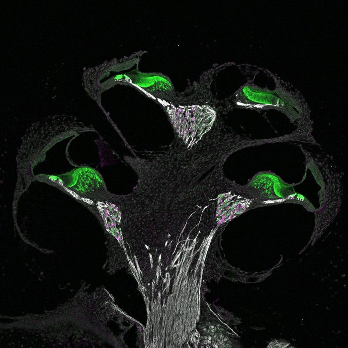
The Organ of Corti is the sensory organ of hearing within the cochlea of the inner ear. It is a structure in the inner spiral sulcus of the cochlear duct and is responsible for converting sound vibrations into electrical signals that are sent to the brain via the auditory nerve.
The Organ of Corti consists of hair cells, which are sensory receptors with hair-like projections called stereocilia on their apical surfaces. These stereocilia are embedded in a gelatinous matrix and are arranged in rows of different heights. When sound vibrations cause the fluid in the cochlea to move, the stereocilia bend, which opens ion channels and triggers nerve impulses that are sent to the brain.
Damage or loss of hair cells in the Organ of Corti can result in hearing loss, making it a critical structure for maintaining normal auditory function.
The inner ear is the innermost part of the ear that contains the sensory organs for hearing and balance. It consists of a complex system of fluid-filled tubes and sacs called the vestibular system, which is responsible for maintaining balance and spatial orientation, and the cochlea, a spiral-shaped organ that converts sound vibrations into electrical signals that are sent to the brain.
The inner ear is located deep within the temporal bone of the skull and is protected by a bony labyrinth. The vestibular system includes the semicircular canals, which detect rotational movements of the head, and the otolith organs (the saccule and utricle), which detect linear acceleration and gravity.
Damage to the inner ear can result in hearing loss, tinnitus (ringing in the ears), vertigo (a spinning sensation), and balance problems.
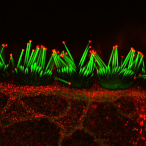
Stereocilia are hair-like projections found in the inner ear, more specifically in the organ of Corti within the cochlea. They are present on the sensory cells known as hair cells and are involved in hearing by converting sound vibrations into electrical signals that can be transmitted to the brain.
Stereocilia are arranged in rows of graded height, with the tallest ones located near the opening of the cochlea (the base) and the shortest ones closer to the apex. When sound waves reach the inner ear, they cause the fluid within the cochlea to move, which in turn causes stereocilia to bend. This bending action triggers the release of chemical signals that stimulate nerve fibers connected to the hair cells, ultimately transmitting information about the sound to the brain.
Damage or loss of stereocilia can result in hearing impairment or deafness, as seen in various forms of hearing disorders and age-related hearing loss.
Hair cells in the ampulla are specialized sensory receptor cells located within the vestibular system of the inner ear. The vestibular system is responsible for detecting movement and maintaining balance. The ampulla is a part of one of the three semicircular canals, fluid-filled structures that sense rotational movements of the head.
Hair cells in the ampulla have hair-like projections called stereocilia on their surface, which are embedded in a gelatinous structure called the cupula. The movement of fluid within the semicircular canal causes the deflection of the stereocilia, leading to the activation of mechanically gated ion channels and generating receptor potentials. These electrical signals are then transmitted to the brain via the vestibular nerve, allowing the brain to interpret head movements and maintain balance.
Damage or loss of hair cells in the ampulla can lead to vestibular dysfunction and balance disorders.
I'm sorry for any confusion, but "hair color" is not a term that has a medical definition in the same way that a term like "myocardial infarction" (heart attack) does. Hair color can be described in various ways, such as being a natural hair color (like blonde, brunette, red, or black) or describing a change in hair color due to factors like aging (gray or white hairs) or hair dye usage.
However, it's worth noting that changes in hair color can sometimes be associated with certain medical conditions. For example, premature graying of the hair before the age of 30 can be a feature of certain genetic disorders or vitamin B12 deficiency. Similarly, some skin conditions like alopecia areata or vitiligo can cause patchy changes in hair color. But these associations don't provide a medical definition for 'hair color'.
Cellular mechanotransduction is the process by which cells convert mechanical stimuli into biochemical signals, resulting in changes in cell behavior and function. This complex process involves various molecular components, including transmembrane receptors, ion channels, cytoskeletal proteins, and signaling molecules. Mechanical forces such as tension, compression, or fluid flow can activate these components, leading to alterations in gene expression, protein synthesis, and cell shape or movement. Cellular mechanotransduction plays a crucial role in various physiological processes, including tissue development, homeostasis, and repair, as well as in pathological conditions such as fibrosis and cancer progression.
Hair diseases is a broad term that refers to various medical conditions affecting the hair shaft, follicle, or scalp. These conditions can be categorized into several types, including:
1. Hair shaft abnormalities: These are conditions that affect the structure and growth of the hair shaft. Examples include trichorrhexis nodosa, where the hair becomes weak and breaks easily, and pili torti, where the hair shaft is twisted and appears sparse and fragile.
2. Hair follicle disorders: These are conditions that affect the hair follicles, leading to hair loss or abnormal growth patterns. Examples include alopecia areata, an autoimmune disorder that causes patchy hair loss, and androgenetic alopecia, a genetic condition that leads to pattern baldness in both men and women.
3. Scalp disorders: These are conditions that affect the scalp, leading to symptoms such as itching, redness, scaling, or pain. Examples include seborrheic dermatitis, psoriasis, and tinea capitis (ringworm of the scalp).
4. Hair cycle abnormalities: These are conditions that affect the normal growth cycle of the hair, leading to excessive shedding or thinning. Examples include telogen effluvium, where a large number of hairs enter the resting phase and fall out, and anagen effluvium, which is typically caused by chemotherapy or radiation therapy.
5. Infectious diseases: Hair follicles can become infected with various bacteria, viruses, or fungi, leading to conditions such as folliculitis, furunculosis, and kerion.
6. Genetic disorders: Some genetic disorders can affect the hair, such as Menkes syndrome, which is a rare inherited disorder that affects copper metabolism and leads to kinky, sparse, and brittle hair.
Proper diagnosis and treatment of hair diseases require consultation with a healthcare professional, often a dermatologist or a trichologist who specializes in hair and scalp disorders.
The lateral line system is a sensory organ found in aquatic animals, such as fish and some aquatic amphibians. It is a series of fluid-filled canals and sensory cells that run along the sides of the body, head, and fins. These sensory cells are called neuromasts and contain hair cells that respond to vibrations and water movements. The lateral line system helps these animals detect movement, pressure changes, and vibrations in their aquatic environment, which aids in schooling behavior, prey detection, and avoiding predators.
Hearing is the ability to perceive sounds by detecting vibrations in the air or other mediums and translating them into nerve impulses that are sent to the brain for interpretation. In medical terms, hearing is defined as the sense of sound perception, which is mediated by the ear and interpreted by the brain. It involves a complex series of processes, including the conduction of sound waves through the outer ear to the eardrum, the vibration of the middle ear bones, and the movement of fluid in the inner ear, which stimulates hair cells to send electrical signals to the auditory nerve and ultimately to the brain. Hearing allows us to communicate with others, appreciate music and sounds, and detect danger or important events in our environment.
Hair removal is the deliberate elimination or reduction of body hair. This can be achieved through various methods, both temporary and permanent. Some common temporary methods include shaving, waxing, tweezing, and depilatory creams. Permanent methods may involve laser hair removal or electrolysis, which target the hair follicle to prevent future growth. It's important to note that some methods can have side effects or risks, so it's recommended to consult with a healthcare professional or dermatologist before starting any new hair removal regimen.
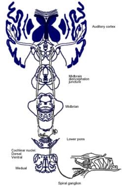
The spiral ganglion is a structure located in the inner ear, specifically within the cochlea. It consists of nerve cell bodies that form the sensory component of the auditory nervous system. The spiral ganglion's neurons are bipolar and have peripheral processes that form synapses with hair cells in the organ of Corti, which is responsible for converting sound vibrations into electrical signals.
The central processes of these neurons then coalesce to form the cochlear nerve, which transmits these electrical signals to the brainstem and ultimately to the auditory cortex for processing and interpretation as sound. Damage to the spiral ganglion or its associated neural structures can lead to hearing loss or deafness.
Auditory brainstem evoked potentials (ABEPs or BAEPs) are medical tests that measure the electrical activity in the auditory pathway of the brain in response to sound stimulation. The test involves placing electrodes on the scalp and recording the tiny electrical signals generated by the nerve cells in the brainstem as they respond to clicks or tone bursts presented through earphones.
The resulting waveform is analyzed for latency (the time it takes for the signal to travel from the ear to the brain) and amplitude (the strength of the signal). Abnormalities in the waveform can indicate damage to the auditory nerve or brainstem, and are often used in the diagnosis of various neurological conditions such as multiple sclerosis, acoustic neuroma, and brainstem tumors.
The test is non-invasive, painless, and takes only a few minutes to perform. It provides valuable information about the functioning of the auditory pathway and can help guide treatment decisions for patients with hearing or balance disorders.
Hair dyes are chemical substances that are used to change the color of hair. They contain various types of dyes, including natural dyes derived from plants and minerals, synthetic dyes, and combinations of both. Hair dyes work by penetrating the outer layer of the hair shaft (the cuticle) and bonding with the hair's pigment (melanin) or depositing new color particles within the hair shaft.
There are three main types of hair dyes: temporary, semi-permanent, and permanent. Temporary hair dyes coat the outside of the hair shaft and wash out after a few shampoos. Semi-perermanent hair dyes penetrate slightly into the hair shaft and fade gradually over several washes. Permanent hair dyes contain chemicals that open the cuticle and allow the dye to penetrate deep into the hair shaft, where it reacts with the hair's natural pigment to create a new color that is resistant to fading and washing out.
It is important to note that some hair dyes may contain potentially harmful chemicals, such as coal tar dyes, para-phenylenediamine (PPD), and resorcinol, which have been linked to allergic reactions, skin irritation, and other health problems. It is recommended to perform a patch test before using any new hair dye product and to follow the manufacturer's instructions carefully to minimize the risk of adverse effects.
Hair preparations refer to cosmetic or grooming products that are specifically formulated to be applied to the hair or scalp for various purposes such as cleansing, conditioning, styling, coloring, or promoting hair growth. These preparations can come in different forms, including shampoos, conditioners, hair masks, serums, gels, mousses, sprays, and dyes. They may contain a wide range of ingredients, such as detergents, moisturizers, proteins, vitamins, minerals, and other nutrients that can help improve the health, appearance, and manageability of the hair. Some hair preparations may also contain medications or natural extracts that have therapeutic properties for treating specific hair or scalp conditions, such as dandruff, dryness, oiliness, thinning, or hair loss.
Transcription Factor Brn-3C, also known as POU4F3, is a protein involved in the regulation of gene expression. It belongs to the class IV POU domain transcription factor family and plays crucial roles in the development, maintenance, and function of inner ear hair cells, which are essential for hearing. Mutations in the Brn-3C gene have been associated with deafness disorders in humans. The protein works by binding to specific DNA sequences in the promoter regions of target genes and controlling their transcription into messenger RNA (mRNA). This process is critical for various cellular functions, including cell growth, differentiation, and survival.
The acoustic maculae, also known as the vestibularocochlear nerve or cranial nerve VIII, are a part of the human body's auditory and vestibular system. The acoustic maculae consist of two main structures: the cochlea and the vestibule.
The cochlea is responsible for hearing and converts sound waves into electrical signals that can be interpreted by the brain. It contains the organ of Corti, which has hair cells that are stimulated by sound vibrations and convert them into nerve impulses.
The vestibule, on the other hand, is responsible for maintaining balance and spatial orientation. It contains two sac-like structures called the utricle and saccule, which contain sensory hair cells that respond to gravity and linear acceleration.
Damage to the acoustic maculae can result in hearing loss, tinnitus (ringing in the ears), or balance disorders.
Electric capacitance is a measure of the amount of electrical charge that a body or system can hold for a given electric potential. In other words, it is a measure of the capacity of a body or system to store an electric charge. The unit of electric capacitance is the farad (F), which is defined as the capacitance of a conductor that, when charged with one coulomb of electricity, has a potential difference of one volt between its surfaces.
In medical terms, electric capacitance may be relevant in the context of electrical stimulation therapies, such as transcutaneous electrical nerve stimulation (TENS) or functional electrical stimulation (FES). In these therapies, electrodes are placed on the skin and a controlled electric current is applied to stimulate nerves or muscles. The electric capacitance of the tissue and electrodes can affect the distribution and intensity of the electric field, which in turn can influence the therapeutic effect.
It is important to note that while electric capacitance is a fundamental concept in physics and engineering, it is not a commonly used term in medical practice or research. Instead, terms such as impedance or resistance are more commonly used to describe the electrical properties of biological tissues.
The vestibular system is a part of the inner ear that contributes to our sense of balance and spatial orientation. It is made up of two main components: the vestibule and the labyrinth.
The vestibule is a bony chamber in the inner ear that contains two important structures called the utricle and saccule. These structures contain hair cells and fluid-filled sacs that help detect changes in head position and movement, allowing us to maintain our balance and orientation in space.
The labyrinth, on the other hand, is a more complex structure that includes the vestibule as well as three semicircular canals. These canals are also filled with fluid and contain hair cells that detect rotational movements of the head. Together, the vestibule and labyrinth work together to provide us with information about our body's position and movement in space.
Overall, the vestibular system plays a crucial role in maintaining our balance, coordinating our movements, and helping us navigate through our environment.
Hearing loss is a partial or total inability to hear sounds in one or both ears. It can occur due to damage to the structures of the ear, including the outer ear, middle ear, inner ear, or nerve pathways that transmit sound to the brain. The degree of hearing loss can vary from mild (difficulty hearing soft sounds) to severe (inability to hear even loud sounds). Hearing loss can be temporary or permanent and may be caused by factors such as exposure to loud noises, genetics, aging, infections, trauma, or certain medical conditions. It is important to note that hearing loss can have significant impacts on a person's communication abilities, social interactions, and overall quality of life.
"Rana catesbeiana" is the scientific name for the American bullfrog, which is not a medical term or concept. It belongs to the animal kingdom, specifically in the order Anura and family Ranidae. The American bullfrog is native to North America and is known for its large size and distinctive loud call.
However, if you are looking for a medical definition, I apologize for any confusion. Please provide more context or specify the term you would like me to define.
Neomycin is an antibiotic drug derived from the bacterium Streptomyces fradiae. It belongs to the class of aminoglycoside antibiotics and works by binding to the 30S subunit of the bacterial ribosome, thereby inhibiting protein synthesis and leading to bacterial cell death. Neomycin is primarily used topically (on the skin or mucous membranes) due to its poor absorption into the bloodstream when taken orally. It is effective against a wide range of gram-positive and gram-negative bacteria. Medical definitions for Neomycin include:
1. An antibiotic (aminoglycoside) derived from Streptomyces fradiae, used primarily for topical application in the treatment of superficial infections, burns, and wounds. It is not usually used systemically due to its potential ototoxicity and nephrotoxicity.
2. A medication (generic name) available as a cream, ointment, solution, or powder, often combined with other active ingredients such as bacitracin and polymyxin B for broader-spectrum antibacterial coverage. Neomycin is used to treat various skin conditions, including eczema, dermatitis, and minor cuts or abrasions.
3. A component of some over-the-counter products (e.g., ear drops, eye drops) intended for the treatment of external otitis, swimmer's ear, or bacterial conjunctivitis. It is crucial to follow the instructions carefully and avoid using neomycin-containing products for extended periods or in larger quantities than recommended, as this may increase the risk of antibiotic resistance and potential side effects.
In summary, Neomycin is an aminoglycoside antibiotic primarily used topically for treating various superficial bacterial infections due to its effectiveness against a wide range of gram-positive and gram-negative bacteria. It should be used cautiously and as directed to minimize the risk of side effects and antibiotic resistance.
Cochlear diseases refer to conditions that affect the structure or function of the cochlea, which is a part of the inner ear responsible for hearing. These diseases can cause various types and degrees of hearing loss, ranging from mild to profound. Some common cochlear diseases include:
1. Cochlear otosclerosis: A condition where there is abnormal bone growth in the cochlea, which can lead to conductive or sensorineural hearing loss.
2. Cochlear Meniere's disease: A disorder that affects the inner ear and causes vertigo, tinnitus, and fluctuating hearing loss.
3. Cochlear damage due to exposure to loud noises: Prolonged or sudden exposure to loud noises can cause permanent cochlear damage and hearing loss.
4. Presbycusis: Age-related hearing loss that affects the cochlea and other structures of the auditory system.
5. Cochlear nerve tumors: Rare benign or malignant growths on the cochlear nerve can cause hearing loss, tinnitus, and balance problems.
6. Infections: Bacterial or viral infections such as meningitis, labyrinthitis, or otitis media can damage the cochlea and lead to hearing loss.
7. Ototoxicity: Certain medications can be toxic to the cochlea and cause hearing loss, tinnitus, or balance problems.
8. Genetic factors: Inherited genetic mutations can cause various types of cochlear diseases, such as connexin 26 deficiency, Waardenburg syndrome, or Usher syndrome.
It is important to note that early diagnosis and treatment of cochlear diseases can help prevent or minimize hearing loss and other complications.
Stria vascularis is a highly vascularized (rich in blood vessels) structure located in the cochlea of the inner ear. It plays a crucial role in the process of hearing by maintaining the endocochlear potential, which is essential for the conversion of sound waves into electrical signals that can be interpreted by the brain. The stria vascularis is composed of three layers: the marginal cells, intermediate cells, and basal cells, which work together to maintain the ionic balance and generate the endocochlear potential. Damage to the stria vascularis can result in hearing loss.
Deafness is a hearing loss that is so severe that it results in significant difficulty in understanding or comprehending speech, even when using hearing aids. It can be congenital (present at birth) or acquired later in life due to various causes such as disease, injury, infection, exposure to loud noises, or aging. Deafness can range from mild to profound and may affect one ear (unilateral) or both ears (bilateral). In some cases, deafness may be accompanied by tinnitus, which is the perception of ringing or other sounds in the ears.
Deaf individuals often use American Sign Language (ASL) or other forms of sign language to communicate. Some people with less severe hearing loss may benefit from hearing aids, cochlear implants, or other assistive listening devices. Deafness can have significant social, educational, and vocational implications, and early intervention and appropriate support services are critical for optimal development and outcomes.
Noise-induced hearing loss (NIHL) is a type of sensorineural hearing loss that occurs due to exposure to harmful levels of noise. The damage can be caused by a one-time exposure to an extremely loud sound or by continuous exposure to lower level sounds over time. NIHL can affect people of all ages and can cause permanent damage to the hair cells in the cochlea, leading to hearing loss, tinnitus (ringing in the ears), and difficulty understanding speech in noisy environments. Prevention measures include avoiding excessive noise exposure, wearing hearing protection, and taking regular breaks from noisy activities.
The basilar membrane is a key structure within the inner ear that plays a crucial role in hearing. It is a narrow, flexible strip of tissue located inside the cochlea, which is the spiral-shaped organ responsible for converting sound waves into neural signals that can be interpreted by the brain.
The basilar membrane runs along the length of the cochlea's duct and is attached to the rigid bony structures at both ends. It varies in width and stiffness along its length, with the widest and most flexible portion located near the entrance of the cochlea and the narrowest and stiffest portion located near the apex.
When sound waves enter the inner ear, they cause vibrations in the fluid-filled cochlear duct. These vibrations are transmitted to the basilar membrane, causing it to flex up and down. The specific pattern of flexion along the length of the basilar membrane depends on the frequency of the sound wave. Higher frequency sounds cause maximum flexion near the base of the cochlea, while lower frequency sounds cause maximum flexion near the apex.
As the basilar membrane flexes, it causes the attached hair cells to bend. This bending stimulates the hair cells to release neurotransmitters, which then activate the auditory nerve fibers. The pattern of neural activity in the auditory nerve encodes the frequency and amplitude of the sound wave, allowing the brain to interpret the sound.
Overall, the basilar membrane is a critical component of the hearing process, enabling us to detect and discriminate different sounds based on their frequency and amplitude.
The cochlear nerve, also known as the auditory nerve, is the sensory nerve that transmits sound signals from the inner ear to the brain. It consists of two parts: the outer spiral ganglion and the inner vestibular portion. The spiral ganglion contains the cell bodies of the bipolar neurons that receive input from hair cells in the cochlea, which is the snail-shaped organ in the inner ear responsible for hearing. These neurons then send their axons to form the cochlear nerve, which travels through the internal auditory meatus and synapses with neurons in the cochlear nuclei located in the brainstem.
Damage to the cochlear nerve can result in hearing loss or deafness, depending on the severity of the injury. Common causes of cochlear nerve damage include acoustic trauma, such as exposure to loud noises, viral infections, meningitis, and tumors affecting the nerve or surrounding structures. In some cases, cochlear nerve damage may be treated with hearing aids, cochlear implants, or other assistive devices to help restore or improve hearing function.
The tectorial membrane is a specialized structure in the inner ear, more specifically in the cochlea. It is a gelatinous, hair-like structure that is located above and parallel to the organ of Corti, which contains the sensory hair cells responsible for hearing. The tectorial membrane is composed of collagen fibers and a glycoprotein matrix.
The main function of the tectorial membrane is to deflect the stereocilia (hair-like projections) of the inner and outer hair cells as sound waves pass through the cochlea, which in turn triggers nerve impulses that are sent to the brain and interpreted as sound. The tectorial membrane moves in response to sound-induced vibrations of the fluid within the cochlea, causing shearing forces on the stereocilia, leading to the initiation of the hearing process.
I'm sorry for any confusion, but "turtles" is not a medical term. It refers to the group of reptiles that have a shell and include various species such as tortoises and terrapins. If you have any medical concerns or questions, I would be happy to try to help with those!
Cochlear microphonic potentials (CMs) are electrical responses that originate from the hair cells in the cochlea, which is a part of the inner ear responsible for hearing. These potentials can be recorded using an electrode placed near the cochlea in response to sound stimulation.
The CMs are considered to be a passive response of the hair cells to the mechanical deflection caused by sound waves. They represent the receptor potential of the outer hair cells and are directly proportional to the sound pressure level. Unlike other electrical responses in the cochlea, such as the action potentials generated by the auditory nerve fibers, CMs do not require the presence of neurotransmitters or synaptic transmission.
Cochlear microphonic potentials have been used in research to study the biophysical properties of hair cells and their response to different types of sound stimuli. However, they are not typically used in clinical audiology due to their small amplitude and susceptibility to interference from other electrical signals in the body.
Cilia are tiny, hair-like structures that protrude from the surface of many types of cells in the body. They are composed of a core bundle of microtubules surrounded by a protein matrix and are covered with a membrane. Cilia are involved in various cellular functions, including movement of fluid or mucus across the cell surface, detection of external stimuli, and regulation of signaling pathways.
There are two types of cilia: motile and non-motile. Motile cilia are able to move in a coordinated manner to propel fluids or particles across a surface, such as those found in the respiratory tract and reproductive organs. Non-motile cilia, also known as primary cilia, are present on most cells in the body and serve as sensory organelles that detect chemical and mechanical signals from the environment.
Defects in cilia structure or function can lead to a variety of diseases, collectively known as ciliopathies. These conditions can affect multiple organs and systems in the body, including the brain, kidneys, liver, and eyes. Examples of ciliopathies include polycystic kidney disease, Bardet-Biedl syndrome, and Meckel-Gruber syndrome.
Endolymph is a specific type of fluid that is found within the inner ear, more specifically in the membranous labyrinth of the inner ear. This fluid plays a crucial role in maintaining balance and hearing functions. It helps in the stimulation of hair cells present in the inner ear which then transmit signals to the brain, enabling us to hear and maintain our balance. Any disturbance or changes in the composition or flow of endolymph can lead to various vestibular disorders and hearing problems.
The cochlear duct, also known as the scala media, is a membranous duct located within the cochlea of the inner ear. It is one of three fluid-filled compartments in the cochlea, along with the vestibular duct (scala vestibuli) and the tympanic duct (scala tympani).
The cochlear duct contains endolymph, a specialized fluid that carries electrical signals to the auditory nerve. The organ of Corti, which is responsible for converting sound vibrations into electrical signals, is located within the cochlear duct.
The cochlear duct runs along the length of the cochlea and is separated from the vestibular duct by Reissner's membrane and from the tympanic duct by the basilar membrane. These membranes help to create a highly sensitive and selective environment for sound perception, allowing us to hear and distinguish different frequencies and intensities of sound.
Scanning electron microscopy (SEM) is a type of electron microscopy that uses a focused beam of electrons to scan the surface of a sample and produce a high-resolution image. In SEM, a beam of electrons is scanned across the surface of a specimen, and secondary electrons are emitted from the sample due to interactions between the electrons and the atoms in the sample. These secondary electrons are then detected by a detector and used to create an image of the sample's surface topography. SEM can provide detailed images of the surface of a wide range of materials, including metals, polymers, ceramics, and biological samples. It is commonly used in materials science, biology, and electronics for the examination and analysis of surfaces at the micro- and nanoscale.
Mechanoreceptors are specialized sensory receptor cells that convert mechanical stimuli such as pressure, tension, or deformation into electrical signals that can be processed and interpreted by the nervous system. They are found in various tissues throughout the body, including the skin, muscles, tendons, joints, and internal organs. Mechanoreceptors can detect different types of mechanical stimuli depending on their specific structure and location. For example, Pacinian corpuscles in the skin respond to vibrations, while Ruffini endings in the joints detect changes in joint angle and pressure. Overall, mechanoreceptors play a crucial role in our ability to perceive and interact with our environment through touch, proprioception (the sense of the position and movement of body parts), and visceral sensation (awareness of internal organ activity).
Regeneration in a medical context refers to the process of renewal, restoration, and growth that replaces damaged or missing cells, tissues, organs, or even whole limbs in some organisms. This complex biological process involves various cellular and molecular mechanisms, such as cell proliferation, differentiation, and migration, which work together to restore the structural and functional integrity of the affected area.
In human medicine, regeneration has attracted significant interest due to its potential therapeutic applications in treating various conditions, including degenerative diseases, trauma, and congenital disorders. Researchers are actively studying the underlying mechanisms of regeneration in various model organisms to develop novel strategies for promoting tissue repair and regeneration in humans.
Examples of regeneration in human medicine include liver regeneration after partial hepatectomy, where the remaining liver lobes can grow back to their original size within weeks, and skin wound healing, where keratinocytes migrate and proliferate to close the wound and restore the epidermal layer. However, the regenerative capacity of humans is limited compared to some other organisms, such as planarians and axolotls, which can regenerate entire body parts or even their central nervous system.
Spontaneous otoacoustic emissions (SOAEs) are low-level sounds that are produced by the inner ear (cochlea) without any external stimulation. They can be recorded in a quiet room using specialized microphones placed inside the ear canal. SOAEs are thought to arise from the motion of the hair cells within the cochlea, which generate tiny currents in response to sound. These currents then cause the surrounding fluid and tissue to vibrate, producing sound waves that can be detected with a microphone.
SOAEs are typically present in individuals with normal hearing, although their presence or absence is not a definitive indicator of hearing ability. They tend to occur at specific frequencies and can vary from person to person. In some cases, SOAEs may be absent or reduced in individuals with hearing loss or damage to the hair cells in the cochlea.
It's worth noting that SOAEs are different from evoked otoacoustic emissions (EOAEs), which are sounds produced by the inner ear in response to external stimuli, such as clicks or tones. Both types of otoacoustic emissions are used in hearing tests and research to assess cochlear function and health.
Efferent neurons are specialized nerve cells that transmit signals from the central nervous system (CNS), which includes the brain and spinal cord, to effector organs such as muscles or glands. These signals typically result in a response or action, hence the term "efferent," derived from the Latin word "efferre" meaning "to carry away."
Efferent neurons are part of the motor pathway and can be further classified into two types:
1. Somatic efferent neurons: These neurons transmit signals to skeletal muscles, enabling voluntary movements and posture maintenance. They have their cell bodies located in the ventral horn of the spinal cord and send their axons through the ventral roots to innervate specific muscle fibers.
2. Autonomic efferent neurons: These neurons are responsible for controlling involuntary functions, such as heart rate, digestion, respiration, and pupil dilation. They have a two-neuron chain arrangement, with the preganglionic neuron having its cell body in the CNS (brainstem or spinal cord) and synapsing with the postganglionic neuron in an autonomic ganglion near the effector organ. Autonomic efferent neurons can be further divided into sympathetic, parasympathetic, and enteric subdivisions based on their functions and innervation patterns.
In summary, efferent neurons are a critical component of the nervous system, responsible for transmitting signals from the CNS to various effector organs, ultimately controlling and coordinating numerous bodily functions and responses.
Alopecia is a medical term that refers to the loss of hair or baldness. It can occur in various parts of the body, but it's most commonly used to describe hair loss from the scalp. Alopecia can have several causes, including genetics, hormonal changes, medical conditions, and aging.
There are different types of alopecia, such as:
* Alopecia Areata: It is a condition that causes round patches of hair loss on the scalp or other parts of the body. The immune system attacks the hair follicles, causing the hair to fall out.
* Androgenetic Alopecia: Also known as male pattern baldness or female pattern baldness, it's a genetic condition that causes gradual hair thinning and eventual hair loss, typically following a specific pattern.
* Telogen Effluvium: It is a temporary hair loss condition caused by stress, medication, pregnancy, or other factors that can cause the hair follicles to enter a resting phase, leading to shedding and thinning of the hair.
The treatment for alopecia depends on the underlying cause. In some cases, such as with telogen effluvium, hair growth may resume without any treatment. However, other forms of alopecia may require medical intervention, including topical treatments, oral medications, or even hair transplant surgery in severe cases.
Gentamicin is an antibiotic that belongs to the class of aminoglycosides. It is used to treat various types of bacterial infections, including:
* Gram-negative bacterial infections, such as those caused by Pseudomonas aeruginosa, Escherichia coli, Klebsiella pneumoniae, and Proteus mirabilis
* Certain Gram-positive bacterial infections, such as those caused by Staphylococcus aureus and Streptococcus pyogenes
Gentamicin works by binding to the 30S subunit of the bacterial ribosome, which inhibits protein synthesis and ultimately leads to bacterial cell death. It is typically given via injection (intramuscularly or intravenously) and is often used in combination with other antibiotics to treat serious infections.
Like all aminoglycosides, gentamicin can cause kidney damage and hearing loss, especially when used for long periods of time or at high doses. Therefore, monitoring of drug levels and renal function is recommended during treatment.
## I am not aware of a medical definition for the term "chinchilla."
A chinchilla is actually a type of rodent that is native to South America. They have thick, soft fur and are often kept as exotic pets or used in laboratory research. If you're looking for information about chinchillas in a medical context, such as their use in research or any potential health concerns related to keeping them as pets, I would be happy to help you try to find more information on those topics.
I must clarify that the term "Guinea Pigs" is not typically used in medical definitions. However, in colloquial or informal language, it may refer to people who are used as the first to try out a new medical treatment or drug. This is known as being a "test subject" or "in a clinical trial."
In the field of scientific research, particularly in studies involving animals, guinea pigs are small rodents that are often used as experimental subjects due to their size, cost-effectiveness, and ease of handling. They are not actually pigs from Guinea, despite their name's origins being unclear. However, they do not exactly fit the description of being used in human medical experiments.
A zebrafish is a freshwater fish species belonging to the family Cyprinidae and the genus Danio. Its name is derived from its distinctive striped pattern that resembles a zebra's. Zebrafish are often used as model organisms in scientific research, particularly in developmental biology, genetics, and toxicology studies. They have a high fecundity rate, transparent embryos, and a rapid development process, making them an ideal choice for researchers. However, it is important to note that providing a medical definition for zebrafish may not be entirely accurate or relevant since they are primarily used in biological research rather than clinical medicine.
The auditory threshold is the minimum sound intensity or loudness level that a person can detect 50% of the time, for a given tone frequency. It is typically measured in decibels (dB) and represents the quietest sound that a person can hear. The auditory threshold can be affected by various factors such as age, exposure to noise, and certain medical conditions. Hearing tests, such as pure-tone audiometry, are used to measure an individual's auditory thresholds for different frequencies.
Pyridinium compounds are organic salts that contain a positively charged pyridinium ion. Pyridinium is a type of cation that forms when pyridine, a basic heterocyclic organic compound, undergoes protonation. The nitrogen atom in the pyridine ring accepts a proton (H+) and becomes positively charged, forming the pyridinium ion.
Pyridinium compounds have the general structure of C5H5NH+X-, where X- is an anion or negatively charged ion. These compounds are often used in research and industry, including as catalysts, intermediates in chemical synthesis, and in pharmaceuticals. Some pyridinium compounds have been studied for their potential therapeutic uses, such as in the treatment of bacterial infections or cancer. However, it is important to note that some pyridinium compounds can also be toxic or reactive, so they must be handled with care.
Sensorineural hearing loss (SNHL) is a type of hearing impairment that occurs due to damage to the inner ear (cochlea) or to the nerve pathways from the inner ear to the brain. It can be caused by various factors such as aging, exposure to loud noises, genetics, certain medical conditions (like diabetes and heart disease), and ototoxic medications.
SNHL affects the ability of the hair cells in the cochlea to convert sound waves into electrical signals that are sent to the brain via the auditory nerve. As a result, sounds may be perceived as muffled, faint, or distorted, making it difficult to understand speech, especially in noisy environments.
SNHL is typically permanent and cannot be corrected with medication or surgery, but hearing aids or cochlear implants can help improve communication and quality of life for those affected.
The otolithic membrane is a part of the inner ear's vestibular system, which contributes to our sense of balance and spatial orientation. It is composed of a gelatinous material containing tiny calcium carbonate crystals called otoconia or otoliths. These crystals provide weight to the membrane, allowing it to detect linear acceleration and gravity-induced head movements.
There are two otolithic membranes in each inner ear, located within the utricle and saccule, two of the three main vestibular organs. The utricle is primarily responsible for detecting horizontal movement and head tilts, while the saccule senses vertical motion and linear acceleration.
Damage to the otolithic membrane can result in balance disorders, vertigo, or dizziness.
"Newborn animals" refers to the very young offspring of animals that have recently been born. In medical terminology, newborns are often referred to as "neonates," and they are classified as such from birth until about 28 days of age. During this time period, newborn animals are particularly vulnerable and require close monitoring and care to ensure their survival and healthy development.
The specific needs of newborn animals can vary widely depending on the species, but generally, they require warmth, nutrition, hydration, and protection from harm. In many cases, newborns are unable to regulate their own body temperature or feed themselves, so they rely heavily on their mothers for care and support.
In medical settings, newborn animals may be examined and treated by veterinarians to ensure that they are healthy and receiving the care they need. This can include providing medical interventions such as feeding tubes, antibiotics, or other treatments as needed to address any health issues that arise. Overall, the care and support of newborn animals is an important aspect of animal medicine and conservation efforts.
The semicircular canals are part of the vestibular system in the inner ear that contributes to the sense of balance and spatial orientation. They are composed of three fluid-filled tubes, each located in a different plane (anterior, posterior, and horizontal) and arranged at approximately right angles to each other. The semicircular canals detect rotational movements of the head, enabling us to maintain our equilibrium during movement.
When the head moves, the fluid within the semicircular canals moves in response to that motion. At the end of each canal is a structure called the ampulla, which contains hair cells with hair-like projections (stereocilia) embedded in a gelatinous substance. As the fluid moves, it bends the stereocilia, stimulating the hair cells and sending signals to the brain via the vestibular nerve. The brain then interprets these signals to determine the direction and speed of head movement, allowing us to maintain our balance and orientation in space.
"Chickens" is a common term used to refer to the domesticated bird, Gallus gallus domesticus, which is widely raised for its eggs and meat. However, in medical terms, "chickens" is not a standard term with a specific definition. If you have any specific medical concern or question related to chickens, such as food safety or allergies, please provide more details so I can give a more accurate answer.
Membrane potential is the electrical potential difference across a cell membrane, typically for excitable cells such as nerve and muscle cells. It is the difference in electric charge between the inside and outside of a cell, created by the selective permeability of the cell membrane to different ions. The resting membrane potential of a typical animal cell is around -70 mV, with the interior being negative relative to the exterior. This potential is generated and maintained by the active transport of ions across the membrane, primarily through the action of the sodium-potassium pump. Membrane potentials play a crucial role in many physiological processes, including the transmission of nerve impulses and the contraction of muscle cells.
Gerbillinae is a subfamily of rodents that includes gerbils, jirds, and sand rats. These small mammals are primarily found in arid regions of Africa and Asia. They are characterized by their long hind legs, which they use for hopping, and their long, thin tails. Some species have adapted to desert environments by developing specialized kidneys that allow them to survive on minimal water intake.
Acoustic stimulation refers to the use of sound waves or vibrations to elicit a response in an individual, typically for the purpose of assessing or treating hearing, balance, or neurological disorders. In a medical context, acoustic stimulation may involve presenting pure tones, speech sounds, or other types of auditory signals through headphones, speakers, or specialized devices such as bone conduction transducers.
The response to acoustic stimulation can be measured using various techniques, including electrophysiological tests like auditory brainstem responses (ABRs) or otoacoustic emissions (OAEs), behavioral observations, or functional imaging methods like fMRI. Acoustic stimulation is also used in therapeutic settings, such as auditory training programs for hearing impairment or vestibular rehabilitation for balance disorders.
It's important to note that acoustic stimulation should be administered under the guidance of a qualified healthcare professional to ensure safety and effectiveness.
Organ culture techniques refer to the methods used to maintain or grow intact organs or pieces of organs under controlled conditions in vitro, while preserving their structural and functional characteristics. These techniques are widely used in biomedical research to study organ physiology, pathophysiology, drug development, and toxicity testing.
Organ culture can be performed using a variety of methods, including:
1. Static organ culture: In this method, the organs or tissue pieces are placed on a porous support in a culture dish and maintained in a nutrient-rich medium. The medium is replaced periodically to ensure adequate nutrition and removal of waste products.
2. Perfusion organ culture: This method involves perfusing the organ with nutrient-rich media, allowing for better distribution of nutrients and oxygen throughout the tissue. This technique is particularly useful for studying larger organs such as the liver or kidney.
3. Microfluidic organ culture: In this approach, microfluidic devices are used to create a controlled microenvironment for organ cultures. These devices allow for precise control over the flow of nutrients and waste products, as well as the application of mechanical forces.
Organ culture techniques can be used to study various aspects of organ function, including metabolism, secretion, and response to drugs or toxins. Additionally, these methods can be used to generate three-dimensional tissue models that better recapitulate the structure and function of intact organs compared to traditional two-dimensional cell cultures.
The vestibulocochlear nerve, also known as the auditory-vestibular nerve or cranial nerve VIII, is a paired peripheral nerve that transmits sensory information from the inner ear to the brain. It has two distinct parts: the cochlear part and the vestibular part.
The cochlear part is responsible for hearing and transmits sound signals from the cochlea to the brain. The vestibular part, on the other hand, is responsible for maintaining balance and spatial orientation by transmitting information about head movement and position from the vestibular apparatus (utricle, saccule, and semicircular canals) in the inner ear to the brain.
Together, these two parts of the vestibulocochlear nerve play a crucial role in our ability to hear and maintain balance. Damage to this nerve can result in hearing loss, tinnitus (ringing in the ears), vertigo (dizziness), or balance problems.
Basic Helix-Loop-Helix (bHLH) transcription factors are a type of proteins that regulate gene expression through binding to specific DNA sequences. They play crucial roles in various biological processes, including cell growth, differentiation, and apoptosis. The bHLH domain is composed of two amphipathic α-helices separated by a loop region. This structure allows the formation of homodimers or heterodimers, which then bind to the E-box DNA motif (5'-CANNTG-3') to regulate transcription.
The bHLH family can be further divided into several subfamilies based on their sequence similarities and functional characteristics. Some members of this family are involved in the development and function of the nervous system, while others play critical roles in the development of muscle and bone. Dysregulation of bHLH transcription factors has been implicated in various human diseases, including cancer and neurodevelopmental disorders.
Batrachoidiformes is an order of primarily marine ray-finned fish that includes the genera Batrachoides, Halophryne, Porichthys, and Thalassophryne. These fish are characterized by having a stout body, large head, and strong, bony mouthparts. They are often called "toadfish" due to their warty skin and toad-like appearance. Some species have the ability to produce sounds, which they use for communication and mating. They are found in tropical and subtropical waters of the Atlantic and Pacific Oceans, as well as in the Mediterranean Sea.
Presbycusis is an age-related hearing loss, typically characterized by the progressive loss of sensitivity to high-frequency sounds. It's a result of natural aging of the auditory system and is often seen as a type of sensorineural hearing loss. The term comes from the Greek words "presbus" meaning old man and "akousis" meaning hearing.
This condition usually develops slowly over many years and can affect both ears equally. Presbycusis can make understanding speech, especially in noisy environments, quite challenging. It's a common condition, and its prevalence increases with age. While it's not reversible, various assistive devices like hearing aids can help manage the symptoms.
Patch-clamp techniques are a group of electrophysiological methods used to study ion channels and other electrical properties of cells. These techniques were developed by Erwin Neher and Bert Sakmann, who were awarded the Nobel Prize in Physiology or Medicine in 1991 for their work. The basic principle of patch-clamp techniques involves creating a high resistance seal between a glass micropipette and the cell membrane, allowing for the measurement of current flowing through individual ion channels or groups of channels.
There are several different configurations of patch-clamp techniques, including:
1. Cell-attached configuration: In this configuration, the micropipette is attached to the outer surface of the cell membrane, and the current flowing across a single ion channel can be measured. This configuration allows for the study of the properties of individual channels in their native environment.
2. Whole-cell configuration: Here, the micropipette breaks through the cell membrane, creating a low resistance electrical connection between the pipette and the inside of the cell. This configuration allows for the measurement of the total current flowing across all ion channels in the cell membrane.
3. Inside-out configuration: In this configuration, the micropipette is pulled away from the cell after establishing a seal, resulting in the exposure of the inner surface of the cell membrane to the solution in the pipette. This configuration allows for the study of the properties of ion channels in isolation from other cellular components.
4. Outside-out configuration: Here, the micropipette is pulled away from the cell after establishing a seal, resulting in the exposure of the outer surface of the cell membrane to the solution in the pipette. This configuration allows for the study of the properties of ion channels in their native environment, but with the ability to control the composition of the extracellular solution.
Patch-clamp techniques have been instrumental in advancing our understanding of ion channel function and have contributed to numerous breakthroughs in neuroscience, pharmacology, and physiology.
Dihydrostreptomycin sulfate is an antibiotic that is derived from streptomycin, a naturally occurring antibiotic produced by the bacterium Streptomyces griseus. Dihydrostreptomycin is a semi-synthetic derivative of streptomycin, in which one of the amino groups has been reduced to a hydroxyl group, resulting in improved water solubility and stability compared to streptomycin.
Dihydrostreptomycin sulfate is used primarily to treat severe infections caused by gram-negative bacteria, such as tuberculosis, typhoid fever, and other bacterial infections that are resistant to other antibiotics. It works by binding to the 30S subunit of the bacterial ribosome, inhibiting protein synthesis and ultimately leading to bacterial cell death.
Like all antibiotics, dihydrostreptomycin sulfate should be used only under the direction of a healthcare provider, as misuse can lead to antibiotic resistance and other serious health consequences.
Aminoglycosides are a class of antibiotics that are derived from bacteria and are used to treat various types of infections caused by gram-negative and some gram-positive bacteria. These antibiotics work by binding to the 30S subunit of the bacterial ribosome, which inhibits protein synthesis and ultimately leads to bacterial cell death.
Some examples of aminoglycosides include gentamicin, tobramycin, neomycin, and streptomycin. These antibiotics are often used in combination with other antibiotics to treat severe infections, such as sepsis, pneumonia, and urinary tract infections.
Aminoglycosides can have serious side effects, including kidney damage and hearing loss, so they are typically reserved for use in serious infections that cannot be treated with other antibiotics. They are also used topically to treat skin infections and prevent wound infections after surgery.
It's important to note that aminoglycosides should only be used under the supervision of a healthcare professional, as improper use can lead to antibiotic resistance and further health complications.
In the context of medicine, particularly in audiology and otolaryngology (ear, nose, and throat specialty), "noise" is defined as unwanted or disturbing sound in the environment that can interfere with communication, rest, sleep, or cognitive tasks. It can also refer to sounds that are harmful to hearing, such as loud machinery noises or music, which can cause noise-induced hearing loss if exposure is prolonged or at high enough levels.
In some medical contexts, "noise" may also refer to non-specific signals or interfering factors in diagnostic tests and measurements that can make it difficult to interpret results accurately.
The vestibular nerve, also known as the vestibulocochlear nerve or cranial nerve VIII, is a pair of nerves that transmit sensory information from the balance-sensing structures in the inner ear (the utricle, saccule, and semicircular canals) to the brain. This information helps the brain maintain balance and orientation of the head in space. The vestibular nerve also plays a role in hearing by transmitting sound signals from the cochlea to the brain.
Developmental gene expression regulation refers to the processes that control the activation or repression of specific genes during embryonic and fetal development. These regulatory mechanisms ensure that genes are expressed at the right time, in the right cells, and at appropriate levels to guide proper growth, differentiation, and morphogenesis of an organism.
Developmental gene expression regulation is a complex and dynamic process involving various molecular players, such as transcription factors, chromatin modifiers, non-coding RNAs, and signaling molecules. These regulators can interact with cis-regulatory elements, like enhancers and promoters, to fine-tune the spatiotemporal patterns of gene expression during development.
Dysregulation of developmental gene expression can lead to various congenital disorders and developmental abnormalities. Therefore, understanding the principles and mechanisms governing developmental gene expression regulation is crucial for uncovering the etiology of developmental diseases and devising potential therapeutic strategies.
Anion transport proteins are specialized membrane transport proteins that facilitate the movement of negatively charged ions, known as anions, across biological membranes. These proteins play a crucial role in maintaining ionic balance and regulating various physiological processes within the body.
There are several types of anion transport proteins, including:
1. Cl-/HCO3- exchangers (also known as anion exchangers or band 3 proteins): These transporters facilitate the exchange of chloride (Cl-) and bicarbonate (HCO3-) ions across the membrane. They are widely expressed in various tissues, including the red blood cells, gastrointestinal tract, and kidneys, where they help regulate pH, fluid balance, and electrolyte homeostasis.
2. Sulfate permeases: These transporters facilitate the movement of sulfate ions (SO42-) across membranes. They are primarily found in the epithelial cells of the kidneys, intestines, and choroid plexus, where they play a role in sulfur metabolism and absorption.
3. Cl- channels: These proteins form ion channels that allow chloride ions to pass through the membrane. They are involved in various physiological processes, such as neuronal excitability, transepithelial fluid transport, and cell volume regulation.
4. Cation-chloride cotransporters: These transporters move both cations (positively charged ions) and chloride anions together across the membrane. They are involved in regulating neuronal excitability, cell volume, and ionic balance in various tissues.
Dysfunction of anion transport proteins has been implicated in several diseases, such as cystic fibrosis (due to mutations in the CFTR Cl- channel), distal renal tubular acidosis (due to defects in Cl-/HCO3- exchangers), and some forms of epilepsy (due to abnormalities in cation-chloride cotransporters).
Electrophysiology is a branch of medicine that deals with the electrical activities of the body, particularly the heart. In a medical context, electrophysiology studies (EPS) are performed to assess abnormal heart rhythms (arrhythmias) and to evaluate the effectiveness of certain treatments, such as medication or pacemakers.
During an EPS, electrode catheters are inserted into the heart through blood vessels in the groin or neck. These catheters can record the electrical activity of the heart and stimulate it to help identify the source of the arrhythmia. The information gathered during the study can help doctors determine the best course of treatment for each patient.
In addition to cardiac electrophysiology, there are also other subspecialties within electrophysiology, such as neuromuscular electrophysiology, which deals with the electrical activity of the nervous system and muscles.
Cell differentiation is the process by which a less specialized cell, or stem cell, becomes a more specialized cell type with specific functions and structures. This process involves changes in gene expression, which are regulated by various intracellular signaling pathways and transcription factors. Differentiation results in the development of distinct cell types that make up tissues and organs in multicellular organisms. It is a crucial aspect of embryonic development, tissue repair, and maintenance of homeostasis in the body.
Physical stimulation, in a medical context, refers to the application of external forces or agents to the body or its tissues to elicit a response. This can include various forms of touch, pressure, temperature, vibration, or electrical currents. The purpose of physical stimulation may be therapeutic, as in the case of massage or physical therapy, or diagnostic, as in the use of reflex tests. It is also used in research settings to study physiological responses and mechanisms.
In a broader sense, physical stimulation can also refer to the body's exposure to physical activity or exercise, which can have numerous health benefits, including improving cardiovascular function, increasing muscle strength and flexibility, and reducing the risk of chronic diseases.
Efferent pathways refer to the neural connections that carry signals from the central nervous system (CNS), which includes the brain and spinal cord, to the peripheral effectors such as muscles and glands. These pathways are responsible for the initiation and control of motor responses, as well as regulating various autonomic functions.
Efferent pathways can be divided into two main types:
1. Somatic efferent pathways: These pathways carry signals from the CNS to the skeletal muscles, enabling voluntary movements and postural control. The final common pathway for somatic motor innervation is the alpha-motor neuron, which synapses directly onto skeletal muscle fibers.
2. Autonomic efferent pathways: These pathways regulate the function of internal organs, smooth muscles, and glands. They are further divided into two subtypes: sympathetic and parasympathetic. The sympathetic system is responsible for the 'fight or flight' response, while the parasympathetic system promotes rest and digestion. Both systems use a two-neuron chain to transmit signals from the CNS to the effector organs. The preganglionic neuron has its cell body in the CNS and synapses with the postganglionic neuron in an autonomic ganglion located near the effector organ. The postganglionic neuron then innervates the target organ or tissue.
In summary, efferent pathways are the neural connections that carry signals from the CNS to peripheral effectors, enabling motor responses and regulating various autonomic functions. They can be divided into somatic and autonomic efferent pathways, with further subdivisions within the autonomic system.
Biophysical processes refer to the physical mechanisms and phenomena that occur within living organisms and their constituent parts, such as cells, tissues, and organs. These processes are governed by the principles of physics and chemistry and play a critical role in maintaining life and enabling biological functions. Examples of biophysical processes include:
1. Diffusion: The passive movement of molecules from an area of high concentration to an area of low concentration, which enables the exchange of gases, nutrients, and waste products between cells and their environment.
2. Osmosis: The diffusion of solvent molecules (usually water) across a semi-permeable membrane from an area of lower solute concentration to an area of higher solute concentration. This process is critical for maintaining cell volume and hydration.
3. Electrochemical gradients: The distribution of ions and charged particles across a membrane, which generates an electrical potential that can drive the movement of molecules and ions across the membrane. This process plays a crucial role in nerve impulse transmission and muscle contraction.
4. Enzyme kinetics: The study of how enzymes catalyze chemical reactions within cells, including the rate of reaction, substrate affinity, and inhibition or activation by other molecules.
5. Cell signaling: The communication between cells through the release and detection of signaling molecules, which can trigger a variety of responses, such as cell division, differentiation, or apoptosis.
6. Mechanical forces: The physical forces exerted by cells and tissues, such as tension, compression, and shear stress, which play a critical role in development, maintenance, and repair of biological structures.
7. Thermodynamics: The study of energy flow and transformation within living systems, including the conversion of chemical energy into mechanical work, heat, or electrical signals.
Understanding biophysical processes is essential for gaining insights into the fundamental mechanisms that underlie life and disease, as well as for developing new diagnostic tools and therapies.
The scalp is the anatomical region located at the upper part of the human head, covering the skull except for the face and the ears. It is made up of several layers: the skin, the connective tissue, the galea aponeurotica (a strong, flat, tendinous sheet), loose areolar tissue, and the periosteum (the highly vascularized innermost layer that attaches directly to the skull bones). The scalp has a rich blood supply and is home to numerous sensory receptors, including those for touch, pain, and temperature. It also contains hair follicles, sebaceous glands, and sweat glands.
Calbindin 2 is a calcium-binding protein that belongs to the calbindin family and is found in various tissues, including the brain and intestines. It has a molecular weight of approximately 28 kDa and plays a crucial role in regulating intracellular calcium levels, neurotransmitter release, and protecting neurons from excitotoxicity. Calbindin 2 is also known as calbindin D-28k or calbindin-D9k, depending on the species and its molecular weight. It has multiple isoforms generated by alternative splicing and is involved in various physiological processes, including muscle contraction, hormone secretion, and cell proliferation. In the nervous system, calbindin 2 is expressed in specific populations of neurons and glial cells, where it functions as a neuroprotective agent and modulates synaptic plasticity.
Molecular motor proteins are a type of protein that convert chemical energy into mechanical work at the molecular level. They play a crucial role in various cellular processes, such as cell division, muscle contraction, and intracellular transport. There are several types of molecular motor proteins, including myosin, kinesin, and dynein.
Myosin is responsible for muscle contraction and movement along actin filaments in the cytoplasm. Kinesin and dynein are involved in intracellular transport along microtubules, moving cargo such as vesicles, organelles, and mRNA to various destinations within the cell.
These motor proteins move in a stepwise fashion, with each step driven by the hydrolysis of adenosine triphosphate (ATP) into adenosine diphosphate (ADP) and inorganic phosphate (Pi). The directionality and speed of movement are determined by the structure and regulation of the motor proteins, as well as the properties of the tracks along which they move.
Calcium is an essential mineral that is vital for various physiological processes in the human body. The medical definition of calcium is as follows:
Calcium (Ca2+) is a crucial cation and the most abundant mineral in the human body, with approximately 99% of it found in bones and teeth. It plays a vital role in maintaining structural integrity, nerve impulse transmission, muscle contraction, hormonal secretion, blood coagulation, and enzyme activation.
Calcium homeostasis is tightly regulated through the interplay of several hormones, including parathyroid hormone (PTH), calcitonin, and vitamin D. Dietary calcium intake, absorption, and excretion are also critical factors in maintaining optimal calcium levels in the body.
Hypocalcemia refers to low serum calcium levels, while hypercalcemia indicates high serum calcium levels. Both conditions can have detrimental effects on various organ systems and require medical intervention to correct.
Auditory pathways refer to the series of structures and nerves in the body that are involved in processing sound and transmitting it to the brain for interpretation. The process begins when sound waves enter the ear and cause vibrations in the eardrum, which then move the bones in the middle ear. These movements stimulate hair cells in the cochlea, a spiral-shaped structure in the inner ear, causing them to release neurotransmitters that activate auditory nerve fibers.
The auditory nerve carries these signals to the brainstem, where they are relayed through several additional structures before reaching the auditory cortex in the temporal lobe of the brain. Here, the signals are processed and interpreted as sounds, allowing us to hear and understand speech, music, and other environmental noises.
Damage or dysfunction at any point along the auditory pathway can lead to hearing loss or impairment.
Myosins are a large family of motor proteins that play a crucial role in various cellular processes, including muscle contraction and intracellular transport. They consist of heavy chains, which contain the motor domain responsible for generating force and motion, and light chains, which regulate the activity of the myosin. Based on their structural and functional differences, myosins are classified into over 35 classes, with classes II, V, and VI being the most well-studied.
Class II myosins, also known as conventional myosins, are responsible for muscle contraction in skeletal, cardiac, and smooth muscles. They form filaments called thick filaments, which interact with actin filaments to generate force and movement during muscle contraction.
Class V myosins, also known as unconventional myosins, are involved in intracellular transport and organelle positioning. They have a long tail that can bind to various cargoes, such as vesicles, mitochondria, and nuclei, and a motor domain that moves along actin filaments to transport the cargoes to their destinations.
Class VI myosins are also unconventional myosins involved in intracellular transport and organelle positioning. They have two heads connected by a coiled-coil tail, which can bind to various cargoes. Class VI myosins move along actin filaments in a unique hand-over-hand motion, allowing them to transport their cargoes efficiently.
Overall, myosins are essential for many cellular functions and have been implicated in various diseases, including cardiovascular diseases, neurological disorders, and cancer.
A "knockout" mouse is a genetically engineered mouse in which one or more genes have been deleted or "knocked out" using molecular biology techniques. This allows researchers to study the function of specific genes and their role in various biological processes, as well as potential associations with human diseases. The mice are generated by introducing targeted DNA modifications into embryonic stem cells, which are then used to create a live animal. Knockout mice have been widely used in biomedical research to investigate gene function, disease mechanisms, and potential therapeutic targets.
In the context of medicine, particularly in the field of auscultation (the act of listening to the internal sounds of the body), "sound" refers to the noises produced by the functioning of the heart, lungs, and other organs. These sounds are typically categorized into two types:
1. **Bradyacoustic sounds**: These are low-pitched sounds that are heard when there is a turbulent flow of blood or when two body structures rub against each other. An example would be the heart sound known as "S1," which is produced by the closure of the mitral and tricuspid valves at the beginning of systole (contraction of the heart's ventricles).
2. **High-pitched sounds**: These are sharper, higher-frequency sounds that can provide valuable diagnostic information. An example would be lung sounds, which include breath sounds like those heard during inhalation and exhalation, as well as adventitious sounds like crackles, wheezes, and pleural friction rubs.
It's important to note that these medical "sounds" are not the same as the everyday definition of sound, which refers to the sensation produced by stimulation of the auditory system by vibrations.
A plant root is not a medical term per se, but it is a term from botany, which is the study of plants. However, I can certainly provide a definition for you.
Plant roots are the underground organs of a plant that typically grow downward into the soil. They serve several important functions, including:
1. Anchorage: Roots help to stabilize the plant and keep it upright in the ground.
2. Absorption: Roots absorb water and nutrients from the soil, which are essential for the plant's growth and development.
3. Conduction: Roots conduct water and nutrients up to the above-ground parts of the plant, such as the stem and leaves.
4. Vegetative reproduction: Some plants can reproduce vegetatively through their roots, producing new plants from root fragments or specialized structures called rhizomes or tubers.
Roots are composed of several different tissues, including the epidermis, cortex, endodermis, and vascular tissue. The epidermis is the outermost layer of the root, which secretes a waxy substance called suberin that helps to prevent water loss. The cortex is the middle layer of the root, which contains cells that store carbohydrates and other nutrients. The endodermis is a thin layer of cells that surrounds the vascular tissue and regulates the movement of water and solutes into and out of the root. The vascular tissue consists of xylem and phloem, which transport water and nutrients throughout the plant.
A "mutant strain of mice" in a medical context refers to genetically engineered mice that have specific genetic mutations introduced into their DNA. These mutations can be designed to mimic certain human diseases or conditions, allowing researchers to study the underlying biological mechanisms and test potential therapies in a controlled laboratory setting.
Mutant strains of mice are created through various techniques, including embryonic stem cell manipulation, gene editing technologies such as CRISPR-Cas9, and radiation-induced mutagenesis. These methods allow scientists to introduce specific genetic changes into the mouse genome, resulting in mice that exhibit altered physiological or behavioral traits.
These strains of mice are widely used in biomedical research because their short lifespan, small size, and high reproductive rate make them an ideal model organism for studying human diseases. Additionally, the mouse genome has been well-characterized, and many genetic tools and resources are available to researchers working with these animals.
Examples of mutant strains of mice include those that carry mutations in genes associated with cancer, neurodegenerative disorders, metabolic diseases, and immunological conditions. These mice provide valuable insights into the pathophysiology of human diseases and help advance our understanding of potential therapeutic interventions.
The ear is the sensory organ responsible for hearing and maintaining balance. It can be divided into three parts: the outer ear, middle ear, and inner ear. The outer ear consists of the pinna (the visible part of the ear) and the external auditory canal, which directs sound waves toward the eardrum. The middle ear contains three small bones called ossicles that transmit sound vibrations from the eardrum to the inner ear. The inner ear contains the cochlea, a spiral-shaped organ responsible for converting sound vibrations into electrical signals that are sent to the brain, and the vestibular system, which is responsible for maintaining balance.
Kanamycin is an aminoglycoside antibiotic that is derived from the bacterium Streptomyces kanamyceticus. It works by binding to the 30S subunit of the bacterial ribosome, thereby inhibiting protein synthesis and leading to bacterial cell death. Kanamycin is primarily used to treat serious infections caused by Gram-negative bacteria, such as Pseudomonas aeruginosa, Escherichia coli, and Klebsiella pneumoniae. It is also used in veterinary medicine to prevent bacterial infections in animals.
Like other aminoglycosides, kanamycin can cause ototoxicity (hearing loss) and nephrotoxicity (kidney damage) with prolonged use or high doses. Therefore, it is important to monitor patients closely for signs of toxicity and adjust the dose accordingly. Kanamycin is not commonly used as a first-line antibiotic due to its potential side effects and the availability of safer alternatives. However, it remains an important option for treating multidrug-resistant bacterial infections.
Usher Syndromes are a group of genetic disorders that are characterized by hearing loss and visual impairment due to retinitis pigmentosa. They are the most common cause of deafblindness in developed countries. There are three types of Usher Syndromes (Type 1, Type 2, and Type 3) which differ in the age of onset, severity, and progression of hearing loss and vision loss.
Type 1 Usher Syndrome is the most severe form, with profound deafness present at birth or within the first year of life, and retinitis pigmentosa leading to significant vision loss by the teenage years. Type 2 Usher Syndrome is characterized by moderate to severe hearing loss beginning in childhood and vision loss due to retinitis pigmentosa starting in adolescence or early adulthood. Type 3 Usher Syndrome has progressive hearing loss that begins in adolescence and vision loss due to retinitis pigmentosa starting in the third decade of life.
The diagnosis of Usher Syndromes is based on a combination of clinical examination, audiological evaluation, and genetic testing. There is currently no cure for Usher Syndromes, but various assistive devices and therapies can help manage the symptoms and improve quality of life.
Immunohistochemistry (IHC) is a technique used in pathology and laboratory medicine to identify specific proteins or antigens in tissue sections. It combines the principles of immunology and histology to detect the presence and location of these target molecules within cells and tissues. This technique utilizes antibodies that are specific to the protein or antigen of interest, which are then tagged with a detection system such as a chromogen or fluorophore. The stained tissue sections can be examined under a microscope, allowing for the visualization and analysis of the distribution and expression patterns of the target molecule in the context of the tissue architecture. Immunohistochemistry is widely used in diagnostic pathology to help identify various diseases, including cancer, infectious diseases, and immune-mediated disorders.
Cell death is the process by which cells cease to function and eventually die. There are several ways that cells can die, but the two most well-known and well-studied forms of cell death are apoptosis and necrosis.
Apoptosis is a programmed form of cell death that occurs as a normal and necessary process in the development and maintenance of healthy tissues. During apoptosis, the cell's DNA is broken down into small fragments, the cell shrinks, and the membrane around the cell becomes fragmented, allowing the cell to be easily removed by phagocytic cells without causing an inflammatory response.
Necrosis, on the other hand, is a form of cell death that occurs as a result of acute tissue injury or overwhelming stress. During necrosis, the cell's membrane becomes damaged and the contents of the cell are released into the surrounding tissue, causing an inflammatory response.
There are also other forms of cell death, such as autophagy, which is a process by which cells break down their own organelles and proteins to recycle nutrients and maintain energy homeostasis, and pyroptosis, which is a form of programmed cell death that occurs in response to infection and involves the activation of inflammatory caspases.
Cell death is an important process in many physiological and pathological processes, including development, tissue homeostasis, and disease. Dysregulation of cell death can contribute to the development of various diseases, including cancer, neurodegenerative disorders, and autoimmune diseases.
S100 calcium binding protein G, also known as calgranulin A or S100A8, is a member of the S100 family of proteins. These proteins are characterized by their ability to bind calcium ions and play a role in intracellular signaling and regulation of various cellular processes.
S100 calcium binding protein G forms a heterodimer with S100 calcium binding protein B (S100A9) and is involved in the inflammatory response, immune function, and tumor growth and progression. The S100A8/A9 heterocomplex has been shown to play a role in neutrophil activation and recruitment, as well as the regulation of cytokine production and cell proliferation.
Elevated levels of S100 calcium binding protein G have been found in various inflammatory conditions, such as rheumatoid arthritis, Crohn's disease, and psoriasis, as well as in several types of cancer, including breast, lung, and colon cancer. Therefore, it has been suggested that S100 calcium binding protein G may be a useful biomarker for the diagnosis and prognosis of these conditions.
KCNQ potassium channels, also known as Kv7 channels, are a type of voltage-gated potassium channel that play important roles in regulating electrical excitability in various tissues, including the heart and nervous system. These channels are composed of several subunits, typically formed by combinations of KCNQ1 to KCNQ5 proteins, which form a pore through which potassium ions can flow in response to changes in membrane voltage.
KCNQ channels are characterized by their slow activation and deactivation kinetics, which contribute to their role in setting the resting membrane potential and modulating the frequency of action potentials in neurons. In the heart, KCNQ channels help to regulate the duration of the cardiac action potential and are therefore important for maintaining normal heart rhythm.
Mutations in KCNQ channel genes have been associated with a variety of inherited disorders, including long QT syndrome, a condition characterized by abnormalities in the electrical repolarization of the heart that can lead to life-threatening arrhythmias. Other diseases associated with KCNQ channel dysfunction include epilepsy, migraine, and various forms of hearing loss.
Afferent neurons, also known as sensory neurons, are a type of nerve cell that conducts impulses or signals from peripheral receptors towards the central nervous system (CNS), which includes the brain and spinal cord. These neurons are responsible for transmitting sensory information such as touch, temperature, pain, sound, and light to the CNS for processing and interpretation. Afferent neurons have specialized receptor endings that detect changes in the environment and convert them into electrical signals, which are then transmitted to the CNS via synapses with other neurons. Once the signals reach the CNS, they are processed and integrated with other information to produce a response or reaction to the stimulus.
Xanthenes are a class of organic compounds that contain a xanthene core, which is a tricyclic compound made up of two benzene rings fused to a central pyran ring. They have the basic structure:
While xanthenes themselves do not have significant medical applications, many of their derivatives are widely used in medicine and research. For example, fluorescein and eosin are xanthene dyes that are commonly used as diagnostic tools in ophthalmology and as stains in histology. Additionally, some xanthene derivatives have been explored for their potential therapeutic benefits, such as anti-inflammatory, antimicrobial, and anticancer activities. However, it is important to note that individual medical definitions would depend on the specific xanthene derivative in question.
Biological models, also known as physiological models or organismal models, are simplified representations of biological systems, processes, or mechanisms that are used to understand and explain the underlying principles and relationships. These models can be theoretical (conceptual or mathematical) or physical (such as anatomical models, cell cultures, or animal models). They are widely used in biomedical research to study various phenomena, including disease pathophysiology, drug action, and therapeutic interventions.
Examples of biological models include:
1. Mathematical models: These use mathematical equations and formulas to describe complex biological systems or processes, such as population dynamics, metabolic pathways, or gene regulation networks. They can help predict the behavior of these systems under different conditions and test hypotheses about their underlying mechanisms.
2. Cell cultures: These are collections of cells grown in a controlled environment, typically in a laboratory dish or flask. They can be used to study cellular processes, such as signal transduction, gene expression, or metabolism, and to test the effects of drugs or other treatments on these processes.
3. Animal models: These are living organisms, usually vertebrates like mice, rats, or non-human primates, that are used to study various aspects of human biology and disease. They can provide valuable insights into the pathophysiology of diseases, the mechanisms of drug action, and the safety and efficacy of new therapies.
4. Anatomical models: These are physical representations of biological structures or systems, such as plastic models of organs or tissues, that can be used for educational purposes or to plan surgical procedures. They can also serve as a basis for developing more sophisticated models, such as computer simulations or 3D-printed replicas.
Overall, biological models play a crucial role in advancing our understanding of biology and medicine, helping to identify new targets for therapeutic intervention, develop novel drugs and treatments, and improve human health.
Electric conductivity, also known as electrical conductance, is a measure of a material's ability to allow the flow of electric current through it. It is usually measured in units of Siemens per meter (S/m) or ohm-meters (Ω-m).
In medical terms, electric conductivity can refer to the body's ability to conduct electrical signals, which is important for various physiological processes such as nerve impulse transmission and muscle contraction. Abnormalities in electrical conductivity can be associated with various medical conditions, including neurological disorders and heart diseases.
For example, in electrocardiography (ECG), the electric conductivity of the heart is measured to assess its electrical activity and identify any abnormalities that may indicate heart disease. Similarly, in electromyography (EMG), the electric conductivity of muscles is measured to diagnose neuromuscular disorders.
Epithelium is the tissue that covers the outer surface of the body, lines the internal cavities and organs, and forms various glands. It is composed of one or more layers of tightly packed cells that have a uniform shape and size, and rest on a basement membrane. Epithelial tissues are avascular, meaning they do not contain blood vessels, and are supplied with nutrients by diffusion from the underlying connective tissue.
Epithelial cells perform a variety of functions, including protection, secretion, absorption, excretion, and sensation. They can be classified based on their shape and the number of cell layers they contain. The main types of epithelium are:
1. Squamous epithelium: composed of flat, scalelike cells that fit together like tiles on a roof. It forms the lining of blood vessels, air sacs in the lungs, and the outermost layer of the skin.
2. Cuboidal epithelium: composed of cube-shaped cells with equal height and width. It is found in glands, tubules, and ducts.
3. Columnar epithelium: composed of tall, rectangular cells that are taller than they are wide. It lines the respiratory, digestive, and reproductive tracts.
4. Pseudostratified epithelium: appears stratified or layered but is actually made up of a single layer of cells that vary in height. The nuclei of these cells appear at different levels, giving the tissue a stratified appearance. It lines the respiratory and reproductive tracts.
5. Transitional epithelium: composed of several layers of cells that can stretch and change shape to accommodate changes in volume. It is found in the urinary bladder and ureters.
Epithelial tissue provides a barrier between the internal and external environments, protecting the body from physical, chemical, and biological damage. It also plays a crucial role in maintaining homeostasis by regulating the exchange of substances between the body and its environment.
C57BL/6 (C57 Black 6) is an inbred strain of laboratory mouse that is widely used in biomedical research. The term "inbred" refers to a strain of animals where matings have been carried out between siblings or other closely related individuals for many generations, resulting in a population that is highly homozygous at most genetic loci.
The C57BL/6 strain was established in 1920 by crossing a female mouse from the dilute brown (DBA) strain with a male mouse from the black strain. The resulting offspring were then interbred for many generations to create the inbred C57BL/6 strain.
C57BL/6 mice are known for their robust health, longevity, and ease of handling, making them a popular choice for researchers. They have been used in a wide range of biomedical research areas, including studies of cancer, immunology, neuroscience, cardiovascular disease, and metabolism.
One of the most notable features of the C57BL/6 strain is its sensitivity to certain genetic modifications, such as the introduction of mutations that lead to obesity or impaired glucose tolerance. This has made it a valuable tool for studying the genetic basis of complex diseases and traits.
Overall, the C57BL/6 inbred mouse strain is an important model organism in biomedical research, providing a valuable resource for understanding the genetic and molecular mechanisms underlying human health and disease.
Auditory evoked potentials (AEP) are medical tests that measure the electrical activity in the brain in response to sound stimuli. These tests are often used to assess hearing function and neural processing in individuals, particularly those who cannot perform traditional behavioral hearing tests.
There are several types of AEP tests, including:
1. Brainstem Auditory Evoked Response (BAER) or Brainstem Auditory Evoked Potentials (BAEP): This test measures the electrical activity generated by the brainstem in response to a click or tone stimulus. It is often used to assess the integrity of the auditory nerve and brainstem pathways, and can help diagnose conditions such as auditory neuropathy and retrocochlear lesions.
2. Middle Latency Auditory Evoked Potentials (MLAEP): This test measures the electrical activity generated by the cortical auditory areas of the brain in response to a click or tone stimulus. It is often used to assess higher-level auditory processing, and can help diagnose conditions such as auditory processing disorders and central auditory dysfunction.
3. Long Latency Auditory Evoked Potentials (LLAEP): This test measures the electrical activity generated by the cortical auditory areas of the brain in response to a complex stimulus, such as speech. It is often used to assess language processing and cognitive function, and can help diagnose conditions such as learning disabilities and dementia.
Overall, AEP tests are valuable tools for assessing hearing and neural function in individuals who cannot perform traditional behavioral hearing tests or who have complex neurological conditions.
A synapse is a structure in the nervous system that allows for the transmission of signals from one neuron (nerve cell) to another. It is the point where the axon terminal of one neuron meets the dendrite or cell body of another, and it is here that neurotransmitters are released and received. The synapse includes both the presynaptic and postsynaptic elements, as well as the cleft between them.
At the presynaptic side, an action potential travels down the axon and triggers the release of neurotransmitters into the synaptic cleft through exocytosis. These neurotransmitters then bind to receptors on the postsynaptic side, which can either excite or inhibit the receiving neuron. The strength of the signal between two neurons is determined by the number and efficiency of these synapses.
Synapses play a crucial role in the functioning of the nervous system, allowing for the integration and processing of information from various sources. They are also dynamic structures that can undergo changes in response to experience or injury, which has important implications for learning, memory, and recovery from neurological disorders.
Perilymph is a type of fluid found in the inner ear, more specifically within the bony labyrinth of the inner ear. It fills the space between the membranous labyrinth and the bony labyrinth in the cochlea and vestibular system. Perilymph is similar in composition to cerebrospinal fluid (CSF) and contains sodium, chloride, and protein ions. Its main function is to protect the inner ear from damage, maintain hydrostatic pressure, and facilitate the transmission of sound waves to the hair cells in the cochlea for hearing.
"CBA" is an abbreviation for a specific strain of inbred mice that were developed at the Cancer Research Institute in London. The "Inbred CBA" mice are genetically identical individuals within the same strain, due to many generations of brother-sister matings. This results in a homozygous population, making them valuable tools for research because they reduce variability and increase reproducibility in experimental outcomes.
The CBA strain is known for its susceptibility to certain diseases, such as autoimmune disorders and cancer, which makes it a popular choice for researchers studying those conditions. Additionally, the CBA strain has been widely used in studies related to transplantation immunology, infectious diseases, and genetic research.
It's important to note that while "Inbred CBA" mice are a well-established and useful tool in biomedical research, they represent only one of many inbred strains available for scientific investigation. Each strain has its own unique characteristics and advantages, depending on the specific research question being asked.
Cell transdifferentiation is a process in which one type of differentiated cell transforms into another type of differentiated cell, without going through the stage of pluripotent stem cells. This process involves a series of genetic and epigenetic changes that result in the activation of new genetic programs and repression of old ones, leading to the acquisition of a new cell identity.
Transdifferentiation is a rare event in nature, but it has been induced in the laboratory through various methods such as gene transfer, chemical treatment, or nuclear transplantation. This process has potential applications in regenerative medicine, tissue engineering, and disease modeling. However, it also raises ethical concerns related to the generation of chimeric organisms and the possibility of uncontrolled cell growth.
Labyrinth diseases refer to conditions that affect the inner ear's labyrinth, which is the complex system of fluid-filled channels and sacs responsible for maintaining balance and hearing. These diseases can cause symptoms such as vertigo (a spinning sensation), dizziness, nausea, hearing loss, and tinnitus (ringing in the ears). Examples of labyrinth diseases include Meniere's disease, labyrinthitis, vestibular neuronitis, and benign paroxysmal positional vertigo. Treatment for these conditions varies depending on the specific diagnosis but may include medications, physical therapy, or surgery.
In the field of medicine, "time factors" refer to the duration of symptoms or time elapsed since the onset of a medical condition, which can have significant implications for diagnosis and treatment. Understanding time factors is crucial in determining the progression of a disease, evaluating the effectiveness of treatments, and making critical decisions regarding patient care.
For example, in stroke management, "time is brain," meaning that rapid intervention within a specific time frame (usually within 4.5 hours) is essential to administering tissue plasminogen activator (tPA), a clot-busting drug that can minimize brain damage and improve patient outcomes. Similarly, in trauma care, the "golden hour" concept emphasizes the importance of providing definitive care within the first 60 minutes after injury to increase survival rates and reduce morbidity.
Time factors also play a role in monitoring the progression of chronic conditions like diabetes or heart disease, where regular follow-ups and assessments help determine appropriate treatment adjustments and prevent complications. In infectious diseases, time factors are crucial for initiating antibiotic therapy and identifying potential outbreaks to control their spread.
Overall, "time factors" encompass the significance of recognizing and acting promptly in various medical scenarios to optimize patient outcomes and provide effective care.
Zebrafish proteins refer to the diverse range of protein molecules that are produced by the organism Danio rerio, commonly known as the zebrafish. These proteins play crucial roles in various biological processes such as growth, development, reproduction, and response to environmental stimuli. They are involved in cellular functions like enzymatic reactions, signal transduction, structural support, and regulation of gene expression.
Zebrafish is a popular model organism in biomedical research due to its genetic similarity with humans, rapid development, and transparent embryos that allow for easy observation of biological processes. As a result, the study of zebrafish proteins has contributed significantly to our understanding of protein function, structure, and interaction in both zebrafish and human systems.
Some examples of zebrafish proteins include:
* Transcription factors that regulate gene expression during development
* Enzymes involved in metabolic pathways
* Structural proteins that provide support to cells and tissues
* Receptors and signaling molecules that mediate communication between cells
* Heat shock proteins that assist in protein folding and protect against stress
The analysis of zebrafish proteins can be performed using various techniques, including biochemical assays, mass spectrometry, protein crystallography, and computational modeling. These methods help researchers to identify, characterize, and understand the functions of individual proteins and their interactions within complex networks.
Notch receptors are a type of transmembrane receptor proteins that play crucial roles in cell-cell communication and regulation of various biological processes, including cell fate determination, differentiation, proliferation, and apoptosis. These receptors are highly conserved across species and are essential for normal development and tissue homeostasis.
The Notch signaling pathway is initiated when the extracellular domain of a Notch receptor on one cell interacts with its ligand (such as Delta or Jagged) on an adjacent cell. This interaction triggers a series of proteolytic cleavage events that release the intracellular domain of the Notch receptor, which then translocates to the nucleus and regulates gene expression by interacting with transcription factors like CSL (CBF1/RBP-Jκ/Su(H)/Lag-1).
There are four known Notch receptors in humans (Notch1-4) that share a similar structure, consisting of an extracellular domain containing multiple epidermal growth factor (EGF)-like repeats, a transmembrane domain, and an intracellular domain. Mutations or dysregulation of the Notch signaling pathway have been implicated in various human diseases, including cancer, cardiovascular disorders, and developmental abnormalities.
Large-conductance calcium-activated potassium channels (BK channels) are a type of ion channel found in the membranes of many types of cells, including excitable cells such as neurons and muscle cells. These channels are characterized by their large conductance to potassium ions (K+), which allows them to significantly impact the electrical excitability of cells.
BK channels are activated by both voltage and intracellular calcium ions (Ca2+). They are therefore also known as Ca2+-activated K+ (KCa) channels. When the membrane potential becomes more positive (depolarized), and/or when intracellular Ca2+ levels rise, BK channels open, allowing K+ to flow out of the cell. This efflux of K+ tends to hyperpolarize the membrane potential, making it more difficult for the cell to generate further action potentials or contractile responses.
BK channels play important roles in regulating a variety of physiological processes, including neuronal excitability, neurotransmitter release, vascular tone, and cardiac electrical activity. Dysfunction of BK channels has been implicated in several diseases, such as hypertension, epilepsy, and chronic pain.
A mutation is a permanent change in the DNA sequence of an organism's genome. Mutations can occur spontaneously or be caused by environmental factors such as exposure to radiation, chemicals, or viruses. They may have various effects on the organism, ranging from benign to harmful, depending on where they occur and whether they alter the function of essential proteins. In some cases, mutations can increase an individual's susceptibility to certain diseases or disorders, while in others, they may confer a survival advantage. Mutations are the driving force behind evolution, as they introduce new genetic variability into populations, which can then be acted upon by natural selection.
Nerve regeneration is the process of regrowth and restoration of functional nerve connections following damage or injury to the nervous system. This complex process involves various cellular and molecular events, such as the activation of support cells called glia, the sprouting of surviving nerve fibers (axons), and the reformation of neural circuits. The goal of nerve regeneration is to enable the restoration of normal sensory, motor, and autonomic functions impaired due to nerve damage or injury.
Confocal microscopy is a powerful imaging technique used in medical and biological research to obtain high-resolution, contrast-rich images of thick samples. This super-resolution technology provides detailed visualization of cellular structures and processes at various depths within a specimen.
In confocal microscopy, a laser beam focused through a pinhole illuminates a small spot within the sample. The emitted fluorescence or reflected light from this spot is then collected by a detector, passing through a second pinhole that ensures only light from the focal plane reaches the detector. This process eliminates out-of-focus light, resulting in sharp images with improved contrast compared to conventional widefield microscopy.
By scanning the laser beam across the sample in a raster pattern and collecting fluorescence at each point, confocal microscopy generates optical sections of the specimen. These sections can be combined to create three-dimensional reconstructions, allowing researchers to study cellular architecture and interactions within complex tissues.
Confocal microscopy has numerous applications in medical research, including studying protein localization, tracking intracellular dynamics, analyzing cell morphology, and investigating disease mechanisms at the cellular level. Additionally, it is widely used in clinical settings for diagnostic purposes, such as analyzing skin lesions or detecting pathogens in patient samples.
Calcium-activated potassium channels are a type of ion channel found in the membranes of cells. These channels are activated by an increase in intracellular calcium levels and play a crucial role in regulating various cellular processes, including electrical excitability, neurotransmitter release, hormone secretion, and vascular tone.
Once activated, calcium-activated potassium channels allow potassium ions (K+) to flow out of the cell, which can lead to membrane hyperpolarization or stabilization of the resting membrane potential. This process helps control the frequency and duration of action potentials in excitable cells such as neurons and muscle fibers.
There are several subtypes of calcium-activated potassium channels, including:
1. Large conductance calcium-activated potassium (BK) channels: These channels have a large single-channel conductance and are activated by both voltage and intracellular calcium. They play essential roles in regulating vascular tone, neurotransmitter release, and neuronal excitability.
2. Small conductance calcium-activated potassium (SK) channels: These channels have a smaller single-channel conductance and are primarily activated by intracellular calcium. They contribute to the regulation of neuronal excitability and neurotransmitter release.
3. Intermediate conductance calcium-activated potassium (IK) channels: These channels have an intermediate single-channel conductance and are activated by both voltage and intracellular calcium. They play a role in regulating epithelial ion transport, smooth muscle cell excitability, and neurotransmitter release.
Dysfunction of calcium-activated potassium channels has been implicated in various pathological conditions, such as hypertension, epilepsy, chronic pain, and neurological disorders.
Transgenic mice are genetically modified rodents that have incorporated foreign DNA (exogenous DNA) into their own genome. This is typically done through the use of recombinant DNA technology, where a specific gene or genetic sequence of interest is isolated and then introduced into the mouse embryo. The resulting transgenic mice can then express the protein encoded by the foreign gene, allowing researchers to study its function in a living organism.
The process of creating transgenic mice usually involves microinjecting the exogenous DNA into the pronucleus of a fertilized egg, which is then implanted into a surrogate mother. The offspring that result from this procedure are screened for the presence of the foreign DNA, and those that carry the desired genetic modification are used to establish a transgenic mouse line.
Transgenic mice have been widely used in biomedical research to model human diseases, study gene function, and test new therapies. They provide a valuable tool for understanding complex biological processes and developing new treatments for a variety of medical conditions.
Amino acid transport systems for acidic amino acids are a group of membrane transporters that facilitate the movement of acidic amino acids across cell membranes. These acidic amino acids include aspartate and glutamate, which have negatively charged side chains at physiological pH.
There are several different transport systems for acidic amino acids, each with distinct properties and functions. Some of these systems are specific to certain types of cells or tissues, while others are more widely distributed.
The two major transport systems for acidic amino acids are known as System xc- and System y+. System xc- is a sodium-independent antiporter that exchanges glutamate for cystine in a 1:1 ratio. This system plays an important role in maintaining the redox balance in cells, as cystine is reduced to cysteine, which is then used for the synthesis of glutathione, a major antioxidant in the body.
System y+, on the other hand, is a sodium-dependent transporter that can transport both basic and acidic amino acids. It plays an important role in the absorption of amino acids from the gut and in the regulation of intracellular pH.
Other transport systems for acidic amino acids include System ASC, which is a sodium-dependent transporter that preferentially transports aspartate and glutamate, and System N, which is a sodium-independent transporter that also prefers aspartate and glutamate.
Overall, the proper functioning of amino acid transport systems for acidic amino acids is essential for maintaining normal cellular metabolism and homeostasis. Dysregulation of these systems has been implicated in various diseases, including neurological disorders, cancer, and cardiovascular disease.
Hearing disorders, also known as hearing impairments or auditory impairments, refer to conditions that affect an individual's ability to hear sounds in one or both ears. These disorders can range from mild to profound and may result from genetic factors, aging, exposure to loud noises, infections, trauma, or certain medical conditions.
There are mainly two types of hearing disorders: conductive hearing loss and sensorineural hearing loss. Conductive hearing loss occurs when there is a problem with the outer or middle ear, preventing sound waves from reaching the inner ear. Causes include earwax buildup, fluid in the middle ear, a perforated eardrum, or damage to the ossicles (the bones in the middle ear).
Sensorineural hearing loss, on the other hand, is caused by damage to the inner ear (cochlea) or the nerve pathways from the inner ear to the brain. This type of hearing loss is often permanent and can be due to aging (presbycusis), exposure to loud noises, genetics, viral infections, certain medications, or head injuries.
Mixed hearing loss is a combination of both conductive and sensorineural components. In some cases, hearing disorders can also involve tinnitus (ringing or other sounds in the ears) or vestibular problems that affect balance and equilibrium.
Early identification and intervention for hearing disorders are crucial to prevent further deterioration and to help individuals develop appropriate communication skills and maintain a good quality of life.
Plasma Membrane Calcium-Transporting ATPases (PMCA) are a type of P-type transmembrane transport proteins located in the plasma membrane of cells. They play a crucial role in maintaining calcium homeostasis within the cell by actively pumping calcium ions (Ca2+) out of the cytoplasm and into the extracellular space, using the energy derived from ATP hydrolysis. This process helps to reduce the intracellular Ca2+ concentration, which is essential for various cellular functions, including signal transduction, muscle contraction, neurotransmitter release, and gene expression. There are four different genes (ATP2B1-4) encoding PMCA isoforms (PMCA1-4), each with distinct expression patterns and biochemical properties, allowing for fine-tuning of calcium regulation in various tissues and cell types.
The Scala Tympani is a part of the inner ear's bony labyrinth, specifically within the cochlea. It is one of the two channels (the other being the Scala Vestibuli) that make up the bony duct of the cochlea, through which sound waves are transmitted to the inner ear.
The Scala Tympani starts at the round window, which is a membrane-covered opening located on the cochlea's outer wall. It runs parallel to the Scala Vestibuli and connects with it at the helicotrema, a small opening at the apex or tip of the cochlea.
When sound waves reach the inner ear, they cause vibrations in the fluid-filled Scala Tympani and Scala Vestibuli, which stimulate hair cells within the organ of Corti, leading to the conversion of mechanical energy into electrical signals that are then transmitted to the brain via the auditory nerve.
It's important to note that any damage or dysfunction in the Scala Tympani or other parts of the inner ear can lead to hearing loss or other auditory disorders.
An action potential is a brief electrical signal that travels along the membrane of a nerve cell (neuron) or muscle cell. It is initiated by a rapid, localized change in the permeability of the cell membrane to specific ions, such as sodium and potassium, resulting in a rapid influx of sodium ions and a subsequent efflux of potassium ions. This ion movement causes a brief reversal of the electrical potential across the membrane, which is known as depolarization. The action potential then propagates along the cell membrane as a wave, allowing the electrical signal to be transmitted over long distances within the body. Action potentials play a crucial role in the communication and functioning of the nervous system and muscle tissue.
In situ hybridization (ISH) is a molecular biology technique used to detect and localize specific nucleic acid sequences, such as DNA or RNA, within cells or tissues. This technique involves the use of a labeled probe that is complementary to the target nucleic acid sequence. The probe can be labeled with various types of markers, including radioisotopes, fluorescent dyes, or enzymes.
During the ISH procedure, the labeled probe is hybridized to the target nucleic acid sequence in situ, meaning that the hybridization occurs within the intact cells or tissues. After washing away unbound probe, the location of the labeled probe can be visualized using various methods depending on the type of label used.
In situ hybridization has a wide range of applications in both research and diagnostic settings, including the detection of gene expression patterns, identification of viral infections, and diagnosis of genetic disorders.
Sensory ganglia are clusters of nerve cell bodies located outside the central nervous system (the brain and spinal cord). They are primarily associated with sensory neurons, which are responsible for transmitting sensory information from various parts of the body to the central nervous system.
In humans, there are two main types of sensory ganglia: dorsal root ganglia and cranial nerve ganglia. Dorsal root ganglia are located along the spinal cord and contain the cell bodies of sensory neurons that innervate the skin, muscles, joints, and other tissues of the body. These neurons transmit information about touch, temperature, pain, and proprioception (the sense of the position and movement of the body).
Cranial nerve ganglia are associated with the cranial nerves, which are responsible for transmitting sensory information from the head and neck to the brain. For example, the trigeminal ganglion is a cranial nerve ganglion that contains the cell bodies of neurons that transmit sensory information from the face, mouth, and other structures of the head.
Overall, sensory ganglia play a critical role in our ability to perceive and interact with the world around us by transmitting important sensory information to the brain for processing.
High-frequency hearing loss is a type of sensorineural hearing impairment in which the ability to hear and discriminate sounds in the higher frequency range (3000 Hz or above) is diminished. This type of hearing loss can make it difficult for individuals to understand speech, especially in noisy environments, as many consonant sounds fall within this frequency range. High-frequency hearing loss can be caused by various factors including aging, exposure to loud noises, genetics, certain medical conditions, and ototoxic medications. It is typically diagnosed through a series of hearing tests, such as pure tone audiometry, and may be treated with hearing aids or other assistive listening devices.
Sensory receptor cells are specialized structures that convert physical stimuli from our environment into electrical signals, which are then transmitted to the brain for interpretation. These receptors can be found in various tissues throughout the body and are responsible for detecting sensations such as touch, pressure, temperature, taste, and smell. They can be classified into two main types: exteroceptors, which respond to stimuli from the external environment, and interoceptors, which react to internal conditions within the body. Examples of sensory receptor cells include hair cells in the inner ear, photoreceptors in the eye, and taste buds on the tongue.
Cell polarity refers to the asymmetric distribution of membrane components, cytoskeleton, and organelles in a cell. This asymmetry is crucial for various cellular functions such as directed transport, cell division, and signal transduction. The plasma membrane of polarized cells exhibits distinct domains with unique protein and lipid compositions that define apical, basal, and lateral surfaces of the cell.
In epithelial cells, for example, the apical surface faces the lumen or external environment, while the basolateral surface interacts with other cells or the extracellular matrix. The establishment and maintenance of cell polarity are regulated by various factors including protein complexes, lipids, and small GTPases. Loss of cell polarity has been implicated in several diseases, including cancer and neurological disorders.
Afferent pathways, also known as sensory pathways, refer to the neural connections that transmit sensory information from the peripheral nervous system to the central nervous system (CNS), specifically to the brain and spinal cord. These pathways are responsible for carrying various types of sensory information, such as touch, temperature, pain, pressure, vibration, hearing, vision, and taste, to the CNS for processing and interpretation.
The afferent pathways begin with sensory receptors located throughout the body, which detect changes in the environment and convert them into electrical signals. These signals are then transmitted via afferent neurons, also known as sensory neurons, to the spinal cord or brainstem. Within the CNS, the information is further processed and integrated with other neural inputs before being relayed to higher cognitive centers for conscious awareness and response.
Understanding the anatomy and physiology of afferent pathways is essential for diagnosing and treating various neurological conditions that affect sensory function, such as neuropathies, spinal cord injuries, and brain disorders.
Central hearing loss is a type of hearing disorder that occurs due to damage or dysfunction in the central auditory pathways of the brain, rather than in the ear itself. This condition can result from various causes, such as stroke, tumors, trauma, infection, or degenerative diseases affecting the brain.
In central hearing loss, the person may have difficulty understanding and processing speech, even when they can hear sounds at normal levels. They might experience problems with sound localization, discriminating between similar sounds, and comprehending complex auditory signals. This type of hearing loss is different from sensorineural or conductive hearing loss, which are related to issues in the outer, middle, or inner ear.
The tympanic membrane, also known as the eardrum, is a thin, cone-shaped membrane that separates the external auditory canal from the middle ear. It serves to transmit sound vibrations from the air to the inner ear, where they are converted into electrical signals that can be interpreted by the brain as sound. The tympanic membrane is composed of three layers: an outer layer of skin, a middle layer of connective tissue, and an inner layer of mucous membrane. It is held in place by several small bones and muscles and is highly sensitive to changes in pressure.
Signal transduction is the process by which a cell converts an extracellular signal, such as a hormone or neurotransmitter, into an intracellular response. This involves a series of molecular events that transmit the signal from the cell surface to the interior of the cell, ultimately resulting in changes in gene expression, protein activity, or metabolism.
The process typically begins with the binding of the extracellular signal to a receptor located on the cell membrane. This binding event activates the receptor, which then triggers a cascade of intracellular signaling molecules, such as second messengers, protein kinases, and ion channels. These molecules amplify and propagate the signal, ultimately leading to the activation or inhibition of specific cellular responses.
Signal transduction pathways are highly regulated and can be modulated by various factors, including other signaling molecules, post-translational modifications, and feedback mechanisms. Dysregulation of these pathways has been implicated in a variety of diseases, including cancer, diabetes, and neurological disorders.
Hair bleaching agents are chemical substances used to lighten the color of hair by reducing the melanin pigments, primarily eumelanin and pheomelanin. These agents typically contain oxidizing agents like hydrogen peroxide or persulfates, which react with the hair's natural color pigments, causing them to lose their color and become lighter. Some bleaching agents may also include reducing agents, surfactants, and other additives to enhance the bleaching process and provide additional benefits, such as conditioning or smoothing the hair.
It is essential to follow proper safety precautions when using hair bleaching agents, as they can cause damage to the hair shaft and irritation to the skin and eyes if not used correctly. It is recommended to perform a strand test before applying the bleach to the entire head and always follow the manufacturer's instructions for use.
"Cell count" is a medical term that refers to the process of determining the number of cells present in a given volume or sample of fluid or tissue. This can be done through various laboratory methods, such as counting individual cells under a microscope using a specialized grid called a hemocytometer, or using automated cell counters that use light scattering and electrical impedance techniques to count and classify different types of cells.
Cell counts are used in a variety of medical contexts, including hematology (the study of blood and blood-forming tissues), microbiology (the study of microscopic organisms), and pathology (the study of diseases and their causes). For example, a complete blood count (CBC) is a routine laboratory test that includes a white blood cell (WBC) count, red blood cell (RBC) count, hemoglobin level, hematocrit value, and platelet count. Abnormal cell counts can indicate the presence of various medical conditions, such as infections, anemia, or leukemia.
Quaternary ammonium compounds (QACs) are a group of disinfectants and antiseptics that contain a nitrogen atom surrounded by four organic groups, resulting in a charged "quat" structure. They are widely used in healthcare settings due to their broad-spectrum activity against bacteria, viruses, fungi, and spores. QACs work by disrupting the cell membrane of microorganisms, leading to their death. Common examples include benzalkonium chloride and cetyltrimethylammonium bromide. It is important to note that some microorganisms have developed resistance to QACs, and they may not be effective against all types of pathogens.
Ion channel gating refers to the process by which ion channels in cell membranes open and close in response to various stimuli, allowing ions such as sodium, potassium, and calcium to flow into or out of the cell. This movement of ions is crucial for many physiological processes, including the generation and transmission of electrical signals in nerve cells, muscle contraction, and the regulation of hormone secretion.
Ion channel gating can be regulated by various factors, including voltage changes across the membrane (voltage-gated channels), ligand binding (ligand-gated channels), mechanical stress (mechanosensitive channels), or other intracellular signals (second messenger-gated channels). The opening and closing of ion channels are highly regulated and coordinated processes that play a critical role in maintaining the proper functioning of cells and organ systems.
Membrane proteins are a type of protein that are embedded in the lipid bilayer of biological membranes, such as the plasma membrane of cells or the inner membrane of mitochondria. These proteins play crucial roles in various cellular processes, including:
1. Cell-cell recognition and signaling
2. Transport of molecules across the membrane (selective permeability)
3. Enzymatic reactions at the membrane surface
4. Energy transduction and conversion
5. Mechanosensation and signal transduction
Membrane proteins can be classified into two main categories: integral membrane proteins, which are permanently associated with the lipid bilayer, and peripheral membrane proteins, which are temporarily or loosely attached to the membrane surface. Integral membrane proteins can further be divided into three subcategories based on their topology:
1. Transmembrane proteins, which span the entire width of the lipid bilayer with one or more alpha-helices or beta-barrels.
2. Lipid-anchored proteins, which are covalently attached to lipids in the membrane via a glycosylphosphatidylinositol (GPI) anchor or other lipid modifications.
3. Monotopic proteins, which are partially embedded in the membrane and have one or more domains exposed to either side of the bilayer.
Membrane proteins are essential for maintaining cellular homeostasis and are targets for various therapeutic interventions, including drug development and gene therapy. However, their structural complexity and hydrophobicity make them challenging to study using traditional biochemical methods, requiring specialized techniques such as X-ray crystallography, nuclear magnetic resonance (NMR) spectroscopy, and single-particle cryo-electron microscopy (cryo-EM).
Electric stimulation, also known as electrical nerve stimulation or neuromuscular electrical stimulation, is a therapeutic treatment that uses low-voltage electrical currents to stimulate nerves and muscles. It is often used to help manage pain, promote healing, and improve muscle strength and mobility. The electrical impulses can be delivered through electrodes placed on the skin or directly implanted into the body.
In a medical context, electric stimulation may be used for various purposes such as:
1. Pain management: Electric stimulation can help to block pain signals from reaching the brain and promote the release of endorphins, which are natural painkillers produced by the body.
2. Muscle rehabilitation: Electric stimulation can help to strengthen muscles that have become weak due to injury, illness, or surgery. It can also help to prevent muscle atrophy and improve range of motion.
3. Wound healing: Electric stimulation can promote tissue growth and help to speed up the healing process in wounds, ulcers, and other types of injuries.
4. Urinary incontinence: Electric stimulation can be used to strengthen the muscles that control urination and reduce symptoms of urinary incontinence.
5. Migraine prevention: Electric stimulation can be used as a preventive treatment for migraines by applying electrical impulses to specific nerves in the head and neck.
It is important to note that electric stimulation should only be administered under the guidance of a qualified healthcare professional, as improper use can cause harm or discomfort.
The PAX2 transcription factor is a protein that plays a crucial role in the development and function of the kidneys and urinary system. It belongs to the PAX family of transcription factors, which are characterized by a highly conserved DNA-binding domain called the paired box. The PAX2 protein helps regulate gene expression during embryonic development, including genes involved in the formation of the nephrons, the functional units of the kidneys.
PAX2 is expressed in the intermediate mesoderm, which gives rise to the kidneys and other organs of the urinary system. It helps to specify the fate of these cells and promote their differentiation into mature kidney structures. In addition to its role in kidney development, PAX2 has also been implicated in the development of the eye, ear, and central nervous system.
Mutations in the PAX2 gene have been associated with various genetic disorders, including renal coloboma syndrome, which is characterized by kidney abnormalities and eye defects. Proper regulation of PAX2 expression is essential for normal development and function of the urinary system and other organs.
Cadherins are a type of cell adhesion molecule that play a crucial role in the development and maintenance of intercellular junctions. They are transmembrane proteins that mediate calcium-dependent homophilic binding between adjacent cells, meaning that they bind to identical cadherin molecules on neighboring cells.
There are several types of cadherins, including classical cadherins, desmosomal cadherins, and protocadherins, each with distinct functions and localization in tissues. Classical cadherins, also known as type I cadherins, are the most well-studied and are essential for the formation of adherens junctions, which help to maintain cell-to-cell contact and tissue architecture.
Desmosomal cadherins, on the other hand, are critical for the formation and maintenance of desmosomes, which are specialized intercellular junctions that provide mechanical strength and stability to tissues. Protocadherins are a diverse family of cadherin-related proteins that have been implicated in various developmental processes, including neuronal connectivity and tissue patterning.
Mutations in cadherin genes have been associated with several human diseases, including cancer, neurological disorders, and heart defects. Therefore, understanding the structure, function, and regulation of cadherins is essential for elucidating their roles in health and disease.
According to the National Institutes of Health (NIH), stem cells are "initial cells" or "precursor cells" that have the ability to differentiate into many different cell types in the body. They can also divide without limit to replenish other cells for as long as the person or animal is still alive.
There are two main types of stem cells: embryonic stem cells, which come from human embryos, and adult stem cells, which are found in various tissues throughout the body. Embryonic stem cells have the ability to differentiate into all cell types in the body, while adult stem cells have more limited differentiation potential.
Stem cells play an essential role in the development and repair of various tissues and organs in the body. They are currently being studied for their potential use in the treatment of a wide range of diseases and conditions, including cancer, diabetes, heart disease, and neurological disorders. However, more research is needed to fully understand the properties and capabilities of these cells before they can be used safely and effectively in clinical settings.
Hair-specific keratins are a type of keratin proteins that are particularly abundant in the structural composition of hair fibers. They are primarily responsible for providing strength, resilience, and elasticity to the hair. Keratins are part of a larger family of fibrous proteins known as intermediate filaments, which also include keratins found in nails, skin, and other epithelial tissues.
Hair-specific keratins are categorized into two types: Type I (acidic keratins) and Type II (basic keratins). These keratin types form heterodimers, which then assemble into intermediate filament structures called protofibrils. Protofibrils further aggregate to create larger intermediate filaments that provide the hair's internal structure.
There are several hair-specific keratin genes, and mutations in these genes can lead to various hair and skin abnormalities, such as hair shaft defects and brittle hair syndromes.
Audiometry, evoked response is a hearing test that measures the brain's response to sound. It is often used to detect hearing loss in infants and young children, as well as in people who are unable to cooperate or communicate during traditional hearing tests.
During the test, electrodes are placed on the scalp to measure the electrical activity produced by the brain in response to sounds presented through earphones. The responses are recorded and analyzed to determine the quietest sounds that can be heard at different frequencies. This information is used to help diagnose and manage hearing disorders.
There are several types of evoked response audiometry, including:
* Auditory Brainstem Response (ABR): measures the electrical activity from the brainstem in response to sound.
* Auditory Steady-State Response (ASSR): measures the brain's response to continuous sounds at different frequencies and loudness levels.
* Auditory Middle Latency Response (AMLR): measures the electrical activity from the auditory cortex in response to sound.
These tests are usually performed in a quiet, sound-treated room and can take several hours to complete.
Green Fluorescent Protein (GFP) is not a medical term per se, but a scientific term used in the field of molecular biology. GFP is a protein that exhibits bright green fluorescence when exposed to light, particularly blue or ultraviolet light. It was originally discovered in the jellyfish Aequorea victoria.
In medical and biological research, scientists often use recombinant DNA technology to introduce the gene for GFP into other organisms, including bacteria, plants, and animals, including humans. This allows them to track the expression and localization of specific genes or proteins of interest in living cells, tissues, or even whole organisms.
The ability to visualize specific cellular structures or processes in real-time has proven invaluable for a wide range of research areas, from studying the development and function of organs and organ systems to understanding the mechanisms of diseases and the effects of therapeutic interventions.
Hypotrichosis is a medical term that refers to a condition characterized by an abnormal lack or sparseness of hair growth. This can apply to the eyebrows, eyelashes, or scalp hair. It's important to note that this is not a complete loss of hair, but rather a significant reduction in hair density. The onset and severity can vary greatly, and it can be inherited or acquired later in life due to various factors such as diseases, burns, or certain medications.
The olivary nucleus is a structure located in the medulla oblongata, which is a part of the brainstem. It consists of two main parts: the inferior olive and the accessory olive. The inferior olive is further divided into several subnuclei.
The olivary nucleus plays an important role in the coordination of movements, particularly in the regulation of fine motor control and rhythmic movements. It receives input from various sources, including the cerebellum, spinal cord, and other brainstem nuclei, and sends output to the cerebellum via the climbing fibers.
Damage to the olivary nucleus can result in a variety of neurological symptoms, including ataxia (loss of coordination), tremors, and dysarthria (speech difficulties). Certain neurodegenerative disorders, such as multiple system atrophy, may also affect the olivary nucleus and contribute to its degeneration.
SOXB1 transcription factors are a subgroup of the SOX (SRY-related HMG box) family of transcription factors, which are characterized by a conserved high mobility group (HMG) box DNA-binding domain. The SOXB1 subfamily includes SOX1, SOX2, and SOX3, which play crucial roles during embryonic development and in the maintenance of stem cells. They regulate gene expression by binding to specific DNA sequences and interacting with other transcription factors and cofactors. SOXB1 proteins have been implicated in various biological processes, such as neurogenesis, eye development, and sex determination. Dysregulation of SOXB1 transcription factors has been associated with several human diseases, including cancer.
I apologize, but I am not able to provide a medical definition for "Egtazic Acid" because it is not a term that is recognized in the field of medicine or pharmacology. It is possible that you may have meant "Egтарic Acid," which also does not have a specific medical meaning, or "Ethylene Glycol Tetraacetic Acid (EGTA)," which is a chemical compound used in research and medicine for its ability to bind calcium ions. If you have any other questions, I would be happy to try to help answer them.
A phenotype is the physical or biochemical expression of an organism's genes, or the observable traits and characteristics resulting from the interaction of its genetic constitution (genotype) with environmental factors. These characteristics can include appearance, development, behavior, and resistance to disease, among others. Phenotypes can vary widely, even among individuals with identical genotypes, due to differences in environmental influences, gene expression, and genetic interactions.
Electron microscopy (EM) is a type of microscopy that uses a beam of electrons to create an image of the sample being examined, resulting in much higher magnification and resolution than light microscopy. There are several types of electron microscopy, including transmission electron microscopy (TEM), scanning electron microscopy (SEM), and reflection electron microscopy (REM).
In TEM, a beam of electrons is transmitted through a thin slice of the sample, and the electrons that pass through the sample are focused to form an image. This technique can provide detailed information about the internal structure of cells, viruses, and other biological specimens, as well as the composition and structure of materials at the atomic level.
In SEM, a beam of electrons is scanned across the surface of the sample, and the electrons that are scattered back from the surface are detected to create an image. This technique can provide information about the topography and composition of surfaces, as well as the structure of materials at the microscopic level.
REM is a variation of SEM in which the beam of electrons is reflected off the surface of the sample, rather than scattered back from it. This technique can provide information about the surface chemistry and composition of materials.
Electron microscopy has a wide range of applications in biology, medicine, and materials science, including the study of cellular structure and function, disease diagnosis, and the development of new materials and technologies.
Exocytosis is the process by which cells release molecules, such as hormones or neurotransmitters, to the extracellular space. This process involves the transport of these molecules inside vesicles (membrane-bound sacs) to the cell membrane, where they fuse and release their contents to the outside of the cell. It is a crucial mechanism for intercellular communication and the regulation of various physiological processes in the body.
The middle ear is the middle of the three parts of the ear, located between the outer ear and inner ear. It contains three small bones called ossicles (the malleus, incus, and stapes) that transmit and amplify sound vibrations from the eardrum to the inner ear. The middle ear also contains the Eustachian tube, which helps regulate air pressure in the middle ear and protects against infection by allowing fluid to drain from the middle ear into the back of the throat.
A cell membrane, also known as the plasma membrane, is a thin semi-permeable phospholipid bilayer that surrounds all cells in animals, plants, and microorganisms. It functions as a barrier to control the movement of substances in and out of the cell, allowing necessary molecules such as nutrients, oxygen, and signaling molecules to enter while keeping out harmful substances and waste products. The cell membrane is composed mainly of phospholipids, which have hydrophilic (water-loving) heads and hydrophobic (water-fearing) tails. This unique structure allows the membrane to be flexible and fluid, yet selectively permeable. Additionally, various proteins are embedded in the membrane that serve as channels, pumps, receptors, and enzymes, contributing to the cell's overall functionality and communication with its environment.
Streptomycin is an antibiotic drug derived from the actinobacterium Streptomyces griseus. It belongs to the class of aminoglycosides and works by binding to the 30S subunit of the bacterial ribosome, thereby inhibiting protein synthesis and leading to bacterial death.
Streptomycin is primarily used to treat a variety of infections caused by gram-negative and gram-positive bacteria, including tuberculosis, brucellosis, plague, tularemia, and certain types of bacterial endocarditis. It is also used as part of combination therapy for the treatment of multidrug-resistant tuberculosis (MDR-TB).
Like other aminoglycosides, streptomycin has a narrow therapeutic index and can cause ototoxicity (hearing loss) and nephrotoxicity (kidney damage) with prolonged use or high doses. Therefore, its use is typically limited to cases where other antibiotics are ineffective or contraindicated.
It's important to note that the use of streptomycin requires careful monitoring of drug levels and kidney function, as well as regular audiometric testing to detect any potential hearing loss.
Phalloidine is not a medical term, but it is often referenced in the field of toxicology and mycology. Phalloidine is a toxin found in certain species of mushrooms, including the death cap (Amanita phalloides) and the destroying angel (Amanita virosa). It is one of the most potent and deadly toxins known to affect humans.
Phalloidine is a cyclic peptide that inhibits the function of actin, a protein involved in cell movement and division. By interfering with actin's normal functioning, phalloidine causes severe damage to the liver, kidneys, and other organs, leading to symptoms such as vomiting, diarrhea, dehydration, electrolyte imbalances, and potentially fatal organ failure.
It is important to note that phalloidine poisoning can be difficult to diagnose and treat, and it often requires prompt medical attention and supportive care to manage the symptoms and prevent long-term damage or death.
Synaptic vesicles are tiny membrane-enclosed sacs within the presynaptic terminal of a neuron, containing neurotransmitters. They play a crucial role in the process of neurotransmission, which is the transmission of signals between nerve cells. When an action potential reaches the presynaptic terminal, it triggers the fusion of synaptic vesicles with the plasma membrane, releasing neurotransmitters into the synaptic cleft. These neurotransmitters can then bind to receptors on the postsynaptic neuron and trigger a response. After release, synaptic vesicles are recycled through endocytosis, allowing them to be refilled with neurotransmitters and used again in subsequent rounds of neurotransmission.
Nerve tissue proteins are specialized proteins found in the nervous system that provide structural and functional support to nerve cells, also known as neurons. These proteins include:
1. Neurofilaments: These are type IV intermediate filaments that provide structural support to neurons and help maintain their shape and size. They are composed of three subunits - NFL (light), NFM (medium), and NFH (heavy).
2. Neuronal Cytoskeletal Proteins: These include tubulins, actins, and spectrins that provide structural support to the neuronal cytoskeleton and help maintain its integrity.
3. Neurotransmitter Receptors: These are specialized proteins located on the postsynaptic membrane of neurons that bind neurotransmitters released by presynaptic neurons, triggering a response in the target cell.
4. Ion Channels: These are transmembrane proteins that regulate the flow of ions across the neuronal membrane and play a crucial role in generating and transmitting electrical signals in neurons.
5. Signaling Proteins: These include enzymes, receptors, and adaptor proteins that mediate intracellular signaling pathways involved in neuronal development, differentiation, survival, and death.
6. Adhesion Proteins: These are cell surface proteins that mediate cell-cell and cell-matrix interactions, playing a crucial role in the formation and maintenance of neural circuits.
7. Extracellular Matrix Proteins: These include proteoglycans, laminins, and collagens that provide structural support to nerve tissue and regulate neuronal migration, differentiation, and survival.
Calcium channels are specialized proteins that span the membrane of cells and allow calcium ions (Ca²+) to flow in and out of the cell. They are crucial for many physiological processes, including muscle contraction, neurotransmitter release, hormone secretion, and gene expression.
There are several types of calcium channels, classified based on their biophysical and pharmacological properties. The most well-known are:
1. Voltage-gated calcium channels (VGCCs): These channels are activated by changes in the membrane potential. They are further divided into several subtypes, including L-type, P/Q-type, N-type, R-type, and T-type. VGCCs play a critical role in excitation-contraction coupling in muscle cells and neurotransmitter release in neurons.
2. Receptor-operated calcium channels (ROCCs): These channels are activated by the binding of an extracellular ligand, such as a hormone or neurotransmitter, to a specific receptor on the cell surface. ROCCs are involved in various physiological processes, including smooth muscle contraction and platelet activation.
3. Store-operated calcium channels (SOCCs): These channels are activated by the depletion of intracellular calcium stores, such as those found in the endoplasmic reticulum. SOCCs play a critical role in maintaining calcium homeostasis and signaling within cells.
Dysregulation of calcium channel function has been implicated in various diseases, including hypertension, arrhythmias, migraine, epilepsy, and neurodegenerative disorders. Therefore, calcium channels are an important target for drug development and therapy.
"Rana pipiens" is not a medical term. It is the scientific name for the Northern Leopard Frog, a species of frog that is native to North America. This frog is commonly found in wetlands and near bodies of water in fields and forests. The Northern Leopard Frog is a smooth-skinned frog with large, well-defined spots on its back and legs. It is a common subject of study in biology and ecology due to its widespread distribution and adaptability to different habitats.
If you have any medical concerns or questions, it's best to consult with a healthcare professional for accurate information.
4-Aminopyridine is a type of medication that is used to treat symptoms of certain neurological disorders, such as multiple sclerosis or spinal cord injuries. It works by blocking the action of potassium channels in nerve cells, which helps to improve the transmission of nerve impulses and enhance muscle function.
The chemical name for 4-Aminopyridine is 4-AP or fampridine. It is available as a prescription medication in some countries and can be taken orally in the form of tablets or capsules. Common side effects of 4-Aminopyridine include dizziness, lightheadedness, and numbness or tingling sensations in the hands or feet.
It is important to note that 4-Aminopyridine should only be used under the supervision of a healthcare professional, as it can have serious side effects if not used properly.
Calcium channels, L-type, are a type of voltage-gated calcium channel that are widely expressed in many excitable cells, including cardiac and skeletal muscle cells, as well as certain neurons. These channels play a crucial role in the regulation of various cellular functions, such as excitation-contraction coupling, hormone secretion, and gene expression.
L-type calcium channels are composed of five subunits: alpha-1, alpha-2, beta, gamma, and delta. The alpha-1 subunit is the pore-forming subunit that contains the voltage sensor and the selectivity filter for calcium ions. It has four repeated domains (I-IV), each containing six transmembrane segments (S1-S6). The S4 segment in each domain functions as a voltage sensor, moving outward upon membrane depolarization to open the channel and allow calcium ions to flow into the cell.
L-type calcium channels are activated by membrane depolarization and have a relatively slow activation and inactivation time course. They are also modulated by various intracellular signaling molecules, such as protein kinases and G proteins. L-type calcium channel blockers, such as nifedipine and verapamil, are commonly used in the treatment of hypertension, angina, and certain cardiac arrhythmias.
I apologize for any confusion, but "lizards" is not a term used in medical definition. Lizards are a group of scaled reptiles constituting the order Squamata, characterized by having skin covered with overlapping scales, external ears, and movable eyelids. They are found worldwide in a variety of habitats, ranging from desert to tropical environments. If you have any questions related to medical terminology or health concerns, I'd be happy to help!
Mollusca is not a medical term per se, but a major group of invertebrate animals that includes snails, clams, octopuses, and squids. However, medically, some mollusks can be relevant as they can act as vectors for various diseases, such as schistosomiasis (transmitted by freshwater snails) and fascioliasis (transmitted by aquatic snails). Therefore, a medical definition might describe Mollusca as a phylum of mostly marine invertebrates that can sometimes play a role in the transmission of certain infectious diseases.
Morphogenesis is a term used in developmental biology and refers to the process by which cells give rise to tissues and organs with specific shapes, structures, and patterns during embryonic development. This process involves complex interactions between genes, cells, and the extracellular environment that result in the coordinated movement and differentiation of cells into specialized functional units.
Morphogenesis is a dynamic and highly regulated process that involves several mechanisms, including cell proliferation, death, migration, adhesion, and differentiation. These processes are controlled by genetic programs and signaling pathways that respond to environmental cues and regulate the behavior of individual cells within a developing tissue or organ.
The study of morphogenesis is important for understanding how complex biological structures form during development and how these processes can go awry in disease states such as cancer, birth defects, and degenerative disorders.
Chelating agents are substances that can bind and form stable complexes with certain metal ions, preventing them from participating in chemical reactions. In medicine, chelating agents are used to remove toxic or excessive amounts of metal ions from the body. For example, ethylenediaminetetraacetic acid (EDTA) is a commonly used chelating agent that can bind with heavy metals such as lead and mercury, helping to eliminate them from the body and reduce their toxic effects. Other chelating agents include dimercaprol (BAL), penicillamine, and deferoxamine. These agents are used to treat metal poisoning, including lead poisoning, iron overload, and copper toxicity.
Vestibular diseases are a group of disorders that affect the vestibular system, which is responsible for maintaining balance and spatial orientation. The vestibular system includes the inner ear and parts of the brain that process sensory information related to movement and position.
These diseases can cause symptoms such as vertigo (a spinning sensation), dizziness, imbalance, nausea, and visual disturbances. Examples of vestibular diseases include:
1. Benign paroxysmal positional vertigo (BPPV): a condition in which small crystals in the inner ear become dislodged and cause brief episodes of vertigo triggered by changes in head position.
2. Labyrinthitis: an inner ear infection that can cause sudden onset of vertigo, hearing loss, and tinnitus (ringing in the ears).
3. Vestibular neuronitis: inflammation of the vestibular nerve that causes severe vertigo, nausea, and imbalance but typically spares hearing.
4. Meniere's disease: a disorder characterized by recurrent episodes of vertigo, tinnitus, hearing loss, and a feeling of fullness in the affected ear.
5. Vestibular migraine: a type of migraine that includes vestibular symptoms such as dizziness, imbalance, and disorientation.
6. Superior canal dehiscence syndrome: a condition in which there is a thinning or absence of bone over the superior semicircular canal in the inner ear, leading to vertigo, sound- or pressure-induced dizziness, and hearing loss.
7. Bilateral vestibular hypofunction: reduced function of both vestibular systems, causing chronic imbalance, unsteadiness, and visual disturbances.
Treatment for vestibular diseases varies depending on the specific diagnosis but may include medication, physical therapy, surgery, or a combination of these approaches.
Labyrinth supporting cells are specialized cells that are located in the inner ear and provide structural and functional support to the sensory hair cells within the labyrinth, which is the complex system of tubes and sacs responsible for maintaining balance and hearing. These supporting cells form a crucial part of the architecture of the inner ear and help to maintain the proper functioning of the sensory hair cells by providing mechanical support, contributing to the development and maintenance of the extracellular matrix, and playing a role in the recycling of neurotransmitters. Additionally, labyrinth supporting cells can also transform into new hair cells in certain circumstances, which has implications for potential regenerative therapies aimed at treating hearing loss and balance disorders.
Large-conductance calcium-activated potassium channels, also known as BK channels, are a type of ion channel that are activated by both voltage and increases in intracellular calcium concentrations. The pore-forming α subunit of the BK channel can be modulated by accessory β subunits, which are referred to as "large-conductance calcium-activated potassium channel beta subunits."
These β subunits are a family of proteins that consist of four members (β1-β4) and play a critical role in regulating the function of BK channels. They can modulate the activation kinetics, voltage dependence, and calcium sensitivity of the BK channel by binding to the α subunit.
The β subunits have distinct expression patterns and functions. For example, the β1 subunit is widely expressed in various tissues, including neurons, smooth muscle cells, and secretory cells, and it can slow down the activation kinetics of BK channels. The β2 subunit is predominantly expressed in neurons and can shift the voltage dependence of BK channel activation to more negative potentials. The β3 subunit is also primarily expressed in neurons and can reduce the calcium sensitivity of BK channels. Finally, the β4 subunit is mainly found in the brain and can inhibit BK channel activity.
Overall, large-conductance calcium-activated potassium channel beta subunits play a crucial role in regulating the function of BK channels, which are involved in various physiological processes, including neuronal excitability, muscle contraction, and hormone secretion.
Physiological adaptation refers to the changes or modifications that occur in an organism's biological functions or structures as a result of environmental pressures or changes. These adaptations enable the organism to survive and reproduce more successfully in its environment. They can be short-term, such as the constriction of blood vessels in response to cold temperatures, or long-term, such as the evolution of longer limbs in animals that live in open environments.
In the context of human physiology, examples of physiological adaptation include:
1. Acclimatization: The process by which the body adjusts to changes in environmental conditions, such as altitude or temperature. For example, when a person moves to a high-altitude location, their body may produce more red blood cells to compensate for the lower oxygen levels, leading to improved oxygen delivery to tissues.
2. Exercise adaptation: Regular physical activity can lead to various physiological adaptations, such as increased muscle strength and endurance, enhanced cardiovascular function, and improved insulin sensitivity.
3. Hormonal adaptation: The body can adjust hormone levels in response to changes in the environment or internal conditions. For instance, during prolonged fasting, the body releases stress hormones like cortisol and adrenaline to help maintain energy levels and prevent muscle wasting.
4. Sensory adaptation: Our senses can adapt to different stimuli over time. For example, when we enter a dark room after being in bright sunlight, it takes some time for our eyes to adjust to the new light level. This process is known as dark adaptation.
5. Aging-related adaptations: As we age, various physiological changes occur that help us adapt to the changing environment and maintain homeostasis. These include changes in body composition, immune function, and cognitive abilities.
Columbidae is the family that includes all pigeons and doves. According to the medical literature, there are no specific medical definitions associated with Columbidae. However, it's worth noting that some species of pigeons and doves are commonly kept as pets or used in research, and may be mentioned in medical contexts related to avian medicine, zoonoses (diseases transmissible from animals to humans), or public health concerns such as bird-related allergies.
Biophysics is a interdisciplinary field that combines the principles and methods of physics with those of biology to study biological systems and phenomena. It involves the use of physical theories, models, and techniques to understand and explain the properties, functions, and behaviors of living organisms and their constituents, such as cells, proteins, and DNA.
Biophysics can be applied to various areas of biology, including molecular biology, cell biology, neuroscience, and physiology. It can help elucidate the mechanisms of biological processes at the molecular and cellular levels, such as protein folding, ion transport, enzyme kinetics, gene expression, and signal transduction. Biophysical methods can also be used to develop diagnostic and therapeutic tools for medical applications, such as medical imaging, drug delivery, and gene therapy.
Examples of biophysical techniques include X-ray crystallography, nuclear magnetic resonance (NMR) spectroscopy, electron microscopy, fluorescence microscopy, atomic force microscopy, and computational modeling. These methods allow researchers to probe the structure, dynamics, and interactions of biological molecules and systems with high precision and resolution, providing insights into their functions and behaviors.
Molecular sequence data refers to the specific arrangement of molecules, most commonly nucleotides in DNA or RNA, or amino acids in proteins, that make up a biological macromolecule. This data is generated through laboratory techniques such as sequencing, and provides information about the exact order of the constituent molecules. This data is crucial in various fields of biology, including genetics, evolution, and molecular biology, allowing for comparisons between different organisms, identification of genetic variations, and studies of gene function and regulation.
The temporal bone is a paired bone that is located on each side of the skull, forming part of the lateral and inferior walls of the cranial cavity. It is one of the most complex bones in the human body and has several important structures associated with it. The main functions of the temporal bone include protecting the middle and inner ear, providing attachment for various muscles of the head and neck, and forming part of the base of the skull.
The temporal bone is divided into several parts, including the squamous part, the petrous part, the tympanic part, and the styloid process. The squamous part forms the lateral portion of the temporal bone and articulates with the parietal bone. The petrous part is the most medial and superior portion of the temporal bone and contains the inner ear and the semicircular canals. The tympanic part forms the lower and anterior portions of the temporal bone and includes the external auditory meatus or ear canal. The styloid process is a long, slender projection that extends downward from the inferior aspect of the temporal bone and serves as an attachment site for various muscles and ligaments.
The temporal bone plays a crucial role in hearing and balance, as it contains the structures of the middle and inner ear, including the oval window, round window, cochlea, vestibule, and semicircular canals. The stapes bone, one of the three bones in the middle ear, is entirely encased within the petrous portion of the temporal bone. Additionally, the temporal bone contains important structures for facial expression and sensation, including the facial nerve, which exits the skull through the stylomastoid foramen, a small opening in the temporal bone.
A larva is a distinct stage in the life cycle of various insects, mites, and other arthropods during which they undergo significant metamorphosis before becoming adults. In a medical context, larvae are known for their role in certain parasitic infections. Specifically, some helminth (parasitic worm) species use larval forms to infect human hosts. These invasions may lead to conditions such as cutaneous larva migrans, visceral larva migrans, or gnathostomiasis, depending on the specific parasite involved and the location of the infection within the body.
The larval stage is characterized by its markedly different morphology and behavior compared to the adult form. Larvae often have a distinct appearance, featuring unsegmented bodies, simple sense organs, and undeveloped digestive systems. They are typically adapted for a specific mode of life, such as free-living or parasitic existence, and rely on external sources of nutrition for their development.
In the context of helminth infections, larvae may be transmitted to humans through various routes, including ingestion of contaminated food or water, direct skin contact with infective stages, or transmission via an intermediate host (such as a vector). Once inside the human body, these parasitic larvae can cause tissue damage and provoke immune responses, leading to the clinical manifestations of disease.
It is essential to distinguish between the medical definition of 'larva' and its broader usage in biology and zoology. In those fields, 'larva' refers to any juvenile form that undergoes metamorphosis before reaching adulthood, regardless of whether it is parasitic or not.
The epidermis is the outermost layer of the skin, composed mainly of stratified squamous epithelium. It forms a protective barrier that prevents water loss and inhibits the entry of microorganisms. The epidermis contains no blood vessels, and its cells are nourished by diffusion from the underlying dermis. The bottom-most layer of the epidermis, called the stratum basale, is responsible for generating new skin cells that eventually move up to replace dead cells on the surface. This process of cell turnover takes about 28 days in adults.
The most superficial part of the epidermis consists of dead cells called squames, which are constantly shed and replaced. The exact rate at which this happens varies depending on location; for example, it's faster on the palms and soles than elsewhere. Melanocytes, the pigment-producing cells, are also located in the epidermis, specifically within the stratum basale layer.
In summary, the epidermis is a vital part of our integumentary system, providing not only physical protection but also playing a crucial role in immunity and sensory perception through touch receptors called Pacinian corpuscles.
A chick embryo refers to the developing organism that arises from a fertilized chicken egg. It is often used as a model system in biological research, particularly during the stages of development when many of its organs and systems are forming and can be easily observed and manipulated. The study of chick embryos has contributed significantly to our understanding of various aspects of developmental biology, including gastrulation, neurulation, organogenesis, and pattern formation. Researchers may use various techniques to observe and manipulate the chick embryo, such as surgical alterations, cell labeling, and exposure to drugs or other agents.
I apologize for the confusion, but "Plant Epidermis" is not a medical term. Instead, it is a term used in botany to describe the outermost layer of cells in plant tissues. The epidermis serves as a protective barrier for the plant, regulating gas exchange and water loss through stomata, and producing cutin, a waxy substance that helps prevent water loss.
In summary, "Plant Epidermis" is a term related to plant biology and not medicine.
Protein synthesis inhibitors are a class of medications or chemical substances that interfere with the process of protein synthesis in cells. Protein synthesis is the biological process by which cells create proteins, essential components for the structure, function, and regulation of tissues and organs. This process involves two main stages: transcription and translation.
Translation is the stage where the genetic information encoded in messenger RNA (mRNA) is translated into a specific sequence of amino acids, resulting in a protein molecule. Protein synthesis inhibitors work by targeting various components of the translation machinery, such as ribosomes, transfer RNAs (tRNAs), or translation factors, thereby preventing or disrupting the formation of new proteins.
These inhibitors have clinical applications in treating various conditions, including bacterial and viral infections, cancer, and autoimmune disorders. Some examples of protein synthesis inhibitors include:
1. Antibiotics: Certain antibiotics, like tetracyclines, macrolides, aminoglycosides, and chloramphenicol, target bacterial ribosomes and inhibit their ability to synthesize proteins, thereby killing or inhibiting the growth of bacteria.
2. Antiviral drugs: Protein synthesis inhibitors are used to treat viral infections by targeting various stages of the viral replication cycle, including protein synthesis. For example, ribavirin is an antiviral drug that can inhibit viral RNA-dependent RNA polymerase and mRNA capping, which are essential for viral protein synthesis.
3. Cancer therapeutics: Some chemotherapeutic agents target rapidly dividing cancer cells by interfering with their protein synthesis machinery. For instance, puromycin is an aminonucleoside antibiotic that can be incorporated into elongating polypeptide chains during translation, causing premature termination and inhibiting overall protein synthesis in cancer cells.
4. Immunosuppressive drugs: Protein synthesis inhibitors are also used as immunosuppressants to treat autoimmune disorders and prevent organ rejection after transplantation. For example, tacrolimus and cyclosporine bind to and inhibit the activity of calcineurin, a protein phosphatase that plays a crucial role in T-cell activation and cytokine production.
In summary, protein synthesis inhibitors are valuable tools for treating various diseases, including bacterial and viral infections, cancer, and autoimmune disorders. By targeting the protein synthesis machinery of pathogens or abnormal cells, these drugs can selectively inhibit their growth and proliferation while minimizing harm to normal cells.
A dose-response relationship in the context of drugs refers to the changes in the effects or symptoms that occur as the dose of a drug is increased or decreased. Generally, as the dose of a drug is increased, the severity or intensity of its effects also increases. Conversely, as the dose is decreased, the effects of the drug become less severe or may disappear altogether.
The dose-response relationship is an important concept in pharmacology and toxicology because it helps to establish the safe and effective dosage range for a drug. By understanding how changes in the dose of a drug affect its therapeutic and adverse effects, healthcare providers can optimize treatment plans for their patients while minimizing the risk of harm.
The dose-response relationship is typically depicted as a curve that shows the relationship between the dose of a drug and its effect. The shape of the curve may vary depending on the drug and the specific effect being measured. Some drugs may have a steep dose-response curve, meaning that small changes in the dose can result in large differences in the effect. Other drugs may have a more gradual dose-response curve, where larger changes in the dose are needed to produce significant effects.
In addition to helping establish safe and effective dosages, the dose-response relationship is also used to evaluate the potential therapeutic benefits and risks of new drugs during clinical trials. By systematically testing different doses of a drug in controlled studies, researchers can identify the optimal dosage range for the drug and assess its safety and efficacy.
"Cells, cultured" is a medical term that refers to cells that have been removed from an organism and grown in controlled laboratory conditions outside of the body. This process is called cell culture and it allows scientists to study cells in a more controlled and accessible environment than they would have inside the body. Cultured cells can be derived from a variety of sources, including tissues, organs, or fluids from humans, animals, or cell lines that have been previously established in the laboratory.
Cell culture involves several steps, including isolation of the cells from the tissue, purification and characterization of the cells, and maintenance of the cells in appropriate growth conditions. The cells are typically grown in specialized media that contain nutrients, growth factors, and other components necessary for their survival and proliferation. Cultured cells can be used for a variety of purposes, including basic research, drug development and testing, and production of biological products such as vaccines and gene therapies.
It is important to note that cultured cells may behave differently than they do in the body, and results obtained from cell culture studies may not always translate directly to human physiology or disease. Therefore, it is essential to validate findings from cell culture experiments using additional models and ultimately in clinical trials involving human subjects.
Synaptic transmission is the process by which a neuron communicates with another cell, such as another neuron or a muscle cell, across a junction called a synapse. It involves the release of neurotransmitters from the presynaptic terminal of the neuron, which then cross the synaptic cleft and bind to receptors on the postsynaptic cell, leading to changes in the electrical or chemical properties of the target cell. This process is critical for the transmission of signals within the nervous system and for controlling various physiological functions in the body.
Bromodeoxyuridine (BrdU) is a synthetic thymidine analog that can be incorporated into DNA during cell replication. It is often used in research and medical settings as a marker for cell proliferation or as a tool to investigate DNA synthesis and repair. When cells are labeled with BrdU and then examined using immunofluorescence or other detection techniques, the presence of BrdU can indicate which cells have recently divided or are actively synthesizing DNA.
In medical contexts, BrdU has been used in cancer research to study tumor growth and response to treatment. It has also been explored as a potential therapeutic agent for certain conditions, such as neurodegenerative diseases, where promoting cell proliferation and replacement of damaged cells may be beneficial. However, its use as a therapeutic agent is still experimental and requires further investigation.
Dyneins are a type of motor protein that play an essential role in the movement of cellular components and structures within eukaryotic cells. They are responsible for generating force and motion along microtubules, which are critical components of the cell's cytoskeleton. Dyneins are involved in various cellular processes, including intracellular transport, organelle positioning, and cell division.
There are several types of dyneins, but the two main categories are cytoplasmic dyneins and axonemal dyneins. Cytoplasmic dyneins are responsible for moving various cargoes, such as vesicles, organelles, and mRNA complexes, toward the minus-end of microtubules, which is usually located near the cell center. Axonemal dyneins, on the other hand, are found in cilia and flagella and are responsible for their movement by sliding adjacent microtubules past each other.
Dyneins consist of multiple subunits, including heavy chains, intermediate chains, light-intermediate chains, and light chains. The heavy chains contain the motor domain that binds to microtubules and hydrolyzes ATP to generate force. Dysfunction in dynein proteins has been linked to various human diseases, such as neurodevelopmental disorders, ciliopathies, and cancer.
Potassium channels are membrane proteins that play a crucial role in regulating the electrical excitability of cells, including cardiac, neuronal, and muscle cells. These channels facilitate the selective passage of potassium ions (K+) across the cell membrane, maintaining the resting membrane potential and shaping action potentials. They are composed of four or six subunits that assemble to form a central pore through which potassium ions move down their electrochemical gradient. Potassium channels can be modulated by various factors such as voltage, ligands, mechanical stimuli, or temperature, allowing cells to fine-tune their electrical properties and respond to different physiological demands. Dysfunction of potassium channels has been implicated in several diseases, including cardiac arrhythmias, epilepsy, and neurodegenerative disorders.
Sebaceous glands are microscopic, exocrine glands that are found in the dermis of mammalian skin. They are attached to hair follicles and produce an oily substance called sebum, which is composed of triglycerides, wax esters, squalene, and metabolites of fat-producing cells (fatty acids, cholesterol). Sebum is released through a duct onto the surface of the skin, where it forms a protective barrier that helps to prevent water loss, keeps the skin and hair moisturized, and has antibacterial properties.
Sebaceous glands are distributed throughout the body, but they are most numerous on the face, scalp, and upper trunk. They can also be found in other areas of the body such as the eyelids (where they are known as meibomian glands), the external ear canal, and the genital area.
Abnormalities in sebaceous gland function can lead to various skin conditions, including acne, seborrheic dermatitis, and certain types of skin cancer.
A mammalian embryo is the developing offspring of a mammal, from the time of implantation of the fertilized egg (blastocyst) in the uterus until the end of the eighth week of gestation. During this period, the embryo undergoes rapid cell division and organ differentiation to form a complex structure with all the major organs and systems in place. This stage is followed by fetal development, which continues until birth. The study of mammalian embryos is important for understanding human development, evolution, and reproductive biology.
Phase-Locking Requires Efficient Ca2+ Extrusion at the Auditory Hair Cell Ribbon Synapse | Journal of Neuroscience
 Stereocilia Bundle Imaging with Nanoscale Resolution in Live Mammalian Auditory Hair Cells | Protocol (Translated to Turkish)
Relationship between Auditory Thresholds, Central Spontaneous Activity, and Hair Cell Loss after Acoustic Trauma<...
gemini Encodes a Zebrafish L-Type Calcium Channel That Localizes at Sensory Hair Cell Ribbon Synapses | Journal of Neuroscience
 Electrical tuning - Wikipedia
 Hearing Tests (for Parents) - Barbara Bush Children's Hospital
Hearing Tests (for Parents) - Akron Children's Hospital
 CDK2 inhibitors as candidate therapeutics for cisplatin- and noise-induced hearing loss
Functional auditory hair cells produced in the mammalian cochlea by in utero gene transfer<...
 Meet Our Scientists | Johns Hopkins Medicine
 Hair cell mechanotransduction regulates spontaneous activity and spiral ganglion subtype specification in the auditory system. ...
 Understand The Implications Of Auditory Damage
 Noise-Exposure Hearing Loss - What to Know
 Cortexi Review: {Buyer Beware} 7 Days Customers Examine Report Exposes? Scam or Legit Read Here!
 How Does Loud Noise Cause Hearing Loss? | NCEH | CDC
컨텐츠 | Nature Portfolio
 MED-EL India
NIOSHTIC-2 Search Results - Full View
 Auditory Brainstem Response Audiometry: Overview, Physiology, Applications
Interaural time difference - Wikipedia
 2023 Council Election | ARO - Association for Research in Otolaryngology
 PPT - Principles of Sensory Systems PowerPoint presentation | free to view - id: 1e36b8-OThiN
 Seminars | About the Program | Amherst College
 Search | Page 57 | Institut Pasteur
 UVA researchers discover how 'hair cells' repair themselves
 A proposal for comprehensive newborn hearing screening to improve identification of deaf and hard-of-hearing children |...
 Thiamine-responsive megaloblastic anemia syndrome: MedlinePlus Genetics
 Function and dysfunction of sensory hair cells and supporting cells | Frontiers Research Topic
 A-Z Faculty: Department of Anesthesiology: Feinberg School of Medicine: Northwestern University
Cochlea29
- Sensory hair cells in the mammalian cochlea convert mechanical stimuli into electrical impulses that subserve audition. (elsevierpure.com)
- Here we show that in utero gene transfer of Atoh1 produces functional supernumerary hair cells in the mouse cochlea. (elsevierpure.com)
- Our results demonstrate that manipulation of cell fate by transcription factor misexpression produces functional sensory cells in the postnatal mammalian cochlea. (elsevierpure.com)
- The mainstay of the human auditory system is the cochlea, a coiled duct measuring about 30 millimeters. (home-remedies-for-you.com)
- The cochlea consists of nerve fibers and specialized cells designed to detect a plethora of sound frequencies and noise levels. (home-remedies-for-you.com)
- Within the inner ear is the cochlea, a delicate structure with hair cells inside. (webmd.com)
- Loud noise damages the hair cells in the cochlea. (webmd.com)
- Loud noise can damage cells and membranes in the cochlea. (cdc.gov)
- The average person is born with about 16,000 hair cells within their cochlea. (cdc.gov)
- When the sound vibrations reach the cochlea, they push against specialized cells known as hair cells. (medel.com)
- The auditory nerve connects the cochlea to the auditory centers of the brain. (medel.com)
- When the hair cells of the cochlea are missing or damaged, this is known as sensorineural hearing loss. (medel.com)
- Auditory brainstem response (ABR) audiometry typically uses a click stimulus that generates a response from the basilar region of the cochlea. (medscape.com)
- The response is believed to originate from afferent activity of the CN VIII fibers (first-order neurons) as they leave the cochlea and enter the internal auditory canal. (medscape.com)
- The vibrations entering the cochlea cause the fluid and hair cells to move, much like the movement of seaweed on the seabed when waves pass over it. (ndcs.org.uk)
- Metal accumulation is regulated in part by the functionality and affinity of these metals for the different transport systems responsible for uptake across the blood-cochlea barrier and their subsequent uptake into the different cells within the inner ear. (cdc.gov)
- The auditory brainstem response (ABR) test tells us how the inner ear, called the cochlea, and the brain pathways for hearing are working. (kyoto2.org)
- In fact, a team of researchers has recently used new findings from research on development of the inner ear to induce a few new cells in the guinea pig cochlea to become hair cells again proving that hair cell regeneration will be possible. (hearingreview.com)
- The good news is that, for the first time in history, there are teams of investigators worldwide exploring the possibility that hair cell regeneration can be induced in the mammal and human cochlea. (hearingreview.com)
- With sensorineural hearing loss (SNHL), the damaged hair cells of the organ of Corti within the cochlea interfere with typical hearing and, as a result, cause impaired language development. (intechopen.com)
- There are tiny, delicate hair cells in your inner ear (cochlea) that move when your ear receives sound waves. (sparrow.org)
- The sound waves then travel through the cochlea (the snail shell looking structure in the image) and transmit sound to the persons brain via the auditory nerve. (hubpages.com)
- Often this means the tiny hair cells in the cochlea are bent, broken, or sheared. (hubpages.com)
- You can see the difference between normal hair cells in the cochlea and damaged hair cells here. (hubpages.com)
- Mn accumulates in the inner ear (cochlea) following acute in vivo treatment and in vitro studies show that uM levels of Mn damage sensory hair cells and spiral ganglion neurons (SGN) in the inner ear. (cdc.gov)
- Tiny sensory hair cells within the cochlea capture the vibrations and transform them into electrical signals. (betterhearing.org)
- The fibers of the cochlear nerve originate from an aggregation of nerve cell bodies in the spiral ganglion, located in the modiolus of the cochlea. (medscape.com)
- The vibrations are amplified via the auditory ossicles and sent to the cochlea in the inner ear. (lu.se)
- The hair cells inside the fluid-filled cochlea react to sounds of different frequencies, 20-20 000 Hz. (lu.se)
Neurons6
- One hallmark embodying the auditory temporal precision is phase-locking, a phenomenon that the timing of spikes fired by single auditory neurons is synchronized and therefore phase-locked to acoustic cycles ( Heil and Peterson, 2017 ). (jneurosci.org)
- Hair cells that respond to high frequency stimuli send information to specific neurons, and the information remains segregated in the brain. (wikipedia.org)
- Thus, information about sound frequency is preserved, rather than being lost as it would if all information from different hair cells converged on the same neuron or group of neurons. (wikipedia.org)
- Loss of hair cells and their innervating neurons is the most frequent cause of hearing impairment. (elsevierpure.com)
- The flow of information through the brain relies on the coordinated activity of thousands or millions of cells, and on ensembles of neurons that are active simultaneously. (amherst.edu)
- Hearing loss correlated with cell death in hair cells, degeneration of spiral neurons and increased macrophage infiltration. (datadryad.org)
Neural11
- Interestingly, hair cell loss was not present in the frequency regions where elevated cochlear thresholds and central hyperactivity were measured, suggesting that subtle changes in hair cell or primary afferent neural function are sufficient for central hyperactivity to be triggered and maintained. (edu.au)
- Damage to the inner ear or auditory neural system is generally permanent. (cdc.gov)
- When the auditory nerve is damaged or missing, this is known as neural hearing loss. (medel.com)
- Vision is known to impact auditory perception and neural mechanisms in vision and audition are tightly coupled, thus, in order to understand how we hear and how CIs affect auditory perception we must consider the integrative effects across these senses. (aro.org)
- What are the basic 7 steps of the auditory system to convert sound to neural signals? (freezingblue.com)
- But perhaps we can do better by ignoring the details and modeling the auditory system as a black box, via a deep neural network (DNN). (stanford.edu)
- However, supporting Schwann and satellite cells, as in all cranial ganglia, are entirely of neural crest origin, apparently arising from the ganglion of the facial nerve (see the image below). (medscape.com)
- In contrast, all the supporting and Schwann cells are derived from neural crest cells, possibly from the VIIth nerve ganglion to which the vestibulocochlear ganglion is initially attached. (medscape.com)
- Sensorineural hearing loss is caused by lesions of either the inner ear (sensory) or the auditory (8th) nerve (neural). (msdmanuals.com)
- Auditory brainstem response is a response to external stimulation that represents the neural electrophysiological activity of the auditory system at the brainstem level. (bvsalud.org)
- The Brainstem Auditory Evoked Response (BAEP) is a complex response to externalstimulation that represents the neural electrophysiological activity of the auditory system at the level of the brainstem, mapping the synapses of the auditory pathways from the cochlear nerve, cochlear nucleus, superior olivary brainstem complex to the inferior colliculus-midbrain 1,2 . (bvsalud.org)
Stimuli2
- Auditory brainstem response (ABR) audiometry is a neurologic test of auditory brainstem function in response to auditory (click) stimuli. (medscape.com)
- Unique among human sensory receptors, the ear's hair cells are not passive, but use an active process that results in a thousandfold amplification of auditory stimuli. (eurekalert.org)
Pathways2
- The positive peaks of the waveforms reflect combined afferent (and likely efferent) activity from axonal pathways in the auditory brain stem. (medscape.com)
- Progenitor cell therapy may also allow functional reorganization of the auditory pathways including primary auditory cortex (Heschl's gyrus). (intechopen.com)
Sensory10
- Sensory hair-cells are required to reliably transmit auditory and vestibular information to the brain. (amherst.edu)
- While the majority of hearing loss is due to damage of sensory hair cells, there is accumulating evidence that in cases of noise-induced hearing loss, the pathology may be due to damage of hair-cell synapses rather than hair cells. (amherst.edu)
- Recently we examined how ensembles of sensory hair cells function in vivo using optical indicators in intact zebrafish. (amherst.edu)
- Therefore synaptically silent hair cells may be an important reserve that acts to maintain sensory function. (amherst.edu)
- For many years, auditory research has placed considerable emphasis on the regeneration of sensory hair cells. (news-medical.net)
- Hair cells and supporting cells of inner ear and lateral line sensory organs evolved from a common cell type to accommodate diverse forms of mechanical input. (frontiersin.org)
- We seek to highlight contemporary research into the function of sensory hair cells and supporting cells in diverse hair cell organs, with consideration of the major questions and impediments to full understanding of sensory processing in the inner ear. (frontiersin.org)
- In preclinical and clinical studies, progenitor cell therapy (cord blood and mesenchymal stem cells) has shown promise in reversing the underlying pathology of SNHL, the loss of cochlear sensory hair cells. (intechopen.com)
- The implant helps those who are deaf because of damage to sensory hair cells in the inner ear, by providing electronic stimulation of the auditory nerve. (talkingpointsmemo.com)
- The sensory taste cells are specialised cells on the tongue, taste buds, which can distinguish between five tastes: sour, sweet, salty, umami and bitter. (lu.se)
Inferior Colliculus4
- Acoustic trauma caused by exposure to a very loud sound increases spontaneous activity in central auditory structures such as the inferior colliculus. (edu.au)
- In this study we further investigated the relationship between cochlear compound action potential threshold loss, cochlear outer and inner hair cell loss, and central hyperactivity in inferior colliculus of guinea pigs. (edu.au)
- The signal travels along the auditory pathway from the cochlear nuclear complex proximally to the inferior colliculus. (medscape.com)
- What is the function of the inferior colliculus in the auditory systme? (freezingblue.com)
Internal auditory canal2
- The peripheral segments of the cochlear and vestibular nerves join at the lateral part of the internal auditory canal (IAC) to form the vestibulocochlear nerve. (medscape.com)
- The "mouth" of the internal auditory canal (IAC) is called the porus acusticus. (medscape.com)
Afferent auditory1
- And finally, route three gets us in the primary afferent auditory and/or vestibular nerve cells. (iflscience.com)
Response to auditory1
- An OAE test uses a small, very sensitive microphone inserted into the ear canal to monitor the faint sounds produced by the outer hair cells in response to auditory stimulation. (kyoto2.org)
Synapses4
- In this study, we combined patch-clamp recording and two-photon Ca 2+ imaging to examine Ca 2+ dynamics in hair cell ribbon synapses in the bullfrog amphibian papilla of both sexes. (jneurosci.org)
- SIGNIFICANCE STATEMENT Hair cell synapses can transmit sound-driven signals precisely in the kHz range. (jneurosci.org)
- This allowed us to reveal the Ca 2+ extrusion mechanisms that are required for phase-locking at auditory hair cell ribbon synapses. (jneurosci.org)
- Recent advances feature the identification of proteins constituting the mechanosensing machinery of hair bundles, how electromechanical amplification arises from prestin, novel mechanisms of signal transmission across specialized synapses and how supporting cells contribute to the homeostasis , maturation and regeneration of hair cell epithelia. (frontiersin.org)
Brainstem responses1
- Hearing function was assessed using distortion product otoacoustic emissions (DPOAE), auditory brainstem responses (ABR) and the compound action potential (CAP). (cdc.gov)
Potentials6
- gem homozygous receptor mutant HCs display normal cell viability, afferent synaptogenesis, and peripheral innervation, yet exhibit strongly reduced extracellular potentials (∼50% of wild-type potentials). (jneurosci.org)
- As an example, hair cells express different sets of ion channels that shape the frequency, gain and time course of receptor potentials to optimize sensitivity to sounds, head or body motions. (frontiersin.org)
- the hair cell portion of hearing is were the acoustical pressure waves are turned into electrical potentials) the electrial potentials transfer on the auditory nerve (I know thier is alot more to it) the electrical potentials are transfered to the brain which actually interpets the sound. (mast-victims.org)
- ECochG generally involves measurement of the stimulus-related cochlear potentials (as opposed to the resting potentials), and often includes measurement of the whole nerve or compound action potential (AP) of the auditory nerve. (audiologyonline.com)
- Although available to the hearing scientist/clinician for over 50 years, ECochG's emergence as a clinical tool (as well as all other auditory evoked potentials) was rekindled in part by the discovery, application and popularity of the auditory brainstem response (ABR). (audiologyonline.com)
- The technical capability of recording cochlear and auditory nerve potentials in humansled to a variety of clinical applications for ECochG. (audiologyonline.com)
Signals5
- Proper perception of sounds in the environment requires auditory signals to be encoded with extraordinary temporal precision up to tens of microseconds, but how it originates from the hearing organs in the periphery is poorly understood. (jneurosci.org)
- As the waves peak, they cause tiny hair cells to bend, which converts the vibrations into electrical signals. (cdc.gov)
- The auditory nerve carries the electrical signals from the inner ear to the brain. (cdc.gov)
- The auditory nerve carries these signals to the brain where they are understood as sound. (ndcs.org.uk)
- This movement triggers electrical signals along the nerve from your ear to your brain (auditory nerve). (sparrow.org)
Stimulation1
- However, previous studies of Ca 2+ handling in auditory hair cells have often been conducted in immature hair cells, with elevated extracellular Ca 2+ concentration, or through steady-state stimulation that may not be physiologically relevant. (jneurosci.org)
Transduction1
- Moreover, the hair cells are capable of mechanoelectrical transduction and show basolateral conductances with age-appropriate specializations. (elsevierpure.com)
Vibrations into electrical2
Basilar membrane2
- Waves move the basilar membrane but not the tectorial membrane resulting in conformational changes in the stereocilia of hair cells. (powershow.com)
- Ignoring details like the basilar membrane transmission line, and inner and outer hair cells, and all sorts of brain structures, can a DNN provide a good enough model? (stanford.edu)
Responses3
- First described by Jewett and Williston in 1971, ABR audiometry is the most common application of auditory evoked responses. (medscape.com)
- Adding separate R and C responses (middle tracing) enhances the cochlear Summating Potential (SP) and auditory nerve Action Potential (AP). (audiologyonline.com)
- Dusp1 gene knock-out caused premature progressive hearing loss, as confirmed by auditory evoked responses in Dusp1-/- mice. (datadryad.org)
Cortex2
- I think this hum effect is a auditory nerve reaction, that stimulates the auditory cortex. (mast-victims.org)
- Talk into the ear and follow the sound's path via the ear canal into the auditory cortex in the temporal lobes of the cerebral cortex. (lu.se)
Impulses2
- These results tell us that auditory-nerve fibers carrying impulses from the ear to the brain can regrow, which is essential to the recovery of hearing, and that the central auditory system in the brain reorganizes itself to maintain its function while the nerve fibers are damaged. (kyoto2.org)
- If the hairs inside your inner ear are bent or broken - this happens as you age or when you are regularly exposed to loud sounds - they can "leak" random electrical impulses to your brain, causing tinnitus. (sparrow.org)
Stereocilia4
- Mammals have long cochleae, and are able to distinguish different sounds by mechanisms such as mechanical tuning, in which the stiffness and length of hair cells' stereocilia makes a given cell best suited to respond to a certain type of stimulus. (wikipedia.org)
- These tiny hair cells are called stereocilia (types of receptors that can detect sound). (cdc.gov)
- These hair-like structures are known as stereocilia, and Shin's new research shows a process they use to repair themselves. (news-medical.net)
- Myosin-XVa is required for tip localization of whirlin and differential elongation of hair-cell stereocilia. (nih.gov)
Pathway3
- So far as microwaves being turned into acoustics, If you look into the auditory pathway -acoustics occur from external sound vibrations, entering the auditory pathway from the outer ear. (mast-victims.org)
- This article discusses the anatomy of the auditory pathway (see the following images), as well as a few physiologic considerations and clinical applications. (medscape.com)
- Illustration of the auditory reflex pathway. (medscape.com)
Thresholds1
- A study by Bramhall et al indicated that in persons with normal pure-tone auditory thresholds, those with a history of greater noise exposure tend to have smaller ABR wave I amplitudes at suprathreshold levels. (medscape.com)
Neuropathy6
- 13 Genetic testing can also identify mild deafness, later-onset childhood deafness, syndromic forms of deafness, risk factors for aminoglycoside-induced deafness, and auditory neuropathy that may not be detected by the current physiologic NBHS. (nature.com)
- Is cochlear microphonic present in auditory neuropathy? (kyoto2.org)
- Conclusion: The presence of the Cochlear Microphonic is a determining finding in the differential diagnosis of Auditory Neuropathy/Dyssynchrony. (kyoto2.org)
- What can be done about auditory neuropathy? (kyoto2.org)
- Its detection in surface recordings has been considered a distinctive sign of outer hair cell integrity in patients with auditory neuropathy. (kyoto2.org)
- How is auditory neuropathy ABR diagnosed? (kyoto2.org)
Centers1
- Later waves may reflect postsynaptic activity in major brainstem auditory centers that concomitantly contribute to waveform peaks and troughs. (medscape.com)
Vestibular system1
- There is evidence however, that the hair cells in our vestibular system can transduce sounds even higher than 20,000 Hz, but the dynamic range would be small. (hearinghealthmatters.org)
Mammalian1
- L-type Ca 2+ channels (LTCCs) drive the bulk of voltage-gated Ca 2+ entry in vertebrate inner ear hair cells (HCs) and are essential for mammalian auditory processing. (jneurosci.org)
Regeneration6
- Hair cell regeneration was not a major topic of research in hearing science until 1986-87. (hearingreview.com)
- Hair cell regeneration occurring after hair cells were destroyed. (hearingreview.com)
- We and others are working hard to understand the molecular chain of events responsible for hair cell regeneration in species where it occurs. (hearingreview.com)
- We have also discovered one gene (and there will be others) that is responsible for turning off the production of hair cells during development and may be involved in preventing regeneration. (hearingreview.com)
- From discovery of molecules that could induce regeneration in laboratory animals, it could be as little as another 10 years until we achieve hair cell regeneration in humans. (hearingreview.com)
- Dr. Cocchiola is internationally published in the area of genetics of hearing loss and auditory hair cell regeneration. (healthyhearing.com)
Synaptic2
- Consistently, phase-locking of synaptic vesicle releases from hair cells was also significantly reduced by blocking PMCA, MCU, or SERCA, but not NCX. (jneurosci.org)
- Here we examine Ca 2+ clearance from hair cell synaptic ribbons in a fully mature preparation at physiological concentration of external Ca 2+ and at physiological temperature. (jneurosci.org)
Organs2
- Although it is not yet known, it will be exciting to see if our data are generalizable to other hair-cell organs and model systems. (amherst.edu)
- We invite primary papers, review papers or commentaries that provide insight into hair cell or supporting cell function and dysfunction - dynamic or homeostatic, and from a diversity of approaches and model organisms and hair cell organs. (frontiersin.org)
Outer hair9
- Then, the machine records the response made by the cochlea's outer hair cells. (kidshealth.org)
- The amplitude differences between the groups could not be attributed to either sex or outer hair cell function variability. (medscape.com)
- The cochlear microphonic is a receptor potential believed to be generated primarily by outer hair cells. (kyoto2.org)
- They seem to have though it was the outer hair cells that were being directly stimulated to produce the effect. (mast-victims.org)
- These hair cells (called inner and outer hair cells) help to transmit the sound waves through the cochlear to the auditory nerve. (hubpages.com)
- Cochlear pathologies were assessed by measuring inner hair cell (IHC) and outer hair cell (OHC) losses. (cdc.gov)
- Generated predominantly by the outer hair cells of the organ of Corti. (audiologyonline.com)
- The longer central fibers, also called the primary auditory fibers, form the cochlear nerve, and the shorter, peripheral fibers extend to the bases of the inner and outer hair cells. (medscape.com)
- The remainder cross the tunnel of Corti to innervate the outer hair cells. (medscape.com)
Frequency5
- Cells with higher oscillating frequencies respond best to higher-frequency sounds, while those with lower frequencies respond best to lower frequency ones. (wikipedia.org)
- Which cells resonate best with a given sound tells the brain what the frequency of the sound is. (wikipedia.org)
- cells in the middle can detect mid-frequency sounds while those at the end of the coil detect low frequency sounds. (home-remedies-for-you.com)
- However, the frequency ranges for which the auditory system can use ITDs and ILDs significantly overlap, and most natural sounds will have both high and low frequency components, so that the auditory system will in most cases have to combine information from both ITDs and ILDs to judge the location of a sound source. (wikipedia.org)
- And like ultrasound, even if there are higher frequency harmonics (within our auditory bandwidth of hearing), they would be easily heard and not be infrasound. (hearinghealthmatters.org)
Loss11
- Our experiments have revealed the proapoptotic function of CDK2 in postmitotic cochlear cells and have identified promising therapeutics for preventing hearing loss. (nih.gov)
- Any kind of damage to the auditory system can result in loss of hearing ability and sense of balance. (home-remedies-for-you.com)
- With more of such exposure, you will lose more hair cells until permanent hearing loss happens. (webmd.com)
- By the time you notice hearing loss, many hair cells have been destroyed and cannot be repaired. (cdc.gov)
- Understanding and harnessing internal mechanisms by which hair cells counteract wear and tear will be crucial in identifying ways to prevent age-related hearing loss. (news-medical.net)
- The ABR (Auditory Brainstem Response) test can be used to detect hearing loss. (kyoto2.org)
- We will present a summary of the effect of hearing loss on auditory development, existing preclinical and clinical data on progenitor cell therapy, and its potential role in the (re)habilitation of non-genetic SNHL. (intechopen.com)
- Auditory disturbances such as hearing loss and tinnitus (ringing in the ears) have been reported by people infected with SARS-CoV-2, the pathogen that causes the disease COVID-19. (iflscience.com)
- MnCl2 in drinking water did not cause hearing impairment or hair cell pathologies by itself and it did not potentiate noise-induced hearing loss or hair cell pathologies. (cdc.gov)
- Very low frequencies or infrasound below 20 Hz have many non-auditory side effects but as far as the literature is concerned, does not cause hearing loss. (hearinghealthmatters.org)
- Conductive hearing loss occurs secondary to lesions in the external auditory canal, tympanic membrane (TM), or middle ear. (msdmanuals.com)
Vivo5
- In particular, sound-evoked spikes in auditory afferent fibers in vivo are phase-locked to sound frequencies up to 5 kHz, but it is not clear how hair cells can handle intracellular Ca 2+ changes with such high speed and efficiency. (jneurosci.org)
- Atonal homologue 1 (encoded by Atoh1, also known as Math1) is a basic helix-loop-helix transcription factor required for hair-cell development, and its misexpression in vitro and in vivo generates hair-cell-like cells. (elsevierpure.com)
- Our research studies leverage the strengths of the zebrafish model system by using genetics and in vivo imaging to dissect the molecular and functional requirements underlying hair-cell synapse function and assembly. (amherst.edu)
- The work on mammals now is at a point where we can induce a small amount of cell division in the inner ear in a dish (that is, in a culture), as well as in vivo (occurring within a living organism). (hearingreview.com)
- The in vitro and in vivo experiments demonstrate superior cancer cell killing and tumor eradication effect of Fe@HRP-ABTS/GOx nanodot under near-infrared (NIR) laser irradiation. (bvsalud.org)
System12
- Hair cell mechanotransduction regulates spontaneous activity and spiral ganglion subtype specification in the auditory system. (logosbio.com)
- The primary function of our auditory system is detection of sound. (home-remedies-for-you.com)
- The auditory system is also responsible for the sense of balance and maintaining equilibrium. (home-remedies-for-you.com)
- To be able to understand the implications of auditory damage, the knowledge of how the auditory system functions is imperative. (home-remedies-for-you.com)
- Damage to the auditory system can result in a condition known as tinnitus. (home-remedies-for-you.com)
- Notably, the visual system is tightly couples to the auditory system. (aro.org)
- I think it's our auditory nervous system that is picking up the signal. (mast-victims.org)
- What is the purpose of the auditory system? (freezingblue.com)
- What is the function of the reticular formation in the auditory system? (freezingblue.com)
- What is the function of the medial geniculate body in the auditory system? (freezingblue.com)
- Our auditory system is wonderfully rich, and there are innumerable ways that it can go wrong. (stanford.edu)
- There are many models that attempt to mimic the structure of the auditory system, with various levels of fidelity. (stanford.edu)
Cochlear hair cells2
- Approximately half of congenital SNHL is hereditary and is the result of genetic mutations causing improper development of cochlear hair cells. (intechopen.com)
- Route one infects and kills cochlear hair cells which are instrumental in hearing. (iflscience.com)
Frequencies3
- Given that phase-locking can be achieved in auditory afferent fibers for frequencies up to ∼5 kHz ( Taberner and Liberman, 2005 ), exocytosis from hair cells must be tightly controlled with sufficient temporal precision required. (jneurosci.org)
- Different cells have different delays for K+ leaving and thus the voltage oscillates at different frequencies. (wikipedia.org)
- The delays can be as short as 0.7 ms or as long as 150 ms, whereas the Ca2+ entry always occurs within about 1 ms. Thus, by varying the length of delay for K+ to leave, cells' ion concentrations can oscillate at specific frequencies. (wikipedia.org)
Stimulus1
- With a combination of voltage-gated calcium channels and calcium sensitive K+ channels, the cells set up an oscillation in voltage and oscillate in response to a depolarizing stimulus. (wikipedia.org)
Receptor1
- Both assays utilized MEC-2 cells, a chronic B cell leukemia cell line, pre-labeled with calcein AM as the target cells, and natural killer (NK) cells engineered to stably express Fc gamma receptor IIIa_ F158 as effector cells. (bvsalud.org)
Function7
- It is high time people start paying enough care to have a healthy auditory function. (mid-day.com)
- Although the ABR provides information regarding auditory function and hearing sensitivity, it is not a substitute for a formal hearing evaluation, and results should be used in conjunction with behavioral audiometry whenever possible. (medscape.com)
- We encourage functional and molecular-genetic studies in the development, mature function, and aging of hair cell epithelia, as well as dysfunctions arising through genetic mutations or ototoxic drug treatment. (frontiersin.org)
- The motor and tail regions of myosin XV are critical for normal structure and function of auditory and vestibular hair cells. (nih.gov)
- She has also given a national presentation and locally published in the area of auditory function of individuals with autism spectrum disorders. (healthyhearing.com)
- Our laboratory uses genetic, cell biological, and biochemical approaches to explore the pathogenesis of polyglutamine neurodegenerative diseases, the function of Math1 in neurodevelopment, and how MECP2 mutations cause postnatal neurodevelopmental disorders. (bcm.edu)
- The proteins in the cell membrane function as pumps, receptors and transporters and regulate which substances find their way into and out of the cell. (lu.se)
Clinical1
- Recognizing the importance of protein interactions for mediating SCA1 pathogenesis, and given that SCA1 shares clinical and pathological features with several other inherited ataxias, we proposed that understanding the interactions of Ataxin-1 as they relate to the interactions of proteins implicated in other ataxias might provide better insight about molecular mechanisms leading to Purkinje cell degeneration and ataxia. (bcm.edu)
Detect2
- These cells allow your brain to detect sounds. (cdc.gov)
- Route two infects and kills vestibular hair cells, tiny hairs that detect changes in movement such as turning your head, speeding up, or slowing down. (iflscience.com)
Cannot be repaired1
- Once damaged, your auditory nerve and cilia cannot be repaired. (kyoto2.org)
Brain2
- In addition to damaging hair cells, noise can also damage the auditory nerve that carries information about sounds to your brain. (cdc.gov)
- Research Summary - In the Scott Lab, we are interested in the workings of the brain at the level of cells and circuits. (amherst.edu)
Regenerate3
- Does auditory nerve regenerate? (kyoto2.org)
- We now know that all vertebrates, except mammals, can regenerate new hair cells in the inner ear after native hair cells are damaged or destroyed. (hearingreview.com)
- I feel that, within 5-10 years, we could easily find out if it s possible to regenerate hair cells at robust levels sufficient to restore hearing in mammals. (hearingreview.com)
Compound2
- The ABR wave I response is the far-field representation of the compound auditory nerve action potential in the distal portion of cranial nerve (CN) VIII. (medscape.com)
- The cochlear microphonic and the summating potential (SP) are generated by the hair cells of the organ of Corti, whereas the compound action potential (AP) of the auditory nerve represents the summed synchronized response of many individual nerve fibers. (kyoto2.org)
Detection1
- They are known as hair cells because the cells are covered in hair-like structures that serve as mechanical antennas for sound detection. (news-medical.net)
Spiral1
- They extend radially from the spiral ganglion to the habenula perforata, a series of tiny holes beneath the inner hair cells. (medscape.com)
Exposure3
- Prolonged exposure to loud noise causes the death of some of these cells. (webmd.com)
- But loud, intense sound exposure can destroy some hair cells in your ear, causing permanent damage even if some healthy hair cells remain and you can hear well enough. (webmd.com)
- Prolonged exposure to loud noise harms hair cells in a variety of ways, and one of those is by damaging the cores of the 'hairs' themselves. (news-medical.net)
Gene therapy1
- Atoh1-based gene therapy to ameliorate auditory and vestibular dysfunction has been proposed. (elsevierpure.com)