Hemangioendothelioma
Hemangioendothelioma, Epithelioid
Kasabach-Merritt Syndrome
Hemangiosarcoma
Neoplasms, Vascular Tissue
Vascular Neoplasms
Hemangioma
Endothelium, Lymphatic
Labyrinth Diseases
Ear Neoplasms
Sarcoma, Kaposi
Blue Toe Syndrome
Spinal Neoplasms
Fatal Outcome
Epithelioid haemangioendothelioma. (1/74)
Epithelioid haemangioendothelioma is a rare pulmonary neoplasm with less than 40 cases described world wide. We describe the only case to have presented with hypertrophic pulmonary osteoarthropathy who has been treated with azathioprine and has remained alive and well with no deterioration in pulmonary function since being diagnosed 16 years ago. The progression of the chest radiograph and spiral CT appearances of this rare neoplasm are described, and current views regarding the cellular origin of the neoplasm, its cytological appearance, clinical presentation and prognosis are discussed. (+info)Aggressive form of pleural epithelioid haemangioendothelioma: complete response after chemotherapy. (2/74)
Epithelioid haemangioendotheliomas are rare tumours of endothelial origin. They can develop in any tissue but occur principally in the lung and liver. Their usual course is a slow progression, so that they can be treated by surgery. In aggressive forms, no treatment has proved efficient to date. This study, describes a case of bilateral pleural epithelioid haemangioendothelioma that extended to the peritoneum. The histological diagnosis was confirmed by both conventional examination and immunohistochemistry. After six courses of carboplatine plus etoposide, a complete response was obtained. The complete remission is still lasting at 18 months after the diagnosis and the patient is healthy. (+info)KIT expression in angiosarcomas and fetal endothelial cells: lack of mutations of exon 11 and exon 17 of C-kit. (3/74)
C-kit proto-oncogene product (KIT, CD117) is a tyrosine kinase growth factor receptor for stem cell factor. This receptor is important for the development and maintenance of hematopoietic stem cells, mast cells, germ cells, melanocytes, and interstitial cells of Cajal and is constitutively expressed in them. Among mesenchymal tumors, KIT seems to be specific for the gastrointestinal stromal tumors, which consistently express this protein. Activating mutations in the tyrosine kinase or juxtamembrane domains of c-kit gene have been found in mastocytoma, seminoma, and gastrointestinal stromal tumors. Following up our initial observation of KIT expression in one angiosarcoma, we examined 50 angiosarcomas, 13 Kaposi sarcomas, 10 epithelioid hemangioendotheliomas, and 31 hemangiomas of different types for KIT expression using a polyclonal antiserum specific to KIT. Adult and fetal tissues and neovascular endothelia in 20 carcinomas were studied for comparison. More than half (56%) of the angiosarcomas representing different clinicopathologic and histologic subtypes and 2 of 13 Kaposi sarcoma were KIT positive. All epithelioid hemangioendotheliomas and hemangiomas were negative, with the exception of two infantile hemangiomas that showed KIT reactivity. The fetal capillary endothelia of lungs, placenta, and soft tissues were also KIT positive, although in soft tissues and placenta, KIT positivity was more prominent in the first trimester. However, endothelia of adult vessels and neovascular capillaries of carcinomas were negative. None of the four KIT-positive angiosarcomas and one KIT-positive Kaposi sarcomas that were studied showed mutations in the juxtamembrane or tyrosine kinase domains of the c-kit gene. These results indicate that KIT expression occurs in a subset of angiosarcomas, and the expression probably represents oncofetal expression (i.e., reversion of the tumor cell phenotype to that of fetal endothelial cells that may show KIT expression). (+info)Malignant vascular tumours of the pleura in "asbestos" workers and endothelial differentiation in malignant mesothelioma. (4/74)
BACKGROUND: Three cases of diffuse malignant vascular tumours of the pleura are described which mimicked malignant mesothelioma clinically and pathologically (so called "pseudomesothelioma"). All had occupational histories of exposure to asbestos. The relationship of these tumours to mesothelioma and asbestos exposure is discussed. METHODS: To examine the histogenetic relationship between mesothelioma and these three tumours an immunohistochemical analysis of vascular marker (CD31, CD34, and Von Willebrand factor) expression was undertaken in 92 cases of pleural mesothelioma, in addition to these three tumours. Electron microscopic fibre analysis of lung tissue was performed on each of the three cases to assess asbestos fibre content. RESULTS: Diffuse pleural epithelioid haemangioendotheliomas may closely resemble malignant mesothelioma clinically and pathologically but, of the 92 pleural mesotheliomas tested, none showed expression of CD31, CD34, and Von Willebrand factor. Although all three cases had claimed exposure to asbestos, ferruginous bodies typical of asbestos were only seen by light microscopy in case 2, and only in this subject was the asbestos fibre content raised in comparison with the range seen in a non-exposed background population. The latent period in the pleural epithelioid haemangioendotheliomas ranged from 18 to 60 years. CONCLUSIONS: Endothelial differentiation does not appear to occur in mesothelioma and therefore should be clearly separated from it. No definite association between pleural epithelioid haemangioendothelioma and exposure to asbestos can be made from this small series but further investigation is warranted. (+info)Epithelioid hemangioendothelioma of the common femoral vein: Case report and review of the literature. (5/74)
A young competitive skier had venous claudication. A stenosis of the left common femoral vein was revealed by means of an examination. Exploration and vein patch angioplasty were performed, and because of both the unusual appearance (focal thickening of vein wall) and the unclear etiology of the lesion, frozen and permanent sections of the wall were obtained. Epithelioid hemangioendothelioma, a rare intravascular sarcoma, was revealed by means of an examination of the permanent sections. Two additional procedures were required to completely excise the epithelioid hemangioendothelioma. We discuss these rare vascular malignancies and include a review of the available literature. Also, oncologic principles important in both the diagnosis and therapy of intravascular sarcomas are discussed. (+info)Pulmonary epithelioid hemangioendothelioma coexistent with pulmonary metastasis of thyroid cancer. (6/74)
We report a 45-year-old man with epithelioid hemangioendothelioma (EH) and simultaneous pulmonary metastasis of thyroid cancer in his lung. Thyroid cancer, and multiple small nodules in both lungs were noted. He underwent total thyroidectomy followed by radiotherapy with 131I. However, 131I scintigraphy showed poor uptake of radionuclide in the nodules, and the size of the nodules remained unchanged. The diagnostic thoracoscopic biopsy showed two types of nodules, some were positive for thyroglobulin and cytokeratin, and others were reactive for factor VIII. The former nodules were diagnosed as pulmonary metastases of thyroid cancer, and the latter EH. (+info)Expression of glucocorticoid receptor and 11beta hydroxysteroid dehydrogenase in a case of pulmonary epithelioid haemangioendothelioma. (7/74)
This report describes a case of pulmonary epithelioid haemangioendothelioma in which the tumour cells expressed the glucocorticoid receptor and 11beta-hydroxysteroid dehydrogenase. The patient, a 15 year old girl, who had no other complaints or past illnesses, was found to have an abnormal shadow on a chest roentgenogram obtained at a school medical examination. Multiple nodular shadows in the bilateral lungs were also confirmed by computerised axial tomography scan. A diagnosis of pulmonary epithelioid haemangioendothelioma was made on the basis of lung biopsy specimens. The tumour cells were immunohistochemically positive for factor VIII related antigen, CD31, and CD34, but not surfactant apoprotein A. In addition, almost all of the tumour cells showed simultaneous expression of the glucocorticoid receptor and 11beta hydroxysteroid dehydrogenase, suggesting that steroid treatment would be effective. (+info)Epithelioid haemangioendothelioma of the rib--a case report. (8/74)
Epithelioid haemangioendothelioma is a rare primary malignant tumour of the bone that accounts for less than 1% of all primary bone malignancies. The case discussed here is of a 35 years old male who presented with gradually increasing left infrascapular mass attached to 10th rib. X-ray showed an expansile lytic lesion in 10th rib. On FNA the diagnOsis of fibrous dysplasia or fibrous-histiocytic lesions was suggested. The lesion was excised along with adjacent rib. Histopathological examination showed features of epithelioid haemangioendothelioma. Immunohistochemistry revealed focal factor VIII related antigen positivity. (+info)Hemangioendothelioma is a rare type of vascular tumor, which means it arises from the endothelial cells that line the blood vessels. It can occur in various parts of the body, but it most commonly involves the soft tissues and bones. Hemangioendotheliomas are often classified as borderline malignant tumors because they can behave either indolently (like a benign tumor) or aggressively (like a malignant tumor), depending on their specific type and location.
There are several subtypes of hemangioendothelioma, including:
1. Epithelioid hemangioendothelioma: This subtype typically affects young adults and can involve various organs, such as the liver, lungs, or soft tissues. It tends to have a more indolent course but can metastasize in some cases.
2. Kaposiform hemangioendothelioma: This is an aggressive subtype that usually occurs in infants and children. It often involves the skin and soft tissues, causing local invasion and consumptive coagulopathy (Kasabach-Merritt phenomenon).
3. Retiform hemangioendothelioma: A rare and low-grade malignant tumor that typically affects the skin and subcutaneous tissue of adults. It has a favorable prognosis with a low risk of metastasis.
4. Papillary intralymphatic angioendothelioma (PILA): This is a rare, slow-growing tumor that usually occurs in the head and neck region of children and young adults. It has an excellent prognosis with no reported cases of metastasis or recurrence after complete surgical resection.
Treatment for hemangioendotheliomas typically involves surgical excision when possible. Other treatment options, such as radiation therapy, chemotherapy, or targeted therapies, may be considered depending on the tumor's location, size, and behavior. Regular follow-up is essential to monitor for potential recurrence or metastasis.
Epithelioid Hemangioendothelioma is a rare type of vascular tumor that can develop in various parts of the body, such as the liver, lungs, bones, and soft tissues. It is characterized by the abnormal growth of endothelial cells, which line the interior surface of blood vessels.
Epithelioid Hemangioendothelioma is classified as a borderline malignant tumor, meaning it has the potential to behave in a benign or malignant manner. The tumor typically grows slowly and may remain localized for an extended period, but it can also metastasize (spread) to other parts of the body.
The epithelioid variant of Hemangioendothelioma is named for its distinctive appearance under a microscope. The tumor cells are large and have an epithelial-like morphology, which means they resemble the cells that make up the outer layer of the skin and other organs.
Clinical presentation and management of Epithelioid Hemangioendothelioma depend on the location and extent of the tumor. Treatment options may include surgery, radiation therapy, chemotherapy, or a combination of these approaches. Regular follow-up is essential to monitor for any signs of recurrence or progression.
Kasabach-Merritt syndrome (KMS) is a rare but serious condition characterized by the combination of a large hemangioma or tufted angioma (benign vascular tumors) with severe thrombocytopenia (low platelet count) and consumptive coagulopathy (a disorder of blood clotting).
The syndrome is named after the two physicians who first described it in 1940. It primarily affects infants, with about 70% of cases diagnosed before the age of one month.
In KMS, the hemangioma or tufted angioma grows rapidly and becomes a consumptive coagulopathy due to platelet trapping within the lesion, leading to profound thrombocytopenia. This can result in bleeding complications, which can be life-threatening if not promptly treated.
The treatment of KMS typically involves a combination of medical management (such as corticosteroids, interferon, and vincristine) and surgical intervention to remove the hemangioma or tufted angioma. In some cases, embolization of the lesion may also be considered.
Hemangiosarcoma is a type of cancer that arises from the cells that line the blood vessels (endothelial cells). It most commonly affects middle-aged to older dogs, but it can also occur in cats and other animals, as well as rarely in humans.
This cancer can develop in various parts of the body, including the skin, heart, spleen, liver, and lungs. Hemangiosarcomas of the skin tend to be more benign and have a better prognosis than those that arise internally.
Hemangiosarcomas are highly invasive and often metastasize (spread) to other organs, making them difficult to treat. The exact cause of hemangiosarcoma is not known, but exposure to certain chemicals, radiation, and viruses may increase the risk of developing this cancer. Treatment options typically include surgery, chemotherapy, and/or radiation therapy, depending on the location and stage of the tumor.
A neoplasm of vascular tissue is an abnormal growth or mass of cells in the blood vessels or lymphatic vessels. These growths can be benign (non-cancerous) or malignant (cancerous). Benign neoplasms, such as hemangiomas and lymphangiomas, are typically not harmful and may not require treatment. However, they can cause symptoms if they grow large enough to press on nearby organs or tissues. Malignant neoplasms, such as angiosarcomas, are cancerous and can invade and destroy surrounding tissue, as well as spread (metastasize) to other parts of the body. Treatment for vascular tissue neoplasms depends on the type, size, location, and stage of the growth, and may include surgery, radiation therapy, chemotherapy, or a combination of these.
Vascular neoplasms are a type of tumor that develops from cells that line the blood vessels or lymphatic vessels. These tumors can be benign (non-cancerous) or malignant (cancerous). Benign vascular neoplasms, such as hemangiomas and lymphangiomas, are usually harmless and may not require treatment unless they cause symptoms or complications. Malignant vascular neoplasms, on the other hand, are known as angiosarcomas and can be aggressive, spreading to other parts of the body and potentially causing serious health problems.
Angiosarcomas can develop in any part of the body but are most commonly found in the skin, particularly in areas exposed to radiation or chronic lymph edema. They can also occur in the breast, liver, spleen, and heart. Treatment for vascular neoplasms depends on the type, location, size, and stage of the tumor, as well as the patient's overall health. Treatment options may include surgery, radiation therapy, chemotherapy, or a combination of these approaches.
A hemangioma is a benign (noncancerous) vascular tumor or growth that originates from blood vessels. It is characterized by an overgrowth of endothelial cells, which line the interior surface of blood vessels. Hemangiomas can occur in various parts of the body, but they are most commonly found on the skin and mucous membranes.
Hemangiomas can be classified into two main types:
1. Capillary hemangioma (also known as strawberry hemangioma): This type is more common and typically appears during the first few weeks of life. It grows rapidly for several months before gradually involuting (or shrinking) on its own, usually within the first 5 years of life. Capillary hemangiomas can be superficial, appearing as a bright red, raised lesion on the skin, or deep, forming a bluish, compressible mass beneath the skin.
2. Cavernous hemangioma: This type is less common and typically appears during infancy or early childhood. It consists of large, dilated blood vessels and can occur in various organs, including the skin, liver, brain, and gastrointestinal tract. Cavernous hemangiomas on the skin appear as a rubbery, bluish mass that does not typically involute like capillary hemangiomas.
Most hemangiomas do not require treatment, especially if they are small and not causing any significant problems. However, in cases where hemangiomas interfere with vital functions, impair vision or hearing, or become infected, various treatments may be considered, such as medication (e.g., corticosteroids, propranolol), laser therapy, surgical excision, or embolization.
The lower extremity, also known as the lower limb, consists of the bones that make up the leg and foot. In humans, these bones include:
1. Femur: This is the thigh bone, and it is the longest and strongest bone in the human body.
2. Patella: Also known as the kneecap, this is a small triangular bone located at the front of the knee joint.
3. Tibia: This is the larger of the two bones in the lower leg, also known as the shin bone.
4. Fibula: This is the smaller of the two bones in the lower leg, located on the lateral side of the tibia.
5. Tarsal bones: These are seven small bones located in the foot, which articulate with the tibia and fibula to form the ankle joint.
6. Metatarsal bones: These are five long bones located in the midfoot, which connect to the phalanges (toes) at one end and the tarsal bones at the other.
7. Phalanges: These are fourteen small bones located in the toes, similar in structure to the phalanges in the fingers of the hand.
Together, these bones provide support, stability, and mobility to the lower extremity, allowing for activities such as walking, running, and jumping.
The endothelium is a thin layer of cells that lines the interior surface of blood vessels and lymphatic vessels. The lymphatic endothelium, specifically, is the type of endothelial cell that forms the walls of lymphatic vessels. These vessels are an important part of the immune system and play a crucial role in transporting fluid, waste products, and immune cells throughout the body.
The lymphatic endothelium helps to regulate the movement of fluids and cells between the tissues and the bloodstream. It also contains specialized structures called valves that help to ensure the unidirectional flow of lymph fluid towards the heart. Dysfunction of the lymphatic endothelium has been implicated in a variety of diseases, including lymphedema, inflammation, and cancer metastasis.
Labyrinth diseases refer to conditions that affect the inner ear's labyrinth, which is the complex system of fluid-filled channels and sacs responsible for maintaining balance and hearing. These diseases can cause symptoms such as vertigo (a spinning sensation), dizziness, nausea, hearing loss, and tinnitus (ringing in the ears). Examples of labyrinth diseases include Meniere's disease, labyrinthitis, vestibular neuronitis, and benign paroxysmal positional vertigo. Treatment for these conditions varies depending on the specific diagnosis but may include medications, physical therapy, or surgery.
Ear neoplasms refer to abnormal growths or tumors that occur in the ear. These growths can be benign (non-cancerous) or malignant (cancerous) and can affect any part of the ear, including the outer ear, middle ear, inner ear, and the ear canal.
Benign ear neoplasms are typically slow-growing and do not spread to other parts of the body. Examples include exostoses, osteomas, and ceruminous adenomas. These types of growths are usually removed surgically for cosmetic reasons or if they cause discomfort or hearing problems.
Malignant ear neoplasms, on the other hand, can be aggressive and may spread to other parts of the body. Examples include squamous cell carcinoma, basal cell carcinoma, and adenoid cystic carcinoma. These types of tumors often require more extensive treatment, such as surgery, radiation therapy, and chemotherapy.
It is important to note that any new growth or change in the ear should be evaluated by a healthcare professional to determine the nature of the growth and develop an appropriate treatment plan.
Kaposi sarcoma (KS) is a type of cancer that causes abnormal growths in the skin, lymph nodes, or other organs. It is caused by the Kaposi sarcoma-associated herpesvirus (KSHV), also known as human herpesvirus 8 (HHV8). There are several forms of KS, including:
1. Classic KS: This form primarily affects older men of Mediterranean, Middle Eastern, or Ashkenazi Jewish descent. It tends to progress slowly and mainly involves the skin.
2. Endemic KS: Found in parts of Africa, this form predominantly affects children and young adults, regardless of their HIV status.
3. Immunosuppression-associated KS: This form is more aggressive and occurs in people with weakened immune systems due to organ transplantation or other causes.
4. Epidemic KS (AIDS-related KS): This is the most common form of KS, seen primarily in people with HIV/AIDS. The widespread use of antiretroviral therapy (ART) has significantly reduced its incidence.
KS lesions can appear as red, purple, or brown spots on the skin and may also affect internal organs such as the lungs, lymph nodes, or gastrointestinal tract. Symptoms vary depending on the location of the lesions but often include fever, fatigue, weight loss, and swelling in the legs or abdomen. Treatment options depend on the extent and severity of the disease and may involve local therapies (e.g., radiation, topical treatments), systemic therapies (e.g., chemotherapy, immunotherapy), or a combination of these approaches.
Blue toe syndrome, also known as acrocyanosis or digital ischemia, is a medical condition characterized by the bluish discoloration of the toes due to insufficient blood supply. This can occur due to various reasons such as chilblains, vasospasms, blood clots in the small arteries of the feet, or certain medications that affect blood flow. Prolonged exposure to cold temperatures, smoking, and underlying health conditions like Raynaud's disease, Buerger's disease, or autoimmune disorders can increase the risk of developing blue toe syndrome. Severe cases may require medical intervention such as medication, surgery, or lifestyle changes to improve blood flow and prevent tissue damage.
Liver neoplasms refer to abnormal growths in the liver that can be benign or malignant. Benign liver neoplasms are non-cancerous tumors that do not spread to other parts of the body, while malignant liver neoplasms are cancerous tumors that can invade and destroy surrounding tissue and spread to other organs.
Liver neoplasms can be primary, meaning they originate in the liver, or secondary, meaning they have metastasized (spread) to the liver from another part of the body. Primary liver neoplasms can be further classified into different types based on their cell of origin and behavior, including hepatocellular carcinoma, cholangiocarcinoma, and hepatic hemangioma.
The diagnosis of liver neoplasms typically involves a combination of imaging studies, such as ultrasound, CT scan, or MRI, and biopsy to confirm the type and stage of the tumor. Treatment options depend on the type and extent of the neoplasm and may include surgery, radiation therapy, chemotherapy, or liver transplantation.
Spinal neoplasms refer to abnormal growths or tumors found within the spinal column, which can be benign (non-cancerous) or malignant (cancerous). These tumors can originate in the spine itself, called primary spinal neoplasms, or they can spread to the spine from other parts of the body, known as secondary or metastatic spinal neoplasms. Spinal neoplasms can cause various symptoms, such as back pain, neurological deficits, and even paralysis, depending on their location and size. Early diagnosis and treatment are crucial to prevent or minimize long-term complications and improve the patient's prognosis.
A fatal outcome is a term used in medical context to describe a situation where a disease, injury, or illness results in the death of an individual. It is the most severe and unfortunate possible outcome of any medical condition, and is often used as a measure of the severity and prognosis of various diseases and injuries. In clinical trials and research, fatal outcome may be used as an endpoint to evaluate the effectiveness and safety of different treatments or interventions.
The thoracic vertebrae are the 12 vertebrae in the thoracic region of the spine, which is the portion between the cervical and lumbar regions. These vertebrae are numbered T1 to T12, with T1 being closest to the skull and T12 connecting to the lumbar region.
The main function of the thoracic vertebrae is to provide stability and support for the chest region, including protection for the vital organs within, such as the heart and lungs. Each thoracic vertebra has costal facets on its sides, which articulate with the heads of the ribs, forming the costovertebral joints. This connection between the spine and the ribcage allows for a range of movements while maintaining stability.
The thoracic vertebrae have a unique structure compared to other regions of the spine. They are characterized by having long, narrow bodies, small bony processes, and prominent spinous processes that point downwards. This particular shape and orientation of the thoracic vertebrae contribute to their role in limiting excessive spinal movement and providing overall trunk stability.
 Epithelioid hemangioendothelioma
Hemangioendothelioma
Crazy Sexy Cancer
Pachydermoperiostosis
Sharon Weiss
John H. Healey
Kris Carr
Neprilysin
List of skin conditions
EHE
List of MeSH codes (C04)
List of cancer types
Averill A. Liebow
Focal nodular hyperplasia
Epithelioid
Sarcoma
International Classification of Diseases for Oncology
Vascular tumor
Epithelioid hemangioendothelioma - Wikipedia
 Epithelioid Hemangioendothelioma Essay | ipl.org
 Epithelioid hemangioendothelioma Archives - Genetic Support Network Victoria (GSNV)
Hemangioendothelioma, Epithelioid | Profiles RNS
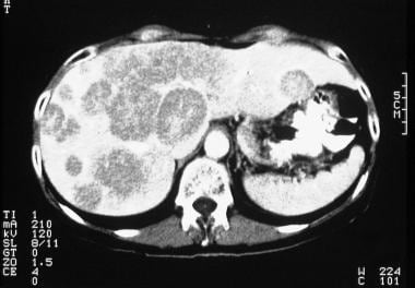
 Liver Metastases Imaging: Practice Essentials, Radiography, Computed Tomography
 Epithelioid Hemangioendothelioma genetic risk Archives | addon.life
Pathology of Nonmesothelial Cancers of the Pleura: Definition, Etiology, Epidemiology
Epithelioid hemangioendothelioma of the thyroid: a case report | Surgical Case Reports | Full Text
 Congenital Vascular Tumors
The 2015 World Health Organization Classification of Tumors of the Pleura: Advances since the 2004 Classification
A case of intravascular epithelioid hemangioendothelioma occurring 14 years after coil embolization for an extracranial...
 Therapeutics and Clinical Risk Management | Volume 16 - Dove Press
Identification of Novel Zoonotic Activity of Bartonella spp., France - Volume 22, Number 3-March 2016 - Emerging Infectious...
 Gary M. Kupfer, MD| Pediatric Hematology Oncology, Pediatric Hematology | MedStar Health
Reactive Angioendotheliomatosis
 Pathology Outlines - Hemangioma
 Serous fluid: Metastatic sarcomas, melanoma, and other non-epithelial neoplasms - CytoJournal
Epithelioid sarcoma
 A Model Research Grants Program for Rare Cancers
 Characteristic contrast‑enhanced ultrasound findings of hepatic epithelioid haemangioendothelioma: A case report and literature...
 Targeted Therapy: Its Status and Promise in Selected Solid Tumors Part I
 Pathologie entlang der sinusoidalen Wegstrecke: sinusendotheliale und perisinusoidale Befunde | Die Pathologie
 WOW! Women On Writing interviews Kris Carr, author of Crazy Sexy Cancer Tips and the TLC Documentary
 Stroke-Like Migraine Attacks After Radiation Therapy Syndrome and Radiation Necrosis After Cerebral Proton Beam Radiation: A...
Success Stories from Alternative Cancer Clinics - Welcome To Cancer Cure Foundation
Links Page - Welcome To Cancer Cure Foundation
 Neurol India: Table of Contents
 Notes on Kris Carr and Crazy Sexy Cancer - Medical Lessons
 John A Goss, M.D. | BCM
Hepatic3
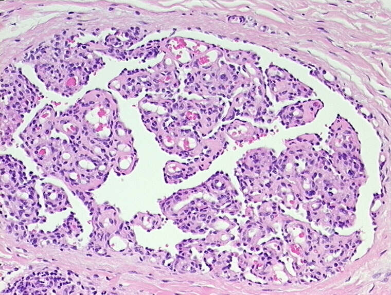
- Hepatic angiosarcomas and epithelioid hemangioendotheliomas can be easily overseen in liver biopsies, if they spread along the sinusoids without detoriation of the acinar architecture and without significant alteration of the surrounding liver cell plates. (springer.com)
- Patients with UICC stages I or II hepatocellular carcinoma, the fibrolamellar variant of hepatocellular carcinoma, or epithelioid hepatic hemangioendothelioma are acceptable candidates for othotopic liver transplantation. (elsevierpure.com)
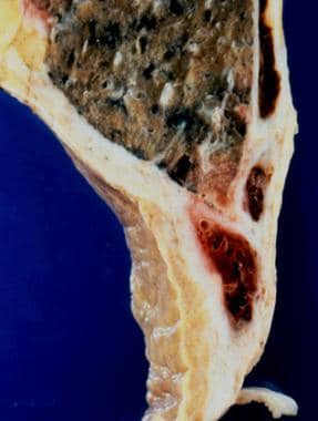
- Hepatic epithelioid hemangioendothelioma: An international multicenter study. (clinic.cat)
Malignant5
- The study group comprised six leiomyosarcomas, five malignant peripheral nerve sheath tumours, two haemangiopericytomas and one epithelioid haemangioendothelioma. (nih.gov)
- Epithelioid sarcoma (ES) is a rare, malignant soft tissue tumor. (logicalimages.com)
- However, our search of the literature did not produce a significant volume of literature, or literature of sufficiently sound methodology, to permit our staff to make a determination regarding liver transplantation for other malignant diagnoses, such as metastatic disease, bile duct carcinoma and epithelioid hemangioendothelioma. (cms.gov)
- Malignant vascular tumors: Epithelioid Hemangioendothelioma. (neoplasiaresearch.com)
- Pulmonary epithelioid hemangioendothelioma (PEH) is a rare, low-to-intermediate malignant tumor of endothelial origin. (e-jyms.org)
Tumor8
- Epithelioid hemangioendothelioma (EHE) is a rare tumor, first characterized by Sharon Weiss and Franz Enzinger in 1982 that both clinically and histologically is intermediate between angiosarcoma and hemangioma. (wikipedia.org)
- Before the initial description of Weiss, the tumor had been reported under a variety of other names, including histiocytoid hemangioendothelioma, intravascular bronchoalveolar tumor (in the lung), and sclerosing cholangiocarcinoma. (wikipedia.org)
- Introduction Foods for Epithelioid Hemangioendothelioma should be personalized for each individual and also must adapt when cancer treatment or tumor genetic change. (addon.life)
- As its name implies, epithelioid hemangioendothelioma is an angiocentric vascular tumor with metastatic potential…These lesions may appear as a solitary, slightly painful mass in either superficial or deep soft tissue. (medicallessons.net)
- Hemangioendothelioma is a vascular tumor with several different morphologic patterns that can include a component of ovoid or spindled cells, but generally lacks an inflammatory component. (neoplasiaresearch.com)
- We present a challenging lymphocyte-rich soft tissue lesion that was not recognized to be an unusual hemangioendothelioma until after several recurrences in the arm of a 63 year-old male, which was originally diagnosed as a follicular dendritic cell tumor instead. (neoplasiaresearch.com)
- The most recent tumor consisted predominantly of epithelioid spindled cells with moderate amounts of bubbly pale to eosinophilic cytoplasm with rare discrete cytoplasmic vacuoles, admixed with a prominent lymphoid infiltrate and occasional erythrocytes. (neoplasiaresearch.com)
- Epithelioid Hemangioendothelioma: A distinctive vascular tumor often mistaken for carcinoma. (neoplasiaresearch.com)
Hemangioendotheliomas2
- An open-label, multicenter, phase II study of bevacizumab for the treatment of angiosarcoma and epithelioid hemangioendotheliomas. (uchicago.edu)
- Genetic studies also led to the finding that WWTR1-CAMTA1 fusions are useful diagnostic markers for epithelioid hemangioendotheliomas, which can present as pleural-based masses. (nih.gov)
YAP1-TFE32
- Novel YAP1-TFE3 fusion defines a distinct subset of epithelioid hemangioendothelioma. (uchicago.edu)
- 15: Puls F, Niblett A, Clarke J, Kindblom LG, McCulloch T. YAP1-TFE3 epithelioid hemangioendothelioma: a case without vasoformation and a new transcript variant. (cancercentrum.se)
Metastatic1
- Mediastinal epithelioid hemangioendothelioma metastatic to lymph nodes and pleural fluid: report of a case. (uchicago.edu)
Fibrosarcoma1
- Recurrent EWSR1-CREB3L1 Gene Fusions in Sclerosing Epithelioid Fibrosarcoma. (lu.se)
Vascular cancer1
- The name of the cancer itself is frightening: epithelioid hemangioendothelioma - a type of vascular cancer inside the blood vessels. (cancure.org)
Intravascular1
- Cutaneous biopsy revealed RAE characterized by the proliferation of epithelioid and spindle-shaped cells in superficial and middermis lining vascular channels, arranged in clusters, and sometimes displaying an intravascular growth pattern. (thedoctorsdoctor.com)
Haemangioendothelioma1
- Haemangiopericytoma and epithelioid haemangioendothelioma were associated with a more favourable prognosis. (nih.gov)
Pulmonary2
- Clinical patterns and outcome in epithelioid hemangioendothelioma with or without pulmonary involvement: insights from an internet registry in the study of a rare cancer. (uchicago.edu)
- Pulmonary epithelioid hemangioendothelioma misconceived as pulmonary metastasis of other malignancies. (e-jyms.org)
Hemangioma1
- ZFP36-FOSB fusion defines a subset of epithelioid hemangioma with atypical features. (uchicago.edu)
Soft tissue1
- Epithelioid hemangioendothelioma of soft tissue: a proposal for risk stratification based on 49 cases. (uchicago.edu)
Diagnosis2
- Epithelioid Hemangioendothelioma: Update on Diagnosis and Treatment. (uchicago.edu)
- The diagnosis was revised to recurrent lymphocyte-rich hemangioendothelioma. (neoplasiaresearch.com)
Angiosarcoma1
- They include less aggressive (epithelioid hemangioendothelioma or EHE) and more aggressive forms (angiosarcoma)s. (sarctrials.org)
Cancer2
- Epithelioid Hemangioendothelioma is a rare form of cancer that affects the blood vessels of an individual and has an unknown etiology. (ipl.org)
- Crazy sexy cancer is a documentary about a young lady, age 31, who was diagnosed with epithelioid Hemangioendothelioma, stage IV cancer on Valentine's day in 2003. (ipl.org)
Organs1
- Epithelioid hemangioendothelioma (EHE) is a rare neoplasm originating from various organs. (elsevierpure.com)
Endothelial1
- Sustained Activation of Endothelial YAP1 Causes Epithelioid Hemangioendothelioma. (umassmed.edu)
MeSH1
- Hemangioendothelioma, Epithelioid" is a descriptor in the National Library of Medicine's controlled vocabulary thesaurus, MeSH (Medical Subject Headings) . (uchicago.edu)
Proliferation1
- Histologic evaluation of the thyroid nodule showed multinodular proliferation of oval to polygonal-shaped epithelioid cells with oval nuclei and eosinophilic cytoplasm arranged in sheet- or cord-like patterns, accompanied by fibrous stroma and marked osseous metaplasia. (springeropen.com)
Mesothelioma1
- First, more detailed study has been performed of histologic subtyping of epithelioid mesothelioma. (nih.gov)
Affects1
- Epithelioid Hemangioendothelioma affects about 0.1 percent of the American population with an overall 5-year survival rate of 55% after a standard primary radical treatment. (ipl.org)
Literature1
- This represents an unusual and potentially confusing pattern of hemangioendothelioma that is not previously well described in the literature. (neoplasiaresearch.com)
Neoplasms2
- Epithelioid vascular tumours are rare vascular neoplasms. (medscape.com)
- 1 It has been used as a tool to identify the vascular origin of neoplasms such as angiosarcomas, Kaposi sarcomas and epithelioid hemangioendothelioma. (roche.com)
Hepatic Epithelioid Hemangioendothelioma1
- 2. CAMTA-1 Expression in 24 Cases of Hepatic Epithelioid Hemangioendothelioma in a Single Institute: Diagnostic Utility for Differential diagnosis from Hepatic Angiosarcoma. (nih.gov)
Sarcoma2
- The EHE Global Patient Registry empowers people with Epithelioid Hemangioendothelioma (EHE) to join together to improve our understanding of this ultra-rare sarcoma. (fightehe.org)
- One of the members of the FOLD Planning Team has been diagnosed with an incredibly rare vascular sarcoma (blood vessel cancer), known as Epithelioid hemangioendothelioma (EHE) . (thefoldcanada.org)
Hemangiopericytoma1
- berkhoffii genotype II from a boy with epithelioid hemangioendothelioma and a dog with hemangiopericytoma. (nih.gov)
Neoplasm2
- Considering the biology and clinically variable natural history of epithelioid hemangioendothelioma, these results suggest that chronic Bartonella infection could have a role in the development of this vascular neoplasm. (nih.gov)
- Background Epithelioid haemangioendothelioma (EHE) is a rare low-grade vascular neoplasm that can arise in the lung, liver, soft tissues or, less commonly, bone. (uni-luebeck.de)
Liver1
- 10. Epithelioid hemangioendotheliomas of the liver and lung in children and adolescents. (nih.gov)
Angiosarcoma2
- This research study is studying a drug as a possible treatment for Angiosarcoma or Epithelioid hemangioendothelioma (EHE). (clinicaltrials.gov)
- When solitary, epithelioid hemangioendothelioma lesions can exhibit a concentric ring or target appearance on contrast-enhanced CT and at MR and when numerous may be indistinguishable from angiosarcoma except for a more slowly advancing course. (johnshopkins.edu)
Diagnosis2
- 8. Epithelioid Hemangioendothelioma: Update on diagnosis and Treatment. (nih.gov)
- Fluorescence in situ hybridization for WWTR1-CAMTA1 has higher sensitivity and specificity for epithelioid hemangioendothelioma diagnosis. (cytopathology.org)
WWTR1-CAMTA11
- 12. Molecular characterization of epithelioid haemangioendotheliomas identifies novel WWTR1- camta1 fusion variants. (nih.gov)
Immunohistochemical3
- Language": "en", "Country": "XG", "Code": "Background Information" }, { "Name": "Principle", "Value": "CD31 (JC70) Mouse Monoclonal Antibody (this antibody) may be used as the primary antibody for immunohistochemical staining of formalin-fixed, paraffin-embedded tissue sections. (roche.com)
- Epithelioid hemangioendothelioma of skin and soft tissues: clinicopathologic and immunohistochemical study of 30 cases. (cytopathology.org)
- Jebastin Thangaiah J, Hanley K, Nomani L, Policarpio-Nicolas ML. Cytologic features and immunohistochemical findings of epithelioid hemangioendothelioma (EHE) in effusion: A case series. (cytopathology.org)
Uncommon1
- [ 1 ] Epithelioid hemangioendothelioma (EHE) is an uncommon vascular tumour that was first described in 1975 by Dail and Liebow in the lung as an aggressive bronchoalveolar cell carcinoma. (medscape.com)
Cancer3
- Epithelioid hemangioendothelioma, an ultra-rare cancer: a consensus paper from the community of experts. (nih.gov)
- Robert (Bob) Thomas Capps Jr. of Steamboat Springs, CO passed away on April 2, 2023 in Nashville, TN at the age of 74 after battling Epithelioid hemangioendothelioma, or EHE, a rare form of cancer. (steamboatpilot.com)
- Feb. 3, 2020) - Today, The EHE Foundation announced it will receive a $450,000 award from the Chan Zuckerberg Initiative (CZI) to drive progress towards treatments and a cure for Epithelioid Hemangioendothelioma (EHE), a rare vascular cancer. (fightehe.org)
Tumors2
- are known to induce vasoproliferative tumors in immunocompromised patients and may play a role in the development of epithelioid hemangioendothelioma in immunocompetent patients. (nih.gov)
- Histologically, the tumors were comprised of dendritic and epithelioid cells that often contained vacuoles representing intracellular lumina. (nih.gov)
Clinical1
- Clinical characteristics of epithelioid hemangioendothelioma: a single-center retrospective study. (nih.gov)
Morphology1
- They are a subtype of mesenchymal tumours, defined by their epithelioid morphology, which differentiates them from other vascular tumours. (medscape.com)
Immunohistochemistry3
- 11. Diagnostic utility of FOSB immunohistochemistry in pseudomyogenic hemangioendothelioma and its histological mimics. (nih.gov)
- Language": "en", "Country": "XG", "Code": "Storage Conditions (Product)" }, { "Name": "Intended Use", "Value": "CD31 (JC70) Mouse Monoclonal Primary Antibody is intended for laboratory use in the detection of the CD31 protein in formalin-fixed, paraffin-embedded human tissue stained in qualitative immunohistochemistry (IHC) on BenchMark IHC/ISH instruments. (roche.com)
- CD31, CD34 and Fli-1 positive immunohistochemistry is strongly indicative of epithelioid lineage. (uni-luebeck.de)
Eosinophilia1
- It is unclear whether the epithelioid hemangioendothelioma is truly neoplastic or an exuberant tissue reaction, nor is it clear if this is equivalent to Kimura's disease (see ANGIOLYMPHOID HYPERPLASIA WITH EOSINOPHILIA ). (nih.gov)
Primary1
- Primary pleural epithelioid hemangioendothelioma. (cytopathology.org)
Histology1
- Histology showed a nodular structure with clusters of epithelioid and spindled cells with a low proliferative index and mitotic count, suspended in a sclerotic stroma. (uni-luebeck.de)
Cells1
- Language": "en", "Country": "XG", "Code": "Content" }, { "Name": "Background Information", "Value": "CD31 has cytoplasmic, membranous expression in non-neoplastic and neoplastic vascular endothelial cells. (roche.com)
Researchers1
- The support of doctors and researchers in the treatment and cure of Epithelioid Hemangioendothelioma (EHE), as well as offering support to EHE patients and families. (igive.com)
Treatment1
- Founded in 2015, The EHE Foundation is a nonprofit, 501c3 organization dedicated to pursuing effective treatment and a cure for Epithelioid Hemangioendothelioma (EHE). (designruleseverything.com)
Patients2
- Bartonella henselae and B. koehlerae bacteremia was documented in two epithelioid hemangioendothelioma patients and B. koehlerae bacteremia in an asymptomatic partner of one of the patients. (nih.gov)
- Management Strategies for Patients With Epithelioid Hemangioendothelioma: Charting an Indolent Disease Course. (nih.gov)
Cases2
- 4. [Atypical epithelioid hemangioendothelioma: a clinicopathological analysis of eight cases]. (nih.gov)
- Epithelioid hemangioendothelioma of soft tissue: a proposal for risk stratification based on 49 cases. (cytopathology.org)
Disease1
- edited by DeVita, Hellman and Rosenberg) off my shelf and looked up Carr's stated disease, epithelioid hemangioendothelioma. (medicallessons.net)
Potential1
- 13. camta1 immunostaining is not useful in differentiating epithelioid hemangioendothelioma from its potential mimickers. (nih.gov)
Features1
- Cytologic features of epithelioid hemangioendothelioma. (cytopathology.org)