Pulmonary Infarction
Bronchopulmonary Sequestration
Surgical Fixation Devices
Thoracostomy
Chondroblastoma
Wounds, Nonpenetrating
Rupture of aortic aneurysm with right-sided haemothorax. (1/130)
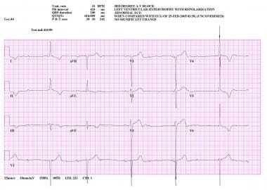
A 62-yr-old male with a history of high blood pressure was admitted for persistent dyspnoea and a right-sided pleural effusion, complicated by a recent episode of shock. There was no history of trauma and the patient denied any thoracic pain. A chest tube was inserted which released nonclotting bloody fluid. A thoracic computed tomographic scan of the chest revealed an aneurysm of the inferior third of the descending thoracic aorta. The patient underwent a successful prosthetic graft replacement. We emphasize that rupture of aortic aneurysms should be considered in the evaluation of spontaneous haemothorax even if it is right-sided and not associated with pain. (+info)Spontaneous haemothorax: a cause of sudden death in von Recklinghausen's disease. (2/130)
Vasculopathy is a relatively frequent but poorly recognised manifestation of von Recklinghausen's neurofibromatosis. One of its more dramatic presentations is as spontaneous haemothorax. Clinicians and pathologists should be aware of this syndrome as a cause of sudden death in patients with neurofibromatosis. (+info)Bilateral hemothorax revealing mediastinal parathyroid adenoma. (3/130)
We report the case of a 63-year-old woman admitted to hospital because of bilateral hemothorax associated with acute respiratory failure and laterotracheal neoformation. A right thoracoscopy biopsy revealed a paratracheal parathyroid adenoma which was responsible for bilateral hemothorax and primary hyperparathyroidism. A curative resection was successfully performed by cervicotomy. (+info)Intrathoracic extramedullary haematopoiesis complicated by massive haemothorax in alpha-thalassaemia. (4/130)
Intrathoracic extramedullary haematopoiesis (EMH) is a rare entity that is usually asymptomatic. A 44 year old man with alpha-thalassaemia is described who developed dyspnoea and massive left sided haemothorax. The haemoglobin disorder was established by Hgb H staining and haemoglobin electrophoretic studies. The DNA analysis revealed it to be a case of double heterozygous terminal codon mutation with the genotype alphaalphaCS/alphaalphaT. Computed tomographic scanning and magnetic resonance imaging of the thorax showed multiple paravertebral masses which were found by thoracoscopic biopsy to be extramedullary haematopoiesis. Although no additional sclerosing pleurodesis or low dose radiation therapy was given, the lung expanded well and there has been no recurrence of haemothorax to date. (+info)Haemoptysis after breath-hold diving. (5/130)
Pulmonary oedema has been described in swimmers and self-contained underwater breathing apparatus (Scuba) divers. This study reports three cases of haemoptysis secondary to alveolar haemorrhage in breath-hold divers. Contributory factors, such as haemodynamic modifications secondary to immersion, cold exposure, exercise and exposure to an increase in ambient pressure, could explain this type of accident. Furthermore, these divers had taken aspirin, which may have aggravated the bleeding. (+info)Spontaneous hemothorax secondary to immature teratoma of the mediastinum. (6/130)
Spontaneous hemothorax in a 20-year-old boy was caused by rupture of an immature teratoma of the mediastinum. The tumor bled spontaneously into the right pleural space. This life-threatening complication necessitated emergency surgery. The unusual cause and the interesting clinical course of spontaneous hemothorax are described. (+info)Spontaneous pneumothorax: outpatient management with intercostal tube drainage. (7/130)
In a series of 104 episodes of pneumothorax 75 percent of episodes were managed successfully on an outpatient basis by observation (23.1 percent) or by intercostal tube drainage using a flutter valve (51.9 percent). The patients for whom this treatment was not successful were admitted to hospital; 17 of them (16.3 percent of 104) were treated surgically. Bleb suturing with a stapling device and dry sponge abrasion of the pleura was the operation of choice. (+info)Mortality associated with odontogenic infection! (8/130)
Odontogenic causes are the most common source for spreading maxillo-facial infections. These infections can develop into life threatening events. However a fatal outcome is fortunately rare and is generally associated with an immunocompromised status. This case report highlights a spreading maxillo-facial infection, which resulted in massive haemorrhage from the subclavian vein into the pleural cavity and subsequent death of a young fit male patient. (+info)Hemothorax is a medical condition characterized by the presence of blood in the pleural space, which is the area between the lungs and the chest wall. This accumulation of blood can occur due to various reasons such as trauma, rupture of a blood vessel, or complications from lung or heart surgery.
The buildup of blood in the pleural space can cause the affected lung to collapse, leading to symptoms such as shortness of breath, chest pain, and cough. In severe cases, hemothorax can be life-threatening if not promptly diagnosed and treated. Treatment options may include chest tube drainage, blood transfusion, or surgery, depending on the severity and underlying cause of the condition.
Thoracic injuries refer to damages or traumas that occur in the thorax, which is the part of the body that contains the chest cavity. The thorax houses vital organs such as the heart, lungs, esophagus, trachea, and major blood vessels. Thoracic injuries can range from blunt trauma, caused by impacts or compressions, to penetrating trauma, resulting from stabbing or gunshot wounds. These injuries may cause various complications, including but not limited to:
1. Hemothorax - bleeding into the chest cavity
2. Pneumothorax - collapsed lung due to air accumulation in the chest cavity
3. Tension pneumothorax - a life-threatening condition where trapped air puts pressure on the heart and lungs, impairing their function
4. Cardiac tamponade - compression of the heart caused by blood or fluid accumulation in the pericardial sac
5. Rib fractures, which can lead to complications like punctured lungs or internal bleeding
6. Tracheobronchial injuries, causing air leaks and difficulty breathing
7. Great vessel injuries, potentially leading to massive hemorrhage and hemodynamic instability
Immediate medical attention is required for thoracic injuries, as they can quickly become life-threatening due to the vital organs involved. Treatment may include surgery, chest tubes, medications, or supportive care, depending on the severity and type of injury.
Pulmonary infarction is the medical term for the death of lung tissue (lung tissue necrosis) due to blocked blood flow. This blockage usually occurs when a clot or a piece of a clot from another part of the body, most commonly from the heart, travels to the lungs and blocks a small pulmonary artery. The lack of oxygen supply to the lung tissue results in inflammation and eventual infarction (tissue death).
The symptoms of pulmonary infarction can vary but often include sudden onset of sharp chest pain, shortness of breath, cough, sometimes with blood-streaked sputum, rapid heart rate, and fever. The diagnosis is typically made based on the patient's medical history, physical examination, imaging tests such as a chest X-ray or CT scan, and occasionally, blood tests to detect D-dimer, a protein fragment that's produced when a blood clot dissolves. Treatment usually involves anticoagulant therapy (blood thinners) to prevent further clots from forming and, in some cases, thrombolytic therapy (clot-busting drugs) to break up existing clots. In severe cases, surgery may be required to remove the clot or infarcted lung tissue.
Bronchopulmonary sequestration is a rare birth defect of the lungs, in which a mass of abnormal lung tissue develops that doesn't function and isn't connected to the tracheobronchial tree (the airways that lead to the lungs). This means that the abnormal tissue receives its blood supply from an anomalous systemic artery instead of the normal pulmonary circulation. The mass may be located within the lung (intralobar sequestration) or outside the lung (extralobar sequestration), and it can occur on either side of the chest.
Intralobar sequestrations are more common than extralobar sequestrations, accounting for about 75% of cases. They are usually found in adults and are located within a normal lung tissue. Extralobar sequestrations, on the other hand, are typically detected earlier in life (often as an incidental finding during prenatal ultrasound) and are surrounded by their own pleural lining, which can make them appear separate from the normal lung tissue.
Symptoms of bronchopulmonary sequestration may include recurrent respiratory infections, coughing up blood (hemoptysis), shortness of breath, or chest pain. Treatment usually involves surgical removal of the abnormal tissue to prevent complications such as infection, bleeding, or the development of malignancy.
Surgical fixation devices are medical implants used in various surgical procedures to provide stability, alignment, and support to fractured or damaged bones, joints, or soft tissues. These devices help promote healing by holding the affected area in the correct position until the body can repair itself. Common types of surgical fixation devices include:
1. Plates: Thin, flat metal pieces contoured to fit against the surface of a bone. They are often held in place with screws and used to stabilize fractures or support weakened bones.
2. Screws: Threaded rods that can be inserted into bones to hold them together or fixate implants such as plates or prosthetic joints.
3. Pins: Smooth or threaded wires used to temporarily or permanently hold bone fragments in place. They are often removed once healing is complete.
4. Intramedullary nails: Long rods placed inside the marrow cavity of a long bone (e.g., femur, tibia) to provide stability and alignment after a fracture.
5. External fixators: Devices attached to the outside of the body with pins or wires that pass through the skin and into the bones. They are used to stabilize complex fractures or injuries when internal fixation is not possible or advisable.
6. Interbody fusion cages: Cylindrical or box-shaped devices placed between two vertebrae during spinal fusion surgery to restore disc height and provide stability while promoting bone growth.
7. Sutures and staples: Used to approximate soft tissue edges (e.g., skin, muscles, ligaments) after surgical repair.
The choice of surgical fixation device depends on various factors, such as the location and severity of the injury, patient age and health status, and surgeon preference.
Thoracostomy is a surgical procedure that involves the creation of an opening into the chest cavity to relieve excessive pressure, drain fluid or air accumulation, or provide access for surgery. It is commonly performed to treat conditions such as pneumothorax (collapsed lung), hemothorax (blood in the chest cavity), pleural effusion (excess fluid in the pleural space), and empyema (pus in the pleural space).
During a thoracostomy, a healthcare professional makes an incision on the chest wall and inserts a tube called a thoracostomy tube or chest tube. The tube is connected to a drainage system that helps remove the air, fluid, or blood from the chest cavity. This procedure can be performed as an emergency treatment or as a planned surgical intervention.
The medical definition of thoracostomy includes the following key components:
1. A surgical procedure
2. Involving the creation of an opening
3. Into the chest cavity (thorax)
4. To relieve pressure, drain fluids or air, or provide access for surgery
5. Often performed with the insertion of a thoracostomy tube or chest tube
6. Used to treat various conditions related to the pleural space and lungs
Thoracoscopy is a surgical procedure in which a thoracoscope, a type of endoscope, is inserted through a small incision between the ribs to examine the lungs and pleural space (the space surrounding the lungs). It allows the surgeon to directly view the chest cavity, take biopsies, and perform various operations. This procedure is often used in the diagnosis and treatment of pleural effusions, lung cancer, and other chest conditions.
Chondroblastoma is a rare, benign (non-cancerous) bone tumor that typically develops in the epiphysis, which is the rounded end of a long bone near a joint. It primarily affects children and adolescents, with around 90% of cases occurring before the age of 20.
The tumor arises from chondroblasts, cells responsible for producing cartilage during bone growth. Chondroblastoma is usually slow-growing and typically causes localized pain, swelling, or tenderness in the affected area. In some cases, it may weaken the bone and lead to fractures.
Treatment generally involves surgical removal of the tumor, followed by curettage (scraping) of the surrounding bone tissue and replacement with bone grafts or substitutes. Recurrence is possible but rare, and long-term prognosis is usually favorable.
Thoracotomy is a surgical procedure that involves making an incision on the chest wall to gain access to the thoracic cavity, which contains the lungs, heart, esophagus, trachea, and other vital organs. The incision can be made on the side (lateral thoracotomy), back (posterolateral thoracotomy), or front (median sternotomy) of the chest wall, depending on the specific surgical indication.
Thoracotomy is performed for various indications, including lung biopsy, lung resection, esophagectomy, heart surgery, and mediastinal mass removal. The procedure allows the surgeon to directly visualize and access the organs within the thoracic cavity, perform necessary procedures, and control bleeding if needed.
After the procedure, the incision is typically closed with sutures or staples, and a chest tube may be placed to drain any accumulated fluid or air from the pleural space around the lungs. The patient will require postoperative care and monitoring in a hospital setting until their condition stabilizes.
Drainage, in medical terms, refers to the removal of excess fluid or accumulated collections of fluids from various body parts or spaces. This is typically accomplished through the use of medical devices such as catheters, tubes, or drains. The purpose of drainage can be to prevent the buildup of fluids that may cause discomfort, infection, or other complications, or to treat existing collections of fluid such as abscesses, hematomas, or pleural effusions. Drainage may also be used as a diagnostic tool to analyze the type and composition of the fluid being removed.
Nonpenetrating wounds are a type of trauma or injury to the body that do not involve a break in the skin or underlying tissues. These wounds can result from blunt force trauma, such as being struck by an object or falling onto a hard surface. They can also result from crushing injuries, where significant force is applied to a body part, causing damage to internal structures without breaking the skin.
Nonpenetrating wounds can cause a range of injuries, including bruising, swelling, and damage to internal organs, muscles, bones, and other tissues. The severity of the injury depends on the force of the trauma, the location of the impact, and the individual's overall health and age.
While nonpenetrating wounds may not involve a break in the skin, they can still be serious and require medical attention. If you have experienced blunt force trauma or suspect a nonpenetrating wound, it is important to seek medical care to assess the extent of the injury and receive appropriate treatment.
Hemoperitoneum is a medical condition characterized by the presence of blood in the peritoneal cavity, which is the space between the lining of the abdominal wall and the organs within it. This can occur due to various reasons such as trauma, rupture of an abdominal aortic aneurysm, ectopic pregnancy, or other conditions that cause bleeding into the abdomen.
The accumulation of blood in the peritoneal cavity can lead to symptoms such as abdominal pain, tenderness, distension, and hypovolemic shock due to blood loss. Hemoperitoneum is a serious medical condition that requires prompt diagnosis and treatment to prevent further complications.
Chest tubes are medical devices that are inserted into the chest cavity to drain fluid, air, or blood. They are typically used to treat conditions such as pneumothorax (collapsed lung), hemothorax (blood in the chest cavity), pleural effusion (excess fluid in the chest cavity), and chylothorax (milky fluid in the chest cavity).
Chest tubes are usually inserted between the ribs and directed into the chest cavity, allowing for drainage of the affected area. The tubes are connected to a collection system that creates negative pressure, which helps to remove the air or fluid from the chest cavity.
The size and number of chest tubes used may vary depending on the severity and location of the condition being treated. Chest tubes are typically removed once the underlying condition has been resolved and the drainage has decreased to a minimal amount.
 Hemothorax
Pleurisy
Eddie August Schneider
Pancreatic fistula
Pulmonary laceration
Thoracostomy
Thoracic endometriosis
Hepatic hydrothorax
Diaphragmatic rupture
Endometriosis
Focused assessment with sonography for trauma
Treatment of lung cancer
Catamenial pneumothorax
Respiratory examination
Hemoptysis
Lung biopsy
Traumatic cardiac arrest
Advanced trauma life support
Thoracic aorta injury
Traumatic aortic rupture
Management of scoliosis
Nuss procedure
Tumor-like disorders of the lung pleura
Walter Whitehead
Polytrauma
Hemopneumothorax
Gunshot wound
Streptokinase
Fibrothorax
Pneumothorax
Hemothorax - Wikipedia
 Hemothorax: MedlinePlus Medical Encyclopedia
 Hemothorax: Background, Anatomy, Pathophysiology
 Hemothorax: Bleeding in the Chest in Dogs
 Practice management guidelines for management of hemothorax and occult pneumothorax
 3D Model: Hemothorax - MSD Manual Consumer Version
 Morbidity and mortality of iatrogenic hemothorax occurring in a cohort of liver transplantation recipients: a multicenter...
Hemothorax - Injuries; Poisoning - MSD Manual Professional Edition
 New Hemothorax Model. (!161) · Merge requests · Pulse Physiology Suite / engine · GitLab
 Hemothorax : Medical Illustration
Hemothorax | Basicmedical Key
 Differential diagnosis of hemothorax
Phoenix Pneumothorax & Hemothorax Attorneys | Misdiagnosis
 occult hemothorax Archives - Emergency Medicine Cases
Questions & Answers
Retained Hemothorax And Empyema | The Trauma Pro
Retained Hemothorax Part 1: Lytics | The Trauma Pro
 Hemothorax: Etiology, diagnosis, and management. | Read by QxMD
 Hemothorax | Hematoma Disposal Treatment. Hospital Prices, Ranking, Reviews - Bookinghealth
 Distinguishing Between a Hemothorax and a Pneumothorax - NEJM Knowledge+
 Chest Trauma
 emDOCs.net - Emergency Medicine EducationManaging a Massive Hemothorax: A Guide to Stabilizing Your Patient - emDOCs.net -...
Ibrutinib-Associated Life-Threatening Hemorrhage with Subcapsular Renal Hematoma and Hemothorax
 Blood in the Chest in Cats | PetMD
 Intrapleural tranexamic acid in persistent malignant hemothorax: a case report - Current Thoracic Surgery
 Live Action Mafia • View topic - Finasteride hospital craniopharyngioma, ship, haemothorax, s
 Clavicle fracture, rib fractures and haemothorax - Image | Evidence-Based Medicine Guidelines
 Thoracentesis | Lima Memorial Health System
Penetrating trauma3
- Hemothorax is most often caused by blunt or penetrating trauma to the chest. (wikipedia.org)
- In blunt traumatic cases, hemothorax typically occurs when rib fracture damages the intercostal vessels or the intraparenchymal pulmonary vessel, while in penetrating trauma, hemothorax occurs due to injuries directly affecting blood vessels in the thoracic wall, lung parenchyma, or the heart. (wikipedia.org)
- Hemothorax is usually a consequence of blunt or penetrating trauma. (medscape.com)
Pneumothorax and Hemothorax3
- Pneumothorax and hemothorax are two commonly misdiagnosed and mistreated traumatic injuries. (sandwegandager.net)
- Having considered the possible complications and narrowed them down, the learner should now be focused on distinguishing between the two most likely complications in this case - pneumothorax and hemothorax - and then determining the best intervention. (nejm.org)
- [1] Pneumothorax and hemothorax are the most common immediate complications associated with blunt chest trauma, [2] however, extrapleural hematoma can also occur and a patient may present immediately or rarely with delayed symptoms. (wajradiology.org)
Hematoma4
- We report an unusual but life-threatening case of hemorrhage with subcapsular renal hematoma and large hemothorax secondary to Ibrutinib. (fortuneonline.org)
- He presented just two weeks after initiating reduced dose of ibrutinib with grade 3 ibrutinib-associated hemorrhage involving subcapsular renal hematoma and hemothorax. (fortuneonline.org)
- Extrapleural hematoma needs to be differentiated from a hemothorax for appropriate management. (wajradiology.org)
- We hereby report a case of a large extrapleural hematoma and associated pleural effusion following trauma being managed as a hemothorax based only on chest radiograph appearance. (wajradiology.org)
Pulmonary3
- Sometimes, a Swan-Ganz catheter causes rupture of the pulmonary artery, causing a massive hemothorax. (wikipedia.org)
- In cases of hemothorax complicating pulmonary embolism treatment, the hemothorax is usually on the side of the original embolism. (wikipedia.org)
- Di Crescenzo V, Laperuta P, Napolitano F, Carlomagno C, Garzi A, Vitale M. Pulmonary sequestration presented as massive left hemothorax and associated with primary lung sarcoma. (medscape.com)
Hemorrhage2
- Massive hemothorax or exsanguinating hemorrhage may result from injury to major arterial or venous structures contained within the thorax or from the heart itself. (medscape.com)
- Intervention 2: Intervention Group (2): receives medical treatment based on WHO modified checklist with focusing on pain management (intervention items are as follows: airway intervention control, internal hemorrhage control and hemothorax diagnosis, etc). (who.int)
Lung5
- Iatrogenic hemothorax can occur as a complication of heart and lung surgery, for example the rupture of lung arteries caused by the placement of catheters, thoracotomy, thoracostomy, or thoracentesis. (wikipedia.org)
- Hemothorax is a collection of blood in the space between the chest wall and the lung (the pleural cavity). (medlineplus.gov)
- The usual cause of hemothorax is laceration of the lung, intercostal vessel, or an internal mammary artery. (msdmanuals.com)
- Depending on the amount of bleeding and the underlying cause, hemothorax may be associated with varying degrees of lung collapse and mediastinal shift. (basicmedicalkey.com)
- 10.26663/cts.2020.00017 Viewed : 2901 - Downloaded : 1443 Hemothorax occurs due to various conditions such as trauma, malignancy, tuberculosis, bullous lung disease, and lung abscess. (tgcd.org.tr)
Traumatic hemothorax1
- Prompt identification and treatment of traumatic hemothorax is an essential part of the care of the injured patient. (medscape.com)
Spontaneous hemothorax3
- Vaziri M, Mehrazma M. Massive spontaneous hemothorax associated with Von Recklinghausen's disease. (medscape.com)
- Spontaneous hemothorax is much less common and results from a variety of pathologic processes. (qxmd.com)
- This article reviews the etiology, diagnosis, and treatment of traumatic and spontaneous hemothorax in modern practice. (qxmd.com)
Massive8
- Massive hemothorax, often defined as over 1.5 liters of blood initially when an intercostal drain is placed, or a bleeding rate greater than .2 liters/hr, can result in shock with two causes: massive bleeding resulting from hypovolemic shock, and venous pressure from the retained blood, impairing blood flow. (wikipedia.org)
- Thoracotomy is the procedure of choice for surgical exploration of the chest when massive hemothorax or persistent bleeding is present. (medscape.com)
- Massive hemothorax is most often defined as rapid accumulation of ≥ 1000 mL of blood. (msdmanuals.com)
- Tantraworasin A, Saeteng S. Massive hemothorax due to intrathoracic extramedullary hematopoiesis in a patient with beta thalassemia hemoglobin E disease. (medscape.com)
- You know immediately that this is a massive hemothorax. (emdocs.net)
- This case report describes an injury to the left inferior phrenic artery caused by blunt trauma, which was complicated by massive hemothorax, and treated with transcatheter arterial embolization (TAE). (biomedcentral.com)
- In the present case, blunt trauma led to left inferior phrenic artery injury associated with massive hemothorax, which was treated with TAE alone. (biomedcentral.com)
- To the author's knowledge, this is the first report of massive hemothorax due to inferior phrenic artery injury treated definitively by TAE. (biomedcentral.com)
Diagnosis4
- A hemothorax is usually an emergency situation requiring rapid diagnosis. (petplace.com)
- Making the diagnosis of a hemothorax is critical in establishing an underling cause. (petplace.com)
- Broderick SR. Hemothorax: Etiology, diagnosis, and management. (medscape.com)
- Hemothorax: Etiology, diagnosis, and management. (qxmd.com)
Iatrogenic5
- Iatrogenic hemothorax is more common in people who have chronic kidney disease in the intensive care unit. (wikipedia.org)
- While Hunter's method was effective in evacuating the hemothorax, the creation of an iatrogenic pneumothorax as a result of the procedure was associated with significant morbidity. (medscape.com)
- Morbidity and mortality of iatrogenic hemothorax occurring in a cohort of liver transplantation recipients: a multicenter observational study. (physiciansweekly.com)
- Hemothorax (HT) is a life-threatening condition, mainly iatrogenic and poorly explored in Liver Transplantation (LT) recipients. (physiciansweekly.com)
- Postoperative hemothorax is mainly due to iatrogenic causes in LT recipients. (physiciansweekly.com)
Pleural effusion1
- Although some authors state that a hematocrit value of at least 50% is necessary to differentiate a hemothorax from a bloody pleural effusion, most do not agree on any specific distinction. (medscape.com)
Laceration1
- Costal osteochondroma presenting as haemothorax and diaphragmatic laceration. (medscape.com)
Fractures4
- In this episode we discuss predicting the sick trauma patient, videolaryngoscopy vs traditional laryngoscopy, Damage Control Resuscitation, Occult Hemothorax, Blunt Thoracic Aorta and Cardiac Injury, Sternal Fractures, Tranexamic Acid, Communication in the trauma bay and much more. (emergencymedicinecases.com)
- A total of seven patients were identified, and most had hemothorax due to rib fractures. (thetraumapro.com)
- If patient vital signs (hypoxia, persistent hypotension), mechanism of injury (penetrating injury), or physical exam (multiple palpable rib fractures, flail segment, significant chest wall ecchymosis or tenderness to palpation) lead you to suspect a hemothorax à Do not let the supine CXR convince you otherwise. (emdocs.net)
- Evidence Central , evidence.unboundmedicine.com/evidence/view/EBMG/456248/all/Clavicle_fracture__rib_fractures_and_haemothorax___Image. (unboundmedicine.com)
Hemothoraces1
- A hemothorax (derived from hemo- [blood] + thorax [chest], plural hemothoraces) is an accumulation of blood within the pleural cavity. (wikipedia.org)
Intercostal2
- A number of surgeons, including John Hunter in 1794, advocated the creation of an intercostal incision and drainage of the hemothorax. (medscape.com)
- By the 1870s, early hemothorax evacuation by trocar and cannula or by intercostal incision was considered standard practice. (medscape.com)
Complication2
- Hemothorax as a complication of costal cartilaginous exostoses. (medscape.com)
- Hemothorax is a common complication of chest trauma, occurring in about one third of cases. (thetraumapro.com)
Pericardial effusion1
- Cowles RA, Rowe DH, Arkovitz MS. Hereditary multiple exostoses of the ribs: an unusual cause of hemothorax and pericardial effusion. (medscape.com)
Persistent1
- The management of this case made us think that intrapleural tranexamic acid may be an alternative in persistent malign hemothorax. (tgcd.org.tr)
Empyema2
- Tube drainage improves ventilation, decreases risk of clotted hemothorax (which can lead to empyema or fibrothorax), and facilitates assessment of ongoing blood loss and diaphragmatic integrity. (msdmanuals.com)
- Reference: Development of posttraumatic empyema in patients with retained hemothorax: Results of a prospective, observational AAST study. (thetraumapro.com)
Workup1
- The upright chest radiograph is the ideal primary diagnostic study in the evaluation of hemothorax (see Workup). (medscape.com)
Bleeding3
- Minor chest trauma can cause hemothorax when the blood's ability to clot is diminished as result either of anticoagulant medications or when there are bleeding disorders such as hemophilia. (wikipedia.org)
- In many cases of hemothorax due to chest trauma, the bleeding will stop on it's own. (petplace.com)
- Huybrechts S, Wojciechowski M, Poot S, Van Reempts P, Ramet J. Hemothorax as presentation of late vitamin-K-deficient bleeding in a 1-month-old infant with homozygous alpha-1-antitrypsin deficiency. (medscape.com)
Complications3
- Complications of a hemothorax include infection within the pleural cavity and the formation of scar tissue. (wikipedia.org)
- Hemothorax and pneumothorax are the two most likely complications in the case presented above. (nejm.org)
- While the injury resulting in hemothorax and pneumothorax occurs during line placement, it usually takes some time for the patient to clinically deteriorate from these complications. (nejm.org)
Malignant1
- In patients with malignant hemothorax, stabilization of the clinical condition and treatment of primary disease is of primary importance. (tgcd.org.tr)
Injury4
- Blunt or penetrating injury involving virtually any intrathoracic structure can result in hemothorax. (medscape.com)
- CVC placement can also result in vascular injury, most commonly through arterial puncture, which can subsequently cause a hemothorax. (nejm.org)
- Inferior phrenic artery injury should be recognized as a rare phenomenon and causative factor for hemothorax. (biomedcentral.com)
- There were no signs of hemothorax or aortic injury. (bvsalud.org)
Blunt or penetrating1
- Most cases of hemothorax are related to blunt or penetrating chest trauma. (qxmd.com)
Patient4
- The patient with hemothorax may experience chest pain, tachypnea, and mild to severe dyspnea, depending on the amount of blood in the pleural cavity and associated pathology. (basicmedicalkey.com)
- Pneumothorax can result in tension physiology as well - though the hemodynamic compromise from this, when a patient is on mechanical ventilation, is usually quicker than with hemothorax. (nejm.org)
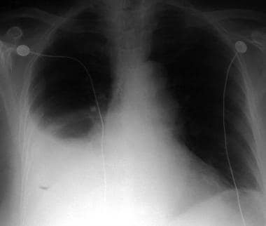
- 4 In the hemodynamically stable patient, a hemothorax is most commonly identified on CXR. (emdocs.net)
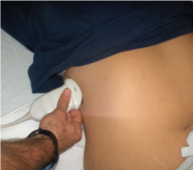
- In the hemodynamically unstable patient, the EFAST or Extended Focused Assessment with Sonography for Trauma is most commonly utilized to identify a hemothorax given its speed of employment . (emdocs.net)
Commonly2
- Pneumo-thorax-air in the pleural cavity - commonly accompanies hemothorax. (basicmedicalkey.com)
- A hemothorax, or a collection of blood in the pleural space, most commonly occurs secondary to penetrating or blunt chest wall trauma. (emdocs.net)
Rupture2
- The most common cause of hemothorax in dogs is chest trauma, although tumors within the thorax (chest cavity) can also result in a hemothorax if they rupture, slowly bleed or invade into a blood vessel causing blood to accumulate in the thorax. (petplace.com)
- Ota H, Kawai H, Matsuo T. Video-assisted minithoracotomy for blunt diaphragmatic rupture presenting as a delayed hemothorax. (medscape.com)
Costal1
- Hemothorax in a child as a result of costal exostosis. (medscape.com)
Differential1
- What are the differential diagnoses for Hemothorax? (medscape.com)
Thoracentesis1
- He underwent thoracentesis revealing hemothorax. (fortuneonline.org)
Intervention2
Diaphragm1
- May J, Ades A. Porous diaphragm syndrome: haemothorax secondary to haemoperitoneum following laparoscopic hysterectomy. (medscape.com)
Treatment3
- The outcome depends on the cause of the hemothorax, the amount of blood loss and how quickly treatment is given. (medlineplus.gov)
- In cases of hemothorax unrelated to trauma, a careful investigation for the underlying source must be performed while treatment is provided. (medscape.com)
- This basic technique has remained the most common form of treatment for hemothorax and other pleural fluid collections to this day. (medscape.com)
Chest pain1
- The symptoms of a hemothorax may include chest pain and difficulty breathing, while the clinical signs may include reduced breath sounds on the affected side and a rapid heart rate. (wikipedia.org)
Anticoagulant therapy1
- In cases caused by anticoagulant therapy, the hemothorax becomes noticeable 4-7 days after anticoagulant therapy is started. (wikipedia.org)
Blood7
- Pleural effusions are given specific names depending on the nature of the fluid: hydrothorax for serous fluid, pyothorax for pus, hemothorax for blood, and urinothorax for urine. (wikipedia.org)
- Hemothorax is the presence of blood in the pleural space. (medscape.com)
- Hemothorax is defined as blood within the chest cavity. (petplace.com)
- Hemothorax occurs when blood accumulates in the pleural cavity. (sandwegandager.net)
- Hemothorax can result in either tension physiology or hypovolemic shock, both of which will take several minutes to hours to manifest as blood has to accumulate in the pleural space. (nejm.org)
- Hemothorax is the medical term used to identify a condition in which blood has collected in the chest cavity, or thorax. (petmd.com)
- Hemothorax, blood collecting between the chest wall and lungs. (clevelandclinic.org)
Symptoms1
- Hemothorax is suspected based on symptoms and physical findings. (msdmanuals.com)
Management1
- Appropriate management of retained hemothorax following initial trauma management is critical, and the best approach remains controversial. (qxmd.com)
Medical1
- Contact us at (602) 648-3200 about any medical malpractice involving pneumothorax or hemothorax you or a loved one may have experienced anywhere in Arizona. (sandwegandager.net)
Tube3
- Patients who had suspected retained hemothorax after tube removal received a CT scan within 14 days . (thetraumapro.com)
- Bottom line: Retained hemothorax turns into a very serious problem in a quarter of trauma patients who have a chest tube inserted. (thetraumapro.com)
- Reference: Evaluation of chest tube administration of tissue plasminogen activator to treat retained hemothorax. (thetraumapro.com)