Hernia, Inguinal
Hernia
Hernia, Diaphragmatic
Hernia, Ventral
Hernia, Abdominal
Hernia, Hiatal
Hernia, Femoral
Hernia, Umbilical
Herniorrhaphy
Surgical Mesh
Hernia, Diaphragmatic, Traumatic
Hernia, Obturator
Polypropylenes
Laparoscopy
Abdominal Wall
Fascia
Suture Techniques
Intestinal Obstruction
Surgical Stomas
Postoperative Complications
Anesthesia, Local
Rectus Abdominis
Ambulatory Surgical Procedures
Round Ligament
Surgical Procedures, Operative
Treatment Outcome
Fundoplication
Gastroesophageal Reflux
Surgical Wound Dehiscence
Testicular Hydrocele
Fetal Diseases
Retrospective Studies
Inguinal Canal
Follow-Up Studies
Reoperation
Diaphragmatic Eventration
Orchiopexy
Spermatic Cord
Polytetrafluoroethylene
Surgical Stapling
Tomography, X-Ray Computed
Prospective Studies
Fascia Lata
Diaphragm
Broad Ligament
Extracorporeal Membrane Oxygenation
Surgical Procedures, Elective
Esophagogastric Junction
Peritoneum
Esophagitis
Ultrasonography, Prenatal
Appendicitis
Abdominal Wound Closure Techniques
Esophagitis, Peptic
Ileostomy
Esophagus
Abnormalities, Multiple
Gestational Age
Omentum
Learning Curve
Surgery, Veterinary
Polyglactin 910
Perineum
Abdomen, Acute
Barium Sulfate
Surgical Procedures, Minimally Invasive
Fibrin Tissue Adhesive
Cryptorchidism
Pregnancy
Skin, Artificial
Radiography, Thoracic
Conversion to Open Surgery
Tensile Strength
Endoscopy
Umbilicus
Rectal Prolapse
Endoscopy, Gastrointestinal
Emergencies
Wounds, Nonpenetrating
Fetoscopy
Prostheses and Implants
Painful incarcerated hernia following a rugby union lineout. (1/559)
Discussion related to hernias in sport usually involves the diagnosis and treatment of chronic musculotendinous groin disruption. A case of acute trauma in an incarcerated inguinal hernia, occurring in a rugby union player during a lineout, is presented. The injury arose as a result of a change in the laws of the game. (+info)Value of scintigraphy in chronic peritoneal dialysis patients. (2/559)
BACKGROUND: A variety of factors can adversely impact chronic peritoneal dialysis (CPD) as an effective renal replacement therapy for patients with end-stage renal disease. These factors include peritonitis, poor clearances, loss of ultrafiltration, and a variety of anatomic problems, such as hernias, peritoneal fluid leaks, loculations, and catheter-related problems caused by omental blockage. This study reviews our experience with peritoneal scintigraphy for the evaluation of some of these difficulties. METHODS: From 1991 to 1996, 50 peritoneal scintigraphy scans were obtained in 48 CPD patients. Indications for scintigraphy were evaluated, and the patients were placed into four groups: group I, abdominal wall swelling; group II, inguinal or genital swelling; group III, pleural fluid; and group IV, poor drainage and/or poor ultrafiltration. A peritoneal scintigraphy protocol was established and the radiotracer isotope that was used was 2.0 mCi of 99mtechnetium sulfur colloid placed in two liters of 2.5% dextrose peritoneal dialysis solution. RESULTS: Ten scans were obtained to study abdominal wall swelling, with seven scans demonstrating leaks; six of these episodes improved with low-volume exchanges. Twenty scans were obtained to evaluate inguinal or genital swelling, and 10 of these had scintigraphic evidence for an inguinal hernia leak (9 of these were surgically corrected). One of four scans obtained to evaluate a pleural fluid collection demonstrated a peritoneal-pleural leak that corrected with a temporary discontinuation of CPD. Sixteen scans were obtained to assess poor drainage and/or ultrafiltration. Five of these scans demonstrated peritoneal location, and all of these patients required transfer to hemodialysis. The other 11 scans were normal; four patients underwent omentectomies, allowing three patients to continue with CPD. CONCLUSION: Peritoneal scintigraphy is useful in the evaluation and assessment of CPD patients who develop anatomical problems (such as anterior abdominal, pleural-peritoneal, inguinal, and genital leaks) and problems with ultrafiltration and/or drainage. (+info)Surgical options in the management of groin hernias. (3/559)
Inguinal and femoral hernias are the most common conditions for which primary care physicians refer patients for surgical management. Hernias usually present as swelling accompanied by pain or a dragging sensation in the groin. Most hernias can be diagnosed based on the history and clinical examination, but ultrasonography may be useful in differentiating a hernia from other causes of groin swelling. Surgical repair is usually advised because of the danger of incarceration and strangulation, particularly with femoral hernias. Three major types of open repair are currently used, and laparoscopic techniques are also employed. The choice of technique depends on several factors, including the type of hernia, anesthetic considerations, cost, period of postoperative disability and the surgeon's expertise. Following initial herniorrhaphy, complication and recurrence rates are generally low. Laparoscopic techniques make it possible for patients to return to normal activities more quickly, but they are more costly than open procedures. In addition, they require general anesthesia, and the long-term hernia recurrence rate with these procedures is unknown. (+info)Management of inguinal herniae in patients on continuous ambulatory peritoneal dialysis: an audit of current UK practice. (4/559)
Patients receiving continuous ambulatory peritoneal dialysis are at increased risk for the development of inguinal herniae, with a reported prevalence of 14%. Elective hernia repair is indicated for these patients as strangulation is associated with a high mortality in this population. There are currently no national guidelines relating to the optimal peri-operative management of these patients, in particular the appropriate pre- and post-operative dialysis regimen. The aim of the current study was to evaluate current practice in the UK by means of a postal questionnaire sent to all centres undertaking renal transplantation. Replies were received from 34/37 centres. The principal study finding was the wide variation in surgical practice between different centres with regard to pre- and post-operative dialysis regimes. Only 44% of centres had an established protocol. Based upon the study findings we have devised a protocol that we hope to see implemented into UK practice. Following its introduction, a re-assessment will be performed and the audit cycle completed. (+info)Inguinal hernia repair: a survey of Canadian practice patterns. (5/559)
OBJECTIVE: To describe the preferences of general surgeons across Canada with respect to hernia repair technique. DESIGN: A survey by mailed questionnaire. PARTICIPANTS: All 1452 fellows of the Royal College of Physicians and Surgeons of Canada currently holding a certificate in general surgery. INTERVENTION: Two mailings of the survey: the first in December 1996, the second to nonrespondents in February 1997. MAIN OUTCOME MEASURES: Surgeons' preference of hernia repair technique for specified indications. This was analysed according to practice setting and geographic location. MAIN RESULTS: Based on 706 completed questionnaires, the preferred techniques for repair of primary inguinal hernias were as follows: 23% Bassini, 20% mesh plug, 16% Lichtenstein, 15% laparoscopic, 11% Shouldice and 11% McVay. Preference for laparoscopic repair increased to 34% for recurrent hernias and 35% for bilateral hernias. The Atlantic provinces had the lowest preference rates for laparoscopic repair and the highest rates for the mesh plug technique. CONCLUSIONS: Most surgeons select the type of repair on the basis of the clinical scenario. Large variations in practice exist between provinces. (+info)Do postal questionnaires change GPs' workload and referral patterns? (6/559)
OBJECTIVE: We aimed to determine changes in workload in general practice associated with the postal administration of a health needs questionnaire. METHOD: We carried out controlled before-and-after intervention study of the effects of delivering a postal questionnaire to assess needs for care for patients with arthropathies of the hip and knee, groin hernia and varicose veins, and to assess health service utilization, general health status and risk factors for cardiovascular disease. The setting was a seven-partner, fundholding, group practice in Avon. The subjects were patients registered with an NHS group practice situated in Backwell and Nailsea, Avon. The outcome measures were the frequency of consultation, home visits and night visits, reasons for consultation, referral to specialist agencies and patterns of prescribing. RESULTS: There was no significant difference between the study and control group in the year before and the year after the postal administration of the questionnaire with respect to changes in overall frequency of consultation, frequency of referral (including type of referral) and frequency of prescribing of non-steroidal anti-inflammatory drugs. In the study group there was a significant (P<0.05) reduction in the number of daytime home visits and prescriptions written for analgesics. Analysis of the records of those who had received a medical examination, in addition to a postal questionnaire, showed that there was no significant difference between the study and control group with respect to frequency of consultation, referral to outside agencies or items prescribed. CONCLUSION: Administration of a health needs questionnaire to patients registered with this general practice was not associated with an increase in consultation frequency or referral, or a change in prescribing patterns. No plausible explanation could be identified for the significant reduction in the number of home visits and prescriptions written for analgesics. It was concluded that these results were a statistical artefact. On the basis of the evidence from this study, GPs can be reassured that the administration of health needs questionnaires of the type used in this study will not result in any increase in workload or costs of care incurred by increased referrals to outside agencies or increased prescribing. (+info)Comparison of laparoscopic vs open modified Shouldice technique in inguinal hernia repair. (7/559)
Inguinal hernia repair has been a common procedure performed by general surgeons. Recently, a newly developed approach has been introduced using the pre-peritoneal laparoscopic repair. The laparoscopic approach allows patients to recover faster, with less pain, however, a disadvantage is the higher cost. We conducted a retrospective study of inguinal hernia repairs performed by one surgeon at the same institution, comparing the laparoscopic technique to the modified Shouldice procedure with regard to surgical time, postoperative recovery time, charge, and time to return to work and to activities. Patients undergoing laparoscopic hernia repairs were able to return to work and to activities sooner than patients undergoing the modified Shouldice procedure. The results obtained in this study showed a higher charge for the laparoscopic procedure, with longer surgical and recovery room time. The more rapid return to work and activities may outweigh the higher charge and longer surgical and recovery room time. (+info)Primary inguinal hernia repair: how audit changed a surgeon's practice. (8/559)
Over 10 years one senior consultant surgeon performed 114 standard plication darn herniorraphies on 92 patients with primary inguinal hernias. These patients were contacted and were reviewed if there was any suspicion of recurrence. Four recurrences were detected, giving an overall recurrence rate of about 3.5%. According to actuarial life-table analysis the risks of recurrence at 1 year, 5 years and 10 years were 0.94%, 3.02% and 9%. This level of recurrence is unacceptable in modern practice and, as a result of the audit, the surgeon changed his technique of primary inguinal hernia repair. (+info)Inguinal hernia, also known as an inguinal rupture or groin hernia, is a protrusion of abdominal-cavity contents through the inguinal canal. The inguinal canal is a passage in the lower abdominal wall that carries the spermatic cord in males and a round ligament in females. Inguinal hernias are more common in men than women.
There are two types of inguinal hernias: direct and indirect. Direct inguinal hernias occur when the abdominal lining and/or fat push through a weakened area in the lower abdominal wall, while indirect inguinal hernias result from a congenital condition where the abdominal lining and/or fat protrude through the internal inguinal ring, a normal opening in the abdominal wall.
Inguinal hernias can cause discomfort or pain, especially during physical activities, coughing, sneezing, or straining. In some cases, incarceration or strangulation of the hernia may occur, leading to serious complications such as bowel obstruction or tissue necrosis, which require immediate medical attention.
Surgical repair is the standard treatment for inguinal hernias, and it can be performed through open or laparoscopic techniques. The goal of surgery is to return the protruding tissues to their proper position and strengthen the weakened abdominal wall with sutures or mesh reinforcement.
A hernia is a protrusion of an organ or tissue through a weakened area in the abdominal wall, often appearing as a bulge beneath the skin. This condition can occur in various parts of the body such as the groin (inguinal hernia), navel (umbilical hernia), or site of a previous surgical incision (incisional hernia). Hernias may cause discomfort or pain, especially when straining, lifting heavy objects, or during bowel movements. In some cases, they may lead to serious complications like intestinal obstruction or strangulation, requiring immediate medical attention.
A diaphragmatic hernia is a type of hernia that occurs when the abdominal organs (such as the stomach, intestines, or liver) protrude through an opening in the diaphragm, the thin muscle that separates the chest and abdominal cavities. This condition can be present at birth (congenital) or acquired due to injury or surgery.
There are two main types of diaphragmatic hernias:
1. Bochdalek hernia: This is a congenital defect that occurs when the posterior portion of the diaphragm fails to close properly during fetal development, creating an opening through which abdominal organs can move into the chest cavity. It is more common on the left side and can lead to pulmonary hypoplasia (underdevelopment of the lungs) and other complications if not detected and treated early.
2. Morgagni hernia: This is a less common type of congenital diaphragmatic hernia that occurs when there is an opening in the anterior portion of the diaphragm, allowing abdominal organs to move into the chest cavity near the sternum. It tends to be asymptomatic and may not be discovered until adulthood.
Acquired diaphragmatic hernias can result from trauma, such as a car accident or penetrating injury, which causes a tear in the diaphragm. In some cases, surgical procedures involving the abdomen or chest can also lead to a diaphragmatic hernia.
Symptoms of a diaphragmatic hernia may include difficulty breathing, chest pain, vomiting, and bowel obstruction. Treatment typically involves surgery to repair the defect in the diaphragm and return the abdominal organs to their proper position.
A ventral hernia is a type of hernia that occurs in the abdominal wall, specifically in the anterior (front) aspect. It can occur due to a weakness or defect in the abdominal wall muscles and fascia, which allows the internal organs or tissues to push through and create a bulge or swelling.
Ventral hernias can be classified into several types based on their location, size, and cause. Some of the common types include:
1. Incisional Hernia - occurs at the site of a previous surgical incision, where the abdominal wall has not healed properly or has become weakened over time.
2. Epigastric Hernia - located in the upper middle part of the abdomen, between the breastbone and the navel.
3. Umbilical Hernia - occurs around the belly button, most commonly seen in infants but can also affect adults.
4. Spigelian Hernia - a rare type of hernia that occurs lateral to the rectus sheath, usually at the level of the semilunar line.
5. Diastasis Recti - a separation of the abdominal muscles in the midline, which can lead to a ventral hernia if not treated.
Symptoms of a ventral hernia may include pain or discomfort, especially when lifting heavy objects, straining, coughing, or during physical activity. In some cases, a hernia may become incarcerated or strangulated, which requires immediate medical attention. Treatment options for ventral hernias typically involve surgical repair, either through open surgery or laparoscopic techniques.
An abdominal hernia refers to the protrusion of an organ or tissue through a weakened area in the abdominal wall, resulting in a bulge. This condition can occur due to various factors such as congenital defects, aging, obesity, pregnancy, persistent coughing, or previous surgeries that have left behind weak spots in the abdominal wall.
There are several types of abdominal hernias, including:
1. Inguinal Hernia: This is the most common type of hernia, occurring when the intestine or bladder protrudes through the inguinal canal in the lower abdomen. Inguinal hernias are more prevalent in men than women.
2. Femoral Hernia: This type of hernia occurs when the intestine or fatty tissue pushes through a weakened area near the femoral artery, located in the upper thigh region. Femoral hernias are more common in women, especially those who are pregnant or obese.
3. Incisional Hernia: This type of hernia develops at the site of a previous abdominal surgery where the abdominal muscles have weakened or failed to heal properly.
4. Umbilical Hernia: An umbilical hernia occurs when the intestine protrudes through the abdominal wall near the navel, often visible as a bulge around the belly button. This type of hernia is more common in infants but can also affect adults, particularly those who are overweight or have had multiple pregnancies.
5. Epigastric Hernia: An epigastric hernia occurs when fatty tissue protrudes through a weakened area between the breastbone and the navel. These hernias are usually small and often painless but can cause discomfort or complications if they become incarcerated or strangulated.
Abdominal hernias can vary in size, from small and barely noticeable to large and severely painful. Symptoms may include a visible bulge, localized pain or discomfort, especially when lifting heavy objects, coughing, or straining during bowel movements. In some cases, hernias may become incarcerated (trapped) or strangulated (blood supply is cut off), which can lead to severe pain, nausea, vomiting, and require immediate medical attention.
Treatment for abdominal hernias typically involves surgical repair, either through open surgery or laparoscopic techniques. The choice of procedure depends on various factors, including the size and location of the hernia, the patient's overall health, and their personal preferences. In some cases, watchful waiting may be recommended for small, asymptomatic hernias, but it is essential to consult with a healthcare professional to determine the best course of action.
A hiatal hernia is a type of hernia that occurs when a part of the stomach protrudes or squeezes through an opening (hiatus) in the diaphragm, the muscular partition between the chest and abdominal cavities. Normally, the esophagus passes through this opening to connect to the stomach, but in a hiatal hernia, a portion of the stomach also moves up into the chest cavity through the hiatus.
There are two main types of hiatal hernias: sliding and paraesophageal. In a sliding hiatal hernia, the junction between the esophagus and stomach (gastroesophageal junction) slides upward into the chest cavity, which is the most common type. Paraesophageal hiatal hernias are less common but can be more severe, as they involve the stomach herniating alongside the esophagus, potentially leading to complications like obstruction or strangulation of the blood supply to the stomach.
Many people with hiatal hernias do not experience symptoms, but some may have heartburn, acid reflux, regurgitation, difficulty swallowing, chest pain, or shortness of breath. Treatment depends on the severity and associated symptoms, ranging from lifestyle modifications and medications to surgical repair in severe cases.
A femoral hernia is a type of hernia that occurs when a portion of the abdominal wall tissue or intestine protrudes through a weakened area in the lower part of the abdominal wall, specifically at the opening of the femoral canal. This canal is located near the groin region and contains blood vessels that pass from the abdomen to the leg.
Femoral hernias are more common in women than men, particularly those who are pregnant, obese, or have a history of multiple pregnancies. Symptoms may include a visible bulge in the inner thigh or groin area, especially when standing, coughing, or straining. Pain or discomfort in the lower abdomen or groin region, particularly during physical activities, is also common.
While some femoral hernias may not cause any symptoms and can be left untreated, they have a higher risk of becoming incarcerated or strangulated compared to other types of hernias. Incarceration occurs when the protruding tissue becomes trapped and cannot be pushed back in, while strangulation happens when the blood supply to the trapped tissue is cut off, leading to tissue death if not treated promptly with surgery.
An umbilical hernia is a type of hernia that occurs at the umbilicus, or belly button. It results from a protrusion of abdominal contents through a weakened area in the abdominal wall surrounding the navel. This condition is common in newborns and infants, especially premature babies, due to incomplete closure of the abdominal muscles during development.
In most cases, umbilical hernias in children close on their own by age 3-4 or by the time they reach school age. However, if the hernia is still present after this age, surgical intervention may be required to prevent potential complications such as incarceration (where the herniated tissue becomes trapped and cannot be pushed back in) or strangulation (where the blood supply to the herniated tissue is cut off, leading to tissue death).
Adults can also develop umbilical hernias, often as a result of increased pressure in the abdomen due to obesity, pregnancy, heavy lifting, or persistent coughing. Umbilical hernias in adults are generally more likely to require surgical repair due to the higher risk of complications.
Herniorrhaphy is a surgical procedure where the herniated tissue or organ is placed back into its original position, and the weakened or damaged muscle wall is repaired. This is typically done to correct a hernia, which is a protrusion of an organ or tissue through a weakened area in the abdominal wall. The surgical incision may be closed with sutures or staples, and sometimes a mesh patch is used to reinforce the repair.
Surgical mesh is a medical device that is used in various surgical procedures, particularly in reconstructive surgery, to provide additional support to weakened or damaged tissues. It is typically made from synthetic materials such as polypropylene or polyester, or from biological materials such as animal tissue or human cadaveric tissue.
The mesh is designed to be implanted into the body, where it can help to reinforce and repair damaged tissues. For example, it may be used in hernia repairs to support the weakened abdominal wall, or in pelvic floor reconstruction surgery to treat conditions such as pelvic organ prolapse or stress urinary incontinence.
Surgical mesh can come in different forms, including sheets, plugs, and patches, and may be either absorbable or non-absorbable. The choice of mesh material and type will depend on the specific surgical indication and the patient's individual needs. It is important for patients to discuss the risks and benefits of surgical mesh with their healthcare provider before undergoing any surgical procedure that involves its use.
A diaphragmatic hernia is a condition in which there is abnormal displacement or protrusion of abdominal organs into the thoracic cavity through an opening or defect in the diaphragm. In traumatic diaphragmatic hernia, this disruption of the diaphragm is caused by trauma, such as a penetrating injury or blunt force trauma from a car accident or fall. The resulting herniation can lead to various complications, including difficulty breathing, digestive problems, and even organ dysfunction. Immediate medical attention is necessary for proper diagnosis and treatment of traumatic diaphragmatic hernia.
A obturator hernia is a type of pelvic hernia that occurs through the obturator foramen, which is an opening in the bony pelvis through which blood vessels and nerves pass. This type of hernia is relatively uncommon and primarily affects women, particularly those who are elderly, thin, or have had multiple pregnancies.
Obturator hernias occur when abdominal contents, such as fat or intestine, protrude through the obturator foramen and become trapped, leading to obstruction of the intestine or strangulation, which can cut off blood flow to the affected tissue. Symptoms may include pain in the thigh or groin, nausea, vomiting, and constipation.
Obturator hernias can be difficult to diagnose due to their nonspecific symptoms and rarity. Diagnosis is often made during surgery to repair the hernia. Treatment typically involves surgical repair of the hernia, which may be performed through an open incision or laparoscopically. Delayed diagnosis and treatment can lead to serious complications, such as bowel necrosis or perforation, so prompt medical attention is necessary if obturator hernia is suspected.
I'm sorry for any confusion, but "Polypropylenes" does not have a medical definition as it is not a medical term. Polypropylene is a type of plastic that is used in various medical and non-medical applications. It is a thermoplastic polymer resin that is produced by the polymerization of propylene.
In the medical field, polypropylene is sometimes used to make single-use surgical instruments, sutures, and medical devices due to its resistance to heat, chemicals, and electricity. It is also biocompatible, meaning it can be safely used in the body without causing adverse reactions. However, "Polypropylenes" as a medical term is not recognized or used in the medical community.
Laparoscopy is a surgical procedure that involves the insertion of a laparoscope, which is a thin tube with a light and camera attached to it, through small incisions in the abdomen. This allows the surgeon to view the internal organs without making large incisions. It's commonly used to diagnose and treat various conditions such as endometriosis, ovarian cysts, infertility, and appendicitis. The advantages of laparoscopy over traditional open surgery include smaller incisions, less pain, shorter hospital stays, and quicker recovery times.
The abdominal wall refers to the group of muscles, fascia (sheaths of connective tissue), and skin that make up the front and sides of the abdomen, extending from the thorax (chest) to the pelvis. It provides protection to the abdominal organs, supports the trunk, and allows for movement of the torso.
The main muscles of the anterior abdominal wall include:
1. Rectus sheaths (Rectus Abdominis): paired vertical muscles running from the pubic symphysis to the xiphoid process and costal cartilages of ribs 5-7.
2. External obliques: thin, irregular muscles that lie over the lower part of the abdomen and run diagonally downward and forward from the lower ribs to the iliac crest (pelvic bone) and pubic tubercle.
3. Internal obliques: thicker muscles that lie under the external obliques, running diagonally upward and forward from the iliac crest to the lower ribs.
4. Transverse abdominis: deepest of the abdominal muscles, lying horizontally across the abdomen, attaching from the lower ribs to the pelvis.
These muscles are interconnected by various layers of fascia and aponeuroses (flat, broad tendons), forming a complex structure that allows for both stability and mobility. The linea alba, a fibrous band, runs down the midline of the anterior abdominal wall, connecting the rectus sheaths.
Damage to the abdominal wall can occur due to trauma, surgery, or various medical conditions, which may require surgical intervention for repair.
A fascia is a band or sheet of connective tissue, primarily collagen, that covers, connects, and separates muscles, organs, and other structures in the body. It provides support and stability, allows for smooth movement between structures, and has the ability to transmit forces throughout the body. Fascia is found throughout the body, and there are several layers of it, including superficial fascia, deep fascia, and visceral fascia. Injury, inflammation, or strain to the fascia can cause pain and restriction of movement.
Suture techniques refer to the various methods used by surgeons to sew or stitch together tissues in the body after an injury, trauma, or surgical incision. The main goal of suturing is to approximate and hold the edges of the wound together, allowing for proper healing and minimizing scar formation.
There are several types of suture techniques, including:
1. Simple Interrupted Suture: This is one of the most basic suture techniques where the needle is passed through the tissue at a right angle, creating a loop that is then tightened to approximate the wound edges. Multiple stitches are placed along the length of the incision or wound.
2. Continuous Locking Suture: In this technique, the needle is passed continuously through the tissue in a zigzag pattern, with each stitch locking into the previous one. This creates a continuous line of sutures that provides strong tension and support to the wound edges.
3. Running Suture: Similar to the continuous locking suture, this technique involves passing the needle continuously through the tissue in a straight line. However, instead of locking each stitch, the needle is simply passed through the previous loop before being tightened. This creates a smooth and uninterrupted line of sutures that can be easily removed after healing.
4. Horizontal Mattress Suture: In this technique, two parallel stitches are placed horizontally across the wound edges, creating a "mattress" effect that provides additional support and tension to the wound. This is particularly useful in deep or irregularly shaped wounds.
5. Vertical Mattress Suture: Similar to the horizontal mattress suture, this technique involves placing two parallel stitches vertically across the wound edges. This creates a more pronounced "mattress" effect that can help reduce tension and minimize scarring.
6. Subcuticular Suture: In this technique, the needle is passed just below the surface of the skin, creating a smooth and barely visible line of sutures. This is particularly useful in cosmetic surgery or areas where minimizing scarring is important.
The choice of suture technique depends on various factors such as the location and size of the wound, the type of tissue involved, and the patient's individual needs and preferences. Proper suture placement and tension are crucial for optimal healing and aesthetic outcomes.
In medical terms, sutures are specialized surgical threads made from various materials such as absorbable synthetic or natural fibers, or non-absorbable materials like nylon or silk. They are used to approximate and hold together the edges of a wound or incision in the skin or other tissues during the healing process. Sutures come in different sizes, types, and shapes, each designed for specific uses and techniques depending on the location and type of tissue being sutured. Properly placed sutures help to promote optimal healing, minimize scarring, and reduce the risk of infection or other complications.
Intestinal obstruction, also known as bowel obstruction, is a medical condition characterized by a blockage that prevents the normal flow of contents through the small intestine or large intestine (colon). This blockage can be caused by various factors such as tumors, adhesions (scar tissue), hernias, inflammation, or impacted feces.
The obstruction can be mechanical, where something physically blocks the intestinal lumen, or functional, where the normal muscular contractions of the bowel are impaired. Mechanical obstructions are more common than functional ones.
Symptoms of intestinal obstruction may include abdominal pain and cramping, nausea and vomiting, bloating, inability to pass gas or have a bowel movement, and abdominal distention. If left untreated, intestinal obstruction can lead to serious complications such as tissue death (necrosis), perforation of the intestine, and sepsis. Treatment typically involves hospitalization, intravenous fluids, nasogastric decompression, and possibly surgery to remove the obstruction.
A laparotomy is a surgical procedure that involves making an incision in the abdominal wall to gain access to the abdominal cavity. This procedure is typically performed to diagnose and treat various conditions such as abdominal trauma, tumors, infections, or inflammatory diseases. The size of the incision can vary depending on the reason for the surgery and the extent of the condition being treated. Once the procedure is complete, the incision is closed with sutures or staples.
The term "laparotomy" comes from the Greek words "lapara," which means "flank" or "side," and "tome," which means "to cut." Together, they describe the surgical procedure that involves cutting into the abdomen to examine its contents.
A surgical stoma, also known simply as a stoma, is a surgically created opening on the surface of the body that allows for the passage of bodily waste. This procedure is typically performed when a person has a malfunctioning or diseased organ in the digestive or urinary system that cannot be effectively treated or repaired.
In a colostomy or ileostomy, which are common types of surgical stomas, a portion of the colon or small intestine is brought through an opening in the abdominal wall to create a new pathway for waste to exit the body. The stoma may be temporary or permanent, depending on the underlying condition and the success of any additional treatments.
After surgery, patients with a stoma will need to wear a pouching system to collect and contain the waste that is expelled through the stoma. This can take some getting used to, but with proper care and support, most people are able to adjust to life with a stoma and maintain a good quality of life.
In medical terms, the "groin" refers to the area where the lower abdomen meets the thigh. It is located on both sides of the body, in front of the upper part of each leg. The groin contains several important structures such as the inguinal canal, which contains blood vessels and nerves, and the femoral artery and vein, which supply blood to and from the lower extremities. Issues in this region, such as pain or swelling, may indicate a variety of medical conditions, including muscle strains, hernias, or infections.
Postoperative complications refer to any unfavorable condition or event that occurs during the recovery period after a surgical procedure. These complications can vary in severity and may include, but are not limited to:
1. Infection: This can occur at the site of the incision or inside the body, such as pneumonia or urinary tract infection.
2. Bleeding: Excessive bleeding (hemorrhage) can lead to a drop in blood pressure and may require further surgical intervention.
3. Blood clots: These can form in the deep veins of the legs (deep vein thrombosis) and can potentially travel to the lungs (pulmonary embolism).
4. Wound dehiscence: This is when the surgical wound opens up, which can lead to infection and further complications.
5. Pulmonary issues: These include atelectasis (collapsed lung), pneumonia, or respiratory failure.
6. Cardiovascular problems: These include abnormal heart rhythms (arrhythmias), heart attack, or stroke.
7. Renal failure: This can occur due to various reasons such as dehydration, blood loss, or the use of certain medications.
8. Pain management issues: Inadequate pain control can lead to increased stress, anxiety, and decreased mobility.
9. Nausea and vomiting: These can be caused by anesthesia, opioid pain medication, or other factors.
10. Delirium: This is a state of confusion and disorientation that can occur in the elderly or those with certain medical conditions.
Prompt identification and management of these complications are crucial to ensure the best possible outcome for the patient.
Recurrence, in a medical context, refers to the return of symptoms or signs of a disease after a period of improvement or remission. It indicates that the condition has not been fully eradicated and may require further treatment. Recurrence is often used to describe situations where a disease such as cancer comes back after initial treatment, but it can also apply to other medical conditions. The likelihood of recurrence varies depending on the type of disease and individual patient factors.
Local anesthesia is a type of anesthesia that numbs a specific area of the body, blocking pain signals from that particular region while allowing the person to remain conscious and alert. It is typically achieved through the injection or application of a local anesthetic drug, which works by temporarily inhibiting the function of nerve fibers carrying pain sensations. Common examples of local anesthetics include lidocaine, prilocaine, and bupivacaine.
Local anesthesia is commonly used for minor surgical procedures, dental work, or other medical interventions where only a small area needs to be numbed. It can also be employed as part of a combined anesthetic technique, such as in conjunction with sedation or regional anesthesia, to provide additional pain relief and increase patient comfort during more extensive surgeries.
The duration of local anesthesia varies depending on the type and dosage of the anesthetic agent used; some last for just a few hours, while others may provide numbness for up to several days. Overall, local anesthesia is considered a safe and effective method for managing pain during various medical procedures.
The rectus abdominis is a paired, flat, and long muscle in the anterior (front) wall of the abdomen. It runs from the pubic symphysis (the joint where the two pubic bones meet in the front of the pelvis) to the xiphoid process (the lower end of the sternum or breastbone) and costal cartilages of the fifth, sixth, and seventh ribs.
The rectus abdominis is responsible for flexing the lumbar spine (lower back), which helps in bending forward or sitting up from a lying down position. It also contributes to maintaining proper posture and stabilizing the pelvis and spine. The muscle's visibility, especially in its lower portion, is often associated with a "six-pack" appearance in well-trained individuals.
Ambulatory surgical procedures, also known as outpatient or same-day surgery, refer to medical operations that do not require an overnight hospital stay. These procedures are typically performed in a specialized ambulatory surgery center (ASC) or in a hospital-based outpatient department. Patients undergoing ambulatory surgical procedures receive anesthesia, undergo the operation, and recover enough to be discharged home on the same day of the procedure.
Examples of common ambulatory surgical procedures include:
1. Arthroscopy (joint scope examination and repair)
2. Cataract surgery
3. Colonoscopy and upper endoscopy
4. Dental surgery, such as wisdom tooth extraction
5. Gallbladder removal (cholecystectomy)
6. Hernia repair
7. Hysteroscopy (examination of the uterus)
8. Minor skin procedures, like biopsies and lesion removals
9. Orthopedic procedures, such as carpal tunnel release or joint injections
10. Pain management procedures, including epidural steroid injections and nerve blocks
11. Podiatric (foot and ankle) surgery
12. Tonsillectomy and adenoidectomy
Advancements in medical technology, minimally invasive surgical techniques, and improved anesthesia methods have contributed to the growth of ambulatory surgical procedures, offering patients a more convenient and cost-effective alternative to traditional inpatient surgeries.
Postoperative pain is defined as the pain or discomfort experienced by patients following a surgical procedure. It can vary in intensity and duration depending on the type of surgery performed, individual pain tolerance, and other factors. The pain may be caused by tissue trauma, inflammation, or nerve damage resulting from the surgical intervention. Proper assessment and management of postoperative pain is essential to promote recovery, prevent complications, and improve patient satisfaction.
The round ligament is a cord-like structure in the female pelvis that extends from the uterus to the labia majora. It is one of the major ligaments that support the uterus and helps to maintain its position within the pelvis. The round ligament is composed of fibrous tissue and smooth muscle, and it plays a role in maintaining the tone and shape of the uterus.
During pregnancy, the round ligament can become stretched and thickened as the uterus grows and expands. This can sometimes cause discomfort or pain, particularly on one side of the pelvis. In some cases, the round ligament may also contribute to the development of certain gynecological conditions, such as uterine prolapse or urinary incontinence.
It is important for healthcare providers to consider the round ligament when evaluating and treating female reproductive health issues, as it can have a significant impact on the function and positioning of the uterus and other pelvic organs.
Operative surgical procedures refer to medical interventions that involve manual manipulation of tissues, structures, or organs in the body, typically performed in an operating room setting under sterile conditions. These procedures are carried out with the use of specialized instruments, such as scalpels, forceps, and scissors, and may require regional or general anesthesia to ensure patient comfort and safety.
Operative surgical procedures can range from relatively minor interventions, such as a biopsy or the removal of a small lesion, to more complex and extensive surgeries, such as open heart surgery or total joint replacement. The specific goals of operative surgical procedures may include the diagnosis and treatment of medical conditions, the repair or reconstruction of damaged tissues or organs, or the prevention of further disease progression.
Regardless of the type or complexity of the procedure, all operative surgical procedures require careful planning, execution, and postoperative management to ensure the best possible outcomes for patients.
I'm sorry for any confusion, but "Phenyl Ethers" is not a recognized medical term. Phenyl ethers are a class of organic compounds consisting of an ether with a phenyl group as one of the components. They are widely used in industry and research, including as solvents, intermediates in chemical synthesis, and pharmaceuticals.
However, if you have any concerns about exposure to certain chemicals or their effects on health, it would be best to consult with a medical professional who can provide advice based on your specific situation and symptoms.
Treatment outcome is a term used to describe the result or effect of medical treatment on a patient's health status. It can be measured in various ways, such as through symptoms improvement, disease remission, reduced disability, improved quality of life, or survival rates. The treatment outcome helps healthcare providers evaluate the effectiveness of a particular treatment plan and make informed decisions about future care. It is also used in clinical research to compare the efficacy of different treatments and improve patient care.
Fundoplication is a surgical procedure in which the upper part of the stomach (the fundus) is wrapped around the lower esophagus and then stitched into place. This procedure strengthens the lower esophageal sphincter, which helps prevent acid reflux from the stomach into the esophagus. It is commonly used to treat gastroesophageal reflux disease (GERD) and paraesophageal hernias.
Gastroesophageal reflux (GER) is the retrograde movement of stomach contents into the esophagus, which can cause discomfort and symptoms. It occurs when the lower esophageal sphincter (a ring of muscle between the esophagus and stomach) relaxes inappropriately, allowing the acidic or non-acidic gastric contents to flow back into the esophagus.
Gastroesophageal reflux becomes gastroesophageal reflux disease (GERD) when it is more severe, persistent, and/or results in complications such as esophagitis, strictures, or Barrett's esophagus. Common symptoms of GERD include heartburn, regurgitation, chest pain, difficulty swallowing, and chronic cough or hoarseness.
Surgical wound dehiscence is a medical condition that refers to the partial or complete separation of layers of a surgical incision after a surgical procedure, leading to the disruption of the wound closure. This can occur due to various factors such as infection, poor nutrition, increased tension on the sutures, hematoma or seroma formation, and patient's underlying health conditions like diabetes or immunodeficiency. Dehiscence may result in the exposure of internal tissues and organs, potentially causing severe complications such as infection, bleeding, or organ dysfunction. Immediate medical attention is required to manage this condition and prevent further complications.
A testicular hydrocele is a type of fluid-filled sac that forms around the testicle (testis), typically in the scrotum. This sac, known as the tunica vaginalis, normally contains a small amount of fluid that helps to lubricate and protect the testicle. However, when an excessive amount of fluid accumulates in this sac, it results in the formation of a hydrocele.
Testicular hydroceles can be congenital (present at birth) or acquired later in life due to various reasons such as injury, inflammation, or infection in the scrotal area. They are usually painless but may cause discomfort or a feeling of heaviness in the scrotum, especially when they become large. In some cases, hydroceles may resolve on their own without treatment, while others may require surgical intervention to drain the fluid and repair the underlying issue.
It is essential to differentiate between hydroceles and other conditions with similar symptoms, such as hernias or tumors, which may require more urgent medical attention. A healthcare professional can perform a physical examination and possibly recommend further testing, like an ultrasound, to confirm the diagnosis of a testicular hydrocele.
A laparoscope is a type of medical instrument called an endoscope, which is used to examine the interior of a body cavity or organ. Specifically, a laparoscope is a long, thin tube with a high-intensity light and a high-resolution camera attached to it. This device allows surgeons to view the abdominal cavity through small incisions, without having to make large, invasive cuts.
During a laparoscopic procedure, the surgeon will insert the laparoscope through a small incision in the abdomen, typically near the navel. The camera sends images back to a monitor, giving the surgeon a clear view of the organs and tissues inside the body. This allows for more precise and less invasive surgical procedures, often resulting in faster recovery times and fewer complications compared to traditional open surgery.
Laparoscopes are commonly used in a variety of surgical procedures, including:
1. Gynecological surgeries (e.g., hysterectomies, ovarian cyst removals)
2. Gallbladder removal (cholecystectomy)
3. Gastrointestinal surgeries (e.g., removing benign or malignant tumors)
4. Hernia repairs
5. Bariatric surgeries for weight loss (e.g., gastric bypass, sleeve gastrectomy)
While laparoscopes provide numerous benefits over open surgery, they still require specialized training and expertise to use effectively and safely.
Fetal diseases are medical conditions or abnormalities that affect a fetus during pregnancy. These diseases can be caused by genetic factors, environmental influences, or a combination of both. They can range from mild to severe and may impact various organ systems in the developing fetus. Examples of fetal diseases include congenital heart defects, neural tube defects, chromosomal abnormalities such as Down syndrome, and infectious diseases such as toxoplasmosis or rubella. Fetal diseases can be diagnosed through prenatal testing, including ultrasound, amniocentesis, and chorionic villus sampling. Treatment options may include medication, surgery, or delivery of the fetus, depending on the nature and severity of the disease.
Retrospective studies, also known as retrospective research or looking back studies, are a type of observational study that examines data from the past to draw conclusions about possible causal relationships between risk factors and outcomes. In these studies, researchers analyze existing records, medical charts, or previously collected data to test a hypothesis or answer a specific research question.
Retrospective studies can be useful for generating hypotheses and identifying trends, but they have limitations compared to prospective studies, which follow participants forward in time from exposure to outcome. Retrospective studies are subject to biases such as recall bias, selection bias, and information bias, which can affect the validity of the results. Therefore, retrospective studies should be interpreted with caution and used primarily to generate hypotheses for further testing in prospective studies.
A colostomy is a surgical procedure that involves creating an opening, or stoma, through the abdominal wall to divert the flow of feces from the colon (large intestine) through this opening and into a pouch or bag worn outside the body. This procedure is typically performed when a portion of the colon has been removed due to disease or injury, such as cancer, inflammatory bowel disease, or trauma.
There are several types of colostomies, including end colostomy, loop colostomy, and double-barrel colostomy, which differ in terms of the location and configuration of the stoma. The type of colostomy performed will depend on the individual's medical condition and the specific goals of the surgery.
After a colostomy, patients will need to learn how to care for their stoma and manage their bowel movements using specialized equipment and techniques. With proper care and management, most people are able to lead active and fulfilling lives after a colostomy.
Tissue adhesions, also known as scar tissue adhesions, are abnormal bands of fibrous tissue that form between two or more internal organs, or between organs and the walls of the chest or abdominal cavity. These adhesions can develop after surgery, infection, injury, radiation, or prolonged inflammation. The fibrous bands can cause pain, restrict movement of the organs, and potentially lead to complications such as bowel obstruction. Treatment options for tissue adhesions may include medication, physical therapy, or surgical intervention to remove the adhesions.
The inguinal canal is a narrow passage in the lower abdominal wall. In males, it allows for the spermatic cord and blood vessels to travel from the abdomen to the scrotum. In females, it provides a pathway for the round ligament of the uterus to pass through. The inguinal canal is located in the groin region, and an inguinal hernia occurs when a portion of the intestine protrudes through this canal.
"Length of Stay" (LOS) is a term commonly used in healthcare to refer to the amount of time a patient spends receiving care in a hospital, clinic, or other healthcare facility. It is typically measured in hours, days, or weeks and can be used as a metric for various purposes such as resource planning, quality assessment, and reimbursement. The length of stay can vary depending on the type of illness or injury, the severity of the condition, the patient's response to treatment, and other factors. It is an important consideration in healthcare management and can have significant implications for both patients and providers.
Follow-up studies are a type of longitudinal research that involve repeated observations or measurements of the same variables over a period of time, in order to understand their long-term effects or outcomes. In medical context, follow-up studies are often used to evaluate the safety and efficacy of medical treatments, interventions, or procedures.
In a typical follow-up study, a group of individuals (called a cohort) who have received a particular treatment or intervention are identified and then followed over time through periodic assessments or data collection. The data collected may include information on clinical outcomes, adverse events, changes in symptoms or functional status, and other relevant measures.
The results of follow-up studies can provide important insights into the long-term benefits and risks of medical interventions, as well as help to identify factors that may influence treatment effectiveness or patient outcomes. However, it is important to note that follow-up studies can be subject to various biases and limitations, such as loss to follow-up, recall bias, and changes in clinical practice over time, which must be carefully considered when interpreting the results.
A reoperation is a surgical procedure that is performed again on a patient who has already undergone a previous operation for the same or related condition. Reoperations may be required due to various reasons, such as inadequate initial treatment, disease recurrence, infection, or complications from the first surgery. The nature and complexity of a reoperation can vary widely depending on the specific circumstances, but it often carries higher risks and potential complications compared to the original operation.
An appendectomy is a surgical procedure in which the vermiform appendix is removed. This procedure is performed when a patient has appendicitis, which is an inflammation of the appendix that can lead to serious complications such as peritonitis or sepsis if not treated promptly. The surgery can be done as an open procedure, in which a single incision is made in the lower right abdomen, or as a laparoscopic procedure, in which several small incisions are made and specialized instruments are used to remove the appendix. In some cases, if the appendix has burst, a more extensive surgery may be required to clean out the abdominal cavity.
Diaphragmatic eventration is a medical condition where the diaphragm, the thin muscle that separates the chest and abdominal cavities and helps with breathing, is abnormally thin and weak. This can cause the diaphragm to move upwards into the chest cavity, which can lead to difficulty breathing and other respiratory symptoms.
In eventration, the affected portion of the diaphragm is usually elevated and may have a transparent or bluish appearance due to the lack of muscle tissue. This condition can be present at birth (congenital) or acquired later in life due to injury or illness.
Mild cases of diaphragmatic eventration may not cause any symptoms and may not require treatment. However, more severe cases may require surgery to repair the damaged diaphragm and improve respiratory function.
Orchiopexy is a surgical procedure in which the testicle (or testicles) that have descended into the scrotum incompletely or not at all (undescended or retractile testes) are fixed into their normal position within the scrotum. This procedure is typically performed on boys, often between the ages of 6 and 12 months, to correct cryptorchidism, a condition where one or both testicles fail to descend into the scrotum.
The main goals of orchiopexy are to:
1. Place the testicle in its proper anatomical location within the scrotum.
2. Fix the testicle in a stable position to prevent retractile testes from moving back into the inguinal canal.
3. Preserve the testicular blood supply and innervation, ensuring normal testicular function and development.
4. Lower the risk of testicular torsion (twisting of the spermatic cord) and malignancy in later life.
Orchiopexy can be performed through an open or laparoscopic approach, depending on the location of the undescended testicle(s). The choice of surgical technique depends on factors such as the patient's age, associated conditions, and surgeon's preference.
The spermatic cord is a fibrous structure that contains the vas deferens, blood vessels, nerves, and lymphatics, which provide passage for these structures between the abdomen and the scrotum in males. It is covered by several layers of protective sheaths, including the internal spermatic fascia, cremasteric fascia, and external spermatic fascia. The spermatic cord allows the testicles to be located outside the body, which helps maintain a cooler temperature for optimal sperm production.
The scrotum is a part of the external male genitalia. It's a sac-like structure made up of several layers of skin and smooth muscle, which hangs down behind and beneath the penis. The primary function of the scrotum is to maintain the testicles at a temperature slightly lower than the core body temperature, which is optimal for sperm production.
The scrotum contains two compartments, each one housing a testicle. It's located in the pubic region and is usually visible externally. The skin of the scrotum is thin and wrinkled, which allows it to expand and contract depending on the temperature, accommodating the shrinking or swelling of the testicles.
Please note that while I strive to provide accurate information, this definition is intended to be a general overview and should not replace professional medical advice.
A surgical wound infection, also known as a surgical site infection (SSI), is defined by the Centers for Disease Control and Prevention (CDC) as an infection that occurs within 30 days after surgery (or within one year if an implant is left in place) and involves either:
1. Purulent drainage from the incision;
2. Organisms isolated from an aseptically obtained culture of fluid or tissue from the incision;
3. At least one of the following signs or symptoms of infection: pain or tenderness, localized swelling, redness, or heat; and
4. Diagnosis of surgical site infection by the surgeon or attending physician.
SSIs can be classified as superficial incisional, deep incisional, or organ/space infections, depending on the depth and extent of tissue involvement. They are a common healthcare-associated infection and can lead to increased morbidity, mortality, and healthcare costs.
Peritoneal diseases refer to a group of conditions that affect the peritoneum, which is the thin, transparent membrane that lines the inner wall of the abdomen and covers the organs within it. The peritoneum has several functions, including providing protection and support to the abdominal organs, producing and absorbing fluids, and serving as a site for the immune system's response to infections and other foreign substances.
Peritoneal diseases can be broadly classified into two categories: infectious and non-infectious. Infectious peritoneal diseases are caused by bacterial, viral, fungal, or parasitic infections that spread to the peritoneum from other parts of the body or through contaminated food, water, or medical devices. Non-infectious peritoneal diseases, on the other hand, are not caused by infections but rather by other factors such as autoimmune disorders, cancer, or chemical irritants.
Some examples of peritoneal diseases include:
1. Peritonitis: Inflammation of the peritoneum due to bacterial or fungal infections, often caused by a ruptured appendix, perforated ulcer, or other abdominal injuries or conditions.
2. Tuberculous peritonitis: A form of peritonitis caused by Mycobacterium tuberculosis, the bacterium that causes tuberculosis (TB).
3. Peritoneal dialysis-associated peritonitis: Infection of the peritoneum in patients undergoing peritoneal dialysis, a type of kidney replacement therapy for patients with end-stage renal disease.
4. Malignant peritoneal mesothelioma: A rare and aggressive form of cancer that affects the mesothelial cells lining the peritoneum, often caused by exposure to asbestos.
5. Systemic lupus erythematosus (SLE): An autoimmune disorder that can cause inflammation and scarring of the peritoneum.
6. Peritoneal carcinomatosis: The spread of cancer cells from other parts of the body to the peritoneum, often seen in patients with advanced ovarian or colorectal cancer.
7. Cirrhotic ascites: Fluid accumulation in the peritoneal cavity due to liver cirrhosis and portal hypertension.
8. Meigs' syndrome: A rare condition characterized by the presence of a benign ovarian tumor, ascites, and pleural effusion.
Polytetrafluoroethylene (PTFE) is not inherently a medical term, but it is a chemical compound with significant uses in the medical field. Medically, PTFE is often referred to by its brand name, Teflon. It is a synthetic fluoropolymer used in various medical applications due to its unique properties such as high resistance to heat, electrical and chemical interaction, and exceptional non-reactivity with body tissues.
PTFE can be found in medical devices like catheters, where it reduces friction, making insertion easier and minimizing trauma. It is also used in orthopedic and dental implants, drug delivery systems, and sutures due to its biocompatibility and non-adhesive nature.
Gastropexy is a surgical procedure in which the stomach is attached to another organ, usually the abdominal wall. This procedure is often performed as a preventative measure for gastric volvulus, a condition where the stomach twists on itself and cuts off its own blood supply. It is also done in animals, particularly dogs, to prevent gastric dilation volvulus (GDV), also known as bloat, which is a life-threatening emergency. In humans, gastropexy is sometimes performed as part of treatment for morbid obesity.
A newborn infant is a baby who is within the first 28 days of life. This period is also referred to as the neonatal period. Newborns require specialized care and attention due to their immature bodily systems and increased vulnerability to various health issues. They are closely monitored for signs of well-being, growth, and development during this critical time.
Cecal diseases refer to medical conditions that affect the cecum, which is a pouch-like structure located at the junction of the small and large intestines. The cecum plays an important role in digestion, particularly in the fermentation of certain types of food.
There are several different types of cecal diseases, including:
1. Cecal volvulus: This is a rare condition in which the cecum twists on itself, cutting off blood flow and causing severe pain and other symptoms.
2. Diverticulitis: This occurs when small pouches called diverticula form in the wall of the cecum and become inflamed or infected.
3. Appendicitis: Although not strictly a cecal disease, the appendix is a small tube-like structure that branches off from the cecum. Inflammation of the appendix (appendicitis) can cause severe pain in the lower right abdomen and may require surgical removal of the appendix.
4. Crohn's disease: This is a chronic inflammatory bowel disease that can affect any part of the digestive tract, including the cecum.
5. Tuberculosis: The cecum can also be affected by tuberculosis, which is a bacterial infection that primarily affects the lungs but can spread to other parts of the body.
6. Cancer: Although rare, cancer can also affect the cecum, leading to symptoms such as abdominal pain, bloating, and changes in bowel habits.
Treatment for cecal diseases depends on the specific condition and its severity. Treatment options may include antibiotics, surgery, or other medical interventions. If you are experiencing symptoms that may be related to a cecal disease, it is important to seek medical attention promptly.
Surgical stapling is a medical technique that uses specialized staplers to place linear staple lines to close surgical incisions, connect or remove organs and tissues during surgical procedures. Surgical staples are made of titanium or stainless steel and can be absorbable or non-absorbable. They provide secure, fast, and accurate wound closure, reducing the risk of infection and promoting faster healing compared to traditional suturing methods.
The surgical stapler consists of a handle, an anvil, and a cartridge containing multiple staples. The device is loaded with staple cartridges and used to approximate tissue edges before deploying the staples. Once the staples are placed, the stapler is removed, leaving the staple line in place.
Surgical stapling has various applications, including gastrointestinal anastomosis, lung resection, vascular anastomosis, and skin closure. It is widely used in different types of surgeries, such as open, laparoscopic, and robotic-assisted procedures. The use of surgical stapling requires proper training and expertise to ensure optimal patient outcomes.
X-ray computed tomography (CT or CAT scan) is a medical imaging method that uses computer-processed combinations of many X-ray images taken from different angles to produce cross-sectional (tomographic) images (virtual "slices") of the body. These cross-sectional images can then be used to display detailed internal views of organs, bones, and soft tissues in the body.
The term "computed tomography" is used instead of "CT scan" or "CAT scan" because the machines take a series of X-ray measurements from different angles around the body and then use a computer to process these data to create detailed images of internal structures within the body.
CT scanning is a noninvasive, painless medical test that helps physicians diagnose and treat medical conditions. CT imaging provides detailed information about many types of tissue including lung, bone, soft tissue and blood vessels. CT examinations can be performed on every part of the body for a variety of reasons including diagnosis, surgical planning, and monitoring of therapeutic responses.
In computed tomography (CT), an X-ray source and detector rotate around the patient, measuring the X-ray attenuation at many different angles. A computer uses this data to construct a cross-sectional image by the process of reconstruction. This technique is called "tomography". The term "computed" refers to the use of a computer to reconstruct the images.
CT has become an important tool in medical imaging and diagnosis, allowing radiologists and other physicians to view detailed internal images of the body. It can help identify many different medical conditions including cancer, heart disease, lung nodules, liver tumors, and internal injuries from trauma. CT is also commonly used for guiding biopsies and other minimally invasive procedures.
In summary, X-ray computed tomography (CT or CAT scan) is a medical imaging technique that uses computer-processed combinations of many X-ray images taken from different angles to produce cross-sectional images of the body. It provides detailed internal views of organs, bones, and soft tissues in the body, allowing physicians to diagnose and treat medical conditions.
Prospective studies, also known as longitudinal studies, are a type of cohort study in which data is collected forward in time, following a group of individuals who share a common characteristic or exposure over a period of time. The researchers clearly define the study population and exposure of interest at the beginning of the study and follow up with the participants to determine the outcomes that develop over time. This type of study design allows for the investigation of causal relationships between exposures and outcomes, as well as the identification of risk factors and the estimation of disease incidence rates. Prospective studies are particularly useful in epidemiology and medical research when studying diseases with long latency periods or rare outcomes.
The digestive system is a series of organs that work together to convert food into nutrients and energy. Digestive system surgical procedures involve operations on any part of the digestive system, including the esophagus, stomach, small intestine, large intestine, liver, pancreas, and gallbladder. These procedures can be performed for a variety of reasons, such as to treat diseases, repair damage, or remove cancerous growths.
Some common digestive system surgical procedures include:
1. Gastric bypass surgery: A procedure in which the stomach is divided into two parts and the smaller part is connected directly to the small intestine, bypassing a portion of the stomach and upper small intestine. This procedure is used to treat severe obesity.
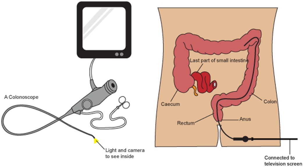
2. Colonoscopy: A procedure in which a flexible tube with a camera on the end is inserted into the rectum and colon to examine the lining for polyps, cancer, or other abnormalities.
3. Colectomy: A procedure in which all or part of the colon is removed, often due to cancer, inflammatory bowel disease, or diverticulitis.
4. Gastrostomy: A procedure in which a hole is made through the abdominal wall and into the stomach to create an opening for feeding. This is often done for patients who have difficulty swallowing.
5. Esophagectomy: A procedure in which all or part of the esophagus is removed, often due to cancer. The remaining esophagus is then reconnected to the stomach or small intestine.
6. Liver resection: A procedure in which a portion of the liver is removed, often due to cancer or other diseases.
7. Pancreatectomy: A procedure in which all or part of the pancreas is removed, often due to cancer or chronic pancreatitis.
8. Cholecystectomy: A procedure in which the gallbladder is removed, often due to gallstones or inflammation.
These are just a few examples of digestive system surgical procedures. There are many other types of operations that can be performed on the digestive system depending on the specific needs and condition of each patient.
Fascia lata is a medical term that refers to the thick, fibrous sheath of connective tissue that envelops and surrounds the thigh muscles (specifically, the quadriceps femoris and hamstrings). It is a type of fascia, which is the soft tissue component of the deep (internal) fascial system.
The fascia lata is continuous with the fascia of the hip and knee joints and plays an important role in providing stability, support, and protection to the muscles and other structures within the thigh. It also helps to facilitate the gliding and movement of muscles and tendons during physical activity.
Injuries or inflammation of the fascia lata can cause pain and discomfort, and may limit mobility and range of motion in the thigh and lower extremity. Conditions such as fascia lata strain, tears, or myofascial pain syndrome may require medical treatment, including physical therapy, medication, or in some cases, surgery.
A diaphragm is a thin, dome-shaped muscle that separates the chest cavity from the abdominal cavity. It plays a vital role in the process of breathing as it contracts and flattens to draw air into the lungs (inhalation) and relaxes and returns to its domed shape to expel air out of the lungs (exhalation).
In addition, a diaphragm is also a type of barrier method of birth control. It is a flexible dome-shaped device made of silicone that fits over the cervix inside the vagina. When used correctly and consistently, it prevents sperm from entering the uterus and fertilizing an egg, thereby preventing pregnancy.
Abdominal injuries refer to damages or traumas that occur in the abdomen, an area of the body that is located between the chest and the pelvis. This region contains several vital organs such as the stomach, liver, spleen, pancreas, small intestine, large intestine, kidneys, and reproductive organs. Abdominal injuries can range from minor bruises and cuts to severe internal bleeding and organ damage, depending on the cause and severity of the trauma.
Common causes of abdominal injuries include:
* Blunt force trauma, such as that caused by car accidents, falls, or physical assaults
* Penetrating trauma, such as that caused by gunshot wounds or stabbing
* Deceleration injuries, which occur when the body is moving at a high speed and suddenly stops, causing internal organs to continue moving and collide with each other or the abdominal wall
Symptoms of abdominal injuries may include:
* Pain or tenderness in the abdomen
* Swelling or bruising in the abdomen
* Nausea or vomiting
* Dizziness or lightheadedness
* Blood in the urine or stool
* Difficulty breathing or shortness of breath
* Rapid heartbeat or low blood pressure
Abdominal injuries can be life-threatening if left untreated, and immediate medical attention is necessary to prevent complications such as infection, internal bleeding, organ failure, or even death. Treatment may include surgery, medication, or other interventions depending on the severity and location of the injury.
The appendix is a small, tube-like structure that projects from the large intestine, located in the lower right quadrant of the abdomen. Its function in humans is not well understood and is often considered vestigial, meaning it no longer serves a necessary purpose. However, in some animals, the appendix plays a role in the immune system. Inflammation of the appendix, known as appendicitis, can cause severe abdominal pain and requires medical attention, often leading to surgical removal of the appendix (appendectomy).
The broad ligament is a wide, flat fold of peritoneum (the serous membrane that lines the abdominal cavity) that supports and suspends the uterus within the pelvic cavity. It consists of two layers - the anterior leaf and the posterior leaf - which enclose and protect various reproductive structures such as the fallopian tubes, ovaries, and blood vessels.
The broad ligament plays a crucial role in maintaining the position and stability of the uterus, allowing for proper functioning of the female reproductive system. It also serves as a conduit for nerves, blood vessels, and lymphatics that supply and drain the uterus and other pelvic organs.
Anomalies or pathologies of the broad ligament, such as cysts, tumors, or inflammation, can potentially lead to various gynecological conditions and symptoms, requiring medical evaluation and intervention if necessary.
Extracorporeal Membrane Oxygenation (ECMO) is a medical procedure that uses a machine to take over the function of the lungs and sometimes also the heart, by pumping and oxygenating the patient's blood outside of their body. This technique is used when a patient's lungs or heart are unable to provide adequate gas exchange or circulation, despite other forms of treatment.
During ECMO, blood is removed from the body through a large catheter or cannula, passed through a membrane oxygenator that adds oxygen and removes carbon dioxide, and then returned to the body through another catheter. This process helps to rest and heal the lungs and/or heart while maintaining adequate oxygenation and circulation to the rest of the body.
ECMO is typically used as a last resort in patients with severe respiratory or cardiac failure who have not responded to other treatments, such as mechanical ventilation or medication. It can be a life-saving procedure, but it also carries risks, including bleeding, infection, and damage to blood vessels or organs.
"Fetal organ maturity" refers to the stage of development and functional competency of the various organs in a fetus. It is the point at which an organ has developed enough to be able to perform its intended physiological functions effectively and sustainably. This maturity is determined by a combination of factors including structural development, cellular differentiation, and biochemical functionality.
Fetal organ maturity is a critical aspect of fetal development, as it directly impacts the newborn's ability to survive and thrive outside the womb. The level of maturity varies among different organs, with some becoming mature earlier in gestation while others continue to develop and mature until birth or even after.
Assessment of fetal organ maturity is often used in clinical settings to determine the optimal time for delivery, particularly in cases where there are risks associated with premature birth. This assessment typically involves a combination of imaging studies, such as ultrasound and MRI, as well as laboratory tests and physical examinations.
Elective surgical procedures are operations that are scheduled in advance because they do not involve a medical emergency. These surgeries are chosen or "elective" based on the patient's and doctor's decision to improve the patient's quality of life or to treat a non-life-threatening condition. Examples include but are not limited to:
1. Aesthetic or cosmetic surgery such as breast augmentation, rhinoplasty, etc.
2. Orthopedic surgeries like knee or hip replacements
3. Cataract surgery
4. Some types of cancer surgeries where the tumor is not spreading or causing severe symptoms
5. Gastric bypass for weight loss
It's important to note that while these procedures are planned, they still require thorough preoperative evaluation and preparation, and carry risks and benefits that need to be carefully considered by both the patient and the healthcare provider.
The esophagogastric junction (EGJ) is the region of the gastrointestinal tract where the esophagus (the tube that carries food from the mouth to the stomach) meets the stomach. It serves as a physiological sphincter, which helps control the direction of flow and prevent reflux of gastric contents back into the esophagus. The EGJ is also known as the gastroesophageal junction or cardia.
The abdomen refers to the portion of the body that lies between the thorax (chest) and the pelvis. It is a musculo-fascial cavity containing the digestive, urinary, and reproductive organs. The abdominal cavity is divided into several regions and quadrants for medical description and examination purposes. These include the upper and lower abdomen, as well as nine quadrants formed by the intersection of the midline and a horizontal line drawn at the level of the umbilicus (navel).
The major organs located within the abdominal cavity include:
1. Stomach - muscular organ responsible for initial digestion of food
2. Small intestine - long, coiled tube where most nutrient absorption occurs
3. Large intestine - consists of the colon and rectum; absorbs water and stores waste products
4. Liver - largest internal organ, involved in protein synthesis, detoxification, and metabolism
5. Pancreas - secretes digestive enzymes and hormones such as insulin
6. Spleen - filters blood and removes old red blood cells
7. Kidneys - pair of organs responsible for filtering waste products from the blood and producing urine
8. Adrenal glands - sit atop each kidney, produce hormones that regulate metabolism, immune response, and stress response
The abdomen is an essential part of the human body, playing a crucial role in digestion, absorption, and elimination of food and waste materials, as well as various metabolic processes.
The peritoneum is the serous membrane that lines the abdominal cavity and covers the abdominal organs. It is composed of a mesothelial cell monolayer supported by a thin, loose connective tissue. The peritoneum has two layers: the parietal peritoneum, which lines the abdominal wall, and the visceral peritoneum, which covers the organs.
The potential space between these two layers is called the peritoneal cavity, which contains a small amount of serous fluid that allows for the smooth movement of the organs within the cavity. The peritoneum plays an important role in the absorption and secretion of fluids and electrolytes, as well as providing a surface for the circulation of immune cells.
In addition, it also provides a route for the spread of infection or malignant cells throughout the abdominal cavity, known as peritonitis. The peritoneum is highly vascularized and innervated, making it sensitive to pain and distention.
Esophagitis is a medical condition characterized by inflammation and irritation of the esophageal lining, which is the muscular tube that connects the throat to the stomach. This inflammation can cause symptoms such as difficulty swallowing, chest pain, heartburn, and acid reflux.
Esophagitis can be caused by various factors, including gastroesophageal reflux disease (GERD), infection, allergies, medications, and chronic vomiting. Prolonged exposure to stomach acid can also cause esophagitis, leading to a condition called reflux esophagitis.
If left untreated, esophagitis can lead to complications such as strictures, ulcers, and Barrett's esophagus, which is a precancerous condition that increases the risk of developing esophageal cancer. Treatment for esophagitis typically involves addressing the underlying cause, managing symptoms, and protecting the esophageal lining to promote healing.
Prenatal ultrasonography, also known as obstetric ultrasound, is a medical diagnostic procedure that uses high-frequency sound waves to create images of the developing fetus, placenta, and amniotic fluid inside the uterus. It is a non-invasive and painless test that is widely used during pregnancy to monitor the growth and development of the fetus, detect any potential abnormalities or complications, and determine the due date.
During the procedure, a transducer (a small handheld device) is placed on the mother's abdomen and moved around to capture images from different angles. The sound waves travel through the mother's body and bounce back off the fetus, producing echoes that are then converted into electrical signals and displayed as images on a screen.
Prenatal ultrasonography can be performed at various stages of pregnancy, including early pregnancy to confirm the pregnancy and detect the number of fetuses, mid-pregnancy to assess the growth and development of the fetus, and late pregnancy to evaluate the position of the fetus and determine if it is head down or breech. It can also be used to guide invasive procedures such as amniocentesis or chorionic villus sampling.
Overall, prenatal ultrasonography is a valuable tool in modern obstetrics that helps ensure the health and well-being of both the mother and the developing fetus.
Appendicitis is a medical condition characterized by inflammation of the appendix, a small finger-like structure that projects from the colon located in the lower right abdomen. The appendix doesn't have a known function, and its removal (appendectomy) does not appear to affect a person's health.
The inflammation of the appendix can be caused by various factors, such as obstruction due to hardened stool, foreign bodies, or tumors. The blockage can lead to increased pressure within the appendix, reduced blood flow, and bacterial growth, resulting in infection and inflammation. If left untreated, appendicitis can progress to peritonitis (inflammation of the lining of the abdominal cavity) or even sepsis, a life-threatening condition.
Common symptoms of appendicitis include:
* Sudden onset of pain in the lower right abdomen, which may start around the navel and shift to the lower right side over several hours
* Pain that worsens with movement, coughing, or sneezing
* Nausea and vomiting
* Loss of appetite
* Fever and chills
* Constipation or diarrhea
* Abdominal swelling or bloating
If you suspect appendicitis, it's essential to seek immediate medical attention. The standard treatment for appendicitis is surgical removal of the appendix (appendectomy), which can be performed as an open surgery or laparoscopically. Antibiotics are also administered to treat any existing infection. Delaying treatment can lead to serious complications, so it's crucial not to ignore symptoms and seek medical help promptly.
An enterostomy is a surgical procedure that creates an opening from the intestine to the abdominal wall, which allows for the elimination of waste from the body. This opening is called a stoma and can be temporary or permanent, depending on the individual's medical condition. There are several types of enterostomies, including colostomy, ileostomy, and jejunostomy, which differ based on the specific location in the intestine where the stoma is created.
The purpose of an enterostomy may vary, but it is often performed to divert the flow of waste away from a diseased or damaged section of the intestine, allowing it to heal. Common reasons for an enterostomy include inflammatory bowel disease, cancer, trauma, and birth defects.
After the surgery, patients will need to wear a pouching system over the stoma to collect waste. They will also require specialized care and education on how to manage their stoma and maintain their overall health. With proper care and support, individuals with an enterostomy can lead active and fulfilling lives.
Abdominal wound closure techniques refer to the methods used to close and repair surgical incisions in the abdomen. The goal of these techniques is to restore the integrity of the abdominal wall, minimize the risk of infection or dehiscence (wound separation), and promote optimal healing. Several abdominal wound closure techniques are available, and the choice of which one to use depends on various factors such as the size and location of the incision, the patient's individual needs and medical history, and the surgeon's preference. Here are some commonly used abdominal wound closure techniques:
1. Continuous running suture: This technique involves using a continuous strand of suture material to close the wound in a single pass. The suture is inserted through the full thickness of the abdominal wall, including the fascia (the strong connective tissue that surrounds the muscles), and then passed continuously along the length of the incision, pulling the edges of the wound together as it goes. This technique can be faster and more efficient than other methods, but it may increase the risk of infection or wound breakdown if not done properly.
2. Interrupted suture: In this technique, the surgeon uses individual stitches placed at regular intervals along the incision to close the wound. Each stitch is tied separately, which can make the closure more secure and reduce the risk of infection or wound breakdown. However, interrupted sutures can be more time-consuming than continuous running sutures.
3. Mass closure: This technique involves using a large, continuous suture to close the entire length of the incision in one pass. The suture is inserted through the full thickness of the abdominal wall and tied at both ends, pulling the edges of the wound together. Mass closure can be faster and more efficient than other methods, but it may increase the risk of infection or wound breakdown if not done properly.
4. Retention sutures: These are additional sutures that are placed deep within the abdominal wall to provide extra support and strength to the closure. They are often used in high-tension areas or in patients who are at increased risk of wound dehiscence, such as those with obesity or diabetes.
5. Layered closure: In this technique, the surgeon closes the incision in multiple layers, starting with the deepest layer of muscle and fascia and working outward to the skin. Each layer is closed separately using either interrupted or continuous sutures. Layered closure can provide added strength and stability to the closure, but it can be more time-consuming than other methods.
6. Skin closure: The final step in wound closure is to close the skin, which can be done using a variety of techniques, including staples, sutures, or surgical glue. The choice of closure method depends on several factors, including the size and location of the incision, the patient's individual needs and preferences, and the surgeon's experience and expertise.
Overall, the choice of wound closure technique depends on several factors, including the size and location of the incision, the patient's individual needs and preferences, and the surgeon's experience and expertise. The goal is to provide a strong, secure, and cosmetically appealing closure that minimizes the risk of infection, wound breakdown, or other complications.
Artificial pneumoperitoneum is a medical condition that refers to the presence of air or gas in the peritoneal cavity, which is the space between the lining of the abdominal wall and the organs within the abdomen. This condition is typically created intentionally during surgical procedures, such as laparoscopy, to provide a working space for the surgeon to perform the operation.
During laparoscopic surgery, a thin tube called a trocar is inserted through a small incision in the abdominal wall, and carbon dioxide gas is pumped into the peritoneal cavity to create a pneumoperitoneum. This allows the surgeon to insert specialized instruments through other small incisions and perform the surgery while visualizing the operative field with a camera.
While artificial pneumoperitoneum is generally safe, there are potential complications that can arise, such as injury to surrounding organs or blood vessels during trocar insertion, subcutaneous emphysema (air trapped under the skin), or gas embolism (gas in the bloodstream). These risks are typically minimized through careful technique and monitoring during the procedure.
Peptic esophagitis is a medical condition that refers to inflammation and damage of the lining of the esophagus caused by stomach acid backing up into the esophagus. This is also known as gastroesophageal reflux disease (GERD). The term "peptic" indicates that digestive enzymes or stomach acids are involved in the cause of the condition.
Peptic esophagitis can cause symptoms such as heartburn, chest pain, difficulty swallowing, and painful swallowing. If left untreated, it can lead to complications like strictures, ulcers, and Barrett's esophagus, which is a precancerous condition. Treatment typically involves lifestyle changes, medications to reduce acid production, and sometimes surgery.
An ileostomy is a surgical procedure in which the end of the small intestine, called the ileum, is brought through an opening in the abdominal wall (stoma) to create a path for waste material to leave the body. This procedure is typically performed when there is damage or removal of the colon, rectum, or anal canal due to conditions such as inflammatory bowel disease (Crohn's disease or ulcerative colitis), cancer, or trauma.
After an ileostomy, waste material from the small intestine exits the body through the stoma and collects in a pouch worn outside the body. The patient needs to empty the pouch regularly, typically every few hours, as the output is liquid or semi-liquid. Ileostomies can be temporary or permanent, depending on the underlying condition and the planned course of treatment. Proper care and management of the stoma and pouch are essential for maintaining good health and quality of life after an ileostomy.
The esophagus is the muscular tube that connects the throat (pharynx) to the stomach. It is located in the midline of the neck and chest, passing through the diaphragm to enter the abdomen and join the stomach. The main function of the esophagus is to transport food and liquids from the mouth to the stomach for digestion.
The esophagus has a few distinct parts: the upper esophageal sphincter (a ring of muscle that separates the esophagus from the throat), the middle esophagus, and the lower esophageal sphincter (another ring of muscle that separates the esophagus from the stomach). The lower esophageal sphincter relaxes to allow food and liquids to enter the stomach and then contracts to prevent stomach contents from flowing back into the esophagus.
The walls of the esophagus are made up of several layers, including mucosa (a moist tissue that lines the inside of the tube), submucosa (a layer of connective tissue), muscle (both voluntary and involuntary types), and adventitia (an outer layer of connective tissue).
Common conditions affecting the esophagus include gastroesophageal reflux disease (GERD), Barrett's esophagus, esophageal cancer, esophageal strictures, and eosinophilic esophagitis.
'Abnormalities, Multiple' is a broad term that refers to the presence of two or more structural or functional anomalies in an individual. These abnormalities can be present at birth (congenital) or can develop later in life (acquired). They can affect various organs and systems of the body and can vary greatly in severity and impact on a person's health and well-being.
Multiple abnormalities can occur due to genetic factors, environmental influences, or a combination of both. Chromosomal abnormalities, gene mutations, exposure to teratogens (substances that cause birth defects), and maternal infections during pregnancy are some of the common causes of multiple congenital abnormalities.
Examples of multiple congenital abnormalities include Down syndrome, Turner syndrome, and VATER/VACTERL association. Acquired multiple abnormalities can result from conditions such as trauma, infection, degenerative diseases, or cancer.
The medical evaluation and management of individuals with multiple abnormalities depend on the specific abnormalities present and their impact on the individual's health and functioning. A multidisciplinary team of healthcare professionals is often involved in the care of these individuals to address their complex needs.
Gestational age is the length of time that has passed since the first day of the last menstrual period (LMP) in pregnant women. It is the standard unit used to estimate the age of a pregnancy and is typically expressed in weeks. This measure is used because the exact date of conception is often not known, but the start of the last menstrual period is usually easier to recall.
It's important to note that since ovulation typically occurs around two weeks after the start of the LMP, gestational age is approximately two weeks longer than fetal age, which is the actual time elapsed since conception. Medical professionals use both gestational and fetal age to track the development and growth of the fetus during pregnancy.
Thoracotomy is a surgical procedure that involves making an incision on the chest wall to gain access to the thoracic cavity, which contains the lungs, heart, esophagus, trachea, and other vital organs. The incision can be made on the side (lateral thoracotomy), back (posterolateral thoracotomy), or front (median sternotomy) of the chest wall, depending on the specific surgical indication.
Thoracotomy is performed for various indications, including lung biopsy, lung resection, esophagectomy, heart surgery, and mediastinal mass removal. The procedure allows the surgeon to directly visualize and access the organs within the thoracic cavity, perform necessary procedures, and control bleeding if needed.
After the procedure, the incision is typically closed with sutures or staples, and a chest tube may be placed to drain any accumulated fluid or air from the pleural space around the lungs. The patient will require postoperative care and monitoring in a hospital setting until their condition stabilizes.
Endoscopy of the digestive system, also known as gastrointestinal (GI) endoscopy, is a medical procedure that allows healthcare professionals to visually examine the inside lining of the digestive tract using a flexible tube with a light and camera attached to it, called an endoscope. This procedure can help diagnose and treat various conditions affecting the digestive system, including gastroesophageal reflux disease (GERD), ulcers, inflammatory bowel disease (IBD), and cancer.
There are several types of endoscopy procedures that focus on different parts of the digestive tract:
1. Esophagogastroduodenoscopy (EGD): This procedure examines the esophagus, stomach, and duodenum (the first part of the small intestine). It is often used to investigate symptoms such as difficulty swallowing, abdominal pain, or bleeding in the upper GI tract.
2. Colonoscopy: This procedure explores the large intestine (colon) and rectum. It is commonly performed to screen for colon cancer, as well as to diagnose and treat conditions like inflammatory bowel disease, diverticulosis, or polyps.
3. Sigmoidoscopy: Similar to a colonoscopy, this procedure examines the lower part of the colon (sigmoid colon) and rectum. It is often used as a screening tool for colon cancer and to investigate symptoms like rectal bleeding or changes in bowel habits.
4. Upper GI endoscopy: This procedure focuses on the esophagus, stomach, and duodenum, using a thin, flexible tube with a light and camera attached to it. It is used to diagnose and treat conditions such as GERD, ulcers, and difficulty swallowing.
5. Capsule endoscopy: This procedure involves swallowing a small capsule containing a camera that captures images of the digestive tract as it passes through. It can help diagnose conditions in the small intestine that may be difficult to reach with traditional endoscopes.
Endoscopy is typically performed under sedation or anesthesia to ensure patient comfort during the procedure. The images captured by the endoscope are displayed on a monitor, allowing the healthcare provider to assess the condition of the digestive tract and make informed treatment decisions.
The omentum, in anatomical terms, refers to a large apron-like fold of abdominal fatty tissue that hangs down from the stomach and loops over the intestines. It is divided into two portions: the greater omentum, which is larger and hangs down further, and the lesser omentum, which is smaller and connects the stomach to the liver.
The omentum has several functions in the body, including providing protection and cushioning for the abdominal organs, assisting with the immune response by containing a large number of immune cells, and helping to repair damaged tissues. It can also serve as a source of nutrients and energy for the body during times of starvation or other stressors.
In medical contexts, the omentum may be surgically mobilized and used to wrap around injured or inflamed tissues in order to promote healing and reduce the risk of infection. This technique is known as an "omentopexy" or "omentoplasty."
Manometry is a medical test that measures pressure inside various parts of the gastrointestinal tract. It is often used to help diagnose digestive disorders such as achalasia, gastroparesis, and irritable bowel syndrome. During the test, a thin, flexible tube called a manometer is inserted through the mouth or rectum and into the area being tested. The tube is connected to a machine that measures and records pressure readings. These readings can help doctors identify any abnormalities in muscle function or nerve reflexes within the digestive tract.
A "learning curve" is not a medical term per se, but rather a general concept that is used in various fields including medicine. It refers to the process of acquiring new skills or knowledge in a specific task or activity, and the improvement in performance that comes with experience and practice over time.
In a medical context, a learning curve may refer to the rate at which healthcare professionals acquire proficiency in a new procedure, technique, or technology. It can also describe how quickly patients learn to manage their own health conditions or treatments. The term is often used to evaluate the effectiveness of training programs and to identify areas where additional education or practice may be necessary.
It's important to note that individuals may have different learning curves depending on factors such as prior experience, innate abilities, motivation, and access to resources. Therefore, it's essential to tailor training and support to the needs of each learner to ensure optimal outcomes.
Veterinary surgery refers to the surgical procedures performed on animals by trained veterinarians or veterinary surgeons. It involves the use of various surgical techniques and tools to diagnose, treat, or prevent diseases and injuries in animals. This can include soft tissue surgeries such as abdominal or thoracic surgeries, orthopedic surgeries for bone and joint issues, neurological surgeries, oncological surgeries for the removal of tumors, and reconstructive surgeries. Veterinary surgeons must complete extensive education and training in order to provide safe and effective surgical care for animals.
Polyglactin 910 is a type of synthetic absorbable suture made from copolymers of lactide and glycolide. It is designed to gradually break down and be absorbed by the body over time, typically within 56 to 70 days after being used in surgical wounds. This property makes it an ideal choice for soft tissue approximation and laceration repairs.
Polyglactin 910 sutures are often used in various surgical procedures, including orthopedic, ophthalmic, cardiovascular, and general surgery. They come in different sizes and forms, such as plain, reverse cutting, and braided, to suit various surgical needs.
The gradual absorption of Polyglactin 910 sutures helps minimize scarring and reduces the need for suture removal procedures. However, it is essential to note that inflammation may occur during the degradation process, which could potentially lead to adverse reactions in some individuals. Proper wound care and follow-up with healthcare professionals are crucial to ensure optimal healing and manage any potential complications.
The perineum is the region between the anus and the genitals. In anatomical terms, it refers to the diamond-shaped area located in the lower part of the pelvis and extends from the coccyx (tailbone) to the pubic symphysis, which is the joint in the front where the two pubic bones meet. This region contains various muscles that support the pelvic floor and contributes to maintaining urinary and fecal continence. The perineum can be further divided into two triangular regions: the urogenital triangle (anterior) and the anal triangle (posterior).
"Acute abdomen" is a medical term used to describe a sudden and severe abdominal pain that requires immediate medical attention. This condition can be caused by various factors such as inflammation, infection, obstruction, or perforation of the abdominal organs. Common causes of acute abdomen include appendicitis, cholecystitis, diverticulitis, intestinal obstruction, and perforated ulcers.
The symptoms of acute abdomen may include severe and localized or generalized abdominal pain, tenderness, rigidity, rebound tenderness, fever, nausea, vomiting, and loss of appetite. The diagnosis of acute abdomen is usually made based on the patient's history, physical examination, laboratory tests, and imaging studies such as X-rays, ultrasound, or CT scan.
Treatment of acute abdomen depends on the underlying cause and may include antibiotics, intravenous fluids, pain management, and surgery in severe cases. Delayed diagnosis and treatment of acute abdomen can lead to serious complications such as sepsis, peritonitis, and even death.
Barium sulfate is a medication that is commonly used as a contrast material in medical imaging procedures, such as X-rays and CT scans. It works by coating the inside of the digestive tract, making it visible on an X-ray or CT scan and allowing doctors to see detailed images of the stomach, intestines, and other parts of the digestive system.
Barium sulfate is a white, chalky powder that is mixed with water to create a thick, milky liquid. It is generally safe and does not cause significant side effects when used in medical imaging procedures. However, it should not be taken by individuals who have a known allergy to barium or who have certain digestive conditions, such as obstructions or perforations of the bowel.
It's important to note that while barium sulfate is an important tool for medical diagnosis, it is not a treatment for any medical condition and should only be used under the direction of a healthcare professional.
Minimally invasive surgical procedures are a type of surgery that is performed with the assistance of specialized equipment and techniques to minimize trauma to the patient's body. This approach aims to reduce blood loss, pain, and recovery time as compared to traditional open surgeries. The most common minimally invasive surgical procedure is laparoscopy, which involves making small incisions (usually 0.5-1 cm) in the abdomen or chest and inserting a thin tube with a camera (laparoscope) to visualize the internal organs.
The surgeon then uses long, slender instruments inserted through separate incisions to perform the necessary surgical procedures, such as cutting, coagulation, or suturing. Other types of minimally invasive surgical procedures include arthroscopy (for joint surgery), thoracoscopy (for chest surgery), and hysteroscopy (for uterine surgery). The benefits of minimally invasive surgical procedures include reduced postoperative pain, shorter hospital stays, quicker return to normal activities, and improved cosmetic results. However, not all surgeries can be performed using minimally invasive techniques, and the suitability of a particular procedure depends on various factors, including the patient's overall health, the nature and extent of the surgical problem, and the surgeon's expertise.
A fibrin tissue adhesive is a type of surgical glue that is used to approximate and secure together cut or wounded tissues in the body during surgical procedures. It is made from fibrin, a protein involved in blood clotting, and is often combined with other substances like thrombin and calcium chloride to promote clot formation and enhance adhesion.
Fibrin tissue adhesives work by mimicking the body's natural clotting process. When applied to the wound site, the fibrinogen component of the adhesive is converted into fibrin by the thrombin component, creating a stable fibrin clot that holds the edges of the wound together. This helps to promote healing and reduce the risk of complications such as bleeding or infection.
Fibrin tissue adhesives are commonly used in various surgical procedures, including dermatologic, ophthalmic, orthopedic, and neurologic surgeries. They offer several advantages over traditional suturing methods, such as reduced operation time, less trauma to the tissues, and improved cosmetic outcomes. However, they may not be suitable for all types of wounds or surgical sites, and their use should be determined by a qualified healthcare professional based on individual patient needs and circumstances.
Cryptorchidism is a medical condition in which one or both of a male infant's testicles fail to descend from the abdomen into the scrotum before birth or within the first year of life. Normally, the testicles descend from the abdomen into the scrotum during fetal development in the second trimester. If the testicles do not descend on their own, medical intervention may be necessary to correct the condition.
Cryptorchidism is a common birth defect, affecting about 3-5% of full-term and 30% of preterm male infants. In most cases, the testicle will descend on its own within the first six months of life. If it does not, treatment may be necessary to prevent complications such as infertility, testicular cancer, and inguinal hernia.
Treatment for cryptorchidism typically involves surgery to bring the testicle down into the scrotum. This procedure is called orchiopexy and is usually performed before the age of 2. In some cases, hormonal therapy may be used as an alternative to surgery. However, this approach has limited success and is generally only recommended in certain situations.
Overall, cryptorchidism is a treatable condition that can help prevent future health problems if addressed early on. Regular check-ups with a pediatrician or healthcare provider can help ensure timely diagnosis and treatment of this condition.
Intestinal perforation is a medical condition that refers to a hole or tear in the lining of the intestine. This can occur anywhere along the gastrointestinal tract, including the small intestine, large intestine (colon), or stomach. Intestinal perforation allows the contents of the intestines, such as digestive enzymes and bacteria, to leak into the abdominal cavity, which can lead to a serious inflammatory response known as peritonitis.
Intestinal perforation can be caused by various factors, including:
* Mechanical trauma (e.g., gunshot wounds, stab wounds)
* Inflammatory bowel disease (e.g., Crohn's disease, ulcerative colitis)
* Diverticulitis
* Appendicitis
* Intestinal obstruction
* Infections (e.g., typhoid fever, tuberculosis)
* Certain medications (e.g., nonsteroidal anti-inflammatory drugs, corticosteroids)
* Radiation therapy
* Ischemic bowel disease (lack of blood flow to the intestines)
Symptoms of intestinal perforation may include sudden abdominal pain, nausea, vomiting, fever, and decreased bowel movements. Treatment typically involves surgery to repair the perforation and remove any damaged tissue. Antibiotics are also administered to prevent infection. In severe cases, a temporary or permanent colostomy or ileostomy may be necessary.
Pregnancy is a physiological state or condition where a fertilized egg (zygote) successfully implants and grows in the uterus of a woman, leading to the development of an embryo and finally a fetus. This process typically spans approximately 40 weeks, divided into three trimesters, and culminates in childbirth. Throughout this period, numerous hormonal and physical changes occur to support the growing offspring, including uterine enlargement, breast development, and various maternal adaptations to ensure the fetus's optimal growth and well-being.
Artificial Skin is a synthetic substitute or equivalent that is used to replace, support, or enhance the function of damaged or absent skin. It can be made from various materials such as biopolymers, composites, or biosynthetic materials. The main purpose of artificial skin is to provide a temporary or permanent covering for wounds, burns, or ulcers that cannot be healed with conventional treatments. Additionally, it may serve as a platform for the delivery of medications or as a matrix for the growth of cells and tissues during skin grafting procedures. Artificial skin must possess properties such as biocompatibility, durability, flexibility, and permeability to air and water vapor in order to promote optimal healing and minimize scarring.
Thoracic radiography is a type of diagnostic imaging that involves using X-rays to produce images of the chest, including the lungs, heart, bronchi, great vessels, and the bones of the spine and chest wall. It is a commonly used tool in the diagnosis and management of various respiratory, cardiovascular, and thoracic disorders such as pneumonia, lung cancer, heart failure, and rib fractures.
During the procedure, the patient is positioned between an X-ray machine and a cassette containing a film or digital detector. The X-ray beam is directed at the chest, and the resulting image is captured on the film or detector. The images produced can help identify any abnormalities in the structure or function of the organs within the chest.
Thoracic radiography may be performed as a routine screening test for certain conditions, such as lung cancer, or it may be ordered when a patient presents with symptoms suggestive of a respiratory or cardiovascular disorder. It is a safe and non-invasive procedure that can provide valuable information to help guide clinical decision making and improve patient outcomes.
In the field of medicine, "time factors" refer to the duration of symptoms or time elapsed since the onset of a medical condition, which can have significant implications for diagnosis and treatment. Understanding time factors is crucial in determining the progression of a disease, evaluating the effectiveness of treatments, and making critical decisions regarding patient care.
For example, in stroke management, "time is brain," meaning that rapid intervention within a specific time frame (usually within 4.5 hours) is essential to administering tissue plasminogen activator (tPA), a clot-busting drug that can minimize brain damage and improve patient outcomes. Similarly, in trauma care, the "golden hour" concept emphasizes the importance of providing definitive care within the first 60 minutes after injury to increase survival rates and reduce morbidity.
Time factors also play a role in monitoring the progression of chronic conditions like diabetes or heart disease, where regular follow-ups and assessments help determine appropriate treatment adjustments and prevent complications. In infectious diseases, time factors are crucial for initiating antibiotic therapy and identifying potential outbreaks to control their spread.
Overall, "time factors" encompass the significance of recognizing and acting promptly in various medical scenarios to optimize patient outcomes and provide effective care.
"Conversion to open surgery" is a medical term that refers to the situation when a surgical procedure, which was initially being performed using minimally invasive techniques (such as laparoscopy or thoracoscopy), needs to be changed to an open approach during the operation. This conversion may be necessary due to various reasons such as unforeseen technical difficulties, excessive bleeding, or discovery of unexpected surgical findings that cannot be safely managed using the minimally invasive approach. The decision to convert to an open surgery is typically made by the operating surgeon in order to ensure the safety and well-being of the patient.
Tensile strength is a material property that measures the maximum amount of tensile (pulling) stress that a material can withstand before failure, such as breaking or fracturing. It is usually measured in units of force per unit area, such as pounds per square inch (psi) or pascals (Pa). In the context of medical devices or biomaterials, tensile strength may be used to describe the mechanical properties of materials used in implants, surgical tools, or other medical equipment. High tensile strength is often desirable in these applications to ensure that the material can withstand the stresses and forces it will encounter during use.
The lumbosacral region is the lower part of the back where the lumbar spine (five vertebrae in the lower back) connects with the sacrum (a triangular bone at the base of the spine). This region is subject to various conditions such as sprains, strains, herniated discs, and degenerative disorders that can cause pain and discomfort. It's also a common site for surgical intervention when non-surgical treatments fail to provide relief.
The mesocolon is a peritoneal fold that attaches the colon to the posterior abdominal wall. It contains blood vessels, lymphatics, and nerves that supply the colon. The mesocolon allows for the mobility and flexibility of the colon within the abdominal cavity. There are several parts of the mesocolon, including the mesentery of the ascending colon (right mesocolon), the transverse mesocolon, and the mesentery of the descending and sigmoid colon (left mesocolon).
Endoscopy is a medical procedure that involves the use of an endoscope, which is a flexible tube with a light and camera at the end, to examine the interior of a body cavity or organ. The endoscope is inserted through a natural opening in the body, such as the mouth or anus, or through a small incision. The images captured by the camera are transmitted to a monitor, allowing the physician to visualize the internal structures and detect any abnormalities, such as inflammation, ulcers, or tumors. Endoscopy can also be used for diagnostic purposes, such as taking tissue samples for biopsy, or for therapeutic purposes, such as removing polyps or performing minimally invasive surgeries.
The umbilicus, also known as the navel, is the scar left on the abdominal wall after the removal of the umbilical cord in a newborn. The umbilical cord connects the developing fetus to the placenta in the uterus during pregnancy, providing essential nutrients and oxygen while removing waste products. After birth, the cord is clamped and cut, leaving behind a small stump that eventually dries up and falls off, leaving the umbilicus. In adults, it typically appears as a slight depression or dimple on the abdomen.
Rectal prolapse is a medical condition where the rectum, which is the lower end of the colon, slips outside the anus, the opening through which stool leaves the body. This usually occurs due to weakened muscles and supporting structures in the pelvic area, often as a result of aging, childbirth, or long-term constipation or diarrhea.
The rectal prolapse can be partial, where only a small portion of the rectum slips outside the anus, or complete, where the entire rectum protrudes. This condition can cause discomfort, pain, bleeding, and difficulty with bowel movements. Treatment options may include dietary changes, medication, or surgical intervention.
Gastrointestinal endoscopy is a medical procedure that allows direct visualization of the inner lining of the digestive tract, which includes the esophagus, stomach, small intestine, large intestine (colon), and sometimes the upper part of the small intestine (duodenum). This procedure is performed using an endoscope, a long, thin, flexible tube with a light and camera at its tip. The endoscope is inserted through the mouth for upper endoscopy or through the rectum for lower endoscopy (colonoscopy), and the images captured by the camera are transmitted to a monitor for the physician to view.
Gastrointestinal endoscopy can help diagnose various conditions, such as inflammation, ulcers, tumors, polyps, or bleeding in the digestive tract. It can also be used for therapeutic purposes, such as removing polyps, taking tissue samples (biopsies), treating bleeding, and performing other interventions to manage certain digestive diseases.
There are different types of gastrointestinal endoscopy procedures, including:
1. Upper Endoscopy (Esophagogastroduodenoscopy or EGD): This procedure examines the esophagus, stomach, and duodenum.
2. Colonoscopy: This procedure examines the colon and rectum.
3. Sigmoidoscopy: A limited examination of the lower part of the colon (sigmoid colon) using a shorter endoscope.
4. Enteroscopy: An examination of the small intestine, which can be performed using various techniques, such as push enteroscopy, single-balloon enteroscopy, or double-balloon enteroscopy.
5. Capsule Endoscopy: A procedure that involves swallowing a small capsule containing a camera, which captures images of the digestive tract as it passes through.
Gastrointestinal endoscopy is generally considered safe when performed by experienced medical professionals. However, like any medical procedure, there are potential risks and complications, such as bleeding, infection, perforation, or adverse reactions to sedatives used during the procedure. Patients should discuss these risks with their healthcare provider before undergoing gastrointestinal endoscopy.
An emergency is a sudden, unexpected situation that requires immediate medical attention to prevent serious harm, permanent disability, or death. Emergencies can include severe injuries, trauma, cardiac arrest, stroke, difficulty breathing, severe allergic reactions, and other life-threatening conditions. In such situations, prompt medical intervention is necessary to stabilize the patient's condition, diagnose the underlying problem, and provide appropriate treatment.
Emergency medical services (EMS) are responsible for providing emergency care to patients outside of a hospital setting, such as in the home, workplace, or public place. EMS personnel include emergency medical technicians (EMTs), paramedics, and other first responders who are trained to assess a patient's condition, provide basic life support, and transport the patient to a hospital for further treatment.
In a hospital setting, an emergency department (ED) is a specialized unit that provides immediate care to patients with acute illnesses or injuries. ED staff includes physicians, nurses, and other healthcare professionals who are trained to handle a wide range of medical emergencies. The ED is equipped with advanced medical technology and resources to provide prompt diagnosis and treatment for critically ill or injured patients.
Overall, the goal of emergency medical care is to stabilize the patient's condition, prevent further harm, and provide timely and effective treatment to improve outcomes and save lives.
Nonpenetrating wounds are a type of trauma or injury to the body that do not involve a break in the skin or underlying tissues. These wounds can result from blunt force trauma, such as being struck by an object or falling onto a hard surface. They can also result from crushing injuries, where significant force is applied to a body part, causing damage to internal structures without breaking the skin.
Nonpenetrating wounds can cause a range of injuries, including bruising, swelling, and damage to internal organs, muscles, bones, and other tissues. The severity of the injury depends on the force of the trauma, the location of the impact, and the individual's overall health and age.
While nonpenetrating wounds may not involve a break in the skin, they can still be serious and require medical attention. If you have experienced blunt force trauma or suspect a nonpenetrating wound, it is important to seek medical care to assess the extent of the injury and receive appropriate treatment.
Fetoscopy is a minimally invasive surgical procedure that allows direct visualization of the fetus and the intrauterine environment through the use of a fiber-optic scope. It is typically performed during the second trimester of pregnancy to diagnose or treat various fetal conditions, such as twin-to-twin transfusion syndrome, congenital diaphragmatic hernia, or spina bifida. The procedure involves inserting a thin tube called a fetoscope through the mother's abdomen and uterus to access the fetus. Fetoscopy can also be used for taking fetal tissue samples for genetic testing.
It is important to note that while fetoscopy can provide valuable information and treatment options, it does carry some risks, including preterm labor, premature rupture of membranes, infection, and bleeding. Therefore, the decision to undergo fetoscopy should be made carefully, in consultation with a medical professional, and based on a thorough evaluation of the potential benefits and risks.
Prostheses: Artificial substitutes or replacements for missing body parts, such as limbs, eyes, or teeth. They are designed to restore the function, appearance, or mobility of the lost part. Prosthetic devices can be categorized into several types, including:
1. External prostheses: Devices that are attached to the outside of the body, like artificial arms, legs, hands, and feet. These may be further classified into:
a. Cosmetic or aesthetic prostheses: Primarily designed to improve the appearance of the affected area.
b. Functional prostheses: Designed to help restore the functionality and mobility of the lost limb.
2. Internal prostheses: Implanted artificial parts that replace missing internal organs, bones, or tissues, such as heart valves, hip joints, or intraocular lenses.
Implants: Medical devices or substances that are intentionally placed inside the body to replace or support a missing or damaged biological structure, deliver medication, monitor physiological functions, or enhance bodily functions. Examples of implants include:
1. Orthopedic implants: Devices used to replace or reinforce damaged bones, joints, or cartilage, such as knee or hip replacements.
2. Cardiovascular implants: Devices that help support or regulate heart function, like pacemakers, defibrillators, and artificial heart valves.
3. Dental implants: Artificial tooth roots that are placed into the jawbone to support dental prostheses, such as crowns, bridges, or dentures.
4. Neurological implants: Devices used to stimulate nerves, brain structures, or spinal cord tissues to treat various neurological conditions, like deep brain stimulators for Parkinson's disease or cochlear implants for hearing loss.
5. Ophthalmic implants: Artificial lenses that are placed inside the eye to replace a damaged or removed natural lens, such as intraocular lenses used in cataract surgery.
Acellular dermis is a type of processed connective tissue graft used in surgical procedures, particularly in reconstructive surgery. It is derived from human or animal skin, but has had the epidermis and cells of the dermis removed, leaving behind the intact extracellular matrix (ECM). This ECM includes proteins such as collagen and elastin, which provide structural support, and growth factors, which can help to stimulate tissue regeneration.
The acellular nature of the graft means that it is less likely to be rejected by the recipient's immune system, making it a useful option for patients who may not be good candidates for autografts (tissue transplanted from another part of their own body) or allografts (tissue transplanted from another person). Acellular dermis can be used to repair and rebuild damaged skin, as well as to augment soft tissue in areas such as the face and breast.
There are several different brands and types of acellular dermis available, each with its own specific composition and indications for use. Some common examples include AlloDerm, FlexHD, and Integra Dermal Regeneration Template. The choice of graft may depend on factors such as the size and location of the defect being treated, as well as the patient's individual needs and medical history.
 Inguinal hernia
Inguinal hernia surgery
Amyand's hernia
Femoral hernia
Hernia
Oluyombo Awojobi
Spigelian hernia
Da Vinci Surgical System
Rudolf Ulrich Krönlein
Oliver Neuville
Male genital examination
Inferior epigastric artery
Artery of round ligament of uterus
Round ligament pain
August von Rothmund
Maroteaux-Lamy syndrome
Complete androgen insensitivity syndrome
Abdominal surgery
Hydrocele testis
William Stewart Halsted
Hernia repair
Dominggus Lim-Duan
Femoral ring
Pubic tubercle
Whitesnake
Polydactyly-myopia syndrome
Erectile dysfunction
Development of the gonads
Lateral inguinal fossa
Inguinal canal
Inguinal hernia - Wikipedia
 Inguinal hernia repair: MedlinePlus Medical Encyclopedia
Inguinal hernia: MedlinePlus Medical Encyclopedia Image
 Inguinal Hernias (for Parents) - CareSource
 Inguinal Hernia in Children | Johns Hopkins Medicine
 Laparoscopic Inguinal Hernia Repair Course | Medtronic
 TEP inguinal hernia repair
 Delivering what inguinal hernia patients really really want | SpringerLink
 Inguinal and Umbilical Hernias | Duke Health
 Bilateral (inguinal) hernia repair</span><i class="icon" aria-hidden=...
Groin hernia (or inguinal hernia)</span><i class="icon" aria-hidden=...
Vesalius Clinical Folios: Large Inguinal Hernia
Vesalius Clinical Folios: Large Inguinal Hernia
 Inguinal hernia, minimally invasive surgery - DocCheck
 Recovering from Inguinal Hernia Surgery | Lifespan
 Inguinal Hernia - Digestive Disorders - Merck Manuals Consumer Version
 Digestive tract with an inset showing an indirect inguinal hernia in a male, with labels - Media Asset - NIDDK
 Inguinal Hernia
 Inguinal Hernia
 Inguinal Hernia Health Remedies - Joyful Belly School of Ayurveda
 Inguinal Hernia | General Surgery | Winfield Hospital | Tewkesbury Road, Longford, Gloucester, GL2 9WH
 Hernias, Inguinal and Umbilical | Iowa State University
 Best Package for Inguinal Hernia Repair Surgery in Mexicali, Mexico
Image: Inguinal Hernia - MSD Manual Consumer Version
 Large Retroperitoneal Liposarcoma Diagnosed upon Radiological Evaluation of Mild Right-Sided Inguinal Hernia
 Inguinal hernia - Vejthani Hospital | JCI Accredited International Hospital in Bangkok, Thailand.
 Inguinal Hernia Surgery (TAPP for Indirect Inguinal Hernia) - Free Laparoscopic Videos
Inguinal Hernia Repairs | Doctor Stock
 Sports Hernia/Inguinal Disruption Surgery: Triathlon Forum: Slowtwitch Forums
Type of hernia8
- This type of hernia begins in the groin area and is more common in premature infants. (dukehealth.org)
- This is the most common type, and a type of hernia that you may be born with. (stanfordchildrens.org)
- In women, this type of hernia can occur if reproductive organs or the small intestine slides into the groin area because of a weakness in the abdominal muscles. (stanfordchildrens.org)
- This type of hernia is caused by weakening of your abdominal muscles over time and is more likely to be seen in adults. (stanfordchildrens.org)
- An inguinal hernia is the most common type of hernia. (placidway.com)
- The choice of therapy depends on the type of hernia. (medical-clinic.org)
- Patient demographics, type of hernia, and operative and postoperative outcomes were analyzed. (eurekamag.com)
- Inguinal Hernia or (Groin Hernia) is the commonest type of hernia and is seen very often at our clinics. (ovihams.com)
Surgical22
- This one day course will focus on advanced surgical techniques for surgeons who wish to increase their knowledge of laparoscopic inguinal hernia repair as a primary procedure. (medtronic.com)
- As general surgeons we strive to obtain results comparable to those of hernia zealots where the reproducibility of the surgical technique, including the type of anaesthesia used, is paramount. (springer.com)
- The way to repair an inguinal hernia is usually with a surgical procedure. (stanfordchildrens.org)
- Inguinal hernia repair is a very common surgical operation that can be performed by open or laparoscopic surgery. (ramsayhealth.co.uk)
- A surgical treatment is the correction of inguinal hernias. (vejthani.com)
- Between April 2011-August 2018, 129 patients (157 groins) underwent surgical treatment for chronic groin pain after inguinal hernia repair. (sages.org)
- This attempt at process improvement demonstrated beneficial effects for patients who had CBT as part of a prehabilitation program prior to a surgical procedure to attempt to relieve groin pain after inguinal hernia repair. (sages.org)
- Most recently, doctors have successfully performed an inguinal hernia operation by laparoscopic surgical method for patients N.T.M (15 years old, HCMC). (fvhospital.com)
- The current practice for hernia repair in South Africa as well as the surgical registrar exposure to laparoscopic repair training is unknown. (scielo.org.za)
- laparoscopy, hernia repair, surgical training. (scielo.org.za)
- 2 As hernias are so common and surgery is the only definitive treatment, it would follow that surgical hernia repair has become one of the most commonly performed general surgical operations globally, with more than 20 million procedures performed annually. (scielo.org.za)
- The objective of this study is to quantify the current practice of inguinal hernia surgery in hospitals in Cape Town, South Africa, affiliated to the University of Cape Town (UCT), a surgical specialist training institution, and to assess trainee exposure to laparoscopic repair technique. (scielo.org.za)
- An incarcerated or strangulated hernia of any kind requires urgent surgical repair. (msdmanuals.com)
- In case of a non-reducible inguinal hernia, usually a general surgeon advises to go for surgical intervention where they apply a mesh sheet to cover the sort of escape route in anterior abdominal wall for the various viscera. (ovihams.com)
- Such surgical hernia treatment has been seen to fail where a relapse of hernia has been seen in many cases. (ovihams.com)
- The good news is that there is absolutely no need to go for any surgical intervention for inguinal hernia as it is completely curable with Homoeopathy. (ovihams.com)
- Common conditions seen are diseases of the thyroid gland and and other head and neck tumors, Breast tumors, gallbladder and biliary stones, Inguinal and umbilical hernia, Anal conditions such as hemorrhoids and fistula-in-ano, acute abdominal emergencies such as appendicitis and diverticulitis, Colon and Rectal diseases, trauma, minor surgical conditions such as skin and soft tissue tumors and cysts, circumcision. (filipinodoctors.org)
- Abdominal wall hernias are among the most common of all surgical problems. (medscape.com)
- Repair of these hernias is one of the most commonly performed surgical procedures in the world. (medscape.com)
- [ 20 ] The issue of observation versus surgical intervention in this asymptomatic or minimally symptomatic population was addressed in two randomized clinical trials, both of which found that there were no significant differences in hernia-related symptoms after long-term follow-up and that watchful waiting did not increase the complication rate. (medscape.com)
- To describe the surgical technique and the short-term results of inguinal hernioplasty with the TAPP technique. (bvsalud.org)
- Academy of Pediatrics, 5 out of 100 chil- to functional disturbance of the testis and dren have inguinal hernias (more boys than surgical treatment leads to improvement of girls), some may not have symptoms until semen quality [ 11 ]. (who.int)
Scrotum13
- In males, indirect hernias follow the same route as the descending testes, which migrate from the abdomen into the scrotum during the development of the urinary and reproductive organs. (wikipedia.org)
- The main sign of an inguinal hernia is a visible bulge under the skin in the groin or scrotum . (kidshealth.org)
- When a baby boy develops in the womb, the testicles travel from the belly to the scrotum through the inguinal canals. (kidshealth.org)
- If one stays open, a boy's intestines can slip into the inguinal canal and into the scrotum, forming a hernia. (kidshealth.org)
- To feel the hernia as it moves into the groin or scrotum, the doctor might have your child stand and cough. (kidshealth.org)
- Before birth, the testes, which are formed in the abdomen, pass through the inguinal canal as they descend into the scrotum. (merckmanuals.com)
- An inguinal hernia extends into the groin and can extend into the scrotum. (merckmanuals.com)
- Inguinal hernias usually cause a painless bulge in the groin or scrotum. (merckmanuals.com)
- In addition, in men, the doctor places a finger into a fold on the upper part of the scrotum and pushes upward toward the abdominal cavity to detect a hernia. (merckmanuals.com)
- The main symptom of an inguinal hernia is a bulge in the groin or scrotum. (alberta.ca)
- In men, an inguinal hernia may extend down into the scrotum and cause an enlarged scrotum. (stanfordchildrens.org)
- The inguinal canal, where the spermatic cord enters the scrotum, is typically where the weak spot in men is located. (vejthani.com)
- If its inguinal, and it looks like it might descend, you have to ask yourself whether you are willing to wait until it appears as a squatter in your scrotum. (slowtwitch.com)
Canal24
- An inguinal hernia or groin hernia, is a hernia (protrusion) of abdominal cavity contents through the inguinal canal. (wikipedia.org)
- The larger size of their inguinal canal, which transmitted the testicle and accommodates the structures of the spermatic cord, might be one reason why men are 25 times more likely to have an inguinal hernia than women. (wikipedia.org)
- Although several mechanisms such as strength of the posterior wall of the inguinal canal and shutter mechanisms compensating for raised intra-abdominal pressure prevent hernia formation in normal individuals, the exact importance of each factor is still under debate. (wikipedia.org)
- Direct inguinal hernias occur medial to the inferior epigastric vessels when abdominal contents herniate through a weak spot in the fascia of the posterior wall of the inguinal canal, which is formed by the transversalis fascia. (wikipedia.org)
- A common type in babies and young kids happens when the intestines slide into a pathway called the inguinal canal in the groin area, rather than staying in the belly. (kidshealth.org)
- Indeed a trans-inguinal approach is easier to teach and perform by residents who need to appreciate the intricacies of the inguinal canal as demonstrated in a randomised fashion by our Dutch colleagues [ 4 ]. (springer.com)
- An inguinal hernia happens at the inguinal canal. (nuffieldhealth.com)
- The inguinal canal contains the spermatic cord, which consists of the vas deferens, blood vessels, nerves, and other structures. (merckmanuals.com)
- An inguinal hernia, also known as a 'groin hernia' is the pushing out of abdominal organs, such as your intestines, through a weak area in your groin, called the inguinal canal. (ramsayhealth.co.uk)
- Protrusion of intestines through the inguinal canal often follows castration of pigs that unknowingly have an inguinal hernia. (iastate.edu)
- This image shows intestines that have pushed through an opening in the abdominal wall into the inguinal canal, which would cause a bulge to form under the skin. (msdmanuals.com)
- Hernias can sometimes develop where connective tissue from the uterus joins to the tissue surrounding the pubic bone in women because the inguinal canal includes a ligament that aids in holding the uterus in place. (vejthani.com)
- An inguinal hernia occurs when an internal organ, usually part of the small intestine, protrudes into the inguinal canal through a tear or weak spot in the abdominal muscles. (health.mil)
- In an inguinal hernia - the most common type - the large or small intestine, omentum, or bladder protrudes into the inguinal canal. (medical-clinic.org)
- Inguinal hernias can be direct (herniation through an area of muscle weakness in the inguinal canal) or indirect (herniation through the inguinal ring). (medical-clinic.org)
- An inguinal hernia results when a structure called the inguinal canal doesn't close properly after birth. (medical-clinic.org)
- If the inguinal canal remains open, other abdominal contents can also pass down it. (medical-clinic.org)
- If no hernia is palpable, the patient should cough or perform a Valsalva maneuver as the examiner palpates the groin (with a finger in the inguinal canal in males). (msdmanuals.com)
- Ultrasound and computed tomography confirmed his vermiform appendix herniated into the right inguinal canal. (biomedcentral.com)
- a Ultrasonography showing non-inflamed appendix (arrow) inside the right inguinal canal. (biomedcentral.com)
- Hirata Precisions, Chiba, Japan) were introduced into the upper right abdominal quadrant to inspect the hernia canal for the absence of appendicitis and reduce the appendix laparoscopically (Fig. 2 ). (biomedcentral.com)
- Medical intensivists, cardiologists, general physicians, and pediatricians also require a working understanding of the femoral sheath and inguinal canal. (medscape.com)
- For a better understanding of the clinical significance of the inguinal canal and the adjacent femoral sheath, this region may be conceptually broken down into osseous, myotendinous, neurovascular, lymphatic, and genitourinary/spermatic cord components. (medscape.com)
- In addition to the inguinal ligament, the inguinal canal is composed of the aponeurosis of the external oblique muscle (anterior wall), the transversalis fascia (posterior wall), and fibers of the internal oblique muscle and the transversus abdominis (roof). (medscape.com)
Complications5
- Repair, however, is generally recommended in females due to the higher rate of femoral hernias (also a type of groin hernia) which have more complications. (wikipedia.org)
- Your doctor will usually recommend inguinal hernia repair surgery if you have an enlarged or painful hernia to relieve your discomfort and to prevent serious complications. (ramsayhealth.co.uk)
- A significant difference was noted in the rate of postoperative complications occurring within 30 days, which was 4.9 % for bilateral compared with 3.9 % for unilateral inguinal hernia ( p = 0.009). (springer.com)
- Background: It is a myth that the student knows the anatomy, causes, sequelae, complications and types of hernia. (amrita.edu)
- Wearing a supportive garment designed to keep hernias in place (hernia truss) doesnt correct the problem or help prevent complications. (ovihams.com)
Bulge12
- The inability to "reduce", or place the bulge back into the abdomen usually means the hernia is 'incarcerated' which requires emergency surgery. (wikipedia.org)
- Most hernias are discovered by seeing or feeling the bulge. (hopkinsmedicine.org)
- Although the hernia won't stay "in" or the bulge will not disappear, this is still considered a reducible hernia because it is possible to compress it. (hopkinsmedicine.org)
- Hernias feel like a soft lump, bulge, or swelling under the skin in the groin or around the belly button. (dukehealth.org)
- An abdominal wall hernia causes a noticeable bulge but little. (merckmanuals.com)
- If the person has an inguinal hernia, coughing will cause the hernia to bulge outward, making it easier for the doctor to detect. (merckmanuals.com)
- The bulge of a hernia is usually easy to feel. (alberta.ca)
- In babies, a hernia may bulge when the child cries or moves around. (alberta.ca)
- An inguinal hernia is a bulge that occurs in your groin region, the area between the lower part of your abdomen and your thigh. (stanfordchildrens.org)
- An inguinal hernia forms when your intestines and the peritoneum push through the muscles and appear as a bulge under your skin. (stanfordchildrens.org)
- Inguinal hernias are dangerous because they tend to keep getting larger and your intestine can get trapped inside the bulge and lose its blood supply. (stanfordchildrens.org)
- If a hernia bulge turns red, purple, or dark, or if you observe any other symptoms or indicators of a strangulated hernia, seek emergency medical attention. (vejthani.com)
Ligament8
- After that, the doctor performed an inguinal hernia surgery, brought the ligament and hernia back to the abdomen and closed the hernia. (fvhospital.com)
- An inguinal hernia is a protrusion of the abdominal contents through an acquired or congenital area of weakness or defect in the abdominal wall just above the inguinal ligament. (msdmanuals.com)
- Inguinal hernias occur above the inguinal ligament. (msdmanuals.com)
- The skin cephalad to the inguinal ligament is backed by Camper fascia, which is a layer of fat-containing tissue of varying thickness and the superficial vessels to the skin that run through it. (medscape.com)
- Likewise, the inguinal ligament, which spans the AIS and the pubic tubercle, can be appreciated in most patients. (medscape.com)
- The inguinal (or Poupart) ligament is a key element in the repair of most inguinofemoral hernias. (medscape.com)
- [ 5 ] When femoral central vascular access is desired, the inguinal ligament may serve as an important landmark in adequately perfused nonobese patients. (medscape.com)
- [ 7 , 8 ] However, failure to stay below the inguinal ligament during attempted percutaneous cannulation of the femoral vessels can lead to serious and even lethal consequences in both adult and pediatric patients (see the image below and Observance of the inguinal ligament in percutaneous femoral vessel cannulation). (medscape.com)
Incarcerated or strangulated hernia2
- This is called an incarcerated or strangulated hernia. (medlineplus.gov)
- An incarcerated or strangulated hernia may need emergency surgery. (stanfordchildrens.org)
Surgery53
- Inguinal hernia repair is surgery to repair a hernia in your groin. (medlineplus.gov)
- During surgery to repair the hernia, the bulging tissue is pushed back into your abdomen. (medlineplus.gov)
- Your health care provider may suggest hernia surgery if you have pain or your hernia bothers you during your everyday activities. (medlineplus.gov)
- If the hernia is not causing you problems, you may not need surgery. (medlineplus.gov)
- Some men may have problems passing urine after hernia surgery. (medlineplus.gov)
- Doctors fix inguinal (IN-gwuh-nul) hernias with surgery. (kidshealth.org)
- Inguinal hernia repair surgery is one of the most common types of surgery done on kids. (kidshealth.org)
- Surgery is needed so that an inguinal hernia doesn't get stuck and cause damage to the intestines. (kidshealth.org)
- The type and location of a hernia determine whether hernia repair surgery is needed. (dukehealth.org)
- If your child is referred to Duke for hernia surgery, they will be cared for by a team of specialists whose main concern is your child's health and well-being. (dukehealth.org)
- Children who need hernia surgery are cared for by a team of pediatric specialists, including pediatric surgeons, pediatric nurses, pediatric anesthesiologists, and other specially trained staff. (dukehealth.org)
- What happens during bilateral hernia repair surgery? (nuffieldhealth.com)
- Surgery is done if hernias are in women, cause symptoms in men, or become strangulated or incarcerated. (merckmanuals.com)
- In men who have an inguinal hernia that does not cause any symptoms, surgery does not need to be done unless symptoms develop. (merckmanuals.com)
- Only surgery can repair an inguinal hernia . (alberta.ca)
- Hernias in babies and young children can be more dangerous and generally need to be repaired with surgery right away. (alberta.ca)
- Inguinal Hernia: Should I Have Surgery Now, or Should I Wait? (alberta.ca)
- A strangulated hernia is a medical emergency that requires immediate surgery. (alberta.ca)
- This is called a strangulated inguinal hernia, and surgery may be needed to correct the problem. (stanfordchildrens.org)
- Adults with symptoms and most children usually have surgery to prevent the possible complication of a strangulated hernia in the future. (stanfordchildrens.org)
- Synthetic mesh may also be used in laparoscopic hernia surgery. (stanfordchildrens.org)
- Inguinal hernia surgery is routinely performed by our expert consultant surgeons at Ramsay Health Care UK to relieve your hernia pain and symptoms. (ramsayhealth.co.uk)
- Our local hospitals offer you rapid and easy access to inguinal hernia appointments and surgery to conveniently evaluate and treat your hernia. (ramsayhealth.co.uk)
- If you have hernia pain or your inguinal hernia is getting larger, your surgeon will most likely recommend inguinal hernia repair surgery, a commonly performed operation. (ramsayhealth.co.uk)
- The cost of an inguinal hernia procedure will depend on whether you have open or keyhole surgery and your Ramsay hospital of choice. (ramsayhealth.co.uk)
- If the hernia is large and causes severe pain and discomfort, y ou may consider Inguinal Hernia surgery Therefore, we provide a cost-effective package for Laparoscopic Inguinal Hernia Repair Surgery in Mexicali, Mexico, at Family Hospital. (placidway.com)
- it is usually treated with less invasive laparoscopic hernia repair surgery. (placidway.com)
- Inguinal Hernia Repair Surgery cost in Mexicali $3,700 on average. (placidway.com)
- Why Choose Mexicali for Keyhole Hernia Surgery? (placidway.com)
- If you are looking for a short distance in Mexico from your hometown, where you'd have quick hernia surgery, then Mexicali is a perfect choice. (placidway.com)
- The city boasts numerous accredited clinics for laparoscopic hernia surgery and experienced hernia repair surgeons providing successful treatment at reasonable prices. (placidway.com)
- Is laparoscopic hernia repair a major surgery? (placidway.com)
- No, laparoscopic inguinal hernia repair surgery is not major surgery. (placidway.com)
- Moreover, it is less invasive than open surgery, performed with small incisions and the laparoscope instrument around to repair the hernia. (placidway.com)
- Inguinal hernia surgery may be advised by your doctor if it is painful or enlarged. (vejthani.com)
- Many studies have shown that Laparoscopic inguinal hernia repair gives better results in terms of recurrence as compared with open hernia surgery but with the added advantage of fewer chances of post-operative, pain, wound infection, and early return to activity. (laparoscopyhospital.com)
- Multi-step surgery showing repair of herniated sac in the inguinal (groin) region. (doctorstock.com)
- For a long time, inguinal hernias have been treated by a traditional open surgery method which has a high potential risk of infection. (fvhospital.com)
- FV Hospital, with the priority of minimally invasive surgery, has applied laparoscopic surgery to treat inguinal hernias in children. (fvhospital.com)
- What's more is that the patient had a left inguinal hernia surgery 12 years ago in China by open surgery method. (fvhospital.com)
- Because open surgery could only check one side, the doctor was unable to detect the inguinal hernia on either side. (fvhospital.com)
- FV's application of endoscopy surgery helps improve the effectiveness of treatment for inguinal hernia, while marking a new milestone in the development of the hospital's endoscopy surgery. (fvhospital.com)
- To quantify the current practice of inguinal hernia surgery in hospitals affiliated to the University of Cape Town (UCT) and to assess trainee exposure to laparoscopic repair. (scielo.org.za)
- Inguinal hernias in our setting are predominantly repaired by open surgery. (scielo.org.za)
- Very limited data exists as to what the burden of inguinal hernia surgery is in South Africa, with even less known about our laparoscopic experience. (scielo.org.za)
- Although a truss doesn't cure a hernia, the device is especially helpful for an elderly or a debilitated patient, for whom any surgery is potentially hazardous. (medical-clinic.org)
- Single-port laparoscopic surgery (SPLS) has been introduced for totally extraperitoneal (TEP) inguinal hernia repair. (eurekamag.com)
- Recurrent hernia, history of lower abdominal surgery, and peritoneal tear during the operation were significantly associated with prolonged operation time. (eurekamag.com)
- Many inguinal hernias are asymptomatic, but some become incarcerated or strangulated, causing pain and requiring immediate surgery. (msdmanuals.com)
- Even the stringent non-reducible inguinal hernia swellings can be completely restored and surgery is absolutely preventable. (ovihams.com)
- Every Doctor I saw for my son said that the treatment for Inguinal Hernia was not possible and that only surgery was the way out. (ovihams.com)
- Laparoscopic inguinal hernia repair originated in the early 1990s as laparoscopy gained a foothold in general surgery. (medscape.com)
- In another study, the authors determined that most patients with a painless inguinal hernia will develop symptoms over time and that surgery is therefore recommended for medically fit patients. (medscape.com)
Occur11
- Inguinal hernias occur more often on the right than left side. (wikipedia.org)
- Predisposition to hernias is genetic and they occur more often in certain families. (wikipedia.org)
- Groin hernias occur most often before the age of one and after the age of fifty. (wikipedia.org)
- Why do inguinal hernias occur? (hopkinsmedicine.org)
- Hernias may not be painful when they first occur. (nuffieldhealth.com)
- Inguinal hernias occur because of a weakening of the muscles in the lower abdomen. (stanfordchildrens.org)
- Inguinal hernias may occur on one or both sides of the body and are much more common in men than women. (stanfordchildrens.org)
- Direct inguinal hernias occur only in men. (stanfordchildrens.org)
- Inguinal hernias are fairly common and occur mainly in men. (ramsayhealth.co.uk)
- Both inguinal and umbilical hernias commonly occur in swine. (iastate.edu)
- If left uncastrated, strangulation of the intestine within an inguinal hernia can occur but the most significant effect of the condition is the economic discount applied to the pig at time of slaughter. (iastate.edu)
Strangulation5
- Significant pain at the hernia site is suggestive of a more severe course, such as incarceration (the hernia cannot be reduced back into the abdomen) and subsequent ischemia and strangulation (when the hernia becomes deprived of blood supply). (wikipedia.org)
- You or your child has an inguinal hernia and symptoms of strangulation , such as nausea, vomiting, fever, tenderness, and severe cramping pain in the groin area . (alberta.ca)
- An inguinal hernia that gets bigger becomes more susceptible to incarceration and strangulation. (ramsayhealth.co.uk)
- This is a medical emergency and is known as hernia strangulation. (ramsayhealth.co.uk)
- Inguinal hernias typically should be repaired electively because of the risk of strangulation, which results in higher morbidity (and possible mortality in older patients). (msdmanuals.com)
Family history of inguinal hernia2
- If you have a family history of inguinal hernia, you may be at a higher risk for one. (stanfordchildrens.org)
- Additional risk factors include being male, white, older, having a family history of inguinal hernia, and having a previous inguinal hernia or hernia repair (such as in childhood). (health.mil)
Symptoms of an Inguinal Hernia1
- What Are the Signs & Symptoms of an Inguinal Hernia? (kidshealth.org)
Abdomen12
- The hernia sac is removed or the hernia is gently pushed back into your abdomen. (medlineplus.gov)
- Some surgeons repair the hernia using laparoscopy (placing a long, thin tube with a light and camera in the abdomen). (kidshealth.org)
- A bilateral (meaning both sides) hernia is a type of inguinal or groin hernia that occurs in both sides of the lower abdomen. (nuffieldhealth.com)
- A hernia can be dangerous because the intestines or other structures within the abdomen can get trapped and have their blood supply cut off (strangulated hernia). (nuffieldhealth.com)
- Small hernias may slide back and forth through the opening in the abdomen and not cause any symptoms. (stanfordchildrens.org)
- Larger hernias may be massaged back into the abdomen. (stanfordchildrens.org)
- An inguinal hernia that can't be moved back into the abdomen is called an incarcerated hernia. (stanfordchildrens.org)
- The surgeon moves the hernia back inside the abdomen and closes the abdominal wall with stitches. (stanfordchildrens.org)
- A surgeon can now see a hernia inside the abdomen and the other side of the abdominal wall. (placidway.com)
- The patient was hemodynamically stable with an abdominal exam that revealed a distended, non-tender abdomen and a large non-reducible left inguinal hernia. (omicsonline.org)
- Next, the hernia sac was reduced into the abdomen via the laparoscopic TEP approach. (biomedcentral.com)
- In both pediatric and adult patients, the inguinal region represents the confluence of the fasciomuscular component of the lower abdomen with the adjacent lower extremities. (medscape.com)
Treatment of Inguinal Hernia1
- The only and most important aspect of treatment of inguinal hernia is management along with Homoeopathic medicines. (ovihams.com)
Intestine11
- If the blood supply of the portion of the intestine caught in the hernia is compromised, the hernia is deemed "strangulated" and gut ischemia and gangrene can result, with potentially fatal consequences. (wikipedia.org)
- Sometimes the intestine can be trapped inside the hernia. (medlineplus.gov)
- These are hernias in which the bulging intestine can be pushed back in. (hopkinsmedicine.org)
- An inguinal hernia is a protrusion of a piece of intestine or another abdominal organ through an opening in the abdominal wall in the groin. (merckmanuals.com)
- Inguinal Hernia in Children A hernia is a protrusion of a piece of the intestine through an abnormal opening in the abdominal wall. (merckmanuals.com)
- Strangulated hernias, which happen when part of the intestine gets trapped in the hernia, are more common in babies and children than in adults. (alberta.ca)
- But if a loop of the intestine is trapped very tightly in the hernia, the blood supply to that part of the intestine can be cut off (strangulated), causing tissue to die. (alberta.ca)
- This is a dangerous situation because the part of your intestine inside the hernia can become strangulated, which is a medical emergency. (stanfordchildrens.org)
- In extreme cases, an incarcerated hernia may cut off the blood supply to part of your intestine. (ramsayhealth.co.uk)
- When tissue, such as a portion of the intestine, pushes through a weak area in the abdominal muscles, it develops into an inguinal hernia. (vejthani.com)
- An inguinal hernia occurs when tissue, such as part of the intestine, protrudes through a weak spot in the abdominal muscles. (ovihams.com)
Reducible7
- A hernia that can be massaged back into place is called a "reducible" hernia. (kidshealth.org)
- Most hernias are reducible. (hopkinsmedicine.org)
- Reducible hernias generally don't cause a patient much pain, but these types of hernias can be mildly uncomfortable at times. (hopkinsmedicine.org)
- If the hernia disappears, it's reducible. (medical-clinic.org)
- For a reducible hernia, temporary relief may result, from moving the protruding organ back into place. (medical-clinic.org)
- A reducible hernia has a much better prognosis as compared to a non-reducible hernia. (ovihams.com)
- In fact, a reducible hernia is very much treatable without any medication also. (ovihams.com)
Diagnosis of inguinal hernia1
- The doctor bases the diagnosis of inguinal hernia on an examination. (merckmanuals.com)
Indirect8
- Inguinal fossae There are two types of inguinal hernia, direct and indirect, which are defined by their relationship to the inferior epigastric vessels. (wikipedia.org)
- There are two types, direct and indirect, depending on exactly where the hernia occurs. (merckmanuals.com)
- Drawing of the digestive tract within the outline of a male body with an inset showing an indirect inguinal hernia in a male. (nih.gov)
- Indirect inguinal hernia in a male. (nih.gov)
- Indirect inguinal hernia. (stanfordchildrens.org)
- There is nothing you can do to prevent an indirect inguinal hernia, the type you are born with. (stanfordchildrens.org)
- Indirect hernias, the more common form, can develop at any age but are especially prevalent in infants under age 1. (medical-clinic.org)
- Inguinal hernias can be direct or indirect. (msdmanuals.com)
Asymptomatic2
- Many hernias are asymptomatic, but some become. (msdmanuals.com)
- however, as many as one third of patients with inguinal hernias are asymptomatic. (medscape.com)
Intestines4
- As the hernia progresses, contents of the abdominal cavity, such as the intestines, can descend into the hernia and run the risk of being pinched within the hernia, causing an intestinal obstruction. (wikipedia.org)
- Inguinal hernias mostly contain the omentum or a part of the small intestines, however, some unusual contents may be an appendicitis, diverticulitis, colon cancer, urinary bladder, ovaries, and rarely malignant lesions. (wikipedia.org)
- An inguinal hernia happens when part of the intestines slips into the groin (where the belly meets the upper leg) instead of staying in the belly as it should. (kidshealth.org)
- Then, the surgeon puts the part of the intestines that are inside the hernia back where it belongs. (kidshealth.org)
Totally extraperitoneal4
- Early recovery after endoscopic totally extraperitoneal (TEP) hernia repair in athletes with inguinal disruption: A prospective cohort study. (iasp-pain.org)
- Aim of this study was to evaluate the effect of endoscopic totally extraperitoneal (TEP) hernia repair in athletes with inguinal disruption. (iasp-pain.org)
- We present a case of a preoperatively diagnosed Amyand's hernia in a man who underwent treatment by simultaneous laparoscopic totally extraperitoneal repair and laparoscopic appendectomy. (biomedcentral.com)
- We recommend laparoscopic appendectomy and totally extraperitoneal hernia repair with mesh after laparoscopic reduction for Amyand's hernia. (biomedcentral.com)
Right inguinal hernia1
- After examination, the doctor diagnosed that N.T.M had a right inguinal hernia with a 2-cm-wide inguinal hole. (fvhospital.com)
Causes Inguinal Hernias2
- What Causes Inguinal Hernias? (kidshealth.org)
- A weakening in the abdominal wall that exists at birth causes inguinal hernias in infants and young children. (vejthani.com)
Previous inguinal hernia2
- Previous inguinal hernia or hernia repair. (vejthani.com)
- Even if your previous inguinal hernia happened while you were a youngster, you are more likely to experience another one. (vejthani.com)
Repairs5
- This has now been well 'dissected' by our North American colleagues at the Abdominal Core Health Quality Collaborative (ACHQC) in their 10 year matched analysis comparing various open pre-peritoneal repairs with a traditional anterior (Lichtenstein) inguinal repair [ 1 ]. (springer.com)
- First-time inguinal hernia repairs from 1998 to 2014 were identified in the Danish Hernia Database. (bmj.com)
- Non-consultants have limited exposure to performing laparoscopic hernia repairs as the primary surgeon. (scielo.org.za)
- The guidelines of the Hernia Interest Group of South Africa recommend three operations that the general surgeon in South Africa should be equally proficient in performing, those being the laparoscopic inguinal hernia repair as the procedure of choice, the Lichtenstein tension-free technique as the preferred method for open repairs, and the Shouldice technique for tissue repairs, where gangrenous bowel is present or a bowel resection is performed. (scielo.org.za)
- approximately 90% of all inguinal hernia repairs are performed on males. (medscape.com)
Femoral hernias1
- Hernia societies provide some guidance, with the current international HerniaSurge guidelines recommending that a laparoscopic inguinal hernia repair be the procedure of choice for primary unilateral and bilateral hernias, for recurrences where the primary repair was an open one, as well as for all femoral hernias, and all females with groin hernias. (scielo.org.za)
Patients with inguinal1
- During the study period, 46 laparoscopic hernioplasties were performed consecutively in patients with inguinal hernia diagnosis and in five of them, videolaparoscopic cholecystectomy was performed in patients with inguinal hernia and symptomatic vesicular lithiasis. (bvsalud.org)
Laparoscopy3
- This video demonstrates laparoscopic inguinal hernia repair by Dr. R K Mishra at World Laparoscopy Hospital. (laparoscopyhospital.com)
- Some patients also had an open approach in addition to laparoscopy, especially if their original hernia repair was performed through an open groin incision. (sages.org)
- The reduced pain after laparoscopic inguinal hernia repair as compared with conventional anterior repair makes laparoscopy the approach of choice for bilateral hernias. (medscape.com)
Occurs in the groin2
- An inguinal hernia is a hernia that occurs in the groin region. (hopkinsmedicine.org)
- An inguinal hernia occurs in the groin (the inguinal area). (merckmanuals.com)
Spermatic2
- In boys, they'll move the hernia away from the spermatic cord, which carries the blood supply for the testicle and the pathway for sperm. (kidshealth.org)
- elongation and varix-like formation of the Inguinal hernia in children and young spermatic veins that form the pampiniform adults is usually the result of patent proces- plexus [ 7 ]. (who.int)
Procedure5
- What is the recovery process after an inguinal hernia procedure? (ramsayhealth.co.uk)
- You can expect to go home the same day or the day after your inguinal hernia procedure. (ramsayhealth.co.uk)
- What is the cost of an inguinal hernia procedure? (ramsayhealth.co.uk)
- This formal quote for your inguinal hernia procedure will be valid for 60 days and includes unlimited aftercare. (ramsayhealth.co.uk)
- Among the 44,898 incident inguinal hernia diagnoses during the surveillance period, 22,349 were followed by an open or laparoscopic inguinal hernia repair procedure. (health.mil)
Lump4
- This produces a lump called a hernia. (nuffieldhealth.com)
- An inguinal hernia can cause a painful hernia lump that is apparent when you stand or cough and goes away when you lie down. (ramsayhealth.co.uk)
- Inguinal hernias usually present as a lump in the groin that goes away with mild pressure or while lying down. (health.mil)
- Usually, the patient reports the appearance of a lump in the inguinal area when he stand or strains. (medical-clinic.org)
Left inguinal2
- The initial radiological findings included a large amount of mesoperitoneum and retroperitoneal air near the proximal sigmoid and a left inguinal hernia containing the distal sigmoid colon suggesting a colonic perforation caused by colonic obstruction. (omicsonline.org)
- Findings include a large amount of air within the proximal sigmoid mesocolon and the retroperitoneum ( Figure 1 ), extensive sigmoid diverticulosis, and a moderate left inguinal hernia containing omental fat and distal sigmoid colon ( Figure 2 ). (omicsonline.org)
Surgeon12
- Your surgeon makes a cut near the hernia. (medlineplus.gov)
- The surgeon uses these tools to repair the hernia. (medlineplus.gov)
- The surgeon closes the opening to help prevent another hernia. (kidshealth.org)
- When a hernia is suspected, the patient is referred to a surgeon to confirm the diagnosis and to discuss management plans with the family. (hopkinsmedicine.org)
- While ultrasounds and various X-ray tests can be used to help determine if a hernia exists, they are rarely necessary and often add additional risks, stress and costs to the child's care that can be avoided by simply having the child examined by an experienced surgeon. (hopkinsmedicine.org)
- The surgeon works through the scope to repair the hernia. (stanfordchildrens.org)
- If a bilateral hernia repair should be attempted in those patients with only a unilateral hernia, these data give the surgeon more information on how to better prepare a patient and obtain consent preoperatively. (springer.com)
- Collected data parameters included age, gender, primary or recurrent, uni- or bilateral hernia, primary surgeon consultant or non-consultant, operative time, and open or laparoscopic technique used. (scielo.org.za)
- The aim of this study was to report our initial experience with SPLS TEP inguinal hernia repair in 100 patients by a single-port laparoscopic surgeon who had no prior experience of conventional TEP hernia repair. (eurekamag.com)
- Between October 2012 and December 2013, 100 patients underwent SPLS TEP inguinal hernia repair by a single surgeon. (eurekamag.com)
- In our experience, the transition from standard hernioplasty to SPLS TEP inguinal hernia repair by an experienced SPLS surgeon was feasible. (eurekamag.com)
- Knowledge of these hernias (usual and unusual) and of protrusions that mimic them is an essential component of the armamentarium of the general and pediatric surgeon. (medscape.com)
Area of the hernia1
- A hernia also may cause swelling and a feeling of heaviness, tugging, or burning in the area of the hernia. (alberta.ca)
Hospital1
- estudio retrospectivo de corte transversalde la base de datos del departamento de estadísticas y de las historias clínicas, de pacientes tratados con hernioplastia laparoscópica TAPP en el Hospital Nacional entre los meses de junio del 2014 a agosto del 2016.El seguimiento de los pacientes se realizó en los controles post operatorios. (bvsalud.org)
Painless1
- Most hernias, however, are painless. (vejthani.com)
Abdominal hernias1
- In 2015 inguinal, femoral and abdominal hernias affected about 18.5 million people. (wikipedia.org)
Unilateral5
- Following repair of a unilateral inguinal hernia, there is a risk of 1 % per year of onset of an inguinal hernia on the other side. (springer.com)
- Of these patients, 10,887 had been operated on because of a unilateral (71.7 %) and 4289 because of a bilateral (28.3 %) inguinal hernia. (springer.com)
- When the laparoscopic technique is used to repair a clinically diagnosed unilateral inguinal hernia, it is possible to also explore the contralateral side. (springer.com)
- Laparoscopic repair was performed for 63.6% of bilateral versus 19.5% of unilateral hernias, 39.4% of recurrent hernias and 45% of hernias in females. (scielo.org.za)
- The choice of repair for primary unilateral inguinal hernias is controversial. (medscape.com)
Bowel6
- Inguinal hernia is the result of an organ, usually bowel, protruding through a weak point or tear in the thin muscular abdominal wall. (medlineplus.gov)
- Inguinal hernias can restrict blood supply to the bowel herniated through the defect, creating a medical emergency. (medlineplus.gov)
- Sometimes the hernia won't be apparent unless the baby is crying, coughing, or having difficulties during bowel movement. (vejthani.com)
- When an older child coughs, stretches during bowel movements, or stands for an extended amount of time, a hernia is likely to be more noticeable. (vejthani.com)
- A strangulated or necrotic hernia requires bowel resection. (medical-clinic.org)
- An incarcerated hernia cannot be reduced and can be the cause of a bowel obstruction. (msdmanuals.com)
Discomfort2
- If the hernia enlarges, it can cause discomfort when coughing or changing position. (merckmanuals.com)
- A 76-year-old Japanese man was referred to our department with a several-week history of right inguinal pain and discomfort in his right femur that worsened with movement. (biomedcentral.com)
Internal organ protrudes2
- An inguinal hernia occurs when an internal organ protrudes through a tear or weak spot in the abdominal muscles. (health.mil)
- When part of an internal organ protrudes through an abnormal opening in the containing wall of its cavity, a hernia results. (medical-clinic.org)
Cough2
- You may be asked to stand and cough to make the hernia appear. (stanfordchildrens.org)
- Besides its role in many serious diseases, smoking often causes a chronic cough that can lead to or aggravate an inguinal hernia. (ovihams.com)
Medial1
- Objectives To evaluate exposure-response relationships between occupational mechanical exposures and first-time lateral and medial inguinal hernia repair and effects of lifestyle factors. (bmj.com)
Underwent2
- Adult patients who underwent inguinal hernia repair during the 12-month study period, at the four UCT affiliated hospitals were included. (scielo.org.za)
- The patient underwent repair of the hernia, sigmoid resection for sigmoid perforation, followed by intra-operative colonoscopy (which confirmed the radiographic findings of a cecal lesion) and right colectomy. (omicsonline.org)
Groin hernias2
- Groin hernias that do not cause symptoms in males do not need to be repaired. (wikipedia.org)
- Groin hernias An abdominal wall hernia is an opening or area of weakness in the abdominal wall through which abdominal contents can protrude. (merckmanuals.com)