Hernia, Inguinal
Hernia
Hernia, Diaphragmatic
Hernia, Ventral
Hernia, Abdominal
Hernia, Hiatal
Hernia, Femoral
Hernia, Umbilical
Umbilical Veins
Umbilical Arteries
Herniorrhaphy
Surgical Mesh
Hernia, Diaphragmatic, Traumatic
Hernia, Obturator
Polypropylenes
Laparoscopy
Fetal Blood
Abdominal Wall
Umbilicus
Human Umbilical Vein Endothelial Cells
Pregnancy
Ultrasonography, Prenatal
Suture Techniques
Fascia
Endothelium, Vascular
Cord Blood Stem Cell Transplantation
Intestinal Obstruction
Surgical Stomas
Postoperative Complications
Fetal Diseases
Gestational Age
Cells, Cultured
Endothelial Cells
Rectus Abdominis
Ultrasonography, Doppler
Anesthesia, Local
Treatment Outcome
Fetus
Single Umbilical Artery
Ambulatory Surgical Procedures
Fetal Growth Retardation
Round Ligament
Surgical Procedures, Operative
Placental Circulation
Retrospective Studies
Fundoplication
Gastroesophageal Reflux
Surgical Wound Dehiscence
Prospective Studies
Testicular Hydrocele
Urachal Cyst
Follow-Up Studies
Blood Flow Velocity
Placenta
Pregnancy Outcome
Heart Rate, Fetal
Maternal-Fetal Exchange
Inguinal Canal
Reoperation
Polytetrafluoroethylene
Fetal Distress
Diaphragmatic Eventration
Orchiopexy
Spermatic Cord
Abnormalities, Multiple
Tomography, X-Ray Computed
Wharton Jelly
Surgical Stapling
Pregnancy Trimester, Third
Fetoscopy
Ultrasonography, Doppler, Color
Fascia Lata
Diaphragm
Neovascularization, Physiologic
Surgery, Veterinary
Pulsatile Flow
Endothelium
Broad Ligament
Prenatal Diagnosis
Extracorporeal Membrane Oxygenation
Fetal Monitoring
Mesenchymal Stromal Cells
Ultrasonography, Doppler, Pulsed
Surgical Procedures, Elective
Omentum
Esophagogastric Junction
Peritoneum
Urachus
Esophagitis
Apgar Score
Appendicitis
Abdominal Wound Closure Techniques
E-Selectin
Vascular Cell Adhesion Molecule-1
Esophagitis, Peptic
Pregnancy Trimester, Second
Ileostomy
Esophagus
Learning Curve
Polyglactin 910
Vascular Endothelial Growth Factor A
Dissection
Antigens, CD34
Perineum
Abdomen, Acute
Cell Movement
Fetal Heart
Barium Sulfate
RNA, Messenger
Surgical Procedures, Minimally Invasive
Biocompatible Materials
Fibrin Tissue Adhesive
Intercellular Adhesion Molecule-1
Endoscopy
Cryptorchidism
Statistics, Nonparametric
Placental Insufficiency
Skin, Artificial
Radiography, Thoracic
Infant, Newborn, Diseases
Conversion to Open Surgery
Pregnancy Complications
Pre-Eclampsia
Tensile Strength
Sheep
Abdominoplasty
Lung
Rectal Prolapse
Endoscopy, Gastrointestinal
Birth Weight
Nitric Oxide Synthase Type III
Angiogenesis Inhibitors
Emergencies
Pregnancy Trimester, First
Meckel Diverticulum
Collagen
Wounds, Nonpenetrating
Reconstructive Surgical Procedures
Neovascularization, Pathologic
Cholecystectomy, Laparoscopic
Prostheses and Implants
A new lethal syndrome of exomphalos, short limbs, and macrogonadism. (1/153)
We report a new lethal multiple congenital abnormality (MCA) syndrome of exomphalos, short limbs, nuchal web, macrogonadism, and facial dysmorphism in seven fetuses (six males and one female) belonging to three unrelated families. X rays showed enlarged and irregular metaphyses with a heterogeneous pattern of mineralisation of the long bones. Pathological examination showed adrenal cytomegaly, hyperplasia of Leydig cells, ovarian stroma cells, and Langherans cells, and renal microcysts. We suggest that this condition is a new autosomal recessive MCA syndrome different from Beckwith-Wiedemann syndrome, especially as no infracytogenetic deletion or uniparental disomy of chromosome 11 was found. (+info)Congenital hernia of the abdominal wall: a differential diagnosis of fetal abdominal wall defects. (2/153)
A 28-year-old woman was referred at 33 weeks of gestation with suspected fetal intestinal atresia. Sonography showed a large extra-abdominal mass on the right of the normal umbilical cord insertion. Following Cesarean section at 36 weeks and immediate surgical treatment, the malformation was not definable either as an omphalocele or as gastroschisis. This reported case involves a previously undocumented malformation of the fetal abdominal wall described as a 'hernia' of the fetal abdominal wall. (+info)Artefactual increasing frequency of omphaloceles in the Northern Netherlands: lessons for systematic analysis of apparent epidemics. (3/153)
BACKGROUND: While monitoring birth defects in a registry, statistically significant increases in prevalence occasionally occur. In the European Registration Of Congenital Anomalies (EUROCAT) in the Northern Netherlands 20000 births are monitored every year. For omphaloceles, a steady increase in the prevalence from 0.86 per 10000 live- and stillbirths in 1981-1983 to 3.11 per 10000 live- and stillbirths in 1994 was seen in the three northern provinces of The Netherlands. METHODS: A stepwise enquiry into this increase, which included checking for misclassification and change in coding and ascertainment when necessary, was done. All cases of omphalocele and associated or similar birth defects registered at the EUROCAT registry were retrieved and if necessary recoded. RESULTS: This study showed that the increase reported previously was not a true time trend. A few cases of e.g. diastasis recti and trisomy 18 were misclassified. The prevalence in more recent years is comparable with that in the rest of Europe, whereas it used to be lower. There was an increase in isolated omphalocele, but the numbers are small. CONCLUSIONS: The stepwise enquiry described should be a standard procedure after noticing an increasing prevalence in a registry. A better subdivision, e.g. in isolated cases versus children with multiple congenital anomalies, before monitoring can contribute to a lower number of false positive signals. (+info)Sonographic features of fetal trisomy 18 at 13 and 14 weeks: four case reports. (4/153)
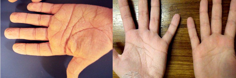
Fetal trisomy 18 is the second most common multiple malformation syndrome. We present four cases of trisomy 18 with multiple sonographic abnormalities at 13 and 14 weeks of gestation. These cases demonstrated that fetal hand deformities can be a tell-tale sign of trisomy 18 with or without increased nuchal translucency at this gestation. (+info)Management of umbilical hernias associated with hepatic cirrhosis and ascites. (5/153)
A series of 35 umbilical herniorraphies in patients with cirrhosis of the liver is reported. In this group there were eight significant complications (22%) and six deaths (16%). There was no evidence in this study of increased likelihood of esophageal variceal bleeding secondary to the interruption of portosystemic collaterals in the umbilical region. An aggressive surgical approach is indicated in cirrhotic patients, with umbilical herniae complicated by incarceration, strangulation, rupture, ulceration, and leakage of ascitic fluid. On the other hand it is recommended, that repair of non-complicated umbilical herniae be delayed until the cirrhosis is stabilized, ascites has diminished and nutrition has been improved. In many instances herniorrhaphy may not be necessary after disappearance of ascites. (+info)Abdominal wall defects: two- versus three-dimensional ultrasonographic diagnosis. (6/153)
We diagnosed 12 cases of abdominal wall defects. The cases diagnosed occurred in 6 fetuses with omphalocele, 3 with gastroschisis, 2 with prune-belly syndrome, and 1 with pentalogy of Cantrell. Except for 1 case of gastroschisis first diagnosed on the basis of three-dimensional ultrasonography at 14 weeks' gestation, all cases were first detected by two-dimensional transabdominal ultrasonography and then reevaluated with three-dimensional ultrasonography using multiplanar and orthogonal plane modes. Although the original diagnosis was accurate on the basis of two-dimensional ultrasonography in 11 of 12 cases, additional information was obtained by three-dimensional scanning in all cases. Our experience suggests that in cases in which abdominal wall defects are first detected by two-dimensional ultrasonographic scanning, the additional information gained by complementary three-dimensional ultrasonographic scanning can be useful for more-efficient counseling and postnatal therapeutic planning. (+info)Prenatal sonographic diagnosis of a twinning epigastric heteropagus. (7/153)
Epigastric heteropagus is a rare type of conjoined twinning which results from an ischemic atrophy of one fetus at an early stage of gestation. We present what we believe to be the first case diagnosed antenatally at 22 weeks' gestation. The pelvis and lower limbs of the ischemic fetus (the parasite) were attached to the epigastrium of the well-developed fetus (the autosite), which had a small omphalocele. Antenatal sonography provided an accurate diagnosis, enabling unnecessary abortion to be avoided. (+info)Anuria, omphalocele, and perinatal lethality in mice lacking the CD34-related protein podocalyxin. (8/153)
Podocalyxin is a CD34-related sialomucin that is expressed at high levels by podocytes, and also by mesothelial cells, vascular endothelia, platelets, and hematopoietic stem cells. To elucidate the function of podocalyxin, we generated podocalyxin-deficient (podxl(-/)-) mice by homologous recombination. Null mice exhibit profound defects in kidney development and die within 24 hours of birth with anuric renal failure. Although podocytes are present in the glomeruli of the podxl(-/)- mice, they fail to form foot processes and slit diaphragms and instead exhibit cell--cell junctional complexes (tight and adherens junctions). The corresponding reduction in permeable, glomerular filtration surface area presumably leads to the observed block in urine production. In addition, podxl(-/)- mice frequently display herniation of the gut (omphalocele), suggesting that podocalyxin may be required for retraction of the gut from the umbilical cord during development. Hematopoietic and vascular endothelial cells develop normally in the podocalyxin-deficient mice, possibly through functional compensation by other sialomucins (such as CD34). Our results provide the first example of an essential role for a sialomucin in development and suggest that defects in podocalyxin could play a role in podocyte dysfunction in renal failure and omphalocele in humans. (+info)Inguinal hernia, also known as an inguinal rupture or groin hernia, is a protrusion of abdominal-cavity contents through the inguinal canal. The inguinal canal is a passage in the lower abdominal wall that carries the spermatic cord in males and a round ligament in females. Inguinal hernias are more common in men than women.
There are two types of inguinal hernias: direct and indirect. Direct inguinal hernias occur when the abdominal lining and/or fat push through a weakened area in the lower abdominal wall, while indirect inguinal hernias result from a congenital condition where the abdominal lining and/or fat protrude through the internal inguinal ring, a normal opening in the abdominal wall.
Inguinal hernias can cause discomfort or pain, especially during physical activities, coughing, sneezing, or straining. In some cases, incarceration or strangulation of the hernia may occur, leading to serious complications such as bowel obstruction or tissue necrosis, which require immediate medical attention.
Surgical repair is the standard treatment for inguinal hernias, and it can be performed through open or laparoscopic techniques. The goal of surgery is to return the protruding tissues to their proper position and strengthen the weakened abdominal wall with sutures or mesh reinforcement.
A hernia is a protrusion of an organ or tissue through a weakened area in the abdominal wall, often appearing as a bulge beneath the skin. This condition can occur in various parts of the body such as the groin (inguinal hernia), navel (umbilical hernia), or site of a previous surgical incision (incisional hernia). Hernias may cause discomfort or pain, especially when straining, lifting heavy objects, or during bowel movements. In some cases, they may lead to serious complications like intestinal obstruction or strangulation, requiring immediate medical attention.
A diaphragmatic hernia is a type of hernia that occurs when the abdominal organs (such as the stomach, intestines, or liver) protrude through an opening in the diaphragm, the thin muscle that separates the chest and abdominal cavities. This condition can be present at birth (congenital) or acquired due to injury or surgery.
There are two main types of diaphragmatic hernias:
1. Bochdalek hernia: This is a congenital defect that occurs when the posterior portion of the diaphragm fails to close properly during fetal development, creating an opening through which abdominal organs can move into the chest cavity. It is more common on the left side and can lead to pulmonary hypoplasia (underdevelopment of the lungs) and other complications if not detected and treated early.
2. Morgagni hernia: This is a less common type of congenital diaphragmatic hernia that occurs when there is an opening in the anterior portion of the diaphragm, allowing abdominal organs to move into the chest cavity near the sternum. It tends to be asymptomatic and may not be discovered until adulthood.
Acquired diaphragmatic hernias can result from trauma, such as a car accident or penetrating injury, which causes a tear in the diaphragm. In some cases, surgical procedures involving the abdomen or chest can also lead to a diaphragmatic hernia.
Symptoms of a diaphragmatic hernia may include difficulty breathing, chest pain, vomiting, and bowel obstruction. Treatment typically involves surgery to repair the defect in the diaphragm and return the abdominal organs to their proper position.
A ventral hernia is a type of hernia that occurs in the abdominal wall, specifically in the anterior (front) aspect. It can occur due to a weakness or defect in the abdominal wall muscles and fascia, which allows the internal organs or tissues to push through and create a bulge or swelling.
Ventral hernias can be classified into several types based on their location, size, and cause. Some of the common types include:
1. Incisional Hernia - occurs at the site of a previous surgical incision, where the abdominal wall has not healed properly or has become weakened over time.
2. Epigastric Hernia - located in the upper middle part of the abdomen, between the breastbone and the navel.
3. Umbilical Hernia - occurs around the belly button, most commonly seen in infants but can also affect adults.
4. Spigelian Hernia - a rare type of hernia that occurs lateral to the rectus sheath, usually at the level of the semilunar line.
5. Diastasis Recti - a separation of the abdominal muscles in the midline, which can lead to a ventral hernia if not treated.
Symptoms of a ventral hernia may include pain or discomfort, especially when lifting heavy objects, straining, coughing, or during physical activity. In some cases, a hernia may become incarcerated or strangulated, which requires immediate medical attention. Treatment options for ventral hernias typically involve surgical repair, either through open surgery or laparoscopic techniques.
An abdominal hernia refers to the protrusion of an organ or tissue through a weakened area in the abdominal wall, resulting in a bulge. This condition can occur due to various factors such as congenital defects, aging, obesity, pregnancy, persistent coughing, or previous surgeries that have left behind weak spots in the abdominal wall.
There are several types of abdominal hernias, including:
1. Inguinal Hernia: This is the most common type of hernia, occurring when the intestine or bladder protrudes through the inguinal canal in the lower abdomen. Inguinal hernias are more prevalent in men than women.
2. Femoral Hernia: This type of hernia occurs when the intestine or fatty tissue pushes through a weakened area near the femoral artery, located in the upper thigh region. Femoral hernias are more common in women, especially those who are pregnant or obese.
3. Incisional Hernia: This type of hernia develops at the site of a previous abdominal surgery where the abdominal muscles have weakened or failed to heal properly.
4. Umbilical Hernia: An umbilical hernia occurs when the intestine protrudes through the abdominal wall near the navel, often visible as a bulge around the belly button. This type of hernia is more common in infants but can also affect adults, particularly those who are overweight or have had multiple pregnancies.
5. Epigastric Hernia: An epigastric hernia occurs when fatty tissue protrudes through a weakened area between the breastbone and the navel. These hernias are usually small and often painless but can cause discomfort or complications if they become incarcerated or strangulated.
Abdominal hernias can vary in size, from small and barely noticeable to large and severely painful. Symptoms may include a visible bulge, localized pain or discomfort, especially when lifting heavy objects, coughing, or straining during bowel movements. In some cases, hernias may become incarcerated (trapped) or strangulated (blood supply is cut off), which can lead to severe pain, nausea, vomiting, and require immediate medical attention.
Treatment for abdominal hernias typically involves surgical repair, either through open surgery or laparoscopic techniques. The choice of procedure depends on various factors, including the size and location of the hernia, the patient's overall health, and their personal preferences. In some cases, watchful waiting may be recommended for small, asymptomatic hernias, but it is essential to consult with a healthcare professional to determine the best course of action.
A hiatal hernia is a type of hernia that occurs when a part of the stomach protrudes or squeezes through an opening (hiatus) in the diaphragm, the muscular partition between the chest and abdominal cavities. Normally, the esophagus passes through this opening to connect to the stomach, but in a hiatal hernia, a portion of the stomach also moves up into the chest cavity through the hiatus.
There are two main types of hiatal hernias: sliding and paraesophageal. In a sliding hiatal hernia, the junction between the esophagus and stomach (gastroesophageal junction) slides upward into the chest cavity, which is the most common type. Paraesophageal hiatal hernias are less common but can be more severe, as they involve the stomach herniating alongside the esophagus, potentially leading to complications like obstruction or strangulation of the blood supply to the stomach.
Many people with hiatal hernias do not experience symptoms, but some may have heartburn, acid reflux, regurgitation, difficulty swallowing, chest pain, or shortness of breath. Treatment depends on the severity and associated symptoms, ranging from lifestyle modifications and medications to surgical repair in severe cases.
A femoral hernia is a type of hernia that occurs when a portion of the abdominal wall tissue or intestine protrudes through a weakened area in the lower part of the abdominal wall, specifically at the opening of the femoral canal. This canal is located near the groin region and contains blood vessels that pass from the abdomen to the leg.
Femoral hernias are more common in women than men, particularly those who are pregnant, obese, or have a history of multiple pregnancies. Symptoms may include a visible bulge in the inner thigh or groin area, especially when standing, coughing, or straining. Pain or discomfort in the lower abdomen or groin region, particularly during physical activities, is also common.
While some femoral hernias may not cause any symptoms and can be left untreated, they have a higher risk of becoming incarcerated or strangulated compared to other types of hernias. Incarceration occurs when the protruding tissue becomes trapped and cannot be pushed back in, while strangulation happens when the blood supply to the trapped tissue is cut off, leading to tissue death if not treated promptly with surgery.
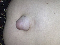
An umbilical hernia is a type of hernia that occurs at the umbilicus, or belly button. It results from a protrusion of abdominal contents through a weakened area in the abdominal wall surrounding the navel. This condition is common in newborns and infants, especially premature babies, due to incomplete closure of the abdominal muscles during development.
In most cases, umbilical hernias in children close on their own by age 3-4 or by the time they reach school age. However, if the hernia is still present after this age, surgical intervention may be required to prevent potential complications such as incarceration (where the herniated tissue becomes trapped and cannot be pushed back in) or strangulation (where the blood supply to the herniated tissue is cut off, leading to tissue death).
Adults can also develop umbilical hernias, often as a result of increased pressure in the abdomen due to obesity, pregnancy, heavy lifting, or persistent coughing. Umbilical hernias in adults are generally more likely to require surgical repair due to the higher risk of complications.
The umbilical veins are blood vessels in the umbilical cord that carry oxygenated and nutrient-rich blood from the mother to the developing fetus during pregnancy. There are typically two umbilical veins, one of which usually degenerates and becomes obliterated, leaving a single functional vein. This remaining vein is known as the larger umbilical vein or the venous duct. It enters the fetal abdomen through the umbilicus and passes through the liver, where it branches off to form the portal sinus. Ultimately, the blood from the umbilical vein mixes with the blood from the inferior vena cava and is pumped to the heart through the right atrium.
It's important to note that after birth, the umbilical veins are no longer needed and undergo involution, becoming the ligamentum teres in the adult.
The umbilical arteries are a pair of vessels that develop within the umbilical cord during fetal development. They carry oxygenated and nutrient-rich blood from the mother to the developing fetus through the placenta. These arteries arise from the internal iliac arteries in the fetus and pass through the umbilical cord to connect with the two umbilical veins within the placenta. After birth, the umbilical arteries become ligaments (the medial umbilical ligaments) that run along the inner abdominal wall.
Herniorrhaphy is a surgical procedure where the herniated tissue or organ is placed back into its original position, and the weakened or damaged muscle wall is repaired. This is typically done to correct a hernia, which is a protrusion of an organ or tissue through a weakened area in the abdominal wall. The surgical incision may be closed with sutures or staples, and sometimes a mesh patch is used to reinforce the repair.
Surgical mesh is a medical device that is used in various surgical procedures, particularly in reconstructive surgery, to provide additional support to weakened or damaged tissues. It is typically made from synthetic materials such as polypropylene or polyester, or from biological materials such as animal tissue or human cadaveric tissue.
The mesh is designed to be implanted into the body, where it can help to reinforce and repair damaged tissues. For example, it may be used in hernia repairs to support the weakened abdominal wall, or in pelvic floor reconstruction surgery to treat conditions such as pelvic organ prolapse or stress urinary incontinence.
Surgical mesh can come in different forms, including sheets, plugs, and patches, and may be either absorbable or non-absorbable. The choice of mesh material and type will depend on the specific surgical indication and the patient's individual needs. It is important for patients to discuss the risks and benefits of surgical mesh with their healthcare provider before undergoing any surgical procedure that involves its use.
A diaphragmatic hernia is a condition in which there is abnormal displacement or protrusion of abdominal organs into the thoracic cavity through an opening or defect in the diaphragm. In traumatic diaphragmatic hernia, this disruption of the diaphragm is caused by trauma, such as a penetrating injury or blunt force trauma from a car accident or fall. The resulting herniation can lead to various complications, including difficulty breathing, digestive problems, and even organ dysfunction. Immediate medical attention is necessary for proper diagnosis and treatment of traumatic diaphragmatic hernia.
A obturator hernia is a type of pelvic hernia that occurs through the obturator foramen, which is an opening in the bony pelvis through which blood vessels and nerves pass. This type of hernia is relatively uncommon and primarily affects women, particularly those who are elderly, thin, or have had multiple pregnancies.
Obturator hernias occur when abdominal contents, such as fat or intestine, protrude through the obturator foramen and become trapped, leading to obstruction of the intestine or strangulation, which can cut off blood flow to the affected tissue. Symptoms may include pain in the thigh or groin, nausea, vomiting, and constipation.
Obturator hernias can be difficult to diagnose due to their nonspecific symptoms and rarity. Diagnosis is often made during surgery to repair the hernia. Treatment typically involves surgical repair of the hernia, which may be performed through an open incision or laparoscopically. Delayed diagnosis and treatment can lead to serious complications, such as bowel necrosis or perforation, so prompt medical attention is necessary if obturator hernia is suspected.
I'm sorry for any confusion, but "Polypropylenes" does not have a medical definition as it is not a medical term. Polypropylene is a type of plastic that is used in various medical and non-medical applications. It is a thermoplastic polymer resin that is produced by the polymerization of propylene.
In the medical field, polypropylene is sometimes used to make single-use surgical instruments, sutures, and medical devices due to its resistance to heat, chemicals, and electricity. It is also biocompatible, meaning it can be safely used in the body without causing adverse reactions. However, "Polypropylenes" as a medical term is not recognized or used in the medical community.
Laparoscopy is a surgical procedure that involves the insertion of a laparoscope, which is a thin tube with a light and camera attached to it, through small incisions in the abdomen. This allows the surgeon to view the internal organs without making large incisions. It's commonly used to diagnose and treat various conditions such as endometriosis, ovarian cysts, infertility, and appendicitis. The advantages of laparoscopy over traditional open surgery include smaller incisions, less pain, shorter hospital stays, and quicker recovery times.
Fetal blood refers to the blood circulating in a fetus during pregnancy. It is essential for the growth and development of the fetus, as it carries oxygen and nutrients from the placenta to the developing tissues and organs. Fetal blood also removes waste products, such as carbon dioxide, from the fetal tissues and transports them to the placenta for elimination.
Fetal blood has several unique characteristics that distinguish it from adult blood. For example, fetal hemoglobin (HbF) is the primary type of hemoglobin found in fetal blood, whereas adults primarily have adult hemoglobin (HbA). Fetal hemoglobin has a higher affinity for oxygen than adult hemoglobin, which allows it to more efficiently extract oxygen from the maternal blood in the placenta.
Additionally, fetal blood contains a higher proportion of reticulocytes (immature red blood cells) and nucleated red blood cells compared to adult blood. These differences reflect the high turnover rate of red blood cells in the developing fetus and the need for rapid growth and development.
Examination of fetal blood can provide important information about the health and well-being of the fetus during pregnancy. For example, fetal blood sampling (also known as cordocentesis or percutaneous umbilical blood sampling) can be used to diagnose genetic disorders, infections, and other conditions that may affect fetal development. However, this procedure carries risks, including preterm labor, infection, and fetal loss, and is typically only performed when there is a significant risk of fetal compromise or when other diagnostic tests have been inconclusive.
The abdominal wall refers to the group of muscles, fascia (sheaths of connective tissue), and skin that make up the front and sides of the abdomen, extending from the thorax (chest) to the pelvis. It provides protection to the abdominal organs, supports the trunk, and allows for movement of the torso.
The main muscles of the anterior abdominal wall include:
1. Rectus sheaths (Rectus Abdominis): paired vertical muscles running from the pubic symphysis to the xiphoid process and costal cartilages of ribs 5-7.
2. External obliques: thin, irregular muscles that lie over the lower part of the abdomen and run diagonally downward and forward from the lower ribs to the iliac crest (pelvic bone) and pubic tubercle.
3. Internal obliques: thicker muscles that lie under the external obliques, running diagonally upward and forward from the iliac crest to the lower ribs.
4. Transverse abdominis: deepest of the abdominal muscles, lying horizontally across the abdomen, attaching from the lower ribs to the pelvis.
These muscles are interconnected by various layers of fascia and aponeuroses (flat, broad tendons), forming a complex structure that allows for both stability and mobility. The linea alba, a fibrous band, runs down the midline of the anterior abdominal wall, connecting the rectus sheaths.
Damage to the abdominal wall can occur due to trauma, surgery, or various medical conditions, which may require surgical intervention for repair.
The umbilicus, also known as the navel, is the scar left on the abdominal wall after the removal of the umbilical cord in a newborn. The umbilical cord connects the developing fetus to the placenta in the uterus during pregnancy, providing essential nutrients and oxygen while removing waste products. After birth, the cord is clamped and cut, leaving behind a small stump that eventually dries up and falls off, leaving the umbilicus. In adults, it typically appears as a slight depression or dimple on the abdomen.
Human Umbilical Vein Endothelial Cells (HUVECs) are a type of primary cells that are isolated from the umbilical cord vein of human placenta. These cells are naturally equipped with endothelial properties and functions, making them an essential tool in biomedical research. HUVECs line the interior surface of blood vessels and play a crucial role in the regulation of vascular function, including angiogenesis (the formation of new blood vessels), coagulation, and permeability. Due to their accessibility and high proliferation rate, HUVECs are widely used in various research areas such as vascular biology, toxicology, drug development, and gene therapy.
Pregnancy is a physiological state or condition where a fertilized egg (zygote) successfully implants and grows in the uterus of a woman, leading to the development of an embryo and finally a fetus. This process typically spans approximately 40 weeks, divided into three trimesters, and culminates in childbirth. Throughout this period, numerous hormonal and physical changes occur to support the growing offspring, including uterine enlargement, breast development, and various maternal adaptations to ensure the fetus's optimal growth and well-being.
Prenatal ultrasonography, also known as obstetric ultrasound, is a medical diagnostic procedure that uses high-frequency sound waves to create images of the developing fetus, placenta, and amniotic fluid inside the uterus. It is a non-invasive and painless test that is widely used during pregnancy to monitor the growth and development of the fetus, detect any potential abnormalities or complications, and determine the due date.
During the procedure, a transducer (a small handheld device) is placed on the mother's abdomen and moved around to capture images from different angles. The sound waves travel through the mother's body and bounce back off the fetus, producing echoes that are then converted into electrical signals and displayed as images on a screen.
Prenatal ultrasonography can be performed at various stages of pregnancy, including early pregnancy to confirm the pregnancy and detect the number of fetuses, mid-pregnancy to assess the growth and development of the fetus, and late pregnancy to evaluate the position of the fetus and determine if it is head down or breech. It can also be used to guide invasive procedures such as amniocentesis or chorionic villus sampling.
Overall, prenatal ultrasonography is a valuable tool in modern obstetrics that helps ensure the health and well-being of both the mother and the developing fetus.
Suture techniques refer to the various methods used by surgeons to sew or stitch together tissues in the body after an injury, trauma, or surgical incision. The main goal of suturing is to approximate and hold the edges of the wound together, allowing for proper healing and minimizing scar formation.
There are several types of suture techniques, including:
1. Simple Interrupted Suture: This is one of the most basic suture techniques where the needle is passed through the tissue at a right angle, creating a loop that is then tightened to approximate the wound edges. Multiple stitches are placed along the length of the incision or wound.
2. Continuous Locking Suture: In this technique, the needle is passed continuously through the tissue in a zigzag pattern, with each stitch locking into the previous one. This creates a continuous line of sutures that provides strong tension and support to the wound edges.
3. Running Suture: Similar to the continuous locking suture, this technique involves passing the needle continuously through the tissue in a straight line. However, instead of locking each stitch, the needle is simply passed through the previous loop before being tightened. This creates a smooth and uninterrupted line of sutures that can be easily removed after healing.
4. Horizontal Mattress Suture: In this technique, two parallel stitches are placed horizontally across the wound edges, creating a "mattress" effect that provides additional support and tension to the wound. This is particularly useful in deep or irregularly shaped wounds.
5. Vertical Mattress Suture: Similar to the horizontal mattress suture, this technique involves placing two parallel stitches vertically across the wound edges. This creates a more pronounced "mattress" effect that can help reduce tension and minimize scarring.
6. Subcuticular Suture: In this technique, the needle is passed just below the surface of the skin, creating a smooth and barely visible line of sutures. This is particularly useful in cosmetic surgery or areas where minimizing scarring is important.
The choice of suture technique depends on various factors such as the location and size of the wound, the type of tissue involved, and the patient's individual needs and preferences. Proper suture placement and tension are crucial for optimal healing and aesthetic outcomes.
A fascia is a band or sheet of connective tissue, primarily collagen, that covers, connects, and separates muscles, organs, and other structures in the body. It provides support and stability, allows for smooth movement between structures, and has the ability to transmit forces throughout the body. Fascia is found throughout the body, and there are several layers of it, including superficial fascia, deep fascia, and visceral fascia. Injury, inflammation, or strain to the fascia can cause pain and restriction of movement.
The endothelium is a thin layer of simple squamous epithelial cells that lines the interior surface of blood vessels, lymphatic vessels, and heart chambers. The vascular endothelium, specifically, refers to the endothelial cells that line the blood vessels. These cells play a crucial role in maintaining vascular homeostasis by regulating vasomotor tone, coagulation, platelet activation, inflammation, and permeability of the vessel wall. They also contribute to the growth and repair of the vascular system and are involved in various pathological processes such as atherosclerosis, hypertension, and diabetes.
A laparotomy is a surgical procedure that involves making an incision in the abdominal wall to gain access to the abdominal cavity. This procedure is typically performed to diagnose and treat various conditions such as abdominal trauma, tumors, infections, or inflammatory diseases. The size of the incision can vary depending on the reason for the surgery and the extent of the condition being treated. Once the procedure is complete, the incision is closed with sutures or staples.
The term "laparotomy" comes from the Greek words "lapara," which means "flank" or "side," and "tome," which means "to cut." Together, they describe the surgical procedure that involves cutting into the abdomen to examine its contents.
Cord blood stem cell transplantation is a medical procedure that involves the infusion of stem cells derived from the umbilical cord blood into a patient. These stem cells, specifically hematopoietic stem cells, have the ability to differentiate into various types of blood cells, including red and white blood cells and platelets.
Cord blood stem cell transplantation is often used as a treatment for patients with various malignant and non-malignant disorders, such as leukemia, lymphoma, sickle cell disease, and metabolic disorders. The procedure involves collecting cord blood from the umbilical cord and placenta after the birth of a baby, processing and testing it for compatibility with the recipient's immune system, and then infusing it into the patient through a vein in a process similar to a blood transfusion.
The advantages of using cord blood stem cells include their availability, low risk of transmission of infectious diseases, and reduced risk of graft-versus-host disease compared to other sources of hematopoietic stem cells, such as bone marrow or peripheral blood. However, the number of stem cells in a cord blood unit is generally lower than that found in bone marrow or peripheral blood, which can limit its use in some patients, particularly adults.
Overall, cord blood stem cell transplantation is an important and promising area of regenerative medicine, offering hope for patients with a wide range of disorders.
In medical terms, sutures are specialized surgical threads made from various materials such as absorbable synthetic or natural fibers, or non-absorbable materials like nylon or silk. They are used to approximate and hold together the edges of a wound or incision in the skin or other tissues during the healing process. Sutures come in different sizes, types, and shapes, each designed for specific uses and techniques depending on the location and type of tissue being sutured. Properly placed sutures help to promote optimal healing, minimize scarring, and reduce the risk of infection or other complications.
Intestinal obstruction, also known as bowel obstruction, is a medical condition characterized by a blockage that prevents the normal flow of contents through the small intestine or large intestine (colon). This blockage can be caused by various factors such as tumors, adhesions (scar tissue), hernias, inflammation, or impacted feces.
The obstruction can be mechanical, where something physically blocks the intestinal lumen, or functional, where the normal muscular contractions of the bowel are impaired. Mechanical obstructions are more common than functional ones.
Symptoms of intestinal obstruction may include abdominal pain and cramping, nausea and vomiting, bloating, inability to pass gas or have a bowel movement, and abdominal distention. If left untreated, intestinal obstruction can lead to serious complications such as tissue death (necrosis), perforation of the intestine, and sepsis. Treatment typically involves hospitalization, intravenous fluids, nasogastric decompression, and possibly surgery to remove the obstruction.
A newborn infant is a baby who is within the first 28 days of life. This period is also referred to as the neonatal period. Newborns require specialized care and attention due to their immature bodily systems and increased vulnerability to various health issues. They are closely monitored for signs of well-being, growth, and development during this critical time.
A surgical stoma, also known simply as a stoma, is a surgically created opening on the surface of the body that allows for the passage of bodily waste. This procedure is typically performed when a person has a malfunctioning or diseased organ in the digestive or urinary system that cannot be effectively treated or repaired.
In a colostomy or ileostomy, which are common types of surgical stomas, a portion of the colon or small intestine is brought through an opening in the abdominal wall to create a new pathway for waste to exit the body. The stoma may be temporary or permanent, depending on the underlying condition and the success of any additional treatments.
After surgery, patients with a stoma will need to wear a pouching system to collect and contain the waste that is expelled through the stoma. This can take some getting used to, but with proper care and support, most people are able to adjust to life with a stoma and maintain a good quality of life.
In medical terms, the "groin" refers to the area where the lower abdomen meets the thigh. It is located on both sides of the body, in front of the upper part of each leg. The groin contains several important structures such as the inguinal canal, which contains blood vessels and nerves, and the femoral artery and vein, which supply blood to and from the lower extremities. Issues in this region, such as pain or swelling, may indicate a variety of medical conditions, including muscle strains, hernias, or infections.
Postoperative complications refer to any unfavorable condition or event that occurs during the recovery period after a surgical procedure. These complications can vary in severity and may include, but are not limited to:
1. Infection: This can occur at the site of the incision or inside the body, such as pneumonia or urinary tract infection.
2. Bleeding: Excessive bleeding (hemorrhage) can lead to a drop in blood pressure and may require further surgical intervention.
3. Blood clots: These can form in the deep veins of the legs (deep vein thrombosis) and can potentially travel to the lungs (pulmonary embolism).
4. Wound dehiscence: This is when the surgical wound opens up, which can lead to infection and further complications.
5. Pulmonary issues: These include atelectasis (collapsed lung), pneumonia, or respiratory failure.
6. Cardiovascular problems: These include abnormal heart rhythms (arrhythmias), heart attack, or stroke.
7. Renal failure: This can occur due to various reasons such as dehydration, blood loss, or the use of certain medications.
8. Pain management issues: Inadequate pain control can lead to increased stress, anxiety, and decreased mobility.
9. Nausea and vomiting: These can be caused by anesthesia, opioid pain medication, or other factors.
10. Delirium: This is a state of confusion and disorientation that can occur in the elderly or those with certain medical conditions.
Prompt identification and management of these complications are crucial to ensure the best possible outcome for the patient.
Fetal diseases are medical conditions or abnormalities that affect a fetus during pregnancy. These diseases can be caused by genetic factors, environmental influences, or a combination of both. They can range from mild to severe and may impact various organ systems in the developing fetus. Examples of fetal diseases include congenital heart defects, neural tube defects, chromosomal abnormalities such as Down syndrome, and infectious diseases such as toxoplasmosis or rubella. Fetal diseases can be diagnosed through prenatal testing, including ultrasound, amniocentesis, and chorionic villus sampling. Treatment options may include medication, surgery, or delivery of the fetus, depending on the nature and severity of the disease.
Gestational age is the length of time that has passed since the first day of the last menstrual period (LMP) in pregnant women. It is the standard unit used to estimate the age of a pregnancy and is typically expressed in weeks. This measure is used because the exact date of conception is often not known, but the start of the last menstrual period is usually easier to recall.
It's important to note that since ovulation typically occurs around two weeks after the start of the LMP, gestational age is approximately two weeks longer than fetal age, which is the actual time elapsed since conception. Medical professionals use both gestational and fetal age to track the development and growth of the fetus during pregnancy.
"Cells, cultured" is a medical term that refers to cells that have been removed from an organism and grown in controlled laboratory conditions outside of the body. This process is called cell culture and it allows scientists to study cells in a more controlled and accessible environment than they would have inside the body. Cultured cells can be derived from a variety of sources, including tissues, organs, or fluids from humans, animals, or cell lines that have been previously established in the laboratory.
Cell culture involves several steps, including isolation of the cells from the tissue, purification and characterization of the cells, and maintenance of the cells in appropriate growth conditions. The cells are typically grown in specialized media that contain nutrients, growth factors, and other components necessary for their survival and proliferation. Cultured cells can be used for a variety of purposes, including basic research, drug development and testing, and production of biological products such as vaccines and gene therapies.
It is important to note that cultured cells may behave differently than they do in the body, and results obtained from cell culture studies may not always translate directly to human physiology or disease. Therefore, it is essential to validate findings from cell culture experiments using additional models and ultimately in clinical trials involving human subjects.
Endothelial cells are the type of cells that line the inner surface of blood vessels, lymphatic vessels, and heart chambers. They play a crucial role in maintaining vascular homeostasis by controlling vasomotor tone, coagulation, platelet activation, and inflammation. Endothelial cells also regulate the transport of molecules between the blood and surrounding tissues, and contribute to the maintenance of the structural integrity of the vasculature. They are flat, elongated cells with a unique morphology that allows them to form a continuous, nonthrombogenic lining inside the vessels. Endothelial cells can be isolated from various tissues and cultured in vitro for research purposes.
Recurrence, in a medical context, refers to the return of symptoms or signs of a disease after a period of improvement or remission. It indicates that the condition has not been fully eradicated and may require further treatment. Recurrence is often used to describe situations where a disease such as cancer comes back after initial treatment, but it can also apply to other medical conditions. The likelihood of recurrence varies depending on the type of disease and individual patient factors.
The rectus abdominis is a paired, flat, and long muscle in the anterior (front) wall of the abdomen. It runs from the pubic symphysis (the joint where the two pubic bones meet in the front of the pelvis) to the xiphoid process (the lower end of the sternum or breastbone) and costal cartilages of the fifth, sixth, and seventh ribs.
The rectus abdominis is responsible for flexing the lumbar spine (lower back), which helps in bending forward or sitting up from a lying down position. It also contributes to maintaining proper posture and stabilizing the pelvis and spine. The muscle's visibility, especially in its lower portion, is often associated with a "six-pack" appearance in well-trained individuals.
Ultrasonography, Doppler refers to a non-invasive diagnostic medical procedure that uses high-frequency sound waves to create real-time images of the movement of blood flow through vessels, tissues, or heart valves. The Doppler effect is used to measure the frequency shift of the ultrasound waves as they bounce off moving red blood cells, which allows for the calculation of the speed and direction of blood flow. This technique is commonly used to diagnose and monitor various conditions such as deep vein thrombosis, carotid artery stenosis, heart valve abnormalities, and fetal heart development during pregnancy. It does not use radiation or contrast agents and is considered safe with minimal risks.
Local anesthesia is a type of anesthesia that numbs a specific area of the body, blocking pain signals from that particular region while allowing the person to remain conscious and alert. It is typically achieved through the injection or application of a local anesthetic drug, which works by temporarily inhibiting the function of nerve fibers carrying pain sensations. Common examples of local anesthetics include lidocaine, prilocaine, and bupivacaine.
Local anesthesia is commonly used for minor surgical procedures, dental work, or other medical interventions where only a small area needs to be numbed. It can also be employed as part of a combined anesthetic technique, such as in conjunction with sedation or regional anesthesia, to provide additional pain relief and increase patient comfort during more extensive surgeries.
The duration of local anesthesia varies depending on the type and dosage of the anesthetic agent used; some last for just a few hours, while others may provide numbness for up to several days. Overall, local anesthesia is considered a safe and effective method for managing pain during various medical procedures.
Treatment outcome is a term used to describe the result or effect of medical treatment on a patient's health status. It can be measured in various ways, such as through symptoms improvement, disease remission, reduced disability, improved quality of life, or survival rates. The treatment outcome helps healthcare providers evaluate the effectiveness of a particular treatment plan and make informed decisions about future care. It is also used in clinical research to compare the efficacy of different treatments and improve patient care.
A fetus is the developing offspring in a mammal, from the end of the embryonic period (approximately 8 weeks after fertilization in humans) until birth. In humans, the fetal stage of development starts from the eleventh week of pregnancy and continues until childbirth, which is termed as full-term pregnancy at around 37 to 40 weeks of gestation. During this time, the organ systems become fully developed and the body grows in size. The fetus is surrounded by the amniotic fluid within the amniotic sac and is connected to the placenta via the umbilical cord, through which it receives nutrients and oxygen from the mother. Regular prenatal care is essential during this period to monitor the growth and development of the fetus and ensure a healthy pregnancy and delivery.
Single umbilical artery (SUA) is a congenital abnormality characterized by the presence of only one umbilical artery in the developing fetus, instead of the usual two. The umbilical cord typically contains two umbilical arteries and one umbilical vein, which are responsible for carrying oxygenated and nutrient-rich blood from the placenta to the growing fetus, as well as transporting deoxygenated and waste-laden blood back to the placenta.
The occurrence of a single umbilical artery can be an isolated finding or associated with other structural or chromosomal abnormalities. The exact cause of SUA is not fully understood, but it has been linked to factors such as maternal age, diabetes, and genetic predisposition.
During prenatal ultrasound examinations, the detection of a single umbilical artery might prompt further evaluation for potential associated anomalies or genetic disorders. In some cases, SUA may not have any significant consequences on fetal development or pregnancy outcomes; however, it can increase the risk for complications such as intrauterine growth restriction, preterm birth, and stillbirth. Therefore, close monitoring of the pregnancy is often recommended when a single umbilical artery is identified.
Postoperative pain is defined as the pain or discomfort experienced by patients following a surgical procedure. It can vary in intensity and duration depending on the type of surgery performed, individual pain tolerance, and other factors. The pain may be caused by tissue trauma, inflammation, or nerve damage resulting from the surgical intervention. Proper assessment and management of postoperative pain is essential to promote recovery, prevent complications, and improve patient satisfaction.
Ambulatory surgical procedures, also known as outpatient or same-day surgery, refer to medical operations that do not require an overnight hospital stay. These procedures are typically performed in a specialized ambulatory surgery center (ASC) or in a hospital-based outpatient department. Patients undergoing ambulatory surgical procedures receive anesthesia, undergo the operation, and recover enough to be discharged home on the same day of the procedure.
Examples of common ambulatory surgical procedures include:
1. Arthroscopy (joint scope examination and repair)
2. Cataract surgery
3. Colonoscopy and upper endoscopy
4. Dental surgery, such as wisdom tooth extraction
5. Gallbladder removal (cholecystectomy)
6. Hernia repair
7. Hysteroscopy (examination of the uterus)
8. Minor skin procedures, like biopsies and lesion removals
9. Orthopedic procedures, such as carpal tunnel release or joint injections
10. Pain management procedures, including epidural steroid injections and nerve blocks
11. Podiatric (foot and ankle) surgery
12. Tonsillectomy and adenoidectomy
Advancements in medical technology, minimally invasive surgical techniques, and improved anesthesia methods have contributed to the growth of ambulatory surgical procedures, offering patients a more convenient and cost-effective alternative to traditional inpatient surgeries.
Fetal growth retardation, also known as intrauterine growth restriction (IUGR), is a condition in which a fetus fails to grow at the expected rate during pregnancy. This can be caused by various factors such as maternal health problems, placental insufficiency, chromosomal abnormalities, and genetic disorders. The fetus may be smaller than expected for its gestational age, have reduced movement, and may be at risk for complications during labor and delivery. It is important to monitor fetal growth and development closely throughout pregnancy to detect any potential issues early on and provide appropriate medical interventions.
The round ligament is a cord-like structure in the female pelvis that extends from the uterus to the labia majora. It is one of the major ligaments that support the uterus and helps to maintain its position within the pelvis. The round ligament is composed of fibrous tissue and smooth muscle, and it plays a role in maintaining the tone and shape of the uterus.
During pregnancy, the round ligament can become stretched and thickened as the uterus grows and expands. This can sometimes cause discomfort or pain, particularly on one side of the pelvis. In some cases, the round ligament may also contribute to the development of certain gynecological conditions, such as uterine prolapse or urinary incontinence.
It is important for healthcare providers to consider the round ligament when evaluating and treating female reproductive health issues, as it can have a significant impact on the function and positioning of the uterus and other pelvic organs.
Operative surgical procedures refer to medical interventions that involve manual manipulation of tissues, structures, or organs in the body, typically performed in an operating room setting under sterile conditions. These procedures are carried out with the use of specialized instruments, such as scalpels, forceps, and scissors, and may require regional or general anesthesia to ensure patient comfort and safety.
Operative surgical procedures can range from relatively minor interventions, such as a biopsy or the removal of a small lesion, to more complex and extensive surgeries, such as open heart surgery or total joint replacement. The specific goals of operative surgical procedures may include the diagnosis and treatment of medical conditions, the repair or reconstruction of damaged tissues or organs, or the prevention of further disease progression.
Regardless of the type or complexity of the procedure, all operative surgical procedures require careful planning, execution, and postoperative management to ensure the best possible outcomes for patients.
Placental circulation refers to the specialized circulatory system that develops during pregnancy to allow for the exchange of nutrients, oxygen, and waste products between the mother's blood and the fetal blood in the placenta. The placenta is a highly vascular organ that grows within the uterus and is connected to the developing fetus via the umbilical cord.
In the maternal side of the placenta, the spiral arteries branch into smaller vessels called the intervillous spaces, where they come in close contact with the fetal blood vessels within the villi (finger-like projections) of the placenta. The intervillous spaces are filled with maternal blood that flows around the villi, allowing for the exchange of gases and nutrients between the two circulations.
On the fetal side, the umbilical cord contains two umbilical arteries that carry oxygen-depleted blood from the fetus to the placenta, and one umbilical vein that returns oxygenated blood back to the fetus. The umbilical arteries branch into smaller vessels within the villi, where they exchange gases and nutrients with the maternal blood in the intervillous spaces.
Overall, the placental circulation is a crucial component of fetal development, allowing for the growing fetus to receive the necessary oxygen and nutrients to support its growth and development.
A laparoscope is a type of medical instrument called an endoscope, which is used to examine the interior of a body cavity or organ. Specifically, a laparoscope is a long, thin tube with a high-intensity light and a high-resolution camera attached to it. This device allows surgeons to view the abdominal cavity through small incisions, without having to make large, invasive cuts.
During a laparoscopic procedure, the surgeon will insert the laparoscope through a small incision in the abdomen, typically near the navel. The camera sends images back to a monitor, giving the surgeon a clear view of the organs and tissues inside the body. This allows for more precise and less invasive surgical procedures, often resulting in faster recovery times and fewer complications compared to traditional open surgery.
Laparoscopes are commonly used in a variety of surgical procedures, including:
1. Gynecological surgeries (e.g., hysterectomies, ovarian cyst removals)
2. Gallbladder removal (cholecystectomy)
3. Gastrointestinal surgeries (e.g., removing benign or malignant tumors)
4. Hernia repairs
5. Bariatric surgeries for weight loss (e.g., gastric bypass, sleeve gastrectomy)
While laparoscopes provide numerous benefits over open surgery, they still require specialized training and expertise to use effectively and safely.
Retrospective studies, also known as retrospective research or looking back studies, are a type of observational study that examines data from the past to draw conclusions about possible causal relationships between risk factors and outcomes. In these studies, researchers analyze existing records, medical charts, or previously collected data to test a hypothesis or answer a specific research question.
Retrospective studies can be useful for generating hypotheses and identifying trends, but they have limitations compared to prospective studies, which follow participants forward in time from exposure to outcome. Retrospective studies are subject to biases such as recall bias, selection bias, and information bias, which can affect the validity of the results. Therefore, retrospective studies should be interpreted with caution and used primarily to generate hypotheses for further testing in prospective studies.
I'm sorry for any confusion, but "Phenyl Ethers" is not a recognized medical term. Phenyl ethers are a class of organic compounds consisting of an ether with a phenyl group as one of the components. They are widely used in industry and research, including as solvents, intermediates in chemical synthesis, and pharmaceuticals.
However, if you have any concerns about exposure to certain chemicals or their effects on health, it would be best to consult with a medical professional who can provide advice based on your specific situation and symptoms.
Fundoplication is a surgical procedure in which the upper part of the stomach (the fundus) is wrapped around the lower esophagus and then stitched into place. This procedure strengthens the lower esophageal sphincter, which helps prevent acid reflux from the stomach into the esophagus. It is commonly used to treat gastroesophageal reflux disease (GERD) and paraesophageal hernias.
Gastroesophageal reflux (GER) is the retrograde movement of stomach contents into the esophagus, which can cause discomfort and symptoms. It occurs when the lower esophageal sphincter (a ring of muscle between the esophagus and stomach) relaxes inappropriately, allowing the acidic or non-acidic gastric contents to flow back into the esophagus.
Gastroesophageal reflux becomes gastroesophageal reflux disease (GERD) when it is more severe, persistent, and/or results in complications such as esophagitis, strictures, or Barrett's esophagus. Common symptoms of GERD include heartburn, regurgitation, chest pain, difficulty swallowing, and chronic cough or hoarseness.
Surgical wound dehiscence is a medical condition that refers to the partial or complete separation of layers of a surgical incision after a surgical procedure, leading to the disruption of the wound closure. This can occur due to various factors such as infection, poor nutrition, increased tension on the sutures, hematoma or seroma formation, and patient's underlying health conditions like diabetes or immunodeficiency. Dehiscence may result in the exposure of internal tissues and organs, potentially causing severe complications such as infection, bleeding, or organ dysfunction. Immediate medical attention is required to manage this condition and prevent further complications.
Prospective studies, also known as longitudinal studies, are a type of cohort study in which data is collected forward in time, following a group of individuals who share a common characteristic or exposure over a period of time. The researchers clearly define the study population and exposure of interest at the beginning of the study and follow up with the participants to determine the outcomes that develop over time. This type of study design allows for the investigation of causal relationships between exposures and outcomes, as well as the identification of risk factors and the estimation of disease incidence rates. Prospective studies are particularly useful in epidemiology and medical research when studying diseases with long latency periods or rare outcomes.
A testicular hydrocele is a type of fluid-filled sac that forms around the testicle (testis), typically in the scrotum. This sac, known as the tunica vaginalis, normally contains a small amount of fluid that helps to lubricate and protect the testicle. However, when an excessive amount of fluid accumulates in this sac, it results in the formation of a hydrocele.
Testicular hydroceles can be congenital (present at birth) or acquired later in life due to various reasons such as injury, inflammation, or infection in the scrotal area. They are usually painless but may cause discomfort or a feeling of heaviness in the scrotum, especially when they become large. In some cases, hydroceles may resolve on their own without treatment, while others may require surgical intervention to drain the fluid and repair the underlying issue.
It is essential to differentiate between hydroceles and other conditions with similar symptoms, such as hernias or tumors, which may require more urgent medical attention. A healthcare professional can perform a physical examination and possibly recommend further testing, like an ultrasound, to confirm the diagnosis of a testicular hydrocele.
A urachal cyst is a rare type of abdominal wall defect that results from the persistent embryonic remnant of the urachus, which is a canal-like structure that connects the bladder to the umbilicus (belly button) during fetal development. This canal normally obliterates and becomes a fibrous cord known as the median umbilical ligament after birth. However, if it fails to do so, it can result in the formation of various urachal anomalies, including a urachal cyst.
A urachal cyst is a fluid-filled sac that forms along any part of the urachus, usually located between the bladder and the umbilicus. These cysts are typically asymptomatic but can become infected or inflamed, leading to symptoms such as abdominal pain, tenderness, fever, and a palpable mass in the lower abdomen. In some cases, urachal cysts may also cause urinary tract infections or bladder irritation. Diagnosis is usually made through imaging studies such as ultrasound, CT scan, or MRI, and treatment typically involves surgical excision of the cyst to prevent complications.
In the field of medicine, "time factors" refer to the duration of symptoms or time elapsed since the onset of a medical condition, which can have significant implications for diagnosis and treatment. Understanding time factors is crucial in determining the progression of a disease, evaluating the effectiveness of treatments, and making critical decisions regarding patient care.
For example, in stroke management, "time is brain," meaning that rapid intervention within a specific time frame (usually within 4.5 hours) is essential to administering tissue plasminogen activator (tPA), a clot-busting drug that can minimize brain damage and improve patient outcomes. Similarly, in trauma care, the "golden hour" concept emphasizes the importance of providing definitive care within the first 60 minutes after injury to increase survival rates and reduce morbidity.
Time factors also play a role in monitoring the progression of chronic conditions like diabetes or heart disease, where regular follow-ups and assessments help determine appropriate treatment adjustments and prevent complications. In infectious diseases, time factors are crucial for initiating antibiotic therapy and identifying potential outbreaks to control their spread.
Overall, "time factors" encompass the significance of recognizing and acting promptly in various medical scenarios to optimize patient outcomes and provide effective care.
Follow-up studies are a type of longitudinal research that involve repeated observations or measurements of the same variables over a period of time, in order to understand their long-term effects or outcomes. In medical context, follow-up studies are often used to evaluate the safety and efficacy of medical treatments, interventions, or procedures.
In a typical follow-up study, a group of individuals (called a cohort) who have received a particular treatment or intervention are identified and then followed over time through periodic assessments or data collection. The data collected may include information on clinical outcomes, adverse events, changes in symptoms or functional status, and other relevant measures.
The results of follow-up studies can provide important insights into the long-term benefits and risks of medical interventions, as well as help to identify factors that may influence treatment effectiveness or patient outcomes. However, it is important to note that follow-up studies can be subject to various biases and limitations, such as loss to follow-up, recall bias, and changes in clinical practice over time, which must be carefully considered when interpreting the results.
Blood flow velocity is the speed at which blood travels through a specific part of the vascular system. It is typically measured in units of distance per time, such as centimeters per second (cm/s) or meters per second (m/s). Blood flow velocity can be affected by various factors, including cardiac output, vessel diameter, and viscosity of the blood. Measuring blood flow velocity is important in diagnosing and monitoring various medical conditions, such as heart disease, stroke, and peripheral vascular disease.
The placenta is an organ that develops in the uterus during pregnancy and provides oxygen and nutrients to the growing baby through the umbilical cord. It also removes waste products from the baby's blood. The placenta attaches to the wall of the uterus, and the baby's side of the placenta contains many tiny blood vessels that connect to the baby's circulatory system. This allows for the exchange of oxygen, nutrients, and waste between the mother's and baby's blood. After the baby is born, the placenta is usually expelled from the uterus in a process called afterbirth.
Pregnancy outcome refers to the final result or status of a pregnancy, including both the health of the mother and the newborn baby. It can be categorized into various types such as:
1. Live birth: The delivery of one or more babies who show signs of life after separation from their mother.
2. Stillbirth: The delivery of a baby who has died in the womb after 20 weeks of pregnancy.
3. Miscarriage: The spontaneous loss of a pregnancy before the 20th week.
4. Abortion: The intentional termination of a pregnancy before the fetus can survive outside the uterus.
5. Ectopic pregnancy: A pregnancy that develops outside the uterus, usually in the fallopian tube, which is not viable and requires medical attention.
6. Preterm birth: The delivery of a baby before 37 weeks of gestation, which can lead to various health issues for the newborn.
7. Full-term birth: The delivery of a baby between 37 and 42 weeks of gestation.
8. Post-term pregnancy: The delivery of a baby after 42 weeks of gestation, which may increase the risk of complications for both mother and baby.
The pregnancy outcome is influenced by various factors such as maternal age, health status, lifestyle habits, genetic factors, and access to quality prenatal care.
A colostomy is a surgical procedure that involves creating an opening, or stoma, through the abdominal wall to divert the flow of feces from the colon (large intestine) through this opening and into a pouch or bag worn outside the body. This procedure is typically performed when a portion of the colon has been removed due to disease or injury, such as cancer, inflammatory bowel disease, or trauma.
There are several types of colostomies, including end colostomy, loop colostomy, and double-barrel colostomy, which differ in terms of the location and configuration of the stoma. The type of colostomy performed will depend on the individual's medical condition and the specific goals of the surgery.
After a colostomy, patients will need to learn how to care for their stoma and manage their bowel movements using specialized equipment and techniques. With proper care and management, most people are able to lead active and fulfilling lives after a colostomy.
Fetal heart rate (FHR) is the number of times a fetus's heart beats in one minute. It is measured through the use of a fetoscope, Doppler ultrasound device, or cardiotocograph (CTG). A normal FHR ranges from 120 to 160 beats per minute (bpm), although it can vary throughout pregnancy and is usually faster than an adult's heart rate. Changes in the FHR pattern may indicate fetal distress, hypoxia, or other conditions that require medical attention. Regular monitoring of FHR during pregnancy, labor, and delivery helps healthcare providers assess fetal well-being and ensure a safe outcome for both the mother and the baby.
"Length of Stay" (LOS) is a term commonly used in healthcare to refer to the amount of time a patient spends receiving care in a hospital, clinic, or other healthcare facility. It is typically measured in hours, days, or weeks and can be used as a metric for various purposes such as resource planning, quality assessment, and reimbursement. The length of stay can vary depending on the type of illness or injury, the severity of the condition, the patient's response to treatment, and other factors. It is an important consideration in healthcare management and can have significant implications for both patients and providers.
Tissue adhesions, also known as scar tissue adhesions, are abnormal bands of fibrous tissue that form between two or more internal organs, or between organs and the walls of the chest or abdominal cavity. These adhesions can develop after surgery, infection, injury, radiation, or prolonged inflammation. The fibrous bands can cause pain, restrict movement of the organs, and potentially lead to complications such as bowel obstruction. Treatment options for tissue adhesions may include medication, physical therapy, or surgical intervention to remove the adhesions.
Maternal-fetal exchange, also known as maternal-fetal transport or placental transfer, refers to the physiological process by which various substances are exchanged between the mother and fetus through the placenta. This exchange includes the transfer of oxygen and nutrients from the mother's bloodstream to the fetal bloodstream, as well as the removal of waste products and carbon dioxide from the fetal bloodstream to the mother's bloodstream.
The process occurs via passive diffusion, facilitated diffusion, and active transport mechanisms across the placental barrier, which is composed of fetal capillary endothelial cells, the extracellular matrix, and the syncytiotrophoblast layer of the placenta. The maternal-fetal exchange is crucial for the growth, development, and survival of the fetus throughout pregnancy.
The inguinal canal is a narrow passage in the lower abdominal wall. In males, it allows for the spermatic cord and blood vessels to travel from the abdomen to the scrotum. In females, it provides a pathway for the round ligament of the uterus to pass through. The inguinal canal is located in the groin region, and an inguinal hernia occurs when a portion of the intestine protrudes through this canal.
An appendectomy is a surgical procedure in which the vermiform appendix is removed. This procedure is performed when a patient has appendicitis, which is an inflammation of the appendix that can lead to serious complications such as peritonitis or sepsis if not treated promptly. The surgery can be done as an open procedure, in which a single incision is made in the lower right abdomen, or as a laparoscopic procedure, in which several small incisions are made and specialized instruments are used to remove the appendix. In some cases, if the appendix has burst, a more extensive surgery may be required to clean out the abdominal cavity.
A reoperation is a surgical procedure that is performed again on a patient who has already undergone a previous operation for the same or related condition. Reoperations may be required due to various reasons, such as inadequate initial treatment, disease recurrence, infection, or complications from the first surgery. The nature and complexity of a reoperation can vary widely depending on the specific circumstances, but it often carries higher risks and potential complications compared to the original operation.
A surgical wound infection, also known as a surgical site infection (SSI), is defined by the Centers for Disease Control and Prevention (CDC) as an infection that occurs within 30 days after surgery (or within one year if an implant is left in place) and involves either:
1. Purulent drainage from the incision;
2. Organisms isolated from an aseptically obtained culture of fluid or tissue from the incision;
3. At least one of the following signs or symptoms of infection: pain or tenderness, localized swelling, redness, or heat; and
4. Diagnosis of surgical site infection by the surgeon or attending physician.
SSIs can be classified as superficial incisional, deep incisional, or organ/space infections, depending on the depth and extent of tissue involvement. They are a common healthcare-associated infection and can lead to increased morbidity, mortality, and healthcare costs.
Polytetrafluoroethylene (PTFE) is not inherently a medical term, but it is a chemical compound with significant uses in the medical field. Medically, PTFE is often referred to by its brand name, Teflon. It is a synthetic fluoropolymer used in various medical applications due to its unique properties such as high resistance to heat, electrical and chemical interaction, and exceptional non-reactivity with body tissues.
PTFE can be found in medical devices like catheters, where it reduces friction, making insertion easier and minimizing trauma. It is also used in orthopedic and dental implants, drug delivery systems, and sutures due to its biocompatibility and non-adhesive nature.
The abdomen refers to the portion of the body that lies between the thorax (chest) and the pelvis. It is a musculo-fascial cavity containing the digestive, urinary, and reproductive organs. The abdominal cavity is divided into several regions and quadrants for medical description and examination purposes. These include the upper and lower abdomen, as well as nine quadrants formed by the intersection of the midline and a horizontal line drawn at the level of the umbilicus (navel).
The major organs located within the abdominal cavity include:
1. Stomach - muscular organ responsible for initial digestion of food
2. Small intestine - long, coiled tube where most nutrient absorption occurs
3. Large intestine - consists of the colon and rectum; absorbs water and stores waste products
4. Liver - largest internal organ, involved in protein synthesis, detoxification, and metabolism
5. Pancreas - secretes digestive enzymes and hormones such as insulin
6. Spleen - filters blood and removes old red blood cells
7. Kidneys - pair of organs responsible for filtering waste products from the blood and producing urine
8. Adrenal glands - sit atop each kidney, produce hormones that regulate metabolism, immune response, and stress response
The abdomen is an essential part of the human body, playing a crucial role in digestion, absorption, and elimination of food and waste materials, as well as various metabolic processes.
A Cesarean section, often referred to as a C-section, is a surgical procedure used to deliver a baby. It involves making an incision through the mother's abdomen and uterus to remove the baby. This procedure may be necessary when a vaginal delivery would put the mother or the baby at risk.
There are several reasons why a C-section might be recommended, including:
* The baby is in a breech position (feet first) or a transverse position (sideways) and cannot be turned to a normal head-down position.
* The baby is too large to safely pass through the mother's birth canal.
* The mother has a medical condition, such as heart disease or high blood pressure, that could make vaginal delivery risky.
* The mother has an infection, such as HIV or herpes, that could be passed to the baby during a vaginal delivery.
* The labor is not progressing and there are concerns about the health of the mother or the baby.
C-sections are generally safe for both the mother and the baby, but like any surgery, they do carry some risks. These can include infection, bleeding, blood clots, and injury to nearby organs. In addition, women who have a C-section are more likely to experience complications in future pregnancies, such as placenta previa or uterine rupture.
If you have questions about whether a C-section is necessary for your delivery, it's important to discuss your options with your healthcare provider.
Fetal distress is a term used to describe situations where a fetus is experiencing problems during labor or delivery that are causing significant physiological changes. These changes may include an abnormal heart rate, decreased oxygen levels, or the presence of meconium (the baby's first stool) in the amniotic fluid. Fetal distress can be caused by a variety of factors, such as problems with the umbilical cord, placental abruption, maternal high blood pressure, or prolonged labor. It is important to monitor fetal well-being during labor and delivery to detect and address any signs of fetal distress promptly. Treatment may include changing the mother's position, administering oxygen, giving intravenous fluids, or performing an emergency cesarean section.
Diaphragmatic eventration is a medical condition where the diaphragm, the thin muscle that separates the chest and abdominal cavities and helps with breathing, is abnormally thin and weak. This can cause the diaphragm to move upwards into the chest cavity, which can lead to difficulty breathing and other respiratory symptoms.
In eventration, the affected portion of the diaphragm is usually elevated and may have a transparent or bluish appearance due to the lack of muscle tissue. This condition can be present at birth (congenital) or acquired later in life due to injury or illness.
Mild cases of diaphragmatic eventration may not cause any symptoms and may not require treatment. However, more severe cases may require surgery to repair the damaged diaphragm and improve respiratory function.
Orchiopexy is a surgical procedure in which the testicle (or testicles) that have descended into the scrotum incompletely or not at all (undescended or retractile testes) are fixed into their normal position within the scrotum. This procedure is typically performed on boys, often between the ages of 6 and 12 months, to correct cryptorchidism, a condition where one or both testicles fail to descend into the scrotum.
The main goals of orchiopexy are to:
1. Place the testicle in its proper anatomical location within the scrotum.
2. Fix the testicle in a stable position to prevent retractile testes from moving back into the inguinal canal.
3. Preserve the testicular blood supply and innervation, ensuring normal testicular function and development.
4. Lower the risk of testicular torsion (twisting of the spermatic cord) and malignancy in later life.
Orchiopexy can be performed through an open or laparoscopic approach, depending on the location of the undescended testicle(s). The choice of surgical technique depends on factors such as the patient's age, associated conditions, and surgeon's preference.
The spermatic cord is a fibrous structure that contains the vas deferens, blood vessels, nerves, and lymphatics, which provide passage for these structures between the abdomen and the scrotum in males. It is covered by several layers of protective sheaths, including the internal spermatic fascia, cremasteric fascia, and external spermatic fascia. The spermatic cord allows the testicles to be located outside the body, which helps maintain a cooler temperature for optimal sperm production.
The scrotum is a part of the external male genitalia. It's a sac-like structure made up of several layers of skin and smooth muscle, which hangs down behind and beneath the penis. The primary function of the scrotum is to maintain the testicles at a temperature slightly lower than the core body temperature, which is optimal for sperm production.
The scrotum contains two compartments, each one housing a testicle. It's located in the pubic region and is usually visible externally. The skin of the scrotum is thin and wrinkled, which allows it to expand and contract depending on the temperature, accommodating the shrinking or swelling of the testicles.
Please note that while I strive to provide accurate information, this definition is intended to be a general overview and should not replace professional medical advice.
Fetal hypoxia is a medical condition that refers to a reduced level of oxygen supply to the fetus. This can occur due to various reasons, such as maternal health problems, complications during pregnancy or delivery, or issues with the placenta. Prolonged fetal hypoxia can lead to serious complications, including brain damage and even fetal death. It is important for healthcare providers to closely monitor fetal oxygen levels during pregnancy and delivery to ensure the well-being of the fetus.
A blood bank is a facility that collects, tests, stores, and distributes blood and blood components for transfusion purposes. It is a crucial part of the healthcare system, as it ensures a safe and adequate supply of blood products to meet the needs of patients undergoing various medical procedures or treatments. The term "blood bank" comes from the idea that collected blood is "stored" or "banked" until it is needed for transfusion.
The primary function of a blood bank is to ensure the safety and quality of the blood supply. This involves rigorous screening and testing of donated blood to detect any infectious diseases, such as HIV, hepatitis B and C, syphilis, and West Nile virus. Blood banks also perform compatibility tests between donor and recipient blood types to minimize the risk of transfusion reactions.
Blood banks offer various blood products, including whole blood, red blood cells, platelets, plasma, and cryoprecipitate. These products can be used to treat a wide range of medical conditions, such as anemia, bleeding disorders, cancer, and trauma. In addition, some blood banks may also provide specialized services, such as apheresis (a procedure that separates specific blood components) and therapeutic phlebotomy (the removal of excess blood).
Blood banks operate under strict regulations and guidelines to ensure the safety and quality of their products and services. These regulations are established by national and international organizations, such as the American Association of Blood Banks (AABB), the World Health Organization (WHO), and the U.S. Food and Drug Administration (FDA).
'Abnormalities, Multiple' is a broad term that refers to the presence of two or more structural or functional anomalies in an individual. These abnormalities can be present at birth (congenital) or can develop later in life (acquired). They can affect various organs and systems of the body and can vary greatly in severity and impact on a person's health and well-being.
Multiple abnormalities can occur due to genetic factors, environmental influences, or a combination of both. Chromosomal abnormalities, gene mutations, exposure to teratogens (substances that cause birth defects), and maternal infections during pregnancy are some of the common causes of multiple congenital abnormalities.
Examples of multiple congenital abnormalities include Down syndrome, Turner syndrome, and VATER/VACTERL association. Acquired multiple abnormalities can result from conditions such as trauma, infection, degenerative diseases, or cancer.
The medical evaluation and management of individuals with multiple abnormalities depend on the specific abnormalities present and their impact on the individual's health and functioning. A multidisciplinary team of healthcare professionals is often involved in the care of these individuals to address their complex needs.
X-ray computed tomography (CT or CAT scan) is a medical imaging method that uses computer-processed combinations of many X-ray images taken from different angles to produce cross-sectional (tomographic) images (virtual "slices") of the body. These cross-sectional images can then be used to display detailed internal views of organs, bones, and soft tissues in the body.
The term "computed tomography" is used instead of "CT scan" or "CAT scan" because the machines take a series of X-ray measurements from different angles around the body and then use a computer to process these data to create detailed images of internal structures within the body.
CT scanning is a noninvasive, painless medical test that helps physicians diagnose and treat medical conditions. CT imaging provides detailed information about many types of tissue including lung, bone, soft tissue and blood vessels. CT examinations can be performed on every part of the body for a variety of reasons including diagnosis, surgical planning, and monitoring of therapeutic responses.
In computed tomography (CT), an X-ray source and detector rotate around the patient, measuring the X-ray attenuation at many different angles. A computer uses this data to construct a cross-sectional image by the process of reconstruction. This technique is called "tomography". The term "computed" refers to the use of a computer to reconstruct the images.
CT has become an important tool in medical imaging and diagnosis, allowing radiologists and other physicians to view detailed internal images of the body. It can help identify many different medical conditions including cancer, heart disease, lung nodules, liver tumors, and internal injuries from trauma. CT is also commonly used for guiding biopsies and other minimally invasive procedures.
In summary, X-ray computed tomography (CT or CAT scan) is a medical imaging technique that uses computer-processed combinations of many X-ray images taken from different angles to produce cross-sectional images of the body. It provides detailed internal views of organs, bones, and soft tissues in the body, allowing physicians to diagnose and treat medical conditions.
Peritoneal diseases refer to a group of conditions that affect the peritoneum, which is the thin, transparent membrane that lines the inner wall of the abdomen and covers the organs within it. The peritoneum has several functions, including providing protection and support to the abdominal organs, producing and absorbing fluids, and serving as a site for the immune system's response to infections and other foreign substances.
Peritoneal diseases can be broadly classified into two categories: infectious and non-infectious. Infectious peritoneal diseases are caused by bacterial, viral, fungal, or parasitic infections that spread to the peritoneum from other parts of the body or through contaminated food, water, or medical devices. Non-infectious peritoneal diseases, on the other hand, are not caused by infections but rather by other factors such as autoimmune disorders, cancer, or chemical irritants.
Some examples of peritoneal diseases include:
1. Peritonitis: Inflammation of the peritoneum due to bacterial or fungal infections, often caused by a ruptured appendix, perforated ulcer, or other abdominal injuries or conditions.
2. Tuberculous peritonitis: A form of peritonitis caused by Mycobacterium tuberculosis, the bacterium that causes tuberculosis (TB).
3. Peritoneal dialysis-associated peritonitis: Infection of the peritoneum in patients undergoing peritoneal dialysis, a type of kidney replacement therapy for patients with end-stage renal disease.
4. Malignant peritoneal mesothelioma: A rare and aggressive form of cancer that affects the mesothelial cells lining the peritoneum, often caused by exposure to asbestos.
5. Systemic lupus erythematosus (SLE): An autoimmune disorder that can cause inflammation and scarring of the peritoneum.
6. Peritoneal carcinomatosis: The spread of cancer cells from other parts of the body to the peritoneum, often seen in patients with advanced ovarian or colorectal cancer.
7. Cirrhotic ascites: Fluid accumulation in the peritoneal cavity due to liver cirrhosis and portal hypertension.
8. Meigs' syndrome: A rare condition characterized by the presence of a benign ovarian tumor, ascites, and pleural effusion.
Wharton's jelly is a gelatinous connective tissue found in the umbilical cord. It surrounds and protects the two umbilical arteries and the single vein that carry oxygenated and deoxygenated blood between the developing fetus and the placenta. The main function of Wharton's jelly is to provide mechanical support and cushioning to these vital cord structures, helping to prevent kinking, compression, or damage during fetal movement and delivery. It contains mucopolysaccharides, proteoglycans, and type III collagen fibers that contribute to its unique, gel-like consistency. Wharton's jelly is also a rich source of mesenchymal stem cells, which have the potential to differentiate into various cell types, making it an area of interest in regenerative medicine research.
Gastropexy is a surgical procedure in which the stomach is attached to another organ, usually the abdominal wall. This procedure is often performed as a preventative measure for gastric volvulus, a condition where the stomach twists on itself and cuts off its own blood supply. It is also done in animals, particularly dogs, to prevent gastric dilation volvulus (GDV), also known as bloat, which is a life-threatening emergency. In humans, gastropexy is sometimes performed as part of treatment for morbid obesity.
Cecal diseases refer to medical conditions that affect the cecum, which is a pouch-like structure located at the junction of the small and large intestines. The cecum plays an important role in digestion, particularly in the fermentation of certain types of food.
There are several different types of cecal diseases, including:
1. Cecal volvulus: This is a rare condition in which the cecum twists on itself, cutting off blood flow and causing severe pain and other symptoms.
2. Diverticulitis: This occurs when small pouches called diverticula form in the wall of the cecum and become inflamed or infected.
3. Appendicitis: Although not strictly a cecal disease, the appendix is a small tube-like structure that branches off from the cecum. Inflammation of the appendix (appendicitis) can cause severe pain in the lower right abdomen and may require surgical removal of the appendix.
4. Crohn's disease: This is a chronic inflammatory bowel disease that can affect any part of the digestive tract, including the cecum.
5. Tuberculosis: The cecum can also be affected by tuberculosis, which is a bacterial infection that primarily affects the lungs but can spread to other parts of the body.
6. Cancer: Although rare, cancer can also affect the cecum, leading to symptoms such as abdominal pain, bloating, and changes in bowel habits.
Treatment for cecal diseases depends on the specific condition and its severity. Treatment options may include antibiotics, surgery, or other medical interventions. If you are experiencing symptoms that may be related to a cecal disease, it is important to seek medical attention promptly.
Surgical stapling is a medical technique that uses specialized staplers to place linear staple lines to close surgical incisions, connect or remove organs and tissues during surgical procedures. Surgical staples are made of titanium or stainless steel and can be absorbable or non-absorbable. They provide secure, fast, and accurate wound closure, reducing the risk of infection and promoting faster healing compared to traditional suturing methods.
The surgical stapler consists of a handle, an anvil, and a cartridge containing multiple staples. The device is loaded with staple cartridges and used to approximate tissue edges before deploying the staples. Once the staples are placed, the stapler is removed, leaving the staple line in place.
Surgical stapling has various applications, including gastrointestinal anastomosis, lung resection, vascular anastomosis, and skin closure. It is widely used in different types of surgeries, such as open, laparoscopic, and robotic-assisted procedures. The use of surgical stapling requires proper training and expertise to ensure optimal patient outcomes.
The third trimester of pregnancy is the final stage of pregnancy that lasts from week 29 until birth, which typically occurs around the 40th week. During this period, the fetus continues to grow and mature, gaining weight rapidly. The mother's body also prepares for childbirth by dilating the cervix and producing milk in preparation for breastfeeding. Regular prenatal care is crucial during this time to monitor the health of both the mother and the developing fetus, as well as to prepare for delivery.
Fetoscopy is a minimally invasive surgical procedure that allows direct visualization of the fetus and the intrauterine environment through the use of a fiber-optic scope. It is typically performed during the second trimester of pregnancy to diagnose or treat various fetal conditions, such as twin-to-twin transfusion syndrome, congenital diaphragmatic hernia, or spina bifida. The procedure involves inserting a thin tube called a fetoscope through the mother's abdomen and uterus to access the fetus. Fetoscopy can also be used for taking fetal tissue samples for genetic testing.
It is important to note that while fetoscopy can provide valuable information and treatment options, it does carry some risks, including preterm labor, premature rupture of membranes, infection, and bleeding. Therefore, the decision to undergo fetoscopy should be made carefully, in consultation with a medical professional, and based on a thorough evaluation of the potential benefits and risks.
Cell adhesion refers to the binding of cells to extracellular matrices or to other cells, a process that is fundamental to the development, function, and maintenance of multicellular organisms. Cell adhesion is mediated by various cell surface receptors, such as integrins, cadherins, and immunoglobulin-like cell adhesion molecules (Ig-CAMs), which interact with specific ligands in the extracellular environment. These interactions lead to the formation of specialized junctions, such as tight junctions, adherens junctions, and desmosomes, that help to maintain tissue architecture and regulate various cellular processes, including proliferation, differentiation, migration, and survival. Disruptions in cell adhesion can contribute to a variety of diseases, including cancer, inflammation, and degenerative disorders.
The digestive system is a series of organs that work together to convert food into nutrients and energy. Digestive system surgical procedures involve operations on any part of the digestive system, including the esophagus, stomach, small intestine, large intestine, liver, pancreas, and gallbladder. These procedures can be performed for a variety of reasons, such as to treat diseases, repair damage, or remove cancerous growths.
Some common digestive system surgical procedures include:
1. Gastric bypass surgery: A procedure in which the stomach is divided into two parts and the smaller part is connected directly to the small intestine, bypassing a portion of the stomach and upper small intestine. This procedure is used to treat severe obesity.
2. Colonoscopy: A procedure in which a flexible tube with a camera on the end is inserted into the rectum and colon to examine the lining for polyps, cancer, or other abnormalities.
3. Colectomy: A procedure in which all or part of the colon is removed, often due to cancer, inflammatory bowel disease, or diverticulitis.
4. Gastrostomy: A procedure in which a hole is made through the abdominal wall and into the stomach to create an opening for feeding. This is often done for patients who have difficulty swallowing.
5. Esophagectomy: A procedure in which all or part of the esophagus is removed, often due to cancer. The remaining esophagus is then reconnected to the stomach or small intestine.
6. Liver resection: A procedure in which a portion of the liver is removed, often due to cancer or other diseases.
7. Pancreatectomy: A procedure in which all or part of the pancreas is removed, often due to cancer or chronic pancreatitis.
8. Cholecystectomy: A procedure in which the gallbladder is removed, often due to gallstones or inflammation.
These are just a few examples of digestive system surgical procedures. There are many other types of operations that can be performed on the digestive system depending on the specific needs and condition of each patient.
Ultrasonography, Doppler, color is a type of diagnostic ultrasound technique that uses the Doppler effect to produce visual images of blood flow in vessels and the heart. The Doppler effect is the change in frequency or wavelength of a wave in relation to an observer who is moving relative to the source of the wave. In this context, it refers to the change in frequency of the ultrasound waves as they reflect off moving red blood cells.
In color Doppler ultrasonography, different colors are used to represent the direction and speed of blood flow. Red typically represents blood flowing toward the transducer (the device that sends and receives sound waves), while blue represents blood flowing away from the transducer. The intensity or brightness of the color is proportional to the velocity of blood flow.
Color Doppler ultrasonography is often used in conjunction with grayscale ultrasound imaging, which provides information about the structure and composition of tissues. Together, these techniques can help diagnose a wide range of conditions, including heart disease, blood clots, and abnormalities in blood flow.
Fascia lata is a medical term that refers to the thick, fibrous sheath of connective tissue that envelops and surrounds the thigh muscles (specifically, the quadriceps femoris and hamstrings). It is a type of fascia, which is the soft tissue component of the deep (internal) fascial system.
The fascia lata is continuous with the fascia of the hip and knee joints and plays an important role in providing stability, support, and protection to the muscles and other structures within the thigh. It also helps to facilitate the gliding and movement of muscles and tendons during physical activity.
Injuries or inflammation of the fascia lata can cause pain and discomfort, and may limit mobility and range of motion in the thigh and lower extremity. Conditions such as fascia lata strain, tears, or myofascial pain syndrome may require medical treatment, including physical therapy, medication, or in some cases, surgery.
A diaphragm is a thin, dome-shaped muscle that separates the chest cavity from the abdominal cavity. It plays a vital role in the process of breathing as it contracts and flattens to draw air into the lungs (inhalation) and relaxes and returns to its domed shape to expel air out of the lungs (exhalation).
In addition, a diaphragm is also a type of barrier method of birth control. It is a flexible dome-shaped device made of silicone that fits over the cervix inside the vagina. When used correctly and consistently, it prevents sperm from entering the uterus and fertilizing an egg, thereby preventing pregnancy.
Physiologic neovascularization is the natural and controlled formation of new blood vessels in the body, which occurs as a part of normal growth and development, as well as in response to tissue repair and wound healing. This process involves the activation of endothelial cells, which line the interior surface of blood vessels, and their migration, proliferation, and tube formation to create new capillaries. Physiologic neovascularization is tightly regulated by a balance of pro-angiogenic and anti-angiogenic factors, ensuring that it occurs only when and where it is needed. It plays crucial roles in various physiological processes, such as embryonic development, tissue regeneration, and wound healing.
Abdominal injuries refer to damages or traumas that occur in the abdomen, an area of the body that is located between the chest and the pelvis. This region contains several vital organs such as the stomach, liver, spleen, pancreas, small intestine, large intestine, kidneys, and reproductive organs. Abdominal injuries can range from minor bruises and cuts to severe internal bleeding and organ damage, depending on the cause and severity of the trauma.
Common causes of abdominal injuries include:
* Blunt force trauma, such as that caused by car accidents, falls, or physical assaults
* Penetrating trauma, such as that caused by gunshot wounds or stabbing
* Deceleration injuries, which occur when the body is moving at a high speed and suddenly stops, causing internal organs to continue moving and collide with each other or the abdominal wall
Symptoms of abdominal injuries may include:
* Pain or tenderness in the abdomen
* Swelling or bruising in the abdomen
* Nausea or vomiting
* Dizziness or lightheadedness
* Blood in the urine or stool
* Difficulty breathing or shortness of breath
* Rapid heartbeat or low blood pressure
Abdominal injuries can be life-threatening if left untreated, and immediate medical attention is necessary to prevent complications such as infection, internal bleeding, organ failure, or even death. Treatment may include surgery, medication, or other interventions depending on the severity and location of the injury.
Veterinary surgery refers to the surgical procedures performed on animals by trained veterinarians or veterinary surgeons. It involves the use of various surgical techniques and tools to diagnose, treat, or prevent diseases and injuries in animals. This can include soft tissue surgeries such as abdominal or thoracic surgeries, orthopedic surgeries for bone and joint issues, neurological surgeries, oncological surgeries for the removal of tumors, and reconstructive surgeries. Veterinary surgeons must complete extensive education and training in order to provide safe and effective surgical care for animals.
Pulsatile flow is a type of fluid flow that occurs in a rhythmic, wave-like pattern, typically seen in the cardiovascular system. It refers to the periodic variation in the volume or velocity of a fluid (such as blood) that is caused by the regular beating of the heart. In pulsatile flow, there are periods of high flow followed by periods of low or no flow, which creates a distinct pattern on a graph or tracing. This type of flow is important for maintaining proper function and health in organs and tissues throughout the body.
The endothelium is the thin, delicate tissue that lines the interior surface of blood vessels and lymphatic vessels. It is a single layer of cells called endothelial cells that are in contact with the blood or lymph fluid. The endothelium plays an essential role in maintaining vascular homeostasis by regulating blood flow, coagulation, platelet activation, immune function, and angiogenesis (the formation of new blood vessels). It also acts as a barrier between the vessel wall and the circulating blood or lymph fluid. Dysfunction of the endothelium has been implicated in various cardiovascular diseases, diabetes, inflammation, and cancer.
The appendix is a small, tube-like structure that projects from the large intestine, located in the lower right quadrant of the abdomen. Its function in humans is not well understood and is often considered vestigial, meaning it no longer serves a necessary purpose. However, in some animals, the appendix plays a role in the immune system. Inflammation of the appendix, known as appendicitis, can cause severe abdominal pain and requires medical attention, often leading to surgical removal of the appendix (appendectomy).
The broad ligament is a wide, flat fold of peritoneum (the serous membrane that lines the abdominal cavity) that supports and suspends the uterus within the pelvic cavity. It consists of two layers - the anterior leaf and the posterior leaf - which enclose and protect various reproductive structures such as the fallopian tubes, ovaries, and blood vessels.
The broad ligament plays a crucial role in maintaining the position and stability of the uterus, allowing for proper functioning of the female reproductive system. It also serves as a conduit for nerves, blood vessels, and lymphatics that supply and drain the uterus and other pelvic organs.
Anomalies or pathologies of the broad ligament, such as cysts, tumors, or inflammation, can potentially lead to various gynecological conditions and symptoms, requiring medical evaluation and intervention if necessary.
Prenatal diagnosis is the medical testing of fetuses, embryos, or pregnant women to detect the presence or absence of certain genetic disorders or birth defects. These tests can be performed through various methods such as chorionic villus sampling (CVS), amniocentesis, or ultrasound. The goal of prenatal diagnosis is to provide early information about the health of the fetus so that parents and healthcare providers can make informed decisions about pregnancy management and newborn care. It allows for early intervention, treatment, or planning for the child's needs after birth.
Extracorporeal Membrane Oxygenation (ECMO) is a medical procedure that uses a machine to take over the function of the lungs and sometimes also the heart, by pumping and oxygenating the patient's blood outside of their body. This technique is used when a patient's lungs or heart are unable to provide adequate gas exchange or circulation, despite other forms of treatment.
During ECMO, blood is removed from the body through a large catheter or cannula, passed through a membrane oxygenator that adds oxygen and removes carbon dioxide, and then returned to the body through another catheter. This process helps to rest and heal the lungs and/or heart while maintaining adequate oxygenation and circulation to the rest of the body.
ECMO is typically used as a last resort in patients with severe respiratory or cardiac failure who have not responded to other treatments, such as mechanical ventilation or medication. It can be a life-saving procedure, but it also carries risks, including bleeding, infection, and damage to blood vessels or organs.
"Fetal organ maturity" refers to the stage of development and functional competency of the various organs in a fetus. It is the point at which an organ has developed enough to be able to perform its intended physiological functions effectively and sustainably. This maturity is determined by a combination of factors including structural development, cellular differentiation, and biochemical functionality.
Fetal organ maturity is a critical aspect of fetal development, as it directly impacts the newborn's ability to survive and thrive outside the womb. The level of maturity varies among different organs, with some becoming mature earlier in gestation while others continue to develop and mature until birth or even after.
Assessment of fetal organ maturity is often used in clinical settings to determine the optimal time for delivery, particularly in cases where there are risks associated with premature birth. This assessment typically involves a combination of imaging studies, such as ultrasound and MRI, as well as laboratory tests and physical examinations.
Fetal monitoring is a procedure used during labor and delivery to assess the well-being of the fetus. It involves the use of electronic devices to measure and record the fetal heart rate and uterine contractions. The information obtained from fetal monitoring can help healthcare providers identify any signs of fetal distress, such as a decreased fetal heart rate, which may indicate the need for interventions or an emergency cesarean delivery.
There are two main types of fetal monitoring: external and internal. External fetal monitoring involves placing sensors on the mother's abdomen to detect the fetal heart rate and uterine contractions. Internal fetal monitoring, which is typically used during high-risk deliveries, involves inserting an electrode into the fetus' scalp to measure the fetal heart rate more accurately.
Fetal monitoring can provide valuable information about the fetus's well-being during labor and delivery, but it is important to note that it has limitations and may not always detect fetal distress in a timely manner. Therefore, healthcare providers must use their clinical judgment and other assessment tools, such as fetal movement counting and visual examination of the fetus, to ensure the safe delivery of the baby.
Mesenchymal Stromal Cells (MSCs) are a type of adult stem cells found in various tissues, including bone marrow, adipose tissue, and umbilical cord blood. They have the ability to differentiate into multiple cell types, such as osteoblasts, chondrocytes, and adipocytes, under specific conditions. MSCs also possess immunomodulatory properties, making them a promising tool in regenerative medicine and therapeutic strategies for various diseases, including autoimmune disorders and tissue injuries. It is important to note that the term "Mesenchymal Stem Cells" has been replaced by "Mesenchymal Stromal Cells" in the scientific community to better reflect their biological characteristics and potential functions.
Ultrasonography, Doppler, Pulsed is a type of diagnostic ultrasound technique that uses the Doppler effect to measure blood flow in the body. In this technique, short bursts of ultrasound are emitted and then listened for as they bounce back off moving red blood cells. By analyzing the frequency shift of the returning sound waves, the velocity and direction of blood flow can be determined. This information is particularly useful in evaluating conditions such as deep vein thrombosis, carotid artery stenosis, and fetal heart abnormalities. Pulsed Doppler ultrasonography provides more detailed information about blood flow than traditional color Doppler imaging, making it a valuable tool for diagnosing and monitoring various medical conditions.
Elective surgical procedures are operations that are scheduled in advance because they do not involve a medical emergency. These surgeries are chosen or "elective" based on the patient's and doctor's decision to improve the patient's quality of life or to treat a non-life-threatening condition. Examples include but are not limited to:
1. Aesthetic or cosmetic surgery such as breast augmentation, rhinoplasty, etc.
2. Orthopedic surgeries like knee or hip replacements
3. Cataract surgery
4. Some types of cancer surgeries where the tumor is not spreading or causing severe symptoms
5. Gastric bypass for weight loss
It's important to note that while these procedures are planned, they still require thorough preoperative evaluation and preparation, and carry risks and benefits that need to be carefully considered by both the patient and the healthcare provider.
The omentum, in anatomical terms, refers to a large apron-like fold of abdominal fatty tissue that hangs down from the stomach and loops over the intestines. It is divided into two portions: the greater omentum, which is larger and hangs down further, and the lesser omentum, which is smaller and connects the stomach to the liver.
The omentum has several functions in the body, including providing protection and cushioning for the abdominal organs, assisting with the immune response by containing a large number of immune cells, and helping to repair damaged tissues. It can also serve as a source of nutrients and energy for the body during times of starvation or other stressors.
In medical contexts, the omentum may be surgically mobilized and used to wrap around injured or inflamed tissues in order to promote healing and reduce the risk of infection. This technique is known as an "omentopexy" or "omentoplasty."
The esophagogastric junction (EGJ) is the region of the gastrointestinal tract where the esophagus (the tube that carries food from the mouth to the stomach) meets the stomach. It serves as a physiological sphincter, which helps control the direction of flow and prevent reflux of gastric contents back into the esophagus. The EGJ is also known as the gastroesophageal junction or cardia.
The peritoneum is the serous membrane that lines the abdominal cavity and covers the abdominal organs. It is composed of a mesothelial cell monolayer supported by a thin, loose connective tissue. The peritoneum has two layers: the parietal peritoneum, which lines the abdominal wall, and the visceral peritoneum, which covers the organs.
The potential space between these two layers is called the peritoneal cavity, which contains a small amount of serous fluid that allows for the smooth movement of the organs within the cavity. The peritoneum plays an important role in the absorption and secretion of fluids and electrolytes, as well as providing a surface for the circulation of immune cells.
In addition, it also provides a route for the spread of infection or malignant cells throughout the abdominal cavity, known as peritonitis. The peritoneum is highly vascularized and innervated, making it sensitive to pain and distention.
The urachus is a vestigial structure in humans, which is a fibrous cord that connects the umbilicus (navel or belly button) to the dome-shaped top of the bladder. In fetal development, the urachus is the passageway for urine to move from the developing bladder to the allantois, an outpouching of the hindgut that ultimately becomes part of the placenta.
After birth, the urachus usually obliterates and turns into a fibrous cord called the median umbilical ligament. However, in some cases, the urachus may not completely obliterate, leading to various congenital abnormalities such as urachal cysts, urachal sinuses, or urachal fistulas. These conditions can cause symptoms like lower abdominal pain, infection, and sometimes even sepsis if left untreated.
It's worth noting that the urachus is not a commonly discussed structure in routine medical practice, but it does have clinical significance in certain pediatric surgical cases and congenital anomalies.
Esophagitis is a medical condition characterized by inflammation and irritation of the esophageal lining, which is the muscular tube that connects the throat to the stomach. This inflammation can cause symptoms such as difficulty swallowing, chest pain, heartburn, and acid reflux.
Esophagitis can be caused by various factors, including gastroesophageal reflux disease (GERD), infection, allergies, medications, and chronic vomiting. Prolonged exposure to stomach acid can also cause esophagitis, leading to a condition called reflux esophagitis.
If left untreated, esophagitis can lead to complications such as strictures, ulcers, and Barrett's esophagus, which is a precancerous condition that increases the risk of developing esophageal cancer. Treatment for esophagitis typically involves addressing the underlying cause, managing symptoms, and protecting the esophageal lining to promote healing.
The Apgar score is a quick assessment of the physical condition of a newborn infant, assessed by measuring heart rate, respiratory effort, muscle tone, reflex irritability, and skin color. It is named after Virginia Apgar, an American anesthesiologist who developed it in 1952. The score is usually given at one minute and five minutes after birth, with a possible range of 0 to 10. Scores of 7 and above are considered normal, while scores of 4-6 indicate moderate distress, and scores below 4 indicate severe distress. The Apgar score can provide important information for making decisions about the need for resuscitation or other medical interventions after birth.
Appendicitis is a medical condition characterized by inflammation of the appendix, a small finger-like structure that projects from the colon located in the lower right abdomen. The appendix doesn't have a known function, and its removal (appendectomy) does not appear to affect a person's health.
The inflammation of the appendix can be caused by various factors, such as obstruction due to hardened stool, foreign bodies, or tumors. The blockage can lead to increased pressure within the appendix, reduced blood flow, and bacterial growth, resulting in infection and inflammation. If left untreated, appendicitis can progress to peritonitis (inflammation of the lining of the abdominal cavity) or even sepsis, a life-threatening condition.
Common symptoms of appendicitis include:
* Sudden onset of pain in the lower right abdomen, which may start around the navel and shift to the lower right side over several hours
* Pain that worsens with movement, coughing, or sneezing
* Nausea and vomiting
* Loss of appetite
* Fever and chills
* Constipation or diarrhea
* Abdominal swelling or bloating
If you suspect appendicitis, it's essential to seek immediate medical attention. The standard treatment for appendicitis is surgical removal of the appendix (appendectomy), which can be performed as an open surgery or laparoscopically. Antibiotics are also administered to treat any existing infection. Delaying treatment can lead to serious complications, so it's crucial not to ignore symptoms and seek medical help promptly.
An enterostomy is a surgical procedure that creates an opening from the intestine to the abdominal wall, which allows for the elimination of waste from the body. This opening is called a stoma and can be temporary or permanent, depending on the individual's medical condition. There are several types of enterostomies, including colostomy, ileostomy, and jejunostomy, which differ based on the specific location in the intestine where the stoma is created.
The purpose of an enterostomy may vary, but it is often performed to divert the flow of waste away from a diseased or damaged section of the intestine, allowing it to heal. Common reasons for an enterostomy include inflammatory bowel disease, cancer, trauma, and birth defects.
After the surgery, patients will need to wear a pouching system over the stoma to collect waste. They will also require specialized care and education on how to manage their stoma and maintain their overall health. With proper care and support, individuals with an enterostomy can lead active and fulfilling lives.
Abdominal wound closure techniques refer to the methods used to close and repair surgical incisions in the abdomen. The goal of these techniques is to restore the integrity of the abdominal wall, minimize the risk of infection or dehiscence (wound separation), and promote optimal healing. Several abdominal wound closure techniques are available, and the choice of which one to use depends on various factors such as the size and location of the incision, the patient's individual needs and medical history, and the surgeon's preference. Here are some commonly used abdominal wound closure techniques:
1. Continuous running suture: This technique involves using a continuous strand of suture material to close the wound in a single pass. The suture is inserted through the full thickness of the abdominal wall, including the fascia (the strong connective tissue that surrounds the muscles), and then passed continuously along the length of the incision, pulling the edges of the wound together as it goes. This technique can be faster and more efficient than other methods, but it may increase the risk of infection or wound breakdown if not done properly.
2. Interrupted suture: In this technique, the surgeon uses individual stitches placed at regular intervals along the incision to close the wound. Each stitch is tied separately, which can make the closure more secure and reduce the risk of infection or wound breakdown. However, interrupted sutures can be more time-consuming than continuous running sutures.
3. Mass closure: This technique involves using a large, continuous suture to close the entire length of the incision in one pass. The suture is inserted through the full thickness of the abdominal wall and tied at both ends, pulling the edges of the wound together. Mass closure can be faster and more efficient than other methods, but it may increase the risk of infection or wound breakdown if not done properly.
4. Retention sutures: These are additional sutures that are placed deep within the abdominal wall to provide extra support and strength to the closure. They are often used in high-tension areas or in patients who are at increased risk of wound dehiscence, such as those with obesity or diabetes.
5. Layered closure: In this technique, the surgeon closes the incision in multiple layers, starting with the deepest layer of muscle and fascia and working outward to the skin. Each layer is closed separately using either interrupted or continuous sutures. Layered closure can provide added strength and stability to the closure, but it can be more time-consuming than other methods.
6. Skin closure: The final step in wound closure is to close the skin, which can be done using a variety of techniques, including staples, sutures, or surgical glue. The choice of closure method depends on several factors, including the size and location of the incision, the patient's individual needs and preferences, and the surgeon's experience and expertise.
Overall, the choice of wound closure technique depends on several factors, including the size and location of the incision, the patient's individual needs and preferences, and the surgeon's experience and expertise. The goal is to provide a strong, secure, and cosmetically appealing closure that minimizes the risk of infection, wound breakdown, or other complications.
E-Selectin, also known as Endothelial Leukocyte Adhesion Molecule 1 (ELAM-1), is a type of cell adhesion molecule mainly expressed on the surface of endothelial cells in response to inflammatory cytokines. It plays a crucial role in the initial recruitment and attachment of leukocytes (white blood cells) to the site of inflammation or injury, facilitating their transendothelial migration into the surrounding tissue. E-Selectin recognizes specific carbohydrate structures on the surface of leukocytes, contributing to the specificity of this adhesive interaction during the inflammatory response.
Artificial pneumoperitoneum is a medical condition that refers to the presence of air or gas in the peritoneal cavity, which is the space between the lining of the abdominal wall and the organs within the abdomen. This condition is typically created intentionally during surgical procedures, such as laparoscopy, to provide a working space for the surgeon to perform the operation.
During laparoscopic surgery, a thin tube called a trocar is inserted through a small incision in the abdominal wall, and carbon dioxide gas is pumped into the peritoneal cavity to create a pneumoperitoneum. This allows the surgeon to insert specialized instruments through other small incisions and perform the surgery while visualizing the operative field with a camera.
While artificial pneumoperitoneum is generally safe, there are potential complications that can arise, such as injury to surrounding organs or blood vessels during trocar insertion, subcutaneous emphysema (air trapped under the skin), or gas embolism (gas in the bloodstream). These risks are typically minimized through careful technique and monitoring during the procedure.
Vascular Cell Adhesion Molecule-1 (VCAM-1) is a glycoprotein expressed on the surface of endothelial cells that plays a crucial role in the inflammatory response. It is involved in the recruitment and adhesion of leukocytes to the site of inflammation. VCAM-1 interacts with integrins on the surface of leukocytes, particularly very late antigen-4 (VLA-4), to facilitate this adhesion process. This interaction leads to the activation of signaling pathways that promote the migration of leukocytes across the endothelial barrier and into the surrounding tissue, where they can contribute to the immune response and resolution of inflammation. Increased expression of VCAM-1 has been associated with various inflammatory diseases, including atherosclerosis, rheumatoid arthritis, and multiple sclerosis.
Peptic esophagitis is a medical condition that refers to inflammation and damage of the lining of the esophagus caused by stomach acid backing up into the esophagus. This is also known as gastroesophageal reflux disease (GERD). The term "peptic" indicates that digestive enzymes or stomach acids are involved in the cause of the condition.
Peptic esophagitis can cause symptoms such as heartburn, chest pain, difficulty swallowing, and painful swallowing. If left untreated, it can lead to complications like strictures, ulcers, and Barrett's esophagus, which is a precancerous condition. Treatment typically involves lifestyle changes, medications to reduce acid production, and sometimes surgery.
The second trimester of pregnancy is the period between the completion of 12 weeks (the end of the first trimester) and 26 weeks (the beginning of the third trimester) of gestational age. It is often considered the most comfortable period for many pregnant women as the risk of miscarriage decreases significantly, and the symptoms experienced during the first trimester, such as nausea and fatigue, typically improve.
During this time, the uterus expands above the pubic bone, allowing more space for the growing fetus. The fetal development in the second trimester includes significant growth in size and weight, formation of all major organs, and the beginning of movement sensations that the mother can feel. Additionally, the fetus starts to hear, swallow and kick, and the skin is covered with a protective coating called vernix.
Prenatal care during this period typically includes regular prenatal appointments to monitor the mother's health and the baby's growth and development. These appointments may include measurements of the uterus, fetal heart rate monitoring, and screening tests for genetic disorders or other potential issues.
An ileostomy is a surgical procedure in which the end of the small intestine, called the ileum, is brought through an opening in the abdominal wall (stoma) to create a path for waste material to leave the body. This procedure is typically performed when there is damage or removal of the colon, rectum, or anal canal due to conditions such as inflammatory bowel disease (Crohn's disease or ulcerative colitis), cancer, or trauma.
After an ileostomy, waste material from the small intestine exits the body through the stoma and collects in a pouch worn outside the body. The patient needs to empty the pouch regularly, typically every few hours, as the output is liquid or semi-liquid. Ileostomies can be temporary or permanent, depending on the underlying condition and the planned course of treatment. Proper care and management of the stoma and pouch are essential for maintaining good health and quality of life after an ileostomy.
The esophagus is the muscular tube that connects the throat (pharynx) to the stomach. It is located in the midline of the neck and chest, passing through the diaphragm to enter the abdomen and join the stomach. The main function of the esophagus is to transport food and liquids from the mouth to the stomach for digestion.
The esophagus has a few distinct parts: the upper esophageal sphincter (a ring of muscle that separates the esophagus from the throat), the middle esophagus, and the lower esophageal sphincter (another ring of muscle that separates the esophagus from the stomach). The lower esophageal sphincter relaxes to allow food and liquids to enter the stomach and then contracts to prevent stomach contents from flowing back into the esophagus.
The walls of the esophagus are made up of several layers, including mucosa (a moist tissue that lines the inside of the tube), submucosa (a layer of connective tissue), muscle (both voluntary and involuntary types), and adventitia (an outer layer of connective tissue).
Common conditions affecting the esophagus include gastroesophageal reflux disease (GERD), Barrett's esophagus, esophageal cancer, esophageal strictures, and eosinophilic esophagitis.
Thoracotomy is a surgical procedure that involves making an incision on the chest wall to gain access to the thoracic cavity, which contains the lungs, heart, esophagus, trachea, and other vital organs. The incision can be made on the side (lateral thoracotomy), back (posterolateral thoracotomy), or front (median sternotomy) of the chest wall, depending on the specific surgical indication.
Thoracotomy is performed for various indications, including lung biopsy, lung resection, esophagectomy, heart surgery, and mediastinal mass removal. The procedure allows the surgeon to directly visualize and access the organs within the thoracic cavity, perform necessary procedures, and control bleeding if needed.
After the procedure, the incision is typically closed with sutures or staples, and a chest tube may be placed to drain any accumulated fluid or air from the pleural space around the lungs. The patient will require postoperative care and monitoring in a hospital setting until their condition stabilizes.
Endoscopy of the digestive system, also known as gastrointestinal (GI) endoscopy, is a medical procedure that allows healthcare professionals to visually examine the inside lining of the digestive tract using a flexible tube with a light and camera attached to it, called an endoscope. This procedure can help diagnose and treat various conditions affecting the digestive system, including gastroesophageal reflux disease (GERD), ulcers, inflammatory bowel disease (IBD), and cancer.
There are several types of endoscopy procedures that focus on different parts of the digestive tract:
1. Esophagogastroduodenoscopy (EGD): This procedure examines the esophagus, stomach, and duodenum (the first part of the small intestine). It is often used to investigate symptoms such as difficulty swallowing, abdominal pain, or bleeding in the upper GI tract.
2. Colonoscopy: This procedure explores the large intestine (colon) and rectum. It is commonly performed to screen for colon cancer, as well as to diagnose and treat conditions like inflammatory bowel disease, diverticulosis, or polyps.
3. Sigmoidoscopy: Similar to a colonoscopy, this procedure examines the lower part of the colon (sigmoid colon) and rectum. It is often used as a screening tool for colon cancer and to investigate symptoms like rectal bleeding or changes in bowel habits.
4. Upper GI endoscopy: This procedure focuses on the esophagus, stomach, and duodenum, using a thin, flexible tube with a light and camera attached to it. It is used to diagnose and treat conditions such as GERD, ulcers, and difficulty swallowing.
5. Capsule endoscopy: This procedure involves swallowing a small capsule containing a camera that captures images of the digestive tract as it passes through. It can help diagnose conditions in the small intestine that may be difficult to reach with traditional endoscopes.
Endoscopy is typically performed under sedation or anesthesia to ensure patient comfort during the procedure. The images captured by the endoscope are displayed on a monitor, allowing the healthcare provider to assess the condition of the digestive tract and make informed treatment decisions.
Manometry is a medical test that measures pressure inside various parts of the gastrointestinal tract. It is often used to help diagnose digestive disorders such as achalasia, gastroparesis, and irritable bowel syndrome. During the test, a thin, flexible tube called a manometer is inserted through the mouth or rectum and into the area being tested. The tube is connected to a machine that measures and records pressure readings. These readings can help doctors identify any abnormalities in muscle function or nerve reflexes within the digestive tract.
A "learning curve" is not a medical term per se, but rather a general concept that is used in various fields including medicine. It refers to the process of acquiring new skills or knowledge in a specific task or activity, and the improvement in performance that comes with experience and practice over time.
In a medical context, a learning curve may refer to the rate at which healthcare professionals acquire proficiency in a new procedure, technique, or technology. It can also describe how quickly patients learn to manage their own health conditions or treatments. The term is often used to evaluate the effectiveness of training programs and to identify areas where additional education or practice may be necessary.
It's important to note that individuals may have different learning curves depending on factors such as prior experience, innate abilities, motivation, and access to resources. Therefore, it's essential to tailor training and support to the needs of each learner to ensure optimal outcomes.
A prolapse is a medical condition where an organ or tissue in the body slips from its normal position and drops down into a lower part of the body. This usually occurs when the muscles and ligaments that support the organ become weak or stretched. The most common types of prolapses include:
* Uterine prolapse: When the uterus slips down into or protrudes out of the vagina.
* Rectal prolapse: When the rectum (the lower end of the colon) slips outside the anus.
* Bladder prolapse (cystocele): When the bladder drops into the vagina.
* Small bowel prolapse (enterocele): When the small intestine bulges into the vagina.
Prolapses can cause various symptoms, such as discomfort, pain, pressure, and difficulty with urination or bowel movements. Treatment options depend on the severity of the prolapse and may include lifestyle changes, physical therapy, medication, or surgery.
Polyglactin 910 is a type of synthetic absorbable suture made from copolymers of lactide and glycolide. It is designed to gradually break down and be absorbed by the body over time, typically within 56 to 70 days after being used in surgical wounds. This property makes it an ideal choice for soft tissue approximation and laceration repairs.
Polyglactin 910 sutures are often used in various surgical procedures, including orthopedic, ophthalmic, cardiovascular, and general surgery. They come in different sizes and forms, such as plain, reverse cutting, and braided, to suit various surgical needs.
The gradual absorption of Polyglactin 910 sutures helps minimize scarring and reduces the need for suture removal procedures. However, it is essential to note that inflammation may occur during the degradation process, which could potentially lead to adverse reactions in some individuals. Proper wound care and follow-up with healthcare professionals are crucial to ensure optimal healing and manage any potential complications.
Vascular Endothelial Growth Factor A (VEGFA) is a specific isoform of the vascular endothelial growth factor (VEGF) family. It is a well-characterized signaling protein that plays a crucial role in angiogenesis, the process of new blood vessel formation from pre-existing vessels. VEGFA stimulates the proliferation and migration of endothelial cells, which line the interior surface of blood vessels, thereby contributing to the growth and development of new vasculature. This protein is essential for physiological processes such as embryonic development and wound healing, but it has also been implicated in various pathological conditions, including cancer, age-related macular degeneration, and diabetic retinopathy. The regulation of VEGFA expression and activity is critical to maintaining proper vascular function and homeostasis.
In medical terms, dissection refers to the separation of the layers of a biological tissue or structure by cutting or splitting. It is often used to describe the process of surgically cutting through tissues, such as during an operation to separate organs or examine their internal structures.
However, "dissection" can also refer to a pathological condition in which there is a separation of the layers of a blood vessel wall by blood, creating a false lumen or aneurysm. This type of dissection is most commonly seen in the aorta and can be life-threatening if not promptly diagnosed and treated.
In summary, "dissection" has both surgical and pathological meanings related to the separation of tissue layers, and it's essential to consider the context in which the term is used.
CD34 is a type of antigen that is found on the surface of certain cells in the human body. Specifically, CD34 antigens are present on hematopoietic stem cells, which are immature cells that can develop into different types of blood cells. These stem cells are found in the bone marrow and are responsible for producing red blood cells, white blood cells, and platelets.
CD34 antigens are a type of cell surface marker that is used in medical research and clinical settings to identify and isolate hematopoietic stem cells. They are also used in the development of stem cell therapies and transplantation procedures. CD34 antigens can be detected using various laboratory techniques, such as flow cytometry or immunohistochemistry.
It's important to note that while CD34 is a useful marker for identifying hematopoietic stem cells, it is not exclusive to these cells and can also be found on other cell types, such as endothelial cells that line blood vessels. Therefore, additional markers are often used in combination with CD34 to more specifically identify and isolate hematopoietic stem cells.
The perineum is the region between the anus and the genitals. In anatomical terms, it refers to the diamond-shaped area located in the lower part of the pelvis and extends from the coccyx (tailbone) to the pubic symphysis, which is the joint in the front where the two pubic bones meet. This region contains various muscles that support the pelvic floor and contributes to maintaining urinary and fecal continence. The perineum can be further divided into two triangular regions: the urogenital triangle (anterior) and the anal triangle (posterior).
"Acute abdomen" is a medical term used to describe a sudden and severe abdominal pain that requires immediate medical attention. This condition can be caused by various factors such as inflammation, infection, obstruction, or perforation of the abdominal organs. Common causes of acute abdomen include appendicitis, cholecystitis, diverticulitis, intestinal obstruction, and perforated ulcers.
The symptoms of acute abdomen may include severe and localized or generalized abdominal pain, tenderness, rigidity, rebound tenderness, fever, nausea, vomiting, and loss of appetite. The diagnosis of acute abdomen is usually made based on the patient's history, physical examination, laboratory tests, and imaging studies such as X-rays, ultrasound, or CT scan.
Treatment of acute abdomen depends on the underlying cause and may include antibiotics, intravenous fluids, pain management, and surgery in severe cases. Delayed diagnosis and treatment of acute abdomen can lead to serious complications such as sepsis, peritonitis, and even death.
Cell movement, also known as cell motility, refers to the ability of cells to move independently and change their location within tissue or inside the body. This process is essential for various biological functions, including embryonic development, wound healing, immune responses, and cancer metastasis.
There are several types of cell movement, including:
1. **Crawling or mesenchymal migration:** Cells move by extending and retracting protrusions called pseudopodia or filopodia, which contain actin filaments. This type of movement is common in fibroblasts, immune cells, and cancer cells during tissue invasion and metastasis.
2. **Amoeboid migration:** Cells move by changing their shape and squeezing through tight spaces without forming protrusions. This type of movement is often observed in white blood cells (leukocytes) as they migrate through the body to fight infections.
3. **Pseudopodial extension:** Cells extend pseudopodia, which are temporary cytoplasmic projections containing actin filaments. These protrusions help the cell explore its environment and move forward.
4. **Bacterial flagellar motion:** Bacteria use a whip-like structure called a flagellum to propel themselves through their environment. The rotation of the flagellum is driven by a molecular motor in the bacterial cell membrane.
5. **Ciliary and ependymal movement:** Ciliated cells, such as those lining the respiratory tract and fallopian tubes, have hair-like structures called cilia that beat in coordinated waves to move fluids or mucus across the cell surface.
Cell movement is regulated by a complex interplay of signaling pathways, cytoskeletal rearrangements, and adhesion molecules, which enable cells to respond to environmental cues and navigate through tissues.
The fetal heart is the cardiovascular organ that develops in the growing fetus during pregnancy. It starts to form around 22 days after conception and continues to develop throughout the first trimester. By the end of the eighth week of gestation, the fetal heart has developed enough to pump blood throughout the body.
The fetal heart is similar in structure to the adult heart but has some differences. It is smaller and more compact, with a four-chambered structure that includes two atria and two ventricles. The fetal heart also has unique features such as the foramen ovale, which is a hole between the right and left atria that allows blood to bypass the lungs, and the ductus arteriosus, a blood vessel that connects the pulmonary artery to the aorta and diverts blood away from the lungs.
The fetal heart is responsible for pumping oxygenated blood from the placenta to the rest of the body and returning deoxygenated blood back to the placenta for re-oxygenation. The rate of the fetal heartbeat is faster than that of an adult, typically ranging from 120 to 160 beats per minute. Fetal heart rate monitoring is a common method used during pregnancy and childbirth to assess the health and well-being of the developing fetus.
Barium sulfate is a medication that is commonly used as a contrast material in medical imaging procedures, such as X-rays and CT scans. It works by coating the inside of the digestive tract, making it visible on an X-ray or CT scan and allowing doctors to see detailed images of the stomach, intestines, and other parts of the digestive system.
Barium sulfate is a white, chalky powder that is mixed with water to create a thick, milky liquid. It is generally safe and does not cause significant side effects when used in medical imaging procedures. However, it should not be taken by individuals who have a known allergy to barium or who have certain digestive conditions, such as obstructions or perforations of the bowel.
It's important to note that while barium sulfate is an important tool for medical diagnosis, it is not a treatment for any medical condition and should only be used under the direction of a healthcare professional.
Messenger RNA (mRNA) is a type of RNA (ribonucleic acid) that carries genetic information copied from DNA in the form of a series of three-base code "words," each of which specifies a particular amino acid. This information is used by the cell's machinery to construct proteins, a process known as translation. After being transcribed from DNA, mRNA travels out of the nucleus to the ribosomes in the cytoplasm where protein synthesis occurs. Once the protein has been synthesized, the mRNA may be degraded and recycled. Post-transcriptional modifications can also occur to mRNA, such as alternative splicing and addition of a 5' cap and a poly(A) tail, which can affect its stability, localization, and translation efficiency.
Minimally invasive surgical procedures are a type of surgery that is performed with the assistance of specialized equipment and techniques to minimize trauma to the patient's body. This approach aims to reduce blood loss, pain, and recovery time as compared to traditional open surgeries. The most common minimally invasive surgical procedure is laparoscopy, which involves making small incisions (usually 0.5-1 cm) in the abdomen or chest and inserting a thin tube with a camera (laparoscope) to visualize the internal organs.
The surgeon then uses long, slender instruments inserted through separate incisions to perform the necessary surgical procedures, such as cutting, coagulation, or suturing. Other types of minimally invasive surgical procedures include arthroscopy (for joint surgery), thoracoscopy (for chest surgery), and hysteroscopy (for uterine surgery). The benefits of minimally invasive surgical procedures include reduced postoperative pain, shorter hospital stays, quicker return to normal activities, and improved cosmetic results. However, not all surgeries can be performed using minimally invasive techniques, and the suitability of a particular procedure depends on various factors, including the patient's overall health, the nature and extent of the surgical problem, and the surgeon's expertise.
Biocompatible materials are non-toxic and non-reacting substances that can be used in medical devices, tissue engineering, and drug delivery systems without causing harm or adverse reactions to living tissues or organs. These materials are designed to mimic the properties of natural tissues and are able to integrate with biological systems without being rejected by the body's immune system.
Biocompatible materials can be made from a variety of substances, including metals, ceramics, polymers, and composites. The specific properties of these materials, such as their mechanical strength, flexibility, and biodegradability, are carefully selected to meet the requirements of their intended medical application.
Examples of biocompatible materials include titanium used in dental implants and joint replacements, polyethylene used in artificial hips, and hydrogels used in contact lenses and drug delivery systems. The use of biocompatible materials has revolutionized modern medicine by enabling the development of advanced medical technologies that can improve patient outcomes and quality of life.
A fibrin tissue adhesive is a type of surgical glue that is used to approximate and secure together cut or wounded tissues in the body during surgical procedures. It is made from fibrin, a protein involved in blood clotting, and is often combined with other substances like thrombin and calcium chloride to promote clot formation and enhance adhesion.
Fibrin tissue adhesives work by mimicking the body's natural clotting process. When applied to the wound site, the fibrinogen component of the adhesive is converted into fibrin by the thrombin component, creating a stable fibrin clot that holds the edges of the wound together. This helps to promote healing and reduce the risk of complications such as bleeding or infection.
Fibrin tissue adhesives are commonly used in various surgical procedures, including dermatologic, ophthalmic, orthopedic, and neurologic surgeries. They offer several advantages over traditional suturing methods, such as reduced operation time, less trauma to the tissues, and improved cosmetic outcomes. However, they may not be suitable for all types of wounds or surgical sites, and their use should be determined by a qualified healthcare professional based on individual patient needs and circumstances.
Wound healing is a complex and dynamic process that occurs after tissue injury, aiming to restore the integrity and functionality of the damaged tissue. It involves a series of overlapping phases: hemostasis, inflammation, proliferation, and remodeling.
1. Hemostasis: This initial phase begins immediately after injury and involves the activation of the coagulation cascade to form a clot, which stabilizes the wound and prevents excessive blood loss.
2. Inflammation: Activated inflammatory cells, such as neutrophils and monocytes/macrophages, infiltrate the wound site to eliminate pathogens, remove debris, and release growth factors that promote healing. This phase typically lasts for 2-5 days post-injury.
3. Proliferation: In this phase, various cell types, including fibroblasts, endothelial cells, and keratinocytes, proliferate and migrate to the wound site to synthesize extracellular matrix (ECM) components, form new blood vessels (angiogenesis), and re-epithelialize the wounded area. This phase can last up to several weeks depending on the size and severity of the wound.
4. Remodeling: The final phase of wound healing involves the maturation and realignment of collagen fibers, leading to the restoration of tensile strength in the healed tissue. This process can continue for months to years after injury, although the tissue may never fully regain its original structure and function.
It is important to note that wound healing can be compromised by several factors, including age, nutrition, comorbidities (e.g., diabetes, vascular disease), and infection, which can result in delayed healing or non-healing chronic wounds.
Intercellular Adhesion Molecule-1 (ICAM-1), also known as CD54, is a transmembrane glycoprotein expressed on the surface of various cell types including endothelial cells, fibroblasts, and immune cells. ICAM-1 plays a crucial role in the inflammatory response and the immune system by mediating the adhesion of leukocytes (white blood cells) to the endothelium, allowing them to migrate into surrounding tissues during an immune response or inflammation.
ICAM-1 contains five immunoglobulin-like domains in its extracellular region and binds to several integrins present on leukocytes, such as LFA-1 (lymphocyte function-associated antigen 1) and Mac-1 (macrophage-1 antigen). This interaction facilitates the firm adhesion of leukocytes to the endothelium, which is a critical step in the extravasation process.
In addition to its role in inflammation and immunity, ICAM-1 has been implicated in several pathological conditions, including atherosclerosis, cancer, and autoimmune diseases. Increased expression of ICAM-1 on endothelial cells is associated with the recruitment of immune cells to sites of injury or infection, making it an important target for therapeutic interventions in various inflammatory disorders.
Endoscopy is a medical procedure that involves the use of an endoscope, which is a flexible tube with a light and camera at the end, to examine the interior of a body cavity or organ. The endoscope is inserted through a natural opening in the body, such as the mouth or anus, or through a small incision. The images captured by the camera are transmitted to a monitor, allowing the physician to visualize the internal structures and detect any abnormalities, such as inflammation, ulcers, or tumors. Endoscopy can also be used for diagnostic purposes, such as taking tissue samples for biopsy, or for therapeutic purposes, such as removing polyps or performing minimally invasive surgeries.
Cryptorchidism is a medical condition in which one or both of a male infant's testicles fail to descend from the abdomen into the scrotum before birth or within the first year of life. Normally, the testicles descend from the abdomen into the scrotum during fetal development in the second trimester. If the testicles do not descend on their own, medical intervention may be necessary to correct the condition.
Cryptorchidism is a common birth defect, affecting about 3-5% of full-term and 30% of preterm male infants. In most cases, the testicle will descend on its own within the first six months of life. If it does not, treatment may be necessary to prevent complications such as infertility, testicular cancer, and inguinal hernia.
Treatment for cryptorchidism typically involves surgery to bring the testicle down into the scrotum. This procedure is called orchiopexy and is usually performed before the age of 2. In some cases, hormonal therapy may be used as an alternative to surgery. However, this approach has limited success and is generally only recommended in certain situations.
Overall, cryptorchidism is a treatable condition that can help prevent future health problems if addressed early on. Regular check-ups with a pediatrician or healthcare provider can help ensure timely diagnosis and treatment of this condition.
Intestinal perforation is a medical condition that refers to a hole or tear in the lining of the intestine. This can occur anywhere along the gastrointestinal tract, including the small intestine, large intestine (colon), or stomach. Intestinal perforation allows the contents of the intestines, such as digestive enzymes and bacteria, to leak into the abdominal cavity, which can lead to a serious inflammatory response known as peritonitis.
Intestinal perforation can be caused by various factors, including:
* Mechanical trauma (e.g., gunshot wounds, stab wounds)
* Inflammatory bowel disease (e.g., Crohn's disease, ulcerative colitis)
* Diverticulitis
* Appendicitis
* Intestinal obstruction
* Infections (e.g., typhoid fever, tuberculosis)
* Certain medications (e.g., nonsteroidal anti-inflammatory drugs, corticosteroids)
* Radiation therapy
* Ischemic bowel disease (lack of blood flow to the intestines)
Symptoms of intestinal perforation may include sudden abdominal pain, nausea, vomiting, fever, and decreased bowel movements. Treatment typically involves surgery to repair the perforation and remove any damaged tissue. Antibiotics are also administered to prevent infection. In severe cases, a temporary or permanent colostomy or ileostomy may be necessary.
Nonparametric statistics is a branch of statistics that does not rely on assumptions about the distribution of variables in the population from which the sample is drawn. In contrast to parametric methods, nonparametric techniques make fewer assumptions about the data and are therefore more flexible in their application. Nonparametric tests are often used when the data do not meet the assumptions required for parametric tests, such as normality or equal variances.
Nonparametric statistical methods include tests such as the Wilcoxon rank-sum test (also known as the Mann-Whitney U test) for comparing two independent groups, the Wilcoxon signed-rank test for comparing two related groups, and the Kruskal-Wallis test for comparing more than two independent groups. These tests use the ranks of the data rather than the actual values to make comparisons, which allows them to be used with ordinal or continuous data that do not meet the assumptions of parametric tests.
Overall, nonparametric statistics provide a useful set of tools for analyzing data in situations where the assumptions of parametric methods are not met, and can help researchers draw valid conclusions from their data even when the data are not normally distributed or have other characteristics that violate the assumptions of parametric tests.
Placental insufficiency is a condition in which the placenta does not provide adequate nutrients and oxygen to the developing fetus. This can occur due to various reasons, such as poor placental development, damage to the placenta, or problems with the blood flow to the placenta. As a result, the fetus may receive less oxygen and nutrients than it needs for proper growth and development, which can lead to a range of complications, including low birth weight, preterm birth, and developmental delays.
The medical definition of placental insufficiency is: "a condition in which the placenta fails to provide adequate support to the developing fetus, resulting in impaired fetal growth and development." This condition can be diagnosed through various tests, such as ultrasound, fetal monitoring, and blood tests, and may require close monitoring and management throughout pregnancy to ensure the best possible outcomes for both the mother and the baby.
Artificial Skin is a synthetic substitute or equivalent that is used to replace, support, or enhance the function of damaged or absent skin. It can be made from various materials such as biopolymers, composites, or biosynthetic materials. The main purpose of artificial skin is to provide a temporary or permanent covering for wounds, burns, or ulcers that cannot be healed with conventional treatments. Additionally, it may serve as a platform for the delivery of medications or as a matrix for the growth of cells and tissues during skin grafting procedures. Artificial skin must possess properties such as biocompatibility, durability, flexibility, and permeability to air and water vapor in order to promote optimal healing and minimize scarring.
Thoracic radiography is a type of diagnostic imaging that involves using X-rays to produce images of the chest, including the lungs, heart, bronchi, great vessels, and the bones of the spine and chest wall. It is a commonly used tool in the diagnosis and management of various respiratory, cardiovascular, and thoracic disorders such as pneumonia, lung cancer, heart failure, and rib fractures.
During the procedure, the patient is positioned between an X-ray machine and a cassette containing a film or digital detector. The X-ray beam is directed at the chest, and the resulting image is captured on the film or detector. The images produced can help identify any abnormalities in the structure or function of the organs within the chest.
Thoracic radiography may be performed as a routine screening test for certain conditions, such as lung cancer, or it may be ordered when a patient presents with symptoms suggestive of a respiratory or cardiovascular disorder. It is a safe and non-invasive procedure that can provide valuable information to help guide clinical decision making and improve patient outcomes.
A "newborn infant" refers to a baby in the first 28 days of life outside of the womb. This period is crucial for growth and development, but also poses unique challenges as the infant's immune system is not fully developed, making them more susceptible to various diseases.
"Newborn diseases" are health conditions that specifically affect newborn infants. These can be categorized into three main types:
1. Congenital disorders: These are conditions that are present at birth and may be inherited or caused by factors such as infection, exposure to harmful substances during pregnancy, or chromosomal abnormalities. Examples include Down syndrome, congenital heart defects, and spina bifida.
2. Infectious diseases: Newborn infants are particularly vulnerable to infections due to their immature immune systems. Common infectious diseases in newborns include sepsis (bloodstream infection), pneumonia, and meningitis. These can be acquired from the mother during pregnancy or childbirth, or from the environment after birth.
3. Developmental disorders: These are conditions that affect the normal growth and development of the newborn infant. Examples include cerebral palsy, intellectual disabilities, and vision or hearing impairments.
It is important to note that many newborn diseases can be prevented or treated with appropriate medical care, including prenatal care, proper hygiene practices, and timely vaccinations. Regular check-ups and monitoring of the newborn's health by a healthcare provider are essential for early detection and management of any potential health issues.
"Conversion to open surgery" is a medical term that refers to the situation when a surgical procedure, which was initially being performed using minimally invasive techniques (such as laparoscopy or thoracoscopy), needs to be changed to an open approach during the operation. This conversion may be necessary due to various reasons such as unforeseen technical difficulties, excessive bleeding, or discovery of unexpected surgical findings that cannot be safely managed using the minimally invasive approach. The decision to convert to an open surgery is typically made by the operating surgeon in order to ensure the safety and well-being of the patient.
Ascites is an abnormal accumulation of fluid in the peritoneal cavity, which is the space between the lining of the abdominal wall and the organs within it. This buildup of fluid can cause the belly to swell and become distended. Ascites can be caused by various medical conditions, including liver cirrhosis, cancer, heart failure, and kidney disease. The accumulation of fluid in the peritoneal cavity can lead to complications such as infection, reduced mobility, and difficulty breathing. Treatment for ascites depends on the underlying cause and may include diuretics, paracentesis (a procedure to remove excess fluid from the abdomen), or treatment of the underlying medical condition.
Pregnancy complications refer to any health problems that arise during pregnancy which can put both the mother and the baby at risk. These complications may occur at any point during the pregnancy, from conception until childbirth. Some common pregnancy complications include:
1. Gestational diabetes: a type of diabetes that develops during pregnancy in women who did not have diabetes before becoming pregnant.
2. Preeclampsia: a pregnancy complication characterized by high blood pressure and damage to organs such as the liver or kidneys.
3. Placenta previa: a condition where the placenta covers the cervix, which can cause bleeding and may require delivery via cesarean section.
4. Preterm labor: when labor begins before 37 weeks of gestation, which can lead to premature birth and other complications.
5. Intrauterine growth restriction (IUGR): a condition where the fetus does not grow at a normal rate inside the womb.
6. Multiple pregnancies: carrying more than one baby, such as twins or triplets, which can increase the risk of premature labor and other complications.
7. Rh incompatibility: a condition where the mother's blood type is different from the baby's, which can cause anemia and jaundice in the newborn.
8. Pregnancy loss: including miscarriage, stillbirth, or ectopic pregnancy, which can be emotionally devastating for the parents.
It is important to monitor pregnancy closely and seek medical attention promptly if any concerning symptoms arise. With proper care and management, many pregnancy complications can be treated effectively, reducing the risk of harm to both the mother and the baby.
Pre-eclampsia is a pregnancy-related disorder, typically characterized by the onset of high blood pressure (hypertension) and damage to organs, such as the kidneys, after the 20th week of pregnancy. It is often accompanied by proteinuria, which is the presence of excess protein in the urine. Pre-eclampsia can lead to serious complications for both the mother and the baby if left untreated or unmanaged.
The exact causes of pre-eclampsia are not fully understood, but it is believed that placental issues, genetic factors, and immune system problems may contribute to its development. Risk factors include first-time pregnancies, history of pre-eclampsia in previous pregnancies, chronic hypertension, obesity, older age (35 or older), and assisted reproductive technology (ART) pregnancies.
Pre-eclampsia can progress to a more severe form called eclampsia, which is characterized by the onset of seizures. HELLP syndrome, another severe complication, involves hemolysis (breaking down of red blood cells), elevated liver enzymes, and low platelet count.
Early detection and management of pre-eclampsia are crucial to prevent severe complications. Regular prenatal care, including frequent blood pressure checks and urine tests, can help identify early signs of the condition. Treatment typically involves close monitoring, medication to lower blood pressure, corticosteroids to promote fetal lung maturity, and, in some cases, delivery of the baby if the mother's or baby's health is at risk.
Tensile strength is a material property that measures the maximum amount of tensile (pulling) stress that a material can withstand before failure, such as breaking or fracturing. It is usually measured in units of force per unit area, such as pounds per square inch (psi) or pascals (Pa). In the context of medical devices or biomaterials, tensile strength may be used to describe the mechanical properties of materials used in implants, surgical tools, or other medical equipment. High tensile strength is often desirable in these applications to ensure that the material can withstand the stresses and forces it will encounter during use.
I apologize, but "sheep" is not a term that has a medical definition. It is a common animal with the scientific name Ovis aries. If you have any medical or health-related questions, I would be happy to try and help answer those for you.
Bupivacaine is a long-acting local anesthetic drug, which is used to cause numbness or loss of feeling in a specific area of the body during certain medical procedures such as surgery, dental work, or childbirth. It works by blocking the nerves that transmit pain signals to the brain.
Bupivacaine is available as a solution for injection and is usually administered directly into the tissue surrounding the nerve to be blocked (nerve block) or into the spinal fluid (epidural). The onset of action of bupivacaine is relatively slow, but its duration of action is long, making it suitable for procedures that require prolonged pain relief.
Like all local anesthetics, bupivacaine carries a risk of side effects such as allergic reactions, nerve damage, and systemic toxicity if accidentally injected into a blood vessel or given in excessive doses. It should be used with caution in patients with certain medical conditions, including heart disease, liver disease, and neurological disorders.
The lumbosacral region is the lower part of the back where the lumbar spine (five vertebrae in the lower back) connects with the sacrum (a triangular bone at the base of the spine). This region is subject to various conditions such as sprains, strains, herniated discs, and degenerative disorders that can cause pain and discomfort. It's also a common site for surgical intervention when non-surgical treatments fail to provide relief.
The mesocolon is a peritoneal fold that attaches the colon to the posterior abdominal wall. It contains blood vessels, lymphatics, and nerves that supply the colon. The mesocolon allows for the mobility and flexibility of the colon within the abdominal cavity. There are several parts of the mesocolon, including the mesentery of the ascending colon (right mesocolon), the transverse mesocolon, and the mesentery of the descending and sigmoid colon (left mesocolon).
Abdominoplasty is a surgical procedure that involves the removal of excess skin and fat from the abdomen and tightening of the abdominal muscles. This procedure is also known as a "tummy tuck." It is typically performed on individuals who have loose or sagging abdominal skin due to weight loss, pregnancy, or aging. The goal of an abdominoplasty is to improve the appearance of the abdomen by making it flatter and more toned.
The procedure involves making an incision across the lower abdomen, just above the pubic area, and another incision around the navel. The skin is then separated from the abdominal muscles, which are tightened using sutures. Excess fat and skin are removed, and the remaining skin is repositioned and sutured closed. In some cases, liposuction may be used to remove additional fat deposits.
Abdominoplasty is typically performed under general anesthesia and requires a hospital stay of one to three days. Recovery time can vary depending on the individual, but most people are able to return to their normal activities within four to six weeks. It is important to note that abdominoplasty is not a substitute for weight loss or exercise, and it is not recommended for individuals who plan to become pregnant in the future.
Varicose veins are defined as enlarged, swollen, and twisting veins often appearing blue or dark purple, which usually occur in the legs. They are caused by weakened valves and vein walls that can't effectively push blood back toward the heart. This results in a buildup of blood, causing the veins to bulge and become varicose.
The condition is generally harmless but may cause symptoms like aching, burning, muscle cramp, or a feeling of heaviness in the legs. In some cases, varicose veins can lead to more serious problems, such as skin ulcers, blood clots, or chronic venous insufficiency. Treatment options include lifestyle changes, compression stockings, and medical procedures like sclerotherapy, laser surgery, or endovenous ablation.
Cell proliferation is the process by which cells increase in number, typically through the process of cell division. In the context of biology and medicine, it refers to the reproduction of cells that makes up living tissue, allowing growth, maintenance, and repair. It involves several stages including the transition from a phase of quiescence (G0 phase) to an active phase (G1 phase), DNA replication in the S phase, and mitosis or M phase, where the cell divides into two daughter cells.
Abnormal or uncontrolled cell proliferation is a characteristic feature of many diseases, including cancer, where deregulated cell cycle control leads to excessive and unregulated growth of cells, forming tumors that can invade surrounding tissues and metastasize to distant sites in the body.
Hematopoietic stem cells (HSCs) are immature, self-renewing cells that give rise to all the mature blood and immune cells in the body. They are capable of both producing more hematopoietic stem cells (self-renewal) and differentiating into early progenitor cells that eventually develop into red blood cells, white blood cells, and platelets. HSCs are found in the bone marrow, umbilical cord blood, and peripheral blood. They have the ability to repair damaged tissues and offer significant therapeutic potential for treating various diseases, including hematological disorders, genetic diseases, and cancer.
A lung is a pair of spongy, elastic organs in the chest that work together to enable breathing. They are responsible for taking in oxygen and expelling carbon dioxide through the process of respiration. The left lung has two lobes, while the right lung has three lobes. The lungs are protected by the ribcage and are covered by a double-layered membrane called the pleura. The trachea divides into two bronchi, which further divide into smaller bronchioles, leading to millions of tiny air sacs called alveoli, where the exchange of gases occurs.
Rectal prolapse is a medical condition where the rectum, which is the lower end of the colon, slips outside the anus, the opening through which stool leaves the body. This usually occurs due to weakened muscles and supporting structures in the pelvic area, often as a result of aging, childbirth, or long-term constipation or diarrhea.
The rectal prolapse can be partial, where only a small portion of the rectum slips outside the anus, or complete, where the entire rectum protrudes. This condition can cause discomfort, pain, bleeding, and difficulty with bowel movements. Treatment options may include dietary changes, medication, or surgical intervention.
Gastrointestinal endoscopy is a medical procedure that allows direct visualization of the inner lining of the digestive tract, which includes the esophagus, stomach, small intestine, large intestine (colon), and sometimes the upper part of the small intestine (duodenum). This procedure is performed using an endoscope, a long, thin, flexible tube with a light and camera at its tip. The endoscope is inserted through the mouth for upper endoscopy or through the rectum for lower endoscopy (colonoscopy), and the images captured by the camera are transmitted to a monitor for the physician to view.
Gastrointestinal endoscopy can help diagnose various conditions, such as inflammation, ulcers, tumors, polyps, or bleeding in the digestive tract. It can also be used for therapeutic purposes, such as removing polyps, taking tissue samples (biopsies), treating bleeding, and performing other interventions to manage certain digestive diseases.
There are different types of gastrointestinal endoscopy procedures, including:
1. Upper Endoscopy (Esophagogastroduodenoscopy or EGD): This procedure examines the esophagus, stomach, and duodenum.
2. Colonoscopy: This procedure examines the colon and rectum.
3. Sigmoidoscopy: A limited examination of the lower part of the colon (sigmoid colon) using a shorter endoscope.
4. Enteroscopy: An examination of the small intestine, which can be performed using various techniques, such as push enteroscopy, single-balloon enteroscopy, or double-balloon enteroscopy.
5. Capsule Endoscopy: A procedure that involves swallowing a small capsule containing a camera, which captures images of the digestive tract as it passes through.
Gastrointestinal endoscopy is generally considered safe when performed by experienced medical professionals. However, like any medical procedure, there are potential risks and complications, such as bleeding, infection, perforation, or adverse reactions to sedatives used during the procedure. Patients should discuss these risks with their healthcare provider before undergoing gastrointestinal endoscopy.
Birth weight refers to the first weight of a newborn infant, usually taken immediately after birth. It is a critical vital sign that indicates the baby's health status and is used as a predictor for various short-term and long-term health outcomes.
Typically, a full-term newborn's weight ranges from 5.5 to 8.8 pounds (2.5 to 4 kg), although normal birth weights can vary significantly based on factors such as gestational age, genetics, maternal health, and nutrition. Low birth weight is defined as less than 5.5 pounds (2.5 kg), while high birth weight is greater than 8.8 pounds (4 kg).
Low birth weight babies are at a higher risk for various medical complications, including respiratory distress syndrome, jaundice, infections, and developmental delays. High birth weight babies may face challenges with delivery, increased risk of obesity, and potential metabolic issues later in life. Regular prenatal care is essential to monitor fetal growth and ensure a healthy pregnancy and optimal birth weight for the baby.
Nitric Oxide Synthase Type III (NOS-III), also known as endothelial Nitric Oxide Synthase (eNOS), is an enzyme responsible for the production of nitric oxide (NO) in the endothelium, the lining of blood vessels. This enzyme catalyzes the conversion of L-arginine to L-citrulline, producing NO as a byproduct. The release of NO from eNOS plays an important role in regulating vascular tone and homeostasis, including the relaxation of smooth muscle cells in the blood vessel walls, inhibition of platelet aggregation, and modulation of immune function. Mutations or dysfunction in NOS-III can contribute to various cardiovascular diseases such as hypertension, atherosclerosis, and erectile dysfunction.
Angiogenesis inhibitors are a class of drugs that block the growth of new blood vessels (angiogenesis). They work by targeting specific molecules involved in the process of angiogenesis, such as vascular endothelial growth factor (VEGF) and its receptors. By blocking these molecules, angiogenesis inhibitors can prevent the development of new blood vessels that feed tumors, thereby slowing or stopping their growth.
Angiogenesis inhibitors are used in the treatment of various types of cancer, including colon, lung, breast, kidney, and ovarian cancer. They may be given alone or in combination with other cancer treatments, such as chemotherapy or radiation therapy. Some examples of angiogenesis inhibitors include bevacizumab (Avastin), sorafenib (Nexavar), sunitinib (Sutent), and pazopanib (Votrient).
It's important to note that while angiogenesis inhibitors can be effective in treating cancer, they can also have serious side effects, such as high blood pressure, bleeding, and damage to the heart or kidneys. Therefore, it's essential that patients receive careful monitoring and management of these potential side effects while undergoing treatment with angiogenesis inhibitors.
An emergency is a sudden, unexpected situation that requires immediate medical attention to prevent serious harm, permanent disability, or death. Emergencies can include severe injuries, trauma, cardiac arrest, stroke, difficulty breathing, severe allergic reactions, and other life-threatening conditions. In such situations, prompt medical intervention is necessary to stabilize the patient's condition, diagnose the underlying problem, and provide appropriate treatment.
Emergency medical services (EMS) are responsible for providing emergency care to patients outside of a hospital setting, such as in the home, workplace, or public place. EMS personnel include emergency medical technicians (EMTs), paramedics, and other first responders who are trained to assess a patient's condition, provide basic life support, and transport the patient to a hospital for further treatment.
In a hospital setting, an emergency department (ED) is a specialized unit that provides immediate care to patients with acute illnesses or injuries. ED staff includes physicians, nurses, and other healthcare professionals who are trained to handle a wide range of medical emergencies. The ED is equipped with advanced medical technology and resources to provide prompt diagnosis and treatment for critically ill or injured patients.
Overall, the goal of emergency medical care is to stabilize the patient's condition, prevent further harm, and provide timely and effective treatment to improve outcomes and save lives.
The first trimester of pregnancy is defined as the period of gestational development that extends from conception (fertilization of the egg by sperm) to the end of the 13th week. This critical phase marks significant transformations in both the mother's body and the growing embryo/fetus.
During the first trimester, the fertilized egg implants into the uterine lining (implantation), initiating a series of complex interactions leading to the formation of the placenta - an organ essential for providing nutrients and oxygen to the developing fetus while removing waste products. Simultaneously, the embryo undergoes rapid cell division and differentiation, giving rise to various organs and systems. By the end of the first trimester, most major structures are present, although they continue to mature and grow throughout pregnancy.
The mother may experience several physiological changes during this time, including:
- Morning sickness (nausea and vomiting)
- Fatigue
- Breast tenderness
- Frequent urination
- Food aversions or cravings
- Mood swings
Additionally, hormonal shifts can cause various symptoms and prepare the body for potential changes in lactation, posture, and pelvic alignment as pregnancy progresses. Regular prenatal care is crucial during this period to monitor both maternal and fetal wellbeing, identify any potential complications early on, and provide appropriate guidance and support throughout the pregnancy.
Meckel's diverticulum is a congenital condition in which a small pouch-like structure protrudes from the wall of the intestine, typically located on the lower portion of the small intestine, near the junction with the large intestine. It is a remnant of the omphalomesenteric duct, which is a vestigial structure that connects the fetal gut to the yolk sac during embryonic development.
Meckel's diverticulum is usually asymptomatic and goes unnoticed. However, in some cases, it can become inflamed or infected, leading to symptoms such as abdominal pain, nausea, vomiting, and blood in the stool. This condition is more common in males than females and is typically diagnosed in children under the age of 2. If left untreated, Meckel's diverticulum can lead to complications such as intestinal obstruction, perforation, or bleeding, which may require surgical intervention.
In the context of medical research, "methods" refers to the specific procedures or techniques used in conducting a study or experiment. This includes details on how data was collected, what measurements were taken, and what statistical analyses were performed. The methods section of a medical paper allows other researchers to replicate the study if they choose to do so. It is considered one of the key components of a well-written research article, as it provides transparency and helps establish the validity of the findings.
Collagen is the most abundant protein in the human body, and it is a major component of connective tissues such as tendons, ligaments, skin, and bones. Collagen provides structure and strength to these tissues and helps them to withstand stretching and tension. It is made up of long chains of amino acids, primarily glycine, proline, and hydroxyproline, which are arranged in a triple helix structure. There are at least 16 different types of collagen found in the body, each with slightly different structures and functions. Collagen is important for maintaining the integrity and health of tissues throughout the body, and it has been studied for its potential therapeutic uses in various medical conditions.
Nonpenetrating wounds are a type of trauma or injury to the body that do not involve a break in the skin or underlying tissues. These wounds can result from blunt force trauma, such as being struck by an object or falling onto a hard surface. They can also result from crushing injuries, where significant force is applied to a body part, causing damage to internal structures without breaking the skin.
Nonpenetrating wounds can cause a range of injuries, including bruising, swelling, and damage to internal organs, muscles, bones, and other tissues. The severity of the injury depends on the force of the trauma, the location of the impact, and the individual's overall health and age.
While nonpenetrating wounds may not involve a break in the skin, they can still be serious and require medical attention. If you have experienced blunt force trauma or suspect a nonpenetrating wound, it is important to seek medical care to assess the extent of the injury and receive appropriate treatment.
Reconstructive surgical procedures are a type of surgery aimed at restoring the form and function of body parts that are defective or damaged due to various reasons such as congenital abnormalities, trauma, infection, tumors, or disease. These procedures can involve the transfer of tissue from one part of the body to another, manipulation of bones, muscles, and tendons, or use of prosthetic materials to reconstruct the affected area. The goal is to improve both the physical appearance and functionality of the body part, thereby enhancing the patient's quality of life. Examples include breast reconstruction after mastectomy, cleft lip and palate repair, and treatment of severe burns.
Equipment design, in the medical context, refers to the process of creating and developing medical equipment and devices, such as surgical instruments, diagnostic machines, or assistive technologies. This process involves several stages, including:
1. Identifying user needs and requirements
2. Concept development and brainstorming
3. Prototyping and testing
4. Design for manufacturing and assembly
5. Safety and regulatory compliance
6. Verification and validation
7. Training and support
The goal of equipment design is to create safe, effective, and efficient medical devices that meet the needs of healthcare providers and patients while complying with relevant regulations and standards. The design process typically involves a multidisciplinary team of engineers, clinicians, designers, and researchers who work together to develop innovative solutions that improve patient care and outcomes.
Pathologic neovascularization is the abnormal growth of new blood vessels in previously avascular tissue or excessive growth within existing vasculature, which occurs as a result of hypoxia, inflammation, or angiogenic stimuli. These newly formed vessels are often disorganized, fragile, and lack proper vessel hierarchy, leading to impaired blood flow and increased vascular permeability. Pathologic neovascularization can be observed in various diseases such as cancer, diabetic retinopathy, age-related macular degeneration, and chronic inflammation. This process contributes to disease progression by promoting tumor growth, metastasis, and edema formation, ultimately leading to tissue damage and organ dysfunction.
Laparoscopic cholecystectomy is a surgical procedure to remove the gallbladder using a laparoscope, a thin tube with a camera, which allows the surgeon to view the internal structures on a video monitor. The surgery is performed through several small incisions in the abdomen, rather than a single large incision used in open cholecystectomy. This approach results in less postoperative pain, fewer complications, and shorter recovery time compared to open cholecystectomy.
The procedure is typically indicated for symptomatic gallstones or chronic inflammation of the gallbladder (cholecystitis), which can cause severe abdominal pain, nausea, vomiting, and fever. Laparoscopic cholecystectomy has become the standard of care for gallbladder removal due to its minimally invasive nature and excellent outcomes.
Cell culture is a technique used in scientific research to grow and maintain cells from plants, animals, or humans in a controlled environment outside of their original organism. This environment typically consists of a sterile container called a cell culture flask or plate, and a nutrient-rich liquid medium that provides the necessary components for the cells' growth and survival, such as amino acids, vitamins, minerals, and hormones.
There are several different types of cell culture techniques used in research, including:
1. Adherent cell culture: In this technique, cells are grown on a flat surface, such as the bottom of a tissue culture dish or flask. The cells attach to the surface and spread out, forming a monolayer that can be observed and manipulated under a microscope.
2. Suspension cell culture: In suspension culture, cells are grown in liquid medium without any attachment to a solid surface. These cells remain suspended in the medium and can be agitated or mixed to ensure even distribution of nutrients.
3. Organoid culture: Organoids are three-dimensional structures that resemble miniature organs and are grown from stem cells or other progenitor cells. They can be used to study organ development, disease processes, and drug responses.
4. Co-culture: In co-culture, two or more different types of cells are grown together in the same culture dish or flask. This technique is used to study cell-cell interactions and communication.
5. Conditioned medium culture: In this technique, cells are grown in a medium that has been conditioned by previous cultures of other cells. The conditioned medium contains factors secreted by the previous cells that can influence the growth and behavior of the new cells.
Cell culture techniques are widely used in biomedical research to study cellular processes, develop drugs, test toxicity, and investigate disease mechanisms. However, it is important to note that cell cultures may not always accurately represent the behavior of cells in a living organism, and results from cell culture experiments should be validated using other methods.
Prostheses: Artificial substitutes or replacements for missing body parts, such as limbs, eyes, or teeth. They are designed to restore the function, appearance, or mobility of the lost part. Prosthetic devices can be categorized into several types, including:
1. External prostheses: Devices that are attached to the outside of the body, like artificial arms, legs, hands, and feet. These may be further classified into:
a. Cosmetic or aesthetic prostheses: Primarily designed to improve the appearance of the affected area.
b. Functional prostheses: Designed to help restore the functionality and mobility of the lost limb.
2. Internal prostheses: Implanted artificial parts that replace missing internal organs, bones, or tissues, such as heart valves, hip joints, or intraocular lenses.
Implants: Medical devices or substances that are intentionally placed inside the body to replace or support a missing or damaged biological structure, deliver medication, monitor physiological functions, or enhance bodily functions. Examples of implants include:
1. Orthopedic implants: Devices used to replace or reinforce damaged bones, joints, or cartilage, such as knee or hip replacements.
2. Cardiovascular implants: Devices that help support or regulate heart function, like pacemakers, defibrillators, and artificial heart valves.
3. Dental implants: Artificial tooth roots that are placed into the jawbone to support dental prostheses, such as crowns, bridges, or dentures.
4. Neurological implants: Devices used to stimulate nerves, brain structures, or spinal cord tissues to treat various neurological conditions, like deep brain stimulators for Parkinson's disease or cochlear implants for hearing loss.
5. Ophthalmic implants: Artificial lenses that are placed inside the eye to replace a damaged or removed natural lens, such as intraocular lenses used in cataract surgery.
Acellular dermis is a type of processed connective tissue graft used in surgical procedures, particularly in reconstructive surgery. It is derived from human or animal skin, but has had the epidermis and cells of the dermis removed, leaving behind the intact extracellular matrix (ECM). This ECM includes proteins such as collagen and elastin, which provide structural support, and growth factors, which can help to stimulate tissue regeneration.
The acellular nature of the graft means that it is less likely to be rejected by the recipient's immune system, making it a useful option for patients who may not be good candidates for autografts (tissue transplanted from another part of their own body) or allografts (tissue transplanted from another person). Acellular dermis can be used to repair and rebuild damaged skin, as well as to augment soft tissue in areas such as the face and breast.
There are several different brands and types of acellular dermis available, each with its own specific composition and indications for use. Some common examples include AlloDerm, FlexHD, and Integra Dermal Regeneration Template. The choice of graft may depend on factors such as the size and location of the defect being treated, as well as the patient's individual needs and medical history.
 Umbilical hernia
Umbilical ring
King Charles Spaniel
Seroma
Spigelian hernia
Maroteaux-Lamy syndrome
Carl Breus
Rob Halford
Hernia
Mucopolysaccharidosis
Beckwith-Wiedemann syndrome
List of Welsh medical pioneers
Mucopolysaccharidosis type I
Congenital generalized lipodystrophy
Linda Blair
Internal hernia
Lateral umbilical fold
Kidnapping of Kamiyah Mobley
Aspartylglucosaminuria
Sister Mary Joseph nodule
Malpuech facial clefting syndrome
List of dog diseases
Femoral hernia
Caudal anaesthesia
Cohen-Gibson syndrome
Mesothelioma
Midgut
Epigastric hernia
Meckel's diverticulum
Silken Windhound
Umbilical hernia - Wikipedia
 Umbilical hernia: MedlinePlus Medical Encyclopedia
 Umbilical Hernia Rupture With Evisceration of Omentum
Umbilical Hernia Repair: Background, Indications, Contraindications
 Umbilical Hernia Surgery (Herniorraphy) - HealthyChildren.org
 Umbilical Hernia: Causes, Symptoms, Diagnosis, and More
 Umbilical Hernia - Leela D'Souza
 Umbilical Hernia Child | Denver Health
 Understanding Pediatric Umbilical Hernias
 Umbilical Hernia in Cats - Cat Hernia | PetMD
 WikiGenes - Hernia, Umbilical
 Inguinal and Umbilical Hernias in Children | University Hospitals
Vesalius Clinical Folios: Incarcerated Umbilical Hernia in a Cirrhotic
 Umbilical Hernias in Dogs � Causes, Symptoms, Treatments, Diagnosis
 Umbilical Hernia in Children - Children's Health Issues - Merck Manuals Consumer Version
 Swelling after umbilical hernia surgery | HealthTap Online Doctor
 LAPAROSCOPIC SPIGELIAN HERNIA WITH RE-RECURRENT UMBILICAL HERNIA REPAIR WITH SLEEVE GASTRECTOMY - A RARE CASE REPORT - SAGES...
 The Intra-Umbilical Approach in Umbilical Hernia
Quick Facts: Umbilical Hernia in Children - MSD Manual Consumer Version
 Reporting a Cholecystectomy and an Umbilical Hernia at the Same Operative Session - KZA
 I Want To Start My Exercise Regiment, But I Have An Umbilical Hernia, Do I Need To Consult My Doctor?
 Hernia Repair - Umbilical | Nova Scotia Wait Time Information
 Searching for new signals for susceptibility to umbilical hernia through genome-wide association analysis in three pig breeds -...
 Umbilical Hernia | Hernia Conditions
 Umbilical Hernia In Children
Umbilical Hernia Belt - RiptGear
 Umbilical Hernia - Everyday Medical
 umbilical hernia definition
- Linguix.com
umbilical hernia definition
- Linguix.com
Umbilical Hernia - Health Tour Odisha
 Purchase - Umbilical Hernia Surgery Recovery
Belly button25
- An umbilical hernia is an outward bulge in the area around the belly button. (medlineplus.gov)
- An umbilical hernia happens when part of a child's intestines bulges through the abdominal wall inside the belly button. (kidshealth.org)
- With an umbilical hernia, the opening is in the middle of the belly button, at a part of the abdominal wall called the umbilical ring. (kidshealth.org)
- The umbilical ring is a muscle that surrounds the belly button. (kidshealth.org)
- The main sign of an umbilical hernia is a visible bump under the skin by the belly button. (kidshealth.org)
- Doctors diagnose an umbilical hernia by looking and feeling for a bump or swelling in the belly button area. (kidshealth.org)
- Hernias feel like a soft lump, bulge, or swelling under the skin in the groin or around the belly button. (dukehealth.org)
- Umbilical hernias are found around the belly button when the abdominal muscle wall doesn't fully close following birth. (dukehealth.org)
- Sometimes more surgery may be done to improve how the belly button looks (for instance, if the hernia was very large or there was a lot of extra skin). (healthwise.net)
- NEWBORN BELLY BUTTON BELT- Baby Umbilical Cord Belly Band: Creative and adorable pattern, this newborn belly band is very lovely, suits for baby, your baby will love them. (babyslists.com)
- The most common symptom of an umbilical hernia is a bulge or lump near your baby's belly button. (beaumont.org)
- Most hernias in kids are either inguinal hernias in the groin area or umbilical hernias in the belly-button area. (kidshealth.org)
- An epigastric hernia is when part of the intestines pushes through the abdominal muscles between the belly button and the chest. (kidshealth.org)
- An umbilical hernia occurs when contents of your pet's abdomen (intestines, organs, and other tissue) bulge through the umbilicus (belly button). (anicira.org)
- An umbilical hernia is a condition where tissue bulges or pokes through the abdominal muscles by the belly button. (lincolnsurgical.com)
- When an infant has an umbilical hernia, his belly button will protrude out at times. (familyeducation.com)
- An umbilical hernia is a protrusion (outward bulging) of the abdominal lining, abdominal fat, or a portion of abdominal organ(s) through the area around the umbilicus (also called the navel or belly button). (wellnessvet.com.hk)
- An Umbilical hernia is an abnormal bulge that can be seen or felt at the umbilicus (belly button). (nsw.gov.au)
- An umbilical hernia is essentially a weakness or pocket that has developed in the stalk of the belly button. (scottsdaletummytuck.com)
- Seeing as the belly button runs through the abdominal wall, an umbilical hernia is a hole that allows the fat inside the abdomen to enter through and bulge into the fat that lies under the skin. (scottsdaletummytuck.com)
- They most often occur near the belly button in infants and some adults (umbilical hernia), In many instances, the hernia involves part of the intestine, but the lining of the abdominal cavity can also herniate. (yellowstonesurgerycenter.com)
- Protrusion at the site of the belly button in the newborn, these hernias tend to resolve without any treatment by around the age of 2-3 years. (thehappysurgeon.com)
- An umbilical hernia occurs when a section of the intestine or abdominal tissue pushes through a weak spot in the abdominal wall near the belly button. (maxindoorgrow.com)
- An umbilical hernia occurs when a baby's abdominal muscles don't fully close around the belly button, allowing a small part of the intestine or abdominal tissue to protrude. (hairstagenovel.com)
- An umbilical hernia presents as a soft swelling or bulge near the baby's belly button or navel. (hairstagenovel.com)
Type of hernia10
- This type of hernia begins in the groin area and is more common in premature infants. (dukehealth.org)
- A common type of hernia people suffer from is fascial hernias. (vitamininsider.com)
- This type of hernia allows a flexed muscle to protrude out of the fascia. (vitamininsider.com)
- Inguinal hernias are the most common type of hernia. (healthline.com)
- This type of hernia is most common in people over 50 years old. (healthline.com)
- This type of hernia can grow and develop in size. (scottsdaletummytuck.com)
- This type of hernia occurs when the abdominal muscles have become separated and widened, creating a "pooch" effect. (scottsdaletummytuck.com)
- This type of hernia can go by various names, and even vary in symptoms. (scottsdaletummytuck.com)
- This type of hernia can be easily repaired with a tummy tuck procedure. (scottsdaletummytuck.com)
- It is generally a common type of hernia in newborns but also in women of reproductive age, after childbirth (umbilical hernia after pregnancy). (chrysoheris.com)
Intestine15
- A hernia is when part of the intestine bulges through the muscle wall that's supposed to hold it in place. (kidshealth.org)
- An umbilical hernia happens when intestine , fat, or fluid pushes through a weak spot or hole in your baby's stomach muscles. (healthwise.net)
- If your child has pain, a swollen belly, or other signs that a part of the intestine is trapped in the hernia ( incarcerated hernia ), surgery will be needed right away. (healthwise.net)
- If left uncastrated, strangulation of the intestine within an inguinal hernia can occur but the most significant effect of the condition is the economic discount applied to the pig at time of slaughter. (iastate.edu)
- In an umbilical hernia, the small intestine can protrude through the opening when the child coughs or strains during a bowel movement. (msdmanuals.com)
- The Salem, Oregon native suffered an umbilical hernia - when part of the intestine protrudes through the umbilical opening in the abdominal muscles - in the offseason, which sidelined her for two months. (azdesertswarm.com)
- Umbilical hernias are harmless unless part of the intestine becomes trapped, which can cause a painful lump that is slow to go away. (beaumont.org)
- If your baby has symptoms of a strangulated hernia, an ultrasound or CT scan may be done to check for a blockage in the intestine. (beaumont.org)
- Hernias happen when part of an organ or tissue in the body (such as a loop of intestine) pushes through an opening or weak spot in a muscle wall. (kidshealth.org)
- An umbilical hernia occurs when part of the intestine protrudes through the umbilical opening in the abdominal muscles. (childrenssurgeryqld.com)
- An umbilical hernia develops when a portion of the intestine breaks through the umbilical opening in the abdominal muscles. (scottsdaletummytuck.com)
- An umbilical hernia, one of the most common problems in the anterior abdominal wall, may appear when part of the intestine bulges out through an opening in the abdominal muscles close to the navel, causing swelling and increased pressure in the abdomen. (chrysoheris.com)
- Your surgeon will then make one incision in order to move the tissue, intestine, or other organs back to their proper place before using stitches to close the site where the hernia had pushed through the abdominal muscles around the navel. (yellowstonesurgerycenter.com)
- The mother needs to keep a close eye on the child's hernia for signs that the intestine has been trapped in the hole and can't able to go back in. (wikihealthnews.com)
- If the hernia becomes incarcerated, the tissue or intestine will cause pain. (wikihealthnews.com)
Recurrence13
- Elective hernia repair was successful without complications and recurrence in 12 out of 17 patients. (nih.gov)
- In the 4 patients that underwent hernia correction during liver transplantation, no complications occurred and 1 patient had a recurrence. (nih.gov)
- If the hernia is treated just by suture, there is a big chance of recurrence during pregnancy. (washingtonindependent.com)
- Pregnancy following umbilical hernia surgery is independently related to ventral hernia recurrence, and the mesh does not reduce this risk. (washingtonindependent.com)
- Few puppies experience recurrence of the hernia and few complications are reported with the procedure. (wellnessvet.com.hk)
- Studies show that the use of mesh in repairing a hernia can reduce the chances of hernia recurrence, as opposed to when not used. (scottsdaletummytuck.com)
- While hernia recurrence is only at most 15%, the use of mesh has proven to decrease this percentage. (scottsdaletummytuck.com)
- Laparoscopy is usually preferred with obese patients, in cases of hernia recurrence or when dealing with severe cases. (chrysoheris.com)
- Additionally, the belt protects the hernia from impact, promotes the healing process, and lowers the chances of recurrence. (ufeelgood.com)
- Due to its innovative features, the infant umbilical hernia belt offers instant relief and ensures stable support for the hernia site, promoting the healing process faster and more comfortably, protecting the hernia from impact, and preventing recurrence. (ufeelgood.com)
- EFFECTIVE UMBILICAL HERNIA SUPPORT FOR BABIES: Designed to provide stable support for reducible infant umbilical hernias, to relieve discomfort associated with hernia, to protect hernia from impact, to promote the healing process, and to prevent recurrence. (ufeelgood.com)
- The main recommendation was to use mesh for repair of umbilical and epigastric hernias to reduce the recurrence rate. (bvsalud.org)
- 1. Hernia recurrence and postoperative seromas diagnosis were based on the patient's physical examination and ultrasound, performed by an experienced radiologist. (who.int)
Complications11
- Most umbilical hernias in infants and children close spontaneously and rarely have complications of gastrointestinal-content incarcerations. (wikipedia.org)
- Two patients of the initially conservative managed group died from complications of the umbilical hernia. (nih.gov)
- Adults are often advised to undergo surgery to prevent complications, especially if the umbilical hernia grows or becomes uncomfortable. (vejthani.com)
- Surgery should prevent the serious complications that a hernia can cause. (newvictoria.co.uk)
- If left untreated, a hernia near your umbilicus can cause serious complications. (newvictoria.co.uk)
- Unless the patient has a very short life expectancy, the possible complications from a hernia that hasn't been repaired far outweigh the risks of surgery. (science-bbs.com)
- Doctors may suggest watchful waiting for complications or surgery to repair the hernia, depending on severity. (healthline.com)
- The application of meshes constitutes a revolution in hernia surgery, as they minimize pain and complications. (chrysoheris.com)
- The decision for the type of surgery (open or laparoscopic) necessary for the treatment of abdominal wall hernias, should be made by a specialized general surgeon based on the profile of each patient and always bearing in mind which method is more effective, minimizes possible complications and offers a better recovery. (chrysoheris.com)
- Complications after umbilical hernia surgery are quite rare. (chrysoheris.com)
- Complications from umbilical hernias are not most common. (wikihealthnews.com)
Cord15
- In the womb, the umbilical cord is attached to the baby through an opening in the baby's abdomen. (medlineplus.gov)
- During pregnancy , the umbilical cord flows through the umbilical ring to bring blood and nutrients to the developing baby. (kidshealth.org)
- The ring of muscle and other tissue that forms where blood vessels in the umbilical cord enter a fetus's body is known as the umbilical ring. (healthwise.net)
- An umbilical hernia can usually be seen after the umbilical cord stump falls off, within a few weeks after birth. (healthwise.net)
- Size:62.00X15.00X0.40cm/24.37X5.89X0 umbilical cord belt baby. (babyslists.com)
- Initiating resuscitation prior to umbilical cord clamping (UCC) may support this transition. (bmj.com)
- Many infants have a small umbilical hernia because the opening for the umbilical cord blood vessels did not close completely. (msdmanuals.com)
- If you think that your child's umbilical cord is showing signs of infection, contact your doctor immediately. (beaumont.org)
- The navel is a congenitally sensitive point, through which all the blood vessels of the umbilical cord pass. (chrysoheris.com)
- Before the umbilical cord falls off, then mother will notice that the area in abdomen seems to come out a little more when the baby cries. (wikihealthnews.com)
- Or maybe, once the cord is fallen, then clearly see that hernia in their navel sticks. (wikihealthnews.com)
- While a mother is pregnant, the umbilical cord is connected to the baby's abdominal muscles through a small hole. (wikihealthnews.com)
- The way the doctor cuts or clamps the cord when the child is born will not affect umbilical and whether an umbilical hernia will form or not. (wikihealthnews.com)
- Do not overlook an umbilical cord hernia before clamping. (erdogan.edu.tr)
- Although longitudinal natural history data are limited, published guidelines are available to assist with decisions related to the pursuit of transplantation and whether to use bone marrow or umbilical cord blood-derived cells. (medscape.com)
Strangulation4
- Obstruction and strangulation of the hernia is rare because the underlying defect in the abdominal wall is larger than in an inguinal hernia of the newborn. (wikipedia.org)
- Umbilical hernia in children in a developing country: Does the season have an effect on the occurrence of strangulation? (samj.org.za)
- Because strangulation is very rare, umbilical hernias are usually not treated and doctors just wait for them to go away on their own. (msdmanuals.com)
- Hernia strangulation is a serious complication, which presents all the above symptoms along with a redness of the area. (chrysoheris.com)
Diagnose an umbilical hernia1
- A hernia specialist can often diagnose an umbilical hernia through physical examination alone. (pristyncare.com)
Abdominal muscles7
- citation needed] Normally, the abdominal muscles converge and fuse at the umbilicus during the formation stage, however, in some cases, there remains a gap where the muscles do not close and through this gap the inner intestines come up and bulge under the skin, giving rise to an umbilical hernia. (wikipedia.org)
- This is called an incisional hernia and can result from surgical scarring or weakness of the abdominal muscles at the surgical site. (healthline.com)
- Before birth, the umbilical blood vessels pass through the umbilical ring (an opening in the abdominal muscles) to provide nourishment to the developing fetus. (wellnessvet.com.hk)
- Essentially, this kind of hernia occurs when the abdominal muscles have become too weak and disconnect from one another. (scottsdaletummytuck.com)
- Umbilical hernias are swollen bumps that appear when part of the intestines stick out through the abdominal muscles near the umbilical. (wikihealthnews.com)
- Homeopathy offers a range of remedies that are believed to strengthen the abdominal muscles, reduce pain and discomfort, and potentially aid in the healing of umbilical hernias. (maxindoorgrow.com)
- Many umbilical hernias in newborns resolve on their own as the baby's abdominal muscles strengthen and close naturally. (hairstagenovel.com)
Abdomen15
- When this area doesn't close completely, it leaves a weak spot in the abdomen, which can lead to a hernia. (medlineplus.gov)
- The doctor may gently try to massage the hernia back into its proper place in the abdomen. (kidshealth.org)
- If the contents of your abdomen push through, this produces a lump called a hernia (see figure 1). (newvictoria.co.uk)
- A hernia can be dangerous because your intestines or other structures within your abdomen can get trapped and have their blood supply cut off (strangulated hernia). (newvictoria.co.uk)
- It is important not to put anything into the abdomen because this could make the hernia larger or cause a serious infection. (beaumont.org)
- An inguinal hernia happens when part of the intestines pushes through an opening in the lower part of the abdomen called the inguinal (IN-gwuh-nul) canal. (kidshealth.org)
- The hernia will form noticeable bulge on the abdomen. (lincolnsurgical.com)
- If you experience vomiting and a sharp pain in the abdomen that gradually feels worse, you may need immediate treatment for a strangulated hernia. (lincolnsurgical.com)
- Many hernias occur in the abdomen between your chest and hips, but they can also appear in the upper thigh and groin areas. (healthline.com)
- A ventral hernia happens when tissue bulges through an opening in the muscles of your abdomen. (healthline.com)
- These hernias cause one to have a protruding bump or bulge in the abdomen area. (scottsdaletummytuck.com)
- In general, an umbilical hernia can be gently pushed or massaged back into its proper place within the abdomen and very rarely it is swollen. (chrysoheris.com)
- Repair of a hernia in the abdomen. (novascotia.ca)
- In some cases, even cannot able to see a bulge, then might be able to feel the hernia in abdomen or umbilical. (wikihealthnews.com)
- The doctor will try to see if they can push the bulge of the hernia back inside of the abdomen. (wikihealthnews.com)
Adults11
- Importantly, an umbilical hernia must be distinguished from a paraumbilical hernia, which occurs in adults and involves a defect in the midline near to the umbilicus, and from omphalocele. (wikipedia.org)
- However, a much smaller number actually had hernias: only 23% of children, 8% of adults, and 15% of pregnant women. (wikipedia.org)
- Inguinal hernias are also common in adults. (dukehealth.org)
- For older children and adults, the hernia will not get better without surgery. (newvictoria.co.uk)
- Unlike hernias seen in adults, these areas are not always considered a weakness in the muscle wall, but a normal area that has not yet closed. (kidshealth.org)
- Adults can also have umbilical hernias. (healthline.com)
- This kind of hernia is seen most often in infants, however it is seen, on occasion, in adults as well. (scottsdaletummytuck.com)
- Most types of hernias develop in adolescents or adults. (drvpareek.com)
- Umbilical hernia treatment in adults - In adults, umbilical hernia is generally treated by surgical repair. (pristyncare.com)
- Umbilical hernia is common, but adults can also get umbilical hernias. (wikihealthnews.com)
- Adults are mostly to have an intestinal blockage as a complication of a trapped hernia. (wikihealthnews.com)
Diagnosis5
- An emergency repair is unavoidable if the hernia is incarcerated or strangulated at the time of diagnosis. (washingtonindependent.com)
- The uncommon presentation described is very rare with only one documented case of abdominal tuberculosis diagnosis with an umbilical hernia. (physiciansweekly.com)
- Further research within hernia registries of the histopathological diagnosis of hernia sacs will be beneficial for surgeons in endemic areas of abdominal tuberculosis. (physiciansweekly.com)
- Doctors base the diagnosis of umbilical hernia on the child's physical examination. (msdmanuals.com)
- Looking at the signs, symptoms, and the outlook of the hernia, the doctor in most cases can identify an umbilical hernia without any other tests for diagnosis. (pristyncare.com)
Pressure on the hernia4
- Symptoms may develop when the contracting abdominal wall causes pressure on the hernia contents. (wikipedia.org)
- The pediatric umbilical hernia truss features a removable foam pad, which provides localized gentle pressure on the hernia. (ufeelgood.com)
- A removable foam pad with soft lining exerts localized gentle pressure on the hernia. (ufeelgood.com)
- Dress your baby in loose, comfortable clothing that doesn't put excessive pressure on the hernia. (hairstagenovel.com)
Types of hernias4
- What Are the Types of Hernias? (kidshealth.org)
- There are different types of hernias, and each needs different levels of medical care. (kidshealth.org)
- Other types of hernias - like hiatal hernias, femoral hernias, and incisional hernias - usually happen in older people, not kids. (kidshealth.org)
- There are several different types of hernias . (healthline.com)
Intestines4
- Protrusion of intestines through the inguinal canal often follows castration of pigs that unknowingly have an inguinal hernia. (iastate.edu)
- In theory, it is possible for intestines to pop out into the hernia. (askavetquestion.com)
- Incarceration happens when the intestines or other abdominal contents become trapped in the hernia. (beaumont.org)
- An umbilical hernia can be repaired if the hernia blocks blood flow to the organs, blocks the intestines, or causes pain. (drvpareek.com)
Surgery for an umbilical hernia1
- There is no agreement on the best time to have surgery for an umbilical hernia in a pregnant woman or expecting to become pregnant. (washingtonindependent.com)
Cases of umbilical hernia2
- Umbilical hernia treatment for infants and children - Most cases of umbilical hernia in children resolve on their own and no treatment is required. (pristyncare.com)
- While surgical intervention is often recommended for severe cases of umbilical hernia, some individuals may prefer non-invasive and natural alternatives. (maxindoorgrow.com)
Treatment of Umbilical Hernia1
- Laparoscopic surgery constitutes the second major revolution in the treatment of umbilical hernia. (chrysoheris.com)
Inguinal and Umbilical Hernias1
- Both inguinal and umbilical hernias commonly occur in swine. (iastate.edu)
Child's1
- If your child is referred to Duke for hernia surgery, they will be cared for by a team of specialists whose main concern is your child's health and well-being. (dukehealth.org)
Occurs9
- citation needed] Ultrasound showing an incarcerated umbilical hernia Ultrasound showing an incarcerated umbilical hernia Ultrasound showing an incarcerated umbilical hernia In some communities mothers routinely push the small bulge back in and tape a coin over the palpable hernia hole until closure occurs. (wikipedia.org)
- An umbilical hernia occurs in 0.08 percent of pregnant women. (washingtonindependent.com)
- An umbilical hernia occurs near or at the bellybutton (umbilicus). (msdmanuals.com)
- A hernia occurs when an organ pushes through an opening in the muscle or tissue that holds it in place. (healthline.com)
- A hiatal hernia occurs when part of your stomach protrudes up through the diaphragm into your chest cavity. (healthline.com)
- A hernia occurs when the inside of the body pushes out a weak spot in a muscle or tissue. (drvpareek.com)
- When the intestinal loop pushes through the abdominal wall an umbilical hernia occurs near the navel. (drvpareek.com)
- A hernia occurs when a piece of an organ sticks through the wall that should normally contain it. (novascotia.ca)
- An umbilical hernia occurs when an internal organ bulges or protrudes through the weak muscle around it near the navel. (pristyncare.com)
Congenital5
- The causes of umbilical hernia are congenital and acquired malformation, but an apparent third cause is really a cause of a different type, a paraumbilical hernia. (wikipedia.org)
- Congenital umbilical hernia is a congenital malformation of the navel (umbilicus). (wikipedia.org)
- Background Infants with congenital diaphragmatic hernia (CDH) often experience hypoxaemia with acidosis immediately after birth. (bmj.com)
- Most umbilical hernias are small and congenital, but they may enlarge and cause symptoms with time from obesity, pregnancy stretching abdominal wall, previous surgeries, and repetitive trauma. (lincolnsurgical.com)
- Umbilical hernias can be congenital (fetal umbilical hernia) or is acquired (umbilical hernia in children). (chrysoheris.com)
Usually painless2
- Umbilical hernias are usually painless. (medlineplus.gov)
- Umbilical hernias are usually painless and don't cause discomfort to the baby. (hairstagenovel.com)
European Hernia Society3
- Guidelines for treatment of umbilical and epigastric hernias from the European Hernia Society and Americas Hernia Society. (bvsalud.org)
- The guideline group consisted of surgeons from Europe and North America including members from the European Hernia Society and the Americas Hernia Society. (bvsalud.org)
- El grupo para la elaboración de la guía estuvo formado por cirujanos europeos y norteamericanos junto con miembros de la European Hernia Society (EHS) y de la America 's Hernia Society (AHS). (bvsalud.org)
Navel2
- An umbilical hernia is a health condition where the abdominal wall behind the navel is damaged. (wikipedia.org)
- The most common symptoms of an umbilical hernia include a lump in the navel, as well as abdominal discomfort and pain, which can be mild or more severe depending on the intra-abdominal pressure. (chrysoheris.com)
Common in newborns and infants2
- Umbilical hernias are most common in newborns and infants younger than 6 months old. (kidshealth.org)
- Umbilical hernias are relatively common in newborns and infants and often resolve on their own as the child grows. (hairstagenovel.com)
Support for the hernia site1
- Durability - The right umbilical hernia belt will need to be durable and supportive while providing relief and stablility support for the hernia site. (consumeradvise.org)
Ventral hernia4
- You may notice that a ventral hernia decreases in size when you're lying down. (healthline.com)
- Although a ventral hernia can be present from birth, it's more commonly acquired at some point during your lifetime. (healthline.com)
- Common factors in ventral hernia formation include obesity, pregnancy, and strenuous activity. (healthline.com)
- Dr. Repta strives to deliver to you among the best ventral hernia surgery Scottsdale has to show for. (scottsdaletummytuck.com)
Intra-abdominal pressure2
- An acquired umbilical hernia directly results from increased intra-abdominal pressure caused by obesity, heavy lifting, a long history of coughing, or multiple pregnancies. (wikipedia.org)
- Because of the steadily growing intra-abdominal pressure, pregnancy may produce an umbilical hernia or make an existing one visible. (washingtonindependent.com)
Defect2
- The fibrous or scar tissues that have formed around the hernia are dissected out or removed, and the defect is closed with sutures. (wellnessvet.com.hk)
- A laparoscopic approach may be considered if the hernia defect is large, or if the patient has an increased risk of wound morbidity . (bvsalud.org)
Laparoscopy3
- Another type of acquired umbilical hernias are incisional hernias, which are hernia developing in a scar following abdominal surgery, e.g. after insertion of laparoscopy trocars through the umbilicus. (wikipedia.org)
- Umbilical hernia treatment: Meshes, laparoscopy or both? (chrysoheris.com)
- An open repair or laparoscopy can be done to repair your umbilical hernia. (drvpareek.com)
Size of the hernia1
- The patient experienced a reduction in the size of the hernia and a decrease in pain and discomfort. (maxindoorgrow.com)
Pediatric5
- Children who need hernia surgery are cared for by a team of pediatric specialists, including pediatric surgeons, pediatric nurses, pediatric anesthesiologists, and other specially trained staff. (dukehealth.org)
- What can I expect from an umbilical hernia pediatric surgery? (beaumont.org)
- In this course, we will discuss best practices and indications for pediatric umbilical hernia repair. (wisc.edu)
- We will be hosting 6-7 sessions on Pediatric Umbilical Hernia Repair across the next few months in order to reach the most providers. (wisc.edu)
- Pediatric Surgery Library , www.pedsurglibrary.com/apsa/view/Pediatric-Surgery-NaT/829196/all/Umbilical_Hernia_Repair. (pedsurglibrary.com)
Occur6
- However, complex instances such as a full-term pregnancy in umbilical hernia, peritonitis owing to skin ulceration, or an imprisoned pregnant uterus inside the hernia rims might occur. (washingtonindependent.com)
- No one can anticipate which hernias will progress or when they will occur. (washingtonindependent.com)
- Ventral hernias can also occur at the site of a surgical incision. (healthline.com)
- Hernias tend to occur as a result of increased pressure in a weakened area of the body. (scottsdaletummytuck.com)
- Adult umbilical hernias often occur over time when abdominal pressure develops. (wikihealthnews.com)
- While umbilical hernias can occur in people of all ages, they are most commonly seen in infants and pregnant women. (maxindoorgrow.com)
Surgical repair2
- Umbilical hernias that do not close or that become strangulated most commonly require surgical repair. (beaumont.org)
- If the hernia has not closed by the time of spaying or neutering, surgical repair of the hernia is recommended. (wellnessvet.com.hk)
Lump2
- The most common symptom of a hernia is a bulge or lump in the affected area. (healthline.com)
- For example, in the case of an inguinal hernia, you may notice a lump on either side of your pubic bone where your groin and thigh meet. (healthline.com)
MeSH13
- Both treatments use mesh to repair the hernia, and our surgeons will determine the approach that is best for your situation. (lincolnsurgical.com)
- Repair of these types of hernia can often be accomplished using your own tissues without the need for mesh. (scottsdaletummytuck.com)
- In order to repair the umbilical hernia, the patient's own body tissue or mesh can be used to stop up the hole. (scottsdaletummytuck.com)
- Mesh is a material that is commonly used when addressing a hernia, and which adds reinforcement to the stitches of the closure. (scottsdaletummytuck.com)
- There are certain hernia procedures where your doctor may recommend using mesh, due to the size or severity of your hernia, however it is not always necessary. (scottsdaletummytuck.com)
- During this surgery, your doctor makes a small incision near your hernia, and a surgical mesh is placed over the hernia site in the muscle. (drvpareek.com)
- The mesh strengthens the area to prevent the hernia from returning. (drvpareek.com)
- The mesh will help prevent the hernia from recurring. (drvpareek.com)
- This type of surgery uses mesh or stitches to repair the hernia. (drvpareek.com)
- Open surgery - The surgeon makes open incisions in the hernia site, pushes in the bulge, and repairs the umbilical hernia with a mesh. (pristyncare.com)
- Laparoscopic surgery - An advanced and more effective form of hernia repair, where the surgeon makes a keyhole, through which the surgeon inserts hernia mesh to seal the abdominal wall. (pristyncare.com)
- Most umbilical and epigastric hernias may be repaired by an open approach with a preperitoneal flat mesh. (bvsalud.org)
- It is recommended that symptomatic umbilical and epigastric hernias are repaired by an open approach with a preperitoneal flat mesh. (bvsalud.org)
Baby's1
- Always follow your pediatrician's advice regarding the treatment and management of your baby's umbilical hernia. (hairstagenovel.com)
Skin over the hernia2
- The skin over the hernia is red or looks darker than usual. (kidshealth.org)
- The skin over the hernia site should appear normal, without any redness or inflammation. (hairstagenovel.com)
Painful4
- The hernia isn't painful and most don't cause any problems. (kidshealth.org)
- The hernia usually isn't painful or dangerous, and it often closes on its own without treatment. (healthwise.net)
- If you are suffering from a painful hernia, you will want to find relief soon. (vitamininsider.com)
- You should also call your pediatrician if the hernia seems stuck out or is painful. (familyeducation.com)
Sutures2
- The umbilical hernia carefully reduced back into the cavity, and the fascia was closed with interrupted vertical mattress sutures to approximate the fascia. (mtsamples.com)
- The umbilical hernia carefully reduced back into the cavity, and the fascia was closed with interrupted vertical mattress sutures to approximate the fascia, and then the wounds were infiltrated with 0.25% Marcaine. (mtsamples.com)
Epigastric6
- Many epigastric (eh-pih-GAS-trik) hernias are small, cause no symptoms, and don't need treatment. (kidshealth.org)
- Umbilical and epigastric hernia repairs are frequently performed surgical procedures with an expected low complication rate. (bvsalud.org)
- The aim was to develop guidelines for the treatment of umbilical and epigastric hernias . (bvsalud.org)
- Literature reporting specifically on umbilical and epigastric hernias was limited in quantity and quality, resulting in a majority of the recommendations being graded as weak, based on low-quality evidence. (bvsalud.org)
- This is the first European and American guideline on the treatment of umbilical and epigastric hernias . (bvsalud.org)
- Concomitant umbilical or epigastric hernia: H0 - without, H1 - present measured using patient records. (who.int)
Discomfort6
- Treatment for cosmetic purposes is not necessary, unless there are health concerns such as pain, discomfort or incarceration of the hernia content. (wikipedia.org)
- Hernias that are symptomless or have limited symptoms, such as slight discomfort or pain, can be evaluated regularly and treated electively after birth and uterine involution. (washingtonindependent.com)
- Closed umbilical hernia repairs are generally less invasive and cause less discomfort after surgery because they do not require an incision. (beaumont.org)
- The infant umbilical hernia belt is designed to provide support for reducible infant umbilical hernia and to relieve discomfort associated with hernia. (ufeelgood.com)
- Umbilical hernias typically do not cause pain or discomfort to the baby. (hairstagenovel.com)
- While changing diapers or bathing, be gentle around the hernia area to prevent any discomfort. (hairstagenovel.com)
Opening in the abdominal2
- A hernia is a small opening in the abdominal wall. (msdmanuals.com)
- Umbilical hernias are due to delayed closure of a small opening in the abdominal wall at the umbilicus. (nsw.gov.au)
Hiatal hernia1
- Hiatal Hernia refers to a surgical operation for the correction of a hernia. (thehappysurgeon.com)
Protrudes2
- The hernia generally appears as a soft swelling beneath the skin and it often protrudes when the puppy is standing, barking, crying, or straining. (wellnessvet.com.hk)
- Umbilical hernias that do not close may require surgery, especially if a portion of an intestinal organ protrudes through it. (wellnessvet.com.hk)
Tissue7
- If it doesn't close, tissue may bulge through the opening, creating a hernia. (healthwise.net)
- If left untreated, an umbilical hernia can become strangulated, meaning the blood supply to the tissue is cut off. (beaumont.org)
- With delicate dissecting scissors free the hernia sac all around from its adhesions with the subcutaneous tissue without opening it. (webop.com)
- Hernia " is the term healthcare professionals use to identify "the medical condition resulting from the bulge of an organ or tissue pushing through an area of weakness in the partition or the wall of muscle that should confine it. (vitamininsider.com)
- Hernias are a protrusion of tissue through a weak spot in the abdominal cavity. (yellowstonesurgerycenter.com)
- The hernia belt works to provide support to the muscles that have been weakened due to the hernia and keep the protruding tissue in its proper place. (consumeradvise.org)
- If the hernia persists beyond the age of 2-4 years or if it becomes incarcerated (the tissue becomes stuck and cannot be pushed back), surgical intervention may be recommended. (hairstagenovel.com)
Incidence3
- In a recent large series, the incidence of an umbilical hernia in pregnant women was as low as 0.08 percent. (washingtonindependent.com)
- The overall incidence of hernias in a normal swine population is low unless matings to a boar that transmits inguinal hernias have occurred. (iastate.edu)
- Certain family lines have a higher incidence of umbilical hernias suggesting at least a partial genetic predisposition to the condition. (wellnessvet.com.hk)
Reducible1
- A hernia that can be massaged back into place is called a "reducible" hernia. (kidshealth.org)
Asymptomatic4
- Hernias may be asymptomatic and present only as a bulge of the umbilicus. (wikipedia.org)
- When the patient has a small and asymptomatic hernia, it may be best to wait until she delivers birth before repairing it. (washingtonindependent.com)
- Asymptomatic hernias may be treated after delivery or a cesarean section (C-section). (washingtonindependent.com)
- State the optimal age for repair of asymptomatic umbilical hernias in children. (wisc.edu)
Prevent the hernia1
- They are useful to those who have hernias and those who want to prevent the hernia from occurring. (consumeradvise.org)
Repair Surgery5
- The type and location of a hernia determine whether hernia repair surgery is needed. (dukehealth.org)
- Emergency hernia repair surgery revealed features of abdominal TB that was confirmed on polymerase chain reaction and histopathology. (physiciansweekly.com)
- Umbilical hernia repair surgery will help restore the sac and strengthen the abdominal wall. (drvpareek.com)
- An umbilical hernia repair surgery is a quick and effortless surgery where the surgeon just needs to push the bulge back into place and strengthen the abdominal wall. (pristyncare.com)
- Umbilical hernia repair surgery is generally a minor procedure and is performed under anesthesia. (hairstagenovel.com)
Pregnancy7
- Umbilical hernia repair during pregnancy may be done with little risk to the mother and the baby. (washingtonindependent.com)
- A hernia might be discovered during the first, second, or third pregnancy. (washingtonindependent.com)
- The correct repair treatment for an umbilical hernia in a woman planning a pregnancy is likewise unknown. (washingtonindependent.com)
- Before planning a pregnancy, it should be suggested that people with significant hernias receive a final repair. (washingtonindependent.com)
- Unfortunately, there is insufficient information about the appropriate gap between hernia repair and pregnancy or delivery. (washingtonindependent.com)
- Patients with an umbilical hernia contemplating pregnancy should be informed of this danger. (washingtonindependent.com)
- Umbilical hernias can also be quite common in women post-pregnancy. (scottsdaletummytuck.com)
Anesthesia2
- Unless they are exceedingly large, hernias can be fixed under local anesthesia. (science-bbs.com)
- You will most likely be given general anesthesia to make sure your body is relaxed and you are asleep during the procedure, but some umbilical hernias can be repaired with only local anesthetic. (yellowstonesurgerycenter.com)
Bulges1
- Then, part of a nearby organ can push into the weak spot so that it bulges and becomes a hernia. (kidshealth.org)
Procedure4
- Surgery to repair the hernia usually is an outpatient procedure, which means that your child can go home the same day the surgery is done. (healthwise.net)
- An umbilical herniotomy is usually a day only procedure and therefore your child will usually be discharged home the day of surgery. (childrenssurgeryqld.com)
- This kind of hernia can be addressed in concurrence with a tummy tuck or mommy makeover procedure. (scottsdaletummytuck.com)
- It is a keyhole procedure to repair the hernia at the umbilicus. (thehappysurgeon.com)
Umbilicus1
- A hernia near your umbilicus is a common condition caused by a weakness in your abdominal wall. (newvictoria.co.uk)
Laparoscopic3
- This article gives information about umbilical hernia laparoscopic and open repair. (drvpareek.com)
- What happens during laparoscopic umbilical hernia repair? (drvpareek.com)
- Our hernia specialists in Chennai provide safe and effective treatment by leveraging open and laparoscopic techniques. (pristyncare.com)
Swollen1
- The hernia gets larger, seems swollen, or is hard. (kidshealth.org)
Paraumbilical hernia1
- This case report is the second documented case of abdominal tuberculosis within a paraumbilical hernia. (physiciansweekly.com)
Infants and children1
- An umbilical hernia is most common in premature infants and children between the ages of 2 and 4. (beaumont.org)