Hernia, Inguinal
Hernia
Hernia, Diaphragmatic
Hernia, Ventral
Hernia, Abdominal
Hernia, Hiatal
Hernia, Femoral
Hernia, Umbilical
Herniorrhaphy
Surgical Mesh
Hernia, Diaphragmatic, Traumatic
Hernia, Obturator
Polypropylenes
Laparoscopy
Abdominal Wall
Fascia
Suture Techniques
Intestinal Obstruction
Surgical Stomas
Postoperative Complications
Anesthesia, Local
Rectus Abdominis
Ambulatory Surgical Procedures
Round Ligament
Surgical Procedures, Operative
Treatment Outcome
Fundoplication
Gastroesophageal Reflux
Surgical Wound Dehiscence
Testicular Hydrocele
Fetal Diseases
Retrospective Studies
Inguinal Canal
Follow-Up Studies
Reoperation
Diaphragmatic Eventration
Orchiopexy
Spermatic Cord
Polytetrafluoroethylene
Surgical Stapling
Tomography, X-Ray Computed
Prospective Studies
Fascia Lata
Diaphragm
Broad Ligament
Extracorporeal Membrane Oxygenation
Surgical Procedures, Elective
Esophagogastric Junction
Peritoneum
Esophagitis
Ultrasonography, Prenatal
Appendicitis
Abdominal Wound Closure Techniques
Esophagitis, Peptic
Ileostomy
Esophagus
Abnormalities, Multiple
Gestational Age
Omentum
Learning Curve
Surgery, Veterinary
Polyglactin 910
Perineum
Abdomen, Acute
Barium Sulfate
Surgical Procedures, Minimally Invasive
Fibrin Tissue Adhesive
Cryptorchidism
Pregnancy
Skin, Artificial
Radiography, Thoracic
Conversion to Open Surgery
Tensile Strength
Endoscopy
Umbilicus
Rectal Prolapse
Endoscopy, Gastrointestinal
Emergencies
Wounds, Nonpenetrating
Fetoscopy
Prostheses and Implants
Incisional hernias in patients with aortic aneurysmal disease: the importance of suture technique. (1/237)
OBJECTIVE: To study the rate of incisional hernia at 12 months in patients undergoing abdominal aortic aneurysm repair compared with others undergoing other surgery through midline incisions. METHODS: A prospective study of 1023 patients, 85 of these with aneurysmal disease. Wounds were continuously closed and the suture technique was monitored by the suture length to wound length ratio. RESULTS: Wound incisions were longer and operations lasted longer in aneurysm patients than in others. Incisional hernia was less common if closure was with a suture length to wound length ratio of at least four. Wounds were closed with a ratio of four or more in 39% (33 of 85) of aneurysm patients and in 59% (546 of 923) of others (p < 0.01). In aneurysm patients no wound dehiscence was recorded, the rate of wound infection was low and incisional hernia occurred in the same amount as in others. CONCLUSIONS: It is concluded that the rate of incisional hernia is similar in patients with abdominal aortic aneurysmal disease and others. Wounds are closed with a less meticulous suture technique in aneurysm patients. (+info)Congenital hernia of the abdominal wall: a differential diagnosis of fetal abdominal wall defects. (2/237)
A 28-year-old woman was referred at 33 weeks of gestation with suspected fetal intestinal atresia. Sonography showed a large extra-abdominal mass on the right of the normal umbilical cord insertion. Following Cesarean section at 36 weeks and immediate surgical treatment, the malformation was not definable either as an omphalocele or as gastroschisis. This reported case involves a previously undocumented malformation of the fetal abdominal wall described as a 'hernia' of the fetal abdominal wall. (+info)Familial phenotype differences in PKD11. (3/237)
Familial phenotype differences in PKD1. BACKGROUND: Mutations within the PKD1 gene are responsible for the most common and most severe form of autosomal dominant polycystic kidney disease (ADPKD). Although it is known that there is a wide range of disease severity within PKD1 families, it is uncertain whether differences in clinical severity also occur among PKD1 families. METHODS: Ten large South Wales ADPKD families with at least 12 affected members were included in the study. From affected members, clinical information was obtained, including survival data and the presence of ADPKD-associated complications. Family members who were at risk of having inherited ADPKD but were proven to be non-affected were included as controls. Linkage and haplotype analysis were performed with highly polymorphic microsatellite markers closely linked to the PKD1 gene. Survival data were analyzed by the Kaplan-Meier method and the log rank test. Logistic regression analysis was used to test for differences in complication rates between families. RESULTS: Haplotype analysis revealed that each family had PKD1-linked disease with a unique disease-associated haplotype. Interfamily differences were observed in overall survival (P = 0.0004), renal survival (P = 0.0001), hypertension prevalence (P = 0.013), and hernia (P = 0.048). Individuals with hypertension had significantly worse overall (P = 0.0085) and renal (P = 0.03) survival compared with those without hypertension. No statistically significant differences in the prevalence of hypertension and hernia were observed among controls. CONCLUSION: We conclude that phenotype differences exist between PKD1 families, which, on the basis of having unique disease-associated haplotypes, are likely to be associated with a heterogeneous range of underlying PKD1 mutations. (+info)The use of TWH catheters in CAPD patients: fourteen-year experience in technique, survival, and complication rates. (4/237)
OBJECTIVE: To evaluate the technique of insertion, complication rates, and survival rates of Toronto-Western Hospital (TWH) peritoneal catheters in continuous ambulatory peritoneal dialysis (CAPD) patients. PATIENTS AND METHODS: A total of 222 TWH catheters were inserted into 203 CAPD patients (113 males and 90 females) with end-stage chronic renal failure during a period of 14 years (1 January 1982 to 31 August 1995). The mean age of the patients was 58.5 years (range 18-86 years). For the first 6 years, the peritoneal cavity approach was performed via a lower midline incision (45 insertions), various other approaches (17 insertions), and, finally, for the last 160 insertions (for approximately 8 years) the transverse paraumbilical incision was exclusively employed. The duration of CAPD ranged between 1 to 151 months (mean time 33.2 months). RESULTS: Regarding early and late complications (namely leakages, obstructions, eviscerations, tunnel infections, herniation, and others), as well as catheter survival, the transverse paraumbilical insertion, compared to other approaches, had the smallest number of complications. Thus, early leakage occurred in 5/222 (2.25%) versus 10/222 (4.5%), obstruction nil versus 2/222 (0.9%), and evisceration nil versus 1/222 (0.45%). In addition, as far as the late complications are concerned: tunnel infections 5/222 (2.22%) versus 13/222 (5.85%), herniations 1/222 (0.45%) versus 16/222 (7.3%), and cuff protrusion nil versus 7/222 (3.1%). Finally, overall peritonitis occurred with a rate of one episode every 21.2 months. Actuarial survival for 1 and 3 years was 75% and 37%, respectively. CONCLUSIONS: The transverse paraumbilical incision seems to be the most advantageous approach in inserting (by "surgical method") theTWH catheters. We found it to be a safe, simple, versatile procedure, giving good results in all parameters concerned. (+info)CAPD in patients with autosomal dominant polycystic kidney disease. (5/237)
OBJECTIVE: To investigate whether there are specific complications to continuous ambulatory peritoneal dialysis (CAPD) in patients with autosomal dominant polycystic kidney disease (ADPKD) due to defects in various wall structures--causing hernia and diverticulitis--and to enlarged kidneys. DESIGN: The clinical experience of CAPD in 26 patients with ADPKD, treated for 11+/-6 months, was studied in retrospect and compared with that of 26 contemporary controls. Medical records were reviewed with respect to survival in this treatment form and any complication. Peritoneal dialysis capacity (PDC), as measured in 21 ADPKD patients and 20 controls, was also evaluated. SETTING: University Hospital. RESULTS: Before initiation of CAPD, enlarged kidneys necessitated nephrectomy in 2 of 26 ADPKD patients; both cases were registered as preparation for transplantation, not for CAPD. Survival in CAPD was similar in ADPKD patients and controls. Hernia was present in 4 ADPKD patients and 2 controls, and required transfer to hemodialysis in 1 patient from each group, temporarily. The incidence of peritonitis was 1 per 20 months in ADPKD patients versus 1 in 27 months in the controls, not significantly different. Peritonitis was caused by colonic bacteria in similar numbers. Residual renal function was 1.9 2.1 mL/min per 1.73 m2 in ADPKD patients versus 1.9+/-1.4 mL/min per 1.73 m2 in the controls. No difference was detected in any of the variables measured by PDC. CONCLUSION: There were no specific problems related to ADPKD. (+info)Exploring the degree of concordance of coded and textual data in answering clinical queries from a clinical data repository. (6/237)
OBJECTIVE: To query a clinical data repository (CDR) for answers to clinical questions to determine whether different types of fields (coded and free text) would yield confirmatory, complementary, or conflicting information and to discuss the issues involved in producing the discrepancies between the fields. METHODS: The appropriate data fields in a subset of a CDR (5,135 patient records) were searched for the answers to three questions related to surgical procedures. Each search included at least one coded data field and at least one free-text field. The identified free-text records were then searched manually to ensure correct interpretation. The fields were then compared to determine whether they agreed with each other, were supportive of each other, contained no entry (absence of data), or were contradictory. RESULTS: The degree of concordance varied greatly according to the field and the question asked. Some fields were not granular enough to answer the question. The free-text fields often gave an answer that was not definitive. Absence of data was most logically interpreted in some cases as lack of completion of data and in others as a negative answer. Even with a question as specific as which side a hernia was on, contradictory data were found in 5 to 8 percent of the records. CONCLUSIONS: Using the data in the CDR to answer clinical questions can yield significantly disparate results depending on the question and which data fields are searched. A database cannot just be queried in automated fashion and the results reported. Both coded and textual fields must be searched to obtain the fullest assessment. This can be expected to result in information that may be confirmatory, complementary, or conflicting. To yield the most accurate information possible, final answers to questions require human judgment and may require the gathering of additional information. (+info)Recurrences in laparoscopic incisional hernia repairs: a personal series and review of the literature. (7/237)
OBJECTIVES: Laparoscopic repair of incisional ventral hernias with ePTFE mesh continues to evolve, with variable reporting of surgical techniques and outcomes. This report of 34 cases discusses, with a literature review of laparoscopic incisional hernia repair, specific factors associated with three recurrences. METHOD: Retrospective analysis and review of the literature. RESULTS: Thirty-two patients (16 female, 16 male), underwent 34 laparoscopic repairs: average age-54 years (27-80), average weight-207 lbs (100-300). Nineteen patients (62%) were undergoing first time repairs, 38% were redo cases and 5 cases (14%) involved previous mesh. Operating times averaged 101 minutes (45-220), and average length of stay was 1.9 days (0.6 days excluding 5 patients who required readmission), with 13 patients (38%) being discharged same-day. Two patients developed cellulitis (6%) treated without patch removal. Two enterotomies occurred (6%) both requiring patch removal. Five patients required readmission (14%), and one patient died postoperative day 29 secondary to end-stage liver disease. Three recurrences developed (9%): one secondary to missed enterotomy with reoperation, patch removal and hernia recurrence; one due to omission of suspension suture fixation; and one recurrence developed in a section of the intact old previous incision that extended beyond the original patch. Follow up has averaged 20 months (4-36). CONCLUSIONS: The laparoscopic repair of ventral and incisional hernias utilizing transabdominal placement of ePTFE patch can achieve excellent results with low morbidity in comparison with open surgical approaches. In reviewing the experience of other investigators, adequate fixation of the mesh, extension to cover the entire previous incision and standardizing the placement interval of the sutures are critical to the success of the repair. (+info)The search for an ideal method of abdominal fascial closure: a meta-analysis. (8/237)
BACKGROUND AND OBJECTIVE: The ideal suture for abdominal fascial closure has yet to be determined. Surgical practice continues to rely largely on tradition rather than high-quality level I evidence. The authors conducted a systematic review and meta-analysis of randomized controlled trials to determine which suture material and technique reduces the odds of incisional hernia. METHODS: MEDLINE and Cochrane Library databases were searched for articles in English published from 1966 to 1998 using the keywords "suture", "abdomen/surgery", and "randomized controlled trials". Randomized controlled trials, trials of adult patients, and trials with a Jadad Quality Score of more than 3, comparing suture materials, technique, or both, were included. Two independent reviewers critically appraised study quality and extracted data. The reviewers were masked to the study site, authors, journal, and date to minimize bias. The primary outcome was postoperative incisional hernia. Secondary outcomes included wound dehiscence, infection, wound pain, and suture sinus formation. RESULTS: The occurrence of incisional hernia was significantly lower when nonabsorbable sutures were used. Suture technique favored nonabsorbable continuous closure. Suture sinuses and wound pain were significantly lower when absorbable sutures were used. There were no differences in the incidence of wound dehiscence or wound infection with respect to suture material or method of closure. Subgroup analyses of individual sutures showed no significant difference in incisional hernia rates between polydioxanone and polypropylene. Polyglactin showed an increased wound failure rate. CONCLUSIONS: Abdominal fascial closure with a continuous nonabsorbable suture had a significantly lower rate of incisional hernia. The ideal suture is nonabsorbable, and the ideal technique is continuous. (+info)Inguinal hernia, also known as an inguinal rupture or groin hernia, is a protrusion of abdominal-cavity contents through the inguinal canal. The inguinal canal is a passage in the lower abdominal wall that carries the spermatic cord in males and a round ligament in females. Inguinal hernias are more common in men than women.
There are two types of inguinal hernias: direct and indirect. Direct inguinal hernias occur when the abdominal lining and/or fat push through a weakened area in the lower abdominal wall, while indirect inguinal hernias result from a congenital condition where the abdominal lining and/or fat protrude through the internal inguinal ring, a normal opening in the abdominal wall.
Inguinal hernias can cause discomfort or pain, especially during physical activities, coughing, sneezing, or straining. In some cases, incarceration or strangulation of the hernia may occur, leading to serious complications such as bowel obstruction or tissue necrosis, which require immediate medical attention.
Surgical repair is the standard treatment for inguinal hernias, and it can be performed through open or laparoscopic techniques. The goal of surgery is to return the protruding tissues to their proper position and strengthen the weakened abdominal wall with sutures or mesh reinforcement.
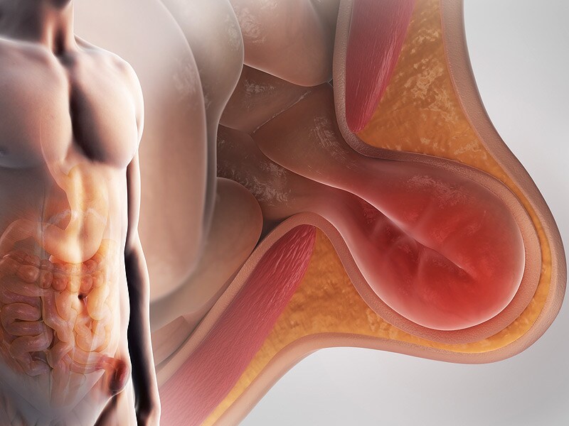
A hernia is a protrusion of an organ or tissue through a weakened area in the abdominal wall, often appearing as a bulge beneath the skin. This condition can occur in various parts of the body such as the groin (inguinal hernia), navel (umbilical hernia), or site of a previous surgical incision (incisional hernia). Hernias may cause discomfort or pain, especially when straining, lifting heavy objects, or during bowel movements. In some cases, they may lead to serious complications like intestinal obstruction or strangulation, requiring immediate medical attention.
A diaphragmatic hernia is a type of hernia that occurs when the abdominal organs (such as the stomach, intestines, or liver) protrude through an opening in the diaphragm, the thin muscle that separates the chest and abdominal cavities. This condition can be present at birth (congenital) or acquired due to injury or surgery.
There are two main types of diaphragmatic hernias:
1. Bochdalek hernia: This is a congenital defect that occurs when the posterior portion of the diaphragm fails to close properly during fetal development, creating an opening through which abdominal organs can move into the chest cavity. It is more common on the left side and can lead to pulmonary hypoplasia (underdevelopment of the lungs) and other complications if not detected and treated early.
2. Morgagni hernia: This is a less common type of congenital diaphragmatic hernia that occurs when there is an opening in the anterior portion of the diaphragm, allowing abdominal organs to move into the chest cavity near the sternum. It tends to be asymptomatic and may not be discovered until adulthood.
Acquired diaphragmatic hernias can result from trauma, such as a car accident or penetrating injury, which causes a tear in the diaphragm. In some cases, surgical procedures involving the abdomen or chest can also lead to a diaphragmatic hernia.
Symptoms of a diaphragmatic hernia may include difficulty breathing, chest pain, vomiting, and bowel obstruction. Treatment typically involves surgery to repair the defect in the diaphragm and return the abdominal organs to their proper position.
A ventral hernia is a type of hernia that occurs in the abdominal wall, specifically in the anterior (front) aspect. It can occur due to a weakness or defect in the abdominal wall muscles and fascia, which allows the internal organs or tissues to push through and create a bulge or swelling.
Ventral hernias can be classified into several types based on their location, size, and cause. Some of the common types include:
1. Incisional Hernia - occurs at the site of a previous surgical incision, where the abdominal wall has not healed properly or has become weakened over time.
2. Epigastric Hernia - located in the upper middle part of the abdomen, between the breastbone and the navel.
3. Umbilical Hernia - occurs around the belly button, most commonly seen in infants but can also affect adults.
4. Spigelian Hernia - a rare type of hernia that occurs lateral to the rectus sheath, usually at the level of the semilunar line.
5. Diastasis Recti - a separation of the abdominal muscles in the midline, which can lead to a ventral hernia if not treated.
Symptoms of a ventral hernia may include pain or discomfort, especially when lifting heavy objects, straining, coughing, or during physical activity. In some cases, a hernia may become incarcerated or strangulated, which requires immediate medical attention. Treatment options for ventral hernias typically involve surgical repair, either through open surgery or laparoscopic techniques.
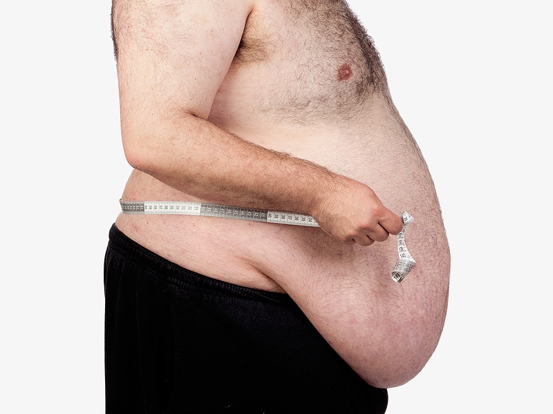
An abdominal hernia refers to the protrusion of an organ or tissue through a weakened area in the abdominal wall, resulting in a bulge. This condition can occur due to various factors such as congenital defects, aging, obesity, pregnancy, persistent coughing, or previous surgeries that have left behind weak spots in the abdominal wall.
There are several types of abdominal hernias, including:
1. Inguinal Hernia: This is the most common type of hernia, occurring when the intestine or bladder protrudes through the inguinal canal in the lower abdomen. Inguinal hernias are more prevalent in men than women.
2. Femoral Hernia: This type of hernia occurs when the intestine or fatty tissue pushes through a weakened area near the femoral artery, located in the upper thigh region. Femoral hernias are more common in women, especially those who are pregnant or obese.
3. Incisional Hernia: This type of hernia develops at the site of a previous abdominal surgery where the abdominal muscles have weakened or failed to heal properly.
4. Umbilical Hernia: An umbilical hernia occurs when the intestine protrudes through the abdominal wall near the navel, often visible as a bulge around the belly button. This type of hernia is more common in infants but can also affect adults, particularly those who are overweight or have had multiple pregnancies.
5. Epigastric Hernia: An epigastric hernia occurs when fatty tissue protrudes through a weakened area between the breastbone and the navel. These hernias are usually small and often painless but can cause discomfort or complications if they become incarcerated or strangulated.
Abdominal hernias can vary in size, from small and barely noticeable to large and severely painful. Symptoms may include a visible bulge, localized pain or discomfort, especially when lifting heavy objects, coughing, or straining during bowel movements. In some cases, hernias may become incarcerated (trapped) or strangulated (blood supply is cut off), which can lead to severe pain, nausea, vomiting, and require immediate medical attention.
Treatment for abdominal hernias typically involves surgical repair, either through open surgery or laparoscopic techniques. The choice of procedure depends on various factors, including the size and location of the hernia, the patient's overall health, and their personal preferences. In some cases, watchful waiting may be recommended for small, asymptomatic hernias, but it is essential to consult with a healthcare professional to determine the best course of action.
A hiatal hernia is a type of hernia that occurs when a part of the stomach protrudes or squeezes through an opening (hiatus) in the diaphragm, the muscular partition between the chest and abdominal cavities. Normally, the esophagus passes through this opening to connect to the stomach, but in a hiatal hernia, a portion of the stomach also moves up into the chest cavity through the hiatus.
There are two main types of hiatal hernias: sliding and paraesophageal. In a sliding hiatal hernia, the junction between the esophagus and stomach (gastroesophageal junction) slides upward into the chest cavity, which is the most common type. Paraesophageal hiatal hernias are less common but can be more severe, as they involve the stomach herniating alongside the esophagus, potentially leading to complications like obstruction or strangulation of the blood supply to the stomach.
Many people with hiatal hernias do not experience symptoms, but some may have heartburn, acid reflux, regurgitation, difficulty swallowing, chest pain, or shortness of breath. Treatment depends on the severity and associated symptoms, ranging from lifestyle modifications and medications to surgical repair in severe cases.
A femoral hernia is a type of hernia that occurs when a portion of the abdominal wall tissue or intestine protrudes through a weakened area in the lower part of the abdominal wall, specifically at the opening of the femoral canal. This canal is located near the groin region and contains blood vessels that pass from the abdomen to the leg.
Femoral hernias are more common in women than men, particularly those who are pregnant, obese, or have a history of multiple pregnancies. Symptoms may include a visible bulge in the inner thigh or groin area, especially when standing, coughing, or straining. Pain or discomfort in the lower abdomen or groin region, particularly during physical activities, is also common.
While some femoral hernias may not cause any symptoms and can be left untreated, they have a higher risk of becoming incarcerated or strangulated compared to other types of hernias. Incarceration occurs when the protruding tissue becomes trapped and cannot be pushed back in, while strangulation happens when the blood supply to the trapped tissue is cut off, leading to tissue death if not treated promptly with surgery.
An umbilical hernia is a type of hernia that occurs at the umbilicus, or belly button. It results from a protrusion of abdominal contents through a weakened area in the abdominal wall surrounding the navel. This condition is common in newborns and infants, especially premature babies, due to incomplete closure of the abdominal muscles during development.
In most cases, umbilical hernias in children close on their own by age 3-4 or by the time they reach school age. However, if the hernia is still present after this age, surgical intervention may be required to prevent potential complications such as incarceration (where the herniated tissue becomes trapped and cannot be pushed back in) or strangulation (where the blood supply to the herniated tissue is cut off, leading to tissue death).
Adults can also develop umbilical hernias, often as a result of increased pressure in the abdomen due to obesity, pregnancy, heavy lifting, or persistent coughing. Umbilical hernias in adults are generally more likely to require surgical repair due to the higher risk of complications.
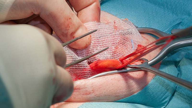
Herniorrhaphy is a surgical procedure where the herniated tissue or organ is placed back into its original position, and the weakened or damaged muscle wall is repaired. This is typically done to correct a hernia, which is a protrusion of an organ or tissue through a weakened area in the abdominal wall. The surgical incision may be closed with sutures or staples, and sometimes a mesh patch is used to reinforce the repair.
Surgical mesh is a medical device that is used in various surgical procedures, particularly in reconstructive surgery, to provide additional support to weakened or damaged tissues. It is typically made from synthetic materials such as polypropylene or polyester, or from biological materials such as animal tissue or human cadaveric tissue.
The mesh is designed to be implanted into the body, where it can help to reinforce and repair damaged tissues. For example, it may be used in hernia repairs to support the weakened abdominal wall, or in pelvic floor reconstruction surgery to treat conditions such as pelvic organ prolapse or stress urinary incontinence.
Surgical mesh can come in different forms, including sheets, plugs, and patches, and may be either absorbable or non-absorbable. The choice of mesh material and type will depend on the specific surgical indication and the patient's individual needs. It is important for patients to discuss the risks and benefits of surgical mesh with their healthcare provider before undergoing any surgical procedure that involves its use.
A diaphragmatic hernia is a condition in which there is abnormal displacement or protrusion of abdominal organs into the thoracic cavity through an opening or defect in the diaphragm. In traumatic diaphragmatic hernia, this disruption of the diaphragm is caused by trauma, such as a penetrating injury or blunt force trauma from a car accident or fall. The resulting herniation can lead to various complications, including difficulty breathing, digestive problems, and even organ dysfunction. Immediate medical attention is necessary for proper diagnosis and treatment of traumatic diaphragmatic hernia.
A obturator hernia is a type of pelvic hernia that occurs through the obturator foramen, which is an opening in the bony pelvis through which blood vessels and nerves pass. This type of hernia is relatively uncommon and primarily affects women, particularly those who are elderly, thin, or have had multiple pregnancies.
Obturator hernias occur when abdominal contents, such as fat or intestine, protrude through the obturator foramen and become trapped, leading to obstruction of the intestine or strangulation, which can cut off blood flow to the affected tissue. Symptoms may include pain in the thigh or groin, nausea, vomiting, and constipation.
Obturator hernias can be difficult to diagnose due to their nonspecific symptoms and rarity. Diagnosis is often made during surgery to repair the hernia. Treatment typically involves surgical repair of the hernia, which may be performed through an open incision or laparoscopically. Delayed diagnosis and treatment can lead to serious complications, such as bowel necrosis or perforation, so prompt medical attention is necessary if obturator hernia is suspected.
I'm sorry for any confusion, but "Polypropylenes" does not have a medical definition as it is not a medical term. Polypropylene is a type of plastic that is used in various medical and non-medical applications. It is a thermoplastic polymer resin that is produced by the polymerization of propylene.
In the medical field, polypropylene is sometimes used to make single-use surgical instruments, sutures, and medical devices due to its resistance to heat, chemicals, and electricity. It is also biocompatible, meaning it can be safely used in the body without causing adverse reactions. However, "Polypropylenes" as a medical term is not recognized or used in the medical community.
Laparoscopy is a surgical procedure that involves the insertion of a laparoscope, which is a thin tube with a light and camera attached to it, through small incisions in the abdomen. This allows the surgeon to view the internal organs without making large incisions. It's commonly used to diagnose and treat various conditions such as endometriosis, ovarian cysts, infertility, and appendicitis. The advantages of laparoscopy over traditional open surgery include smaller incisions, less pain, shorter hospital stays, and quicker recovery times.
The abdominal wall refers to the group of muscles, fascia (sheaths of connective tissue), and skin that make up the front and sides of the abdomen, extending from the thorax (chest) to the pelvis. It provides protection to the abdominal organs, supports the trunk, and allows for movement of the torso.
The main muscles of the anterior abdominal wall include:
1. Rectus sheaths (Rectus Abdominis): paired vertical muscles running from the pubic symphysis to the xiphoid process and costal cartilages of ribs 5-7.
2. External obliques: thin, irregular muscles that lie over the lower part of the abdomen and run diagonally downward and forward from the lower ribs to the iliac crest (pelvic bone) and pubic tubercle.
3. Internal obliques: thicker muscles that lie under the external obliques, running diagonally upward and forward from the iliac crest to the lower ribs.
4. Transverse abdominis: deepest of the abdominal muscles, lying horizontally across the abdomen, attaching from the lower ribs to the pelvis.
These muscles are interconnected by various layers of fascia and aponeuroses (flat, broad tendons), forming a complex structure that allows for both stability and mobility. The linea alba, a fibrous band, runs down the midline of the anterior abdominal wall, connecting the rectus sheaths.
Damage to the abdominal wall can occur due to trauma, surgery, or various medical conditions, which may require surgical intervention for repair.
A fascia is a band or sheet of connective tissue, primarily collagen, that covers, connects, and separates muscles, organs, and other structures in the body. It provides support and stability, allows for smooth movement between structures, and has the ability to transmit forces throughout the body. Fascia is found throughout the body, and there are several layers of it, including superficial fascia, deep fascia, and visceral fascia. Injury, inflammation, or strain to the fascia can cause pain and restriction of movement.
Suture techniques refer to the various methods used by surgeons to sew or stitch together tissues in the body after an injury, trauma, or surgical incision. The main goal of suturing is to approximate and hold the edges of the wound together, allowing for proper healing and minimizing scar formation.
There are several types of suture techniques, including:
1. Simple Interrupted Suture: This is one of the most basic suture techniques where the needle is passed through the tissue at a right angle, creating a loop that is then tightened to approximate the wound edges. Multiple stitches are placed along the length of the incision or wound.
2. Continuous Locking Suture: In this technique, the needle is passed continuously through the tissue in a zigzag pattern, with each stitch locking into the previous one. This creates a continuous line of sutures that provides strong tension and support to the wound edges.
3. Running Suture: Similar to the continuous locking suture, this technique involves passing the needle continuously through the tissue in a straight line. However, instead of locking each stitch, the needle is simply passed through the previous loop before being tightened. This creates a smooth and uninterrupted line of sutures that can be easily removed after healing.
4. Horizontal Mattress Suture: In this technique, two parallel stitches are placed horizontally across the wound edges, creating a "mattress" effect that provides additional support and tension to the wound. This is particularly useful in deep or irregularly shaped wounds.
5. Vertical Mattress Suture: Similar to the horizontal mattress suture, this technique involves placing two parallel stitches vertically across the wound edges. This creates a more pronounced "mattress" effect that can help reduce tension and minimize scarring.
6. Subcuticular Suture: In this technique, the needle is passed just below the surface of the skin, creating a smooth and barely visible line of sutures. This is particularly useful in cosmetic surgery or areas where minimizing scarring is important.
The choice of suture technique depends on various factors such as the location and size of the wound, the type of tissue involved, and the patient's individual needs and preferences. Proper suture placement and tension are crucial for optimal healing and aesthetic outcomes.
In medical terms, sutures are specialized surgical threads made from various materials such as absorbable synthetic or natural fibers, or non-absorbable materials like nylon or silk. They are used to approximate and hold together the edges of a wound or incision in the skin or other tissues during the healing process. Sutures come in different sizes, types, and shapes, each designed for specific uses and techniques depending on the location and type of tissue being sutured. Properly placed sutures help to promote optimal healing, minimize scarring, and reduce the risk of infection or other complications.
Intestinal obstruction, also known as bowel obstruction, is a medical condition characterized by a blockage that prevents the normal flow of contents through the small intestine or large intestine (colon). This blockage can be caused by various factors such as tumors, adhesions (scar tissue), hernias, inflammation, or impacted feces.
The obstruction can be mechanical, where something physically blocks the intestinal lumen, or functional, where the normal muscular contractions of the bowel are impaired. Mechanical obstructions are more common than functional ones.
Symptoms of intestinal obstruction may include abdominal pain and cramping, nausea and vomiting, bloating, inability to pass gas or have a bowel movement, and abdominal distention. If left untreated, intestinal obstruction can lead to serious complications such as tissue death (necrosis), perforation of the intestine, and sepsis. Treatment typically involves hospitalization, intravenous fluids, nasogastric decompression, and possibly surgery to remove the obstruction.
A laparotomy is a surgical procedure that involves making an incision in the abdominal wall to gain access to the abdominal cavity. This procedure is typically performed to diagnose and treat various conditions such as abdominal trauma, tumors, infections, or inflammatory diseases. The size of the incision can vary depending on the reason for the surgery and the extent of the condition being treated. Once the procedure is complete, the incision is closed with sutures or staples.
The term "laparotomy" comes from the Greek words "lapara," which means "flank" or "side," and "tome," which means "to cut." Together, they describe the surgical procedure that involves cutting into the abdomen to examine its contents.
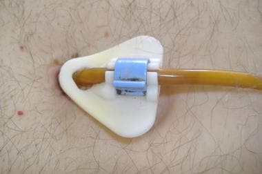
A surgical stoma, also known simply as a stoma, is a surgically created opening on the surface of the body that allows for the passage of bodily waste. This procedure is typically performed when a person has a malfunctioning or diseased organ in the digestive or urinary system that cannot be effectively treated or repaired.
In a colostomy or ileostomy, which are common types of surgical stomas, a portion of the colon or small intestine is brought through an opening in the abdominal wall to create a new pathway for waste to exit the body. The stoma may be temporary or permanent, depending on the underlying condition and the success of any additional treatments.
After surgery, patients with a stoma will need to wear a pouching system to collect and contain the waste that is expelled through the stoma. This can take some getting used to, but with proper care and support, most people are able to adjust to life with a stoma and maintain a good quality of life.
In medical terms, the "groin" refers to the area where the lower abdomen meets the thigh. It is located on both sides of the body, in front of the upper part of each leg. The groin contains several important structures such as the inguinal canal, which contains blood vessels and nerves, and the femoral artery and vein, which supply blood to and from the lower extremities. Issues in this region, such as pain or swelling, may indicate a variety of medical conditions, including muscle strains, hernias, or infections.
Postoperative complications refer to any unfavorable condition or event that occurs during the recovery period after a surgical procedure. These complications can vary in severity and may include, but are not limited to:
1. Infection: This can occur at the site of the incision or inside the body, such as pneumonia or urinary tract infection.
2. Bleeding: Excessive bleeding (hemorrhage) can lead to a drop in blood pressure and may require further surgical intervention.
3. Blood clots: These can form in the deep veins of the legs (deep vein thrombosis) and can potentially travel to the lungs (pulmonary embolism).
4. Wound dehiscence: This is when the surgical wound opens up, which can lead to infection and further complications.
5. Pulmonary issues: These include atelectasis (collapsed lung), pneumonia, or respiratory failure.
6. Cardiovascular problems: These include abnormal heart rhythms (arrhythmias), heart attack, or stroke.
7. Renal failure: This can occur due to various reasons such as dehydration, blood loss, or the use of certain medications.
8. Pain management issues: Inadequate pain control can lead to increased stress, anxiety, and decreased mobility.
9. Nausea and vomiting: These can be caused by anesthesia, opioid pain medication, or other factors.
10. Delirium: This is a state of confusion and disorientation that can occur in the elderly or those with certain medical conditions.
Prompt identification and management of these complications are crucial to ensure the best possible outcome for the patient.
Recurrence, in a medical context, refers to the return of symptoms or signs of a disease after a period of improvement or remission. It indicates that the condition has not been fully eradicated and may require further treatment. Recurrence is often used to describe situations where a disease such as cancer comes back after initial treatment, but it can also apply to other medical conditions. The likelihood of recurrence varies depending on the type of disease and individual patient factors.
Local anesthesia is a type of anesthesia that numbs a specific area of the body, blocking pain signals from that particular region while allowing the person to remain conscious and alert. It is typically achieved through the injection or application of a local anesthetic drug, which works by temporarily inhibiting the function of nerve fibers carrying pain sensations. Common examples of local anesthetics include lidocaine, prilocaine, and bupivacaine.
Local anesthesia is commonly used for minor surgical procedures, dental work, or other medical interventions where only a small area needs to be numbed. It can also be employed as part of a combined anesthetic technique, such as in conjunction with sedation or regional anesthesia, to provide additional pain relief and increase patient comfort during more extensive surgeries.
The duration of local anesthesia varies depending on the type and dosage of the anesthetic agent used; some last for just a few hours, while others may provide numbness for up to several days. Overall, local anesthesia is considered a safe and effective method for managing pain during various medical procedures.
The rectus abdominis is a paired, flat, and long muscle in the anterior (front) wall of the abdomen. It runs from the pubic symphysis (the joint where the two pubic bones meet in the front of the pelvis) to the xiphoid process (the lower end of the sternum or breastbone) and costal cartilages of the fifth, sixth, and seventh ribs.
The rectus abdominis is responsible for flexing the lumbar spine (lower back), which helps in bending forward or sitting up from a lying down position. It also contributes to maintaining proper posture and stabilizing the pelvis and spine. The muscle's visibility, especially in its lower portion, is often associated with a "six-pack" appearance in well-trained individuals.
Ambulatory surgical procedures, also known as outpatient or same-day surgery, refer to medical operations that do not require an overnight hospital stay. These procedures are typically performed in a specialized ambulatory surgery center (ASC) or in a hospital-based outpatient department. Patients undergoing ambulatory surgical procedures receive anesthesia, undergo the operation, and recover enough to be discharged home on the same day of the procedure.
Examples of common ambulatory surgical procedures include:
1. Arthroscopy (joint scope examination and repair)
2. Cataract surgery
3. Colonoscopy and upper endoscopy
4. Dental surgery, such as wisdom tooth extraction
5. Gallbladder removal (cholecystectomy)
6. Hernia repair
7. Hysteroscopy (examination of the uterus)
8. Minor skin procedures, like biopsies and lesion removals
9. Orthopedic procedures, such as carpal tunnel release or joint injections
10. Pain management procedures, including epidural steroid injections and nerve blocks
11. Podiatric (foot and ankle) surgery
12. Tonsillectomy and adenoidectomy
Advancements in medical technology, minimally invasive surgical techniques, and improved anesthesia methods have contributed to the growth of ambulatory surgical procedures, offering patients a more convenient and cost-effective alternative to traditional inpatient surgeries.
Postoperative pain is defined as the pain or discomfort experienced by patients following a surgical procedure. It can vary in intensity and duration depending on the type of surgery performed, individual pain tolerance, and other factors. The pain may be caused by tissue trauma, inflammation, or nerve damage resulting from the surgical intervention. Proper assessment and management of postoperative pain is essential to promote recovery, prevent complications, and improve patient satisfaction.
The round ligament is a cord-like structure in the female pelvis that extends from the uterus to the labia majora. It is one of the major ligaments that support the uterus and helps to maintain its position within the pelvis. The round ligament is composed of fibrous tissue and smooth muscle, and it plays a role in maintaining the tone and shape of the uterus.
During pregnancy, the round ligament can become stretched and thickened as the uterus grows and expands. This can sometimes cause discomfort or pain, particularly on one side of the pelvis. In some cases, the round ligament may also contribute to the development of certain gynecological conditions, such as uterine prolapse or urinary incontinence.
It is important for healthcare providers to consider the round ligament when evaluating and treating female reproductive health issues, as it can have a significant impact on the function and positioning of the uterus and other pelvic organs.
Operative surgical procedures refer to medical interventions that involve manual manipulation of tissues, structures, or organs in the body, typically performed in an operating room setting under sterile conditions. These procedures are carried out with the use of specialized instruments, such as scalpels, forceps, and scissors, and may require regional or general anesthesia to ensure patient comfort and safety.
Operative surgical procedures can range from relatively minor interventions, such as a biopsy or the removal of a small lesion, to more complex and extensive surgeries, such as open heart surgery or total joint replacement. The specific goals of operative surgical procedures may include the diagnosis and treatment of medical conditions, the repair or reconstruction of damaged tissues or organs, or the prevention of further disease progression.
Regardless of the type or complexity of the procedure, all operative surgical procedures require careful planning, execution, and postoperative management to ensure the best possible outcomes for patients.
I'm sorry for any confusion, but "Phenyl Ethers" is not a recognized medical term. Phenyl ethers are a class of organic compounds consisting of an ether with a phenyl group as one of the components. They are widely used in industry and research, including as solvents, intermediates in chemical synthesis, and pharmaceuticals.
However, if you have any concerns about exposure to certain chemicals or their effects on health, it would be best to consult with a medical professional who can provide advice based on your specific situation and symptoms.
Treatment outcome is a term used to describe the result or effect of medical treatment on a patient's health status. It can be measured in various ways, such as through symptoms improvement, disease remission, reduced disability, improved quality of life, or survival rates. The treatment outcome helps healthcare providers evaluate the effectiveness of a particular treatment plan and make informed decisions about future care. It is also used in clinical research to compare the efficacy of different treatments and improve patient care.
Fundoplication is a surgical procedure in which the upper part of the stomach (the fundus) is wrapped around the lower esophagus and then stitched into place. This procedure strengthens the lower esophageal sphincter, which helps prevent acid reflux from the stomach into the esophagus. It is commonly used to treat gastroesophageal reflux disease (GERD) and paraesophageal hernias.
Gastroesophageal reflux (GER) is the retrograde movement of stomach contents into the esophagus, which can cause discomfort and symptoms. It occurs when the lower esophageal sphincter (a ring of muscle between the esophagus and stomach) relaxes inappropriately, allowing the acidic or non-acidic gastric contents to flow back into the esophagus.
Gastroesophageal reflux becomes gastroesophageal reflux disease (GERD) when it is more severe, persistent, and/or results in complications such as esophagitis, strictures, or Barrett's esophagus. Common symptoms of GERD include heartburn, regurgitation, chest pain, difficulty swallowing, and chronic cough or hoarseness.
Surgical wound dehiscence is a medical condition that refers to the partial or complete separation of layers of a surgical incision after a surgical procedure, leading to the disruption of the wound closure. This can occur due to various factors such as infection, poor nutrition, increased tension on the sutures, hematoma or seroma formation, and patient's underlying health conditions like diabetes or immunodeficiency. Dehiscence may result in the exposure of internal tissues and organs, potentially causing severe complications such as infection, bleeding, or organ dysfunction. Immediate medical attention is required to manage this condition and prevent further complications.
A testicular hydrocele is a type of fluid-filled sac that forms around the testicle (testis), typically in the scrotum. This sac, known as the tunica vaginalis, normally contains a small amount of fluid that helps to lubricate and protect the testicle. However, when an excessive amount of fluid accumulates in this sac, it results in the formation of a hydrocele.
Testicular hydroceles can be congenital (present at birth) or acquired later in life due to various reasons such as injury, inflammation, or infection in the scrotal area. They are usually painless but may cause discomfort or a feeling of heaviness in the scrotum, especially when they become large. In some cases, hydroceles may resolve on their own without treatment, while others may require surgical intervention to drain the fluid and repair the underlying issue.
It is essential to differentiate between hydroceles and other conditions with similar symptoms, such as hernias or tumors, which may require more urgent medical attention. A healthcare professional can perform a physical examination and possibly recommend further testing, like an ultrasound, to confirm the diagnosis of a testicular hydrocele.
A laparoscope is a type of medical instrument called an endoscope, which is used to examine the interior of a body cavity or organ. Specifically, a laparoscope is a long, thin tube with a high-intensity light and a high-resolution camera attached to it. This device allows surgeons to view the abdominal cavity through small incisions, without having to make large, invasive cuts.
During a laparoscopic procedure, the surgeon will insert the laparoscope through a small incision in the abdomen, typically near the navel. The camera sends images back to a monitor, giving the surgeon a clear view of the organs and tissues inside the body. This allows for more precise and less invasive surgical procedures, often resulting in faster recovery times and fewer complications compared to traditional open surgery.
Laparoscopes are commonly used in a variety of surgical procedures, including:
1. Gynecological surgeries (e.g., hysterectomies, ovarian cyst removals)
2. Gallbladder removal (cholecystectomy)
3. Gastrointestinal surgeries (e.g., removing benign or malignant tumors)
4. Hernia repairs
5. Bariatric surgeries for weight loss (e.g., gastric bypass, sleeve gastrectomy)
While laparoscopes provide numerous benefits over open surgery, they still require specialized training and expertise to use effectively and safely.
Fetal diseases are medical conditions or abnormalities that affect a fetus during pregnancy. These diseases can be caused by genetic factors, environmental influences, or a combination of both. They can range from mild to severe and may impact various organ systems in the developing fetus. Examples of fetal diseases include congenital heart defects, neural tube defects, chromosomal abnormalities such as Down syndrome, and infectious diseases such as toxoplasmosis or rubella. Fetal diseases can be diagnosed through prenatal testing, including ultrasound, amniocentesis, and chorionic villus sampling. Treatment options may include medication, surgery, or delivery of the fetus, depending on the nature and severity of the disease.
Retrospective studies, also known as retrospective research or looking back studies, are a type of observational study that examines data from the past to draw conclusions about possible causal relationships between risk factors and outcomes. In these studies, researchers analyze existing records, medical charts, or previously collected data to test a hypothesis or answer a specific research question.
Retrospective studies can be useful for generating hypotheses and identifying trends, but they have limitations compared to prospective studies, which follow participants forward in time from exposure to outcome. Retrospective studies are subject to biases such as recall bias, selection bias, and information bias, which can affect the validity of the results. Therefore, retrospective studies should be interpreted with caution and used primarily to generate hypotheses for further testing in prospective studies.
A colostomy is a surgical procedure that involves creating an opening, or stoma, through the abdominal wall to divert the flow of feces from the colon (large intestine) through this opening and into a pouch or bag worn outside the body. This procedure is typically performed when a portion of the colon has been removed due to disease or injury, such as cancer, inflammatory bowel disease, or trauma.
There are several types of colostomies, including end colostomy, loop colostomy, and double-barrel colostomy, which differ in terms of the location and configuration of the stoma. The type of colostomy performed will depend on the individual's medical condition and the specific goals of the surgery.
After a colostomy, patients will need to learn how to care for their stoma and manage their bowel movements using specialized equipment and techniques. With proper care and management, most people are able to lead active and fulfilling lives after a colostomy.
Tissue adhesions, also known as scar tissue adhesions, are abnormal bands of fibrous tissue that form between two or more internal organs, or between organs and the walls of the chest or abdominal cavity. These adhesions can develop after surgery, infection, injury, radiation, or prolonged inflammation. The fibrous bands can cause pain, restrict movement of the organs, and potentially lead to complications such as bowel obstruction. Treatment options for tissue adhesions may include medication, physical therapy, or surgical intervention to remove the adhesions.
The inguinal canal is a narrow passage in the lower abdominal wall. In males, it allows for the spermatic cord and blood vessels to travel from the abdomen to the scrotum. In females, it provides a pathway for the round ligament of the uterus to pass through. The inguinal canal is located in the groin region, and an inguinal hernia occurs when a portion of the intestine protrudes through this canal.
"Length of Stay" (LOS) is a term commonly used in healthcare to refer to the amount of time a patient spends receiving care in a hospital, clinic, or other healthcare facility. It is typically measured in hours, days, or weeks and can be used as a metric for various purposes such as resource planning, quality assessment, and reimbursement. The length of stay can vary depending on the type of illness or injury, the severity of the condition, the patient's response to treatment, and other factors. It is an important consideration in healthcare management and can have significant implications for both patients and providers.
Follow-up studies are a type of longitudinal research that involve repeated observations or measurements of the same variables over a period of time, in order to understand their long-term effects or outcomes. In medical context, follow-up studies are often used to evaluate the safety and efficacy of medical treatments, interventions, or procedures.
In a typical follow-up study, a group of individuals (called a cohort) who have received a particular treatment or intervention are identified and then followed over time through periodic assessments or data collection. The data collected may include information on clinical outcomes, adverse events, changes in symptoms or functional status, and other relevant measures.
The results of follow-up studies can provide important insights into the long-term benefits and risks of medical interventions, as well as help to identify factors that may influence treatment effectiveness or patient outcomes. However, it is important to note that follow-up studies can be subject to various biases and limitations, such as loss to follow-up, recall bias, and changes in clinical practice over time, which must be carefully considered when interpreting the results.
A reoperation is a surgical procedure that is performed again on a patient who has already undergone a previous operation for the same or related condition. Reoperations may be required due to various reasons, such as inadequate initial treatment, disease recurrence, infection, or complications from the first surgery. The nature and complexity of a reoperation can vary widely depending on the specific circumstances, but it often carries higher risks and potential complications compared to the original operation.
An appendectomy is a surgical procedure in which the vermiform appendix is removed. This procedure is performed when a patient has appendicitis, which is an inflammation of the appendix that can lead to serious complications such as peritonitis or sepsis if not treated promptly. The surgery can be done as an open procedure, in which a single incision is made in the lower right abdomen, or as a laparoscopic procedure, in which several small incisions are made and specialized instruments are used to remove the appendix. In some cases, if the appendix has burst, a more extensive surgery may be required to clean out the abdominal cavity.
Diaphragmatic eventration is a medical condition where the diaphragm, the thin muscle that separates the chest and abdominal cavities and helps with breathing, is abnormally thin and weak. This can cause the diaphragm to move upwards into the chest cavity, which can lead to difficulty breathing and other respiratory symptoms.
In eventration, the affected portion of the diaphragm is usually elevated and may have a transparent or bluish appearance due to the lack of muscle tissue. This condition can be present at birth (congenital) or acquired later in life due to injury or illness.
Mild cases of diaphragmatic eventration may not cause any symptoms and may not require treatment. However, more severe cases may require surgery to repair the damaged diaphragm and improve respiratory function.
Orchiopexy is a surgical procedure in which the testicle (or testicles) that have descended into the scrotum incompletely or not at all (undescended or retractile testes) are fixed into their normal position within the scrotum. This procedure is typically performed on boys, often between the ages of 6 and 12 months, to correct cryptorchidism, a condition where one or both testicles fail to descend into the scrotum.
The main goals of orchiopexy are to:
1. Place the testicle in its proper anatomical location within the scrotum.
2. Fix the testicle in a stable position to prevent retractile testes from moving back into the inguinal canal.
3. Preserve the testicular blood supply and innervation, ensuring normal testicular function and development.
4. Lower the risk of testicular torsion (twisting of the spermatic cord) and malignancy in later life.
Orchiopexy can be performed through an open or laparoscopic approach, depending on the location of the undescended testicle(s). The choice of surgical technique depends on factors such as the patient's age, associated conditions, and surgeon's preference.
The spermatic cord is a fibrous structure that contains the vas deferens, blood vessels, nerves, and lymphatics, which provide passage for these structures between the abdomen and the scrotum in males. It is covered by several layers of protective sheaths, including the internal spermatic fascia, cremasteric fascia, and external spermatic fascia. The spermatic cord allows the testicles to be located outside the body, which helps maintain a cooler temperature for optimal sperm production.
The scrotum is a part of the external male genitalia. It's a sac-like structure made up of several layers of skin and smooth muscle, which hangs down behind and beneath the penis. The primary function of the scrotum is to maintain the testicles at a temperature slightly lower than the core body temperature, which is optimal for sperm production.
The scrotum contains two compartments, each one housing a testicle. It's located in the pubic region and is usually visible externally. The skin of the scrotum is thin and wrinkled, which allows it to expand and contract depending on the temperature, accommodating the shrinking or swelling of the testicles.
Please note that while I strive to provide accurate information, this definition is intended to be a general overview and should not replace professional medical advice.
A surgical wound infection, also known as a surgical site infection (SSI), is defined by the Centers for Disease Control and Prevention (CDC) as an infection that occurs within 30 days after surgery (or within one year if an implant is left in place) and involves either:
1. Purulent drainage from the incision;
2. Organisms isolated from an aseptically obtained culture of fluid or tissue from the incision;
3. At least one of the following signs or symptoms of infection: pain or tenderness, localized swelling, redness, or heat; and
4. Diagnosis of surgical site infection by the surgeon or attending physician.
SSIs can be classified as superficial incisional, deep incisional, or organ/space infections, depending on the depth and extent of tissue involvement. They are a common healthcare-associated infection and can lead to increased morbidity, mortality, and healthcare costs.
Peritoneal diseases refer to a group of conditions that affect the peritoneum, which is the thin, transparent membrane that lines the inner wall of the abdomen and covers the organs within it. The peritoneum has several functions, including providing protection and support to the abdominal organs, producing and absorbing fluids, and serving as a site for the immune system's response to infections and other foreign substances.
Peritoneal diseases can be broadly classified into two categories: infectious and non-infectious. Infectious peritoneal diseases are caused by bacterial, viral, fungal, or parasitic infections that spread to the peritoneum from other parts of the body or through contaminated food, water, or medical devices. Non-infectious peritoneal diseases, on the other hand, are not caused by infections but rather by other factors such as autoimmune disorders, cancer, or chemical irritants.
Some examples of peritoneal diseases include:
1. Peritonitis: Inflammation of the peritoneum due to bacterial or fungal infections, often caused by a ruptured appendix, perforated ulcer, or other abdominal injuries or conditions.
2. Tuberculous peritonitis: A form of peritonitis caused by Mycobacterium tuberculosis, the bacterium that causes tuberculosis (TB).
3. Peritoneal dialysis-associated peritonitis: Infection of the peritoneum in patients undergoing peritoneal dialysis, a type of kidney replacement therapy for patients with end-stage renal disease.
4. Malignant peritoneal mesothelioma: A rare and aggressive form of cancer that affects the mesothelial cells lining the peritoneum, often caused by exposure to asbestos.
5. Systemic lupus erythematosus (SLE): An autoimmune disorder that can cause inflammation and scarring of the peritoneum.
6. Peritoneal carcinomatosis: The spread of cancer cells from other parts of the body to the peritoneum, often seen in patients with advanced ovarian or colorectal cancer.
7. Cirrhotic ascites: Fluid accumulation in the peritoneal cavity due to liver cirrhosis and portal hypertension.
8. Meigs' syndrome: A rare condition characterized by the presence of a benign ovarian tumor, ascites, and pleural effusion.
Polytetrafluoroethylene (PTFE) is not inherently a medical term, but it is a chemical compound with significant uses in the medical field. Medically, PTFE is often referred to by its brand name, Teflon. It is a synthetic fluoropolymer used in various medical applications due to its unique properties such as high resistance to heat, electrical and chemical interaction, and exceptional non-reactivity with body tissues.
PTFE can be found in medical devices like catheters, where it reduces friction, making insertion easier and minimizing trauma. It is also used in orthopedic and dental implants, drug delivery systems, and sutures due to its biocompatibility and non-adhesive nature.
Gastropexy is a surgical procedure in which the stomach is attached to another organ, usually the abdominal wall. This procedure is often performed as a preventative measure for gastric volvulus, a condition where the stomach twists on itself and cuts off its own blood supply. It is also done in animals, particularly dogs, to prevent gastric dilation volvulus (GDV), also known as bloat, which is a life-threatening emergency. In humans, gastropexy is sometimes performed as part of treatment for morbid obesity.
A newborn infant is a baby who is within the first 28 days of life. This period is also referred to as the neonatal period. Newborns require specialized care and attention due to their immature bodily systems and increased vulnerability to various health issues. They are closely monitored for signs of well-being, growth, and development during this critical time.
Cecal diseases refer to medical conditions that affect the cecum, which is a pouch-like structure located at the junction of the small and large intestines. The cecum plays an important role in digestion, particularly in the fermentation of certain types of food.
There are several different types of cecal diseases, including:
1. Cecal volvulus: This is a rare condition in which the cecum twists on itself, cutting off blood flow and causing severe pain and other symptoms.
2. Diverticulitis: This occurs when small pouches called diverticula form in the wall of the cecum and become inflamed or infected.
3. Appendicitis: Although not strictly a cecal disease, the appendix is a small tube-like structure that branches off from the cecum. Inflammation of the appendix (appendicitis) can cause severe pain in the lower right abdomen and may require surgical removal of the appendix.
4. Crohn's disease: This is a chronic inflammatory bowel disease that can affect any part of the digestive tract, including the cecum.
5. Tuberculosis: The cecum can also be affected by tuberculosis, which is a bacterial infection that primarily affects the lungs but can spread to other parts of the body.
6. Cancer: Although rare, cancer can also affect the cecum, leading to symptoms such as abdominal pain, bloating, and changes in bowel habits.
Treatment for cecal diseases depends on the specific condition and its severity. Treatment options may include antibiotics, surgery, or other medical interventions. If you are experiencing symptoms that may be related to a cecal disease, it is important to seek medical attention promptly.
Surgical stapling is a medical technique that uses specialized staplers to place linear staple lines to close surgical incisions, connect or remove organs and tissues during surgical procedures. Surgical staples are made of titanium or stainless steel and can be absorbable or non-absorbable. They provide secure, fast, and accurate wound closure, reducing the risk of infection and promoting faster healing compared to traditional suturing methods.
The surgical stapler consists of a handle, an anvil, and a cartridge containing multiple staples. The device is loaded with staple cartridges and used to approximate tissue edges before deploying the staples. Once the staples are placed, the stapler is removed, leaving the staple line in place.
Surgical stapling has various applications, including gastrointestinal anastomosis, lung resection, vascular anastomosis, and skin closure. It is widely used in different types of surgeries, such as open, laparoscopic, and robotic-assisted procedures. The use of surgical stapling requires proper training and expertise to ensure optimal patient outcomes.
X-ray computed tomography (CT or CAT scan) is a medical imaging method that uses computer-processed combinations of many X-ray images taken from different angles to produce cross-sectional (tomographic) images (virtual "slices") of the body. These cross-sectional images can then be used to display detailed internal views of organs, bones, and soft tissues in the body.
The term "computed tomography" is used instead of "CT scan" or "CAT scan" because the machines take a series of X-ray measurements from different angles around the body and then use a computer to process these data to create detailed images of internal structures within the body.
CT scanning is a noninvasive, painless medical test that helps physicians diagnose and treat medical conditions. CT imaging provides detailed information about many types of tissue including lung, bone, soft tissue and blood vessels. CT examinations can be performed on every part of the body for a variety of reasons including diagnosis, surgical planning, and monitoring of therapeutic responses.
In computed tomography (CT), an X-ray source and detector rotate around the patient, measuring the X-ray attenuation at many different angles. A computer uses this data to construct a cross-sectional image by the process of reconstruction. This technique is called "tomography". The term "computed" refers to the use of a computer to reconstruct the images.
CT has become an important tool in medical imaging and diagnosis, allowing radiologists and other physicians to view detailed internal images of the body. It can help identify many different medical conditions including cancer, heart disease, lung nodules, liver tumors, and internal injuries from trauma. CT is also commonly used for guiding biopsies and other minimally invasive procedures.
In summary, X-ray computed tomography (CT or CAT scan) is a medical imaging technique that uses computer-processed combinations of many X-ray images taken from different angles to produce cross-sectional images of the body. It provides detailed internal views of organs, bones, and soft tissues in the body, allowing physicians to diagnose and treat medical conditions.
Prospective studies, also known as longitudinal studies, are a type of cohort study in which data is collected forward in time, following a group of individuals who share a common characteristic or exposure over a period of time. The researchers clearly define the study population and exposure of interest at the beginning of the study and follow up with the participants to determine the outcomes that develop over time. This type of study design allows for the investigation of causal relationships between exposures and outcomes, as well as the identification of risk factors and the estimation of disease incidence rates. Prospective studies are particularly useful in epidemiology and medical research when studying diseases with long latency periods or rare outcomes.
The digestive system is a series of organs that work together to convert food into nutrients and energy. Digestive system surgical procedures involve operations on any part of the digestive system, including the esophagus, stomach, small intestine, large intestine, liver, pancreas, and gallbladder. These procedures can be performed for a variety of reasons, such as to treat diseases, repair damage, or remove cancerous growths.
Some common digestive system surgical procedures include:
1. Gastric bypass surgery: A procedure in which the stomach is divided into two parts and the smaller part is connected directly to the small intestine, bypassing a portion of the stomach and upper small intestine. This procedure is used to treat severe obesity.
2. Colonoscopy: A procedure in which a flexible tube with a camera on the end is inserted into the rectum and colon to examine the lining for polyps, cancer, or other abnormalities.
3. Colectomy: A procedure in which all or part of the colon is removed, often due to cancer, inflammatory bowel disease, or diverticulitis.
4. Gastrostomy: A procedure in which a hole is made through the abdominal wall and into the stomach to create an opening for feeding. This is often done for patients who have difficulty swallowing.
5. Esophagectomy: A procedure in which all or part of the esophagus is removed, often due to cancer. The remaining esophagus is then reconnected to the stomach or small intestine.
6. Liver resection: A procedure in which a portion of the liver is removed, often due to cancer or other diseases.
7. Pancreatectomy: A procedure in which all or part of the pancreas is removed, often due to cancer or chronic pancreatitis.
8. Cholecystectomy: A procedure in which the gallbladder is removed, often due to gallstones or inflammation.
These are just a few examples of digestive system surgical procedures. There are many other types of operations that can be performed on the digestive system depending on the specific needs and condition of each patient.
Fascia lata is a medical term that refers to the thick, fibrous sheath of connective tissue that envelops and surrounds the thigh muscles (specifically, the quadriceps femoris and hamstrings). It is a type of fascia, which is the soft tissue component of the deep (internal) fascial system.
The fascia lata is continuous with the fascia of the hip and knee joints and plays an important role in providing stability, support, and protection to the muscles and other structures within the thigh. It also helps to facilitate the gliding and movement of muscles and tendons during physical activity.
Injuries or inflammation of the fascia lata can cause pain and discomfort, and may limit mobility and range of motion in the thigh and lower extremity. Conditions such as fascia lata strain, tears, or myofascial pain syndrome may require medical treatment, including physical therapy, medication, or in some cases, surgery.
A diaphragm is a thin, dome-shaped muscle that separates the chest cavity from the abdominal cavity. It plays a vital role in the process of breathing as it contracts and flattens to draw air into the lungs (inhalation) and relaxes and returns to its domed shape to expel air out of the lungs (exhalation).
In addition, a diaphragm is also a type of barrier method of birth control. It is a flexible dome-shaped device made of silicone that fits over the cervix inside the vagina. When used correctly and consistently, it prevents sperm from entering the uterus and fertilizing an egg, thereby preventing pregnancy.
Abdominal injuries refer to damages or traumas that occur in the abdomen, an area of the body that is located between the chest and the pelvis. This region contains several vital organs such as the stomach, liver, spleen, pancreas, small intestine, large intestine, kidneys, and reproductive organs. Abdominal injuries can range from minor bruises and cuts to severe internal bleeding and organ damage, depending on the cause and severity of the trauma.
Common causes of abdominal injuries include:
* Blunt force trauma, such as that caused by car accidents, falls, or physical assaults
* Penetrating trauma, such as that caused by gunshot wounds or stabbing
* Deceleration injuries, which occur when the body is moving at a high speed and suddenly stops, causing internal organs to continue moving and collide with each other or the abdominal wall
Symptoms of abdominal injuries may include:
* Pain or tenderness in the abdomen
* Swelling or bruising in the abdomen
* Nausea or vomiting
* Dizziness or lightheadedness
* Blood in the urine or stool
* Difficulty breathing or shortness of breath
* Rapid heartbeat or low blood pressure
Abdominal injuries can be life-threatening if left untreated, and immediate medical attention is necessary to prevent complications such as infection, internal bleeding, organ failure, or even death. Treatment may include surgery, medication, or other interventions depending on the severity and location of the injury.
The appendix is a small, tube-like structure that projects from the large intestine, located in the lower right quadrant of the abdomen. Its function in humans is not well understood and is often considered vestigial, meaning it no longer serves a necessary purpose. However, in some animals, the appendix plays a role in the immune system. Inflammation of the appendix, known as appendicitis, can cause severe abdominal pain and requires medical attention, often leading to surgical removal of the appendix (appendectomy).
The broad ligament is a wide, flat fold of peritoneum (the serous membrane that lines the abdominal cavity) that supports and suspends the uterus within the pelvic cavity. It consists of two layers - the anterior leaf and the posterior leaf - which enclose and protect various reproductive structures such as the fallopian tubes, ovaries, and blood vessels.
The broad ligament plays a crucial role in maintaining the position and stability of the uterus, allowing for proper functioning of the female reproductive system. It also serves as a conduit for nerves, blood vessels, and lymphatics that supply and drain the uterus and other pelvic organs.
Anomalies or pathologies of the broad ligament, such as cysts, tumors, or inflammation, can potentially lead to various gynecological conditions and symptoms, requiring medical evaluation and intervention if necessary.
Extracorporeal Membrane Oxygenation (ECMO) is a medical procedure that uses a machine to take over the function of the lungs and sometimes also the heart, by pumping and oxygenating the patient's blood outside of their body. This technique is used when a patient's lungs or heart are unable to provide adequate gas exchange or circulation, despite other forms of treatment.
During ECMO, blood is removed from the body through a large catheter or cannula, passed through a membrane oxygenator that adds oxygen and removes carbon dioxide, and then returned to the body through another catheter. This process helps to rest and heal the lungs and/or heart while maintaining adequate oxygenation and circulation to the rest of the body.
ECMO is typically used as a last resort in patients with severe respiratory or cardiac failure who have not responded to other treatments, such as mechanical ventilation or medication. It can be a life-saving procedure, but it also carries risks, including bleeding, infection, and damage to blood vessels or organs.
"Fetal organ maturity" refers to the stage of development and functional competency of the various organs in a fetus. It is the point at which an organ has developed enough to be able to perform its intended physiological functions effectively and sustainably. This maturity is determined by a combination of factors including structural development, cellular differentiation, and biochemical functionality.
Fetal organ maturity is a critical aspect of fetal development, as it directly impacts the newborn's ability to survive and thrive outside the womb. The level of maturity varies among different organs, with some becoming mature earlier in gestation while others continue to develop and mature until birth or even after.
Assessment of fetal organ maturity is often used in clinical settings to determine the optimal time for delivery, particularly in cases where there are risks associated with premature birth. This assessment typically involves a combination of imaging studies, such as ultrasound and MRI, as well as laboratory tests and physical examinations.
Elective surgical procedures are operations that are scheduled in advance because they do not involve a medical emergency. These surgeries are chosen or "elective" based on the patient's and doctor's decision to improve the patient's quality of life or to treat a non-life-threatening condition. Examples include but are not limited to:
1. Aesthetic or cosmetic surgery such as breast augmentation, rhinoplasty, etc.
2. Orthopedic surgeries like knee or hip replacements
3. Cataract surgery
4. Some types of cancer surgeries where the tumor is not spreading or causing severe symptoms
5. Gastric bypass for weight loss
It's important to note that while these procedures are planned, they still require thorough preoperative evaluation and preparation, and carry risks and benefits that need to be carefully considered by both the patient and the healthcare provider.
The esophagogastric junction (EGJ) is the region of the gastrointestinal tract where the esophagus (the tube that carries food from the mouth to the stomach) meets the stomach. It serves as a physiological sphincter, which helps control the direction of flow and prevent reflux of gastric contents back into the esophagus. The EGJ is also known as the gastroesophageal junction or cardia.
The abdomen refers to the portion of the body that lies between the thorax (chest) and the pelvis. It is a musculo-fascial cavity containing the digestive, urinary, and reproductive organs. The abdominal cavity is divided into several regions and quadrants for medical description and examination purposes. These include the upper and lower abdomen, as well as nine quadrants formed by the intersection of the midline and a horizontal line drawn at the level of the umbilicus (navel).
The major organs located within the abdominal cavity include:
1. Stomach - muscular organ responsible for initial digestion of food
2. Small intestine - long, coiled tube where most nutrient absorption occurs
3. Large intestine - consists of the colon and rectum; absorbs water and stores waste products
4. Liver - largest internal organ, involved in protein synthesis, detoxification, and metabolism
5. Pancreas - secretes digestive enzymes and hormones such as insulin
6. Spleen - filters blood and removes old red blood cells
7. Kidneys - pair of organs responsible for filtering waste products from the blood and producing urine
8. Adrenal glands - sit atop each kidney, produce hormones that regulate metabolism, immune response, and stress response
The abdomen is an essential part of the human body, playing a crucial role in digestion, absorption, and elimination of food and waste materials, as well as various metabolic processes.
The peritoneum is the serous membrane that lines the abdominal cavity and covers the abdominal organs. It is composed of a mesothelial cell monolayer supported by a thin, loose connective tissue. The peritoneum has two layers: the parietal peritoneum, which lines the abdominal wall, and the visceral peritoneum, which covers the organs.
The potential space between these two layers is called the peritoneal cavity, which contains a small amount of serous fluid that allows for the smooth movement of the organs within the cavity. The peritoneum plays an important role in the absorption and secretion of fluids and electrolytes, as well as providing a surface for the circulation of immune cells.
In addition, it also provides a route for the spread of infection or malignant cells throughout the abdominal cavity, known as peritonitis. The peritoneum is highly vascularized and innervated, making it sensitive to pain and distention.
Esophagitis is a medical condition characterized by inflammation and irritation of the esophageal lining, which is the muscular tube that connects the throat to the stomach. This inflammation can cause symptoms such as difficulty swallowing, chest pain, heartburn, and acid reflux.
Esophagitis can be caused by various factors, including gastroesophageal reflux disease (GERD), infection, allergies, medications, and chronic vomiting. Prolonged exposure to stomach acid can also cause esophagitis, leading to a condition called reflux esophagitis.
If left untreated, esophagitis can lead to complications such as strictures, ulcers, and Barrett's esophagus, which is a precancerous condition that increases the risk of developing esophageal cancer. Treatment for esophagitis typically involves addressing the underlying cause, managing symptoms, and protecting the esophageal lining to promote healing.
Prenatal ultrasonography, also known as obstetric ultrasound, is a medical diagnostic procedure that uses high-frequency sound waves to create images of the developing fetus, placenta, and amniotic fluid inside the uterus. It is a non-invasive and painless test that is widely used during pregnancy to monitor the growth and development of the fetus, detect any potential abnormalities or complications, and determine the due date.
During the procedure, a transducer (a small handheld device) is placed on the mother's abdomen and moved around to capture images from different angles. The sound waves travel through the mother's body and bounce back off the fetus, producing echoes that are then converted into electrical signals and displayed as images on a screen.
Prenatal ultrasonography can be performed at various stages of pregnancy, including early pregnancy to confirm the pregnancy and detect the number of fetuses, mid-pregnancy to assess the growth and development of the fetus, and late pregnancy to evaluate the position of the fetus and determine if it is head down or breech. It can also be used to guide invasive procedures such as amniocentesis or chorionic villus sampling.
Overall, prenatal ultrasonography is a valuable tool in modern obstetrics that helps ensure the health and well-being of both the mother and the developing fetus.
Appendicitis is a medical condition characterized by inflammation of the appendix, a small finger-like structure that projects from the colon located in the lower right abdomen. The appendix doesn't have a known function, and its removal (appendectomy) does not appear to affect a person's health.
The inflammation of the appendix can be caused by various factors, such as obstruction due to hardened stool, foreign bodies, or tumors. The blockage can lead to increased pressure within the appendix, reduced blood flow, and bacterial growth, resulting in infection and inflammation. If left untreated, appendicitis can progress to peritonitis (inflammation of the lining of the abdominal cavity) or even sepsis, a life-threatening condition.
Common symptoms of appendicitis include:
* Sudden onset of pain in the lower right abdomen, which may start around the navel and shift to the lower right side over several hours
* Pain that worsens with movement, coughing, or sneezing
* Nausea and vomiting
* Loss of appetite
* Fever and chills
* Constipation or diarrhea
* Abdominal swelling or bloating
If you suspect appendicitis, it's essential to seek immediate medical attention. The standard treatment for appendicitis is surgical removal of the appendix (appendectomy), which can be performed as an open surgery or laparoscopically. Antibiotics are also administered to treat any existing infection. Delaying treatment can lead to serious complications, so it's crucial not to ignore symptoms and seek medical help promptly.
An enterostomy is a surgical procedure that creates an opening from the intestine to the abdominal wall, which allows for the elimination of waste from the body. This opening is called a stoma and can be temporary or permanent, depending on the individual's medical condition. There are several types of enterostomies, including colostomy, ileostomy, and jejunostomy, which differ based on the specific location in the intestine where the stoma is created.
The purpose of an enterostomy may vary, but it is often performed to divert the flow of waste away from a diseased or damaged section of the intestine, allowing it to heal. Common reasons for an enterostomy include inflammatory bowel disease, cancer, trauma, and birth defects.
After the surgery, patients will need to wear a pouching system over the stoma to collect waste. They will also require specialized care and education on how to manage their stoma and maintain their overall health. With proper care and support, individuals with an enterostomy can lead active and fulfilling lives.
Abdominal wound closure techniques refer to the methods used to close and repair surgical incisions in the abdomen. The goal of these techniques is to restore the integrity of the abdominal wall, minimize the risk of infection or dehiscence (wound separation), and promote optimal healing. Several abdominal wound closure techniques are available, and the choice of which one to use depends on various factors such as the size and location of the incision, the patient's individual needs and medical history, and the surgeon's preference. Here are some commonly used abdominal wound closure techniques:
1. Continuous running suture: This technique involves using a continuous strand of suture material to close the wound in a single pass. The suture is inserted through the full thickness of the abdominal wall, including the fascia (the strong connective tissue that surrounds the muscles), and then passed continuously along the length of the incision, pulling the edges of the wound together as it goes. This technique can be faster and more efficient than other methods, but it may increase the risk of infection or wound breakdown if not done properly.
2. Interrupted suture: In this technique, the surgeon uses individual stitches placed at regular intervals along the incision to close the wound. Each stitch is tied separately, which can make the closure more secure and reduce the risk of infection or wound breakdown. However, interrupted sutures can be more time-consuming than continuous running sutures.
3. Mass closure: This technique involves using a large, continuous suture to close the entire length of the incision in one pass. The suture is inserted through the full thickness of the abdominal wall and tied at both ends, pulling the edges of the wound together. Mass closure can be faster and more efficient than other methods, but it may increase the risk of infection or wound breakdown if not done properly.
4. Retention sutures: These are additional sutures that are placed deep within the abdominal wall to provide extra support and strength to the closure. They are often used in high-tension areas or in patients who are at increased risk of wound dehiscence, such as those with obesity or diabetes.
5. Layered closure: In this technique, the surgeon closes the incision in multiple layers, starting with the deepest layer of muscle and fascia and working outward to the skin. Each layer is closed separately using either interrupted or continuous sutures. Layered closure can provide added strength and stability to the closure, but it can be more time-consuming than other methods.
6. Skin closure: The final step in wound closure is to close the skin, which can be done using a variety of techniques, including staples, sutures, or surgical glue. The choice of closure method depends on several factors, including the size and location of the incision, the patient's individual needs and preferences, and the surgeon's experience and expertise.
Overall, the choice of wound closure technique depends on several factors, including the size and location of the incision, the patient's individual needs and preferences, and the surgeon's experience and expertise. The goal is to provide a strong, secure, and cosmetically appealing closure that minimizes the risk of infection, wound breakdown, or other complications.
Artificial pneumoperitoneum is a medical condition that refers to the presence of air or gas in the peritoneal cavity, which is the space between the lining of the abdominal wall and the organs within the abdomen. This condition is typically created intentionally during surgical procedures, such as laparoscopy, to provide a working space for the surgeon to perform the operation.
During laparoscopic surgery, a thin tube called a trocar is inserted through a small incision in the abdominal wall, and carbon dioxide gas is pumped into the peritoneal cavity to create a pneumoperitoneum. This allows the surgeon to insert specialized instruments through other small incisions and perform the surgery while visualizing the operative field with a camera.
While artificial pneumoperitoneum is generally safe, there are potential complications that can arise, such as injury to surrounding organs or blood vessels during trocar insertion, subcutaneous emphysema (air trapped under the skin), or gas embolism (gas in the bloodstream). These risks are typically minimized through careful technique and monitoring during the procedure.
Peptic esophagitis is a medical condition that refers to inflammation and damage of the lining of the esophagus caused by stomach acid backing up into the esophagus. This is also known as gastroesophageal reflux disease (GERD). The term "peptic" indicates that digestive enzymes or stomach acids are involved in the cause of the condition.
Peptic esophagitis can cause symptoms such as heartburn, chest pain, difficulty swallowing, and painful swallowing. If left untreated, it can lead to complications like strictures, ulcers, and Barrett's esophagus, which is a precancerous condition. Treatment typically involves lifestyle changes, medications to reduce acid production, and sometimes surgery.
An ileostomy is a surgical procedure in which the end of the small intestine, called the ileum, is brought through an opening in the abdominal wall (stoma) to create a path for waste material to leave the body. This procedure is typically performed when there is damage or removal of the colon, rectum, or anal canal due to conditions such as inflammatory bowel disease (Crohn's disease or ulcerative colitis), cancer, or trauma.
After an ileostomy, waste material from the small intestine exits the body through the stoma and collects in a pouch worn outside the body. The patient needs to empty the pouch regularly, typically every few hours, as the output is liquid or semi-liquid. Ileostomies can be temporary or permanent, depending on the underlying condition and the planned course of treatment. Proper care and management of the stoma and pouch are essential for maintaining good health and quality of life after an ileostomy.
The esophagus is the muscular tube that connects the throat (pharynx) to the stomach. It is located in the midline of the neck and chest, passing through the diaphragm to enter the abdomen and join the stomach. The main function of the esophagus is to transport food and liquids from the mouth to the stomach for digestion.
The esophagus has a few distinct parts: the upper esophageal sphincter (a ring of muscle that separates the esophagus from the throat), the middle esophagus, and the lower esophageal sphincter (another ring of muscle that separates the esophagus from the stomach). The lower esophageal sphincter relaxes to allow food and liquids to enter the stomach and then contracts to prevent stomach contents from flowing back into the esophagus.
The walls of the esophagus are made up of several layers, including mucosa (a moist tissue that lines the inside of the tube), submucosa (a layer of connective tissue), muscle (both voluntary and involuntary types), and adventitia (an outer layer of connective tissue).
Common conditions affecting the esophagus include gastroesophageal reflux disease (GERD), Barrett's esophagus, esophageal cancer, esophageal strictures, and eosinophilic esophagitis.
'Abnormalities, Multiple' is a broad term that refers to the presence of two or more structural or functional anomalies in an individual. These abnormalities can be present at birth (congenital) or can develop later in life (acquired). They can affect various organs and systems of the body and can vary greatly in severity and impact on a person's health and well-being.
Multiple abnormalities can occur due to genetic factors, environmental influences, or a combination of both. Chromosomal abnormalities, gene mutations, exposure to teratogens (substances that cause birth defects), and maternal infections during pregnancy are some of the common causes of multiple congenital abnormalities.
Examples of multiple congenital abnormalities include Down syndrome, Turner syndrome, and VATER/VACTERL association. Acquired multiple abnormalities can result from conditions such as trauma, infection, degenerative diseases, or cancer.
The medical evaluation and management of individuals with multiple abnormalities depend on the specific abnormalities present and their impact on the individual's health and functioning. A multidisciplinary team of healthcare professionals is often involved in the care of these individuals to address their complex needs.
Gestational age is the length of time that has passed since the first day of the last menstrual period (LMP) in pregnant women. It is the standard unit used to estimate the age of a pregnancy and is typically expressed in weeks. This measure is used because the exact date of conception is often not known, but the start of the last menstrual period is usually easier to recall.
It's important to note that since ovulation typically occurs around two weeks after the start of the LMP, gestational age is approximately two weeks longer than fetal age, which is the actual time elapsed since conception. Medical professionals use both gestational and fetal age to track the development and growth of the fetus during pregnancy.
Thoracotomy is a surgical procedure that involves making an incision on the chest wall to gain access to the thoracic cavity, which contains the lungs, heart, esophagus, trachea, and other vital organs. The incision can be made on the side (lateral thoracotomy), back (posterolateral thoracotomy), or front (median sternotomy) of the chest wall, depending on the specific surgical indication.
Thoracotomy is performed for various indications, including lung biopsy, lung resection, esophagectomy, heart surgery, and mediastinal mass removal. The procedure allows the surgeon to directly visualize and access the organs within the thoracic cavity, perform necessary procedures, and control bleeding if needed.
After the procedure, the incision is typically closed with sutures or staples, and a chest tube may be placed to drain any accumulated fluid or air from the pleural space around the lungs. The patient will require postoperative care and monitoring in a hospital setting until their condition stabilizes.
Endoscopy of the digestive system, also known as gastrointestinal (GI) endoscopy, is a medical procedure that allows healthcare professionals to visually examine the inside lining of the digestive tract using a flexible tube with a light and camera attached to it, called an endoscope. This procedure can help diagnose and treat various conditions affecting the digestive system, including gastroesophageal reflux disease (GERD), ulcers, inflammatory bowel disease (IBD), and cancer.
There are several types of endoscopy procedures that focus on different parts of the digestive tract:
1. Esophagogastroduodenoscopy (EGD): This procedure examines the esophagus, stomach, and duodenum (the first part of the small intestine). It is often used to investigate symptoms such as difficulty swallowing, abdominal pain, or bleeding in the upper GI tract.
2. Colonoscopy: This procedure explores the large intestine (colon) and rectum. It is commonly performed to screen for colon cancer, as well as to diagnose and treat conditions like inflammatory bowel disease, diverticulosis, or polyps.
3. Sigmoidoscopy: Similar to a colonoscopy, this procedure examines the lower part of the colon (sigmoid colon) and rectum. It is often used as a screening tool for colon cancer and to investigate symptoms like rectal bleeding or changes in bowel habits.
4. Upper GI endoscopy: This procedure focuses on the esophagus, stomach, and duodenum, using a thin, flexible tube with a light and camera attached to it. It is used to diagnose and treat conditions such as GERD, ulcers, and difficulty swallowing.
5. Capsule endoscopy: This procedure involves swallowing a small capsule containing a camera that captures images of the digestive tract as it passes through. It can help diagnose conditions in the small intestine that may be difficult to reach with traditional endoscopes.
Endoscopy is typically performed under sedation or anesthesia to ensure patient comfort during the procedure. The images captured by the endoscope are displayed on a monitor, allowing the healthcare provider to assess the condition of the digestive tract and make informed treatment decisions.
The omentum, in anatomical terms, refers to a large apron-like fold of abdominal fatty tissue that hangs down from the stomach and loops over the intestines. It is divided into two portions: the greater omentum, which is larger and hangs down further, and the lesser omentum, which is smaller and connects the stomach to the liver.
The omentum has several functions in the body, including providing protection and cushioning for the abdominal organs, assisting with the immune response by containing a large number of immune cells, and helping to repair damaged tissues. It can also serve as a source of nutrients and energy for the body during times of starvation or other stressors.
In medical contexts, the omentum may be surgically mobilized and used to wrap around injured or inflamed tissues in order to promote healing and reduce the risk of infection. This technique is known as an "omentopexy" or "omentoplasty."
Manometry is a medical test that measures pressure inside various parts of the gastrointestinal tract. It is often used to help diagnose digestive disorders such as achalasia, gastroparesis, and irritable bowel syndrome. During the test, a thin, flexible tube called a manometer is inserted through the mouth or rectum and into the area being tested. The tube is connected to a machine that measures and records pressure readings. These readings can help doctors identify any abnormalities in muscle function or nerve reflexes within the digestive tract.
A "learning curve" is not a medical term per se, but rather a general concept that is used in various fields including medicine. It refers to the process of acquiring new skills or knowledge in a specific task or activity, and the improvement in performance that comes with experience and practice over time.
In a medical context, a learning curve may refer to the rate at which healthcare professionals acquire proficiency in a new procedure, technique, or technology. It can also describe how quickly patients learn to manage their own health conditions or treatments. The term is often used to evaluate the effectiveness of training programs and to identify areas where additional education or practice may be necessary.
It's important to note that individuals may have different learning curves depending on factors such as prior experience, innate abilities, motivation, and access to resources. Therefore, it's essential to tailor training and support to the needs of each learner to ensure optimal outcomes.
Veterinary surgery refers to the surgical procedures performed on animals by trained veterinarians or veterinary surgeons. It involves the use of various surgical techniques and tools to diagnose, treat, or prevent diseases and injuries in animals. This can include soft tissue surgeries such as abdominal or thoracic surgeries, orthopedic surgeries for bone and joint issues, neurological surgeries, oncological surgeries for the removal of tumors, and reconstructive surgeries. Veterinary surgeons must complete extensive education and training in order to provide safe and effective surgical care for animals.
Polyglactin 910 is a type of synthetic absorbable suture made from copolymers of lactide and glycolide. It is designed to gradually break down and be absorbed by the body over time, typically within 56 to 70 days after being used in surgical wounds. This property makes it an ideal choice for soft tissue approximation and laceration repairs.
Polyglactin 910 sutures are often used in various surgical procedures, including orthopedic, ophthalmic, cardiovascular, and general surgery. They come in different sizes and forms, such as plain, reverse cutting, and braided, to suit various surgical needs.
The gradual absorption of Polyglactin 910 sutures helps minimize scarring and reduces the need for suture removal procedures. However, it is essential to note that inflammation may occur during the degradation process, which could potentially lead to adverse reactions in some individuals. Proper wound care and follow-up with healthcare professionals are crucial to ensure optimal healing and manage any potential complications.
The perineum is the region between the anus and the genitals. In anatomical terms, it refers to the diamond-shaped area located in the lower part of the pelvis and extends from the coccyx (tailbone) to the pubic symphysis, which is the joint in the front where the two pubic bones meet. This region contains various muscles that support the pelvic floor and contributes to maintaining urinary and fecal continence. The perineum can be further divided into two triangular regions: the urogenital triangle (anterior) and the anal triangle (posterior).
"Acute abdomen" is a medical term used to describe a sudden and severe abdominal pain that requires immediate medical attention. This condition can be caused by various factors such as inflammation, infection, obstruction, or perforation of the abdominal organs. Common causes of acute abdomen include appendicitis, cholecystitis, diverticulitis, intestinal obstruction, and perforated ulcers.
The symptoms of acute abdomen may include severe and localized or generalized abdominal pain, tenderness, rigidity, rebound tenderness, fever, nausea, vomiting, and loss of appetite. The diagnosis of acute abdomen is usually made based on the patient's history, physical examination, laboratory tests, and imaging studies such as X-rays, ultrasound, or CT scan.
Treatment of acute abdomen depends on the underlying cause and may include antibiotics, intravenous fluids, pain management, and surgery in severe cases. Delayed diagnosis and treatment of acute abdomen can lead to serious complications such as sepsis, peritonitis, and even death.
Barium sulfate is a medication that is commonly used as a contrast material in medical imaging procedures, such as X-rays and CT scans. It works by coating the inside of the digestive tract, making it visible on an X-ray or CT scan and allowing doctors to see detailed images of the stomach, intestines, and other parts of the digestive system.
Barium sulfate is a white, chalky powder that is mixed with water to create a thick, milky liquid. It is generally safe and does not cause significant side effects when used in medical imaging procedures. However, it should not be taken by individuals who have a known allergy to barium or who have certain digestive conditions, such as obstructions or perforations of the bowel.
It's important to note that while barium sulfate is an important tool for medical diagnosis, it is not a treatment for any medical condition and should only be used under the direction of a healthcare professional.
Minimally invasive surgical procedures are a type of surgery that is performed with the assistance of specialized equipment and techniques to minimize trauma to the patient's body. This approach aims to reduce blood loss, pain, and recovery time as compared to traditional open surgeries. The most common minimally invasive surgical procedure is laparoscopy, which involves making small incisions (usually 0.5-1 cm) in the abdomen or chest and inserting a thin tube with a camera (laparoscope) to visualize the internal organs.
The surgeon then uses long, slender instruments inserted through separate incisions to perform the necessary surgical procedures, such as cutting, coagulation, or suturing. Other types of minimally invasive surgical procedures include arthroscopy (for joint surgery), thoracoscopy (for chest surgery), and hysteroscopy (for uterine surgery). The benefits of minimally invasive surgical procedures include reduced postoperative pain, shorter hospital stays, quicker return to normal activities, and improved cosmetic results. However, not all surgeries can be performed using minimally invasive techniques, and the suitability of a particular procedure depends on various factors, including the patient's overall health, the nature and extent of the surgical problem, and the surgeon's expertise.
A fibrin tissue adhesive is a type of surgical glue that is used to approximate and secure together cut or wounded tissues in the body during surgical procedures. It is made from fibrin, a protein involved in blood clotting, and is often combined with other substances like thrombin and calcium chloride to promote clot formation and enhance adhesion.
Fibrin tissue adhesives work by mimicking the body's natural clotting process. When applied to the wound site, the fibrinogen component of the adhesive is converted into fibrin by the thrombin component, creating a stable fibrin clot that holds the edges of the wound together. This helps to promote healing and reduce the risk of complications such as bleeding or infection.
Fibrin tissue adhesives are commonly used in various surgical procedures, including dermatologic, ophthalmic, orthopedic, and neurologic surgeries. They offer several advantages over traditional suturing methods, such as reduced operation time, less trauma to the tissues, and improved cosmetic outcomes. However, they may not be suitable for all types of wounds or surgical sites, and their use should be determined by a qualified healthcare professional based on individual patient needs and circumstances.
Cryptorchidism is a medical condition in which one or both of a male infant's testicles fail to descend from the abdomen into the scrotum before birth or within the first year of life. Normally, the testicles descend from the abdomen into the scrotum during fetal development in the second trimester. If the testicles do not descend on their own, medical intervention may be necessary to correct the condition.
Cryptorchidism is a common birth defect, affecting about 3-5% of full-term and 30% of preterm male infants. In most cases, the testicle will descend on its own within the first six months of life. If it does not, treatment may be necessary to prevent complications such as infertility, testicular cancer, and inguinal hernia.
Treatment for cryptorchidism typically involves surgery to bring the testicle down into the scrotum. This procedure is called orchiopexy and is usually performed before the age of 2. In some cases, hormonal therapy may be used as an alternative to surgery. However, this approach has limited success and is generally only recommended in certain situations.
Overall, cryptorchidism is a treatable condition that can help prevent future health problems if addressed early on. Regular check-ups with a pediatrician or healthcare provider can help ensure timely diagnosis and treatment of this condition.
Intestinal perforation is a medical condition that refers to a hole or tear in the lining of the intestine. This can occur anywhere along the gastrointestinal tract, including the small intestine, large intestine (colon), or stomach. Intestinal perforation allows the contents of the intestines, such as digestive enzymes and bacteria, to leak into the abdominal cavity, which can lead to a serious inflammatory response known as peritonitis.
Intestinal perforation can be caused by various factors, including:
* Mechanical trauma (e.g., gunshot wounds, stab wounds)
* Inflammatory bowel disease (e.g., Crohn's disease, ulcerative colitis)
* Diverticulitis
* Appendicitis
* Intestinal obstruction
* Infections (e.g., typhoid fever, tuberculosis)
* Certain medications (e.g., nonsteroidal anti-inflammatory drugs, corticosteroids)
* Radiation therapy
* Ischemic bowel disease (lack of blood flow to the intestines)
Symptoms of intestinal perforation may include sudden abdominal pain, nausea, vomiting, fever, and decreased bowel movements. Treatment typically involves surgery to repair the perforation and remove any damaged tissue. Antibiotics are also administered to prevent infection. In severe cases, a temporary or permanent colostomy or ileostomy may be necessary.
Pregnancy is a physiological state or condition where a fertilized egg (zygote) successfully implants and grows in the uterus of a woman, leading to the development of an embryo and finally a fetus. This process typically spans approximately 40 weeks, divided into three trimesters, and culminates in childbirth. Throughout this period, numerous hormonal and physical changes occur to support the growing offspring, including uterine enlargement, breast development, and various maternal adaptations to ensure the fetus's optimal growth and well-being.
Artificial Skin is a synthetic substitute or equivalent that is used to replace, support, or enhance the function of damaged or absent skin. It can be made from various materials such as biopolymers, composites, or biosynthetic materials. The main purpose of artificial skin is to provide a temporary or permanent covering for wounds, burns, or ulcers that cannot be healed with conventional treatments. Additionally, it may serve as a platform for the delivery of medications or as a matrix for the growth of cells and tissues during skin grafting procedures. Artificial skin must possess properties such as biocompatibility, durability, flexibility, and permeability to air and water vapor in order to promote optimal healing and minimize scarring.
Thoracic radiography is a type of diagnostic imaging that involves using X-rays to produce images of the chest, including the lungs, heart, bronchi, great vessels, and the bones of the spine and chest wall. It is a commonly used tool in the diagnosis and management of various respiratory, cardiovascular, and thoracic disorders such as pneumonia, lung cancer, heart failure, and rib fractures.
During the procedure, the patient is positioned between an X-ray machine and a cassette containing a film or digital detector. The X-ray beam is directed at the chest, and the resulting image is captured on the film or detector. The images produced can help identify any abnormalities in the structure or function of the organs within the chest.
Thoracic radiography may be performed as a routine screening test for certain conditions, such as lung cancer, or it may be ordered when a patient presents with symptoms suggestive of a respiratory or cardiovascular disorder. It is a safe and non-invasive procedure that can provide valuable information to help guide clinical decision making and improve patient outcomes.
In the field of medicine, "time factors" refer to the duration of symptoms or time elapsed since the onset of a medical condition, which can have significant implications for diagnosis and treatment. Understanding time factors is crucial in determining the progression of a disease, evaluating the effectiveness of treatments, and making critical decisions regarding patient care.
For example, in stroke management, "time is brain," meaning that rapid intervention within a specific time frame (usually within 4.5 hours) is essential to administering tissue plasminogen activator (tPA), a clot-busting drug that can minimize brain damage and improve patient outcomes. Similarly, in trauma care, the "golden hour" concept emphasizes the importance of providing definitive care within the first 60 minutes after injury to increase survival rates and reduce morbidity.
Time factors also play a role in monitoring the progression of chronic conditions like diabetes or heart disease, where regular follow-ups and assessments help determine appropriate treatment adjustments and prevent complications. In infectious diseases, time factors are crucial for initiating antibiotic therapy and identifying potential outbreaks to control their spread.
Overall, "time factors" encompass the significance of recognizing and acting promptly in various medical scenarios to optimize patient outcomes and provide effective care.
"Conversion to open surgery" is a medical term that refers to the situation when a surgical procedure, which was initially being performed using minimally invasive techniques (such as laparoscopy or thoracoscopy), needs to be changed to an open approach during the operation. This conversion may be necessary due to various reasons such as unforeseen technical difficulties, excessive bleeding, or discovery of unexpected surgical findings that cannot be safely managed using the minimally invasive approach. The decision to convert to an open surgery is typically made by the operating surgeon in order to ensure the safety and well-being of the patient.
Tensile strength is a material property that measures the maximum amount of tensile (pulling) stress that a material can withstand before failure, such as breaking or fracturing. It is usually measured in units of force per unit area, such as pounds per square inch (psi) or pascals (Pa). In the context of medical devices or biomaterials, tensile strength may be used to describe the mechanical properties of materials used in implants, surgical tools, or other medical equipment. High tensile strength is often desirable in these applications to ensure that the material can withstand the stresses and forces it will encounter during use.
The lumbosacral region is the lower part of the back where the lumbar spine (five vertebrae in the lower back) connects with the sacrum (a triangular bone at the base of the spine). This region is subject to various conditions such as sprains, strains, herniated discs, and degenerative disorders that can cause pain and discomfort. It's also a common site for surgical intervention when non-surgical treatments fail to provide relief.
The mesocolon is a peritoneal fold that attaches the colon to the posterior abdominal wall. It contains blood vessels, lymphatics, and nerves that supply the colon. The mesocolon allows for the mobility and flexibility of the colon within the abdominal cavity. There are several parts of the mesocolon, including the mesentery of the ascending colon (right mesocolon), the transverse mesocolon, and the mesentery of the descending and sigmoid colon (left mesocolon).
Endoscopy is a medical procedure that involves the use of an endoscope, which is a flexible tube with a light and camera at the end, to examine the interior of a body cavity or organ. The endoscope is inserted through a natural opening in the body, such as the mouth or anus, or through a small incision. The images captured by the camera are transmitted to a monitor, allowing the physician to visualize the internal structures and detect any abnormalities, such as inflammation, ulcers, or tumors. Endoscopy can also be used for diagnostic purposes, such as taking tissue samples for biopsy, or for therapeutic purposes, such as removing polyps or performing minimally invasive surgeries.
The umbilicus, also known as the navel, is the scar left on the abdominal wall after the removal of the umbilical cord in a newborn. The umbilical cord connects the developing fetus to the placenta in the uterus during pregnancy, providing essential nutrients and oxygen while removing waste products. After birth, the cord is clamped and cut, leaving behind a small stump that eventually dries up and falls off, leaving the umbilicus. In adults, it typically appears as a slight depression or dimple on the abdomen.
Rectal prolapse is a medical condition where the rectum, which is the lower end of the colon, slips outside the anus, the opening through which stool leaves the body. This usually occurs due to weakened muscles and supporting structures in the pelvic area, often as a result of aging, childbirth, or long-term constipation or diarrhea.
The rectal prolapse can be partial, where only a small portion of the rectum slips outside the anus, or complete, where the entire rectum protrudes. This condition can cause discomfort, pain, bleeding, and difficulty with bowel movements. Treatment options may include dietary changes, medication, or surgical intervention.
Gastrointestinal endoscopy is a medical procedure that allows direct visualization of the inner lining of the digestive tract, which includes the esophagus, stomach, small intestine, large intestine (colon), and sometimes the upper part of the small intestine (duodenum). This procedure is performed using an endoscope, a long, thin, flexible tube with a light and camera at its tip. The endoscope is inserted through the mouth for upper endoscopy or through the rectum for lower endoscopy (colonoscopy), and the images captured by the camera are transmitted to a monitor for the physician to view.
Gastrointestinal endoscopy can help diagnose various conditions, such as inflammation, ulcers, tumors, polyps, or bleeding in the digestive tract. It can also be used for therapeutic purposes, such as removing polyps, taking tissue samples (biopsies), treating bleeding, and performing other interventions to manage certain digestive diseases.
There are different types of gastrointestinal endoscopy procedures, including:
1. Upper Endoscopy (Esophagogastroduodenoscopy or EGD): This procedure examines the esophagus, stomach, and duodenum.
2. Colonoscopy: This procedure examines the colon and rectum.
3. Sigmoidoscopy: A limited examination of the lower part of the colon (sigmoid colon) using a shorter endoscope.
4. Enteroscopy: An examination of the small intestine, which can be performed using various techniques, such as push enteroscopy, single-balloon enteroscopy, or double-balloon enteroscopy.
5. Capsule Endoscopy: A procedure that involves swallowing a small capsule containing a camera, which captures images of the digestive tract as it passes through.
Gastrointestinal endoscopy is generally considered safe when performed by experienced medical professionals. However, like any medical procedure, there are potential risks and complications, such as bleeding, infection, perforation, or adverse reactions to sedatives used during the procedure. Patients should discuss these risks with their healthcare provider before undergoing gastrointestinal endoscopy.
An emergency is a sudden, unexpected situation that requires immediate medical attention to prevent serious harm, permanent disability, or death. Emergencies can include severe injuries, trauma, cardiac arrest, stroke, difficulty breathing, severe allergic reactions, and other life-threatening conditions. In such situations, prompt medical intervention is necessary to stabilize the patient's condition, diagnose the underlying problem, and provide appropriate treatment.
Emergency medical services (EMS) are responsible for providing emergency care to patients outside of a hospital setting, such as in the home, workplace, or public place. EMS personnel include emergency medical technicians (EMTs), paramedics, and other first responders who are trained to assess a patient's condition, provide basic life support, and transport the patient to a hospital for further treatment.
In a hospital setting, an emergency department (ED) is a specialized unit that provides immediate care to patients with acute illnesses or injuries. ED staff includes physicians, nurses, and other healthcare professionals who are trained to handle a wide range of medical emergencies. The ED is equipped with advanced medical technology and resources to provide prompt diagnosis and treatment for critically ill or injured patients.
Overall, the goal of emergency medical care is to stabilize the patient's condition, prevent further harm, and provide timely and effective treatment to improve outcomes and save lives.
Nonpenetrating wounds are a type of trauma or injury to the body that do not involve a break in the skin or underlying tissues. These wounds can result from blunt force trauma, such as being struck by an object or falling onto a hard surface. They can also result from crushing injuries, where significant force is applied to a body part, causing damage to internal structures without breaking the skin.
Nonpenetrating wounds can cause a range of injuries, including bruising, swelling, and damage to internal organs, muscles, bones, and other tissues. The severity of the injury depends on the force of the trauma, the location of the impact, and the individual's overall health and age.
While nonpenetrating wounds may not involve a break in the skin, they can still be serious and require medical attention. If you have experienced blunt force trauma or suspect a nonpenetrating wound, it is important to seek medical care to assess the extent of the injury and receive appropriate treatment.
Fetoscopy is a minimally invasive surgical procedure that allows direct visualization of the fetus and the intrauterine environment through the use of a fiber-optic scope. It is typically performed during the second trimester of pregnancy to diagnose or treat various fetal conditions, such as twin-to-twin transfusion syndrome, congenital diaphragmatic hernia, or spina bifida. The procedure involves inserting a thin tube called a fetoscope through the mother's abdomen and uterus to access the fetus. Fetoscopy can also be used for taking fetal tissue samples for genetic testing.
It is important to note that while fetoscopy can provide valuable information and treatment options, it does carry some risks, including preterm labor, premature rupture of membranes, infection, and bleeding. Therefore, the decision to undergo fetoscopy should be made carefully, in consultation with a medical professional, and based on a thorough evaluation of the potential benefits and risks.
Prostheses: Artificial substitutes or replacements for missing body parts, such as limbs, eyes, or teeth. They are designed to restore the function, appearance, or mobility of the lost part. Prosthetic devices can be categorized into several types, including:
1. External prostheses: Devices that are attached to the outside of the body, like artificial arms, legs, hands, and feet. These may be further classified into:
a. Cosmetic or aesthetic prostheses: Primarily designed to improve the appearance of the affected area.
b. Functional prostheses: Designed to help restore the functionality and mobility of the lost limb.
2. Internal prostheses: Implanted artificial parts that replace missing internal organs, bones, or tissues, such as heart valves, hip joints, or intraocular lenses.
Implants: Medical devices or substances that are intentionally placed inside the body to replace or support a missing or damaged biological structure, deliver medication, monitor physiological functions, or enhance bodily functions. Examples of implants include:
1. Orthopedic implants: Devices used to replace or reinforce damaged bones, joints, or cartilage, such as knee or hip replacements.
2. Cardiovascular implants: Devices that help support or regulate heart function, like pacemakers, defibrillators, and artificial heart valves.
3. Dental implants: Artificial tooth roots that are placed into the jawbone to support dental prostheses, such as crowns, bridges, or dentures.
4. Neurological implants: Devices used to stimulate nerves, brain structures, or spinal cord tissues to treat various neurological conditions, like deep brain stimulators for Parkinson's disease or cochlear implants for hearing loss.
5. Ophthalmic implants: Artificial lenses that are placed inside the eye to replace a damaged or removed natural lens, such as intraocular lenses used in cataract surgery.
Acellular dermis is a type of processed connective tissue graft used in surgical procedures, particularly in reconstructive surgery. It is derived from human or animal skin, but has had the epidermis and cells of the dermis removed, leaving behind the intact extracellular matrix (ECM). This ECM includes proteins such as collagen and elastin, which provide structural support, and growth factors, which can help to stimulate tissue regeneration.
The acellular nature of the graft means that it is less likely to be rejected by the recipient's immune system, making it a useful option for patients who may not be good candidates for autografts (tissue transplanted from another part of their own body) or allografts (tissue transplanted from another person). Acellular dermis can be used to repair and rebuild damaged skin, as well as to augment soft tissue in areas such as the face and breast.
There are several different brands and types of acellular dermis available, each with its own specific composition and indications for use. Some common examples include AlloDerm, FlexHD, and Integra Dermal Regeneration Template. The choice of graft may depend on factors such as the size and location of the defect being treated, as well as the patient's individual needs and medical history.
 Ventral hernia repair: MedlinePlus Medical Encyclopedia
 ventral hernia repair Tags - SAGES
 Medial incisional ventral hernia repair with Adhesix® autoadhesive mesh: descriptive study
 Abdominoplasty, Suction Lipectomy, and Ventral Hernia Repair - Medical Clinical Policy Bulletins | Aetna
Advanced Ventral Hernia Repair Session from the SAGES Video Library
Vesalius Clinical Folios: Giant Ventral Hernia
 Evolution and advances in laparoscopic ventral and incisional hernia repair
 Abdominal (Ventral) Hernias | Comprehensive Hernia Center |University Hospitals Cleveland Medical Center | Cleveland, OH |...
 Ventral hernia repair under neuraxial anesthesia | springermedizin.at
 ICD-10 Code for Other and unspecified ventral hernia with obstruction, without gangrene- K43.6- Codify by AAPC
 Implementation of a Patient-Tailored Opioid Prescribing Guideline in Ventral Hernia Surgery. - International Association for...
 Polyvinylidene Fluoride-coated Polypropylene Mesh for Intraperitoneal Onlay Mesh (IPOM) Repair of Ventral and Incisional...
 A Novel Technique of Minimally Invasive Retro-Rectus Repair of Ventral Hernia- Modified eTEP: FIRMST2020-AB36 - ScienceOpen
 Hernia Ventral
Ventral Hernia Repair
 Ventral Hernias - Hernia Surgery
 Causes of Ventral Hernia
 Ventral Hernia Repair : Medical Illustration
 Hernia: Types, Symptoms, Causes, and More
 Hernia Repair & Surgery Scottsdale | Umbilical & Ventral
 Ventral Hernia Repair - Florida Surgical Clinic
 Karın Duvarı F. / Ventral Hernia - LAPAROSKOPiK
 Laparoscopic Ventral/Incisional Hernia Repair | Norwest GI
 Ventral or Incisional Hernia Surgery in India
 Ventral/Incisional Hernia Blog Posts - NYC Hernia
Impact of BMI on Open Ventral Hernia Repair
 Management Briefs eBrief-no173 -- Robot-assisted Procedures in General Surgery: Cholecystectomy, Inguinal and Ventral Hernia...
 Ventral Hernias | Advanced Laparoscopic Associates
 Robotic assisted TAPP ventral hernia repair with mesh
Incisional hernias13
- Primary ventral hernias and ventral incisional hernias have been a challenge for surgeons throughout the ages. (wjgnet.com)
- In the current era, incisional hernias have increased in prevalence due to the very high number of laparotomies performed in the 20th century. (wjgnet.com)
- This method of repair gathered popularity among surgeons rapidly as a method of repairing ventral and/or incisional hernias [ 8 ]. (omicsonline.org)
- Ventral or incisional hernias specifically occur at the site of a previous surgical incision. (healthtrip.com)
- Fortunately, there are effective surgical procedures to treat ventral or incisional hernias, and India has emerged as a preferred destination for hernia surgery due to its high-quality medical facilities and skilled surgeons. (healthtrip.com)
- The surgical treatment of ventral or incisional hernias typically involves hernia repair surgery. (healthtrip.com)
- 96% of ventral hernias with defects of larger than 2 cm and 95% of incisional hernias were repaired with mesh. (smu.ac.za)
- Since median incisions in the abdomen are frequent for abdominal exploratory surgery, ventral incisional hernias are often also classified as ventral hernias due to their location. (wikipedia.org)
- Clinically, incisional hernias present as a bulge or protrusion at or near the area of a surgical incision. (wikipedia.org)
- While incisional hernias can occur at any incision, they tend to occur more commonly along a straight line from the xiphoid process of the sternum straight down to the pubis, and are more complex in these regions. (wikipedia.org)
- Incisional hernias are usually caused by a weakness of the surgical wounds, which may be caused by hematoma, seroma, or infection, all of which result in decreased wound healing. (wikipedia.org)
- Traditional "open" repair of incisional hernias can be quite difficult and complicated. (wikipedia.org)
- Regeneration by autologous tissue stem cells is a unique method for repair of large incisional hernias. (wikipedia.org)
Epigastric Hernia5
- An epigastric hernia happens when the intestines or tissue bulge through the muscle midline above the umbilicus. (wustl.edu)
- An epigastric hernia is ventral hernia in the upper abdomen above the umbilicus. (floridasurgicalclinic.com)
- Epigastric Hernia - a hernia above the belly button (umbilicus) which is commonly present at birth. (brownsurgicalassociates.org)
- We report a case of a Meckel's diverticulum presenting in a spontaneous ventral (epigastric) hernia. (elsevierpure.com)
- ULTRA-BREATHABLE FABRIC - Sporting a simple yet sleek look, this epigastric hernia belt for men and women boasts the finest cotton blend fabric. (storepaperoomates.com)
Abdomen14
- A ventral hernia is a sac (pouch) formed from the inner lining of your belly (abdomen) that pushes through a hole in the abdominal wall. (medlineplus.gov)
- Then the contents of the hernia, such as intestines, will be gently pushed back into the abdomen. (medlineplus.gov)
- A ventral hernia happens when the contents of the abdomen bulge through a weak spot in the muscles of the abdominal wall. (wustl.edu)
- Many hernias occur in the abdomen between your chest and hips, but they can also appear in the upper thigh and groin areas. (healthline.com)
- A ventral hernia happens when tissue bulges through an opening in the muscles of your abdomen. (healthline.com)
- These hernias cause one to have a protruding bump or bulge in the abdomen area. (scottsdaletummytuck.com)
- Seeing as the belly button runs through the abdominal wall, an umbilical hernia is a hole that allows the fat inside the abdomen to enter through and bulge into the fat that lies under the skin. (scottsdaletummytuck.com)
- If patients notice increasing or constant pain surrounding the hernia, redness of the abdomen, or difficulty passing stool they need to be evaluated as soon as possible. (floridasurgicalclinic.com)
- A ventral hernia occurs in the abdominal wall, as the word ventral refers to the abdomen. (nychernia.com)
- A ventral hernia occurs in the abdomen wall and can be congenital or acquired (i.e., after an operation). (va.gov)
- During a laparoscopic hernia repair procedure, a surgeon first makes a few small incisions in the abdomen and then fills the cavity with gas for ideal visualization of the hernia and surrounding structures. (lap-associates.com)
- Then, the surgeon uses small tools inserted through the incisions in the abdomen, and a small camera, to repair the hernia using either a mesh material or sutures. (lap-associates.com)
- A Spigelian hernia is one that occurs on the side of the abdomen. (brownsurgicalassociates.org)
- The surgical mesh is placed into the abdomen underneath the abdominal muscles through small incisions to the side of the hernia. (wikipedia.org)
Surgery33
- Not all hernias can be repaired with laparoscopic surgery. (medlineplus.gov)
- Without surgery, there is a risk that some fat or part of the intestine will get stuck (incarcerated) in the hernia and become impossible to push back in. (medlineplus.gov)
- A specific risk of ventral hernia surgery is injury to the bowel (small or large intestine). (medlineplus.gov)
- Following surgery, there is a low risk that the hernia may come back. (medlineplus.gov)
- Even though minimally invasive surgery and hernia repair have evolved rapidly, general surgeons have yet to develop the ideal, standardized method that adequately decreases common postoperative complications, such as wound failure, hernia recurrence and pain. (wjgnet.com)
- Implementation of a Patient-Tailored Opioid Prescribing Guideline in Ventral Hernia Surgery. (iasp-pain.org)
- Most people with ventral hernias will need surgery. (wustl.edu)
- There are different types of hernia repair surgery, including open and minimally invasive approaches. (wustl.edu)
- Doctors may suggest watchful waiting for complications or surgery to repair the hernia, depending on severity. (healthline.com)
- If the hernia hasn't gone away by 5 years of age, surgery can be performed to correct it. (healthline.com)
- Treatments to repair a hernia typically call for surgery. (scottsdaletummytuck.com)
- Dr. Repta strives to deliver some of the top hernia surgery Scottsdale has available. (scottsdaletummytuck.com)
- Dr. Repta seeks to perform some of the best hernia and umbilical hernia surgery Scottsdale can provide. (scottsdaletummytuck.com)
- Dr. Repta strives to deliver to you among the best ventral hernia surgery Scottsdale has to show for. (scottsdaletummytuck.com)
- Dr. Repta works to administer what is among the top umbilical hernia surgery Scottsdale can provide. (scottsdaletummytuck.com)
- Patients need emergent surgery for strangulated hernias. (floridasurgicalclinic.com)
- India has become a popular destination for hernia surgery due to several factors, including highly trained surgeons, state-of-the-art medical facilities, cost-effectiveness, and a robust healthcare infrastructure. (healthtrip.com)
- Hernia surgery in India is more affordable compared to many other countries, making it a cost-effective option for international patients. (healthtrip.com)
- Patients often experience shorter waiting times for hernia surgery in India, allowing for timely treatment. (healthtrip.com)
- Before undergoing ventral or incisional hernia surgery in India, it's important to be well-prepared for the procedure and recovery. (healthtrip.com)
- This systematic review sought to help clinicians, patients, and policymakers better assess the appropriateness of robot-assisted compared to other surgical approaches by assessing the literature on three common general surgery operations: cholecystectomy, inguinal hernia repair, and incisional hernia repair. (va.gov)
- Robot-assisted surgery for cholecystectomy, inguinal hernia repair, and ventral hernia repair is associated with longer OR times, in general, and the strength-of-evidence ranged from high to low, depending on the procedure. (va.gov)
- In patients in the highest BMI categories, should ventral hernia repair be postponed until after bariatric surgery ? (medscape.com)
- Ventral hernias can occur after a previous surgery because of weakness of the tissue around the surgical scar-in this case, it is called an incisional hernia. (lap-associates.com)
- Dr Iraniha hernia repair specialist performed the surgery procedure in an outpatient setting and post-operatively, patient required just a few days of narcotics and pain killers. (totalherniarepaircenter.com)
- Call Dr. Iraniha Hernia Repair Specialist and get same day or next day appointment for surgical and surgery procedures. (totalherniarepaircenter.com)
- An incisional hernia is one that occurs at a previous incision site or an incompletely healed surgery site. (brownsurgicalassociates.org)
- Robot-assisted hernia repair - The robotic approach to hernia surgery utilizes small incisions similar to the laparoscopic approach. (brownsurgicalassociates.org)
- At Advanced Surgical of North Texas in McKinney, Texas, the experienced general surgery team specializes in ventral hernia surgery and uses many surgical approaches to repair the bulging tissue. (advsntx.com)
- In this surgery, the rectum is restored to its normal position by using ventral mesh. (medicinenet.com)
- Whether your hernia is the result of one of these conditions, a medical surgery, or even a pregnancy, our umbilical hernia wrap will provide immediate pain relief no matter where you are. (storepaperoomates.com)
- Virtually any prior abdominal operation can develop an incisional hernia at the scar area (provided adequate healing does not occur due to infection), including large abdominal procedures such as intestinal or vascular surgery, and small incisions, such as (appendix removal or abdominal exploratory surgery). (wikipedia.org)
- Laparoscopic incisional hernia repair is a new method of surgery for this condition. (wikipedia.org)
Surgical20
- Ventral hernias often occur at the site of an old surgical cut (incision). (medlineplus.gov)
- Ventral hernias can also occur at the site of a surgical incision. (healthline.com)
- This is called an incisional hernia and can result from surgical scarring or weakness of the abdominal muscles at the surgical site. (healthline.com)
- This breed of hernia appears at or near the site of an old surgical scar. (scottsdaletummytuck.com)
- Patients who believe they have a ventral hernia should contact Dr. Jenna Kazil at the Florida Surgical Clinic located in Bradenton, Manatee County, FL. (floridasurgicalclinic.com)
- A ventral or incisional hernia is a type of hernia that occurs at the site of a prior surgical incision. (healthtrip.com)
- Gain a clear understanding of the surgical procedure recommended by your surgeon-whether it's an open hernia repair or laparoscopic hernia repair. (healthtrip.com)
- There is evidence that a number of post-operative events are lower for ventral hernia repair - specifically, post-operative complications and surgical site infections (as compared to open approach) - but these both had low certainty of evidence. (va.gov)
- If you are experiencing severe pain in the area of a suspected hernia, it could be a surgical emergency causing your tissue to lose its blood supply. (lap-associates.com)
- If a ventral hernia is not causing you any profound symptoms, you may be able to wait some time before surgical treatment (this may be helpful if you are anticipating a different medical event or if you would prefer to plan around a life event). (lap-associates.com)
- Ventral hernias are definitively treated using a surgical procedure. (lap-associates.com)
- At ALA, our surgical specialists excel at treating ventral hernias using a minimally-invasive, laparoscopic approach. (lap-associates.com)
- Whenever possible, our surgeons strive to use state-of-the-art methods, such as robotics, to have greater precision and control during a hernia repair and improve surgical recovery outcomes. (lap-associates.com)
- Ventral hernias can range in complexity from small hernias requiring a relatively simple surgical repair to very large hernias requiring a complex abdominal wall reconstruction. (brownsurgicalassociates.org)
- The Brown Surgical Associates Comprehensive Hernia Center has surgeons who specialize in all types of ventral hernia repair. (brownsurgicalassociates.org)
- Inguinal hernia remains a very common surgical problem with a lifetime risk of nearly 25% in men and 3% in women. (basicmedicalkey.com)
- Surgical history focusing on prior surgeries, including all abdominal or pelvic procedures and prior inguinal/femoral hernia surgeries, may alter the operative approach. (basicmedicalkey.com)
- Ventral hernias don't get better on their own, so they require surgical intervention. (advsntx.com)
- An incisional hernia is a type of hernia caused by an incompletely-healed surgical wound. (wikipedia.org)
- A mesh infection after this type of hernia repair most frequently requires a complete removal of the mesh and ultimately results in surgical failure. (wikipedia.org)
Called an incisional hernia1
- This type of hernia is also called an incisional hernia. (medlineplus.gov)
Bulge10
- By Rhonda Buckholtz Hernias occur when the contents of a body cavity bulge out of the area where the. (aapc.com)
- A primary abdominal wall hernia happens when the intestines or tissue bulge through the muscle at a naturally weak spot in the abdominal wall. (wustl.edu)
- An incisional hernia happens when the intestines or tissue bulge through the muscle at a past incision site. (wustl.edu)
- A ventral hernia can become stuck (also called incarcerated), meaning the contents of the bulge cannot be massaged back inside the abdominal wall. (wustl.edu)
- The most common symptom of a hernia is a bulge or lump in the affected area. (healthline.com)
- The most common symptom of any hernia is a bulge in the area of herniation. (floridasurgicalclinic.com)
- Often, at least initially, patients are able to push the bulge back in or "reduce" the hernia. (floridasurgicalclinic.com)
- Over time, this weakened area can develop into a hernia, where the abdominal contents protrude through the weakened abdominal wall, creating a visible bulge or lump. (healthtrip.com)
- In most patients symptomatic ventral hernias present with a bulge and/or pain at the hernia site when engaging abdominal wall core musculature. (brownsurgicalassociates.org)
- If a bulge is present, it is important to note its relative location to the inguinal ligament, as femoral hernias reside inferior to the inguinal ligament. (basicmedicalkey.com)
Complications8
- Postoperative complications and hernia recurrences were recorded. (nih.gov)
- What are the complications of ventral hernias? (wustl.edu)
- For inguinal and ventral hernia repairs, some post-operative complications may be lower with the robot-assisted approach as compared to open. (va.gov)
- This report identifies the disturbing link between obesity and complications after repair of a ventral hernia. (medscape.com)
- One important suggestion is that there should be a preoperative discussion with obese patients who have a ventral hernia about the increased risk for postoperative complications. (medscape.com)
- This technique can allow a surgeon to repair a ventral hernia using mesh or sutures with extremely fine control of all movements, and it has been associated with shorter hospital stays and fewer complications compared to a traditional open repair. (lap-associates.com)
- Data extracted from charts included demographics, number and type of previous abdominal operations, number of previous hernia repairs, defect and mesh size, postoperative complications, and follow-up. (elsevierpure.com)
- In this manner, the weakened tissue of the original hernia is never re-incised to perform the repair, and one can minimize the potential for wound complications such as infections. (wikipedia.org)
Diagnose a ventral hernia2
- To diagnose a ventral hernia, your doctor will ask about your medical history and symptoms, perform a physical examination, and in some cases order other tests. (wustl.edu)
- The best way to diagnose a ventral hernia is to contact your physician for a physical exam. (brownsurgicalassociates.org)
Common type of hernia2
- Inguinal hernias are the most common type of hernia. (healthline.com)
- Ventral hernias are one common type of hernia that can occur in both males and females. (lap-associates.com)
Laparoscopy2
- The evolution of laparoscopy and ventral hernia repair will be reviewed, from the rectoscopy of the 4th century to the advent of laparoscopy, from suture repair to the evolution of mesh reinforcement. (wjgnet.com)
- With the advent of laparoscopy, techniques such as transabdominal preperitoneal (TAPP) and total extraperitoneal (TEP) have maintained the low morbidity rates, with advantages of less postoperative pain, earlier return to regular activity, as well as the ability to detect occult contralateral hernia. (basicmedicalkey.com)
Incision4
- In this traditional approach, a single, larger incision is made near the hernia site. (healthtrip.com)
- Open hernia repair - This is done by making a small incision directly over the hernia. (brownsurgicalassociates.org)
- INSTANT PAIN RELIEF - By providing localized pressure at the incision site, this umbilical hernia support belt also provides instant pain relief whenever needed. (storepaperoomates.com)
- The belt comes with a firm adjustable and removable compression pad that provides localized pressure to the hernia site or incision wound in order to speed up the recovery. (storepaperoomates.com)
Type of hernia occurs2
- Another name for ventral hernias is abdominal wall hernias, since this type of hernia occurs somewhere in the abdominal wall. (wustl.edu)
- This type of hernia occurs when the abdominal muscles have become separated and widened, creating a "pooch" effect. (scottsdaletummytuck.com)
Robotic2
- He underwent a robotic assisted Trans Abdominal Pre-Peritoneal (TAPP) ventral hernia repair with primary closure and placement of a mesh. (totalherniarepaircenter.com)
- Hernias that are unable to be repaired laparoscopically may be amenable to repair with a minimally invasive approach using the robotic platform. (brownsurgicalassociates.org)
Giant Ventral Hernia1
- A 67-year-old woman with body mass index (BMI) 51 kg/m 2 , hospitalized for SARS-CoV-2-related interstitial pneumonia and renal failure, presented with acute abdominal pain, nausea, vomiting, and abdominal tenderness secondary to giant ventral hernia strangulation. (springermedizin.at)
Symptomatic ventral hernias1
- Patients with symptomatic ventral hernias require operative repair. (floridasurgicalclinic.com)
Belly button5
- An umbilical hernia occurs near the belly button. (wustl.edu)
- This means the hernia occurs somewhere in a straight line above your belly button. (wustl.edu)
- An umbilical hernia is essentially a weakness or pocket that has developed in the stalk of the belly button. (scottsdaletummytuck.com)
- For instance, and umbilical hernia occurs in or near the belly button. (nychernia.com)
- An umbilical hernia is one that occurs at the belly button (umbilicus). (brownsurgicalassociates.org)
Open hernia1
- To help prepare patients for a ventral (abdominal) hernia repair, the Comprehensive Hernia Center at University Hospitals offers patient education and care instructions for both minimally invasive (laparoscopic) and open hernia procedures. (uhhospitals.org)
Performed using a laparo1
- A ventral hernia repair is usually performed using a laparoscopic technique, under general anaesthesia. (brisbanesurgeon.com.au)
Approach to ventral2
- Zurück zum Zitat Samel S, Hakky SM, Tukanova K, Huf S, Markar S, Purkayastha S. Hybrid approach to ventral wall hernia repair: a single-institution cohort study. (springermedizin.at)
- Should surgeons modify their approach to ventral hernia repair in obese patients? (medscape.com)
Repairs5
- Most ventral hernia repairs are done on an outpatient basis. (medlineplus.gov)
- Its main use is for ventral hernia repairs as an on-lay mesh. (omicsonline.org)
- There are two types of treatment for ventral hernia repairs- laparoscopic and open. (floridasurgicalclinic.com)
- For inguinal and ventral hernia repairs, Medline also was searched from 2010 to 2020. (va.gov)
- Given the prevalence, nearly 20 million inguinal hernia repairs are performed each year worldwide. (basicmedicalkey.com)
Symptoms11
- Sometimes, smaller hernias with no symptoms can be watched. (medlineplus.gov)
- If the hernia used to be reducible, but no longer seems to be, or if the symptoms of the hernia get worse, that could be a sign of a serious complication requiring urgent medical attention. (wustl.edu)
- Some types of hernia, such as hiatal hernias, can have more specific symptoms. (healthline.com)
- In many cases, hernias have no symptoms. (healthline.com)
- This type of hernia can go by various names, and even vary in symptoms. (scottsdaletummytuck.com)
- What are the symptoms of a ventral hernia? (lap-associates.com)
- If you have a ventral hernia, you may be experiencing an array of symptoms, ranging from those causing you a slight nuisance to those causing significant distress. (lap-associates.com)
- To learn more about our minimally-invasive ventral hernia repair or incisional hernia repair techniques, or to be evaluated for symptoms of a ventral hernia, contact us today. (lap-associates.com)
- Elective hernia repair - If the hernia is causing significant symptoms your surgeon may recommend hernia repair on a non-emergent basis. (brownsurgicalassociates.org)
- Our patient presented with signs and symptoms consistent with a small bowel obstruction secondary to an incarcerated hernia, and underwent emergent laparotomy. (elsevierpure.com)
- Everyday Medical's abdominal hernia support band was designed for optimal performance, comfort and pain relief from all umbilical, navel and incisional hernia symptoms. (storepaperoomates.com)
Elective2
- Although some anesthetists and surgeons may be reluctant to use regional anesthesia for both emergent and elective ventral hernia repair, this may represent an excellent option in obese patients with a high respiratory risk. (springermedizin.at)
- The public sector had higher rates of current smokers undergoing elective ventral hernia repair. (smu.ac.za)
Groin3
- For example, in the case of an inguinal hernia, you may notice a lump on either side of your pubic bone where your groin and thigh meet. (healthline.com)
- Examples include a herniated disk in the back or an inguinal hernia in the groin. (floridasurgicalclinic.com)
- An inguinal hernia occurs in the groin area. (va.gov)
Occurs7
- A hernia occurs when an organ pushes through an opening in the muscle or tissue that holds it in place. (healthline.com)
- A hiatal hernia occurs when part of your stomach protrudes up through the diaphragm into your chest cavity. (healthline.com)
- Essentially, this kind of hernia occurs when the abdominal muscles have become too weak and disconnect from one another. (scottsdaletummytuck.com)
- A hernia occurs when an organ or fatty tissue squeezes through a weak spot or opening in the surrounding muscle or connective tissue. (healthtrip.com)
- A ventral hernia occurs when a weak area of the abdominal wall allows a portion of your intestinal tissue or other abdominal tissue to push through your abdominal muscle layer. (lap-associates.com)
- A ventral hernia occurs at a weak point on the front of the abdominal wall musculature. (brownsurgicalassociates.org)
- A parastomal hernia is one that occurs at the site of a prior or current ostomy. (brownsurgicalassociates.org)
Emergent1
- Emergent hernia repair - If there are signs of bowel stuck within the hernia this may require emergent repair as the blood flow to the bowel may become compromised. (brownsurgicalassociates.org)
Diastasis3
- Aetna considers repair of a diastasis recti, defined as a thinning out of the anterior abdominal wall fascia, not medically necessary because, according to the clinical literature, it does not represent a "true" hernia and is of no clinical significance. (aetna.com)
- e TEP , umbilical hernia & diastasis recti. (laparoskopik.com)
- This is associated with pain and discomfort during activity and straining who found to have a large periumbilical ventral hernia with large diastasis recti. (totalherniarepaircenter.com)
Procedure3
- Ventral hernia repair is a procedure to repair a ventral hernia. (medlineplus.gov)
- This kind of hernia can be addressed in concurrence with a tummy tuck or mommy makeover procedure. (scottsdaletummytuck.com)
- This type of hernia can be easily repaired with a tummy tuck procedure. (scottsdaletummytuck.com)
Inguinal hernias4
- Inguinal hernias are more common in men because the testicles descend through the inguinal canal shortly after birth. (healthline.com)
- Learn more about inguinal hernias. (healthline.com)
- Traditionally, inguinal hernias have been repaired using an open technique with low morbidity. (basicmedicalkey.com)
- Regarding imaging, the diagnosis of inguinal hernias remains a clinical diagnosis. (basicmedicalkey.com)
Reducible hernia2
Diagnosis1
- You may be familiar with the diagnosis of a hernia, but you may be unaware that there are different kinds of hernias. (lap-associates.com)
Underwent2
- We performed a retrospective descriptive study with prospective data collection from 125 patients who underwent prosthetic repair of medial incisional ventral hernia-M1-M5 classification according to European Hernia Society (EHS)-with self-adhesive mesh Adhesix ® between 2013 and 2021. (nih.gov)
- Ninety-seven patients underwent laparoscopic ventral hernia repair (50 men and 47 women). (elsevierpure.com)
Repair a hernia1
- A hernia will not go away on its own, and there are no nonsurgical options that will fix or repair a hernia. (wustl.edu)
Defect3
- Detecting a ventral hernia during an exam is typically done by having the patient lie down while the surgeon palpates the abdominal wall feeling for the hernia defect. (brownsurgicalassociates.org)
- The hernia defect is then closed with a combination of suture material and mesh. (brownsurgicalassociates.org)
- Laparoscopic hernia repair - This is done with several small incisions and a laparoscopic telescope is used to visualize the hernia defect. (brownsurgicalassociates.org)
Bowel3
- If the herniated tissue or bowel twists off its blood supply this can cause tissue death or necrosis which is known as a strangulated hernia. (floridasurgicalclinic.com)
- Rarely a ventral hernia may become strangulated with the contents (either fat or bowel) becoming stuck within the hernia and losing its blood supply. (brownsurgicalassociates.org)
- In the early stages, a ventral hernia may not cause any discomfort, but you may experience pain from the bulging tissue after lifting something heavy or during a bowel movement. (advsntx.com)
Typically2
- Hernias typically result from a combination of muscle weakness and strain. (healthline.com)
- Ventral hernias are typically treated surgically. (lap-associates.com)
Recurrence rate2
- The use of synthetic meshes has reduced the recurrence rate in incisional hernia repair by up to 60% [ 1 - 4 ]. (omicsonline.org)
- The published recurrence rate after laparoscopic ventral hernia repair is much less than the rate of recurrence via the open approach. (elsevierpure.com)
Obese1
- Patients in this study were significantly more obese than the general population, reminding us that obesity is a risk factor for ventral hernia. (medscape.com)
Surgeons2
- To avoid this problem, surgeons often recommend repairing the ventral hernia. (medlineplus.gov)
- India is known for its pool of highly skilled and experienced surgeons who specialize in hernia surgeries, ensuring safe and effective procedures. (healthtrip.com)
Midline1
- A female patient with the excessive body mass has been undergone 2 lapapotomic operations, and a big ventral hernia has been formed along the midline. (best-in-surgery.com)
Femoral hernia1
- Entre las hernias abdominales están la hernia inguinal (HERNIA FEMORAL, HERNIA INGUINAL) y la HERNIA VENTRAL. (bvsalud.org)
Congenital2
- Other causes of hernias include trauma or congenital hernias that may or may not present in childhood. (floridasurgicalclinic.com)
- Not all ventral hernias are from incisions, as some may be caused by other trauma or congenital problems. (wikipedia.org)
Appointment1
- Call 1-216-844-7874 to schedule your appointment with a hernia specialist at a UH location close to you. (uhhospitals.org)