Histiocytoma, Benign Fibrous
Histiocytoma, Malignant Fibrous
Histiocytoma
Soft Tissue Neoplasms
Histiocytic Sarcoma
Sarcoma
Liposarcoma
Retroperitoneal Neoplasms
Femoral Neoplasms
Angiomatosis
Myxosarcoma
Piezosurgery
Vascular Neoplasms
Heart Neoplasms
Tracheobronchomegaly
Fatal Outcome
Dog Diseases
Neoplasm Regression, Spontaneous
Giant Cell Tumors
Histiocytes
Maxillary Sinus Neoplasms
Immunohistochemistry
Dermatofibrosarcoma
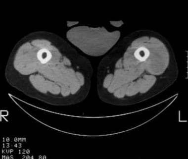
Tomography, X-Ray Computed
Osteosarcoma
Cystadenocarcinoma
Leiomyosarcoma
Neoplasms, Multiple Primary
Chondrosarcoma
Gene expression profiling of human sarcomas: insights into sarcoma biology. (1/114)
Sarcomas are a biologically complex group of tumors of mesenchymal origin. By using gene expression microarray analysis, we aimed to find clues into the cellular differentiation and oncogenic pathways active in these tumors as well as potential biomarkers and therapeutic targets. We examined 181 tumors representing 16 classes of human bone and soft tissue sarcomas on a 12,601-feature cDNA microarray. Remarkably, 2,766 probes differentially expressed across this sample set clearly delineated the various tumor classes. Several genes of potential biological and therapeutic interest were associated with each sarcoma type, including specific tyrosine kinases, transcription factors, and homeobox genes. We also identified subgroups of tumors within the liposarcomas, leiomyosarcomas, and malignant fibrous histiocytomas. We found significant gene ontology correlates for each tumor group and identified similarity to normal tissues by Gene Set Enrichment Analysis. Mutation analysis done on 275 tumor samples revealed that the high expression of epidermal growth factor receptor (EGFR) in certain tumors was not associated with gene mutations. Finally, to further the investigation of human sarcoma biology, we have created an online, publicly available, searchable database housing the data from the gene expression profiles of these tumors (http://watson.nhgri.nih.gov/sarcoma), allowing the user to interactively explore this data set in depth. (+info)Extended pedicle rectus abdominis myocutaneous flap for thigh reconstruction. (2/114)
A rectus abdominis myocutaneous flap can provide a large amount of tissue for defect coverage. Rarely a flabby and redundant abdominal tissue was used as a huge extended flap. We report a case of recurrence malignant fibrous histiocytoma of the thigh which was radically resected. The resultant massive defect was success reconstructed with an extended pedicle inferiorly based rectus abdominis myocutaneous flap. (+info)Proposal of a new grading system for malignant fibrous histiocytomas. (3/114)
The proposed grading system for malignant fibrous histiocytomas (MFH) comprises 3 grades of malignancy. Analogous to other grading systems, the system includes the factors of mitotic rate and necrosis. In addition to these two factors, the concept of cellularity was included. The prognostic relevance of the grading systems published by Costa, Coindre, van Unnik, Pezzi and Tsujimoto as well as the grading system proposed by the present study was tested on 161 MFH. The results showed that all grading systems tested produced clearly significant differences (p < 0.01) with regard to the survival estimated for patients with various grades of malignancy. These results revealed the superiority of systems that use 3 grades of malignancy over a 2-grade classification. The proposed grading system yielded a lower percentage of grade II tumours (37%) than the grading systems of Coindre (60%) and van Unnik (70%). In the multivariate analysis of all grading systems, the proposed grading system was the only one to show prognostic relevance (p < 0. 05). (+info)Postirradiation sarcoma of the sphenoid bone--a case report. (4/114)
INTRODUCTION: The development of secondary tumours as a result of radiation therapy is a rare but serious complication. CLINICAL PICTURE: This is a case report of a 45-year-old Chinese male who developed postirradiation sarcoma of the sphenoid bone in less than 5 years after radiation therapy for Stage T3N1M0 nasopharyngeal carcinoma. DISCUSSION: In the literature, the only case of postirradiation osteosarcoma of the sphenoid bone was after radiation therapy for craniopharyngioma. There was no previously reported case of postirradiation sarcoma of the sphenoid bone after radiation therapy for nasopharyngeal carcinoma. CONCLUSION: This is the first case of postirradiation malignant fibrous histiocytoma of the sphenoid to be reported. Of about 3000 patients treated with radiotherapy for nasopharyngeal carcinoma over a 10-year period in Singapore, only 1 patient developed postirradiation tumour of the sphenoid bone. (+info)Abdominal aorta and inferior vena cava thromboses in advanced stage of malignant fibrous histiocytoma. (5/114)
Asymptomatic simultaneous thrombosis of abdominal aorta and inferior vena cava is a rare complication in advanced malignancy. We described an incidental finding of this clinical entity in our patient who presented with advance stage of malignant fibrous hystiocytoma of soft tissue and pathological fracture. The radiological evaluation with spiral computed tomography scan of abdominal aorta and inferior vena cava are presented and the subsequent management highlighted. (+info)Retroperitoneal soft tissue sarcomas: prognosis and treatment of primary and recurrent disease in 117 patients. (6/114)
BACKGROUND: The objective of this study was to define prognostic factors for patients with primary soft tissue sarcomas (STS) arising from the retroperitoneum. PATIENTS AND METHODS: One hundred and seventeen consecutive patients, resected in our institutions between July 1972 and November 2002, were reviewed. RESULTS: The prognostic factors predicting survival were incomplete resection, a tumor of high grade (G3), metastases to lymph nodes and distant metastasis. Patients with a malignant fibrous histiocytoma (MFH) or a malignant peripheral nerve sheath tumor (MPNST) had a worse prognosis than those patients with other tumors. The prognostic factors predicting local recurrence were incomplete resection and high grade (G3). The prognostic factors predicting metastasis were incomplete resection, lymph node metastasis at the time of the resection of the primary tumor and tumor histology. CONCLUSION: Since only complete tumor resection offers a chance for cure, it is mandatory, and local control remains the most significant challenge in the management of retroperitoneal sarcomas. Other therapies can support surgical treatment, depending on the tumor localization and histological entity. The management of patients with a STS should be provided by a specialized team of surgeons, oncologists and radiotherapists, and patients should be enrolled in a treatment study whenever possible. (+info)Retroperitoneal liposarcomas: follow-up analysis of dedifferentiation after clinicopathologic reexamination of 86 liposarcomas and malignant fibrous histiocytomas. (7/114)
BACKGROUND: Dedifferentiated liposarcoma (DDL) juxtapose components of well-differentiated liposarcoma (WDL) and nonlipogenic sarcoma. Malignant fibrous histiocytoma (MFH) is no longer considered a homogeneous entity, but rather as the common morphologic appearance of various subtypes of sarcomas. The objectives of the current retrospective study were: 1( to analyze the relation between DDLs and tumors previously diagnosed as MFHs; 2) to trace the evolution of liposarcomas, and 3) to assess the consequences of dedifferentiation. METHODS: Between 1974 and 2001, 86 patients with retroperitoneal liposarcoma (61 patients) or MFH (25 patients) underwent surgery at Institut Bergonie in Bordeaux, France. Histologic review was performed and tumors reclassified as WDL or DDL were retained for further clinicohistologic study. Subsequently, initial presentation and all recurrences were analyzed. RESULTS: The 61 liposarcomas consisted of 21 WDLs and 35 DDLs; 17 MFHs were reclassified as DDL. In all, approximately half of the retroperitoneal liposarcomas and MFHs were found to be DDLs. The DDLs presented with a smaller size (20 cm vs.30 cm; P = .05) but a lower rate of complete resection (72% vs.90%; P = .015) and remission (72% vs. 100%; P = .0015). Dedifferentiated recurrence was evidenced in 7 WDLs. Ten DDLs presented with metastatic evolution. DDLs demonstrated a tendency toward lower rates of 5-year overall survival (55% vs. 82%; P = .075). CONCLUSIONS: Most occurrences of retroperitoneal liposarcomas and MFHs are in fact DDLs and dedifferentiated recurrence of WDLs is frequent. Retroperitoneal DDLs present a lower rate of complete remission than WDLs and a risk of metastatic recurrence. Therefore, extensive histologic analysis of WDLs is required to identify an undifferentiated component and avoid misdiagnosis of DDL. (+info)Dedifferentiated central chondrosarcoma. (8/114)
BACKGROUND: The prognosis for patients who develop dedifferentiation of central chondrosarcoma traditionally has been poor. Because not much has been reported about this rare lesion, many uncertainties remain about prognostic factors. METHODS: In this retrospective study, the clinical, radiographic, and histologic features and the treatments in 123 patients from the Rizzoli Institute were reviewed in an attempt to define which factors may be related to outcome in patients with dedifferentiated central chondrosarcoma. RESULTS: Among 123 patients who were included in this study, 109 patients were treated at the Rizzoli Institute, and 14 patients were seen in consultation. There were 66 males and 57 females, and their average age was 59 years. The femur (62 patients), pelvis (28 patients), and humerus (20 patients) were the most common locations. Radiographically, a soft tissue mass was present in 87% of patients, and a bimorphic pattern was appreciated in 53% of patients. Histologically, the cartilaginous component was considered Grade 1 in 63% of patients and Grade 2 in 37% of patients. In most patients, the dedifferentiated component showed the features of an osteosarcoma (92 patients), followed by fibrosarcoma (19 patients), and malignant fibrous histiocytoma (9 patients). For 101 patients, surgery was a component of their definitive management. In 25 patients, surgery was combined with chemotherapy. The 2-year and 5-year survival rates were 34% and 24%, respectively. The median survival was 13 months (95% confidence interval, 9-17 months). CONCLUSIONS: Metastatic disease at diagnosis, malignant fibrous histiocytoma dedifferentiation, and a high percentage of dedifferentiated component were related to poorer outcomes. There was no statistical evidence of any beneficial effect from adjuvant chemotherapy. (+info)Benign fibrous histiocytoma (BFH) is a common benign tumor of the skin and superficial soft tissues. It primarily affects middle-aged adults and is more prevalent in men than women. The exact cause of BFH is unknown, but it's thought to arise from dermal fibroblasts or histiocytes.
Medical Definition: Benign Fibrous Histiocytoma (BFH) is a benign, slowly growing, solitary cutaneous or subcutaneous nodular tumor predominantly composed of a mixture of fibroblastic and histiocytic-like cells. The tumor typically presents as a well-circumscribed, firm, dome-shaped papule or nodule, ranging in size from a few millimeters to several centimeters. Histologically, BFH is characterized by the proliferation of spindle-shaped fibroblasts and histiocytes arranged in a storiform pattern, along with variable amounts of collagen deposition, multinucleated giant cells, and hemosiderin deposits. The lesion usually has a pushing border with no invasion into the surrounding tissues. BFH generally follows a benign clinical course, with local recurrence being uncommon following complete surgical excision.
Malignant fibrous histiocytoma (MFH) is not a specific type of histiocytoma; rather, it is a type of soft tissue sarcoma. Histiocytomas are benign tumors that arise from cells called histiocytes, which are part of the immune system. MFH, on the other hand, is a malignant (cancerous) tumor that can arise in various types of soft tissues, such as muscle, fat, tendons, and ligaments.
MFH was once thought to originate from histiocytes, but more recent research suggests that it may actually arise from undifferentiated mesenchymal cells, which are capable of developing into a variety of different cell types. MFH is the most common type of soft tissue sarcoma in adults over the age of 50 and typically presents as a painless mass in the extremities or retroperitoneum (the area in the back of the abdomen).
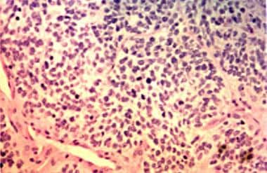
The tumor is characterized by the presence of fibroblastic and histiocytic-like cells, which can be quite pleomorphic (varied in shape and size) and may contain numerous mitotic figures (indicating rapid cell division). Treatment typically involves surgical excision, often followed by radiation therapy and/or chemotherapy. The prognosis for MFH depends on several factors, including the tumor's location, size, grade (degree of differentiation), and the patient's age and overall health.
Histiocytoma is a general term used to describe a group of disorders characterized by an abnormal accumulation or proliferation of histiocytes, which are a type of immune cell. These cells normally play a role in fighting infection and helping to heal wounds. However, when they multiply excessively, they can form tumors known as histiocytomas.
There are several types of histiocytomas, including:
1. Cutaneous histiocytoma: This is the most common type of histiocytoma, which typically appears as a small, raised, hairless, and pink or red bump on the skin. It usually affects dogs, but can also occur in cats and rarely in humans. These tumors are benign and often regress spontaneously within a few months.
2. Systemic histiocytoses: These are less common types of histiocytomas that involve multiple organs and tissues throughout the body. They can be further classified into several subtypes, such as Langerhans cell histiocytosis (LCH), Erdheim-Chester disease (ECD), and malignant histiocytosis. These conditions can range from benign to malignant and may require aggressive treatment, including chemotherapy or radiation therapy.
It is important to note that while histiocytomas are generally benign, they can sometimes mimic other more serious conditions. Therefore, it is essential to have any suspicious growths evaluated by a veterinarian or healthcare professional for proper diagnosis and management.
Soft tissue neoplasms refer to abnormal growths or tumors that develop in the soft tissues of the body. Soft tissues include muscles, tendons, ligaments, fascia, nerves, blood vessels, fat, and synovial membranes (the thin layer of cells that line joints and tendons). Neoplasms can be benign (non-cancerous) or malignant (cancerous), and their behavior and potential for spread depend on the specific type of neoplasm.
Benign soft tissue neoplasms are typically slow-growing, well-circumscribed, and rarely spread to other parts of the body. They can often be removed surgically with a low risk of recurrence. Examples of benign soft tissue neoplasms include lipomas (fat tumors), schwannomas (nerve sheath tumors), and hemangiomas (blood vessel tumors).
Malignant soft tissue neoplasms, on the other hand, can grow rapidly, invade surrounding tissues, and may metastasize (spread) to distant parts of the body. They are often more difficult to treat than benign neoplasms and require a multidisciplinary approach, including surgery, radiation therapy, and chemotherapy. Examples of malignant soft tissue neoplasms include sarcomas, such as rhabdomyosarcoma (arising from skeletal muscle), leiomyosarcoma (arising from smooth muscle), and angiosarcoma (arising from blood vessels).
It is important to note that soft tissue neoplasms can occur in any part of the body, and their diagnosis and treatment require a thorough evaluation by a healthcare professional with expertise in this area.
Histiocytic sarcoma is a rare type of cancer that originates from histiocytes, which are cells that are part of the immune system and found in various tissues throughout the body. These cells normally function to help fight infection and remove foreign substances. In histiocytic sarcoma, there is an abnormal accumulation and proliferation of these cells, leading to the formation of tumors.
Histiocytic sarcoma can affect people of any age but is more commonly found in adults, with a slight male predominance. It can occur in various parts of the body, such as the lymph nodes, skin, soft tissues, and internal organs like the spleen, liver, and lungs. The exact cause of histiocytic sarcoma remains unknown, but it is not considered to be hereditary.
The symptoms of histiocytic sarcoma depend on the location and extent of the tumor(s). Common signs include swollen lymph nodes, fatigue, fever, weight loss, night sweats, and pain or discomfort in the affected area. Diagnosis typically involves a combination of imaging studies (like CT scans, PET scans, or MRI), biopsies, and laboratory tests to confirm the presence of histiocytic sarcoma and assess its extent.
Treatment for histiocytic sarcoma usually involves a multidisciplinary approach, including surgery, radiation therapy, and chemotherapy. The choice of treatment depends on several factors, such as the location and stage of the disease, the patient's overall health, and their personal preferences. Clinical trials may also be an option for some patients, allowing them to access new and experimental therapies.
Prognosis for histiocytic sarcoma is generally poor, with a five-year survival rate of approximately 15-30%. However, outcomes can vary significantly depending on individual factors, such as the patient's age, the extent of the disease at diagnosis, and the effectiveness of treatment. Continued research is necessary to improve our understanding of this rare cancer and develop more effective therapies for those affected.
Sarcoma is a type of cancer that develops from certain types of connective tissue (such as muscle, fat, fibrous tissue, blood vessels, or nerves) found throughout the body. It can occur in any part of the body, but it most commonly occurs in the arms, legs, chest, and abdomen.
Sarcomas are classified into two main groups: bone sarcomas and soft tissue sarcomas. Bone sarcomas develop in the bones, while soft tissue sarcomas develop in the soft tissues of the body, such as muscles, tendons, ligaments, fat, blood vessels, and nerves.
Sarcomas can be further classified into many subtypes based on their specific characteristics, such as the type of tissue they originate from, their genetic makeup, and their appearance under a microscope. The different subtypes of sarcoma have varying symptoms, prognoses, and treatment options.
Overall, sarcomas are relatively rare cancers, accounting for less than 1% of all cancer diagnoses in the United States each year. However, they can be aggressive and may require intensive treatment, such as surgery, radiation therapy, and chemotherapy.
Skull neoplasms refer to abnormal growths or tumors that develop within the skull. These growths can be benign (non-cancerous) or malignant (cancerous). They can originate from various types of cells, such as bone cells, nerve cells, or soft tissues. Skull neoplasms can cause various symptoms depending on their size and location, including headaches, seizures, vision problems, hearing loss, and neurological deficits. Treatment options include surgery, radiation therapy, and chemotherapy. It is important to note that a neoplasm in the skull can also refer to metastatic cancer, which has spread from another part of the body to the skull.
Liposarcoma is a type of soft tissue sarcoma, which is a cancer that develops in the soft tissues of the body, such as fat, muscle, nerves, blood vessels, and fibrous tissues. Specifically, liposarcoma arises from fat cells (adipocytes) or their precursors.
There are several subtypes of liposarcoma, which differ in their appearance under the microscope, genetic features, and clinical behavior. These include well-differentiated, dedifferentiated, myxoid, round cell, and pleomorphic liposarcomas. The most common sites for liposarcoma are the thigh, retroperitoneum (the area behind the abdominal cavity), and the buttock.
Liposarcomas can grow slowly or rapidly, and they may spread to other parts of the body (metastasize) through the bloodstream or lymphatic system. Treatment typically involves surgical removal of the tumor, often followed by radiation therapy and/or chemotherapy. The prognosis for liposarcoma depends on several factors, including the type and grade of the tumor, its size and location, and whether it has spread to other parts of the body.
Retroperitoneal neoplasms refer to abnormal growths or tumors that develop in the retroperitoneal space. This is the area located behind the peritoneum, which is the membrane that lines the abdominal cavity and covers the abdominal organs. The retroperitoneal space contains several vital structures such as the kidneys, adrenal glands, pancreas, aorta, and lymphatic vessels.
Retroperitoneal neoplasms can be benign or malignant (cancerous). Malignant retroperitoneal neoplasms are often aggressive and can invade surrounding tissues and organs, leading to various complications. Common types of retroperitoneal neoplasms include lymphomas, sarcomas, and metastatic tumors from other primary sites. Symptoms may vary depending on the size and location of the tumor but can include abdominal or back pain, weight loss, and swelling in the legs. Diagnosis typically involves imaging studies such as CT scans or MRI, followed by a biopsy to determine the type and grade of the tumor. Treatment options may include surgery, radiation therapy, chemotherapy, or a combination of these approaches.
Femoral neoplasms refer to abnormal growths or tumors that develop in the femur, which is the long thigh bone in the human body. These neoplasms can be benign (non-cancerous) or malignant (cancerous). Benign femoral neoplasms are slow-growing and rarely spread to other parts of the body, while malignant neoplasms are aggressive and can invade nearby tissues and organs, as well as metastasize (spread) to distant sites.
There are various types of femoral neoplasms, including osteochondromas, enchondromas, chondrosarcomas, osteosarcomas, and Ewing sarcomas, among others. The specific type of neoplasm is determined by the cell type from which it arises and its behavior.
Symptoms of femoral neoplasms may include pain, swelling, stiffness, or weakness in the thigh, as well as a palpable mass or limited mobility. Diagnosis typically involves imaging studies such as X-rays, CT scans, or MRI, as well as biopsy to determine the type and grade of the tumor. Treatment options may include surgery, radiation therapy, chemotherapy, or a combination of these approaches, depending on the type, size, location, and stage of the neoplasm.
Angiomatosis is a medical term that refers to a benign condition characterized by the proliferation of blood vessels in various tissues and organs. It is typically composed of small, tangled blood vessels called capillaries, which can form clusters or networks. The condition can affect skin, internal organs, bones, and other tissues.
Angiomatosis is often asymptomatic and may be discovered incidentally during medical imaging or surgical procedures. In some cases, it may cause symptoms such as pain, swelling, or bleeding, depending on the location and extent of the lesions.
While angiomatosis is generally a benign condition, in rare cases, it can be associated with malignant tumors or other medical conditions. Treatment options for angiomatosis depend on the size, location, and symptoms of the lesions and may include observation, medication, or surgical removal.
Myxosarcoma is a very rare type of soft tissue sarcoma, a cancer that develops in the soft tissues of the body, such as fat, muscle, nerves, blood vessels, and fibrous tissues. Myxosarcomas are characterized by the presence of mucoid or gelatinous material in the tumor, which is composed of an abnormal accumulation of acid mucopolysaccharides. These tumors typically affect adults, with a peak incidence in the sixth to seventh decade of life. They usually occur in the extremities, particularly the lower limbs, and can also arise in the retroperitoneum or other deep soft tissues. Myxosarcomas are classified into several subtypes based on their histological features, with the most common being the myxofibrosarcoma. Treatment typically involves surgical resection with wide margins, often followed by radiation therapy and/or chemotherapy. The prognosis for patients with myxosarcoma depends on several factors, including the size and location of the tumor, the histological grade, and the patient's age and overall health.
Piezosurgery is a type of surgical procedure that uses ultrasonic vibrations to cut through bone tissue while minimizing damage to surrounding soft tissues. It is often used in oral and maxillofacial surgery, such as during dental implant placement or jaw osteotomies. The piezoelectric instrument generates high-frequency microvibrations that selectively cut mineralized tissue like bone, while leaving adjacent soft tissues largely unaffected. This allows for precise cuts with less trauma and bleeding compared to traditional surgical techniques, potentially resulting in faster healing times and reduced postoperative discomfort.
Vascular neoplasms are a type of tumor that develops from cells that line the blood vessels or lymphatic vessels. These tumors can be benign (non-cancerous) or malignant (cancerous). Benign vascular neoplasms, such as hemangiomas and lymphangiomas, are usually harmless and may not require treatment unless they cause symptoms or complications. Malignant vascular neoplasms, on the other hand, are known as angiosarcomas and can be aggressive, spreading to other parts of the body and potentially causing serious health problems.
Angiosarcomas can develop in any part of the body but are most commonly found in the skin, particularly in areas exposed to radiation or chronic lymph edema. They can also occur in the breast, liver, spleen, and heart. Treatment for vascular neoplasms depends on the type, location, size, and stage of the tumor, as well as the patient's overall health. Treatment options may include surgery, radiation therapy, chemotherapy, or a combination of these approaches.
Heart neoplasms are abnormal growths or tumors that develop within the heart tissue. They can be benign (noncancerous) or malignant (cancerous). Benign tumors, such as myxomas and rhabdomyomas, are typically slower growing and less likely to spread, but they can still cause serious complications if they obstruct blood flow or damage heart valves. Malignant tumors, such as angiosarcomas and rhabdomyosarcomas, are fast-growing and have a higher risk of spreading to other parts of the body. Symptoms of heart neoplasms can include shortness of breath, chest pain, fatigue, and irregular heart rhythms. Treatment options depend on the type, size, and location of the tumor, and may include surgery, radiation therapy, or chemotherapy.
Bone neoplasms are abnormal growths or tumors that develop in the bone. They can be benign (non-cancerous) or malignant (cancerous). Benign bone neoplasms do not spread to other parts of the body and are rarely a threat to life, although they may cause problems if they grow large enough to press on surrounding tissues or cause fractures. Malignant bone neoplasms, on the other hand, can invade and destroy nearby tissue and may spread (metastasize) to other parts of the body.
There are many different types of bone neoplasms, including:
1. Osteochondroma - a benign tumor that develops from cartilage and bone
2. Enchondroma - a benign tumor that forms in the cartilage that lines the inside of the bones
3. Chondrosarcoma - a malignant tumor that develops from cartilage
4. Osteosarcoma - a malignant tumor that develops from bone cells
5. Ewing sarcoma - a malignant tumor that develops in the bones or soft tissues around the bones
6. Giant cell tumor of bone - a benign or occasionally malignant tumor that develops from bone tissue
7. Fibrosarcoma - a malignant tumor that develops from fibrous tissue in the bone
The symptoms of bone neoplasms vary depending on the type, size, and location of the tumor. They may include pain, swelling, stiffness, fractures, or limited mobility. Treatment options depend on the type and stage of the tumor but may include surgery, radiation therapy, chemotherapy, or a combination of these treatments.
Genital neoplasms in males refer to abnormal growths or tumors that develop in the male reproductive organs. These can be benign (non-cancerous) or malignant (cancerous).
Malignant genital neoplasms are often referred to as genital cancers. The most common types of male genital cancers include:
1. Penile Cancer: This occurs when cancer cells form in the tissues of the penis.
2. Testicular Cancer: This forms in the testicles (testes), which are located inside the scrotum.
3. Prostate Cancer: This is a common cancer in men, forming in the prostate gland, which is part of the male reproductive system that helps make semen.
4. Scrotal Cancer: This is a rare form of cancer that forms in the skin or tissue of the scrotum.
5. Penile Intraepithelial Neoplasia (PeIN): This is not cancer, but it is considered a pre-cancerous condition of the penis.
Early detection and treatment of genital neoplasms can significantly improve the prognosis. Regular self-examinations and medical check-ups are recommended, especially for individuals with risk factors such as smoking, HIV infection, or a family history of these cancers.
Tracheobronchomegaly is a rare condition characterized by an abnormal dilatation or widening of the trachea and bronchi, which are the airway tubes leading to the lungs. This condition is also known as Mounier-Kuhn syndrome. It is typically associated with recurrent respiratory infections, coughing, and difficulty breathing, especially during physical exertion. The exact cause of tracheobronchomegaly is not well understood, but it may be related to a congenital abnormality or connective tissue disorder. Diagnosis is often made through imaging studies such as chest X-rays or CT scans. Treatment typically involves managing symptoms and preventing complications, and may include bronchodilators, antibiotics, and respiratory therapy. In severe cases, surgery may be necessary to repair or reinforce the airway walls.
A fatal outcome is a term used in medical context to describe a situation where a disease, injury, or illness results in the death of an individual. It is the most severe and unfortunate possible outcome of any medical condition, and is often used as a measure of the severity and prognosis of various diseases and injuries. In clinical trials and research, fatal outcome may be used as an endpoint to evaluate the effectiveness and safety of different treatments or interventions.
There is no medical definition for "dog diseases" as it is too broad a term. However, dogs can suffer from various health conditions and illnesses that are specific to their species or similar to those found in humans. Some common categories of dog diseases include:
1. Infectious Diseases: These are caused by viruses, bacteria, fungi, or parasites. Examples include distemper, parvovirus, kennel cough, Lyme disease, and heartworms.
2. Hereditary/Genetic Disorders: Some dogs may inherit certain genetic disorders from their parents. Examples include hip dysplasia, elbow dysplasia, progressive retinal atrophy (PRA), and degenerative myelopathy.
3. Age-Related Diseases: As dogs age, they become more susceptible to various health issues. Common age-related diseases in dogs include arthritis, dental disease, cancer, and cognitive dysfunction syndrome (CDS).
4. Nutritional Disorders: Malnutrition or improper feeding can lead to various health problems in dogs. Examples include obesity, malnutrition, and vitamin deficiencies.
5. Environmental Diseases: These are caused by exposure to environmental factors such as toxins, allergens, or extreme temperatures. Examples include heatstroke, frostbite, and toxicities from ingesting harmful substances.
6. Neurological Disorders: Dogs can suffer from various neurological conditions that affect their nervous system. Examples include epilepsy, intervertebral disc disease (IVDD), and vestibular disease.
7. Behavioral Disorders: Some dogs may develop behavioral issues due to various factors such as anxiety, fear, or aggression. Examples include separation anxiety, noise phobias, and resource guarding.
It's important to note that regular veterinary care, proper nutrition, exercise, and preventative measures can help reduce the risk of many dog diseases.
Spontaneous neoplasm regression is a rare and somewhat controversial phenomenon in which a tumor or malignancy appears to decrease in size or disappear without any treatment or with treatment that is typically not expected to produce such an effect. This can occur through various mechanisms, including immune-mediated processes, apoptosis (programmed cell death), differentiation of cancer cells into normal cells, and angiogenesis inhibition (preventing the growth of new blood vessels that feed the tumor).
Spontaneous regression of neoplasms is not well understood and is considered unpredictable. It has been reported in various types of cancers, including neuroblastoma, melanoma, renal cell carcinoma, and others. However, it should be noted that spontaneous regression does not imply a cure, as the tumor may still recur or metastasize later on.
In summary, spontaneous neoplasm regression refers to the partial or complete disappearance of a malignancy without any specific treatment or with treatment that is not typically associated with such an effect.
In the context of human anatomy, the thigh is the part of the lower limb that extends from the hip to the knee. It is the upper and largest portion of the leg and is primarily composed of the femur bone, which is the longest and strongest bone in the human body, as well as several muscles including the quadriceps femoris (front thigh), hamstrings (back thigh), and adductors (inner thigh). The major blood vessels and nerves that supply the lower limb also pass through the thigh.
Giant cell tumors (GCTs) are a type of benign or rarely malignant bone tumor that is characterized by the presence of multinucleated giant cells. These tumors typically affect adults between the ages of 20 and 40, and they can occur in any bone, but they most commonly involve the long bones near the knee joint.
GCTs are composed of three types of cells: mononuclear stromal cells, which produce the matrix of the tumor; multinucleated osteoclast-like giant cells, which resemble the bone-resorbing cells found in normal bone; and macrophages, which are part of the body's immune system.
The mononuclear stromal cells produce a variety of growth factors that stimulate the formation and activity of the osteoclast-like giant cells, leading to localized bone destruction. The tumor may cause pain, swelling, and limited mobility in the affected area.
While GCTs are typically benign, they can be aggressive and locally destructive, with a tendency to recur after surgical removal. In some cases, GCTs may undergo malignant transformation, leading to the development of sarcomas. Treatment options for GCTs include curettage (scraping out) of the tumor, followed by bone grafting or the use of a cement spacer to fill the defect, and/or adjuvant therapy with radiation or chemotherapy.
A fibroma is a benign (non-cancerous) tumor that consists primarily of fibrous or connective tissue. It can occur in various parts of the body, including the skin, mouth, and internal organs. The term "fibroma" is often used to describe any benign fibrous growth, but there are specific types of fibromas such as dermatofibroma (found in the skin), oral fibroma (found in the mouth), and benign fibrous histiocytoma (found in soft tissues).
It's important to note that while fibromas are generally harmless, they can cause discomfort or problems depending on their size and location. If a fibroma is causing issues or there's concern about its growth or malignancy, it should be evaluated by a healthcare professional for potential removal or further assessment.
Histiocytes are a type of immune cell that are part of the mononuclear phagocyte system. They originate from monocytes, which are derived from hematopoietic stem cells in the bone marrow. Histiocytes play an important role in the immune system by engulfing and destroying foreign substances, such as bacteria and viruses, as well as removing dead cells and other debris from the body. They can be found in various tissues throughout the body, including the skin, lymph nodes, spleen, and liver.
Histiocytes include several different types of cells, such as macrophages, dendritic cells, and Langerhans cells. These cells have different functions but all play a role in the immune response. For example, macrophages are involved in inflammation and tissue repair, while dendritic cells are important for presenting antigens to T cells and initiating an immune response.
Abnormal accumulations or dysfunction of histiocytes can lead to various diseases, such as histiocytosis, which is a group of disorders characterized by the abnormal proliferation and accumulation of histiocytes in various tissues.
Maxillary sinus neoplasms refer to abnormal growths or tumors that develop in the maxillary sinuses, which are located in the upper part of your cheekbones, below your eyes. These growths can be benign (non-cancerous) or malignant (cancerous).
Benign neoplasms may include conditions such as an osteoma (a benign bone tumor), a papilloma (a benign growth of the lining of the sinus), or a fibrous dysplasia (a condition where bone is replaced by fibrous tissue).
Malignant neoplasms, on the other hand, can be primary (originating in the maxillary sinuses) or secondary (spreading to the maxillary sinuses from another site in the body). Common types of malignant tumors that arise in the maxillary sinus include squamous cell carcinoma, adenocarcinoma, and mucoepidermoid carcinoma.
Symptoms of maxillary sinus neoplasms may include nasal congestion, nosebleeds, facial pain or numbness, vision changes, and difficulty swallowing or speaking. Treatment options depend on the type, size, and location of the tumor but may include surgery, radiation therapy, chemotherapy, or a combination of these approaches.
Immunohistochemistry (IHC) is a technique used in pathology and laboratory medicine to identify specific proteins or antigens in tissue sections. It combines the principles of immunology and histology to detect the presence and location of these target molecules within cells and tissues. This technique utilizes antibodies that are specific to the protein or antigen of interest, which are then tagged with a detection system such as a chromogen or fluorophore. The stained tissue sections can be examined under a microscope, allowing for the visualization and analysis of the distribution and expression patterns of the target molecule in the context of the tissue architecture. Immunohistochemistry is widely used in diagnostic pathology to help identify various diseases, including cancer, infectious diseases, and immune-mediated disorders.
Orbital neoplasms refer to abnormal growths or tumors that develop in the orbit, which is the bony cavity that contains the eyeball, muscles, nerves, fat, and blood vessels. These neoplasms can be benign (non-cancerous) or malignant (cancerous), and they can arise from various types of cells within the orbit.
Orbital neoplasms can cause a variety of symptoms depending on their size, location, and rate of growth. Common symptoms include protrusion or displacement of the eyeball, double vision, limited eye movement, pain, swelling, and numbness in the face. In some cases, orbital neoplasms may not cause any noticeable symptoms, especially if they are small and slow-growing.
There are many different types of orbital neoplasms, including:
1. Optic nerve glioma: a rare tumor that arises from the optic nerve's supportive tissue.
2. Orbital meningioma: a tumor that originates from the membranes covering the brain and extends into the orbit.
3. Lacrimal gland tumors: benign or malignant growths that develop in the lacrimal gland, which produces tears.
4. Orbital lymphangioma: a non-cancerous tumor that arises from the lymphatic vessels in the orbit.
5. Rhabdomyosarcoma: a malignant tumor that develops from the skeletal muscle cells in the orbit.
6. Metastatic tumors: cancerous growths that spread to the orbit from other parts of the body, such as the breast, lung, or prostate.
The diagnosis and treatment of orbital neoplasms depend on several factors, including the type, size, location, and extent of the tumor. Imaging tests, such as CT scans and MRI, are often used to visualize the tumor and determine its extent. A biopsy may also be performed to confirm the diagnosis and determine the tumor's type and grade. Treatment options include surgery, radiation therapy, chemotherapy, or a combination of these approaches.
Skin neoplasms refer to abnormal growths or tumors in the skin that can be benign (non-cancerous) or malignant (cancerous). They result from uncontrolled multiplication of skin cells, which can form various types of lesions. These growths may appear as lumps, bumps, sores, patches, or discolored areas on the skin.
Benign skin neoplasms include conditions such as moles, warts, and seborrheic keratoses, while malignant skin neoplasms are primarily classified into melanoma, squamous cell carcinoma, and basal cell carcinoma. These three types of cancerous skin growths are collectively known as non-melanoma skin cancers (NMSCs). Melanoma is the most aggressive and dangerous form of skin cancer, while NMSCs tend to be less invasive but more common.
It's essential to monitor any changes in existing skin lesions or the appearance of new growths and consult a healthcare professional for proper evaluation and treatment if needed.
Dermatofibrosarcoma protuberans (DFSP) is a rare type of skin cancer that begins in the middle layer of the skin known as the dermis. It often appears as a scar or bruise that does not go away and may grow slowly over time, sometimes spreading to deeper tissues and other parts of the body. DFSP can be difficult to treat if it has spread, but when caught early, it is usually curable with surgery.
DFSP is characterized by the growth of abnormal fibroblasts, which are cells that produce collagen, a protein that helps make up connective tissues in the body. The exact cause of DFSP is not known, but it has been linked to genetic mutations and previous injuries or surgeries to the skin.
Treatment for DFSP typically involves surgical removal of the tumor, along with a margin of healthy tissue around it. In some cases, radiation therapy or targeted therapy may also be used to help ensure that all cancer cells have been removed. Regular follow-up care is important to monitor for any signs of recurrence or spread of the disease.
X-ray computed tomography (CT or CAT scan) is a medical imaging method that uses computer-processed combinations of many X-ray images taken from different angles to produce cross-sectional (tomographic) images (virtual "slices") of the body. These cross-sectional images can then be used to display detailed internal views of organs, bones, and soft tissues in the body.
The term "computed tomography" is used instead of "CT scan" or "CAT scan" because the machines take a series of X-ray measurements from different angles around the body and then use a computer to process these data to create detailed images of internal structures within the body.
CT scanning is a noninvasive, painless medical test that helps physicians diagnose and treat medical conditions. CT imaging provides detailed information about many types of tissue including lung, bone, soft tissue and blood vessels. CT examinations can be performed on every part of the body for a variety of reasons including diagnosis, surgical planning, and monitoring of therapeutic responses.
In computed tomography (CT), an X-ray source and detector rotate around the patient, measuring the X-ray attenuation at many different angles. A computer uses this data to construct a cross-sectional image by the process of reconstruction. This technique is called "tomography". The term "computed" refers to the use of a computer to reconstruct the images.
CT has become an important tool in medical imaging and diagnosis, allowing radiologists and other physicians to view detailed internal images of the body. It can help identify many different medical conditions including cancer, heart disease, lung nodules, liver tumors, and internal injuries from trauma. CT is also commonly used for guiding biopsies and other minimally invasive procedures.
In summary, X-ray computed tomography (CT or CAT scan) is a medical imaging technique that uses computer-processed combinations of many X-ray images taken from different angles to produce cross-sectional images of the body. It provides detailed internal views of organs, bones, and soft tissues in the body, allowing physicians to diagnose and treat medical conditions.
Osteosarcoma is defined as a type of cancerous tumor that arises from the cells that form bones (osteoblasts). It's the most common primary bone cancer, and it typically develops in the long bones of the body, such as the arms or legs, near the growth plates. Osteosarcoma can metastasize (spread) to other parts of the body, including the lungs, making it a highly malignant form of cancer. Symptoms may include bone pain, swelling, and fractures. Treatment usually involves a combination of surgery, chemotherapy, and/or radiation therapy.
Cystadenocarcinoma is a type of tumor that arises from the epithelial lining of a cyst, and it has the potential to invade surrounding tissues and spread (metastasize) to other parts of the body. It typically affects glandular organs such as the ovaries, pancreas, and salivary glands.
Cystadenocarcinomas can be classified into two types: serous and mucinous. Serous cystadenocarcinomas produce a watery fluid, while mucinous cystadenocarcinomas produce a thick, mucus-like fluid. Both types of tumors can be benign or malignant, but malignant cystadenocarcinomas are more aggressive and have a higher risk of metastasis.
Symptoms of cystadenocarcinoma depend on the location and size of the tumor. In some cases, there may be no symptoms until the tumor has grown large enough to cause pain or other problems. Treatment typically involves surgical removal of the tumor, along with any affected surrounding tissue. Chemotherapy and radiation therapy may also be used in some cases to help prevent recurrence or spread of the cancer.
Leiomyosarcoma is a type of cancer that arises from the smooth muscle cells, which are responsible for the involuntary contractions of various organs and blood vessels. It most commonly occurs in the uterus, soft tissues (such as muscles and fat), and the gastrointestinal tract.
Leiomyosarcomas can vary in their aggressiveness and may spread to other parts of the body (metastasize) through the bloodstream or lymphatic system. The prognosis for leiomyosarcoma depends on several factors, including the location and size of the tumor, the patient's age and overall health, and the extent of metastasis. Treatment typically involves surgical removal of the tumor, along with radiation therapy and/or chemotherapy to help prevent recurrence or spread of the cancer.
Multiple primary neoplasms refer to the occurrence of more than one primary malignant tumor in an individual, where each tumor is unrelated to the other and originates from separate cells or organs. This differs from metastatic cancer, where a single malignancy spreads to multiple sites in the body. Multiple primary neoplasms can be synchronous (occurring at the same time) or metachronous (occurring at different times). The risk of developing multiple primary neoplasms increases with age and is associated with certain genetic predispositions, environmental factors, and lifestyle choices such as smoking and alcohol consumption.
Chondrosarcoma is a type of cancer that develops in the cartilaginous tissue, which is the flexible and smooth connective tissue found in various parts of the body such as the bones, ribs, and nose. It is characterized by the production of malignant cartilage cells that can invade surrounding tissues and spread to other parts of the body (metastasis).
Chondrosarcomas are typically slow-growing tumors but can be aggressive in some cases. They usually occur in adults over the age of 40, and men are more commonly affected than women. The most common sites for chondrosarcoma development include the bones of the pelvis, legs, and arms.
Treatment for chondrosarcoma typically involves surgical removal of the tumor, along with radiation therapy or chemotherapy in some cases. The prognosis for chondrosarcoma depends on several factors, including the size and location of the tumor, the grade of malignancy, and whether it has spread to other parts of the body.
Mediastinal neoplasms refer to abnormal growths or tumors located in the mediastinum, which is the central compartment of the thoracic cavity that lies between the lungs and contains various vital structures such as the heart, esophagus, trachea, blood vessels, lymph nodes, and nerves. Mediastinal neoplasms can be benign (non-cancerous) or malignant (cancerous), and they can arise from any of the tissues or organs within the mediastinum.
Benign mediastinal neoplasms may include thymomas, lipomas, neurofibromas, or teratomas, among others. These tumors are typically slow-growing and rarely spread to other parts of the body. However, they can still cause symptoms or complications by compressing adjacent structures within the mediastinum, such as the airways, blood vessels, or nerves.
Malignant mediastinal neoplasms are cancerous tumors that can invade and destroy surrounding tissues and may spread (metastasize) to other parts of the body. Common types of malignant mediastinal neoplasms include thymic carcinomas, lymphomas, germ cell tumors, and neuroendocrine tumors. These tumors often require aggressive treatment, such as surgery, radiation therapy, and chemotherapy, to control their growth and spread.
It is important to note that mediastinal neoplasms can present with various symptoms depending on their location, size, and type. Some patients may be asymptomatic, while others may experience cough, chest pain, difficulty breathing, hoarseness, or swallowing difficulties. A thorough diagnostic workup, including imaging studies and biopsies, is necessary to confirm the diagnosis and determine the best course of treatment for mediastinal neoplasms.
 Sarcoma
Sharon Weiss
Margaret Ransone Murray
Hardcastle syndrome
Vaccine-associated sarcoma
Atypical fibroxanthoma
Fibrosarcoma
Doege-Potter syndrome
Histiocytoma
Betacellulin
Epiregulin
Angiomatoid fibrous histiocytoma
Low-grade myofibroblastic sarcoma
Myxofibrosarcoma
Nodular fasciitis
Ewing sarcoma
Liposarcoma
Werner syndrome
TSPAN31
CD34
List of skin conditions
Vulvar tumors
List of diseases (M)
List of MeSH codes (C04)
Fibroblastic and myofibroblastic tumors
List of cancer types
Proliferative fasciitis and proliferative myositis
Soft tissue sarcoma in cats and dogs
Index of oncology articles
Bone tumor
 Pleomorphic Sarcoma (Malignant Fibrous Histiocytoma) of Soft Tissue Imaging: Practice Essentials, Radiography, Computed...
Mfhas1 MGI Mouse Gene Detail - MGI:1098644 - malignant fibrous histiocytoma amplified sequence 1
 Malignant Fibrous Histiocytoma Global Clinical Trials Review, H2, 2016
Development of Chronic Myelocytic Leukemia after Chemotherapy for Malignant Fibrous Histiocytoma | Acta Haematologica | Karger...
 Malignant fibrous histiocytoma - causes, side effects and treatments at NaturalPedia.com
What Is Malignant Fibrous Histiocytoma? Symptoms & Treatment - Hdkino.org
 Primary pulmonary synovial sarcoma diagnosed ten years after treatment for malignant fibrous histiocytoma
Malignant fibrous histiocytoma
 Malignant Fibrous Histiocytoma (Skin) - Symptoms, Treatment & Support - Without a Ribbon
Myxoid malignant fibrous histiocytoma of the bladder<...
 October 1977 - Volume 60 - Issue 4 : Plastic and Reconstructive Surgery
A case of intracranial malignant fibrous histiocytoma | Sarrami | Journal of Research in Medical Sciences
Sarcoma - Wikipedia
Treatment of Malignant Fibrous Histiocytoma and Atypical Fibrous Xanthomas with Micrographic Surgery<...
 British Medical Journal: 298 (6675) | The BMJ
![Malignant fibrous histiocytoma of bones: an important differential diagnosis of malignant bone neoplasms]. | AMB Rev Assoc Med...](data:image/png;base64,iVBORw0KGgoAAAANSUhEUgAAABAAAAAQCAYAAAAf8/9hAAACpklEQVQ4jaXTe2iNcRzH8ffz7Jzncp4ZZyZjZ6bmEvPPxrDIJSnJZZJIbn8Qk1CI2pQ7JX8h5fKHW4o/3EuaWnJvrUNqiFZssTWcs83z7DnPc87XH88Ziv98//z1+7369v38vvCfpQBY0w6LeHb2RCHl+fg9vaiGxuyq0ZQMzef0tSeoOSqGEQaR4Go4QghAPBvxHRzbBddjUGGUhXMmULN0KhVlxQDMLC+iZt9VEt+6MPNMCIwAAEj7aSrGxFg5r5KVCyoZGM2lx3Y5dbGB6jnlLJs7nvIxMZbvOE9TvAUjagXtAygVu2Tk3P3ieb6IiPjpjBw9Vy+F02uFYWulaEadnLn2WEREJCOyetdFYdRGMSfXSQ4ABVV7Jo4rYtWCiVyvf8nizWe4cuMZdkYwBlh863a4fa+JR/EWyscWs37pFAxD4/7T96hB/2lKhuYDcORsPW+aWzHz+6FrIRDB1MMY0Vwe3I9Td+IuALMmjyIjZAGBkiFRACKmBqb+77hyDQw9DED8TRukvCygKpQWFwCQyUb0zxIwtQD40NoZPAVQdY3S2EAAkj9cfo/3b0DJ+h8/fwdVCYA8S6d0WNBBbHB/6LJJeWmUPkiB3pQPPQ7RPDMAviQglBMAtuNSs/cqyW6HO8fXcaB2CZYWwk78IOWlcb72UDjA4tihFRzcOg+Ato4EaigIkUjVbmHEBimbf1Diza0iIvKupV2qN52W/MrtsuPoDUl22dJXl2+9EHP8NtEn1YnSB0i6F6fbwYronKxdwupFkwCwbZdIRKe9s5sLN59z6W4jr962oZs6OVrk91dGhEg/A8f1WbPzPI3Nnzi8ZT7NLe2cuPyQWw2vSXQmwdAwLIM/xvP3NmYyGXptl+GxAlo7kvi2S9gyCIfVX0sEwTb+BBBpI5QgousUAAAAAElFTkSuQmCC) Malignant fibrous histiocytoma of bones: an important differential diagnosis of malignant bone neoplasms]. | AMB Rev Assoc Med...
Malignant fibrous histiocytoma of bones: an important differential diagnosis of malignant bone neoplasms]. | AMB Rev Assoc Med...
 A new complication of retained surgical gauze: development of malignant fibrous histiocytoma - report of a case with a...
 Late development of a malignant fibrous histiocytoma at the site of a giant cell tumor<...
 https://www.cancer.gov/types/bone/patient/osteosarcoma-treatment-pdq
 Rhabdomyosarcoma : Wheeless' Textbook of Orthopaedics
![AIKOU T[au] - Search Results - PubMed](data:image/png;base64,iVBORw0KGgoAAAANSUhEUgAAABAAAAAQCAMAAAAoLQ9TAAAARVBMVEVHcEwoU45gYmYAUpQAUpRPYGVgYmZLXnJgYmYAUZUAUpRJXnIAUpQAUpRgYmYAUpRgYmZgYmZhYmYAUpQAUpQAUpRgYmaDiPJuAAAAFXRSTlMADOJ+6QewGO8/uTRqtH7GdFJ11p1bCL3TAAAAZUlEQVQYlV2PVw7AIAxDTeney7n/UcsoldX3E+VJOAboEi7MBpHWMs1ADlG8u7UYWauwyZFeRQVPOhG2o+aiwhByJxUx91Jxhje3iJSqGfHuLKI0+0TpXvY1twCOPlFh5pa/++MB0vIOBm+1zaoAAAAASUVORK5CYII=) AIKOU T[au] - Search Results - PubMed
AIKOU T[au] - Search Results - PubMed
 Rare Cancer Support Forum • Index page
Rare Cancer Support Forum • Index page
 Edward C. McCarron, MD| Surgical Oncology | MedStar Health
 Cancer Info | Vanderbilt-Ingram Cancer Center
 Chondrosarcoma differential diagnosis - wikidoc
Tumors23
- Tumors are further classified by WHO as benign, intermediate, or malignant, and intermediate tumors are designated as either locally aggressive or rarely metastasizing. (medscape.com)
- These subtypes are as follows: Osteosarcoma Chondrosarcoma Poorly differentiated round/spindle cell tumors (includes Ewing sarcoma) Hemangioendothelioma Angiosarcoma Fibrosarcoma/myofibrosarcoma Chordoma Adamantinoma Other: Liposarcoma Leiomyosarcoma Malignant peripheral nerve sheath tumor Rhabdomyosarcoma Synovial sarcoma Malignant solitary fibrous tumor. (wikipedia.org)
- Fibrous tumors of the soft tissue are usually benign, but some fibrous neoplasms such as dermatofibrosarcoma protuberans (DFSP), atypical fibroxanthoma (AFX), and malignant fibrohistiocytoma (MFH) can be very destructive locally with a high recurrence rate after local excision. (elsevierpure.com)
- Sarcomas comprise about 15% of malignant tumors in dogs, making them among the most common canine cancers you'll see. (wearethecure.org)
- Generally, sarcomas are malignant tumors that affect the "connective tissues" in the body. (wearethecure.org)
- [ 2 ] Tumors are further classified as benign, intermediate, or malignant, and intermediate tumors are designated as either locally aggressive or rarely metastasizing. (medscape.com)
- Sarcomatoid renal cell carcinoma (SRCC) is currently defined in the 2004 World Health Organization (WHO) classification of renal tumors as any histologic type of renal cell carcinoma (RCC) containing foci of high-grade malignant spindle cells. (medscape.com)
- Another group of tumors once thought to arise from cells around blood vessels, hemangiopericytoma, is now termed solitary fibrous tumor/hemangiopericytoma, since the tumors all appear to be very similar in terms of their biology. (sarctrials.org)
- The majority of nasal tumors are malignant. (vin.com)
- For primary cardiac tumors, about 75% are benign and 25% are malignant. (acc.org)
- Of the malignant tumors, about 75% are sarcoma. (acc.org)
- The purpose of this paper is to summarize our experience with the evaluation and treatment of primary malignant cardiac tumors. (acc.org)
- Primary malignant cardiac tumors tend to occur in young patients with a mean age of 44 years and are evenly divided between the sexes. (acc.org)
- We perform cardiac magnetic resonance angiography, chest and abdominal computed tomography, as well as a whole body positron emission tomography on all patients suspected of primary malignant cardiac tumors. (acc.org)
- These tumors also are referred to as sarcomatous, spindled or diffuse malignant fibrous mesothelioma. (pleuralmesothelioma.com)
- Under a microscope, sarcomatoid tissue samples can appear similar to sarcoma, fibrosarcoma, fibrous pleurisy, malignant fibrous histiocytoma, localized fibrous tumors, pleural liposarcoma or metastatic renal cell carcinoma. (pleuralmesothelioma.com)
- Primary malignant bone tumors are much less common than metastatic bone tumors, particularly in adults. (msdmanuals.com)
- Overview of Bone and Joint Tumors Bone tumors may be benign or malignant. (msdmanuals.com)
- Malignant tumors may be primary or metastatic. (msdmanuals.com)
- some are malignant primary tumors (eg, osteosarcoma. (msdmanuals.com)
- in current practice, it can be more reliably distinguished histologically from similar lesions, such as desmoid tumors, malignant fibrous histiocytoma, malignant schwannoma, and high-grade osteosarcoma. (medscape.com)
- These pitfalls in diagnosis are most significant when they involve various tumors with a malignant potential. (medscape.com)
- Abstract The incidence of primary malignant bone tumors is low, and clinical cognition is insufficient. (bvsalud.org)
Sarcoma6
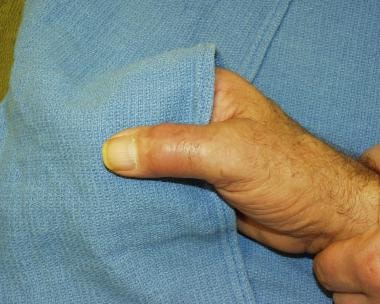
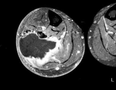
- Undifferentiated pleomorphic sarcoma (UPS), previously called malignant fibrous histiocytoma (MFH), is a soft tissue sarcoma (STS) that can occur anywhere in the body, but it usually occurs in the extremities (especially the thighs) or back of the abdomen (see the image below). (medscape.com)
- A malignant fibrous histiocytoma (MFH) is a type of soft tissue sarcoma (malignant tumor). (naturalpedia.com)
- Also called pleomorphic undifferentiated sarcoma, malignant fibrous histiocytoma usually occurs in the legs, arms, or abdomen. (hdkino.org)
- Although the most common soft tissue sarcoma of adulthood, malignant fibrous histiocytoma (MFH) is an extremely rare tumor of the urinary bladder. (johnshopkins.edu)
- A sarcoma is a malignant tumor, a type of cancer that arises from transformed cells of mesenchymal (connective tissue) origin. (wikipedia.org)
- Osteosarcoma and undifferentiated pleomorphic sarcoma (UPS) of bone are diseases in which malignant (cancer) cells form in bone. (cancer.gov)
Benign3
- Benign fibrous histiocytoma is a benign tumor composed of a mixture of fibroblastic and histiocytic cells. (alliedacademies.org)
- The diagnosis of benign fibrous histiocytoma located in the deeper tissues is clinically difficult and is confirmed histopathologically after excision. (alliedacademies.org)
- After detailed clinical and laboratory examinations, the lesion was excised in toto under general anesthesia, and histopathology revealed it to be a benign fibrous histiocytoma. (alliedacademies.org)
Osteosarcoma1
- Osteosarcoma is the most common malignant primary bone tumor (if one considers myeloma a marrow cell tumor and not a primary bone tumor) and is highly malignant. (msdmanuals.com)
Bone4
- Malignant fibrous histiocytoma replaces healthy bone tissue with cancer cells. (hdkino.org)
- Malignant fibrous histiocytoma of bones: an important differential diagnosis of malignant bone neoplasms]. (bvsalud.org)
- UPS (formerly called malignant fibrous histiocytoma [MFH]) is a rare type of bone cancer that usually starts in soft tissue, but it may form in bone. (cancer.gov)
- Fibrosarcoma has also been noted to arise from preexisting lesions, such as bone infarcts and lesions associated with fibrous dysplasia, chronic osteomyelitis, and Paget disease, as well as in previously irradiated areas of bone. (medscape.com)
Lesion2
- At the age of 28, a 33-year-old male was diagnosed with malignant fibrous histiocytoma (MFH) with a primary lesion in the right maxillary sinus. (karger.com)
- The histological features of this rare lesion and the importance of the differential diagnosis from other similar appearing malignant lesions will be discussed. (bmj.com)
Dermatofibrosarcoma protuberans1
- The most important diagnostic distinction is the separation of this tumor from aggressive forms of fibrohistiocytic neoplasms like dermatofibrosarcoma protuberans and malignant fibrous histiocytoma. (alliedacademies.org)
Diagnosis1
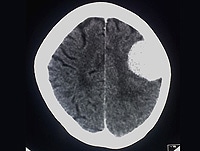
- In the autopsy, histological and immunohistochemical study of the brain revealed the diagnosis of malignant fibrous histiocytoma (MFH). (ac.ir)
Fibrosarcoma1
- Fibrosarcoma is a tumor of mesenchymal cell origin that is composed of malignant fibroblasts in a collagen background. (medscape.com)
Giant Cell Tumor2
- A case of late development of a high grade malignant fibrous histiocytoma at the site of a previously surgically treated giant cell tumor is reported. (uthscsa.edu)
- After chemotherapy, the tumor was resected and histologically showed no evidence of a recurrent giant cell tumor, only a high grade malignant fibrous histiocytoma. (uthscsa.edu)
Myxoid2
- Brendler, Charles B. / Myxoid malignant fibrous histiocytoma of the bladder . (johnshopkins.edu)
- Histopathologic study of the surgical piece revealed a malignant mesenchymal tumor of high cellular density arrayed in sheets and adopting different patterns in which fusiform, epithelioid, highly collagenized, and myxoid areas alternated. (isciii.es)
Neoplasms1
- Historically, the term malignant fibrous histiocytoma (MFH) was applied to pleomorphic spindle cell neoplasms with fibroblastic and histiocytic differentiation. (medscape.com)
Morphologic1
- RR CC was named for its morphologic resemblance to pediatric malignant rhabdoid tumor (MRT) of the kidney, which is a highly aggressive tumor characterized by cells that resemble rhabdomyoblasts and by genetic alterations involving chromosome 22, particularly the hSNF5/INI1 gene on 22q11.2. (medscape.com)
Symptoms1
- What are the symptoms of malignant fibrous histiocytoma? (hdkino.org)
Atypical1
- BROWN, MARCD & SWANSON, NEILA 1989, ' Treatment of Malignant Fibrous Histiocytoma and Atypical Fibrous Xanthomas with Micrographic Surgery ', The Journal of Dermatologic Surgery and Oncology , vol. 15, no. 12, pp. 1287-1293. (elsevierpure.com)
Multiple myeloma1
- Additionally, UPS has been associated with hematopoietic diseases such as non-Hodgkin lymphoma , Hodgkin lymphoma , multiple myeloma , and malignant histiocytosis. (medscape.com)
Originates1
- It originates in fibrous tissue that forms tendons and ligaments, and covers bones and other parts of the body. (naturalpedia.com)
Recurrent1
- This report describes the case of a 48 year old woman with a recurrent fibrous histiocytoma with prominent vasculature, which over a three year period recurred on two occasions, showing more progressive features of the aneurysmal variant. (bmj.com)
Sarcomas3
- Soft tissue sarcomas are a heterogeneous group of solid malignant tumours which represent ~1% of all new cancer cases in Europe and the United States ( 1 ). (spandidos-publications.com)
- Fibrosarcomas are rare soft tissue sarcomas originating from the intra- and intermuscular fibrous tissues, fascia and tendons and account for ~3% of all soft tissue sarcomas. (spandidos-publications.com)
- In addition, malignant mesotheliomas (ICD-O-2, M9050-9055) have been tabulated with respect to their three principal sites of Sarcomas and mesotheliomas by site occurrence (Table 4.1). (who.int)
Tumours1
- For this reason, the CD-ROM accompanying malignant vascular tumours. (who.int)
Clinical3
- GlobalData's clinical trial report, "Malignant Fibrous Histiocytoma Global Clinical Trials Review, H2, 2016' provides an overview of Malignant Fibrous Histiocytoma clinical trials scenario. (globaldata.com)
- This report provides top line data relating to the clinical trials on Malignant Fibrous Histiocytoma. (globaldata.com)
- Furthermore, it is adenoma (in 30.1% of cases) at a mean age very important to differentiate between bet of 22.2 years, fibrocystic disease (27.4%) nign and malignant breast diseases in view at a mean age of 30.2 years, invasive carcit of the clinical similarities between them [ 3 ]. (who.int)
Primary1
- Primary visceral malignant fibrous histiocytoma (MFH) is a rare disease, and few cases have been reported in the English literature. (biomedcentral.com)
Bones1
- Malignant fibrous histiocytoma of the skin is a rare type of soft tissue cancer but may also develop in the bones. (withoutaribbon.org)
Treatment1
- Surgery to remove the tumor is the first line of treatment for malignant histiocytoma. (hdkino.org)
Cancer2
- Malignant Fibrous Histiocytoma is rare cancer, meaning it is not as well known as other forms of cancer. (withoutaribbon.org)
- If you suffer from rare cancer such as Malignant Fibrous Histiocytoma, we can help and support you through your journey thanks to the generous donations we receive. (withoutaribbon.org)
Development1
- There is no well-known cause however certain hereditary and genetic mutations along with the history of radiotherapy are the important contributing risk factors for the development of malignant fibrous histiocytoma. (withoutaribbon.org)
Rare1
- Aneurysmal fibrous histiocytoma is a rare variant of cutaneous fibrous histiocytoma that results from blood vessel proliferation and haemorrhage into a fibrous histiocytoma. (bmj.com)
Surgical1
- In this article, we describe for the first time a case of malign abdominal fibrous histiocytoma associated with a surgical sponge forgotten in the abdominal cavity a long time ago. (biomedcentral.com)
Case1
- We describe a case of intracranial malignant fibrous histiocytoma which had infiltrated pons, cerebellum and basal surface of left temporal lobe without any visible mass. (ac.ir)
Skin1
- What is Malignant Fibrous Histiocytoma of the Skin? (withoutaribbon.org)