Hyperostosis, Sternocostoclavicular
Sternoclavicular Joint
Sternocostal Joints
Hyperostosis, Diffuse Idiopathic Skeletal
Acquired Hyperostosis Syndrome
Hyperostosis Frontalis Interna
Exostoses
Melorheostosis
Paleopathology
Papilloma, Inverted
Hyperostosis, Cortical, Congenital
Spinal Osteophytosis
Paranasal Sinuses
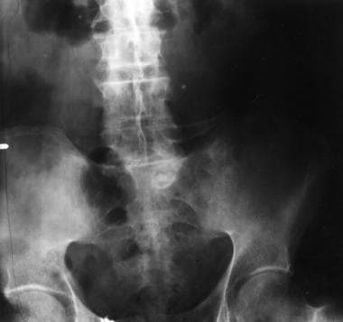
The sternoclavicular syndrome: experience from a district general hospital and results of a national postal survey. (1/8)
OBJECTIVE: To report our local experience of the sternoclavicular syndrome and sample the experience of other rheumatologists in the UK. METHODS: We studied case records of 23 patients referred to the Southend rheumatology clinic and data obtained from a postal questionnaire survey of British rheumatologists. RESULTS: We describe 58 cases (20 males and 38 females, mean age 47.2 yr). The disease was unilateral in 40 patients. Shoulder and/or arm pain (38 cases) with limitation of shoulder movements was an important presenting feature; other presenting features were anterior chest wall pain (14 cases) and neck pain (15 cases). Peripheral joint involvement was seen in 12 cases. Skin rash was reported in 12 cases (psoriasis, 6; acne, 2; none had pustulosis). No patients had symptoms or signs of sacroiliitis, and HLA-B27 was negative in 22 out of 23 patients. 99Technetium scintiscanning showed increased uptake in the sternoclavicular region in 31/34 patients (91.1%), but not in the sacroiliac areas. Plain radiographs were abnormal in 18 cases (sclerosis, 9; erosions, 2; soft tissue swelling, 2; bony expansion, 5). CT and/or MRI scans (available in 27 cases) showed erosions in 12 and osteitis in 18. Available histology showed a variable picture, including inflammation, bone erosion, sterile osteomyelitis and fibrosis. The majority of patients (45) were treated with non-steroidal anti-inflammatory drugs: 12 received steroids and 10 received disease-modifying anti-rheumatic drugs (methotrexate, 4; sulphasalazine, 6). Follow-up information was available for 38 patients, of whom 14 became asymptomatic and 24 had chronic disease with intermittent flares. CONCLUSION: Sternoclavicular disease is not uncommon in the UK. It can present with pain in the shoulder, neck or anterior chest wall, and may be underdiagnosed. Our results do not show a link with acne or pustulosis. Features of spondyloarthropathies, such as sacroiliitis and HLA-B27 positivity, were rare in this survey. (+info)Sternocostoclavicular hyperostosis: its progression and radiological features. A study of 12 cases. (2/8)
Twelve cases of sternocostoclavicular hyperostosis were followed up over four to 16 years. The patients underwent repeated radiological examinations of the sternocostoclavicular joints and the sternum, and the extrasternal osseous manifestations of the disease were studied to show changes in the radiological features during long term follow up. Five of 12 (41%) patients had extrasternal manifestations. With the exception of one patient extrasternal manifestations were first detected by scintigraphs because they were asymptomatic. With respect to the sternal manifestations the initial radiological diagnosis was made during an acute phase while painful swelling over the sternum and decreased mobility of the shoulders occurred. The radiological examinations showed the signs of a proliferative destructive arthritis in most patients. In contrast with the frequent occurrences of clinical symptoms, the radiological signs of progression take several years to become detectable. There are no specific bacteriological, serological or histological findings. Usually a permanent increase in the erythrocyte sedimentation rates is found. Sternocostoclavicular hyperostosis is a slowly progressing disease, characterised by a chronic aseptic destructive sternoclavicular arthritis with a reactive low turnover sclerosis that begins in a similar way to an enthesopathy and ends after several decades with total ankylosis. The radiological identification of retrosternal proliferation of soft tissue by computed tomography was found to be a valuable criterion for the differential diagnosis from other benign hyperostotic processes of the sternoclavicular region. (+info)In SAPHO syndrome anti-TNF-alpha therapy may induce persistent amelioration of osteoarticular complaints, but may exacerbate cutaneous manifestations. (3/8)
OBJECTIVES: SAPHO syndrome (synovitis, acne, pustulosis, hyperostosis and osteitis) is a rare disease combining skin, bone and joint manifestations. In recent years new therapeutic strategies have been tried, among them TNF-alpha-blocking agents. We report our experience with infliximab in four cases of SAPHO syndrome refractory to conventional therapies. METHODS: Between 2002 and 2005, four cases of SAPHO syndrome (two females and two males; mean age 49.7 yr) responding poorly to conventional drugs were treated with infliximab. The dose was 5 mg/kg, according to the protocol used in spondyloarthropathies, with infusions at 0, 2 and 6 weeks followed by 6 weeks intervals. No active cutaneous manifestations were present at the time of starting therapy. RESULTS: Complete remission of osteoarticular involvement was achieved after the second or third infusion, and the positive response was maintained for up to 12 months. A patient relapsed after discontinuation of infliximab, because of infectious complication. Palmoplantaris pustulosis relapsed in two patients after three and six infusions, respectively; there was slight improvement after discontinuation of anti-TNF-alpha drugs. CONCLUSIONS: Infliximab seems to be a very effective therapy for osteoarticular complaints of SAPHO syndrome. Cutaneous involvement responded less favourably, palmoplantaris pustulosis relapse being a possible complication. (+info)Calcitonin treatment for intersternocostoclavicular ossification: clinical experience in two cases. (4/8)
Intersternocostoclavicular ossification is a benign arthro-osteitis of the upper anterior chest of unknown cause. Two patients with acute exacerbation of this disorder were successfully treated with intramuscular injections of an eel calcitonin analogue (40 units three times a week). Besides symptomatic relief of local pain and swelling, serial scintigrams showed quantitative improvement in radiophosphonate uptake. The rapid alleviation of pain implies that the hormone has a central analgesic effect, in addition to its direct influence on bone cells and antiinflammatory action. In one patient the disease was associated with palmoplantar pustulosis, which was cured with oral colchicine, whereas the other patient did not have such skin lesions. Despite a hypothetical link between palmoplantar pustulosis and intersternocostoclavicular ossification, colchicine had no beneficial impact on the bone pain. Salmon calcitonin delivered by nasal spray was tried for the second patient but failed, probably because of insufficient drug delivery. The initial favourable results described here warrant future use of calcitonin injection on a larger number of patients with intersternocostoclavicular ossification. (+info)SAPHO syndrome: misdiagnosed and operated. (5/8)
SAPHO is a rare disorder that results in synovitis, acne, pustulosis, hyperostosis and osteitis. Patients with this syndrome typically present with musculoskeletal complaints, frequently localized to the anterior chest wall. However, diagnosis can be difficult in case of involvement of only one symptomatic bone without skin lesions. Awareness of SAPHO syndrome is necessary for accurate diagnosis and to prevent inappropriate and unnecessary treatment. (+info)Clinical and radiologic evolution of synovitis, acne, pustulosis, hyperostosis, and osteitis syndrome: a single center study of a cohort of 71 subjects. (6/8)
(+info)The psychological burden of an initially unexplained illness: patients with sternocostoclavicular hyperostosis before and after delayed diagnosis. (7/8)
(+info)Sternocostoclavicular hyperostosis presenting with thoracic sinus formation. (8/8)
Sternocostoclavicular hyperostosis (SCCH) is a condition which is well described in the Japanese literature but is rare in Western Europe. It is characterised by pain and swelling in the upper anterior part of the chest, which tends to be progressive. A patient is described with bilateral chronic discharging sinuses over the anterior ends of the clavicles in whom the diagnosis appeared to be one of SCCH. (+info)Hyperostosis, sternocostoclavicular, is a medical condition characterized by the abnormal thickening and hardening of the bone tissue in the sternocostoclavicular joint and surrounding areas. The sternocostoclavicular joint is where the clavicle (collarbone) meets the sternum (breastbone) and manubrium, and costae (ribs). This condition can result in pain, stiffness, and limited range of motion in the affected area. The exact cause of hyperostosis, sternocostoclavicular, is not fully understood, but it may be associated with trauma, inflammation, or genetic factors. In some cases, this condition may be asymptomatic and only discovered during imaging studies performed for other reasons. Treatment options typically include pain management, physical therapy, and in some cases, surgery to remove the excess bone growth.
Hyperostosis is a medical term that refers to an excessive growth or abnormal thickening of bone tissue. It can occur as a result of various conditions, such as inflammation, injury, or genetic disorders. The extra bone growth can cause pain, stiffness, and limited mobility in the affected area. In some cases, hyperostosis can also lead to deformities and other complications.
There are several types of hyperostosis, including:
1. Diffuse idiopathic skeletal hyperostosis (DISH): This is a condition that affects the spine, causing calcification and stiffening of the ligaments and bone spurs to form along the edges of the vertebrae. It is often asymptomatic but can cause pain and stiffness in some cases.
2. Flat bone hyperostosis: This type of hyperostosis affects the flat bones of the body, such as the skull, ribs, and pelvis. It can be caused by various conditions, including Paget's disease, fibrous dysplasia, and certain types of cancer.
3. Focal hyperostosis: This refers to localized areas of bone overgrowth that can occur in response to injury, infection, or inflammation. Examples include heterotopic ossification (the formation of bone in soft tissues) and Freiberg's infarction (a condition that affects the joint surface of the metatarsal bones in the foot).
4. Hyperostosis frontalis interna: This is a benign condition that causes thickening of the inner table of the frontal bone in the skull. It is more common in women and often asymptomatic but can cause headaches and other symptoms in some cases.
Treatment for hyperostosis depends on the underlying cause and severity of the condition. In some cases, no treatment may be necessary. However, if the condition causes pain or limits mobility, various treatments may be recommended, such as medication, physical therapy, or surgery.
The sternoclavicular joint is the joint where the clavicle (collarbone) meets the sternum (breastbone). It is the only joint that connects the upper limb to the trunk of the body. This joint allows for movement in multiple directions, including elevation and depression of the shoulder, as well as some degree of protraction and retraction. The sternoclavicular joint is supported by several ligaments, which provide stability and strength to the joint.
The sternocostal joints are the articulations between the sternum (breastbone) and the costal cartilages of the true ribs (the first seven pairs of ribs). Specifically, the manubrium (the superior portion of the sternum) articulates with the second to seventh costal cartilages, while the body of the sternum articulates with the lower costal cartilages of the fifth to seventh ribs. These joints are synovial joints and allow for some degree of movement during respiration, contributing to the expansion and contraction of the thoracic cavity. The primary motion at these joints is a gliding or sliding motion.
Diffuse Idiopathic Hyperostosis (DIH), also known as Forestier's Disease, is a non-inflammatory skeletal disorder characterized by the abnormal thickening and hardening (hyperostosis) of the bony portions of the spine and/or other parts of the skeleton. In DIH, there is an excessive formation of new bone along the edges of these bones, particularly at the sites where ligaments attach to the bones.
The term "idiopathic" indicates that the cause of this condition is currently unknown, while "diffuse" refers to its widespread involvement of multiple skeletal areas. The exact pathogenesis of DIH remains unclear; however, it has been suggested that there might be a connection with abnormal bone metabolism and/or localized inflammation.
DIH primarily affects middle-aged and older adults, with men being more commonly affected than women. Common symptoms include stiffness, pain, and limited mobility in the spine and joints. In some cases, DIH may also lead to complications such as spinal stenosis or nerve compression due to the excessive bone growth.
It is important to note that while hyperostosis can be a feature of various medical conditions, the term "Diffuse Idiopathic Skeletal Hyperostosis" specifically refers to this distinct clinical entity characterized by the widespread involvement of the skeleton and the absence of inflammation or other underlying causes.
Osteitis is a medical term that refers to the inflammation of bone tissue. It can occur as a result of various conditions, such as infection (osteomyelitis), trauma, or autoimmune disorders. The symptoms of osteitis may include pain, swelling, warmth, and redness in the affected area, as well as fever and general malaise. Treatment typically involves addressing the underlying cause of the inflammation, which may involve antibiotics for infection or anti-inflammatory medications for other causes. In some cases, surgery may be necessary to remove infected or damaged bone tissue.
Acquired hyperostosis syndrome is not a widely recognized medical term, and it may refer to several different conditions that involve abnormal bone growth or hardening. One possible condition that might be referred to as acquired hyperostosis syndrome is diffuse idiopathic skeletal hyperostosis (DISH).
Diffuse idiopathic skeletal hyperostosis is a non-inflammatory condition that affects the spine and other parts of the body. It is characterized by the calcification and ossification of ligaments and entheses, which are the sites where tendons or ligaments attach to bones. This process can lead to the formation of bony spurs or growths, called osteophytes, along the spine and other affected areas.
The exact cause of DISH is not known, but it is more common in older adults, males, and people with certain medical conditions such as diabetes and obesity. The symptoms of DISH can vary widely depending on the severity and location of the bone growths. Some people may experience stiffness, pain, or limited mobility in the affected areas, while others may have no symptoms at all.
It is important to note that there are many other conditions that can cause abnormal bone growth or hardening, so a proper medical evaluation is necessary to determine the underlying cause of any symptoms. If you have concerns about acquired hyperostosis syndrome or any other medical condition, you should speak with your healthcare provider for further guidance.
Hyperostosis Frontalis Interna (HFI) is a medical condition characterized by an abnormal thickening or overgrowth of the inner table of the frontal bone, which is the bone that forms the forehead. This condition most commonly affects middle-aged to older women. The exact cause of HFI is not known, but it may be associated with hormonal factors, as it is more common in women who have gone through menopause.
In HFI, the overgrowth of bone can cause a raised, bumpy, or irregular appearance on the forehead, and can sometimes lead to headaches or other symptoms. However, many people with HFI do not experience any symptoms at all. The diagnosis of HFI is typically made based on imaging studies such as X-rays or CT scans, which show the characteristic thickening of the frontal bone.
While HFI is not a life-threatening condition, it can cause cosmetic concerns and may require treatment in some cases. Treatment options for HFI include medication to manage symptoms such as headaches, as well as surgical removal of the excess bone in severe cases.
Exostoses are benign (noncancerous) bone growths that develop on the surface of a bone, usually in response to repeated stress or friction. They are often small and smooth, but can become larger and more irregular over time. In some cases, they may cause pain or discomfort, especially if they continue to grow and put pressure on nearby nerves, muscles, or other bones.
Exostoses can occur in various parts of the body, but they are most commonly found in the long bones of the arms and legs, as well as in the small bones of the feet. They may also develop in response to chronic irritation or injury, such as from jogging or playing sports that involve a lot of running or jumping.
In some cases, exostoses may be surgically removed if they cause persistent pain or other symptoms. However, in many cases, they do not require treatment and can be left alone. If you are concerned about any bone growths or other unusual symptoms, it is always best to consult with a healthcare professional for an accurate diagnosis and treatment plan.
Melorheostosis is a very rare, progressive bone disorder characterized by the thickening and hardening of the bones' outer covering (periosteum). The name "melorheostosis" means "melting bones," which describes the appearance of the long bones on X-rays. It resembles dripping candle wax flowing down the shafts of the bones.
The condition typically affects one side of the body, often involving the legs and arms, but can also affect the skull, spine, and ribs. The symptoms can vary widely, depending on the location and extent of bone involvement. They may include bone pain, deformities, limited mobility, joint stiffness, and skin changes over the affected bones.
The exact cause of melorheostosis is unknown, but it is not a hereditary condition. It is thought to be related to abnormal blood vessel formation during fetal development, leading to improper bone growth and development. There is no known cure for melorheostosis, but various treatments can help manage symptoms and improve quality of life. These may include pain management, physical therapy, surgery, and other supportive measures.
Ankylosis is a medical term that refers to the abnormal joining or fusion of bones, typically in a joint. This can occur as a result of various conditions such as injury, infection, or inflammatory diseases like rheumatoid arthritis. The fusion of bones can restrict movement and cause stiffness in the affected joint. In some cases, ankylosis can lead to deformity and disability if not treated promptly and effectively.
There are different types of ankylosis depending on the location and extent of bone fusion. For instance, when it affects the spine, it is called "ankylosing spondylitis," which is a chronic inflammatory disease that can cause stiffness and pain in the joints between the vertebrae.
Treatment for ankylosis depends on the underlying cause and severity of the condition. In some cases, physical therapy or surgery may be necessary to restore mobility and function to the affected joint.
Paleopathology is the study of ancient diseases and injuries as recorded in bones, mummies, and other archaeological remains. It is an interdisciplinary field that combines knowledge from pathology, epidemiology, anthropology, and archaeology to understand the health and disease patterns of past populations. The findings of paleopathology can provide valuable insights into the evolution of diseases, the effectiveness of ancient medical practices, and the impact of environmental and social factors on human health over time. Examples of conditions that may be studied in paleopathology include infectious diseases (such as tuberculosis or leprosy), nutritional deficiencies, trauma, cancer, and genetic disorders.
Inverted papilloma is a specific type of benign (non-cancerous) growth that occurs in the mucosal lining of the nasal cavity or paranasal sinuses. It is also known as schneiderian papilloma or cylindrical cell papilloma.
This condition is characterized by the growth of finger-like projections (papillae) that invert or grow inward into the underlying tissue, hence the name "inverted." The lesions are usually composed of an outer layer of stratified squamous epithelium and an inner core of connective tissue.
Inverted papillomas can cause symptoms such as nasal congestion, nosebleeds, sinus pressure, and difficulty breathing through the nose. In some cases, they may also lead to more serious complications, including recurrence after removal and a small risk of malignant transformation into squamous cell carcinoma.
It is important to note that while inverted papillomas are benign, they can still cause significant problems due to their location and tendency to recur. Therefore, they typically require surgical removal and close follow-up with an otolaryngologist (ear, nose, and throat specialist).
Congenital cortical hyperostosis is a rare, inherited bone disorder that is characterized by abnormal thickening of the outer layer of bones (cortical hyperostosis). This condition primarily affects the skull and long bones of the arms and legs. The exact cause of congenital cortical hyperostosis is not fully understood, but it is believed to be related to mutations in certain genes that regulate bone growth and development.
The symptoms of congenital cortical hyperostosis can vary widely from person to person, depending on the severity and location of the bone abnormalities. Some common features of this condition include:
* A thickened skull, which may cause a prominent forehead or a misshapen head
* Abnormally thick and dense long bones in the arms and legs, which can make them heavy and difficult to move
* Delayed growth and development
* Increased risk of fractures
* Pain and stiffness in the affected bones
Congenital cortical hyperostosis is typically diagnosed based on a combination of clinical symptoms, imaging studies (such as X-rays or CT scans), and genetic testing. There is no cure for this condition, but treatment may involve pain management, physical therapy, and surgery to correct any bone deformities. In some cases, the symptoms of congenital cortical hyperostosis may improve over time, but in others, they may persist throughout life.
Spinal osteophytosis, also known as spinal osteophyte formation or bone spurs on the spine, refers to the abnormal growth of bony projections along the vertebral column's margins. These bony outgrowths develop due to degenerative changes, inflammation, or injury in the joints between the vertebrae (facet joints) and can cause stiffness, pain, and reduced mobility. In some cases, spinal osteophytosis may lead to complications such as spinal stenosis or nerve compression.
Paranasal sinuses are air-filled cavities in the skull that surround the nasal cavity. There are four pairs of paranasal sinuses, including the maxillary, frontal, ethmoid, and sphenoid sinuses. These sinuses help to warm, humidify, and filter the air we breathe. They also contribute to our voice resonance and provide a slight cushioning effect for the skull. The openings of the paranasal sinuses lead directly into the nasal cavity, allowing mucus produced in the sinuses to drain into the nose. Infections or inflammation of the paranasal sinuses can result in conditions such as sinusitis.
The skull is the bony structure that encloses and protects the brain, the eyes, and the ears. It is composed of two main parts: the cranium, which contains the brain, and the facial bones. The cranium is made up of several fused flat bones, while the facial bones include the upper jaw (maxilla), lower jaw (mandible), cheekbones, nose bones, and eye sockets (orbits).
The skull also provides attachment points for various muscles that control chewing, moving the head, and facial expressions. Additionally, it contains openings for blood vessels, nerves, and the spinal cord to pass through. The skull's primary function is to protect the delicate and vital structures within it from injury and trauma.
The cervical vertebrae are the seven vertebrae that make up the upper part of the spine, also known as the neck region. They are labeled C1 to C7, with C1 being closest to the skull and C7 connecting to the thoracic vertebrae in the chest region. The cervical vertebrae have unique structures to allow for a wide range of motion in the neck while also protecting the spinal cord and providing attachment points for muscles and ligaments.
 SAPHO syndrome
List of MeSH codes (C17)
List of MeSH codes (C05)
 Psoriatic Arthritis Imaging: Practice Essentials, Radiography, Computed Tomography
 Die kindliche Osteomyelitis | Die Orthopädie
SAPHO syndrome - Wikipedia
 What causes an enlarged sternoclavicular joint? - wren-clothing.com
 Diagnostics | September 2016 - Browse Articles
 Other Problems | Rib Injury Clinic
Browse Items · NEOMED Bibliography Database
Arthritis, Juvenile | Profiles RNS
Rheumatic Diseases | Profiles RNS
 MeSH Browser
 Genetic variants in FBLIM1 gene do not contribute to SAPHO syndrome and chronic recurrent multifocal osteomyelitis in typical...
![Afzelius P[au] - Search Results - PubMed](data:image/png;base64,iVBORw0KGgoAAAANSUhEUgAAABAAAAAQCAMAAAAoLQ9TAAAARVBMVEVHcEwoU45gYmYAUpQAUpRPYGVgYmZLXnJgYmYAUZUAUpRJXnIAUpQAUpRgYmYAUpRgYmZgYmZhYmYAUpQAUpQAUpRgYmaDiPJuAAAAFXRSTlMADOJ+6QewGO8/uTRqtH7GdFJ11p1bCL3TAAAAZUlEQVQYlV2PVw7AIAxDTeney7n/UcsoldX3E+VJOAboEi7MBpHWMs1ADlG8u7UYWauwyZFeRQVPOhG2o+aiwhByJxUx91Jxhje3iJSqGfHuLKI0+0TpXvY1twCOPlFh5pa/++MB0vIOBm+1zaoAAAAASUVORK5CYII=) Afzelius P[au] - Search Results - PubMed
Afzelius P[au] - Search Results - PubMed
Psoriatic Arthritis Imaging: Practice Essentials, Radiography, Computed Tomography
MeSH Browser
MeSH Browser
 DeCS
DeCS
Code System Concept
 Pesquisa | Portal Regional da BVS
 Chronic Recurrent Multifocal Osteomyelitis - A Rare Clinical Presentation and Review of Literature | Journal of Orthopaedic...
 Arthritis, Rheumatoid | Profiles RNS
Descriptors in 2013 MeSH. Preferred term only. December 14, 2012
Osteitis10
- Beretta-Piccoli BC et al (2000) Synovitis, acne, pustulosis, hyperostosis, osteitis (SAPHO) syndrome in childhood: a report of ten cases and review of the literature. (springer.com)
- Dihlmann W (1993) Acquired hyperostosis syndrome (so-called pustular arthro-osteitis). (springer.com)
- Since then, a number of associations between skin conditions and osteoarticular disorders have been reported under a variety of names, including sternocostoclavicular hyperostosis, pustulotic arthro-osteitis, and acne-associated spondyloarthropathy. (wikipedia.org)
- The term SAPHO (an acronym for synovitis, acne, pustulosis, hyperostosis, osteitis) was coined in 1987 to represent this spectrum of inflammatory bone disorders that may or may not be associated with dermatologic pathology. (wikipedia.org)
- Acne-pustulosis-hyperostosis-osteitis syndrome. (wikipedia.org)
- Underlying aetiologies include osteoarthritis of the sternoclavicular joint, condensing osteitis, spontaneous dislocation of the sternoclavicular joint, and sternocostoclavicular hyperostosis. (wren-clothing.com)
- Syndrome of synovitis acne pustulosis hyperostosis osteitis (SAPHO) and chronic recurrent multifocal osteomyelitis (CRMO) present two diseases of a dermatologic and rheumatologic spectrum that are variable in manifestation und therapeutic response. (biomedcentral.com)
- Therefore, we aimed to clinically characterize a patient group with syndrome of synovitis acne pustulosis hyperostosis osteitis (SAPHO) ( n = 47) and chronic recurrent multifocal osteomyelitis (CRMO)/ chronic non-bacterial osteomyelitis (CNO) ( n = 9) and analyze a CRMO candidate gene. (biomedcentral.com)
- however, osteitis with hyperostosis is considered as a pathognomonic lesion of SAPHO syndrome [ 3 ]. (biomedcentral.com)
- Moreover, it can present as a sclerosing osteitis originating from the bone marrow, a hyperostosis with increased bone cuff formed by the periost, an ossification of ligaments, accompanied by osteolysis or an erosion of a joint. (biomedcentral.com)
Sternoclavicular joint2
- Anterior chest wall (most common site, 65-90% of patients):[citation needed] Hyperostosis, sclerosis and bone hypertrophy especially involving the sternoclavicular joint, often with a soft tissue component. (wikipedia.org)
- Other unusual often described as atypical conditions affecting the SC joint include Sterno- costoclavicular hyperostosis (SCCH) a chronic inflammatory disorder which presents with erythema, swelling, and pain of the sternoclavicular joint. (ribinjuryclinic.com)
Pustulotic1
- Freyschmidt J et al (1998) The bullhead sign: scintigraphic pattern of sternocostoclavicular hyperostosis and pustulotic arthroosteitis. (springer.com)
Acquired hyperost1
- Dihlmann W et al (1988) The acquired hyperostosis syndrome. (springer.com)
SAPHO1
- most SAPHO patients had sterno-costo-clavicular hyperostosis. (biomedcentral.com)
Cases1
- Synthesis of 13 personal observations of sternocostoclavicular hyperostosis and 300 cases from the literature. (springer.com)
Sclerosis2
- Anterior chest wall (most common site, 65-90% of patients):[citation needed] Hyperostosis, sclerosis and bone hypertrophy especially involving the sternoclavicular joint, often with a soft tissue component. (wikipedia.org)
- A rare rheumatologic disease characterised by predominantly bilateral, chronic, sterile inflammation and progressive sclerosis and hyperostosis of the sternocostoclavicular joint, with adjacent soft tissue ossification, in the absence of other joint involvement. (cdc.gov)
Soft tissue ossification1
- A rare, benign rheumatologic disorder or syndrome characterized by hyperostosis and soft tissue ossification between the clavicles and the anterior part of the upper ribs. (nih.gov)
Syndrome1
- Acne-pustulosis-hyperostosis-osteitis syndrome. (wikipedia.org)
Term1
- The term SAPHO (an acronym for synovitis, acne, pustulosis, hyperostosis, osteitis) was coined in 1987 to represent this spectrum of inflammatory bone disorders that may or may not be associated with dermatologic pathology. (wikipedia.org)