Hypertensive Encephalopathy
Brain Diseases
Brain Edema
Hepatic Encephalopathy
Hypertension
Blood-Brain Barrier
Magnetic Resonance Imaging
Brain
Wernicke Encephalopathy
Encephalopathy, Bovine Spongiform
Hypertensive encephalopathy in a patient with retroperitoneal fibrosis. (1/43)
A patient presented with retroperitoneal fibrosis but without any ureteric obstruction. The diagnosis was made by an abdominal CT scan and also at laparotomy. Post-operatively, she developed hypertensive encephalopathy. An isotope renogram with captopril was abnormal but not diagnostic of renal artery stenosis. The patient's condition improved with steroid and antihypertensive treatment. A follow-up CT scan showed complete resolution of peri-aortic thickening. A causative link is postulated between retroperitoneal fibrosis, trauma during laparotomy, and onset of acute hypertension. (+info)Unusual MR findings of the brain stem in arterial hypertension. (2/43)
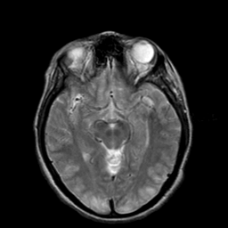
MR imaging findings have been reported in only a few cases of severe arterial hypertension. We report two cases of severe paroxysmal arterial hypertension associated with unusual brain stem hyperintensity. The lesions improved dramatically after stabilization of blood pressure, suggesting that edema could be the main cause of the MR imaging-observed hyperintensity. (+info)Functional recovery despite prolonged bilateral loss of somatosensory evoked potentials: report on two patients. (3/43)
A bilateral loss of short latency somatosensory evoked potentials (SSEPs) after head trauma or non-traumatic brain damage is normally associated with a deleterious neurological outcome. An adequate recovery in reported in two deeply comatose patients with head trauma or severe hypertensive encephalopathy despite prolonged bilateral loss of SSEPs over days, found in repeated recordings. Hence, a bilateral loss of SSEPs should not be considered alone for prediction of outcome in cerebral injury. (+info)Posterior reversible encephalopathy syndrome: utility of fluid-attenuated inversion recovery MR imaging in the detection of cortical and subcortical lesions. (4/43)
BACKGROUND AND PURPOSE: Posterior reversible encephalopathy syndrome (PRES) is typically characterized by headache, altered mental functioning, seizures, and visual loss associated with imaging findings of bilateral subcortical and cortical edema with a predominantly posterior distribution. Our goal was to determine whether fluid-attenuated inversion recovery (FLAIR) imaging improves the ability to detect subtle peripheral lesions of PRES, as compared with conventional MR techniques. METHODS: Sixteen patients with clinical and imaging findings consistent with PRES were studied. Thirteen patients had undergone transplantation and had cyclosporin A neurotoxicity. Fast-FLAIR images were compared with spin-echo proton density- and T2-weighted images. RESULTS: FLAIR imaging improved diagnostic confidence and conspicuity of the T2 hyperintense lesions of PRES, typically in the subcortical white matter of the parietooccipital regions bilaterally. On all 23 abnormal MR studies, FLAIR was judged superior to proton density- and T2-weighted images for the detection of PRES in the supratentorial brain. In a mean of 6.7 of 23 studies, FLAIR findings prompted a raise in the grade of disease severity. FLAIR also showed cortical involvement in 94% of patients with PRES and in a mean of 46% of the total lesion burden. In four cases, subtle lesions were virtually undetectable without FLAIR. Brain stem or cerebellar disease was encountered in 56% of patients. CONCLUSION: FLAIR improves the ability to diagnose and detect subcortical and cortical lesions in PRES as compared with proton density- and T2-weighted spin-echo images. We therefore believe that FLAIR should be performed in patients with suspected PRES to allow more confident recognition of the often subtle imaging abnormalities. (+info)Reversible posterior leukoencephalopathy syndrome in a patient with hypertensive encephalopathy--case report. (5/43)
A 58-year-old male presented with reversible posterior leukoencephalopathy syndrome (RPLS) manifesting as headache, papilledema, and renal hypertension. T2-weighted magnetic resonance (MR) imaging showed hyperintensity lesions in the medulla, pons, bilateral thalami, and bilateral deep white matter of the parieto-occipital lobes. The pons was swollen. Diffusion-weighted MR imaging did not show increased intensity in these lesions. The lesions disappeared with improvement of clinical symptoms after treatment for hypertension. These findings suggested the lesions were vasogenic edema and the diagnosis was RPLS. T2-weighted and diffusion-weighted MR imaging are useful modalities to differentiate RPLS from other central nervous system abnormalities such as infarction, multiple sclerosis, and central pontine myelinolysis. The clinical and neuroradiological findings of RPLS can be reversed by timely initiation of treatment for the causative factor. (+info)Reversible posterior leukoencephalopathy syndrome: a report of 2 cases. (6/43)
Reversible posterior leukoencephalopathy syndrome (RPLE) is an increasingly recognised disorder, most commonly associated with malignant hypertension, toxaemia of pregnancy or the use of immunosuppressive agents. Two cases of RPLE syndrome occurring in the setting of accelerated hypertension and eclampsia are described. Both patients had seizures, altered sensorium and typical findings on neuroimaging. They had complete clinical and radiological recovery. The clinical course, pathophysiology and neuroimaging features of RPLE syndrome are discussed. (+info)Selective renal embolisation for renovascular hypertension? (7/43)
An 11 year old girl developed hypertensive encephalopathy and renal failure from reflux nephropathy. Resection of her shrunken left kidney did not control her hypertension. Two selective arterial embolisations of the scarred right lower pole produced only transient benefit, but a heminephrectomy gave good control. Embolisation may delay definitive treatment. (+info)Two cases of malignant hypertension with reversible diffuse leukoencephalopathy exhibiting a reversible nocturnal blood pressure "riser" pattern. (8/43)
We report two cases of malignant hypertension with reversible diffuse leukoencephalopathy demonstrating a nocturnal blood pressure (BP) rising pattern ("riser" pattern). Case 1 was a 54-year-old man diagnosed with malignant hypertension who presented with diffuse leukoencephalopathy and nocturnal BP rise during the acute phase. These abnormal findings diminished after treatment of hypertension. Case 2 was a 50-year-old woman diagnosed with malignant hypertension in association with leukoencephalopathy, heart failure and acute renal failure. She also presented with a "riser" pattern during the acute phase. In contrast to case 1, the leukoencephalopathy and "riser" pattern in case 2 were not improved even after 1 month of treatment. Following intensive antihypertensive treatment, renal failure was improved in case 1, but renal failure was not improved after 1 month in case 2. In conclusion, a possible explanation of this phenomenon is that a causative volume overload due to renal dysfunction produced the temporal leukoencephalopathy-like brain edema and "riser" pattern in these cases. (+info)Hypertensive encephalopathy is a serious neurological condition that occurs due to extremely high blood pressure, which is not adequately controlled. This leads to the leakage of fluid and blood into the brain (cerebral edema) and disrupts the normal functioning of the brain. Symptoms may include severe headache, nausea, vomiting, confusion, seizures, visual disturbances, and in severe cases, coma. Immediate medical attention is required to reduce blood pressure and prevent potential long-term damage or even death.
Brain diseases, also known as neurological disorders, refer to a wide range of conditions that affect the brain and nervous system. These diseases can be caused by various factors such as genetics, infections, injuries, degeneration, or structural abnormalities. They can affect different parts of the brain, leading to a variety of symptoms and complications.
Some examples of brain diseases include:
1. Alzheimer's disease - a progressive degenerative disorder that affects memory and cognitive function.
2. Parkinson's disease - a movement disorder characterized by tremors, stiffness, and difficulty with coordination and balance.
3. Multiple sclerosis - a chronic autoimmune disease that affects the nervous system and can cause a range of symptoms such as vision loss, muscle weakness, and cognitive impairment.
4. Epilepsy - a neurological disorder characterized by recurrent seizures.
5. Brain tumors - abnormal growths in the brain that can be benign or malignant.
6. Stroke - a sudden interruption of blood flow to the brain, which can cause paralysis, speech difficulties, and other neurological symptoms.
7. Meningitis - an infection of the membranes surrounding the brain and spinal cord.
8. Encephalitis - an inflammation of the brain that can be caused by viruses, bacteria, or autoimmune disorders.
9. Huntington's disease - a genetic disorder that affects muscle coordination, cognitive function, and mental health.
10. Migraine - a neurological condition characterized by severe headaches, often accompanied by nausea, vomiting, and sensitivity to light and sound.
Brain diseases can range from mild to severe and may be treatable or incurable. They can affect people of all ages and backgrounds, and early diagnosis and treatment are essential for improving outcomes and quality of life.
Brain edema is a medical condition characterized by the abnormal accumulation of fluid in the brain, leading to an increase in intracranial pressure. This can result from various causes, such as traumatic brain injury, stroke, infection, brain tumors, or inflammation. The swelling of the brain can compress vital structures, impair blood flow, and cause neurological symptoms, which may range from mild headaches to severe cognitive impairment, seizures, coma, or even death if not treated promptly and effectively.
Hepatic encephalopathy (HE) is a neuropsychiatric syndrome associated with liver dysfunction and/or portosystemic shunting. It results from the accumulation of toxic substances, such as ammonia and inflammatory mediators, which are normally metabolized by the liver. HE can present with a wide range of symptoms, including changes in sleep-wake cycle, altered mental status, confusion, disorientation, asterixis (flapping tremor), and in severe cases, coma. The diagnosis is based on clinical evaluation, neuropsychological testing, and exclusion of other causes of cognitive impairment. Treatment typically involves addressing the underlying liver dysfunction, reducing ammonia production through dietary modifications and medications, and preventing further episodes with lactulose or rifaximin therapy.
Hypertension is a medical term used to describe abnormally high blood pressure in the arteries, often defined as consistently having systolic blood pressure (the top number in a blood pressure reading) over 130 mmHg and/or diastolic blood pressure (the bottom number) over 80 mmHg. It is also commonly referred to as high blood pressure.
Hypertension can be classified into two types: primary or essential hypertension, which has no identifiable cause and accounts for about 95% of cases, and secondary hypertension, which is caused by underlying medical conditions such as kidney disease, hormonal disorders, or use of certain medications.
If left untreated, hypertension can lead to serious health complications such as heart attack, stroke, heart failure, and chronic kidney disease. Therefore, it is important for individuals with hypertension to manage their condition through lifestyle modifications (such as healthy diet, regular exercise, stress management) and medication if necessary, under the guidance of a healthcare professional.
The Blood-Brain Barrier (BBB) is a highly specialized, selective interface between the central nervous system (CNS) and the circulating blood. It is formed by unique endothelial cells that line the brain's capillaries, along with tight junctions, astrocytic foot processes, and pericytes, which together restrict the passage of substances from the bloodstream into the CNS. This barrier serves to protect the brain from harmful agents and maintain a stable environment for proper neural function. However, it also poses a challenge in delivering therapeutics to the CNS, as most large and hydrophilic molecules cannot cross the BBB.
Medical Definition:
Magnetic Resonance Imaging (MRI) is a non-invasive diagnostic imaging technique that uses a strong magnetic field and radio waves to create detailed cross-sectional or three-dimensional images of the internal structures of the body. The patient lies within a large, cylindrical magnet, and the scanner detects changes in the direction of the magnetic field caused by protons in the body. These changes are then converted into detailed images that help medical professionals to diagnose and monitor various medical conditions, such as tumors, injuries, or diseases affecting the brain, spinal cord, heart, blood vessels, joints, and other internal organs. MRI does not use radiation like computed tomography (CT) scans.
The brain is the central organ of the nervous system, responsible for receiving and processing sensory information, regulating vital functions, and controlling behavior, movement, and cognition. It is divided into several distinct regions, each with specific functions:
1. Cerebrum: The largest part of the brain, responsible for higher cognitive functions such as thinking, learning, memory, language, and perception. It is divided into two hemispheres, each controlling the opposite side of the body.
2. Cerebellum: Located at the back of the brain, it is responsible for coordinating muscle movements, maintaining balance, and fine-tuning motor skills.
3. Brainstem: Connects the cerebrum and cerebellum to the spinal cord, controlling vital functions such as breathing, heart rate, and blood pressure. It also serves as a relay center for sensory information and motor commands between the brain and the rest of the body.
4. Diencephalon: A region that includes the thalamus (a major sensory relay station) and hypothalamus (regulates hormones, temperature, hunger, thirst, and sleep).
5. Limbic system: A group of structures involved in emotional processing, memory formation, and motivation, including the hippocampus, amygdala, and cingulate gyrus.
The brain is composed of billions of interconnected neurons that communicate through electrical and chemical signals. It is protected by the skull and surrounded by three layers of membranes called meninges, as well as cerebrospinal fluid that provides cushioning and nutrients.
Wernicke Encephalopathy is a neuropsychiatric disorder that is caused by a deficiency of thiamine (vitamin B1). It is characterized by a classic triad of symptoms: confusion, oculomotor dysfunction (such as nystagmus and ophthalmoplegia), and gait ataxia. Other symptoms can include memory loss, apathy, and hypothermia.
Wernicke Encephalopathy is most commonly seen in alcoholics due to poor nutrition, but it can also occur in people with conditions that cause malabsorption or increased thiamine requirements, such as AIDS, cancer, and chronic diarrhea. Immediate treatment with thiamine replacement therapy is necessary to prevent progression of the disease and potential permanent neurological damage. If left untreated, Wernicke Encephalopathy can lead to Korsakoff's syndrome, a chronic memory disorder.
Bovine spongiform encephalopathy (BSE), also known as "mad cow disease," is a progressive neurodegenerative disorder that affects cattle. It is caused by prions, which are misfolded proteins that can cause other proteins in the brain to also misfold and accumulate, leading to brain damage and degeneration. The disease is named for the sponge-like appearance of the brain tissue that results from this degenerative process.
BSE is a zoonotic disease, which means that it can be transmitted from animals to humans. In humans, BSE is known as variant Creutzfeldt-Jakob disease (vCJD) and is caused by consuming contaminated beef products. The symptoms of vCJD include rapidly progressing dementia, neurological symptoms such as muscle spasms and difficulty coordinating movements, and physical deterioration leading to death.
It's important to note that the use of certain growth promoters in cattle feed and the practice of feeding cattle meat and bone meal have been banned in many countries in order to prevent the spread of BSE. Additionally, strict controls on the inspection and testing of beef products have been implemented to ensure their safety.
SHR (Spontaneously Hypertensive Rats) are an inbred strain of rats that were originally developed through selective breeding for high blood pressure. They are widely used as a model to study hypertension and related cardiovascular diseases, as well as neurological disorders such as stroke and dementia.
Inbred strains of animals are created by mating genetically identical individuals (siblings or offspring) for many generations, resulting in a population that is highly homozygous at all genetic loci. This means that the animals within an inbred strain are essentially genetically identical to one another, which makes them useful for studying the effects of specific genes or environmental factors on disease processes.
SHR rats develop high blood pressure spontaneously, without any experimental manipulation, and show many features of human hypertension, such as increased vascular resistance, left ventricular hypertrophy, and renal dysfunction. They also exhibit a number of behavioral abnormalities, including hyperactivity, impulsivity, and cognitive deficits, which make them useful for studying the neurological consequences of hypertension and other cardiovascular diseases.
Overall, inbred SHR rats are an important tool in biomedical research, providing a valuable model for understanding the genetic and environmental factors that contribute to hypertension and related disorders.
 Hypertensive encephalopathy
 Hypertensive Encephalopathy Differential Diagnoses
 American Journal of Case Reports | A case of brainstem hypertensive encephalopathy - Article abstract #881909
 Hypertensive Encephalopathy | Case Files: Emergency Medicine, 5e | AccessEmergency Medicine | McGraw Hill Medical
 Permissive hypertension for stroke: How it works and more
 DailyMed - ALYMSYS- bevacizumab-maly injection, solution
 Bevacizumab Side Effects: Common, Severe, Long Term
 Neurological Emergencies | Hindawi
 Cholinesterase Inhibitors: Part 4: The Cholinergic Toxidrome Section 7: Differential Diagnosis of the Cholinergic Toxidrome |...
 Dr. Steven Levi, MD, Cardiology Specialist - Haddon Heights, NJ | Sharecare
 Index of Published Newsletters - Canada.ca
 Posterior Leukoencephalopathy Syndrome - Ontology Report - Rat Genome Database
 Preeclampsia: MedlinePlus Genetics
 Stat Consult: Hypertensive emergency/ hypertensive urgency - Clinical Advisor
 Computed tomography and magnetic resonance imaging in adult-onset leukodystrophy - PubMed
 Hydrochlorothiazide And Candesartan (Atacand HCT) - Side Effects, Interactions, Uses, Dosage, Warnings