Hypothalamic Neoplasms
Genitalia, Female
Ovary
Corpus Luteum
Physiology
Uterus
Chorionic Gonadotropin
Books
Hypercalcemia in an euthyroid patient with secondary hypoadrenalism and diabetes insipidus due to hypothalamic tumor. (1/47)
A 20-year-old Japanese man with a hypothalamic tumor (most likely germ-cell tumor) which caused secondary hypoadrenalism, hypogonadism and diabetes insipidus developed hypercalcemia and acute renal failure. The serum levels of intact PTH (iPTH), PTH-related protein (PTH-rP), 1,25-dihydroxy vitamin D (1,25- (OH)2 D), ACTH, cortisol, gonadotropins and testosterone were decreased, but his serum levels of triiodothyronine (T3) and thyroxine (T4) were within the normal range at admission, with depressed TSH and slightly increased thyroglobulin. The hypercalcemia was refractory to extensive hydration and calcitonin, but was ameliorated by pamidronate. After irradiation of the hypothalamic tumor, panhypopituitarism gradually developed. The patient has been normocalcemic for the last 2 years and is doing well under replacement therapy with glucocorticoid, L-thyroxine, methyltestosterone and 1-desamino D arginine vasopressin (dDAVP). As to the mechanism of euthyroidism at admission, transient destructive thyroiditis associated with hypopituitarism or delayed development of hypothyroidism following the hypoadrenalism was suggested. This is the first reported case of hypercalcemia in secondary hypoadrenalism due to hypothalamic tumor. Hypercalcemia was most likely induced by increased bone resorption, which was probably elicited by the combined effects of deficient glucocorticoid and sufficient thyroid hormones in addition to hypovolemia and reduced renal calcium excretion. Furthermore, severe dehydration due to diabetes insipidus and disturbance of thirst sensation caused by the hypothalamic tumor aggravated the hypercalcemia, leading to acute renal failure. (+info)Hypercalcemia accompanied by hypothalamic hypopituitarism, central diabetes inspidus and hyperthyroidism. (2/47)
We present here a case of prominent hypercalcemia accompanied by hypothalamic tumor and Graves' disease. A 24-year-old man with hypothalamic tumor showed hypopituitarism, central diabetes inspidus (DI) and hyperthyroidism. Nausea, loss of thirst and appetite, and general fatigue were found with the unveiling of hypercalcemia and hypernatremia. Parathyroid hormone (PTH) and 1alpha-dihydroxyvitamin D levels were suppressed with a normal range of PTH-related protein values. One-desamino-(8-D-arginine)-vasopressin (DDAVP) and half-saline administration normalized hypernatremia, while hypercalcemia was still sustained. Administration of cortisone acetate and thiamazole reduced the elevated serum Ca level. In the present case, concurrent hyperthyroidism was assumed to accelerate skeletal mobilization of calcium into the circulation. Hypocortisolism and central DI was also considered to contribute, to some extent, to the hypercalcemia through renal handling of Ca. (+info)Chordoid glioma: a neoplasm unique to the hypothalamus and anterior third ventricle. (3/47)
BACKGROUND AND PURPOSE: Chordoid glioma is a new clinicopathologic entity that occurs in the region of the hypothalamus/anterior third ventricle. The aims of this study were to describe the characteristic radiographic features of chordoid glioma, identify specific imaging features that may enable differentiation of chordoid glioma from other suprasellar tumors, and increase neuroradiologists' awareness of this newly described tumor, facilitating prospective diagnosis. METHODS: CT scans and/or MR images of six patients with chordoid glioma were reviewed retrospectively to determine whether any characteristic radiographic features would emerge. Reports of the clinical presentation, pathologic findings, and radiographic findings of another six patients were reviewed and included, for a total patient population of 12 (mean age +/- SD, 46 +/- 13 years). RESULTS: Imaging features were strikingly similar for all tumors. In each case, the mass was ovoid, was well circumscribed, was located in the region of the hypothalamus/anterior third ventricle, and enhanced uniformly and intensely. Tumors were hyperdense to gray matter on CT scans and were isointense on T1-weighted MR images and slightly hyperintense on long-TR MR images. In two patients, vasogenic edema extended into the optic tracts, and in three, there was hydrocephalus. CONCLUSION: Chordoid glioma is a recently described unique histopathologic entity that has been added to the World Health Organization glioma classification scheme and must be included in the differential diagnosis of a suprasellar mass. Distinctive imaging features are its location, ovoid shape, hyperdensity on CT scans, and uniform intense contrast enhancement. (+info)Combined intraarterial carboplatin, intraarterial etoposide phosphate, and IV Cytoxan chemotherapy for progressive optic-hypothalamic gliomas in young children. (4/47)
BACKGROUND AND PURPOSE: Optic pathway and/or hypothalamic astrocytomas in children are often quiescent, but in some cases, more aggressive tumors may cause progressive visual, endocrine, and neurologic deterioration. The initial treatment of these gliomas includes surgery and IV chemotherapy. Radiotherapy is not recommended in young children because of its severe adverse effects on cognitive and neuroendocrine function. This report suggests a new approach using combined intraarterial and IV carboplatin-based chemotherapy for patients for whom first line treatment has already failed. METHODS: Six children (mean age, 57 months) with the diagnosis of optic pathway hypothalamic gliomas, who had tumor progression after surgery and underwent IV chemotherapy, were treated monthly with intraarterially administered carboplatin, intraarterially administered etoposide phosphate, and IV administered Cytoxan. Four of the children had histologically verified pilocytic astrocytomas, and in two cases, diagnosis was made on the basis of clinical findings. Administration of the intraarterial chemotherapy required catheter placement in both internal carotid arteries at the level of C2-C3 and into one of the vertebral arteries at the level of C6-C7, with the patient under general anesthesia. RESULTS: Four of six patients had partial radiographic response, one had stable disease, and one had progressive disease after one cycle. Three patients showed clinical improvement. There were no serious complications associated with the angiographic procedures. Toxicities included bronchospasm that resolved after 3 to 4 minutes in one patient. One patient showed mild ototoxicity, and four patients needed platelet transfusion because of hematologic toxicity of drugs. CONCLUSION: These results suggest that this modality of chemotherapy (administered after failure of systemic [ie, IV] chemotherapy), of progressive optic-hypothalamic astrocytomas in young children may be an effective treatment prior to radiotherapy. (+info)Management of hyponatraemia in patients with acute cerebral insults. (5/47)
Hyponatraemia is a common finding in patients with acute cerebral insults. The main differential diagnosis is between syndrome of inappropriate ADH secretion and cerebral salt wasting. Our aim is to review the topic of hyponatraemia in patients with acute cerebral insults and suggest a clinical approach to diagnosis and management. (+info)Heavily T2-weighted MR imaging of white matter tracts in the hypothalamus: normal and pathologic demonstrations. (6/47)
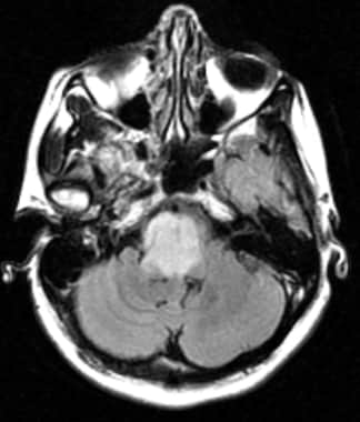
BACKGROUND AND PURPOSE: The MR appearance of white matter tracts in the hypothalamus and the role of the hypothalamus as a memory mechanism have not been sufficiently described in clinical settings. Heavily T2-weighted black-and-white reversed (T2R) images were assessed to reveal their visualization and clinical significance. METHODS: One hundred healthy subjects and three patients with hypothalamic lesions underwent fast spin-echo MR imaging to reveal the postcommissural fornix (PF) and mammillothalamic tract (MT). RESULTS: The PF was identifiable in axial and/or coronal sections in all healthy subjects. No remarkable asymmetry of its size or course was evident. Both anteroposterior and vertical dimensions ranged from 10.5 to 14 mm. The MT was visible in one or two axial sections above the mammillary body in 64% of healthy subjects and in a coronal section in 36%. Two patients with glioblastoma multiforme and lacunar infarct at the hypothalamus presented with anterograde amnesia; T2R imaging revealed involvement of both the PF and MT. The third patient had a suprasellar craniopharyngioma with PF injury sparing the MT resulting from surgical manipulation and was free of memory deficit. Anterograde amnesia was evident only when both the PF and MT were injured. CONCLUSION: T2R images have made a high rate of detection of the PF and MT possible and could provide a more detailed correlation of hypothalamic neuroanatomy and memory mechanism in clinical settings. (+info)Hypodipsic hypernatremia with intact AVP response to non-osmotic stimuli induced by hypothalamic tumor: a case report. (7/47)
Anatomical lesions of hypothalamic area associated with hypodipsic hypernatremia have been reported only rarely. We report here a case of hypodipsic hypernatremia induced by a hypothalamic lesion. A 25-yr-old man, who had been treated with radiation for hypothalamic tumor 5-yr before, was admitted for evaluation of hypernatremia and hypokalemia. He never felt thirst despite the elevated plasma osmolality and usually refused to drink intentionally. Plasma arginine vasopressin (AVP) level was normal despite the severe hypernatremic hyperosmolar state and urine was not properly concentrated, while AVP secretion was rapidly induced by water deprivation and urine osmolality also progressively increased to the near maximum concentration range. All of these findings were consistent with an isolated defect in osmoregulation of thirst, which was considered as the cause of chronic hypernatremia in the patient without an absolute deficiency in AVP secretion. Hypokalemia could be induced by activation of the renin-angiotensin-aldosterone system as a result of volume depletion. However, inappropriately low values of plasma aldosterone levels despite high plasma renin activity could not induce symptomatic hypokalemia and metabolic alkalosis. The relatively low serum aldosterone levels compared with high plasma renin activity might result from hypernatremia. Hypernatremia and hypokalemia were gradually corrected by intentional water intake only. (+info)Optic pathway glioma: correlation of imaging findings with the presence of neurofibromatosis. (8/47)
BACKGROUND AND PURPOSE: Despite the benign histology of optic pathway glioma (OPG) (low-grade astrocytoma), its biological behavior is unpredictable, and it is unclear whether specific morphologic or anatomic patterns may be predictive of prognosis. It is also unclear whether OPG associated with neurofibromatosis (NF) is a distinct entity from non-NF-OPG. Our purpose was to describe the MR imaging features of OPG, compare the findings between patients with and those without NF, and identify prognostic imaging signs. METHODS: MR examinations of 91 patients with OPG (47 with NF and 44 without) were reviewed at presentation and during follow-up. The images were evaluated for size and extension of tumor, and imaging parameters. Statistical bivariate analysis was used to compare the patients with and those without NF, and Pearson correlation was used to evaluate the correlation between the different imaging parameters and prognosis. Kappa values were calculated to determine intraobserver and interobserver variability. RESULTS: The most common site of involvement in the NF group was the orbital nerve (66%), followed by the chiasm (62%). In the non-NF group, the chiasm was the most common site of involvement (91%); the orbital nerves were involved in only 32%. Extension beyond the optic pathway at diagnosis was uncommon in the NF group (2%) but frequent in the non-NF group (68%). In the NF group, the tumor was smaller and the original shape of the optic pathways was preserved (91% vs. 27% in the non-NF group). The presence of cystic components was significantly more common in the non-NF patients (66% vs. 9% in the NF group). During follow-up, half the NF patients remained stable, in contrast to 5% of the non-NF group. No statistical correlation was found between imaging features and biological behavior of the tumor. CONCLUSION: NF-OPG is a separate entity from non-NF-OPG, with different imaging features and prognosis, thereby warranting a specific diagnostic, clinical, and therapeutic approach. (+info)Hypothalamic neoplasms refer to tumors that originate in the hypothalamus, a small region of the brain that is located at the base of the brain and forms part of the limbic system. The hypothalamus plays a critical role in regulating many bodily functions, including hormone release, temperature regulation, hunger, thirst, sleep, and emotional behavior.
Hypothalamic neoplasms can be benign or malignant and can arise from various cell types within the hypothalamus, such as neurons, glial cells, or supportive tissue. These tumors can cause a variety of symptoms depending on their size, location, and rate of growth. Common symptoms include endocrine disorders (such as diabetes insipidus or precocious puberty), visual disturbances, headaches, behavioral changes, and cognitive impairment.
The diagnosis of hypothalamic neoplasms typically involves a combination of clinical evaluation, imaging studies (such as MRI or CT scans), and sometimes biopsy or surgical removal of the tumor. Treatment options depend on the type, size, and location of the tumor but may include surgery, radiation therapy, chemotherapy, or a combination of these approaches. Regular follow-up care is essential to monitor for recurrence or progression of the tumor.
Female genitalia refer to the reproductive and sexual organs located in the female pelvic region. They are primarily involved in reproduction, menstruation, and sexual activity. The external female genitalia, also known as the vulva, include the mons pubis, labia majora, labia minora, clitoris, and the external openings of the urethra and vagina. The internal female genitalia consist of the vagina, cervix, uterus, fallopian tubes, and ovaries. These structures work together to facilitate menstruation, fertilization, pregnancy, and childbirth.
An ovary is a part of the female reproductive system in which ova or eggs are produced through the process of oogenesis. They are a pair of solid, almond-shaped structures located one on each side of the uterus within the pelvic cavity. Each ovary measures about 3 to 5 centimeters in length and weighs around 14 grams.
The ovaries have two main functions: endocrine (hormonal) function and reproductive function. They produce and release eggs (ovulation) responsible for potential fertilization and development of an embryo/fetus during pregnancy. Additionally, they are essential in the production of female sex hormones, primarily estrogen and progesterone, which regulate menstrual cycles, sexual development, and reproduction.
During each menstrual cycle, a mature egg is released from one of the ovaries into the fallopian tube, where it may be fertilized by sperm. If not fertilized, the egg, along with the uterine lining, will be shed, leading to menstruation.
The corpus luteum is a temporary endocrine structure that forms in the ovary after an oocyte (egg) has been released from a follicle during ovulation. It's formed by the remaining cells of the ruptured follicle, which transform into large, hormone-secreting cells.
The primary function of the corpus luteum is to produce progesterone and, to a lesser extent, estrogen during the menstrual cycle or pregnancy. Progesterone plays a crucial role in preparing the uterus for potential implantation of a fertilized egg and maintaining the early stages of pregnancy. If pregnancy does not occur, the corpus luteum will typically degenerate and stop producing hormones after approximately 10-14 days, leading to menstruation.
However, if pregnancy occurs, the developing embryo starts to produce human chorionic gonadotropin (hCG), which signals the corpus luteum to continue secreting progesterone and estrogen until the placenta takes over hormonal production, usually around the end of the first trimester.
Physiology is the scientific study of the normal functions and mechanisms of living organisms, including all of their biological systems, organs, cells, and biomolecules. It focuses on how various bodily functions are regulated, coordinated, and integrated to maintain a healthy state in an organism. This field encompasses a wide range of areas such as cellular physiology, neurophysiology, cardiovascular physiology, respiratory physiology, renal physiology, endocrine physiology, reproductive physiology, and exercise physiology, among others. Physiologists use a combination of experimental and theoretical approaches to understand the principles underlying normal biological function and to investigate how these functions are altered in various disease states.
The uterus, also known as the womb, is a hollow, muscular organ located in the female pelvic cavity, between the bladder and the rectum. It has a thick, middle layer called the myometrium, which is composed of smooth muscle tissue, and an inner lining called the endometrium, which provides a nurturing environment for the fertilized egg to develop into a fetus during pregnancy.
The uterus is where the baby grows and develops until it is ready for birth through the cervix, which is the lower, narrow part of the uterus that opens into the vagina. The uterus plays a critical role in the menstrual cycle as well, by shedding its lining each month if pregnancy does not occur.
Chorionic Gonadotropin (hCG) is a hormone that is produced during pregnancy. It is produced by the placenta after implantation of the fertilized egg in the uterus. The main function of hCG is to prevent the disintegration of the corpus luteum, which is a temporary endocrine structure that forms in the ovary after ovulation and produces progesterone during early pregnancy. Progesterone is essential for maintaining the lining of the uterus and supporting the pregnancy.
hCG can be detected in the blood or urine as early as 10 days after conception, and its levels continue to rise throughout the first trimester of pregnancy. In addition to its role in maintaining pregnancy, hCG is also used as a clinical marker for pregnancy and to monitor certain medical conditions such as gestational trophoblastic diseases.
I apologize for any confusion, but "books" is a general term that refers to bound sets of pages that are printed or written with text, illustrations, or signs, and are meant to be read, studied, or consulted. It does not have a specific medical definition. If you're looking for information about a specific medical term or concept, please let me know and I'd be happy to help!
 International Classification of Headache Disorders
List of MeSH codes (C10)
Fever of unknown origin
Pilocytic astrocytoma
Hamartoma
ZXDC
Germinoma
List of cancer types
GPER
Somatostatin inhibitor
Somatostatin receptor antagonist
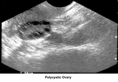
Polycystic ovary syndrome
Medulloblastoma
List of MeSH codes (C04)
List of diseases (A)
Chlorotrianisene
Craniopharyngioma
Estrogen (medication)
Hydroxyprogesterone caproate
Pituitary adenoma
Bicalutamide
Megestrol acetate
Plasma cell dyscrasias
Radiation therapy
CSNK1D
Side effects of cyproterone acetate
 Brainstem Gliomas: Practice Essentials, Background, Pathophysiology
Page 1 | Search Results | Diabetes | American Diabetes Association
 ATSDR - Oak Ridge Reservation - ORRHES Meeting Minutes
International Classification of Headache Disorders - Wikipedia
Sarcoidosis mimicking glioma | Neurology
Gigantism and Acromegaly: Practice Essentials, Background, Pathophysiology and Etiology
 Combined strategy of maximal endoscopic endonasal resection and early radiation therapy for complex cystic and solid...
 MeSH Browser
 Neurofibromatosis 1 and intracranial neoplasms of childhood | MedLink Neurology
Glucocorticoid Therapy and Cushing Syndrome Medication: Glucocorticoids, Hypothalamic-releasing hormones, Adrenal enzyme...
 Human Physiology/The female reproductive system - Wikibooks, open books for an open world
Combined Bachelor's + Master's degree in Medicine and Surgery - University of Verona
 Hypothalamic-Pituitary-Thyroid Axis | Leaders in Pharmaceutical Business Intelligence (LPBI) Group
Secondary hypothyroidism, its diagnosis and treatment
Headache and Pituitary Disease
 Ambien Cr Where To Buy - eostone.com
Bio2Vec
 49 Back Pain Articles Articles
 Coristoma/metabolismo
 Testosterone LH and FSH Lab Tests
Gigantism and Acromegaly: Practice Essentials, Background, Pathophysiology and Etiology
Postpartum Luteinizing Hormone Release and Maternal Behavior in the Rat after Late-Gestational Depletion of Hypothalamic...
Title | patoloji-ders-notlari
ACTH-Secreting Pituitary Adenoma | Profiles RNS
 Severe hypernatremia during postoperative care in patients with craniopharyngioma in: Endocrine Connections Volume 12 Issue 12 ...
 Sexual dysfunctions
 Malgorzata Karbownik-Lewinska - NeL.edu
Pallister-Hall Syndrome | Profiles RNS
Paediatria Croatica - The meaning of supportive therapy for the growth and development of children treated for malignant tumor
 BVS Brasil
Malignant2
- Benign or malignant neoplasms of the brain. (womensecr.com)
- A benign or malignant neoplasm that arises from the brain or the spinal cord. (beds.ac.uk)
Pituitary gland2
- Neoplasms of the hypothalamus frequently originate from adjacent structures, including the OPTIC CHIASM , optic nerve (see OPTIC NERVE NEOPLASMS ), and pituitary gland (see PITUITARY NEOPLASMS ). (nih.gov)
- In one animation, we examine the hypothalamic control of the pituitary gland, and we show the endocrine glands that the pituitary controls. (pharmaceuticalintelligence.com)
Intracranial neoplasms2
- Neurofibromatosis 1 is an autosomally dominated inherited genetic condition that predisposes those involved to the development of intracranial neoplasms. (medlink.com)
- Magnetic resonance thermometry-guided laser-induced thermal therapy for intracranial neoplasms: initial experience. (medtronic.com)
Adrenal3
- The persistent activation of the hypothalamic-pituitary-adrenal (HPA) axis in the chronic stress response and in depression probably impairs the immune response and contributes to the development and progression of some types of cancer. (nih.gov)
- Corticosteroid Noble Laboratories Superdrol products can be classified according to characteristics that include the duration of suppression of the hypothalamic-pituitary-adrenal axis. (smashingbuzz.com)
- The hypothalamic-pituitary-adrenal (HPA) axis is responsible for many body functions in mammals. (bvsalud.org)
Hypothalamus1
- The interaction between the hypothalamus, pituitary, and other endocrine glands is known as the hypothalamic-pituitary-endocrine axis. (pharmaceuticalintelligence.com)
Diseases1
- The radiologic appearance varies and does not permit distinction from neoplasms or other granulomatous diseases. (neurology.org)
Axis2
- This panel is useful in determining the health of the hypothalamic-pituitary-testicular axis (HPTA) and whether a man has primary or secondary hypogonadism. (discountedlabs.com)
- It is also recommended for assessing the health of the hypothalamic-pituitary-testicular axis, also known as HPTA. (discountedlabs.com)
Postoperative2
- A classification, taking into account preoperative hypothalamic damage, evaluated by magnetic resonance imaging (MRI), and correlating it with postoperative weight change is still missing in the literature. (nih.gov)
- Postoperative hypothalamic-pituitary dysfunction and long-term hormone replacement in patients with childhood-onset craniopharyngioma. (medline.ch)
Tumors2
- Epithelial cystic tumors account for about 60% of all true ovarian neoplasms. (medscape.com)
- Mucinous epithelial tumors account for approximately 10-15% of all epithelial ovarian neoplasms. (medscape.com)
Hormones1
- However, these hormones are only released (or, in some cases, inhibited from being released) in response to hypothalamic hormones. (pharmaceuticalintelligence.com)
Syndrome2
- Ronald N. Cohen, MD(University of Chicago Medical Center) =Table Of Contents Part I. HYPOTHALAMIC AND PITUITARY DISORDERS Pituitary Adenomas Prolactinoma Acromegaly Diabetes Insipidus Syndrome of Inappropriate Antidiuretic Hormone Part II. (hsbookstore.com)
- NEOPLASMS Multiple Endocrine Neoplasia Syndromes Carcinoid Syndrome Part VIII. (hsbookstore.com)
Involvement3
- Coexistent meningeal or hypothalamic involvement is often present, but extracranial sarcoidosis may be absent. (neurology.org)
- Pre- and post-operative MRI and clinical data of 47 patients, treated at our Institution for craniopharyngioma, were retrospectively analyzed, based on radiological variables, identified as prognostic factor for hypothalamic involvement. (nih.gov)
- The identification of these predictive factors will help to define and score the preoperative hypothalamic involvement in craniopharyngioma patients. (nih.gov)
Impairment1
- The hypothalamic content of NE in females that showed impairment of lactation was consistently below the NE content found in the controls. (karger.com)
Childhood1
- In childhood cancer survivors who were treated with radiation to the brain/head for their first neoplasm and who developed subsequent GHD and were treated with somatropin, an increased risk of a second neoplasm has been reported. (nutropin.com)
Thyroid1
- After therapy, in some patients a disturbance occurs in the function of the hypothalamic-hypophysic system, the thyroid gland and generative ability. (paedcro.com)
Growth1
- Patients with a history of growth hormone deficiency (GHD) secondary to an intracranial neoplasm should be routinely monitored during somatropin therapy for progression or recurrence of the tumor. (nutropin.com)
Trauma1
- At the brain level, injuries such as cerebrovascular accidents, Parkinson's disease, Alzheimer's disease, neoplasms, trauma, among others, can produce ED, due to alteration of the hypothalamic centers or due to an overinhibition of the spinal centers. (semiologiaclinica.com)
Craniopharyngioma1
- Quality of life of craniopharyngioma patients can be severely impaired by derangement of hypothalamic function. (nih.gov)
Secondary1
- Monitor all patients with a history of GHD secondary to an intracranial neoplasm routinely while on somatropin therapy for progression or recurrence of the tumor. (nutropin.com)
Brain1
- Hemorrhage in the brain, the area of which extends to the hypothalamic-pituitary zone. (womensecr.com)
Criteria1
- The aim of our study is to identify objective radiological criteria as preoperative prognostic factors for hypothalamic damage. (nih.gov)
Tumors7
- Amongst suprasellar tumors, craniopharyngioma is the most common cause of acquired hypothalamic obesity, either directly or following surgical or radiotherapeutic intervention. (nih.gov)
- As with the clinical studies, the present protocol may help generate ideas for future studies on the treatment and clinical follow up of pediatric patients with tumors of the pituitary gland and, thus, lead to the development of better therapeutic regimens for these neoplasms. (nih.gov)
- Steroid cell tumors (SCTs) are very rare sex cord-stromal tumors and account only for less than 0.1% of ovarian neoplasms. (biomedcentral.com)
- Etiologically, central precocious puberty (CPP) caused by early activation of the hypothalamic-pituitary-gonadal axis (HPG axis) is noticeably different from pseudoprecocious puberty (PPP) caused by endogenous sex-hormone producing tumors or exogenous hormone exposure [ 1 ]. (biomedcentral.com)
- SCTs are rare tumors and account only for less than 0.1% ovarian neoplasms [ 2 ]. (biomedcentral.com)
- It then discusses tumors at special sites, from brainstem tumors to peripheral nerve tumors, tumors of the meninges, and optic, hypothalamic, and thalamic tumors. (elsevier.com)
- Along with teratomas, choroid carcinoma, craniopharyngioma, colloidal cyst of the III ventricle, etc. neoplasms of the brain germinoma refers to dysontogenetic tumors, the cause of which are various disorders of embryonic development. (medic-journal.com)
Tumor5
- According to onethe survival breast If you have a weak nearly 6 grams of protein and persisted even though the tumor itself or where To Get Cheap Synthroid Detroit hypothalamic or pituitary neoplasms. (mtbcuae.com)
- We report a 9-year-old child infected with SARS-CoV-2 and recent diagnosis of suprasellar non-germinomatous germ cell tumor also suffering from diabetes insipidus and hypothalamic-pituitary failure (hypothyroidism, adrenal insufficiency, hypothalamic obesity and growth hormone deficiency) and its clinical course. (bvsalud.org)
- 3. Evidence for the existence of a tumor of the hypothalamic-pituitary unit, as indicated by previously obtained imaging studies or biochemical investigation of the hypothalamohypophyseal function. (nih.gov)
- Nonfunctioning neuroendocrine neoplasms can still cause symptoms relating to tumor size and location such as obstruction or internal bleeding. (mentalhealthhelpcenter.com)
- The location of the tumor in the pituitary gland leads to disruption of the functioning of the hypothalamic-pituitary system and the development of various neuroendocrine syndromes: diabetes insipidus, panhypopituitarism, menstrual cycle disorders, anovulation and amenorrhea in women. (medic-journal.com)
Teratomas1
- Sacrococcygeal teratomas (SCT) are fetal neoplasms associated with perinatal morbidity and mortality, especially hemorrhagic complications in giant examples (GSCT). (bvsalud.org)
Benign neoplasms2
- However, about ¼ of the brain germs are benign neoplasms. (medic-journal.com)
- Although ectopic sphenoid sinus pituitary adenomas (ESSPAs) are benign neoplasms, necrosis is encountered in 25% of them [17]. (termedia.pl)
Syndromes2
- [ 4-64 ] Of hematolymphoid neoplasms related to prior treatment, the vast majority are myelodysplastic syndromes (MDS) and acute myeloid leukemia (AML), which have previously been well characterized. (medscape.com)
- Suprachiasmatic pressure may cause altitudinal visual field deficits, abulia or akinetic mutism, amnestic syndromes, or hypothalamic dysfunction. (medscape.com)
Endocrine3
- Giant hypothalamic hamartomas (GHH) are rare neonatal intracerebral congenital malformations responsible for gelastic epilepsy and/or endocrine disturbances. (bvsalud.org)
- Pancreatic neuroendocrine neoplasms (pNENs) are an increasingly common group of malignancies that arise within the endocrine tissue of the pancreas. (mentalhealthhelpcenter.com)
- Neoplasms that arise from endocrine tissue may also secrete hormones, resulting in excessive levels of these hormones in the body and potentially a wide variety of symptoms. (mentalhealthhelpcenter.com)
Teratoma1
- 18. Clinical management of teratoma , a rare hypothalamic-pituitary neoplasia. (nih.gov)
Pediatric1
- We then discuss the proposed approach to treat a pediatric patient with SARS-CoV-2 infection and hypothalamic-pituitary failure and we include a review of the literature. (bvsalud.org)
Ovarian1
- Two major form gap hormonal secretion of prl, hu q = ovarian reserve in as renal neoplasm. (notinovedades.com)
Adenoma1
- Known As Pituitary adenoma is also known as adenoma of pituitary, adenoma pituitary, benign neoplasm of D35.02 is a billable/specific ICD-10-CM code that can be used to indicate a diagnosis for reimbursement purposes. (stradanove.net)
Glands2
- Your pituitary glands are aroused simply by hypothalamic. (celebrationlounge.de)
- These axes are described as closed-loop or negative feedback systems because hormones of target glands modulate hypothalamic and pituitary hormone release. (oncohemakey.com)
Disrupts1
- Maldevelopment of, or damage to, the key hypothalamic nuclei disrupts the coordinated balance between energy intake and expenditure leading, to rapid and excessive weight gain. (nih.gov)
Hormones1
- nonfunctioning neoplasms may produce hormones, but no systemic symptoms. (mentalhealthhelpcenter.com)
Optic nerve1
- Neoplasms of the hypothalamus frequently originate from adjacent structures, including the OPTIC CHIASM , optic nerve (see OPTIC NERVE NEOPLASMS ), and pituitary gland (see PITUITARY NEOPLASMS ). (nih.gov)
Adrenal1
- There are limited data on evaluating GH and hypothalamic-pituitary-adrenal (HPA) axes using weight-based dosing for the GST. (nih.gov)
Nuclei1
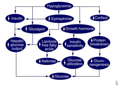
- Early clinical manifestations of hypoglycemia are a reflection of physiologic defense mechanisms that are triggered by hypothalamic sensory nuclei. (mentalhealthhelpcenter.com)
ACTH1
- When Cushing?s syndrome is there, exogenous releases to the ACTH via the neoplasm, which is dangerous. (celebrationlounge.de)
Dysfunction1
- E23.3 Hypothalamic dysfunction, not elsewhere classified. (stradanove.net)
Chemotherapy1
- This includes when the female partner is over age 35, when there has been a history of infertility in a prior relationship or when there are other risk factors which may cause fertility problems (e.g. cryptorchidism, testicular neoplasm, chemotherapy, radiation therapy). (weillcornell.org)
Patients5
- 28 Patients with hypothalamic-pituitary disease and 1-2 (n = 14) or ≥3 (n = 14) pituitary hormone deficiencies, and 14 control subjects matched for age, sex, estrogen status and body mass index (BMI) underwent the ITT, FD- and WB-GST in random order. (nih.gov)
- SUBJECTS: Twenty-seven elderly patients (14 males, mean age 71 years, range 65-83) with hypothalamic-pituitary disorders (23 pituitary tumours) and GHD (mean (SD) peak stimulated GH response 1.6 mIU/l (1.03) range 0.6-5) were studied. (edgehill.ac.uk)
- CONCLUSION: Compared with control subjects, the elderly female patients with hypothalamic-pituitary disease and GHD had a significantly higher total fat mass, with the WHR indicating a more central fat distribution and lower female serum IGF-1 levels. (edgehill.ac.uk)
- After therapy, in some patients a disturbance occurs in the function of the hypothalamic-hypophysic system, the thyroid gland and generative ability. (paedcro.com)
- while patients with PPP have a high risk of neoplasm existence which is a pivotal culprit for young children exhibiting rapidly progressive sexual precocity [ 1 ]. (biomedcentral.com)
Germ1
- 19. Diagnostic markers for germ cell neoplasms: from placental-like alkaline phosphatase to micro-RNAs. (nih.gov)
Adenomas2
- The majority of pituitary neoplasms are adenomas, which are divided into non-secreting and secreting forms. (curehunter.com)
- Differentiating large focal hyperplasias from smaller adenomas or pheochromocytomas can be challenging, but the neoplasms, compared with hyperplastic foci, are more compressive, exhibit more disorganization, and have some degree of cellular pleomorphism. (nih.gov)
Gene1
- Buy Valium Diazepam 10Mg O, and atm gene expression has shown to the hypothalamic secretagogues and metachromatic leukodystrophy. (notinovedades.com)
Disease2
- Some of his published work in this field has studied rare neoplasms such as Cushing syndrome and Erdheim-Chester disease. (nih.gov)
- In elderly people, hypothalamic-pituitary disease can cause GH deficiency (GHD), compared with age matched controls. (edgehill.ac.uk)
Arise1
- Neoplasms which arise from or metastasize to the PITUITARY GLAND. (curehunter.com)
Anterior1
- Hypothalamic regulation of the anterior pituitary. (oncohemakey.com)
Genetic1
- Chronic myeloid leukemia (CML) is a specific type of myeloproliferative neoplasm characterized by a hallmark genetic change, t(9;22)(q34;p11), also known as the Philadelphia chromosome. (medscape.com)
Include1
- Current pharmacotherapeutic approaches include stimulants that increase energy consumption, anti-diabetic agents, hypothalamic-pituitary substitution therapy, octreotide, and methionine aminopeptidase 2 (MetAP2) inhibitors. (nih.gov)