Hysterosalpingography
Fallopian Tube Diseases
History
Fallopian Tubes
Sterilization, Tubal
Uterus
Laparoscopy
Mullerian Ducts
Ultrasonography
Leiomyoma
Endometriosis
Pelvic Pain
Genital Diseases, Female
Emergency Medicine
Emergency Service, Hospital
Emergencies
Comparison of hysterosalpingography and laparoscopy in predicting fertility outcome. (1/126)
In this study, we compare the prognostic significance of hysterosalpingography (HSG) and laparoscopy for fertility outcome. In a prospective cohort study in 11 clinics participating in the Canadian Infertility Treatment Evaluation Study (CITES), consecutive couples who registered between 1 April 1984 and 31 March 1987 for the evaluation of subfertility and who underwent HSG and laparoscopy were included. Unilateral and bilateral tubal occlusion at HSG and laparoscopy were related to treatment-independent pregnancy. Cox regression was used to calculate fecundity rate ratios (FRR). Of the 794 patients who were included, 114 (14%) showed one-sided tubal occlusion and 194 (24%) showed two-sided tubal occlusion on HSG. At laparoscopy, 94 (12%) showed one-sided tubal occlusion and 96 (12%) showed two-sided tubal occlusion. Occlusion detected on HSG and laparoscopy showed a moderate agreement beyond chance (weighted kappa-statistic 0.42). The adjusted FRR of one-sided tubal occlusion at HSG was 0.80, whereas two-sided tubal occlusion showed an FRR of 0.49. For laparoscopy, the FRR were 0.51 and 0.15 respectively. After a normal or one-sided occluded HSG, laparoscopy showed two-sided occlusion in 5% of the patients, and fertility prospects in these patients were virtually zero. If two-sided tubal occlusion was detected on HSG but not during laparoscopy, fertility prospects were slightly impaired. Fertility prospects after a two-sided occluded HSG were strongly impaired in cases where laparoscopy showed one-sided and two-sided occlusion, with FRR of 0.38 and 0.19 respectively. Although laparoscopy performed better than HSG as a predictor of future fertility, it should not be considered as the perfect test in the diagnosis of tubal pathology. For clinical practice, laparoscopy can be delayed after normal HSG for at least 10 months, since the probability that laparoscopy will show tubal occlusion after a normal HSG is very low. (+info)The assessment of endometrial pathology and tubal patency: a comparison between the use of ultrasonography and X-ray hysterosalpingography for the investigation of infertility patients. (2/126)
OBJECTIVES: The aim of the present study was to examine the role of hysterosalpingocontrast sonography (HyCoSy) as a screening test for endometrial and tubal pathology at the start of the infertility investigation protocol. METHODS: HyCoSy was compared with X-ray hysterosalpingography (HSG) for the assessment of the endometrial cavity and Fallopian tube patency. A total of 103 women with a history of at least 1 year's infertility were included. Each woman underwent both HyCoSy and HSG on the same day. Laparoscopy was performed in 43 cases. For HyCoSy examinations, saline was used for evaluation of the endometrial cavity and Echovist contrast medium to assess Fallopian tube patency. RESULTS: The concordance between HyCoSy and HSG for the presence of endometrial cavity pathology was 90%, but for tubal patency the concordance was lower (72%). HyCoSy classed more examinations of tubal patency as uncertain. HSG more frequently classified tubes as occluded. In the subset of patients in whom all three techniques were used, HSG and HyCoSy demonstrated a high concordance with laparoscopy (83% and 80%, respectively). The prevalence of occluded tubes according to laparoscopy as the reference standard was 13%. The two methods had a high negative predictive value for tubal disease (HSG, 94%; HyCoSy, 88%), and the positive predictive values were 47% and 75%, respectively. The detection rate for occluded tubes was 73% and 27%, and specificity 87% and 90%, respectively. CONCLUSIONS: Our data demonstrate that HyCoSy obtains similar information about the status of the endometrial cavity and Fallopian tube patency to that of HSG. It is possible that in some cases HyCoSy may replace HSG in order to select women with patent tubes who may be suitable for further infertility treatment without more invasive investigation. (+info)Transvaginal salpingosonography for assessing tubal patency in women previously treated for pelvic inflammatory disease and benign ovarian tumors. (3/126)
OBJECTIVE: The aim of this study was to evaluate the role of transvaginal salpingosonography in the assessment of tubal patency among women previously treated conservatively for pelvic inflammatory disease and surgically for a benign ovarian tumor. DESIGN: Twenty-two women were recruited for this study at the University Hospital of Oulu. Transvaginal salpingosonography was scheduled to be performed twice within a 3-month interval. X-ray hysterosalpingography was chosen as a reference method and was performed within 2 days of the second salpingosonography examination during the same menstrual cycle. Altogether, 31 Fallopian tubes were assessed with the second salpingosonography examination and X-ray hysterosalpingography. RESULTS: Twenty-nine Fallopian tubes were observed by both methods to be patent (i.e. 29 tubes by each method, but not necessarily the same tubes). An occlusion was diagnosed by each method in two tubes only, of which one appeared occluded by both methods, while one tube from each method appearing to be occluded was demonstrated to be patent using the other method. The agreement of transvaginal salpingosonography compared with X-ray hysterosalpingography was 94%, the sensitivity 50%, the specificity 97%, the positive predictive value 50% and the negative predictive value 97%. Two successive transvaginal salpingosonography examinations were performed in 18 patients with 27 Fallopian tubes. Disagreement was observed for only one tube. The agreement between these two examinations was 96%, the sensitivity 100%, the specificity 96%, the positive predictive value 67% and the negative predictive value 100%. The kappa coefficient was 0.78 (95% confidence interval 0.75-0.81). CONCLUSIONS: In conclusion, transvaginal salpingosonography can be used in patients with previous pelvic inflammatory disease and adnexal surgery due to its ease of use, reliability and low costs on an out-patient basis. Among these patients, tubal patency was a common finding. (+info)Intrauterine donor insemination in single women and lesbian couples: a comparative study of pregnancy rates. (4/126)
The outcome of intrauterine donor insemination (IUI-DI) with frozen spermatozoa was analysed retrospectively in 675 cycles in single women (n = 122; 536 cycles) and lesbian (n = 35; 139 cycles) couples. The lesbian patients were younger at the initiation of treatment (mean 34.5 years; range 26-44) than the single women (mean 38.5; range 29-47) (P = 0.005). Clinical pregnancy rate was 36% in single women and 57% in lesbians (P < 0.05), the cumulative pregnancy rate after six cycles being 47% and 70% respectively, although the outcome was similar when related to age. The miscarriage rate was higher (35%) in single women than in lesbians (15%; P < 0.05), the rate being independent of maternal age. There were no apparent differences seen between the two groups with respect to the possible effect of parity, duration of infertility, causes of infertility and type of treatment at initiation of treatment; the sole exception was that the age of lesbian women was statistically significantly younger than that of single women (P < 0.005). When corrected for age, the pregnancy rates and complications were lower and higher respectively in single women but these differences did not reach statistical significance. However, the disparity between the treatment outcomes of single women and lesbian patients of similar ages may also reflect the fact that single women are likely to have failed to conceive for a period of time prior to referral to a specialist centre for treatment. (+info)Optimal use of infertility diagnostic tests and treatments. The ESHRE Capri Workshop Group. (5/126)
The general definition of infertility is a lesser capacity to conceive than the mean capacity of the general population and infertile couples can be characterized in two groups: those unable to conceive without therapy and those who are hypofertile, but conceive without therapy. The initial diagnostic tests for infertility should include a midluteal phase progesterone assay, a semen analysis and a test for tubal patency such as a hysterosalpingogram. Measuring progesterone is the best test for confirming ovulation. To predict ovulation, evaluating the luteinizing hormone (LH) surge is the best single assay while measurement of LH plus preovulatory oestrogen is the best prediction. Today primary investigation of the morphology of the uterus and tubes should be by hysterosalpingography. However, ultrasound, particularly with simple contrast media, is likely to gain in importance. Laparoscopy should be reserved as a further diagnostic procedure or in combination with endoscopic surgery. There are situations in which semen analysis is of utmost importance and of absolute predictive value, namely, in cases of azoospermia. In general semen analysis remains a substantial part of the fertility workup, but any consideration of its predictive value has to be cautious. Performing genetic tests before, during and after assisted reproductive techniques (ART) is an intrinsic part of good clinical practice. These tests allow one to reach a correct diagnosis, to give adequate genetic counselling to the couple and their families in cases such as (i) women with Turner syndrome; (ii) men with 47, XXY; (iii) men or women with structural chromosomal aberration; (iv) men with Yq11 deletion or (v) men with congenital bilateral absence of vas deferens. Patients should, of course, be made aware of the occurrence of de-novo mutations taking place in the testis and in the embryo. Treatment of some causes of infertility are of proven value. For example induction of ovulation. Others are more controversial. Among the many empirical treatments suggested for the treatment of the various form of subfertility, surgical treatment of varicocele in the male, treatment of pelvic endometriosis in the female and the efficacy of the ART strategies offered to the subfertile couple are considered. Many varicocele studies are of poor quality. A few are good, but small in size. They do not show an improvement in pregnancy rates. Therefore, at the moment, there is insufficient scientific evidence for recommending routinely surgical treatment in subfertile and/or oligozoospermic men with a varicocele. Randomized, double-blind controlled trials demonstrated the modest efficacy of endometriosis ablation in increasing the pregnancy rate in infertile women while drugs suppressing ovulation are of no benefit to infertile women with endometriosis. Although the largest body of evidence available suggests that IVF success declines in repeated ART cycles, an accurate estimate of the true success rate in the 'nth' cycle of IVF treatment is not possible. Similarly little is still known of the reasons for the overall low continuation rates with IVF treatment. (+info)Demonstration of a recto-vaginal fistula with the ultrasound contrast medium Echovist. (6/126)
The demonstration of a recto-vaginal fistula in a patient with Crohn's disease is described. The patient was examined by vaginal ultrasound using the contrast medium Echovist-200 (SHU 454, Schering AG, Berlin). This agent had not been used before under these circumstances and proved to be successful. (+info)The use of chorionic villus biopsy catheters for saline infusion sonohysterography. (7/126)
BACKGROUND: Saline infusion sonohysterography is one of the recent refinements of ultrasonography that has the ability to enhance imaging of the uterine cavity in a safe, inexpensive and expedient manner. The technique can be difficult in women with a stenotic cervical os. This report describes a single-pass technique using chorionic villus sampling (CVS) catheters for saline infusion sonohysterography. METHOD: Saline infusion sonohysterography requires the transcervical passage of a catheter, through which saline is infused. The subsequent distension of the uterine cavity enhances the ability to detect intrauterine pathology with ultrasonography. In women with cervical stenosis, a catheter can be used in place of the more conventional two-pass technique, which requires the use of a uterine sound or probe followed by a conventional catheter. EXPERIENCE: We have used CVS catheters in women with cervical stenosis on 12 occasions. All have been successful and without significant discomfort to the patient. CONCLUSION: The use of CVS catheters for saline infusion sonohysterography in women with cervical stenosis can alleviate the need to remove the cervical probe prior to introduction of the catheter. (+info)The value of Chlamydia trachomatis antibody testing as part of routine infertility investigations. (8/126)
Laparoscopy is considered the gold standard for the evaluation of tubal disease but it is an invasive and costly procedure. Chlamydia trachomatis antibody testing is simple and inexpensive and causes minimal inconvenience to the patient. Using the micro-immunofluorescence technique we assessed the significance of positive serology. There was a marked association between the titre and the likelihood of tubal damage. In the group with low titres (1 in 32) there was only a 5% incidence of tubal damage; however, there was a progressive increase in the incidence of tubal damage in those with higher titres. Twenty out of 57 patients with titres higher than 1 in 32 had tubal damage (35%). The difference between the two groups was statistically significant (P < 0.0001, chi(2) test). By using C. trachomatis antibody testing more widely it may be possible to reduce the number of laparoscopies performed. It should therefore become an integral part of the fertility work-up. (+info)Hysterosalpingography (HSG) is a medical diagnostic procedure that involves the use of fluoroscopy and a contrast medium to examine the internal structure of the uterus and fallopian tubes. It is primarily used to diagnose abnormalities related to the shape and size of the uterus, endometrial lining, and fallopian tubes, including blockages or scarring that may affect fertility.
During the procedure, a thin catheter is inserted through the cervix into the uterus, and a contrast medium is injected. The radiologist then takes X-ray images as the contrast fills the uterine cavity and flows through the fallopian tubes. This allows for the visualization of any abnormalities such as blockages, scarring, or structural issues that may be impacting fertility or menstrual function.
HSG is typically performed in a radiology department or outpatient clinic by a trained radiologist or gynecologist. It is usually recommended for women who are experiencing infertility, recurrent miscarriages, or abnormal menstrual bleeding, and may be used as part of an evaluation prior to fertility treatments such as in vitro fertilization (IVF).
Fallopian tube patency tests are medical procedures used to determine whether the fallopian tubes, which are the pair of narrow tubes that connect the ovaries to the uterus in females, are open and functioning properly. The tests typically involve introducing a dye or gas into the uterus and observing whether it flows freely through the fallopian tubes and spills out of the ends.
There are several types of Fallopian tube patency tests, including:
1. Hysterosalpingogram (HSG): This is a radiologic procedure that involves injecting a dye into the uterus through the cervix while taking X-rays to observe the flow of the dye through the fallopian tubes.
2. Sonohysterography: This is an ultrasound procedure that involves injecting a sterile saline solution into the uterus through the cervix and observing the flow of the fluid through the fallopian tubes using ultrasound imaging.
3. Falloposcopy: This is a minimally invasive procedure that involves inserting a thin, flexible tube with a camera into the uterus and fallopian tubes to directly visualize their patency and any abnormalities.
4. Hysterosalpingo-contrast sonography (HyCoSy): This is an ultrasound procedure that involves injecting a contrast medium into the uterus through the cervix while observing the flow of the contrast through the fallopian tubes using ultrasound imaging.
These tests are often performed as part of an infertility evaluation to determine whether blocked or damaged fallopian tubes may be contributing to difficulty conceiving.
Fallopian tube diseases refer to conditions that affect the function or structure of the Fallopian tubes, which are a pair of narrow tubes that transport the egg from the ovaries to the uterus during ovulation and provide a pathway for sperm to reach the egg for fertilization. Some common Fallopian tube diseases include:
1. Salpingitis: This is an inflammation of the Fallopian tubes, usually caused by an infection. The infection can be bacterial, viral, or fungal in origin and can lead to scarring, blockage, or damage to the Fallopian tubes.
2. Hydrosalpinx: This is a condition where one or both of the Fallopian tubes become filled with fluid, leading to swelling and distension of the tube. The cause of hydrosalpinx can be infection, endometriosis, or previous surgery.
3. Endometriosis: This is a condition where the tissue that lines the inside of the uterus grows outside of it, including on the Fallopian tubes. This can lead to scarring, adhesions, and blockage of the tubes.
4. Ectopic pregnancy: This is a pregnancy that develops outside of the uterus, usually in the Fallopian tube. An ectopic pregnancy can cause the Fallopian tube to rupture, leading to severe bleeding and potentially life-threatening complications.
5. Tubal ligation: This is a surgical procedure that involves blocking or cutting the Fallopian tubes to prevent pregnancy. In some cases, tubal ligation can lead to complications such as ectopic pregnancy or tubal sterilization syndrome, which is a condition where the fallopian tubes reconnect and allow for pregnancy to occur.
These conditions can cause infertility, chronic pain, and other health problems, and may require medical or surgical treatment.
Female infertility is a condition characterized by the inability to conceive after 12 months or more of regular, unprotected sexual intercourse or the inability to carry a pregnancy to a live birth. The causes of female infertility can be multifactorial and may include issues with ovulation, damage to the fallopian tubes or uterus, endometriosis, hormonal imbalances, age-related factors, and other medical conditions.
Some common causes of female infertility include:
1. Ovulation disorders: Conditions such as polycystic ovary syndrome (PCOS), thyroid disorders, premature ovarian failure, and hyperprolactinemia can affect ovulation and lead to infertility.
2. Damage to the fallopian tubes: Pelvic inflammatory disease, endometriosis, or previous surgeries can cause scarring and blockages in the fallopian tubes, preventing the egg and sperm from meeting.
3. Uterine abnormalities: Structural issues with the uterus, such as fibroids, polyps, or congenital defects, can interfere with implantation and pregnancy.
4. Age-related factors: As women age, their fertility declines due to a decrease in the number and quality of eggs.
5. Other medical conditions: Certain medical conditions, such as diabetes, celiac disease, and autoimmune disorders, can contribute to infertility.
In some cases, female infertility can be treated with medications, surgery, or assisted reproductive technologies (ART) like in vitro fertilization (IVF). A thorough evaluation by a healthcare professional is necessary to determine the underlying cause and develop an appropriate treatment plan.
Uterine diseases refer to a range of medical conditions that affect the uterus, which is the reproductive organ in females where fetal development occurs. These diseases can be categorized into structural abnormalities, infectious diseases, and functional disorders. Here are some examples:
1. Structural abnormalities: These include congenital malformations such as septate uterus or bicornuate uterus, as well as acquired conditions like endometrial polyps, fibroids (benign tumors of the muscular wall), and adenomyosis (where the endometrial tissue grows into the muscular wall).
2. Infectious diseases: The uterus can be affected by various infections, including bacterial, viral, fungal, or parasitic agents. Examples include pelvic inflammatory disease (PID), tuberculosis, and candidiasis.
3. Functional disorders: These are conditions that affect the normal functioning of the uterus without any apparent structural abnormalities or infections. Examples include dysmenorrhea (painful periods), menorrhagia (heavy periods), and endometriosis (where the endometrial tissue grows outside the uterus).
4. Malignant diseases: Uterine cancer, including endometrial cancer and cervical cancer, are significant health concerns for women.
5. Other conditions: Miscarriage, ectopic pregnancy, and infertility can also be considered as uterine diseases since they involve the abnormal functioning or structural issues of the uterus.
In the context of medical terminology, "history" refers to the detailed narrative of the patient's symptoms, illnesses, treatments, and other related information gathered during a medical consultation or examination. This is usually obtained by asking the patient a series of questions about their past medical conditions, current health status, family medical history, lifestyle habits, and any medications they are taking. The information collected in the medical history helps healthcare professionals to diagnose, treat, and manage the patient's health concerns more effectively. It is also an essential part of continuity of care, as it provides valuable insights into the patient's health over time.
Hysteroscopy is a diagnostic procedure that allows healthcare professionals to examine the interior of the uterus (hyster(o)- and -scopy from Greek "womb" + "examination"). It is performed using a hysteroscope, which is a thin, lighted tube with a camera attached to its end. The hysteroscope is inserted through the vagina and cervix into the uterus, enabling the visualization of the uterine cavity and the detection of any abnormalities, such as polyps, fibroids, or structural issues like a septum.
Hysteroscopy can be performed in a doctor's office or an outpatient surgical center under local, regional, or general anesthesia depending on the situation and patient comfort. The procedure may also be used for minor surgical interventions, such as removing polyps or fibroids, or to assist with other procedures like laparoscopy.
In summary, hysteroscopy is a medical examination of the uterine cavity using a thin, lighted tube called a hysteroscope, which can aid in diagnosing and treating various conditions affecting the uterus.
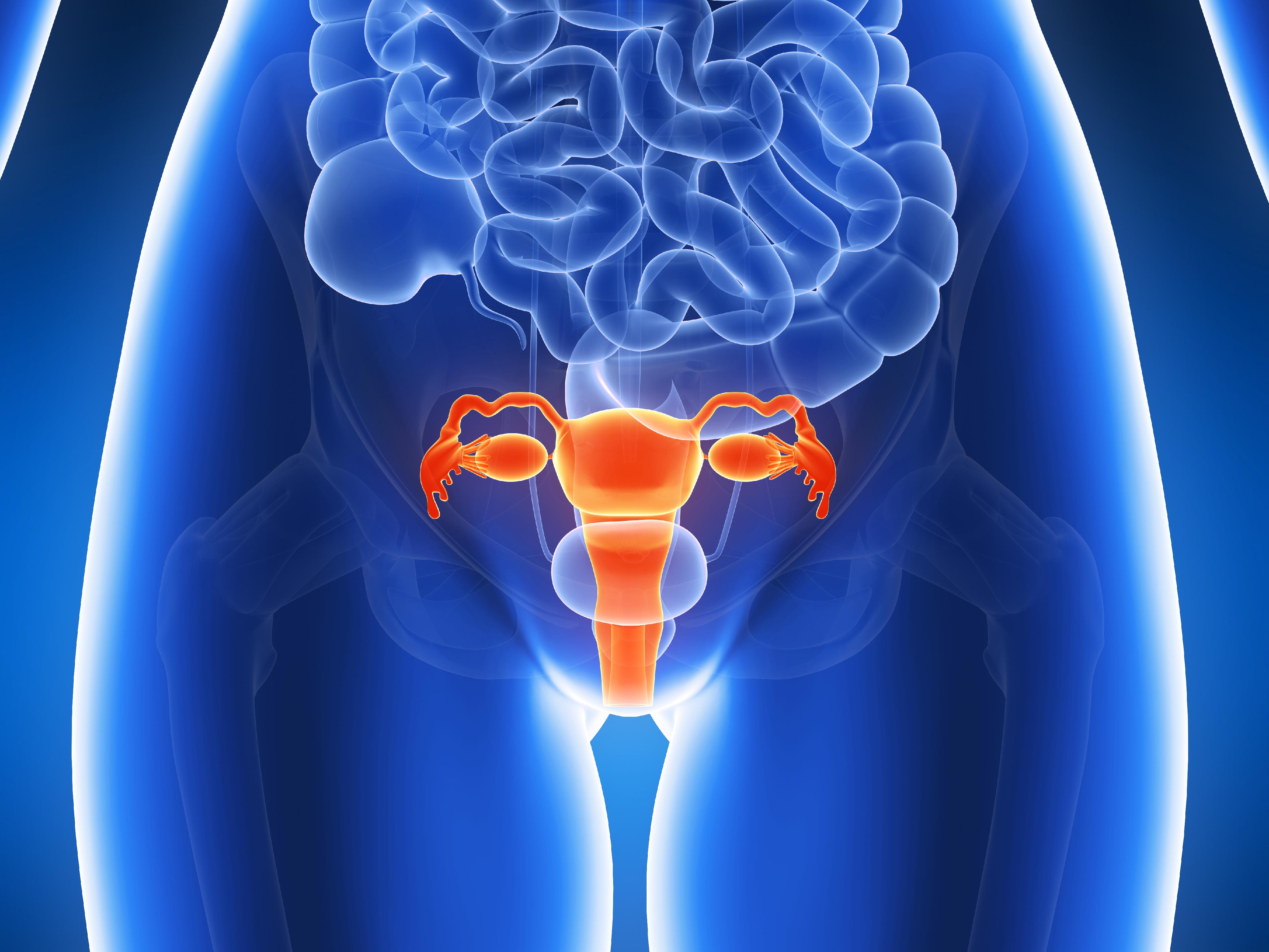
The Fallopian tubes, also known as uterine tubes or oviducts, are a pair of slender tubular structures in the female reproductive system. They play a crucial role in human reproduction by providing a passageway for the egg (ovum) from the ovary to the uterus (womb).
Each Fallopian tube is typically around 7.6 to 10 centimeters long and consists of four parts: the interstitial part, the isthmus, the ampulla, and the infundibulum. The fimbriated end of the infundibulum, which resembles a fringe or frill, surrounds and captures the released egg from the ovary during ovulation.
Fertilization usually occurs in the ampulla when sperm meets the egg after sexual intercourse. Once fertilized, the zygote (fertilized egg) travels through the Fallopian tube toward the uterus for implantation and further development. The cilia lining the inner surface of the Fallopian tubes help propel the egg and the zygote along their journey.
In some cases, abnormalities or blockages in the Fallopian tubes can lead to infertility or ectopic pregnancies, which are pregnancies that develop outside the uterus, typically within the Fallopian tube itself.
Tubal sterilization, also known as female sterilization or tubal ligation, is a permanent form of birth control for women. It involves blocking, sealing, or removing the fallopian tubes, which prevents the sperm from reaching and fertilizing the egg. This procedure can be performed surgically through various methods such as cutting and tying the tubes, using clips or rings to block them, or removing a portion of the tube (known as a partial salpingectomy). Tubal sterilization is considered a highly effective form of contraception with a low failure rate. However, it does not protect against sexually transmitted infections and should be combined with condom use for that purpose. It's important to note that tubal sterilization is a permanent procedure and cannot be easily reversed.
A myoma, also known as a leiomyoma or fibroid, is a benign (noncancerous) tumor that originates from the smooth muscle cells in the wall of a visceral organ. The term "myoma" is often used to describe these growths when they occur in the uterus, where they are typically referred to as uterine fibroids. Uterine fibroids can vary in size, shape, and location within the uterine wall. They are quite common, especially among women of reproductive age, and may not always cause symptoms. However, in some cases, they can lead to issues such as heavy menstrual bleeding, pelvic pain, or infertility. Myomas can also occur in other organs, like the gastrointestinal tract, but they are most frequently found in the uterus.
The uterus, also known as the womb, is a hollow, muscular organ located in the female pelvic cavity, between the bladder and the rectum. It has a thick, middle layer called the myometrium, which is composed of smooth muscle tissue, and an inner lining called the endometrium, which provides a nurturing environment for the fertilized egg to develop into a fetus during pregnancy.
The uterus is where the baby grows and develops until it is ready for birth through the cervix, which is the lower, narrow part of the uterus that opens into the vagina. The uterus plays a critical role in the menstrual cycle as well, by shedding its lining each month if pregnancy does not occur.
Tissue adhesions, also known as scar tissue adhesions, are abnormal bands of fibrous tissue that form between two or more internal organs, or between organs and the walls of the chest or abdominal cavity. These adhesions can develop after surgery, infection, injury, radiation, or prolonged inflammation. The fibrous bands can cause pain, restrict movement of the organs, and potentially lead to complications such as bowel obstruction. Treatment options for tissue adhesions may include medication, physical therapy, or surgical intervention to remove the adhesions.
Laparoscopy is a surgical procedure that involves the insertion of a laparoscope, which is a thin tube with a light and camera attached to it, through small incisions in the abdomen. This allows the surgeon to view the internal organs without making large incisions. It's commonly used to diagnose and treat various conditions such as endometriosis, ovarian cysts, infertility, and appendicitis. The advantages of laparoscopy over traditional open surgery include smaller incisions, less pain, shorter hospital stays, and quicker recovery times.
Müllerian ducts are a pair of embryonic structures found in female mammals, including humans. They give rise to the female reproductive system during fetal development. In females, the Müllerian ducts develop into the fallopian tubes, uterus, cervix, and upper part of the vagina.
In males, the regression of Müllerian ducts is induced by a hormone called anti-Müllerian hormone (AMH), produced by the developing testes. In the absence of AMH or if it fails to function properly, the Müllerian ducts may persist and lead to conditions known as persistent Müllerian duct syndrome (PMDS) or Müllerian remnants in males.
In summary, Müllerian ducts are essential structures for female reproductive system development, and their regression is crucial for male reproductive organ formation.
Ultrasonography, also known as sonography, is a diagnostic medical procedure that uses high-frequency sound waves (ultrasound) to produce dynamic images of organs, tissues, or blood flow inside the body. These images are captured in real-time and can be used to assess the size, shape, and structure of various internal structures, as well as detect any abnormalities such as tumors, cysts, or inflammation.
During an ultrasonography procedure, a small handheld device called a transducer is placed on the patient's skin, which emits and receives sound waves. The transducer sends high-frequency sound waves into the body, and these waves bounce back off internal structures and are recorded by the transducer. The recorded data is then processed and transformed into visual images that can be interpreted by a medical professional.
Ultrasonography is a non-invasive, painless, and safe procedure that does not use radiation like other imaging techniques such as CT scans or X-rays. It is commonly used to diagnose and monitor conditions in various parts of the body, including the abdomen, pelvis, heart, blood vessels, and musculoskeletal system.
The medical definition of "Habitual Abortion" refers to a woman who has three or more consecutive pregnancies that end in spontaneous miscarriages before 20 weeks of gestation. The cause of habitual abortions can be difficult to determine and may involve genetic, anatomical, hormonal, or immune system factors. Treatment is often aimed at addressing any underlying issues that may be contributing to the recurrent miscarriages. It's important to note that the terminology has changed over time and the term "recurrent pregnancy loss" is now more commonly used in place of "habitual abortion".
Leiomyoma is a benign (non-cancerous) tumor that originates from the smooth muscle cells. It most commonly occurs in the uterus, where it is also known as a fibroid, but can also develop in other parts of the body such as the skin, gastrointestinal tract, and genitourinary system. Leiomyomas are typically slow-growing and often cause no symptoms, although they can lead to various complications depending on their size and location. Treatment options for leiomyomas include surveillance, medication, or surgical removal.
The vagina is the canal that joins the cervix (the lower part of the uterus) to the outside of the body. It also is known as the birth canal because babies pass through it during childbirth. The vagina is where sexual intercourse occurs and where menstrual blood exits the body. It has a flexible wall that can expand and retract. During sexual arousal, the vaginal walls swell with blood to become more elastic in order to accommodate penetration.
It's important to note that sometimes people use the term "vagina" to refer to the entire female genital area, including the external structures like the labia and clitoris. But technically, these are considered part of the vulva, not the vagina.
Uterine neoplasms refer to abnormal growths in the uterus, which can be benign (non-cancerous) or malignant (cancerous). These growths can originate from different types of cells within the uterus, leading to various types of uterine neoplasms. The two main categories of uterine neoplasms are endometrial neoplasms and uterine sarcomas.
Endometrial neoplasms develop from the endometrium, which is the inner lining of the uterus. Most endometrial neoplasms are classified as endometrioid adenocarcinomas, arising from glandular cells in the endometrium. Other types include serous carcinoma, clear cell carcinoma, and mucinous carcinoma.
Uterine sarcomas, on the other hand, are less common and originate from the connective tissue (stroma) or muscle (myometrium) of the uterus. Uterine sarcomas can be further divided into several subtypes, such as leiomyosarcoma, endometrial stromal sarcoma, and undifferentiated uterine sarcoma.
Uterine neoplasms can cause various symptoms, including abnormal vaginal bleeding or discharge, pelvic pain, and difficulty urinating or having bowel movements. The diagnosis typically involves a combination of imaging tests (such as ultrasound, CT, or MRI scans) and tissue biopsies to determine the type and extent of the neoplasm. Treatment options depend on the type, stage, and patient's overall health but may include surgery, radiation therapy, chemotherapy, or hormone therapy.
Endometriosis is a medical condition in which tissue similar to the lining of the uterus (endometrium) grows outside the uterine cavity, most commonly on the ovaries, fallopian tubes, and the pelvic peritoneum. This misplaced endometrial tissue continues to act as it would inside the uterus, thickening, breaking down, and bleeding with each menstrual cycle. However, because it is outside the uterus, this blood and tissue have no way to exit the body and can lead to inflammation, scarring, and the formation of adhesions (tissue bands that bind organs together).
The symptoms of endometriosis may include pelvic pain, heavy menstrual periods, painful intercourse, and infertility. The exact cause of endometriosis is not known, but several theories have been proposed, including retrograde menstruation (the backflow of menstrual blood through the fallopian tubes into the pelvic cavity), genetic factors, and immune system dysfunction.
Endometriosis can be diagnosed through a combination of methods, such as medical history, physical examination, imaging tests like ultrasound or MRI, and laparoscopic surgery with tissue biopsy. Treatment options for endometriosis include pain management, hormonal therapies, and surgical intervention to remove the misplaced endometrial tissue. In severe cases, a hysterectomy (removal of the uterus) may be recommended, but this is typically considered a last resort due to its impact on fertility and quality of life.
Pelvic pain is defined as discomfort or unpleasant sensation in the lower abdominal region, below the belly button, and between the hips. It can be acute (sudden and lasting for a short time) or chronic (persisting for months or even years), and it may be steady or intermittent, mild or severe. The pain can have various causes, including musculoskeletal issues, nerve irritation, infection, inflammation, or organic diseases in the reproductive, urinary, or gastrointestinal systems. Accurate diagnosis often requires a thorough medical evaluation to determine the underlying cause and develop an appropriate treatment plan.
Genital diseases in females refer to various medical conditions that affect the female reproductive system, including the vulva, vagina, cervix, uterus, and ovaries. These conditions can be caused by bacterial, viral, or fungal infections, hormonal imbalances, or structural abnormalities. Some common examples of genital diseases in females include bacterial vaginosis, yeast infections, sexually transmitted infections (STIs) such as chlamydia, gonorrhea, and human papillomavirus (HPV), pelvic inflammatory disease (PID), endometriosis, uterine fibroids, ovarian cysts, and vulvar or vaginal cancer. Symptoms of genital diseases in females can vary widely depending on the specific condition but may include abnormal vaginal discharge, pain or discomfort during sex, irregular menstrual bleeding, painful urination, and pelvic pain. It is important for women to receive regular gynecological care and screenings to detect and treat genital diseases early and prevent complications.
Emergency medicine is a medical specialty that focuses on the diagnosis and treatment of acute illnesses or injuries that require immediate medical attention. This can include conditions such as severe trauma, cardiac arrest, stroke, respiratory distress, and other life-threatening situations. Emergency medicine physicians, also known as emergency doctors or ER doctors, are trained to provide rapid assessment, diagnosis, and treatment in a fast-paced and often unpredictable environment. They work closely with other healthcare professionals, such as nurses, paramedics, and specialists, to ensure that patients receive the best possible care in a timely manner. Emergency medicine is a critical component of the healthcare system, providing essential services for patients who require immediate medical attention, 24 hours a day, 7 days a week.
An emergency service in a hospital is a department that provides immediate medical or surgical care for individuals who are experiencing an acute illness, injury, or severe symptoms that require immediate attention. The goal of an emergency service is to quickly assess, stabilize, and treat patients who require urgent medical intervention, with the aim of preventing further harm or death.
Emergency services in hospitals typically operate 24 hours a day, 7 days a week, and are staffed by teams of healthcare professionals including physicians, nurses, physician assistants, nurse practitioners, and other allied health professionals. These teams are trained to provide rapid evaluation and treatment for a wide range of medical conditions, from minor injuries to life-threatening emergencies such as heart attacks, strokes, and severe infections.
In addition to providing emergency care, hospital emergency services also serve as a key point of entry for patients who require further hospitalization or specialized care. They work closely with other departments within the hospital, such as radiology, laboratory, and critical care units, to ensure that patients receive timely and appropriate treatment. Overall, the emergency service in a hospital plays a crucial role in ensuring that patients receive prompt and effective medical care during times of crisis.
An emergency is a sudden, unexpected situation that requires immediate medical attention to prevent serious harm, permanent disability, or death. Emergencies can include severe injuries, trauma, cardiac arrest, stroke, difficulty breathing, severe allergic reactions, and other life-threatening conditions. In such situations, prompt medical intervention is necessary to stabilize the patient's condition, diagnose the underlying problem, and provide appropriate treatment.
Emergency medical services (EMS) are responsible for providing emergency care to patients outside of a hospital setting, such as in the home, workplace, or public place. EMS personnel include emergency medical technicians (EMTs), paramedics, and other first responders who are trained to assess a patient's condition, provide basic life support, and transport the patient to a hospital for further treatment.
In a hospital setting, an emergency department (ED) is a specialized unit that provides immediate care to patients with acute illnesses or injuries. ED staff includes physicians, nurses, and other healthcare professionals who are trained to handle a wide range of medical emergencies. The ED is equipped with advanced medical technology and resources to provide prompt diagnosis and treatment for critically ill or injured patients.
Overall, the goal of emergency medical care is to stabilize the patient's condition, prevent further harm, and provide timely and effective treatment to improve outcomes and save lives.
Dysmenorrhea is a medical term that refers to painful menstrual cramps and discomfort during menstruation. It's one of the most common gynecological complaints among women of reproductive age. There are two types of dysmenorrhea: primary and secondary.
1. Primary Dysmenorrhea: This type is more common and occurs in women who have had normal, pelvic anatomy. The pain is caused by strong contractions of the uterus due to the production of prostaglandins (hormone-like substances that are involved in inflammation and pain). Primary dysmenorrhea usually starts soon after menarche (the beginning of menstruation) and tends to improve with age, particularly after childbirth.
2. Secondary Dysmenorrhea: This type is less common and occurs due to an underlying medical condition affecting the reproductive organs, such as endometriosis, uterine fibroids, pelvic inflammatory disease (PID), or adenomyosis. The pain associated with secondary dysmenorrhea tends to worsen over time and may be accompanied by other symptoms like irregular menstrual bleeding, pain during intercourse, or chronic pelvic pain.
Treatment for dysmenorrhea depends on the type and underlying cause. For primary dysmenorrhea, nonsteroidal anti-inflammatory drugs (NSAIDs) such as ibuprofen or naproxen can help alleviate pain by reducing prostaglandin production. Hormonal birth control methods like oral contraceptives and intrauterine devices (IUDs) may also be prescribed to reduce menstrual pain. For secondary dysmenorrhea, treatment typically involves addressing the underlying medical condition causing the pain.
 Hysterosalpingography
 Hysterosalpingography (HSG) презентация
 Hysterosalpingography | Profiles RNS
 Emergent Treatment of Endometriosis: Background, Laboratory Studies, Imaging Studies
 MR-hysterosalpingography: technique and clinical application - Russian Electronic Journal of Radiology
 Female Infertility | PCOS | Miscarriage | MedlinePlus
 Chandra C. Shenoy, M.D. - Médicos y personal médico - Mayo Clinic
 Sonohysterogram: Procedure, Fertility, Pain & Side Effects
 Role of Gynecologist in Hysterosalpingography: An Easy Test for Evaluation for Evaluation of Tubal Factor in Infertile Woman,...
 Dr. Antoaneta Mueller, MD, Obstetrics & Gynecology Specialist - Lakewood, CA | Sharecare
 Sutter Capitol Pavilion X-Ray Walk-in Clinic | Sutter Health
 Dr Constantine Girio-Fragkoulakis | Medicine and Population Health | The University of Sheffield
 Infertility in women Information | Mount Sinai - New York
 Infertility | Sparrow
 Do I need to see a specialist other than my gynecologist to diagnose and treat fibroids? - HealthyWomen
 The Journal of Contemporary Dental Practice
 Tests for Gynecologic Disorders - Women's Health Issues - MSD Manual Consumer Version
 Uterine Prolapse