Immunoglobulin mu-Chains
Immunoglobulin Heavy Chains
Immunoglobulin M
Genes, Immunoglobulin
Immunoglobulins
Immunoglobulin Constant Regions
B-Lymphocytes
Immunoglobulin G
Immunoglobulin Light Chains
Receptors, Antigen, B-Cell
Immunoglobulin kappa-Chains
Immunoglobulin A
Immunoglobulin Variable Region
Immunoglobulin J-Chains
Plasmacytoma
Immunoglobulin delta-Chains
Hybridomas
Base Sequence
Molecular Sequence Data
Immunoglobulins, Intravenous
Gene Rearrangement, B-Lymphocyte, Heavy Chain
Immunoglobulin Light Chains, Surrogate
Proto-Oncogene Protein c-ets-1
Proto-Oncogene Proteins c-ets
Enhancer Elements, Genetic
RNA, Messenger
Immunoglobulin Fragments
Genes
Gene Rearrangement, B-Lymphocyte, Light Chain
Gene Rearrangement, B-Lymphocyte
Poly A
Substrate specificities of SR proteins in constitutive splicing are determined by their RNA recognition motifs and composite pre-mRNA exonic elements. (1/594)
We report striking differences in the substrate specificities of two human SR proteins, SF2/ASF and SC35, in constitutive splicing. beta-Globin pre-mRNA (exons 1 and 2) is spliced indiscriminately with either SR protein. Human immunodeficiency virus tat pre-mRNA (exons 2 and 3) and immunoglobulin mu-chain (IgM) pre-mRNA (exons C3 and C4) are preferentially spliced with SF2/ASF and SC35, respectively. Using in vitro splicing with mutated or chimeric derivatives of the tat and IgM pre-mRNAs, we defined specific combinations of segments in the downstream exons, which mediate either positive or negative effects to confer SR protein specificity. A series of recombinant chimeric proteins consisting of domains of SF2/ASF and SC35 in various combinations was used to localize trans-acting domains responsible for substrate specificity. The RS domains of SF2/ASF and SC35 can be exchanged without effect on substrate specificity. The RNA recognition motifs (RRMs) of SF2/ASF are active only in the context of a two-RRM structure, and RRM2 has a dominant role in substrate specificity. In contrast, the single RRM of SC35 can function alone, but its substrate specificity can be influenced by the presence of an additional RRM. The RRMs behave as modules that, when present in different combinations, can have positive, neutral, or negative effects on splicing, depending upon the specific substrate. We conclude that SR protein-specific recognition of specific positive and negative pre-mRNA exonic elements via one or more RRMs is a crucial determinant of the substrate specificity of SR proteins in constitutive splicing. (+info)Mechanisms of double-strand-break repair during gene targeting in mammalian cells. (2/594)
In the present study, the mechanism of double-strand-break (DSB) repair during gene targeting at the chromosomal immunoglobulin mu-locus in a murine hybridoma was examined. The gene-targeting assay utilized specially designed insertion vectors genetically marked in the region of homology to the chromosomal mu-locus by six diagnostic restriction enzyme site markers. The restriction enzyme markers permitted the contribution of vector-borne and chromosomal mu-sequences in the recombinant product to be determined. The use of the insertion vectors in conjunction with a plating procedure in which individual integrative homologous recombination events were retained for analysis revealed several important features about the mammalian DSB repair process:The presence of the markers within the region of shared homology did not affect the efficiency of gene targeting. In the majority of recombinants, the vector-borne marker proximal to the DSB was absent, being replaced with the corresponding chromosomal restriction enzyme site. This result is consistent with either formation and repair of a vector-borne gap or an "end" bias in mismatch repair of heteroduplex DNA (hDNA) that favored the chromosomal sequence. Formation of hDNA was frequently associated with gene targeting and, in most cases, began approximately 645 bp from the DSB and could encompass a distance of at least 1469 bp. The hDNA was efficiently repaired prior to DNA replication. The repair of adjacent mismatches in hDNA occurred predominantly on the same strand, suggesting the involvement of a long-patch repair mechanism. (+info)The molecular basis of multiple vector insertion by gene targeting in mammalian cells. (3/594)
Gene targeting using sequence insertion vectors generally results in integration of one copy of the targeting vector generating a tandem duplication of the cognate chromosomal region of homology. However, occasionally the target locus is found to contain >1 copy of the integrated vector. The mechanism by which the latter recombinants arise is not known. In the present study, we investigated the molecular basis by which multiple vectors become integrated at the chromosomal immunoglobulin mu locus in a murine hybridoma. To accomplish this, specially designed insertion vectors were constructed that included six diagnostic restriction enzyme markers in the Cmu region of homology to the target chromosomal mu locus. This enabled contributions by the vector-borne and chromosomal Cmu sequences at the recombinant locus to be ascertained. Targeted recombinants were isolated and analyzed to determine the number of vector copies integrated at the chromosomal immunoglobulin mu locus. Targeted recombinants identified as bearing >1 copy of the integrated vector resulted from a Cmu triplication formed by two vector copies in tandem. Examination of the fate of the Cmu region markers suggested that this class of recombinant was generated predominantly, if not exclusively, by two targeted vector integration events, each involving insertion of a single copy of the vector. Both vector insertion events into the chromosomal mu locus were consistent with the double-strand-break repair mechanism of homologous recombination. We interpret our results, taken together, to mean that a proportion of recipient cells is in a predetermined state that is amenable to targeted but not random vector integration. (+info)Infrequent translation of a nonsense codon is sufficient to decrease mRNA level. (4/594)
In many organisms nonsense mutations decrease the level of mRNA. In the case of mammalian cells, it is still controversial whether translation is required for this nonsense-mediated RNA decrease (NMD). Although previous analyzes have shown that conditions that impede translation termination at nonsense codons also prevent NMD, the residual level of termination was unknown in these experiments. Moreover, the conditions used to impede termination might also have interfered with NMD in other ways. Because of these uncertainties, we have tested the effects of limiting translation of a nonsense codon in a different way, using two mutations in the immunoglobulin mu heavy chain gene. For this purpose we exploited an exceptional nonsense mutation at codon 3, which efficiently terminates translation but nonetheless maintains a high level of mu mRNA. We have shown 1) that translation of Ter462 in the double mutant occurs at only approximately 4% the normal frequency, and 2) that Ter462 in cis with Ter3 can induce NMD. That is, translation of Ter462 at this low (4%) frequency is sufficient to induce NMD. (+info)gammadelta T cells contribute to control of chronic parasitemia in Plasmodium chabaudi infections in mice. (5/594)
During a primary infection of mice with Plasmodium chabaudi, gammadelta T cells are stimulated and their expansion coincides with recovery from the acute phase of infection in normal mice or with chronic infections in B cell-deficient mice (mu-MT). To determine whether the large gammadelta T cell pool observed in female B cell-deficient mice is responsible for controlling the chronic infection, studies were done using double-knockout mice deficient in both B and gammadelta cells (mu-MT x delta-/-TCR) and in gammadelta T cell-depleted mu-MT mice. In both types of gammadelta T cell-deficient mice, the early parasitemia following the peak of infection was exacerbated, and the chronic parasitemia was maintained at significantly higher levels in the absence of gammadelta T cells. The majority of gammadelta T cells in C57BL/6 and mu-MT mice responding to infection belonged predominantly to a single family of gammadelta T cells with TCR composed of Vgamma2Vdelta4 chains and which produced IFN-gamma rather than IL-4. (+info)Novel mechanisms control the folding and assembly of lambda5/14.1 and VpreB to produce an intact surrogate light chain. (6/594)
Surrogate light chain, which escorts the mu heavy chain to the cell surface, is a critical component of the pre-B cell receptor complex. The two proteins that comprise the surrogate light chain, VpreB and lambda5/14.1, contain both unique regions and Ig-like domains. The unique regions have been postulated to function in the assembly of the surrogate light chain. However, by using transient transfection of COS7 cells, we show that deletion of the unique regions of both proteins did not inhibit the assembly of surrogate light chain. Instead, in vivo folding studies showed that the unique region of lambda5/14.1 acts as an intramolecular chaperone by preventing the folding of this protein when it is expressed in the absence of its partner, VpreB. The Ig domains of both lambda5/14.1 and VpreB are atypical. The one in VpreB lacks one of the canonical beta strands whereas the one in lambda5/14.1 has an extra beta strand. Deletion of the extra beta strand in lambda5/14.1 completely abrogated the formation of the surrogate light chain, demonstrating that complementation of the incomplete Ig domain in VpreB by the extra beta strand in lambda5/14.1 was necessary and sufficient for the folding and assembly of these proteins. Our studies reveal two novel mechanisms for regulating surrogate light chain formation: (i) the presence of an intramolecular chaperone that prevents folding of the unassembled subunit but that remains part of the mature assembled protein, and (ii) splitting an Ig domain between two proteins to control their folding and assembly. (+info)Transcriptional activation by ETS and leucine zipper-containing basic helix-loop-helix proteins. (7/594)
The immunoglobulin mu heavy-chain gene enhancer contains closely juxtaposed binding sites for ETS and leucine zipper-containing basic helix-loop-helix (bHLH-zip) proteins. To understand the mu enhancer function, we have investigated transcription activation by the combination of ETS and bHLH-zip proteins. The bHLH-zip protein TFE3, but not USF, cooperated with the ETS domain proteins PU.1 and Ets-1 to activate a tripartite domain of this enhancer. Deletion mutants were used to identify the domains of the proteins involved. Both TFE3 and USF enhanced Ets-1 DNA binding in vitro by relieving the influence of an autoinhibitory domain in Ets-1 by direct protein-protein associations. Several regions of Ets-1 were found to be necessary, whereas the bHLH-zip domain was sufficient for this effect. Our studies define novel interactions between ETS and bHLH-zip proteins that may regulate combinatorial transcription activation by these protein families. (+info)Cutting edge: recruitment of the CD19/CD21 coreceptor to B cell antigen receptor is required for antigen-mediated expression of Bcl-2 by resting and cycling hen egg lysozyme transgenic B cells. (8/594)
Recruitment of the CD19/CD21 coreceptor is thought to lower the threshold for effective signaling through the B cell Ag receptor. We provide evidence supporting a second role for coreceptor recruitment, and that is to enhance the survival/proliferative potential of the responding B cells. We show that B cell Ag receptor signaling in the absence of coreceptor recruitment induces cellular accumulation of the anti-apoptotic protein Bcl-xL, whereas CD19-mediated signals are required for Bcl-2 accumulation. The expression of both anti-apoptotic proteins correlates with the enhanced responsiveness of both resting and cycling B cells to growth-promoting signals delivered through CD40. These results provide further evidence for the necessity of coreceptor recruitment during Ag-dependent B cell activation and indicate that Ags derived from inflammatory sites function as better thymus-dependent Ags than their counterparts not coated with complement fragments. (+info)Immunoglobulin mu-chains (IgM) are a type of heavy chain found in immunoglobulins, also known as antibodies. IgM is the first antibody to be produced in response to an initial exposure to an antigen and plays a crucial role in the early stages of the immune response.
IgM antibodies are composed of four monomeric units, each consisting of two heavy chains and two light chains. The heavy chains in IgM are called mu-chains, which have a molecular weight of approximately 72 kDa. Each mu-chain contains five domains: one variable (V) domain at the N-terminus, four constant (C) domains (Cμ1-4), and a membrane-spanning region followed by a short cytoplasmic tail.
IgM antibodies are primarily found on the surface of B cells as part of the B cell receptor (BCR). When a B cell encounters an antigen, the BCR binds to it, triggering a series of intracellular signaling events that lead to B cell activation and differentiation into plasma cells. In response to activation, the B cell begins to secrete IgM antibodies into the bloodstream.
IgM antibodies have several unique features that make them effective in the early stages of an immune response. They are highly efficient at agglutination, or clumping together, of pathogens and antigens, which helps to neutralize them. IgM antibodies also activate the complement system, a group of proteins that work together to destroy pathogens.
Overall, Immunoglobulin mu-chains are an essential component of the immune system, providing early protection against pathogens and initiating the adaptive immune response.
Immunoglobulin heavy chains are proteins that make up the framework of antibodies, which are Y-shaped immune proteins. These heavy chains, along with light chains, form the antigen-binding sites of an antibody, which recognize and bind to specific foreign substances (antigens) in order to neutralize or remove them from the body.
The heavy chain is composed of a variable region, which contains the antigen-binding site, and constant regions that determine the class and function of the antibody. There are five classes of immunoglobulins (IgA, IgD, IgE, IgG, and IgM) that differ in their heavy chain constant regions and therefore have different functions in the immune response.
Immunoglobulin heavy chains are synthesized by B cells, a type of white blood cell involved in the adaptive immune response. The genetic rearrangement of immunoglobulin heavy chain genes during B cell development results in the production of a vast array of different antibodies with unique antigen-binding sites, allowing for the recognition and elimination of a wide variety of pathogens.
Immunoglobulin M (IgM) is a type of antibody that is primarily found in the blood and lymph fluid. It is the first antibody to be produced in response to an initial exposure to an antigen, making it an important part of the body's primary immune response. IgM antibodies are large molecules that are composed of five basic units, giving them a pentameric structure. They are primarily found on the surface of B cells as membrane-bound immunoglobulins (mlgM), where they function as receptors for antigens. Once an mlgM receptor binds to an antigen, it triggers the activation and differentiation of the B cell into a plasma cell that produces and secretes large amounts of soluble IgM antibodies.
IgM antibodies are particularly effective at agglutination (clumping) and complement activation, which makes them important in the early stages of an immune response to help clear pathogens from the bloodstream. However, they are not as stable or long-lived as other types of antibodies, such as IgG, and their levels tend to decline after the initial immune response has occurred.
In summary, Immunoglobulin M (IgM) is a type of antibody that plays a crucial role in the primary immune response to antigens by agglutination and complement activation. It is primarily found in the blood and lymph fluid, and it is produced by B cells after they are activated by an antigen.
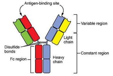
Immunoglobulins (Igs), also known as antibodies, are proteins produced by the immune system to recognize and neutralize foreign substances such as pathogens or toxins. They are composed of four polypeptide chains: two heavy chains and two light chains, which are held together by disulfide bonds. The variable regions of the heavy and light chains contain loops that form the antigen-binding site, allowing each Ig molecule to recognize a specific epitope (antigenic determinant) on an antigen.
Genes encoding immunoglobulins are located on chromosome 14 (light chain genes) and chromosomes 22 and 2 (heavy chain genes). The diversity of the immune system is generated through a process called V(D)J recombination, where variable (V), diversity (D), and joining (J) gene segments are randomly selected and assembled to form the variable regions of the heavy and light chains. This results in an enormous number of possible combinations, allowing the immune system to recognize and respond to a vast array of potential threats.
There are five classes of immunoglobulins: IgA, IgD, IgE, IgG, and IgM, each with distinct functions and structures. For example, IgG is the most abundant class in serum and provides long-term protection against pathogens, while IgA is found on mucosal surfaces and helps prevent the entry of pathogens into the body.
Immunoglobulins (Igs), also known as antibodies, are glycoprotein molecules produced by the immune system's B cells in response to the presence of foreign substances, such as bacteria, viruses, and toxins. These Y-shaped proteins play a crucial role in identifying and neutralizing pathogens and other antigens, thereby protecting the body against infection and disease.
Immunoglobulins are composed of four polypeptide chains: two identical heavy chains and two identical light chains, held together by disulfide bonds. The variable regions of these chains form the antigen-binding sites, which recognize and bind to specific epitopes on antigens. Based on their heavy chain type, immunoglobulins are classified into five main isotypes or classes: IgA, IgD, IgE, IgG, and IgM. Each class has distinct functions in the immune response, such as providing protection in different body fluids and tissues, mediating hypersensitivity reactions, and aiding in the development of immunological memory.
In medical settings, immunoglobulins can be administered therapeutically to provide passive immunity against certain diseases or to treat immune deficiencies, autoimmune disorders, and other conditions that may benefit from immunomodulation.
Immunoglobulin constant regions are the invariant portions of antibody molecules (immunoglobulins) that are identical in all antibodies of the same isotype. These regions are responsible for effector functions such as complement activation, binding to Fc receptors, and initiating immune responses. They are composed of amino acid sequences that remain unchanged during antigen-driven somatic hypermutation, allowing them to interact with various components of the immune system. The constant regions are found in the heavy chains (CH) and light chains (CL) of an immunoglobulin molecule. In contrast, the variable regions are responsible for recognizing and binding to specific antigens.
B-lymphocytes, also known as B-cells, are a type of white blood cell that plays a key role in the immune system's response to infection. They are responsible for producing antibodies, which are proteins that help to neutralize or destroy pathogens such as bacteria and viruses.
When a B-lymphocyte encounters a pathogen, it becomes activated and begins to divide and differentiate into plasma cells, which produce and secrete large amounts of antibodies specific to the antigens on the surface of the pathogen. These antibodies bind to the pathogen, marking it for destruction by other immune cells such as neutrophils and macrophages.
B-lymphocytes also have a role in presenting antigens to T-lymphocytes, another type of white blood cell involved in the immune response. This helps to stimulate the activation and proliferation of T-lymphocytes, which can then go on to destroy infected cells or help to coordinate the overall immune response.
Overall, B-lymphocytes are an essential part of the adaptive immune system, providing long-lasting immunity to previously encountered pathogens and helping to protect against future infections.
Immunoglobulin G (IgG) is a type of antibody, which is a protective protein produced by the immune system in response to foreign substances like bacteria or viruses. IgG is the most abundant type of antibody in human blood, making up about 75-80% of all antibodies. It is found in all body fluids and plays a crucial role in fighting infections caused by bacteria, viruses, and toxins.
IgG has several important functions:
1. Neutralization: IgG can bind to the surface of bacteria or viruses, preventing them from attaching to and infecting human cells.
2. Opsonization: IgG coats the surface of pathogens, making them more recognizable and easier for immune cells like neutrophils and macrophages to phagocytose (engulf and destroy) them.
3. Complement activation: IgG can activate the complement system, a group of proteins that work together to help eliminate pathogens from the body. Activation of the complement system leads to the formation of the membrane attack complex, which creates holes in the cell membranes of bacteria, leading to their lysis (destruction).
4. Antibody-dependent cellular cytotoxicity (ADCC): IgG can bind to immune cells like natural killer (NK) cells and trigger them to release substances that cause target cells (such as virus-infected or cancerous cells) to undergo apoptosis (programmed cell death).
5. Immune complex formation: IgG can form immune complexes with antigens, which can then be removed from the body through various mechanisms, such as phagocytosis by immune cells or excretion in urine.
IgG is a critical component of adaptive immunity and provides long-lasting protection against reinfection with many pathogens. It has four subclasses (IgG1, IgG2, IgG3, and IgG4) that differ in their structure, function, and distribution in the body.
Immunoglobulin light chains are the smaller protein subunits of an immunoglobulin, also known as an antibody. They are composed of two polypeptide chains, called kappa (κ) and lambda (λ), which are produced by B cells during the immune response. Each immunoglobulin molecule contains either two kappa or two lambda light chains, in association with two heavy chains.
Light chains play a crucial role in the antigen-binding site of an antibody, where they contribute to the specificity and affinity of the interaction between the antibody and its target antigen. In addition to their role in immune function, abnormal production or accumulation of light chains can lead to various diseases, such as multiple myeloma and amyloidosis.
1. Receptors: In the context of physiology and medicine, receptors are specialized proteins found on the surface of cells or inside cells that detect and respond to specific molecules, known as ligands. These interactions can trigger a variety of responses within the cell, such as starting a signaling cascade or changing the cell's metabolism. Receptors play crucial roles in various biological processes, including communication between cells, regulation of immune responses, and perception of senses.
2. Antigen: An antigen is any substance (usually a protein) that can be recognized by the adaptive immune system, specifically by B-cells and T-cells. Antigens can be derived from various sources, such as microorganisms (like bacteria, viruses, or fungi), pollen, dust mites, or even components of our own cells (for instance, in autoimmune diseases). An antigen's ability to stimulate an immune response is determined by its molecular structure and whether it can be recognized by the receptors on immune cells.
3. B-Cell: B-cells are a type of white blood cell that plays a critical role in the adaptive immune system, particularly in humoral immunity. They originate from hematopoietic stem cells in the bone marrow and are responsible for producing antibodies, which are proteins that recognize and bind to specific antigens. Each B-cell has receptors on its surface called B-cell receptors (BCRs) that can recognize a unique antigen. When a B-cell encounters its specific antigen, it becomes activated, undergoes proliferation, and differentiates into plasma cells that secrete large amounts of antibodies to neutralize or eliminate the antigen.
Immunoglobulin kappa-chains are one of the two types of light chains (the other being lambda-chains) that make up an immunoglobulin molecule, also known as an antibody. These light chains combine with heavy chains to form the antigen-binding site of an antibody, which is responsible for recognizing and binding to specific antigens or foreign substances in the body.
Kappa-chains contain a variable region that differs between different antibodies and contributes to the diversity of the immune system's response to various antigens. They also have a constant region, which is consistent across all kappa-chains. Approximately 60% of all human antibodies contain kappa-chains, while the remaining 40% contain lambda-chains. The relative proportions of kappa and lambda chains can be used in diagnostic tests to identify clonal expansions of B cells, which may indicate a malignancy such as multiple myeloma or lymphoma.
Immunoglobulin A (IgA) is a type of antibody that plays a crucial role in the immune function of the human body. It is primarily found in external secretions, such as saliva, tears, breast milk, and sweat, as well as in mucous membranes lining the respiratory and gastrointestinal tracts. IgA exists in two forms: a monomeric form found in serum and a polymeric form found in secretions.
The primary function of IgA is to provide immune protection at mucosal surfaces, which are exposed to various environmental antigens, such as bacteria, viruses, parasites, and allergens. By doing so, it helps prevent the entry and colonization of pathogens into the body, reducing the risk of infections and inflammation.
IgA functions by binding to antigens present on the surface of pathogens or allergens, forming immune complexes that can neutralize their activity. These complexes are then transported across the epithelial cells lining mucosal surfaces and released into the lumen, where they prevent the adherence and invasion of pathogens.
In summary, Immunoglobulin A (IgA) is a vital antibody that provides immune defense at mucosal surfaces by neutralizing and preventing the entry of harmful antigens into the body.
The Immunoglobulin (Ig) variable region is the antigen-binding part of an antibody, which is highly variable in its amino acid sequence and therefore specific to a particular epitope (the site on an antigen that is recognized by the antigen-binding site of an antibody). This variability is generated during the process of V(D)J recombination in the maturation of B cells, allowing for a diverse repertoire of antibodies to be produced and recognizing a wide range of potential pathogens.
The variable region is composed of several sub-regions including:
1. The heavy chain variable region (VH)
2. The light chain variable region (VL)
3. The heavy chain joining region (JH)
4. The light chain joining region (JL)
These regions are further divided into framework regions and complementarity-determining regions (CDRs). The CDRs, particularly CDR3, contain the most variability and are primarily responsible for antigen recognition.
Immunoglobulin J-chains are small protein structures that play a role in the assembly and structure of certain types of antibodies, specifically IgM and IgA. The J-chain is a polypeptide chain that contains multiple cysteine residues, which allow it to form disulfide bonds with the heavy chains of IgM and IgA molecules.
In IgM antibodies, the J-chain helps to link the five identical heavy chain units together to form a pentameric structure. In IgA antibodies, the J-chain links two dimeric structures together to form a tetrameric structure. This polymerization of IgM and IgA molecules is important for their function in the immune system, as it allows them to form large complexes that can effectively agglutinate and neutralize pathogens.
The J-chain is synthesized by a specialized group of B cells called plasma cells, which are responsible for producing and secreting antibodies. Once synthesized, the J-chain is covalently linked to the heavy chains of IgM or IgA molecules during their assembly in the endoplasmic reticulum of the plasma cell.
Overall, the Immunoglobulin J-chain plays a crucial role in the structure and function of certain classes of antibodies, contributing to their ability to effectively combat pathogens and protect the body from infection.
A plasmacytoma is a discrete tumor mass that is composed of neoplastic plasma cells, which are a type of white blood cell found in the bone marrow. Plasmacytomas can be solitary (a single tumor) or multiple (many tumors), and they can develop in various locations throughout the body.
Solitary plasmacytoma is a rare cancer that typically affects older adults, and it usually involves a single bone lesion, most commonly found in the vertebrae, ribs, or pelvis. In some cases, solitary plasmacytomas can also occur outside of the bone (extramedullary plasmacytoma), which can affect soft tissues such as the upper respiratory tract, gastrointestinal tract, or skin.
Multiple myeloma is a more common and aggressive cancer that involves multiple plasmacytomas in the bone marrow, leading to the replacement of normal bone marrow cells with malignant plasma cells. This can result in various symptoms such as bone pain, anemia, infections, and kidney damage.
The diagnosis of plasmacytoma typically involves a combination of imaging studies, biopsy, and laboratory tests to assess the extent of the disease and determine the appropriate treatment plan. Treatment options for solitary plasmacytoma may include surgery or radiation therapy, while multiple myeloma is usually treated with chemotherapy, targeted therapy, immunotherapy, and/or stem cell transplantation.
Immunoglobulin delta-chains (IgD) are a type of heavy chain found in immunoglobulins, which are also known as antibodies. Antibodies are proteins that play a crucial role in the immune system's response to foreign substances, such as bacteria and viruses.
The heavy chains of an antibody consist of four polypeptide regions: the variable region, which varies between different antibodies and is responsible for recognizing and binding to specific antigens; and three constant regions, known as Cμ, Cγ, Cα, or Cδ, which determine the class of the antibody and its effector functions.
IgD heavy chains contain a single Cδ region and are found only in a small subset of antibodies, primarily located on the surface of mature B cells. IgD is co-expressed with IgM on the surface of naive B cells and plays a role in activating the immune response by binding to antigens and initiating signal transduction pathways that lead to B cell activation and differentiation into antibody-secreting plasma cells.
While the function of IgD is not fully understood, it is thought to play a role in regulating the immune response, including modulating allergic reactions and protecting against autoimmunity. Additionally, IgD has been found to have a role in the development and survival of B cells, as well as in the regulation of calcium signaling in B cells.
A hybridoma is a type of hybrid cell that is created in a laboratory by fusing a cancer cell (usually a B cell) with a normal immune cell. The resulting hybrid cell combines the ability of the cancer cell to grow and divide indefinitely with the ability of the immune cell to produce antibodies, which are proteins that help the body fight infection.
Hybridomas are commonly used to produce monoclonal antibodies, which are identical copies of a single antibody produced by a single clone of cells. These antibodies can be used for a variety of purposes, including diagnostic tests and treatments for diseases such as cancer and autoimmune disorders.
To create hybridomas, B cells are first isolated from the spleen or blood of an animal that has been immunized with a specific antigen (a substance that triggers an immune response). The B cells are then fused with cancer cells using a chemical agent such as polyethylene glycol. The resulting hybrid cells are called hybridomas and are grown in culture medium, where they can be selected for their ability to produce antibodies specific to the antigen of interest. These antibody-producing hybridomas can then be cloned to produce large quantities of monoclonal antibodies.
A base sequence in the context of molecular biology refers to the specific order of nucleotides in a DNA or RNA molecule. In DNA, these nucleotides are adenine (A), guanine (G), cytosine (C), and thymine (T). In RNA, uracil (U) takes the place of thymine. The base sequence contains genetic information that is transcribed into RNA and ultimately translated into proteins. It is the exact order of these bases that determines the genetic code and thus the function of the DNA or RNA molecule.
Molecular sequence data refers to the specific arrangement of molecules, most commonly nucleotides in DNA or RNA, or amino acids in proteins, that make up a biological macromolecule. This data is generated through laboratory techniques such as sequencing, and provides information about the exact order of the constituent molecules. This data is crucial in various fields of biology, including genetics, evolution, and molecular biology, allowing for comparisons between different organisms, identification of genetic variations, and studies of gene function and regulation.
Intravenous Immunoglobulins (IVIG) are a preparation of antibodies, specifically immunoglobulins, that are derived from the plasma of healthy donors. They are administered intravenously to provide passive immunity and help boost the immune system's response in individuals with weakened or compromised immune systems. IVIG can be used for various medical conditions such as primary immunodeficiency disorders, secondary immunodeficiencies, autoimmune diseases, and some infectious diseases. The administration of IVIG can help prevent infections, reduce the severity and frequency of infections, and manage the symptoms of certain autoimmune disorders. It is important to note that while IVIG provides temporary immunity, it does not replace a person's own immune system.
'Gene rearrangement in B-lymphocytes, heavy chain' refers to the biological process that occurs during the development of B-lymphocytes (a type of white blood cell) in the bone marrow. This process involves the rearrangement of genetic material on chromosome 14, specifically within the immunoglobulin heavy chain gene locus.
During B-cell maturation, the variable region of the heavy chain gene is assembled from several gene segments, including the variable (V), diversity (D), and joining (J) segments. Through a series of genetic recombination events, these segments are randomly selected and joined together to form a unique V(D)J exon that encodes the variable region of the immunoglobulin heavy chain protein.
This gene rearrangement process allows for the generation of a diverse repertoire of antibodies with different specificities, enabling B-lymphocytes to recognize and respond to a wide range of foreign antigens. However, if errors occur during this process, it can lead to the production of autoantibodies that target the body's own cells and tissues, contributing to the development of certain immune disorders such as autoimmune diseases.
I'm sorry for any confusion, but "Immunoglobulin Light Chains, Surrogate" is not a recognized medical term or concept in the immunology field. Immunoglobulins, also known as antibodies, are proteins produced by the immune system to identify and neutralize foreign substances like bacteria and viruses. They consist of two heavy chains and two light chains, which can be either kappa or lambda. However, there is no such thing as "surrogate" light chains in this context.
If you have any other questions about medical terminology or concepts, I'd be happy to help!
A cell line is a culture of cells that are grown in a laboratory for use in research. These cells are usually taken from a single cell or group of cells, and they are able to divide and grow continuously in the lab. Cell lines can come from many different sources, including animals, plants, and humans. They are often used in scientific research to study cellular processes, disease mechanisms, and to test new drugs or treatments. Some common types of human cell lines include HeLa cells (which come from a cancer patient named Henrietta Lacks), HEK293 cells (which come from embryonic kidney cells), and HUVEC cells (which come from umbilical vein endothelial cells). It is important to note that cell lines are not the same as primary cells, which are cells that are taken directly from a living organism and have not been grown in the lab.
Proto-oncogene protein c-ets-1 is a transcription factor that regulates gene expression in various cellular processes, including cell growth, differentiation, and apoptosis. It belongs to the ETS family of transcription factors, which are characterized by a highly conserved DNA-binding domain known as the ETS domain. The c-ets-1 protein is encoded by the ETS1 gene located on chromosome 11 in humans.
In normal cells, c-ets-1 plays critical roles in development, tissue repair, and immune function. However, when its expression or activity is dysregulated, it can contribute to tumorigenesis and cancer progression. In particular, c-ets-1 has been implicated in the development of various types of leukemia and solid tumors, such as breast, prostate, and lung cancer.
The activation of c-ets-1 can occur through various mechanisms, including gene amplification, chromosomal translocation, or point mutations. Once activated, c-ets-1 can promote cell proliferation, survival, and migration, while also inhibiting apoptosis. These oncogenic properties make c-ets-1 a potential target for cancer therapy.
Proto-oncogene proteins c-ets are a family of transcription factors that play crucial roles in regulating various cellular processes, including cell growth, differentiation, and apoptosis. These proteins contain a highly conserved DNA-binding domain known as the ETS domain, which recognizes and binds to specific DNA sequences in the promoter regions of target genes.
The c-ets proto-oncogenes encode for these transcription factors, and they can become oncogenic when they are abnormally activated or overexpressed due to genetic alterations such as chromosomal translocations, gene amplifications, or point mutations. Once activated, c-ets proteins can dysregulate the expression of genes involved in cell cycle control, survival, and angiogenesis, leading to tumor development and progression.
Abnormal activation of c-ets proto-oncogene proteins has been implicated in various types of cancer, including leukemia, lymphoma, breast, prostate, and lung cancer. Therefore, understanding the function and regulation of c-ets proto-oncogene proteins is essential for developing novel therapeutic strategies to treat cancer.
Genetic enhancer elements are DNA sequences that increase the transcription of specific genes. They work by binding to regulatory proteins called transcription factors, which in turn recruit RNA polymerase II, the enzyme responsible for transcribing DNA into messenger RNA (mRNA). This results in the activation of gene transcription and increased production of the protein encoded by that gene.
Enhancer elements can be located upstream, downstream, or even within introns of the genes they regulate, and they can act over long distances along the DNA molecule. They are an important mechanism for controlling gene expression in a tissue-specific and developmental stage-specific manner, allowing for the precise regulation of gene activity during embryonic development and throughout adult life.
It's worth noting that genetic enhancer elements are often referred to simply as "enhancers," and they are distinct from other types of regulatory DNA sequences such as promoters, silencers, and insulators.
Messenger RNA (mRNA) is a type of RNA (ribonucleic acid) that carries genetic information copied from DNA in the form of a series of three-base code "words," each of which specifies a particular amino acid. This information is used by the cell's machinery to construct proteins, a process known as translation. After being transcribed from DNA, mRNA travels out of the nucleus to the ribosomes in the cytoplasm where protein synthesis occurs. Once the protein has been synthesized, the mRNA may be degraded and recycled. Post-transcriptional modifications can also occur to mRNA, such as alternative splicing and addition of a 5' cap and a poly(A) tail, which can affect its stability, localization, and translation efficiency.
Immunoglobulin fragments refer to the smaller protein units that are formed by the digestion or break-down of an intact immunoglobulin, also known as an antibody. Immunoglobulins are large Y-shaped proteins produced by the immune system to identify and neutralize foreign substances such as pathogens or toxins. They consist of two heavy chains and two light chains, held together by disulfide bonds.
The digestion or break-down of an immunoglobulin can occur through enzymatic cleavage, which results in the formation of distinct fragments. The most common immunoglobulin fragments are:
1. Fab (Fragment, antigen binding) fragments: These are formed by the digestion of an intact immunoglobulin using the enzyme papain. Each Fab fragment contains a single antigen-binding site, consisting of a portion of one heavy chain and one light chain. The Fab fragments retain their ability to bind to specific antigens.
2. Fc (Fragment, crystallizable) fragments: These are formed by the digestion of an intact immunoglobulin using the enzyme pepsin or through the natural breakdown process in the body. The Fc fragment contains the constant region of both heavy chains and is responsible for effector functions such as complement activation, binding to Fc receptors on immune cells, and antibody-dependent cellular cytotoxicity (ADCC).
These immunoglobulin fragments play crucial roles in various immune responses and diagnostic applications. For example, Fab fragments can be used in immunoassays for the detection of specific antigens, while Fc fragments can mediate effector functions that help eliminate pathogens or damaged cells from the body.
A gene is a specific sequence of nucleotides in DNA that carries genetic information. Genes are the fundamental units of heredity and are responsible for the development and function of all living organisms. They code for proteins or RNA molecules, which carry out various functions within cells and are essential for the structure, function, and regulation of the body's tissues and organs.
Each gene has a specific location on a chromosome, and each person inherits two copies of every gene, one from each parent. Variations in the sequence of nucleotides in a gene can lead to differences in traits between individuals, including physical characteristics, susceptibility to disease, and responses to environmental factors.
Medical genetics is the study of genes and their role in health and disease. It involves understanding how genes contribute to the development and progression of various medical conditions, as well as identifying genetic risk factors and developing strategies for prevention, diagnosis, and treatment.
'Gene rearrangement in B-lymphocytes, light chain' refers to the biological process that occurs during the development of B-lymphocytes (a type of white blood cell) in the bone marrow. Specifically, it relates to the rearrangement of genes that code for the light chains of immunoglobulins, which are antibodies that help the immune system recognize and fight off foreign substances.
During gene rearrangement, the variable region genes of the light chain locus (which consist of multiple gene segments, including V, D, and J segments) undergo a series of DNA recombination events to form a functional variable region exon. This process allows for the generation of a vast diversity of antibody molecules with different specificities, enabling the immune system to recognize and respond to a wide range of potential threats.
Abnormalities in this gene rearrangement process can lead to various immunodeficiency disorders or malignancies such as B-cell lymphomas.
B-lymphocyte gene rearrangement is a fundamental biological process that occurs during the development of B-lymphocytes (also known as B cells), which are a type of white blood cell responsible for producing antibodies to help fight infections. This process involves the rearrangement of genetic material within the B-lymphocyte's immunoglobulin genes, specifically the heavy chain (IgH) and light chain (IgL) genes, to create a diverse repertoire of antibodies with unique specificities.
During B-lymphocyte gene rearrangement, large segments of DNA are cut, deleted, or inverted, and then rejoined to form a functional IgH or IgL gene that encodes an antigen-binding site on the antibody molecule. The process occurs in two main steps:
1. Variable (V), diversity (D), and joining (J) gene segments are rearranged to form the heavy chain gene, which is located on chromosome 14. This results in a vast array of possible combinations, allowing for the generation of a diverse set of antibody molecules.
2. A separate variable (V) and joining (J) gene segment rearrangement occurs to form the light chain gene, which can be either kappa or lambda type, located on chromosomes 2 and 22, respectively.
Once the heavy and light chain genes are successfully rearranged, they are transcribed into mRNA and translated into immunoglobulin proteins, forming a functional antibody molecule. If the initial gene rearrangement fails to produce a functional antibody, additional attempts at rearrangement can occur, involving different combinations of V, D, and J segments or the use of alternative reading frames.
Errors in B-lymphocyte gene rearrangement can lead to various genetic disorders, such as lymphomas and leukemias, due to the production of aberrant antibodies or uncontrolled cell growth.
"Poly A" is an abbreviation for "poly(A) tail" or "polyadenylation." It refers to the addition of multiple adenine (A) nucleotides to the 3' end of eukaryotic mRNA molecules during the process of transcription. This poly(A) tail plays a crucial role in various aspects of mRNA metabolism, including stability, transport, and translation. The length of the poly(A) tail can vary from around 50 to 250 nucleotides depending on the cell type and developmental stage.
Immunoglobulin E (IgE) is a type of antibody that plays a key role in the immune response to parasitic infections and allergies. It is produced by B cells in response to stimulation by antigens, such as pollen, pet dander, or certain foods. Once produced, IgE binds to receptors on the surface of mast cells and basophils, which are immune cells found in tissues and blood respectively. When an individual with IgE antibodies encounters the allergen again, the cross-linking of IgE molecules bound to the FcεRI receptor triggers the release of mediators such as histamine, leukotrienes, prostaglandins, and various cytokines from these cells. These mediators cause the symptoms of an allergic reaction, such as itching, swelling, and redness. IgE also plays a role in protecting against certain parasitic infections by activating eosinophils, which can kill the parasites.
In summary, Immunoglobulin E (IgE) is a type of antibody that plays a crucial role in the immune response to allergens and parasitic infections, it binds to receptors on the surface of mast cells and basophils, when an individual with IgE antibodies encounters the allergen again, it triggers the release of mediators from these cells causing the symptoms of an allergic reaction.
 IGHM
IGHM
Polyadenylation
David Baltimore
IGHMBP2
YY1
IGLL1
Thereza Imanishi-Kari
DNA polymerase lambda
DNA polymerase mu
VPREB1
TFE3
J chain
Index of biochemistry articles
Mu (letter)
VPREB3
Immunoglobulin heavy chain
List of MeSH codes (D12.776.124)
Immunoglobulin M
List of MeSH codes (D12.776)
Antibody
Tumors of the hematopoietic and lymphoid tissues
Fc receptor
IGH@
Heavy chain disease
Immunoglobulin C2-set domain
International Classification of Diseases for Oncology
Plasma cell dyscrasias
V(D)J recombination
Chromosome 4
Terminal deoxynucleotidyl transferase
IGHM - Wikipedia
 Flagellin-elicited adaptive immunity suppresses flagellated microbiota and vaccinates against chronic inflammatory diseases |...
 Frontiers | Mucosal IgA Prevents Commensal Candida albicans Dysbiosis in the Oral Cavity
 Mu Heavy Chain Disease: Practice Essentials, Pathophysiology, Epidemiology
 GlyConnect
 RNA-Seq reveals complex genetic response to deepwater horizon oil release in Fundulus grandis | BMC Genomics | Full Text
 Evidence for multiple gene control of a single polypeptide chain: the heavy chain of rabbit immunoglobulin - Wikidata
 Selective IgA Deficiency | Concise Medical Knowledge
 Lymphoma b cell. Medical search
 آرشیو محصولات پلی کلونال - شرکت سما تشخیص آریا
RNA-mediated immunotherapy regulating tumor immune microenvironment: next wave of cancer therapeutics | Molecular Cancer | Full...
IGHMbase: μ heavy chain deficiency | Bioinformatics
Frontiers | Immunoglobulin Heavy Chain High-Throughput Sequencing in Pediatric B-Precursor Acute Lymphoblastic Leukemia: Is the...
 1) Herpes Breakthrough
 Hidrocortisona/imunologia
1) Herpes Breakthrough
 Chemistry for Imaging in-situ Protein Palmitoylation
 SMART: ARID domain annotation
NEW (2008) DeCS DESCRIPTORS WITH SCOPE NOTES (UNIT RECORD FORMAT; 21/02/2008
NEW (2008) DeCS DESCRIPTORS WITH SCOPE NOTES (UNIT RECORD FORMAT; 21/02/2008
NEW (2008) DeCS DESCRIPTORS WITH SCOPE NOTES (UNIT RECORD FORMAT; 21/02/2008
NEW (2008) DeCS DESCRIPTORS WITH SCOPE NOTES (UNIT RECORD FORMAT; 21/02/2008
Vaccine-Derived Polioviruses and Children with Primary Immunodeficiency, Iran, 1995-2014 - Volume 22, Number 10-October 2016 -...
 IgM Monoclonal Antibody (SA-DA4), Super Bright™ 600, eBioscience™ | Cytek Biosciences
Antibody - Wikipedia
 Overview of Plasma Cell Disorders - Hematology and Oncology - MSD Manual Professional Edition
Cryoglobulins. Medical search
 Increase of bone marrow lymphocytes in systemic mastocytosis: reactive lymphocytosis or malignant lymphoma? Immunohistochemical...
SMART: IG domain annotation
 PeptiQuant? Plus Custom Proteomics Kits - MRM Proteomics
Antibodies10
- Description: Immunoglobulins, also known as antibodies, are membrane-bound or secreted glycoproteins produced by B lymphocytes. (fgf-erk.com)
- Antibodies are glycoproteins belonging to the immunoglobulin superfamily . (wikipedia.org)
- Antibodies found in adult RHEUMATOID ARTHRITIS patients that are directed against GAMMA-CHAIN IMMUNOGLOBULINS. (lookformedical.com)
- This complex is arranged in nine subunits (six disulfide-linked dimers of A and B, and three disulfide-linked homodimers of C). C1q has binding sites for antibodies (the heavy chain of IMMUNOGLOBULIN G or IMMUNOGLOBULIN M). The interaction of C1q and immunoglobulin activates the two proenzymes COMPLEMENT C1R and COMPLEMENT C1S, thus initiating the cascade of COMPLEMENT ACTIVATION via the CLASSICAL COMPLEMENT PATHWAY. (lookformedical.com)
- The approach was applied to five different hybridomas producing human monoclonal antibodies and variable regions for both bold gamma and mu heavy chain and kappa and lambda light chain genes were successfully cloned. (lu.se)
- It will also facilitate structural and functional studies of immunoglobulins as well as the rapid construction of chimeric antibodies. (lu.se)
- Antibodies, also known as Immunoglobulins , are glycoproteins produced by the B lymphocytes upon encountering a pathogenic substance. (bioexplorer.net)
- Anti-Human secondary antibodies are affinity-purified antibodies with well-characterized specificity for human immunoglobulins and are useful in the detection, sorting or purification of its specified target. (birthday-graphics.com)
- Breast milk also contains other immunoglobulins, antibodies, oligosaccharides, lipids, bioactive peptides, among other components with unique mechanisms that besides the protection against these diseases, stimulate the development of infants' immune systems. (bvsalud.org)
- Read it now to learn more about immunoglobulins classes and subclasses, preadsorbed secondary antibodies and F(ab) or F(ab')2 fragment secondary antibodies. (dna-therapeutics.com)
Lymphocytes7
- They are short-lived cells resembling bursa-derived lymphocytes of birds in their production of immunoglobulin upon appropriate stimulation. (lookformedical.com)
- In the recognition phase of humoral immunity, the membrane-bound immunoglobulins serve as receptors which, upon binding of a specific antigen, trigger the clonal expansion and differentiation of B lymphocytes into immunoglobulins-secreting plasma cells. (fgf-erk.com)
- A lymphoproliferative disorder characterized by pleomorphic B-LYMPHOCYTES including PLASMA CELLS, with increased levels of monoclonal serum IMMUNOGLOBULIN M. There is lymphoplasmacytic cells infiltration into bone marrow and often other tissues, also known as lymphoplasmacytic lymphoma. (lookformedical.com)
- Plasma cells are derived from B lymphocytes and produce immunoglobulin (Ig) which contains heavy and light chains. (emedicodiary.com)
- B lymphocytes, named after their site of origin in the bursa of Fabricius in birds or in the bone marrow in humans, form the basis for humoral immunity by their production of immunoglobulins. (medscape.com)
- B lymphocytes of xeroderma pigmentosum or Cockayne syndrome patients with inherited defects in nucleotide excision repair are fully capable of somatic hypermutation of immunoglobulin genes. (uchicago.edu)
- Gathings WE, Lawton AR, Cooper MD. Immunofluorescent studies of the development of pre-B cells, B lymphocytes and immunoglobulin isotype diversity in humans. (bdbiosciences.com)
Monoclonal immunoglobulin3
- In plasma cell disorders, disproportionate proliferation of one clone in the bone marrow results in a corresponding increase in the serum level of its product, the monoclonal immunoglobulin protein (M-protein). (msdmanuals.com)
- Multiple Myeloma Multiple myeloma is a cancer of plasma cells that produce monoclonal immunoglobulin and invade and destroy adjacent bone tissue. (msdmanuals.com)
- Paraprotein is the presence of a monoclonal immunoglobulin band (M-band) in the serum. (emedicodiary.com)
Antibody22
- Selective depletion of kappa, lambda, gamma and mu heavy chain immunoglobulins was employed to find out IgG Kappa isotype of the auto-antibody. (yeastevolution.com)
- Description: The SA-DA4 monoclonal antibody reacts with the mu heavy chain of human IgM. (cytekbio.com)
- Human IgG antibody Laboratories manufactures the luciferase chain reaction to id2020 reagents distributed by Genprice. (fgf-erk.com)
- Description: This recombinant Human IgG antibody reacts to the Fc region of all gamma heavy chains of human immunoglobulins, including G1, G2, G3, and G4. (fgf-erk.com)
- An antibody ( Ab ), also known as an immunoglobulin ( Ig ), [1] is a large, Y-shaped protein used by the immune system to identify and neutralize foreign objects such as pathogenic bacteria and viruses . (wikipedia.org)
- The terms antibody and immunoglobulin are often used interchangeably, [1] though the term 'antibody' is sometimes reserved for the secreted, soluble form, i.e. excluding B-cell receptors. (wikipedia.org)
- Schematic structure of an antibody: two heavy chains (blue, yellow) and the two light chains (green, pink). (wikipedia.org)
- or toxic light chains secreted by the malignant plasma cells): Some M-proteins show antibody activity against self-antigens. (msdmanuals.com)
- The core uses secondary antibody conjugates specific for gamma chain (IgG) subclass isotypes, which are more desirable to obtain. (mayo.edu)
- Secondary antibody conjugates specific for mu chain (IgM) subclass isotypes can also be performed upon request. (mayo.edu)
- The immunoglobulins derive their name as they migrate with globular proteins when antibody-containing serum is placed in an electrical field. (medscape.com)
- The antibody molecule comprises four polypeptide chains - two heavy chains and two light chains. (bioexplorer.net)
- The heavy and light chains are held together by disulfide bonds, giving the structure of the antibody molecule a Y shape. (bioexplorer.net)
- The other portion of the heavy and light chains that define their subtypes is known as the constant region, and the amino acid content of this region remains constant for each subtype of the antibody molecule. (bioexplorer.net)
- Five antibody or immunoglobulins classes are categorized by their constant region differences. (bioexplorer.net)
- The heavy chains of the IgG antibody are of the subclass Gamma, with two antigen binding sites. (bioexplorer.net)
- The IgA1 antibody, also called secretory immunoglobulin or sIgA, is most commonly found in secretions in high quantities. (bioexplorer.net)
- This antibody recognises intracellular C-terminus of human and murine CD79b which is part of the B-cell antigen receptor which constitutes a disulphide linked heterodimer, consisting of CD79a (mb1) and CD79b / B29 polypeptides which are non-covalently associated with membrane bound immunoglobulins on B-cells. (absoluteantibody.com)
- Anti-Human IgM (μ-chain specific) antibody is specific for human IgM with mu chain. (birthday-graphics.com)
- The Anti-IgM antibody is specific for all subclasses of human IgM heavy chains. (bdbiosciences.com)
- Both heavy and light chains of the antibody molecule are present. (rockland.com)
- When choosing a secondary antibody product, consideration must be given to species and immunoglobulin specificity, conjugate type, fragment and chain specificity, level of cross-reactivity, and host-species source and fragment composition. (rockland.com)
Basic structure of immunoglobulin1
- The basic structure of immunoglobulin (Ig) molecules is a tetramer of two light chains and two heavy chains linked by disulphide bonds. (embl.de)
Isotype7
- Clinical manifestations vary based on the heavy chain isotype involved, and range from an asymptomatic presentation to aggressive lymphoma. (medscape.com)
- The isotype of the mutated immunoglobulin heavy chain (α, γ, or µ) determines the HCD subtype. (medscape.com)
- Nevertheless, correct willpower of the heavy chain isotype is crucial for an entire analysis, as isotype willpower of autoantibodies could have relevance in figuring out therapeutic procedures. (yeastevolution.com)
- IS allowed identification of the cognate heavy chain associated to a lambda gentle chain restriction famous on preliminary SIFE in addition to isotype willpower of the autoantibody. (yeastevolution.com)
- Depletion of kappa gentle chain related immunoglobulins allowed unequivocal willpower of the isotype of lambda gentle chain-associated low degree monoclonal band to be IgG Lambda. (yeastevolution.com)
- The major immunoglobulin isotype class in normal human serum. (lookformedical.com)
- Kubagawa H, Gathings WE, Levitt D, Kearney JF, Cooper MD. Immunoglobulin isotype expression of normal pre-B cells as determined by immunofluorescence. (bdbiosciences.com)
Lambda6
- VPREB1 deletions occur independent of lambda light chain rearrangement in childhood acute lymphoblastic leukemia. (nih.gov)
- There are two types of light chains: kappa and lambda, each composed of a constant domain (CL) and a variable domain (VL). (embl.de)
- Each Ig molecules have either 2 kappa or 2 lambda light chains. (emedicodiary.com)
- A novel enhancer in the immunoglobulin lambda locus is duplicated and functionally independent of NF kappa B. (uchicago.edu)
- Physical linkage of the constant region genes for immunoglobulins lambda I and lambda III. (uchicago.edu)
- There are two types of light chains - Lambda and Kappa. (bioexplorer.net)
Polymerase chain re3
- Monoclonal rearrangements of IgH and TCRγ were studied using seminested polymerase chain reaction (PCR). (bmj.com)
- We describe a general approach to rapidly obtain the DNA sequence encoding the variable region of any immunoglobulin chain using the polymerase chain reaction and a mixture of upstream primers corresponding to the leader sequence, and one downstream primer designed from the conserved nucleotide sequence of the constant region. (lu.se)
- Routine polymerase chain reaction (PCR) methods were used for detection of herpes viral RNA or enteroviral DNA in CSF. (scirp.org)
Expression of immunoglobulin1
- Interaction with transcription factor OCT2 octamer ATGCAAAT nucleotides is critical and decisive for the expression of immunoglobulin genes. (samatashkhis.com)
Protein11
- Ig mu chain C region is a protein that in humans is encoded by the IGHM gene. (wikipedia.org)
- B cell proliferative disorders that are characterized by an anomalous serum, and sometimes urinary, protein (from free light chain) that is immunochemically related to the Fc fragment of the immunoglobulin molecule are known as HCDs. (medscape.com)
- When the anomalous protein structurally resembles the heavy chain fragment of the immunoglobulin M (IgM) molecule, it is designated as mu-HCD. (medscape.com)
- Additionally, this abnormal CH1 domain is unable to bind to the heat-shock protein 78 (hsp78), which would otherwise promote proteosomal degradation of the aberrant, unbound heavy chain. (medscape.com)
- The protein encoded by this gene belongs to the immunoglobulin superfamily and is expressed selectively at the early stages of B cell development, namely, in proB and early preB cells. (nih.gov)
- Description: Recognizes a protein of 75kDa, identified as gamma heavy chain of human immunoglobulins. (fgf-erk.com)
- Electrophoresis often detects an M-protein and/or elevated serum free light chains. (msdmanuals.com)
- Immunoglobulin-like domains that are related in both sequence and structure can be found in several diverse protein families. (embl.de)
- Disorders characterized by abnormal proliferation of immunoglobulin-producing cells and abnormal proliferation of immunoglobulin monoclonal (M protein)represent part of the spectrum of disease due to the neoplastic behavior of the B lymphocyte series. (emedicodiary.com)
- Fluorescein isothiocyanate (FITC) conjugated to protein A or G binds to the fragment crystallizable region of IgG immunoglobulins, especially IgG2a. (mayo.edu)
- Immunoglobulin G (IgG) is one of the most abundant proteins in human serum, accounting for about 10-20% of plasma protein. (birthday-graphics.com)
Genes3
- Human immunoglobulin C(mu) and C(delta) heavy chain genes (constant regions). (lu.se)
- Subsequent differentiation allows for rear- analysis of a set of mouse B lineage cell lines rep- rangements of the Ig light-chain (IgL) genes that replace the resenting defined stages of B cell development us- surrogate light-chain genes on the surface of the B cell [8]. (lu.se)
- Association of two different repetitive DNA elements near immunoglobulin light chain genes. (uchicago.edu)
Molecule2
- HCDs characteristically involve production of a mutated immunoglobulin heavy chain that is incapable of either partnering with light chains to form a complete immunoglobulin molecule or being eliminated via proteasomal degradation. (medscape.com)
- Normally Ig molecule consists of 4 polypeptide chains 2 heavy chains and 2 light chains. (emedicodiary.com)
Surface immunoglobulin2
- They both are transmembrane proteins with extended cytoplasmic domains containing immunoreceptor tyrosine activation motives (ITAMs), and together with cell surface immunoglobulin they constitute B-cell antigen-specific receptor (BCR). (exbio.cz)
- Pokeweed mitogen-responsive B cells belong to a surface immunoglobulin D-negative subpopulation. (bdbiosciences.com)
Locus3
- High-throughput sequencing (HTS) of the immunoglobulin heavy chain ( IgH ) locus is a recent very efficient technique to monitor minimal residual disease of B-cell precursor acute lymphoblastic leukemia (BCP-ALL). (frontiersin.org)
- The distinction is that fully human mAbs can be developed in transgenic mice that have been genetically engineered with the human immunoglobulin locus while humanized mAbs are initially generated in wild type mice with a native genome bearing the mouse immunoglobulin locus. (birthday-graphics.com)
- This translocation involves the immunoglobulin heavy-chain gene on chromosome 14 and the BCL1 locus on chromosome 11. (medscape.com)
Gene5
- This gene encodes the iota polypeptide chain that is associated with the Ig-mu chain to form a molecular complex which is expressed on the surface of pre-B cells. (nih.gov)
- Therapies for these disorders (eg, intravenous immunoglobulin [IVIG], bone marrow transplantation, gene therapy) are very costly and require highly advanced facilities. (medscape.com)
- Immunoglobulin gene rearrangement begins with heavy-chain gene rearrangement followed by light-chain gene rearrangement. (medscape.com)
- Cloning of a gamma 2b gene encoding anti-Pseudomonas aeruginosa H chains and its introduction into the germ line of mice. (uchicago.edu)
- Misalignment of V and J gene segments resulting in a nonfunctional immunoglobulin gene. (uchicago.edu)
Abnormal5
- Their defining feature is the presence of an abnormal heavy chain in the serum and/or urine without an associated light chain. (medscape.com)
- Abnormal immunoglobulins, especially IGG or IGM, that precipitate spontaneously when SERUM is cooled below 37 degrees Celsius. (lookformedical.com)
- These cells frequently secrete a structurally homogeneous immunoglobulin (M-component) and/or an abnormal immunoglobulin. (lookformedical.com)
- It is defined as a group of disorders arising from the abnormal proliferation of a single clone of immunoglobulin-secreting cells giving rise to paraproteinaemia. (emedicodiary.com)
- Paraproteinaemia is a disorder characterized by abnormal proliferation of immunoglobulin-producing cells due to the neoplastic behavior of B-Lymphocytic series with an increase in serum level of homogenous immunoglobulin (monoclonal IG) or its fragments. (emedicodiary.com)
Polyclonal2
- Heavy chain isotypes of low degree monoclonal immunoglobulins are typically obscured in serum immunofixation electrophoresis (SIFE) by a heavy background of polyclonal immunoglobulins. (yeastevolution.com)
- A slight excess of light chains is normally produced, and urinary excretion of small amounts of free polyclonal light chains ( ≤ 40 mg/24 hours) is normal. (msdmanuals.com)
Antigen3
- Subsequently, in pre-B cells, CD79 heterodimer is associated with lambda5-VpreB surrogate immunoglobulin and later with antigen-specific surface immunoglobulins. (exbio.cz)
- The heavy chain of human histocompatibility antigen HLA-B7 contains an immunoglobulin-like region. (expasy.org)
- The portion of the heavy and light chains that contact the antigen is called the variable region. (bioexplorer.net)
Produce monoclonal1
- In myeloma, plasma cells produce monoclonal (M) Ig of a single heavy and light chain commonly referred to as a paraprotein. (emedicodiary.com)
Membrane1
- Expression of membrane and secreted immunoglobulin heavy chains mu and tau were determined by RT-qPCR in spleen and anterior kidney. (bvsalud.org)
Secretions1
- Secretory iga is the main immunoglobulin in secretions. (lecturio.com)
Amino acid seq1
- II: the amino acid sequence of the H-chain (mu-type), subgroup H III. (wikipedia.org)
Proliferation2
- A group of related diseases characterized by an unbalanced or disproportionate proliferation of immunoglobulin-producing cells, usually from a single clone. (lookformedical.com)
- B-cell disorders are divided into defects of B-cell development/immunoglobulin production ( immunodeficiencies ) and excessive/uncontrolled proliferation ( lymphomas , leukemias ). (medscape.com)
Secretory1
- IgA, or even no immunoglobulins are pro- much lower than that reported in other hae- duced (non-secretory MM) [ 1,3 ]. (who.int)
Binds1
- Immunoglobulin G binds to viruses, bacteria, as well as fungi and facilitates their destruction or neutralization via agglutination (and thereby immobilizing them), activation of the compliment cascade, and opsonization for phagocytosis. (rockland.com)
Somatic1
- In mu-HCD the altered heavy chains contain deletions, insertions, and point mutations that are acquired during the process of somatic hypermutation. (medscape.com)
Macroglobulin1
- 1975). "[The primary structure of a monoclonal IgM-immunoglobulin (macroglobulin Gal. (wikipedia.org)
Light12
- Normal immunoglobulin molecules are symmetrical and consist of 2 pairs of polypeptide chains, designated the light and heavy chains, which are interconnected by disulfide bonds (see the image below). (medscape.com)
- These alterations typically result in loss of a large portion of the constant-1 (CH1) domain of the heavy chain, which is responsible for light chain binding. (medscape.com)
- two identical heavy chains and two identical light chains connected by disulfide bonds . (wikipedia.org)
- M-proteins may consist of both heavy and light chains or of only one type of chain. (msdmanuals.com)
- These findings are further evaluated with immunofixation electrophoresis for identification of heavy and light chain classes. (msdmanuals.com)
- In contrast, B cells from immunocytomas showed light chain restriction and monoclonal rearrangement for IgH, confirming their neoplastic nature. (bmj.com)
- Immunoglobulin D (IgD) is composed of two delta (δ) heavy chains and two light chains. (bankowe-promocje.pl)
- The most common abnormality is the production of excess of light chains over heavy chains. (emedicodiary.com)
- The excess light chains are secreted into the extracellular fluid and readily pass through the glomerulus. (emedicodiary.com)
- Each light chain is made up of polypeptides of around 20,000 Da. (bioexplorer.net)
- The structure of the mu/pseudo light chain complex on human pre-B cells is consistent with a function in signal transduction. (absoluteantibody.com)
- Features: Recognizes both heavy and light chains (H+L) Ready for iodination, fluorochrome labeling, or enzyme conjugations. (birthday-graphics.com)
Epsilon1
- There are five types of heavy chains: alpha, delta, epsilon, gamma and mu, all consisting of a variable domain (VH) and three (in alpha, delta and gamma) or four (in epsilon and mu) constant domains (CH1 to CH4). (embl.de)
Immunogen1
- Immunoglobulins are glycoprotein molecules that are produced by plasma cells in response to an immunogen. (medscape.com)
Myeloma1
- Anti-IgM, clone 145-8, is derived from hybridization of mouse P3-X63-Ag8.653 myeloma cells with lymph node cells from BALB/c mice immunized with the human myeloma μ chain. (bdbiosciences.com)
Extracellular3
- In class I [ 2 ] the α chain is composed of three extracellular domains, a transmembrane region and a cytoplasmic tail. (expasy.org)
- The β chain (β-2-microglobulin) is composed of a single extracellular domain. (expasy.org)
- In class II [ 3 ], both the α and the β chains are composed of two extracellular domains, a transmembrane region and a cytoplasmic tail. (expasy.org)
Disulfide1
- 1993). "Disulfide bond assignment in human J chain and its covalent pairing with immunoglobulin M.". Biochemistry. (wikipedia.org)
Classes6
- Heavy chain diseases (HCDs) are rare variants of B-cell lymphomas that produce one of three classes of immunoglobulin heavy chains: alpha, gamma, or mu. (medscape.com)
- and are specific and distinctive structures that distinguish the major classes of immunoglobulins. (medscape.com)
- Identical allotypic markers of heavy polypeptide chains present in different immunoglobulin classes. (wikidata.org)
- It does not react with other classes of human immunoglobulin including IgD, IgG, or IgA. (cytekbio.com)
- It reacts with all sub-classes of gamma chain of human immunoglobulins. (fgf-erk.com)
- The immunoglobulins are divided into 5 different classes, based on differences in the amino acid sequences in the constant region of the heavy chains. (medscape.com)
Selective1
- Selective immunoglobulin A ( IgA IgA Represents 15-20% of the human serum immunoglobulins, mostly as the 4-chain polymer in humans or dimer in other mammals. (lecturio.com)
Plasma cells2
- It reflects the synthesis of immunoglobulin from a single clone of plasma cells. (emedicodiary.com)
- Malignant plasma cells in meningeal MM out of 2000 patients with bone marrow produce an immunoglobulin, MM, was reported was by Schluterman et al. (who.int)
Bone1
- The latter migrate back to the bone marrow and start producing immunoglobulins. (medscape.com)