Intracranial Hypotension
Blood Patch, Epidural
Subdural Effusion
Hypotension
Myelography
Cerebrospinal Fluid Rhinorrhea
Cerebrospinal Fluid Pressure
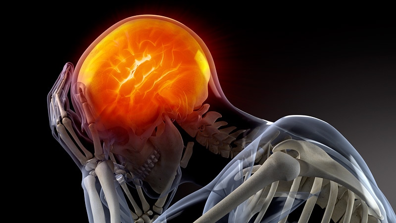
Headache
Dura Mater
Meninges
Hematoma, Subdural
Encephalocele
Cerebrospinal Fluid
Headache Disorders
Hypotension, Controlled
Spinal Puncture
Arachnoid Cysts
Superior Sagittal Sinus
Intracranial Pressure
Cerebrospinal Fluid Otorrhea
Hypotension, Orthostatic
Magnetic Resonance Imaging
Spinal Nerve Roots
Cervical Vertebrae
Hydrocephalus
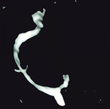
Giant cervical epidural veins after lumbar puncture in a case of intracranial hypotension. (1/87)
A 29-year-old woman presented with dilated epidural veins and incapacitating headache after undergoing a lumbar puncture. Two months later, the results of follow-up MR imaging were normal. These findings suggest that temporary dilation of the epidural vein may occur in association with post-lumbar puncture intracranial hypotension syndrome. In these cases, it seems useful to confirm whether the patient has recently undergone a lumbar puncture. (+info)Intracranial hypotension due to cerebrospinal fluid leakage detected by radioisotope cisternography. (2/87)
Seven patients, six females and one male aged 26 to 39 years old, presented with headache in the upright posture, which was completely relieved in the recumbent posture. Radioisotope cisternography with technetium-99m-human serum albumin detected cerebrospinal fluid (CSF) leakage at the cervicothoracic level in six patients, and at the high cervical level in one patient. The diagnosis was intracranial hypotension due to spontaneous CSF leakage. Complete bed rest for more than 2 weeks resulted in complete resolution of the headache in all patients, and follow-up cisternography showed no leakage. Radioisotope cisternography is useful for the diagnosis of spontaneous CSF leakage, and complete bed rest for more than 2 weeks may be the best method of treatment. (+info)Spontaneous intracranial hypotension associated with bilateral chronic subdural hematomas--case report. (3/87)
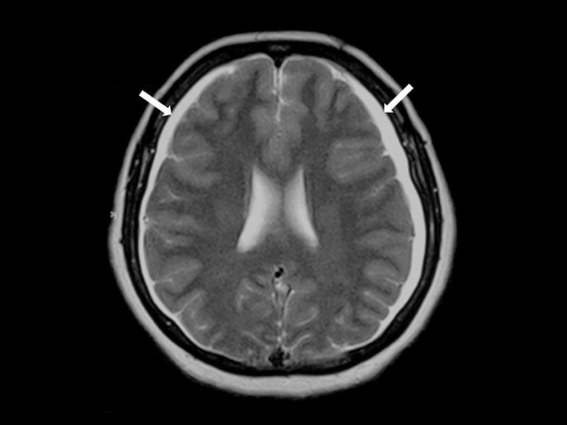
A 34-year-old female presented with spontaneous intracranial hypotension (SIH) manifesting as severe postural headache and meningism. Magnetic resonance (MR) imaging with gadolinium showed diffuse pachymeningeal enhancement. She developed bilateral chronic subdural hematomas 4 weeks after the onset of the symptoms. MR imaging showed descent of the midline structures of the brain. The bilateral chronic subdural hematomas were surgically drained, with no remarkable pressure. Postoperative MR imaging showed complete resolution of the pachymeningeal enhancement and relevation of the midline structures of the brain. SIH is an uncommon and probably unrecognized condition because of the usually benign course. However, this case emphasizes that SIH is not entirely benign. SIH should be considered if there is no identifiable risk for intracranial hemorrhage, particularly in young patients. Neurosurgical intervention for the treatment of the underlying cerebrospinal fluid leak may be required if SIH persists. (+info)Cervical MR imaging in postural headache: MR signs and pathophysiological implications. (4/87)
BACKGROUND AND PURPOSE: Postural headache most often occurs after lumbar puncture as post-lumbar puncture headache (PLPH) or, rarely, spontaneously as spontaneous intracranial hypotension headache (SIHH). In this prospective study, we used spinal MR imaging to determine the findings that would assist in the diagnosis of PLPH and SIHH and that would further our pathophysiological understanding of postural headache. METHODS: The study group consisted of 15 healthy volunteers and 20 patients with postural headache: nine with SIHH and 11 with PLPH. The craniocervical junction and the cervical spine were studied using T2-weighted fast spin-echo and T1-weighted spin-echo sequences in the axial and sagittal planes. Follow-up studies were performed in 13 patients. RESULTS: Dilatation of the anterior internal vertebral venous plexus was the most constant finding, present in 17 (85%) of 20 patients with postural headache. Spinal hygromas, whose location as subdural or epidural could not be exactly determined, were present in 14 patients (70%). A focal fluid collection was detected in the retrospinal region at the C1-C2 level in six patients with SIHH and in four patients with PLPH (50%). Tonsillar descent was detected in only one patient, and subtentorial hygroma in five patients. No abnormalities were found in the volunteers. CONCLUSION: The MR signs of dilatation of the venous plexus, presence of spinal hygromas, and presence of retrospinal fluid collections can help to establish the diagnosis of intracranial hypotension. They are probably the result of decreased CSF volume, with the retrospinal fluid collections being a transudate from the venous plexus rather than frank extravasation. Resolution of these signs parallels resolution of the headache. (+info)Clinical comparison of the Spiegelberg parenchymal transducer and ventricular fluid pressure. (5/87)
The Spiegelberg brain pressure catheter is a low cost implantable intracranial pressure measuring system which has the unique ability to perform regular automatic zeroing. A new version of the catheter has become available with a subdural bolt fixation to allow insertion of the device into the brain parenchyma. The accuracy of this system has been evaluated in comparison with a ventricular fluid pressure method in a series of patients to determine its accuracy and utility in the clinical environment. Hourly readings from the Spiegelberg system have been compared with those obtained using a standard pressure transducer connected to an external ventricular drain. Measurements continued while there was a clinical need for CSF drainage. Eleven patients were recruited to the study and data were recorded for periods ranging from 40 to 111 hours. A good agreement between the two systems was obtained. In 10 cases the mean difference was less than +/-1.5 mm Hg and the dynamic changes in value were contemporaneous. In one case an intracerebral haemorrhage developed around the tips of the Spiegelberg catheter and significant differences occurred between the two methods of measurement. In conclusion, the Spiegelberg parenchymal transducer provides an accurate measurement of intracranial pressure when compared with ventricular pressure. The transducer was found to be robust in the clinical environment and very popular with the nursing staff. Further studies may determine whether the complication rate of this system is comparable with other available devices. (+info)Symptomatic spinal extramedullary mass lesion secondary to chronic overdrainage of ventricular fluid--case report. (6/87)
A 69-year-old man presented with progressive nuchal pain and spastic gait 2 years after undergoing ventriculoperitoneal (VP) shunting for a pineal astrocytoma with obstructive hydrocephalus. The neurological manifestations were compatible with radiculomyelopathy caused by an upper cervical lesion. Magnetic resonance imaging showed an enhanced extramedullary mass lesion tightly constricting the upper cervical spinal cord. The pressure of the shunt system was 150 mmH2O, and lumbar puncture revealed normal cerebrospinal fluid (CSF) pressure of 170 mmH2O. After removal of the shunt system, the clinical symptoms and neuroradiological findings markedly improved. This symptomatic spinal mass lesion was thought to be formed secondary to chronic depletion of ventricular CSF through the VP shunt. (+info)Intracranial hypotension as a cause of radiculopathy from cervical epidural venous engorgement: case report. (7/87)
We describe the case of a 40-year-old man with spontaneous intracranial hypotension who presented with cervical radiculopathy associated with epidural venous engorgement. Epidural venous engorgement can occur secondary to intracranial hypotension and manifests intracranially as pachymeningeal venous engorgement. In the cervical spine, two cases of epidural venous engorgement due to intracranial hypotension have been reported in the literature, and neither patient presented with symptoms related to nerve compression. Epidural venous engorgement should be considered in the differential diagnosis of an enhancing epidural mass in the cervical spine. Diagnostic clues include sparing of the anterior midline and posterior aspects of the epidural space and, if present, pulsation artifact. (+info)Quantitative analysis of continuous intracranial pressure recordings in symptomatic patients with extracranial shunts. (8/87)
OBJECTIVES: To explore the outcome of management of possible shunt related symptoms using intracranial pressure (ICP) monitoring, and to identify potential methodological limitations with the current strategies of ICP assessment. METHODS: The distribution of persistent symptoms related to extracranial shunt treatment was compared before and after management of shunt failure in 69 consecutive hydrocephalic cases. Management was heavily based on ICP monitoring (calculation of mean ICP and visual determination of plateau waves). After the end of patient management, all ICP curves were re-evaluated using a quantitative method and software (Sensometrics pressure analyser). The ICP curves were presented as a matrix of numbers of ICP elevations (20 to 35 mm Hg) or depressions (-10 to -5 mm Hg) of different durations (0.5, 1, or 5 minutes). The numbers of ICP elevations/depressions standardised to 10 hours recording time were calculated to allow comparisons of ICP between individuals. RESULTS: After ICP monitoring and management of the putative shunt related symptoms, the symptoms remained unchanged in as many as 58% of the cases, with the highest percentages in those patients with ICP considered normal or too low at the time of ICP monitoring. The quantitative analysis revealed a high frequency of ICP elevations (20 to 35 mm Hg lasting 0.5 to 1 minute) and ICP depressions (-10 to -5 mm Hg lasting 0.5, 1, or 5 minutes), particularly in patients with ICP considered normal. CONCLUSIONS: The value of continuous ICP monitoring with ICP analysis using current criteria appears doubtful in the management of possible shunt related symptoms. This may reflect limitations in the strategies of ICP analysis. Calculation of the exact numbers of ICP elevations and depressions may provide a more accurate description of the ICP profile. (+info)Intracranial hypotension is a medical condition characterized by reduced pressure within the cranial cavity (the space containing brain and cerebrospinal fluid). This can occur due to several reasons, most commonly being a spontaneous or traumatic CSF leak (cerebrospinal fluid leak) from the dural membrane that surrounds the brain and spinal cord. The decrease in CSF pressure can cause various symptoms such as headaches (often positional), nausea, vomiting, neck pain, blurred vision, ringing in the ears, and cognitive impairment. Treatment typically involves identifying and addressing the underlying cause, which may include bed rest, hydration, caffeine, epidural blood patch procedures, or surgical repair of CSF leaks.
A blood patch, epidural is a medical procedure used to treat a post-dural puncture headache (PDPH), which can occur after a lumbar puncture or spinal anesthesia. During the procedure, a small amount of the patient's own blood is withdrawn and injected into the epidural space, forming a clot that seals the dural tear and alleviates the headache.
The blood patch procedure involves several steps:
1. The patient is typically placed in a lateral decubitus position (lying on their side) to widen the intervertebral space.
2. The area is cleaned and prepared for the injection, similar to other sterile procedures.
3. Using a local anesthetic, the skin and underlying tissues are numbed to minimize discomfort during the procedure.
4. A thin needle is inserted into the epidural space, usually at the same level as the original dural puncture.
5. Once the needle is in the correct position, a small amount of blood (usually around 10-20 mL) is drawn from a vein in the patient's arm.
6. The withdrawn blood is then slowly injected into the epidural space through the needle.
7. After the injection, the needle is removed, and the patient is monitored for any adverse reactions or complications.
The clot formed by the injected blood helps to seal the dural tear, preventing cerebrospinal fluid (CSF) from leaking into the epidural space and causing a headache. The blood patch procedure typically provides rapid relief from PDPH, with most patients experiencing significant improvement within 30 minutes to an hour after the injection. However, in some cases, multiple blood patches may be required to achieve complete resolution of the headache.
A subdural effusion is an abnormal accumulation of fluid in the potential space between the dura mater (the outermost layer of the meninges that covers the brain and spinal cord) and the arachnoid membrane (one of the three layers of the meninges that surround the brain and spinal cord) in the subdural space.
Subdural effusions can occur due to various reasons, including head trauma, infection, or complications from neurosurgical procedures. The fluid accumulation may result from bleeding (subdural hematoma), inflammation, or increased cerebrospinal fluid pressure. Depending on the underlying cause and the amount of fluid accumulated, subdural effusions can cause various symptoms, such as headaches, altered mental status, or neurological deficits.
Subdural effusions are often asymptomatic and may resolve independently; however, in some cases, medical intervention might be necessary to alleviate the pressure on the brain or address the underlying condition. Imaging techniques like computed tomography (CT) or magnetic resonance imaging (MRI) scans are typically used to diagnose and monitor subdural effusions.
Hypotension is a medical term that refers to abnormally low blood pressure, usually defined as a systolic blood pressure less than 90 millimeters of mercury (mm Hg) or a diastolic blood pressure less than 60 mm Hg. Blood pressure is the force exerted by the blood against the walls of the blood vessels as the heart pumps blood.
Hypotension can cause symptoms such as dizziness, lightheadedness, weakness, and fainting, especially when standing up suddenly. In severe cases, hypotension can lead to shock, which is a life-threatening condition characterized by multiple organ failure due to inadequate blood flow.
Hypotension can be caused by various factors, including certain medications, medical conditions such as heart disease, endocrine disorders, and dehydration. It is important to seek medical attention if you experience symptoms of hypotension, as it can indicate an underlying health issue that requires treatment.
Myelography is a medical imaging technique used to examine the spinal cord and surrounding structures, such as the spinal nerves, intervertebral discs, and the spinal column. This procedure involves the injection of a contrast dye into the subarachnoid space, which is the area surrounding the spinal cord filled with cerebrospinal fluid (CSF). The dye outlines the spinal structures, making them visible on X-ray or CT scan images.
The primary purpose of myelography is to diagnose various spinal conditions, including herniated discs, spinal stenosis, tumors, infection, and traumatic injuries. It can help identify any compression or irritation of the spinal cord or nerves that may be causing pain, numbness, weakness, or other neurological symptoms.
The procedure typically requires the patient to lie flat on their stomach or side while the radiologist inserts a thin needle into the subarachnoid space, usually at the lower lumbar level. Once the contrast dye is injected, the patient will be repositioned for various X-ray views or undergo a CT scan to capture detailed images of the spine. After the procedure, patients may experience headaches, nausea, or discomfort at the injection site, but these symptoms usually resolve within a few days.
Cerebrospinal fluid (CSF) rhinorrhea is a condition where the cerebrospinal fluid, which surrounds and protects the brain and spinal cord, leaks through the nasal cavity. This occurs due to a defect or opening in the skull base or the thin bone that separates the brain from the nasal cavity, known as the cribriform plate.
CSF rhinorrhea can result from trauma, surgery, or spontaneously due to increased pressure in the brain. It is important to diagnose and treat this condition promptly because it increases the risk of meningitis, an infection of the membranes covering the brain and spinal cord. Treatment options include bed rest, hydration, stool softeners, and sometimes surgical repair of the defect.
Cerebrospinal Fluid Pressure (CSFP) is the pressure exerted by the cerebrospinal fluid (CSF), a clear, colorless fluid that surrounds and protects the brain and spinal cord. CSF acts as a cushion for the brain, allowing it to float within the skull and protecting it from trauma.
The normal range of CSFP is typically between 6 and 18 cm of water (cm H2O) when measured in the lateral decubitus position (lying on one's side). Elevated CSFP can be a sign of various medical conditions, such as hydrocephalus, meningitis, or brain tumors. Conversely, low CSFP may indicate dehydration or other underlying health issues.
It is important to monitor and maintain normal CSFP levels, as abnormal pressure can lead to serious neurological complications, including damage to the optic nerve, cognitive impairment, and even death in severe cases. Regular monitoring of CSFP may be necessary for individuals with conditions that affect CSF production or absorption.
A headache is defined as pain or discomfort in the head, scalp, or neck. It can be a symptom of various underlying conditions such as stress, sinus congestion, migraine, or more serious issues like meningitis or concussion. Headaches can vary in intensity, ranging from mild to severe, and may be accompanied by other symptoms such as nausea, vomiting, or sensitivity to light and sound. There are over 150 different types of headaches, including tension headaches, cluster headaches, and sinus headaches, each with their own specific characteristics and causes.
Dura Mater is the thickest and outermost of the three membranes (meninges) that cover the brain and spinal cord. It provides protection and support to these delicate structures. The other two layers are called the Arachnoid Mater and the Pia Mater, which are thinner and more delicate than the Dura Mater. Together, these three layers form a protective barrier around the central nervous system.
The meninges are the protective membranes that cover the brain and spinal cord. They consist of three layers: the dura mater (the outermost, toughest layer), the arachnoid mater (middle layer), and the pia mater (the innermost, delicate layer). These membranes provide protection and support to the central nervous system, and contain blood vessels that supply nutrients and remove waste products. Inflammation or infection of the meninges is called meningitis, which can be a serious medical condition requiring prompt treatment.
A subdural hematoma is a type of hematoma (a collection of blood) that occurs between the dura mater, which is the outermost protective covering of the brain, and the brain itself. It is usually caused by bleeding from the veins located in this potential space, often as a result of a head injury or trauma.
Subdural hematomas can be classified as acute, subacute, or chronic based on their rate of symptom progression and the time course of their appearance on imaging studies. Acute subdural hematomas typically develop and cause symptoms rapidly, often within hours of the head injury. Subacute subdural hematomas have a more gradual onset of symptoms, which can occur over several days to a week after the trauma. Chronic subdural hematomas may take weeks to months to develop and are often seen in older adults or individuals with chronic alcohol abuse, even after minor head injuries.
Symptoms of a subdural hematoma can vary widely depending on the size and location of the hematoma, as well as the patient's age and overall health. Common symptoms include headache, altered mental status, confusion, memory loss, weakness or numbness, seizures, and in severe cases, coma or even death. Treatment typically involves surgical evacuation of the hematoma, along with management of any underlying conditions that may have contributed to its development.
An Encephalocele is a type of neural tube defect that occurs when the bones of the skull do not close completely during fetal development. This results in a sac-like protrusion of the brain and the membranes that cover it through an opening in the skull. The sac may be visible on the scalp, forehead, or back of the head, and can vary in size. Encephaloceles can cause a range of symptoms, including developmental delays, intellectual disabilities, vision problems, and seizures, depending on the severity and location of the defect. Treatment typically involves surgical repair of the encephalocele soon after birth to prevent further damage to the brain and improve outcomes.
Cerebrospinal fluid (CSF) is a clear, colorless fluid that surrounds and protects the brain and spinal cord. It acts as a shock absorber for the central nervous system and provides nutrients to the brain while removing waste products. CSF is produced by specialized cells called ependymal cells in the choroid plexus of the ventricles (fluid-filled spaces) inside the brain. From there, it circulates through the ventricular system and around the outside of the brain and spinal cord before being absorbed back into the bloodstream. CSF analysis is an important diagnostic tool for various neurological conditions, including infections, inflammation, and cancer.
Headache disorders refer to a group of conditions characterized by recurrent headaches that cause significant distress and impairment in daily functioning. The most common types of headache disorders are tension-type headaches, migraines, and cluster headaches.
Tension-type headaches are typically described as a dull, aching sensation around the head and neck, often accompanied by tightness or pressure. Migraines, on the other hand, are usually characterized by moderate to severe throbbing pain on one or both sides of the head, often accompanied by nausea, vomiting, sensitivity to light and sound, and visual disturbances.
Cluster headaches are relatively rare but extremely painful, with attacks lasting from 15 minutes to three hours and occurring several times a day for weeks or months. They typically affect one side of the head and are often accompanied by symptoms such as redness and tearing of the eye, nasal congestion, and sweating on the affected side of the face.
Headache disorders can have a significant impact on quality of life, and effective treatment often requires a multidisciplinary approach that may include medication, lifestyle changes, and behavioral therapies.
Controlled hypotension is a medical procedure in which the healthcare provider intentionally lowers the patient's blood pressure during surgery. This is done to reduce bleeding and improve surgical conditions. The goal is to maintain the patient's blood pressure at a level that is lower than their normal resting blood pressure, but high enough to ensure adequate blood flow to vital organs such as the heart and brain. Controlled hypotension is closely monitored and managed throughout the surgery to minimize risks and ensure the best possible outcomes for the patient.
A spinal puncture, also known as a lumbar puncture or a spinal tap, is a medical procedure in which a thin, hollow needle is inserted between two vertebrae in the lower back to extract cerebrospinal fluid (CSF) from the subarachnoid space. This procedure is typically performed to diagnose conditions affecting the central nervous system, such as meningitis, encephalitis, or subarachnoid hemorrhage, by analyzing the CSF for cells, chemicals, bacteria, or viruses. Additionally, spinal punctures can be used to administer medications or anesthetics directly into the CSF space, such as in the case of epidural anesthesia during childbirth.
The medical definition of a spinal puncture is: "A diagnostic and therapeutic procedure that involves introducing a thin needle into the subarachnoid space, typically at the lumbar level, to collect cerebrospinal fluid or administer medications."
The epidural space is the potential space located outside the dura mater, which is the outermost of the three membranes covering the brain and spinal cord (the meninges). This space runs the entire length of the spinal canal and contains fatty tissue, blood vessels, and nerve roots. It is often used as a route for administering anesthesia during childbirth or surgery, as well as for pain management in certain medical conditions. The injection of medications into this space is called an epidural block.
Cerebral veins are the blood vessels that carry deoxygenated blood from the brain to the dural venous sinuses, which are located between the layers of tissue covering the brain. The largest cerebral vein is the superior sagittal sinus, which runs along the top of the brain. Other major cerebral veins include the straight sinus, transverse sinus, sigmoid sinus, and cavernous sinus. These veins receive blood from smaller veins called venules that drain the surface and deep structures of the brain. The cerebral veins play an important role in maintaining normal circulation and pressure within the brain.
Enophthalmos is a medical term that refers to the abnormal positioning of the eyeball within its socket, resulting in a posterior or backward displacement of the eye. This condition can occur due to various reasons such as trauma, surgical procedures, or diseases that affect the orbital tissues, including cancer, inflammation, or infection. Enophthalmos may lead to cosmetic concerns and visual disturbances, depending on its severity. A thorough examination by an ophthalmologist or an oculoplastic surgeon is necessary for accurate diagnosis and management of this condition.
An Arachnoid cyst is a type of abnormal fluid-filled sac that develops between the brain or spinal cord and the arachnoid membrane, which is one of the three layers that cover and protect the central nervous system. These cysts are filled with cerebrospinal fluid (CSF), which is the same fluid that surrounds and cushions the brain and spinal cord.
Arachnoid cysts can vary in size and may be present at birth or develop later in life due to trauma, infection, or other factors. While many arachnoid cysts are asymptomatic and do not cause any problems, larger cysts or those that grow or shift over time can put pressure on the brain or spinal cord, leading to a range of neurological symptoms such as headaches, seizures, hearing or vision changes, balance or coordination difficulties, and cognitive impairments.
Treatment for arachnoid cysts depends on their size, location, and associated symptoms. In some cases, observation and monitoring may be sufficient, while in others, surgical intervention may be necessary to drain the cyst or create a connection between it and the surrounding CSF space to relieve pressure.
The Superior Sagittal Sinus is a medical term that refers to a venous sinus (a channel for blood flow) located in the superior part (highest portion) of the sagittal suture, which is the line along the top of the skull where the two parietal bones join in the middle. It runs from front to back, starting at the frontal bone and ending at the occipital bone, and it receives blood from veins that drain the cerebral hemispheres (the right and left halves of the brain).
The Superior Sagittal Sinus is an important structure in the circulatory system of the brain as it plays a critical role in draining venous blood from the cranial cavity. It also contains valveless venous channels that allow for the flow of cerebrospinal fluid (CSF) between the intracranial and extracranial compartments.
It is worth noting that any damage to this structure, such as through trauma or infection, can lead to serious neurological complications, including increased intracranial pressure, seizures, and even death.
A ventriculostomy is a medical procedure in which an opening is made into one of the cerebral ventricles, the fluid-filled spaces within the brain, to relieve pressure or to obtain cerebrospinal fluid (CSF) for diagnostic testing. This is typically performed using a catheter known as an external ventricular drain (EVD). The EVD is inserted through a burr hole in the skull and into the ventricle, allowing CSF to drain out and be measured or tested. Ventriculostomy may be necessary in the management of various conditions that can cause increased intracranial pressure, such as hydrocephalus, brain tumors, or traumatic brain injuries.
Intracranial pressure (ICP) is the pressure inside the skull and is typically measured in millimeters of mercury (mmHg). It's the measurement of the pressure exerted by the cerebrospinal fluid (CSF), blood, and brain tissue within the confined space of the skull.
Normal ICP ranges from 5 to 15 mmHg in adults when lying down. Intracranial pressure may increase due to various reasons such as bleeding in the brain, swelling of the brain, increased production or decreased absorption of CSF, and brain tumors. Elevated ICP is a serious medical emergency that can lead to brain damage or even death if not promptly treated. Symptoms of high ICP may include severe headache, vomiting, altered consciousness, and visual changes.
Cerebrospinal fluid (CSF) otorrhea is a condition characterized by the leakage of cerebrospinal fluid from the inner ear into the external auditory canal of the ear. CSF is a clear, colorless fluid that surrounds and protects the brain and spinal cord. It is normally contained within the subarachnoid space, which is a space between the arachnoid membrane and the pia mater that surrounds the brain and spinal cord.
CSF otorrhea can occur as a result of a head injury, skull base fracture, or surgical procedure involving the ear or surrounding structures. It can also be caused by congenital defects or tumors in the area. CSF otorrhea is a serious condition that requires prompt medical attention, as it can lead to meningitis or other complications if left untreated.
Diagnosis of CSF otorrhea typically involves a physical examination and testing of any fluid draining from the ear for beta-2 transferrin, a protein that is present in CSF but not in other bodily fluids. Imaging studies such as CT or MRI scans may also be used to help identify the underlying cause of the CSF leak. Treatment may involve bed rest, hydration, and antibiotics to prevent infection. In some cases, surgery may be necessary to repair the site of the CSF leak.
Orthostatic hypotension is a type of low blood pressure that occurs when you stand up from a sitting or lying position. The drop in blood pressure causes a brief period of lightheadedness or dizziness, and can even cause fainting in some cases. This condition is also known as postural hypotension.
Orthostatic hypotension is caused by a rapid decrease in blood pressure when you stand up, which reduces the amount of blood that reaches your brain. Normally, when you stand up, your body compensates for this by increasing your heart rate and constricting blood vessels to maintain blood pressure. However, if these mechanisms fail or are impaired, orthostatic hypotension can occur.
Orthostatic hypotension is more common in older adults, but it can also affect younger people who have certain medical conditions or take certain medications. Some of the risk factors for orthostatic hypotension include dehydration, prolonged bed rest, pregnancy, diabetes, heart disease, Parkinson's disease, and certain neurological disorders.
If you experience symptoms of orthostatic hypotension, it is important to seek medical attention. Your healthcare provider can perform tests to determine the underlying cause of your symptoms and recommend appropriate treatment options. Treatment may include lifestyle changes, such as increasing fluid intake, avoiding alcohol and caffeine, and gradually changing positions from lying down or sitting to standing up. In some cases, medication may be necessary to manage orthostatic hypotension.
Medical Definition:
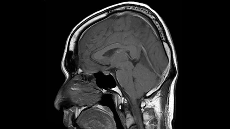
Magnetic Resonance Imaging (MRI) is a non-invasive diagnostic imaging technique that uses a strong magnetic field and radio waves to create detailed cross-sectional or three-dimensional images of the internal structures of the body. The patient lies within a large, cylindrical magnet, and the scanner detects changes in the direction of the magnetic field caused by protons in the body. These changes are then converted into detailed images that help medical professionals to diagnose and monitor various medical conditions, such as tumors, injuries, or diseases affecting the brain, spinal cord, heart, blood vessels, joints, and other internal organs. MRI does not use radiation like computed tomography (CT) scans.
Spinal nerve roots are the initial parts of spinal nerves that emerge from the spinal cord through the intervertebral foramen, which are small openings between each vertebra in the spine. These nerve roots carry motor, sensory, and autonomic fibers to and from specific regions of the body. There are 31 pairs of spinal nerve roots in total, with 8 cervical, 12 thoracic, 5 lumbar, 5 sacral, and 1 coccygeal pair. Each root has a dorsal (posterior) and ventral (anterior) ramus that branch off to form the peripheral nervous system. Irritation or compression of these nerve roots can result in pain, numbness, weakness, or loss of reflexes in the affected area.
The cervical vertebrae are the seven vertebrae that make up the upper part of the spine, also known as the neck region. They are labeled C1 to C7, with C1 being closest to the skull and C7 connecting to the thoracic vertebrae in the chest region. The cervical vertebrae have unique structures to allow for a wide range of motion in the neck while also protecting the spinal cord and providing attachment points for muscles and ligaments.
Hydrocephalus is a medical condition characterized by an abnormal accumulation of cerebrospinal fluid (CSF) within the brain, leading to an increase in intracranial pressure and potentially causing damage to the brain tissues. This excessive buildup of CSF can result from either overproduction or impaired absorption of the fluid, which typically causes the ventricles (fluid-filled spaces) inside the brain to expand and put pressure on surrounding brain structures.
The condition can be congenital, present at birth due to genetic factors or abnormalities during fetal development, or acquired later in life as a result of injuries, infections, tumors, or other disorders affecting the brain's ability to regulate CSF flow and absorption. Symptoms may vary depending on age, severity, and duration but often include headaches, vomiting, balance problems, vision issues, cognitive impairment, and changes in behavior or personality.
Treatment for hydrocephalus typically involves surgically implanting a shunt system that diverts the excess CSF from the brain to another part of the body where it can be absorbed, such as the abdominal cavity. In some cases, endoscopic third ventriculostomy (ETV) might be an alternative treatment option, creating a new pathway for CSF flow within the brain. Regular follow-ups with neurosurgeons and other healthcare professionals are essential to monitor the condition and make any necessary adjustments to the treatment plan.
The thoracic vertebrae are the 12 vertebrae in the thoracic region of the spine, which is the portion between the cervical and lumbar regions. These vertebrae are numbered T1 to T12, with T1 being closest to the skull and T12 connecting to the lumbar region.
The main function of the thoracic vertebrae is to provide stability and support for the chest region, including protection for the vital organs within, such as the heart and lungs. Each thoracic vertebra has costal facets on its sides, which articulate with the heads of the ribs, forming the costovertebral joints. This connection between the spine and the ribcage allows for a range of movements while maintaining stability.
The thoracic vertebrae have a unique structure compared to other regions of the spine. They are characterized by having long, narrow bodies, small bony processes, and prominent spinous processes that point downwards. This particular shape and orientation of the thoracic vertebrae contribute to their role in limiting excessive spinal movement and providing overall trunk stability.
 Orthostatic headache
Intracranial pressure
James Edward Cottrell
Chiropractic
Cerebrospinal fluid
Wouter Schievink
Epidural blood patch
Brain herniation
John River (rapper)
Tarlov cyst
Acetazolamide
Dural ectasia
Cerebrospinal fluid leak
Australian funnel-web spider
Postural orthostatic tachycardia syndrome
Cerebral edema
Bothrops
Yūichirō Umehara
Nausea
Thunderclap headache
Animal model of ischemic stroke
Tinnitus
Myoclonus
Pseudosubarachnoid hemorrhage
Subarachnoid hemorrhage
Visual impairment due to intracranial pressure
Non-invasive measurement of intracranial pressure
Cushing reflex
List of MeSH codes (C10)
Vegetative state
 What is intracranial hypotension?
Cerebral Angiographic Findings of Spontaneous Intracranial Hypotension | American Journal of Neuroradiology
Diagnostic Criteria for Spontaneous Spinal CSF Leaks and Intracranial Hypotension | American Journal of Neuroradiology
Delayed diagnosis of bilateral subdural effusions complicating intracranial hypotension in a patient presenting with post...
 SIH-EBP Score Calculator for Spontaneous Intracranial Hypotension
 Postural tremor as a manifestation of spontaneous intracranial hypotension | AVESİS
 Cerebral Venous Thrombosis: Background, Etiology, Epidemiology
 spontaneous intracranial hypotension | Taber's Medical Dictionary
 Spontaneous Intracranial Hypotension Archives - North Cyprus IVF
 "Spontaneous dural tear leading to intracranial hypotension and tonsill" by Aqueel H. Pabaney, Farhan A. Mirza et al.
 Free ESMINT Webinar on Spontaneous Intracranial Hypotension - Part 2 | ESMINT
Dural enhancement and cerebral displacement secondary to intracranial hypotension | Neurology
A Review of Spontaneous Intracranial Hypotension<...
 Spontaneous Intracranial Hypotension: A Case Report of Non-targeted Epidural Blood Patch Treatment | Scitechnol
Sagging brain causing postural loss of consciousness: a case of severe spontaneous intracranial hypotension | Practical...
 Asia Pacific Journal of Pain A review of the diagnosis and management of spontaneous intracranial hypotension
 Article By Diseases
| Bentham Science
Article By Diseases
| Bentham Science
Orthostatic headache - Wikipedia
 Multidisciplinary consensus guideline for the diagnosis and management of spontaneous intracranial hypotension. | J Neurol...
 A two-level large-volume epidural blood patch protocol for spontaneous intracranial hypotension: retrospective analysis of risk...
Predicting high-flow spinal CSF leaks in spontaneous intracranial hypotension using a spinal MRI-based algorithm: Have repeat...
 Patterns of cerebrospinal fluid (CSF) distribution in patients with spontaneous intracranial hypotension: Assessed with...
Search Results | jns Journals
![Fishman R[au] - Search Results - PubMed](data:image/png;base64,iVBORw0KGgoAAAANSUhEUgAAABAAAAAQCAMAAAAoLQ9TAAAARVBMVEVHcEwoU45gYmYAUpQAUpRPYGVgYmZLXnJgYmYAUZUAUpRJXnIAUpQAUpRgYmYAUpRgYmZgYmZhYmYAUpQAUpQAUpRgYmaDiPJuAAAAFXRSTlMADOJ+6QewGO8/uTRqtH7GdFJ11p1bCL3TAAAAZUlEQVQYlV2PVw7AIAxDTeney7n/UcsoldX3E+VJOAboEi7MBpHWMs1ADlG8u7UYWauwyZFeRQVPOhG2o+aiwhByJxUx91Jxhje3iJSqGfHuLKI0+0TpXvY1twCOPlFh5pa/++MB0vIOBm+1zaoAAAAASUVORK5CYII=) Fishman R[au] - Search Results - PubMed
Fishman R[au] - Search Results - PubMed
 Meetings & Courses • ESNR
 PDF) Behavioral and Learning Changes Secondary to Chiropractic Care to Reduce Subluxations in a Child with Attention Deficit...
Park J[au] - Search Results - PubMed
 Thieme E-Books & E-Journals - Radiologie up2date / Neuroradiologie
Dimitry Y. Baranov | Faculty | About Us | Perelman School of Medicine | Perelman School of Medicine at the University of...
Spontaneous23
- Summary: We report a case of spontaneous intracranial hypotension that underwent cerebral angiography. (ajnr.org)
- Spontaneous intracranial hypotension (SIH) is being recognized with increasing frequency primarily because of the identification of the various MR imaging features. (ajnr.org)
- Predicts the response to the first epidural blood patching in patients with spontaneous intracranial hypotension. (mdapp.co)
- The SIH-EBP is a prognostic scoring system for the response to the first epidural blood patching (EBP) in patients with spontaneous intracranial hypotension (SIH) based on clinical and radiological features. (mdapp.co)
- The SIH-EBP score is a simple and validated scoring system that could be used to predict the response to the first EBP in patients with spontaneous intracranial hypotension (SIH) with high accuracy. (mdapp.co)
- Spontaneous intracranial hypotension (SIH) is a syndrome caused by low cerebrospinal fluid (CSF) pressure due to leakage of CSF. (istanbul.edu.tr)
- Nursing Central , nursing.unboundmedicine.com/nursingcentral/view/Tabers-Dictionary/762787/all/spontaneous_intracranial_hypotension. (unboundmedicine.com)
- Spontaneous dural tear leading to intracranial hypotension and tonsill" by Aqueel H. Pabaney, Farhan A. Mirza et al. (aku.edu)
- Case Presentation: The Patient was diagnosed with Spontaneous Intracranial Hypotension secondary to CSF leaks, objectively demonstrated by MR Myelogram with intrathecal contrast. (aku.edu)
- On Monday, 7 March 2022 from 5 PM - 6 PM CET, we will host the second and final session of the ESMINT Webinar Mini-Series on Spontaneous Intracranial Hypotension . (esmint.eu)
- Purpose of Review: Spontaneous intracranial hypotension (SIH) is an underdiagnosed phenomenon predominantly presenting with low cerebrospinal fluid (CSF) pressure and postural headache in setting of CSF leak. (elsevierpure.com)
- The rise in the incidence rate of spontaneous intracranial hypotension (SIH) has increased the burden on physicians to accurately diagnose and treat patients presenting with SIH. (scitechnol.com)
- Multidisciplinary consensus guideline for the diagnosis and management of spontaneous intracranial hypotension. (bvsalud.org)
- We aimed to create a multidisciplinary consensus clinical guideline for best practice in the diagnosis , investigation and management of spontaneous intracranial hypotension (SIH) due to cerebrospinal fluid leak based on current evidence and consensus from a multidisciplinary specialist interest group (SIG). (bvsalud.org)
- Background We report a retrospective analysis of a two-level, variable-volume epidural blood patch (EBP) technique for the treatment of spontaneous intracranial hypotension (SIH). (bmj.com)
- Predicting high-flow spinal CSF leaks in spontaneous intracranial hypotension using a spinal MRI-based algorithm: Have repeat CT myelograms been reduced? (elsevierpure.com)
- Dive into the research topics of 'Predicting high-flow spinal CSF leaks in spontaneous intracranial hypotension using a spinal MRI-based algorithm: Have repeat CT myelograms been reduced? (elsevierpure.com)
- Spinal CSF leaks cause spontaneous intracranial hypotension (SIH). (thejns.org)
- Increasingly, EBPs are used to treat patients with spontaneous intracranial hypotension. (duke.edu)
- Cerebellar hemorrhages are occasionally reported in patients following supratentorial surgery, spinal surgery, and in patients with spontaneous intracranial hypotension. (medscape.com)
- A back of the head headache, often accompanied by neck pain , can also be a sign of a low-pressure headache, otherwise known as spontaneous intracranial hypotension (SIH). (healthline.com)
- Cerebral vein and dural sinus thrombosis (CVT) is a rare but important complication of spontaneous intracranial hypotension (SIH). (surgicalneurologyint.com)
- Spontaneous intracranial hypotension (SIH) is an unusual neurological situation caused by low cerebrospinal fluid (CSF) volume, and the symptoms are characterized by an orthostatic headache. (surgicalneurologyint.com)
Epidural2
- The epidural blood patch (EBP) is one of the most effective treatments for intracranial hypotension. (duke.edu)
- Anesthesiologists are familiar with performing EBPs for the treatment of dural puncture-associated intracranial hypotension following spinal anesthesia, complicated epidural analgesia, and diagnostic lumbar puncture. (duke.edu)
Cerebral1
- Development of obstructive hydrocephalus from ventricular compression may lead to increased intracranial pressure and decreased cerebral perfusion pressure. (medscape.com)
Secondary2
- This is thought to be secondary to decreased intracranial pressure and subsequent dilation of the venous system to attempt to replace the lost intracranial CSF volume. (ajnr.org)
- Barkoula D, Bontozoglou N, Gatzonis S, Sakas D. Intracranial hypotension syndrome in a patient due to suboccipital craniectomy secondary to Chiari type malformation. (wjgnet.com)
Hypertension1
- Clinical short-term outcome at discharge was unchanged in 14 patients and improved in 25 patients, and 19 patients had signs of rebound intracranial hypertension. (thejns.org)
Surgical2
- Surgical complications are possible and include headaches, intracranial hypotension, spinal fluid leak and implant site infection. (medtronic.com)
- Renewed interest in the anatomy of this region arose due to advances in surgical approaches to tumors of the petroclival region and the need to explain the abducent palsies seen in trauma, intracranial hypotension, and aneurysms. (medscape.com)
Subdural1
- Chronic hypotension may be associated with subdural hematomas (see HEMATOMA, SUBDURAL) or hygromas. (bvsalud.org)
Hydrocephalus1
- Among his accomplishments in neuroscience research and biomedical engineering are his investigation of the cerebrovascular response to hydrocephalus and the invention of a unique device for control of intracranial pressure (ICP) pulsatility to increase blood flow. (hopkinsmedicine.org)
Bradycardia1
- Avoid coadministration with other drugs that decrease pulse or blood pressure to mitigate risk of excessive bradycardia and hypotension. (medscape.com)
Pressure3
- This fluid acts as a cushion for the brain and is responsible for maintaining pressure called the intracranial pressure. (ndtv.com)
- Escape of this fluid leads to a fall in pressure, a condition referred to as intracranial hypotension. (ndtv.com)
- The intake of caffeine is recognized in helping to ameliorate symptoms because it helps to maintain intracranial pressure. (ndtv.com)
Diagnostic1
- The purpose of this narrative review is to discuss both procedural and diagnostic considerations of EBP for the various presentations of intracranial hypotension and allow the clinician to tailor treatment for the patient, especially in the setting of diagnostic dilemmas. (duke.edu)
Risk1
- Coadministration of nitrates or nitric oxide donors is contraindicated due to risk of hypotension. (medscape.com)
Cerebrospinal fluid leak1
- We aimed to create a multidisciplinary consensus clinical guideline for best practice in the diagnosis , investigation and management of spontaneous intracranial hypotension (SIH) due to cerebrospinal fluid leak based on current evidence and consensus from a multidisciplinary specialist interest group (SIG). (bvsalud.org)
Idiopathic3
- The link between idiopathic intracranial hypertension, fibromyalgia, and CFS: exploration of a shared pathophysiology (2018) Hulens et al. (s4me.info)
- The link between idiopathic intracranial hypertension, fibromyalgia, and chronic fatigue syndrome: exploration of a shared pathophysiology (This. (s4me.info)
- CVT can mimic the syndrome of idiopathic increased intracranial pressure (so-called pseudotumor cerebri). (symptoma.com)
Headache4
- Spontaneous intracranial hypotension (SIH) is an unusual neurological situation caused by low cerebrospinal fluid (CSF) volume, and the symptoms are characterized by an orthostatic headache. (surgicalneurologyint.com)
- If Chiari Malformation is found in someone with Thunderclap Headache, make sure it is not Spontaneous Intracranial Hypotension. (severe-headache-expert.com)
- Diagnosis is usually suspected based on the postural dependency of the headache, although in many cases the diagnosis of intracranial hypotension is not considered for some time. (kpaddock.com)
- Conclusion Epidural blood patch procedures can provide sustained improvement in headache symptoms in selected patients with spontaneous intracranial hypotension, but an untargeted approach has a lower success rate than reported in other case series. (rcpe.ac.uk)
Venous3
- Cerebral venous thrombosis in spontaneous intracranial hypotension . (symptoma.com)
- Intracranial hypotension Isolated cortical venous thrombosis has been associated with intracranial hypotension syndrome, but only rarely. (symptoma.com)
- We speculate that intracranial hypotension during the operation might have decreased venous flow and contributed to straight sinus thrombosis. (symptoma.com)
Leak2
- Intracranial hypotension attributable to a spontaneous spinal cerebrospinal fluid (CSF) leak is an increasingly recognized cause of postural headaches. (nih.gov)
- Of the patients with a meningocele, one had a partially empty sella and none had imaging evidence of CSF leak or intracranial hypotension. (ajnr.org)
Syndrome2
- Spontaneous intracranial hypotension (SIH) is a syndrome of postural headaches aggravated by sitting or standing and relieved by recumbence. (eurorad.org)
- 3] Watanabe A, Horikoshi T, Uchida M, Koizumi H, Yagishita T, Kinouchi H. (2009) Diagnostic value of spinal MR imaging in spontaneous intracranial hypotension syndrome. (eurorad.org)
Diagnosis2
- To discuss common myths and misperceptions about spontaneous intracranial hypotension (SIH), focusing on common issues related to diagnosis and treatment, and to review the evidence that contradicts and clarifies these myths. (medscape.com)
- A diagnosis of spontaneous intracranial hypotension was made, and the patient was treated with bed rest and acetaminophen with codeine. (asahq.org)
Hypovolemia1
- 30 , 31 , 35 CSF hypovolemia (craniospinal hypotension) is a disorder that is frequently associated with dural defects, occasionally with intraspinal fluid collection of variable longitudinal extent and rarely with red blood cells (RBCs) or xanthochromia in the CSF. (ajnr.org)
Subdural3
- 5. [Bilateral intracranial subdural hematoma following lumbar puncture: report of a case]. (nih.gov)
- 14. [Intracranial hypotension with subdural hematoma following lumbar puncture: case report]. (nih.gov)
- Chronic hypotension may be associated with subdural hematomas (see HEMATOMA, SUBDURAL) or hygromas. (bvsalud.org)
Thrombosis1
- Cerebral vein and dural sinus thrombosis (CVT) is a rare but important complication of spontaneous intracranial hypotension (SIH). (surgicalneurologyint.com)
Spinal5
- Spontaneous intracranial hypotension with spinal myelography imaging findings. (eurorad.org)
- It is characterized by reduced CSF pressure in the intracranial cavity caused by CSF leaks through small defects of the meninges into the spinal canal without previous lumbar puncture or an interventional procedure or surgery on the neural axis [1-4]. (eurorad.org)
- 2] Jocelyn H. Medina, Kevin Abrams, Steven Falcone, Rita G. Bhatia (2010) Spinal Imaging Findings in Spontaneous Intracranial Hypotension. (eurorad.org)
- Spinal CSF leaks cause spontaneous intracranial hypotension (SIH). (thejns.org)
- Initial presentation suggested spontaneous intracranial hypotension (SIH) since there was no history of epidural or spinal anesthesia, or trauma or surgery to her back or neck. (symptoma.com)
Neurosurg1
- From white to blue light: evolution of endoscope-assisted intracranial tumor neurosurgery and expansion to intraaxial tumors J Neurosurg. (usc.edu)
Severe3
- 18. Severe intracranial and intraspinal subarachnoid hemorrhage after lumbar puncture: a rare case report. (nih.gov)
- Intracranial Hypotension After Severe Covid-19. (cdc.gov)
- Severe hypotension and seizures have been reported following rapid IV administration, particularly with concomitant use of fentanyl (see DOSAGE AND ADMINISTRATION for complete information). (nih.gov)
Meningitis2
- Dural injury recognized intraoperatively permits immediate repair, but unnoticed damage may cause postoperative pleural effusion, intracranial hypotension, meningitis, or pneumocephalus. (hindawi.com)
- Symptoms develop from the accumulation of pleural fluid, the reduced volume of cerebrospinal fluid (CSF), the presence of intracranial air, or the development of meningitis, with outcomes ranging from benign to catastrophic. (hindawi.com)
Patients4
- A similar finding has also been reported in patients with craniospinal hypotension. (ajnr.org)
- It is a relatively common imaging finding in patients with spontaneous intracranial hypotension seen in up to 14% of patients according to a large retrospective study 1 . (radiopaedia.org)
- Diffuse Calvarial Hyperostosis in Patients with Spontaneous Intracranial Hypotension. (radiopaedia.org)
- Performance of a machine-learning algorithm to predict hypotension in mechanically ventilated patients with COVID-19 admitted to the intensive care unit: a cohort study. (cdc.gov)
Migraine1
- Increased intracranial pressure in migraine? (s4me.info)
Lumbar puncture2
- Intracranial hypotension, which is most commonly caused by lumbar puncture, can lead to intense meningeal enhancement, which resolves on its own once the intracranial hypotension has been corrected. (johnshopkins.edu)
- 15. Life-threatening intracranial hypotension after diagnostic lumbar puncture. (nih.gov)
Infusion1
- Insights into the natural history of spontaneous intracranial hypotension from infusion testing. (uni-freiburg.de)
Chronic1
- It is hypothesized that hyperostosis occurs due to chronic loss of CSF thus it is more likely to be seen in long-standing intracranial hypotension. (radiopaedia.org)
Brain2
- According to the Monro-Kellie doctrine, the intracranial volume of CSF is inversely related to the brain blood volume [1]. (eurorad.org)
- INTRACRANIAL HYPOTENSION (IH) is a condition in which there is negative pressure within the brain cavity. (kpaddock.com)
Rarely1
- [1] Rarely, however, is it spontaneous, a condition called "spontaneous intracranial hypotension"(SIH). (asahq.org)