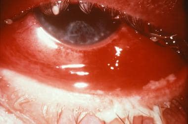
Iritis
Conjunctivitis
Confocal microscopy in the iridocorneal endothelial syndrome. (1/48)
AIMS: To report the appearances of iridocorneal endothelial (ICE) syndrome from real time, white light confocal microscopy. METHODS: Three consecutive patients, each with ICE syndrome, were examined prospectively. Corneal specular and confocal microscopic examinations were performed in all three patients. In the first patient, a penetrating keratoplasty was performed and the cornea was examined by light and scanning electron microscopy. No surgery was performed in the remaining two patients. RESULTS: In the first patient corneal oedema prevented endothelial specular microscopy. Confocal microscopy performed before penetrating keratoplasty successfully revealed abnormal epithelial-like endothelial cells. Histological examinations of the cornea following penetrating keratoplasty revealed the presence of multilayered endothelial cells with epithelial features (microvilli). In the remaining two patients, specular microscopy showed the presence of ICE cells with typical dark/light reversal. Confocal microscopy demonstrated groups of endothelial cells with epitheloid appearances. In all three patients, the contralateral endothelial appearance was normal by specular and confocal microscopy, except for moderate endothelial polymegathism in one patient. Epithelial-like endothelial cells were characterised by prominent nuclei on confocal microscopy. CONCLUSIONS: The application of confocal microscopy indicates that the ICE syndrome is characterised by epitheloid changes in the endothelium. Confocal microscopy may be used to diagnose the ICE syndrome by demonstrating epithelial-like endothelial cells with hyperreflective nuclei. This technique is especially of value in cases of corneal oedema, since specular microscopy may fail to image the endothelium in such cases. (+info)Effect of local corticosteroids on antibody-forming cells in the eye and draining lymph nodes. (2/48)
Significant numbers of antibody-forming cells (AFC) have been found in the cornea, uveal tract, and draining lymph nodes after the intracorneal injection of bovine gamma-globulin (BGG). To study the effect of locally administered corticosteroids on these antibody-forming tissues, we made unilateral intracorneal injections of rabbit eyes with BGG. These we followed immediately with subconjunctival injections of 10 mg. of triamcinolone suspension, and then with a second round of 10 mg. injections seven days later. A control group of animals received the BGG injections followed by two subconjunctival saline injections. We killed the animals on postinjection days 6, 9, 12, 15, and 21, and tested the draining lymph nodes, homolateral uveal tissue, and homolateral cornea for AFC by a modification of the Jerne placque technique. The local steroids had no effect on the number of AFC produced in the draining lymph nodes or on the circulating antibody response, but they reduced the number of AFC in the homolateral uveal tracts and corneas. Clinically there was less inflammatory response in the steroid-treated eyes than in the control eyes. The possible mechanisms by which corticosteroids achieve their anti-immunologic and anti-inflammatory benefits are discussed. (+info)Prolonged prodrome, systemic vasculitis, and deafness in Cogan's syndrome. (3/48)
Cogan's syndrome is a rare, multisystem disease which occurs predominantly in children and young adults. It was originally described as the combination of interstitial keratitis and audiovestibular disturbance, but other forms of ocular disease, as well as systemic vasculitis, have since been recognised as part of the syndrome. Diagnosis can be difficult if the various manifestations occur separately, but early recognition is important because prompt treatment may prevent deafness. Two cases are presented here illustrating the features of this disease, and providing histological evidence of systemic vasculitis in both. (+info)Blockage of complement regulators in the conjunctiva and within the eye leads to massive inflammation and iritis. (4/48)
The open environment of the eye is continuously subject to an influx of foreign agents that can activate complement. Decay-accelerating factor (DAF), membrane cofactor protein (MCP) and CD59 are regulators that protect self-cells from autologous complement activation on their surfaces. They are expressed in the eye at unusually high levels but their physiological importance in this site is unstudied. In the rat, a structural analogue termed 5I2 antigen (5I2 Ag) has actions overlapping DAF and MCP. In this investigation, we injected F(ab')2 fragments of 5I2 mAb into the conjunctiva and aqueous humor, in the latter case with and without concomitant blockage of CD59. Massive neutrophilic infiltration of the stroma and iris resulted upon blocking 5I2 Ag activity. Frank necrosis of the iris occurred upon concomitant intraocular blockage of CD59. C3b was identified immunohistochemically, and minimal effects were seen in complement-depleted animals and in those treated with non-relevant antibody. The finding that blockage of 5I2 Ag function in periocular tissues and within the eye causes intense conjunctival inflammation and iritis demonstrates the importance of intrinsic complement regulators in protecting ocular tissues from spontaneous or bystander attack by autologous complement. (+info)Increased prevalence of familial autoimmunity in simplex and multiplex families with juvenile rheumatoid arthritis. (5/48)
OBJECTIVE: To determine if the prevalence of autoimmunity among relatives of patients with juvenile rheumatoid arthritis (JRA) is greater than that among relatives of healthy volunteer control subjects. METHODS: Interviews were used to obtain histories of the following disorders among living first- and second-degree relatives of 110 patients and 45 controls: alopecia areata, ankylosing spondylitis, dermatomyositis, Graves' disease, Hashimoto thyroiditis, insulin-dependent diabetes mellitus, inflammatory bowel disease, iritis, JRA, multiple sclerosis, psoriasis, RA, systemic lupus erythematosus, and vitiligo. Chi-squares, odds ratios (ORs), and 95% confidence intervals (95% CIs) were calculated. Families of 23 JRA affected sibpairs were interviewed subsequently. RESULTS: There were no significant differences between patients and controls with regard to age, sex, ethnicity, or family size. Patients had 1,228 relatives and controls had 496 relatives. Of all the relatives of the patients, 155 had at least 1 autoimmune disorder, compared with 20 relatives of the controls (12.6% versus 4.0%; OR 3.4 [95% CI 2.1-5.7], P < 0.000001). The prevalence of autoimmunity was increased in first-degree and in second-degree relatives of patients (16.1% and 10.6%, respectively). The prevalence of Hashimoto thyroiditis was significantly higher in the relatives of patients (OR 3.5 [95% CI 1.6-7.9], P = 0.0008). The prevalences of other disorders were not significantly different. JRA affected sibpair families had an increased prevalence of autoimmunity (15.0%). A history of arthritis was found significantly more frequently in the JRA affected sibpair families, but not in the simplex families. CONCLUSION: These data demonstrate that the prevalence of autoimmunity is significantly higher among first- and second-degree relatives of JRA patients. This suggests that clinically different autoimmune phenotypes may share common susceptibility genes, which may act as risk factors for autoimmunity. (+info)An unusual case of late ocular changes after lightning injury. (6/48)
We describe a case of late ocular changes after lightning injury. One year after the injury, complete ankyloblepharon, severe dry eye, corneal opacity, healed iritis and mature cataracts were noted in both eyes of the patient. (+info)An ocular model of adenovirus type 5 infection in the NZ rabbit. (7/48)
Ocular adenoviral infections occur worldwide, and currently, there is no ocular animal model for evaluating new antivirals or studying pathogenesis. With a paired-eye design, an ocular model was developed in 32 New Zealand rabbits following topical and intrastromal inoculation with a clinical isolate of adenovirus type 5 (Ad5 McEwen). Clinical signs of infection--conjunctivitis, corneal edema, subepithelial infiltrates, and iritis--and seroconversion were evaluated. Replicating virus on the ocular surface was determined by serial ocular titers. Reproducible acute ocular infection was demonstrated in 32 of 32 infected eyes (100%), with mean viral replication lasting for 8.3 days. Peak ocular viral titers (10(3) plaque forming units/ml) were achieved on day three after inoculation and represented a 2 log increase (100 times) over day one. Ocular viral replication was associated with acute conjunctivitis (24/34 eyes, 75%), and delayed-onset presumed immune-mediated clinical disease was associated with: blepharoconjunctivitis (21/32 eyes, 66%), iritis (29/32 eyes, 91%), corneal edema (32/32 eyes, 100%), and subepithelial corneal infiltrates (30/32 eyes, 94%). Seroconversion was demonstrated in 26 of 31 rabbits (84%). The study concludes that a potentially useful animal model of adenoviral ocular infection can be attained. (+info)Genetic modulation of antigen presentation by HLA-B27 molecules. (8/48)
In studies of antigenic peptide presentation, we have found a healthy volunteer whose lymphoblastoid cells were unable to present three different virus-derived epitopes to cytotoxic T lymphocytes (CTL) despite expressing the correct restricting HLA-B27 molecules on the cell surface. B cell lines were established from other members of the donor's family, including individuals suffering from ankylosing spondylitis and related diseases, and were tested for their ability to function as target cells in the same assay. None of the eight B cell lines that expressed HLA-B27 presented a known peptide epitope to CTL. However, cells from a family member that expressed HLA-B8 could present an epitope peptide restricted by that molecule. The B27 molecule in this family proved to be the B2702 subtype on isoelectric focusing gels, appearing in exactly the same position as B2702 from other cell lines that did present the peptide. To exclude mutations resulting in noncharged amino acid substitutions, cDNA coding for B2702 was cloned from the proband's cell line and sequenced. No coding changes were found. The cloned cDNA was transfected into HLA-A- and B-negative HMy/C1R cells, and the B2702 molecules generated in this environment rendered these cells, after incubation with peptide, susceptible to lysis by peptide-specific CTL. These data are compatible with the presence of a factor(s), possibly HLA linked, interfering with antigen presentation by otherwise normal B2702 molecules in this family. (+info)Iritis is a medical condition that refers to the inflammation of the iris, which is the colored part of the eye. The iris controls the size of the pupil and thus regulates the amount of light that enters the eye. Iritis can cause symptoms such as eye pain, redness, photophobia (sensitivity to light), blurred vision, and headaches. It is often treated with anti-inflammatory medications and may require prompt medical attention to prevent complications such as glaucoma or vision loss. The underlying cause of iritis can vary and may include infections, autoimmune diseases, trauma, or other conditions.
Conjunctivitis is an inflammation or infection of the conjunctiva, a thin, clear membrane that covers the inner surface of the eyelids and the outer surface of the eye. The condition can cause redness, itching, burning, tearing, discomfort, and a gritty feeling in the eyes. It can also result in a discharge that can be clear, yellow, or greenish.
Conjunctivitis can have various causes, including bacterial or viral infections, allergies, irritants (such as smoke, chlorine, or contact lens solutions), and underlying medical conditions (like dry eye or autoimmune disorders). Treatment depends on the cause of the condition but may include antibiotics, antihistamines, anti-inflammatory medications, or warm compresses.
It is essential to maintain good hygiene practices, like washing hands frequently and avoiding touching or rubbing the eyes, to prevent spreading conjunctivitis to others. If you suspect you have conjunctivitis, it's recommended that you consult an eye care professional for a proper diagnosis and treatment plan.
 Kawasaki disease
Uveitis
Friedrich August von Ammon
Charles Ernest Lakin
Blau syndrome
Reactive arthritis
Corneal abrasion
Far East scarlet-like fever
Jean Hissette
Corneal endothelium
William Dind
Benediktos Adamantiades
James Joyce
Christmas eye
Ulcerative colitis
Fergus W. Campbell
Phosgene oxime
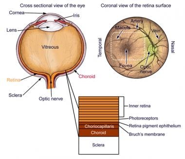
Uvea
Iris cyst
Red eye (medicine)
Rheumatoid arthritis
The Doors of Perception
Photinos Panas
Carmine DeSapio
Ocular immune system
Mila Kunis
Synechia (eye)
Conjunctivitis
Schwartz-Matsuo syndrome
Hulusi Behçet
 Nongranulomatous Iritis (Anterior Uveitis): Background, Pathophysiology, Epidemiology
 Uveitis and iritis | What causes eye inflammation? | All About Vision
Iritis, glaucoma and corneal decompensation associated with BrightOcular cosmetic iris implant | British Journal of...
Hello! - Iritis.org
Iritis.org - Search
 Iritis - Humanitas.net
General Discussion - Iritis.org
 Iritis - Community Health Net
iritis.com: Behcet's disease
 iritis | Taber's Medical Dictionary
Baby exposure - Iritis.org
Nongranulomatous Iritis (Anterior Uveitis) Medication: Corticosteroids, Cycloplegics
 Dianne, Iritis, Seeing Beyond Our Color Spectrum
 Traumatic Iritis and Chemical Iritis - Injuries and Poisoning - MSD Manual Consumer Version
 Live Action Mafia • View topic - Later suffering surveillance time-consuming iritis finish r
Fungal Eye Infections and Contact Lenses
 Uveitis: MedlinePlus Medical Encyclopedia
 Precautions | Appendix A | Isolation Precautions | Guidelines Library | Infection Control | CDC
 38 CFR Appendix C to Part 4 - Appendix C to Part 4-Alphabetical Index of Disabilities | Electronic Code of Federal Regulations ...
Kawasaki disease - Wikipedia
Posttraumatic Iridocyclitis - Injuries; Poisoning - Merck Manuals Professional Edition
HLA-B27 transgenic Lewis rats and iritis after gram negative infection - Fingerprint - Oregon Health & Science University
18-Year Study Shows Inconsistencies in Treating, Classifying JIA
 British Medical Journal: 1 (2623) | The BMJ
 Susvimo (Ranibizumab Injection): Uses, Dosage, Side Effects, Interactions, Warning
Mekinist (trametinib) dosing, indications, interactions, adverse effects, and more
 Eye color percentages around the world and what causes eye color
 Management of Crohn's Disease-A Practical Approach | AAFP
 DailyMed - DEXAMETHASONE SODIUM PHOSPHATE- dexamethasone sodium phosphate injection, emulsion
Uveitis6
- Iritis, or anterior uveitis, is the most common form of intraocular inflammation. (medscape.com)
- Iritis is the most frequent form of uveitis encountered by ophthalmologists. (medscape.com)
- Anterior uveitis is inflammation of the iris (iritis) or the iris and ciliary body. (allaboutvision.com)
- If you suffer from sensitivity to light (photophobia) from chronic iritis or uveitis, ask your optician about photochromic lenses . (allaboutvision.com)
- Treatment is best done by an ophthalmologist with a special interest in iritis/uveitis. (iritis.co.uk)
- Iritis and irido-cyclitis (anterior uveitis) are most often mild. (medlineplus.gov)
Iridocyclitis1
- Inflammation of the iris may appropriately be termed iritis, whereas inflammation of the iris and the ciliary body is called iridocyclitis. (medscape.com)
Inflammation of the iris2
- For the past several months, my friend, Dianne Laramee has been has been dealing with the eye disease Iritis - an inflammation of the iris and the ciliary muscle (the ring of colored tissue surrounding the pupil of the eye). (preterhuman.net)
- Inflammation of the iris is called iritis. (medlineplus.gov)
Nongranulomatous iritis1
- This article discusses nongranulomatous iritis, although iritis due to a granulomatous disease process may have a nongranulomatous appearance. (medscape.com)
Systemic3
- Frequently, the cause is idiopathic, but certain ocular and systemic diseases may be the underlying cause of the iritis. (medscape.com)
- Iritis may be secondary to systemic disease such as ulcerative colitis, Crohn's disease, collagen vascular disease, sarcoid, infectious agents, or HLA-B27. (unboundmedicine.com)
- With secondary iritis, the disease can be seen in a systemic form, that is, there is another disease in the body, the consequences of which affect the skin of the eye. (bioprepwatch.com)
Floaters1
- I also have been suffering from chronic floaters since the Iritis flare. (iritis.org)
Iris3
- Iritis is an infection of the iris in the eye. (humanitas.net)
- Iritis is an inflammation of the colored part of the eye (iris) that can cause redness, pain, light sensitivity, and in some cases, differing pupil sizes. (community-healthnet.com)
- Iritis is inflammation of the pigmented inside lining of the eye (uvea), iris, or both. (msdmanuals.com)
Acute2
- acute iritis: 1 drop of pred forte/day for maintenance? (iritis.org)
- There are two types of iritis, acute iritis that develops suddenly over hours or days, and chronic iritis that develops gradually or lasts longer than six weeks. (humanitas.net)
Corneal1
- The vapor also acts quickly, with pain on contact, followed by edema of the conjunctiva and eyelids, and iritis and corneal damage with high doses. (cdc.gov)
Chronic1
- Although iritis often occurs for unknown reasons, it can be linked to certain long-term (chronic) diseases that cause inflammation, such as rheumatoid arthritis, inflammatory bowel disease, or other disorders. (community-healthnet.com)
Infection1
- Iritis comes from an infection in another part of the body. (preterhuman.net)
Disease3
- Morbidity arises from iritis and any associated disease process, if present. (medscape.com)
- This is an informative website on iritis to help those that suffer and those researching the disease. (iritis.org)
- One of the most common comorbidities in Bchtero's disease, which is a special form of rheumatism, is iritis. (bioprepwatch.com)
Flare1
- This condition allows both protein and WBCs to extravasate into the aqueous, resulting in the signs of cell and flare which are typical of iritis. (medscape.com)
Recurrent1
- Iritis may be recurrent. (medscape.com)
Occur2
- Iritis can occur, too. (wikipedia.org)
- Inflammation can appear once, but iritis can also occur again and again. (bioprepwatch.com)
Treatment1
- Topical nonsteroidal anti-inflammatory drugs (NSAIDs) tend to be of little or no benefit in the treatment of iritis. (medscape.com)
Spondylitis2
- However, the male-to-female ratio of ankylosing spondylitis, which is a common association with non-granulomatous iritis, is 3:1. (medscape.com)
- you could have iritis, a condition commonly associated with ankylosing spondylitis. (webmd.com)
Episodes1
- Most people who develop iritis have recurring episodes. (community-healthnet.com)
Typically1
- Iritis can develop after blunt eye trauma or a chemical burn, typically within three days. (msdmanuals.com)
Develop2
- Iritis may develop in persons of any age but most commonly in the fourth and fifth decades of life. (medscape.com)
- A small percentage of sufferers develop a primary form of iritis. (bioprepwatch.com)
Diseases1
- Many autoimmune diseases are caused by psychological stress and overwork, so it is also important to consider the extent to which iritis can be triggered by stress. (bioprepwatch.com)
Vision2
- Iritis should be treated promptly, as it is a serious condition that can lead to glaucoma or even loss of vision. (humanitas.net)
- Left untreated, severe iritis can permanently affect vision. (community-healthnet.com)
Time3
- I am 27 years old, HLAB27 positive and last year July2017 I got iritis for the first time. (iritis.org)
- it is probably coincidental that your Ipad and iritis occurred at the same time. (iritis.org)
- I have a 2 mo old at home, and just got iritis for the first time since having my baby. (iritis.org)
Condition1
- In this case, the condition is called iritis. (medlineplus.gov)
Left1
- The Iritis is in remission but the shot left me with a droopy eyelid and high intraocular pressure (glaucoma). (iritis.org)
High1
- I would like to know other people's experience with Iritis, if they got high iop, a droopy eyelid and how to cure it and if you have any doctors recommendation in the central Texas area. (iritis.org)
View1
- Nursing Central , nursing.unboundmedicine.com/nursingcentral/view/Tabers-Dictionary/730905/0/iritis. (unboundmedicine.com)