Jaw Diseases
Jaw Neoplasms
Bisphosphonate-Associated Osteonecrosis of the Jaw
Masticatory Muscles
Jaw, Edentulous
Jaw Cysts
Mandible
Mandibular Diseases
Jaw Relation Record
Diphosphonates
Temporomandibular Joint
Maxilla
Pterygoid Muscles
Branchial Region
Temporal Muscle
Maxillary Diseases
Dentition
Bone Density Conservation Agents
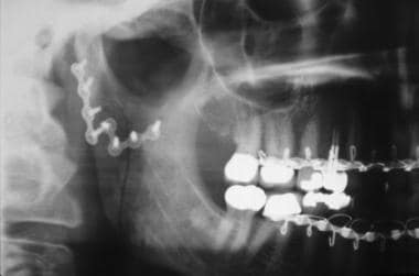
Radiography, Panoramic
Hyoid Bone
Fibrous Dysplasia of Bone
Fibroma, Ossifying
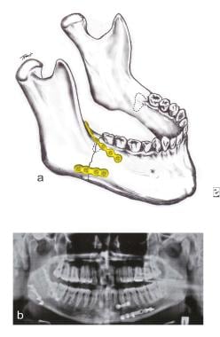
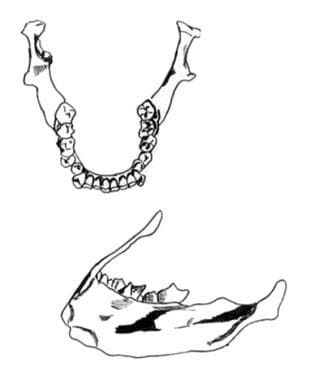
Relationship between accessory foramina and tumour spread in the lateral mandibular surface. (1/97)
The spread of tumour cells to the mandible has been well recognised and invasion of the edentulous alveolar ridge by tumour through accessory foramina has been documented. Tumour infiltration can also occur through the lateral cortical plate, but the number and distribution of accessory foramina on this surface has not been reported. Lateral surfaces of 89 mandibles were examined and accessory foramina which showed a direct communication with the underlying cancellous bone were charted. It was found that the number of accessory foramina varied greatly from specimen to specimen. Only 70.8 % of mandibles showed foramina in the coronoid, sigmoid and condylar sections; of these 93.7 % exhibited foramina in the condylar section, 23.8% in the coronoid and only 19 % in the sigmoid section. This finding confirms that the current practice of conserving part of the ascending ramus posterior to the coronoid process following surgery is sound. Similarly in the rest of the lateral surface, foramina were present in the upper third section in 97.8 % of mandibles, 61.8% in the lower third and 58.4 % in the middle third sections. This result justifies the principle of rim resection in appropriate cases and the recognition that the alveolar section is commonly invaded before the rest of the body. The number and distribution of foramina may be of greater significance following radiotherapy when the foramina could provide multiple direct channels for invasion of tumour cells from the lateral surface to the medulla. (+info)Post-extraction remodeling of the adult mandible. (2/97)
Following tooth loss, the mandible shows an extensive loss of bone in some individuals. This may pose a significant problem in the prosthodontic restoration of function and esthetics. The many factors which have been proposed as being responsible for the inter-individual variation in post-extraction remodeling mean that a perfunctory analysis of the literature, in which well-controlled, relevant studies are scarce, may not provide the whole story. This article reviews the local and systemic factors which may play a role in the post-extraction remodeling of the mandible. Since severe residual ridge resorption may occur even when the bone status in the rest of the skeleton is good and vice versa, it is concluded that local functional factors are of paramount significance. It is now essential to determine how they can be modified and applied to help maintain ridge height and quality in our aging, edentulous population. (+info)Oral healthcare in transition in Eastern Europe. (3/97)
Big changes have occurred in the oral healthcare delivery systems of most Eastern European countries since the fall of the Berlin wall in 1989 and the demise of communism in the former USSR in 1991. In the new situation it was necessary to reform the political and social systems including healthcare. Reforms were started to improve the economy and, in comparison with Western Europe, the generally lower living standards. It is difficult to obtain comprehensive data on oral healthcare in Eastern European countries but this paper reports data from nine countries and provides a 'macro' view of the current situation in these countries. Many countries seem to have adopted a Bismarckian model for the provision of oral healthcare based on a sickness insurance system. (+info)A multi-centre study of Osseotite implants supporting mandibular restorations: a 3-year report. (4/97)
This multi-centre study evaluated the performance of the Osseotite implant in the mandibular arch. Osseotite implants (n = 688) were placed in 172 patients; 43.5% were placed in the anterior mandible and 66.5% in the posterior mandible. Fifteen per cent of the implants were placed in soft bone, 56.9% in normal bone and 28.1% in dense bone. During placement, 49.9% of the implants were identified as having a tight fit, 48.6% a firm fit and 1.5% a loose fit. About one-third of the implants (32.4%) were short (10 mm in length or less). After 36 months, only 5 implants had been lost, for a cumulative survival rate of 99.3%. The 3-year results of this study indicate a high degree of predictability with placement of Osseotite implants in the mandibular arch. (+info)Good occlusal practice in removable prosthodontics. (5/97)
The loss of teeth may result in patients experiencing problems of a functional, aesthetic and psychological nature. This section addresses the very important subject of occlusal considerations for partial and complete dentures. The occlusion is particularly important given the bearing that occlusal factors have, especially on edentulous patients. (+info)The effect of increasing vertical dimension of occlusion on facial aesthetics. (6/97)
AIM: To investigate the effect of increasing the vertical dimension of occlusion on facial aesthetics. SETTING: General practice. METHOD: Questionnaires were sent to 96 patients who had been treated in the practice during the period of July 1998 to December 2000, resulting in an overall 72% response rate. All these patients had had their occlusal vertical dimension increased. Photographs of patients were taken before, during and after treatment. The questionnaire asked their opinion on the effects of the treatment on their facial features. To obtain an objective view to substantiate the opinions of the patients, a panel of five judges reviewed the before and after photographs and filled in their own questionnaires. RESULTS: Of the patients who responded to the questionnaire, 79.7% said they looked younger after the treatment. The panel thought 81.2% of the patients treated whose photographs they reviewed looked younger. CONCLUSION: Increasing the vertical dimension of occlusion can have far reaching effects on facial aesthetics, not just on the peri-oral areas but on the whole face. (+info)The milled implant bar: an alternative to spark erosion. (7/97)
Patients who cannot tolerate total coverage of the hard palate or whose maxillary arches are poorly formed, because of congenital, developmental or surgical defects, may be unable to wear a conventional complete denture. These patients can be successfully treated with implant-supported prostheses that cover only a minimal amount of palatal tissue. With spark-eroded castings, very precise restorations can be constructed to fit such implant supports. However, these castings are so expensive that cost precludes their use for many patients. This article presents an alternative approach, developed with the refined techniques used for removable partial dentures, which can yield results similar to those for spark-eroded castings at a fraction of the cost. The clinical and laboratory procedures involved in this technique are described. (+info)Cell proliferation and expression of Cbfa-1 in a peripheral osteo-chondroma arising from the mandibular oral mucosa of an edentulous alveolar ridge. (8/97)
This report describes the proliferation and the expression of Cbfa-1 in a rare case of peripheral osteo-chondroma arising from the mandibular oral mucosa of an edentulous alveolar ridge. Histologically, the lesion consisted of mesenchymal cells with either bone or cartilage tissue in the center. Almost all the tumor cells were reactive for PCNA, however, only the cells around the bone and cartilage tissues were reactive for Cbfa-1. These results suggest that both the bone and the cartilage tissues in this case were produced by mesenchymal cells that originated from the peripheral periosteum of the alveolar ridge. Furthermore, we have shown that immunohistochemical staining for PCNA and Cbfa-1 can be used to investigate lesions with bone or cartilage formation and to distinguish between those produced by osteogenic cells from those that are just reactive and produced by dystrophic calcification. (+info)In medical terms, the jaw is referred to as the mandible (in humans and some other animals), which is the lower part of the face that holds the lower teeth in place. It's a large, horseshoe-shaped bone that forms the lower jaw and serves as a attachment point for several muscles that are involved in chewing and moving the lower jaw.
In addition to the mandible, the upper jaw is composed of two bones known as the maxillae, which fuse together at the midline of the face to form the upper jaw. The upper jaw holds the upper teeth in place and forms the roof of the mouth, as well as a portion of the eye sockets and nasal cavity.
Together, the mandible and maxillae allow for various functions such as speaking, eating, and breathing.
Jaw diseases refer to a variety of conditions that affect the temporomandibular joint (TMJ) and the surrounding muscles, as well as dental disorders that can impact the jaw. Some common examples include:
1. Temporomandibular Joint Disorders (TMD): These are problems with the TMJ and the muscles that control jaw movement. Symptoms may include pain, clicking or popping sounds, and limited movement of the jaw.
2. Osteonecrosis of the Jaw: This is a condition where bone in the jaw dies due to lack of blood supply. It can be caused by radiation therapy, chemotherapy, or certain medications.
3. Dental Cavities: These are holes in the teeth caused by bacteria. If left untreated, they can cause pain, infection, and damage to the jawbone.
4. Periodontal Disease: This is an infection of the gums and bones that support the teeth. Advanced periodontal disease can lead to loss of teeth and damage to the jawbone.
5. Jaw Fractures: These are breaks in the jawbone, often caused by trauma.
6. Oral Cancer: This is a type of cancer that starts in the mouth or throat. If not treated early, it can spread to the jaw and other parts of the body.
7. Cysts and Tumors: These are abnormal growths in the jawbone or surrounding tissues. While some are benign (non-cancerous), others can be malignant (cancerous).
8. Osteomyelitis: This is an infection of the bone, often occurring in the lower jaw. It can cause pain, swelling, and fever.
9. Oral Thrush: This is a fungal infection that causes white patches on the inside of the mouth. If left untreated, it can spread to the jaw and other parts of the body.
10. Sinusitis: Inflammation of the sinuses can sometimes cause pain in the upper jaw.
Jaw neoplasms refer to abnormal growths or tumors in the jawbone (mandible) or maxilla (upper jaw). These growths can be benign (non-cancerous) or malignant (cancerous). Benign neoplasms are not considered life-threatening, but they can still cause problems by invading nearby tissues and causing damage. Malignant neoplasms, on the other hand, can spread to other parts of the body and can be life-threatening if not treated promptly and effectively.
Jaw neoplasms can present with various symptoms such as swelling, pain, loose teeth, numbness or tingling in the lips or tongue, difficulty chewing or swallowing, and jaw stiffness or limited movement. The diagnosis of jaw neoplasms typically involves a thorough clinical examination, imaging studies such as X-rays, CT scans, or MRI, and sometimes a biopsy to determine the type and extent of the tumor.
Treatment options for jaw neoplasms depend on several factors, including the type, size, location, and stage of the tumor, as well as the patient's overall health and medical history. Treatment may involve surgery, radiation therapy, chemotherapy, or a combination of these modalities. Regular follow-up care is essential to monitor for recurrence or metastasis (spread) of the neoplasm.
Jaw abnormalities, also known as maxillofacial abnormalities, refer to any structural or functional deviations from the normal anatomy and physiology of the jaw bones (mandible and maxilla) and the temporomandibular joint (TMJ). These abnormalities can be present at birth (congenital) or acquired later in life due to various factors such as trauma, infection, tumors, or degenerative diseases.
Examples of jaw abnormalities include:
1. Micrognathia: a condition where the lower jaw is underdeveloped and appears recessed or small.
2. Prognathism: a condition where the lower jaw protrudes forward beyond the normal position.
3. Maxillary hypoplasia/aplasia: a condition where the upper jaw is underdeveloped or absent.
4. Mandibular hypoplasia/aplasia: a condition where the lower jaw is underdeveloped or absent.
5. Condylar hyperplasia: a condition where one or both of the condyles (the rounded ends of the mandible that articulate with the skull) continue to grow abnormally, leading to an asymmetrical jaw and facial deformity.
6. TMJ disorders: conditions affecting the temporomandibular joint, causing pain, stiffness, and limited movement.
7. Jaw tumors or cysts: abnormal growths that can affect the function and structure of the jaw bones.
Jaw abnormalities can cause various problems, including difficulty with chewing, speaking, breathing, and swallowing, as well as aesthetic concerns. Treatment options may include orthodontic treatment, surgery, or a combination of both, depending on the severity and nature of the abnormality.
Bisphosphonate-associated osteonecrosis of the jaw (BAONJ) is a medical condition characterized by the death of bone tissue in the jaw due to the use of bisphosphonate medications. Bisphosphonates are commonly prescribed for the treatment and prevention of bone diseases such as osteoporosis, Paget's disease, and metastatic cancer that has spread to the bones.
BAONJ typically occurs after a dental procedure, such as tooth extraction or oral surgery, that causes trauma to the jawbone. The use of bisphosphonates can interfere with the body's ability to heal from this trauma, leading to the death of bone tissue in the jaw. Symptoms of BAONJ may include pain, swelling, numbness, and exposed bone in the mouth.
The risk of developing BAONJ is low but increases with higher doses and longer durations of bisphosphonate use. Dental care before starting bisphosphonate therapy and regular dental check-ups during treatment are recommended to reduce the risk of developing BAONJ. If BAONJ does develop, treatment may include antibiotics, pain management, and surgical debridement or removal of necrotic bone tissue.
Masticatory muscles are a group of skeletal muscles responsible for the mastication (chewing) process in humans and other animals. They include:
1. Masseter muscle: This is the primary muscle for chewing and is located on the sides of the face, running from the lower jawbone (mandible) to the cheekbone (zygomatic arch). It helps close the mouth and elevate the mandible during chewing.
2. Temporalis muscle: This muscle is situated in the temporal region of the skull, covering the temple area. It assists in closing the jaw, retracting the mandible, and moving it sideways during chewing.
3. Medial pterygoid muscle: Located deep within the cheek, near the angle of the lower jaw, this muscle helps move the mandible forward and grind food during chewing. It also contributes to closing the mouth.
4. Lateral pterygoid muscle: Found inside the ramus (the vertical part) of the mandible, this muscle has two heads - superior and inferior. The superior head helps open the mouth by pulling the temporomandibular joint (TMJ) downwards, while the inferior head assists in moving the mandible sideways during chewing.
These muscles work together to enable efficient chewing and food breakdown, preparing it for swallowing and digestion.
"Edentulous jaw" is a medical term used to describe a jaw that is missing all of its natural teeth. The term "edentulous" is derived from the Latin word "edentulus," which means "without teeth." This condition can affect either the upper jaw (maxilla) or the lower jaw (mandible), or both, resulting in a significant impact on an individual's ability to eat, speak, and maintain proper facial structure.
Edentulism is often associated with aging, as tooth loss becomes more common in older adults due to factors like gum disease, tooth decay, and injury. However, it can also affect younger individuals who have lost their teeth due to various reasons. Dental professionals typically recommend the use of dentures or dental implants to restore oral function and aesthetics for patients with edentulous jaws.
A jaw cyst is a pathological cavity filled with fluid or semi-fluid material, which forms within the jaw bones. They are typically classified as odontogenic (developing from tooth-forming tissues) or non-odontogenic (developing from other tissues). The most common types of odontogenic jaw cysts include dentigerous cysts (formed around the crown of an unerupted tooth) and follicular cysts (formed from the inflammation of a developing tooth's tissue). Non-odontogenic cysts, such as nasopalatine duct cysts and keratocystic odontogenic tumors, can also occur in the jaw bones. Jaw cysts may cause symptoms like swelling, pain, or displacement of teeth, but some may not present any symptoms until they grow large enough to be detected on a radiographic examination. Treatment typically involves surgical removal of the cyst and, if necessary, reconstruction of the affected bone.
Osteonecrosis is a medical condition characterized by the death of bone tissue due to the disruption of blood supply. Also known as avascular necrosis, this process can lead to the collapse of the bone and adjacent joint surfaces, resulting in pain, limited mobility, and potential deformity if left untreated. Osteonecrosis most commonly affects the hips, shoulders, and knees, but it can occur in any bone. The condition may be caused by trauma, corticosteroid use, alcohol abuse, certain medical conditions (like sickle cell disease or lupus), or for no apparent reason (idiopathic).
The masseter muscle is a strong chewing muscle in the jaw. It is a broad, thick, quadrilateral muscle that extends from the zygomatic arch (cheekbone) to the lower jaw (mandible). The masseter muscle has two distinct parts: the superficial part and the deep part.
The superficial part of the masseter muscle originates from the lower border of the zygomatic process of the maxilla and the anterior two-thirds of the inferior border of the zygomatic arch. The fibers of this part run almost vertically downward to insert on the lateral surface of the ramus of the mandible and the coronoid process.
The deep part of the masseter muscle originates from the deep surface of the zygomatic arch and inserts on the medial surface of the ramus of the mandible, blending with the temporalis tendon.
The primary function of the masseter muscle is to elevate the mandible, helping to close the mouth and clench the teeth together during mastication (chewing). It also plays a role in stabilizing the jaw during biting and speaking. The masseter muscle is one of the most powerful muscles in the human body relative to its size.
The mandible, also known as the lower jaw, is the largest and strongest bone in the human face. It forms the lower portion of the oral cavity and plays a crucial role in various functions such as mastication (chewing), speaking, and swallowing. The mandible is a U-shaped bone that consists of a horizontal part called the body and two vertical parts called rami.
The mandible articulates with the skull at the temporomandibular joints (TMJs) located in front of each ear, allowing for movements like opening and closing the mouth, protrusion, retraction, and side-to-side movement. The mandible contains the lower teeth sockets called alveolar processes, which hold the lower teeth in place.
In medical terminology, the term "mandible" refers specifically to this bone and its associated structures.
Edentulous partially refers to a condition where some teeth are missing in the jaw but not all. In other words, it is a state of having fewer teeth than normal for that particular dental arch. A dental arch can be either the upper or lower jaw.
In medical terms, "edentulous" means lacking teeth. So, when we say "jaw, edentulous, partially," it indicates a jaw that has some missing teeth. This condition is different from being completely edentulous, which refers to having no teeth at all in the dental arch.
Being edentulous or partially edentulous can impact an individual's ability to eat, speak, and affect their overall quality of life. Dental professionals often recommend various treatment options, such as dentures, bridges, or implants, to restore functionality and aesthetics for those who are partially edentulous.
Bite force refers to the amount of force or pressure that can be exerted by the teeth and jaw when biting down or clenching together. It is a measure of an individual's maximum biting strength, typically expressed in units such as pounds (lb) or newtons (N). Bite force is an important factor in various biological and medical contexts, including oral health, nutrition, and the study of animal behavior and evolution.
In humans, bite force can vary widely depending on factors such as age, sex, muscle strength, and dental health. On average, a healthy adult human male may have a maximum bite force of around 150-200 pounds (670-890 newtons), while an adult female may have a bite force of around 100-130 pounds (445-578 newtons). However, these values can vary significantly from person to person.
Abnormalities in bite force can be indicative of various medical conditions or injuries, such as temporomandibular joint disorders (TMD), muscle weakness, or neurological disorders affecting the facial muscles. Assessing and measuring bite force may also be useful in evaluating the effectiveness of dental treatments or appliances, such as dentures or orthodontic devices.
A jaw fracture, also known as a mandibular fracture, is a break in the lower jawbone. It can occur at any point along the bone, from the condyle (the rounded end that articulates with the skull) to the symphysis (the area where the two halves of the jaw meet in the front).
Jaw fractures are typically caused by trauma, such as a direct blow to the face during sports injuries, traffic accidents, or physical assaults. They can also result from falls, particularly in older adults with osteoporosis.
Symptoms of jaw fractures may include pain, swelling, bruising, difficulty speaking, chewing, or opening the mouth wide, and malocclusion (the teeth do not fit together properly when biting down). In some cases, there may be visible deformity or mobility in the jaw.
Diagnosis of jaw fractures typically involves a thorough physical examination, dental X-rays, CT scans, or other imaging studies to assess the location and severity of the fracture. Treatment may involve immobilization with wires or braces, pain management, antibiotics to prevent infection, and in some cases, surgery to realign and stabilize the bone fragments.
Mandibular diseases refer to conditions that affect the mandible, or lower jawbone. These diseases can be classified as congenital (present at birth) or acquired (developing after birth). They can also be categorized based on the tissues involved, such as bone, muscle, or cartilage. Some examples of mandibular diseases include:
1. Mandibular fractures: These are breaks in the lower jawbone that can result from trauma or injury.
2. Osteomyelitis: This is an infection of the bone and surrounding tissues, which can affect the mandible.
3. Temporomandibular joint (TMJ) disorders: These are conditions that affect the joint that connects the jawbone to the skull, causing pain and limited movement.
4. Mandibular tumors: These are abnormal growths that can be benign or malignant, and can develop in any of the tissues of the mandible.
5. Osteonecrosis: This is a condition where the bone tissue dies due to lack of blood supply, which can affect the mandible.
6. Cleft lip and palate: This is a congenital deformity that affects the development of the face and mouth, including the lower jawbone.
7. Mandibular hypoplasia: This is a condition where the lower jawbone does not develop properly, leading to a small or recessed chin.
8. Developmental disorders: These are conditions that affect the growth and development of the mandible, such as condylar hyperplasia or hemifacial microsomia.
A Jaw Relation Record (also known as a "mounted cast" or "articulated record") is a dental term used to describe the process of recording and replicating the precise spatial relationship between the upper and lower jaws. This information is crucial in various dental treatments, such as designing and creating dental restorations, dentures, or orthodontic appliances.
The Jaw Relation Record typically involves these steps:
1. Determining the optimal jaw position (occlusion) during a clinical procedure called "bite registration." This is done by using various materials like waxes, silicones, or impression compounds to record the relationship between the upper and lower teeth in a static position or at specific movements.
2. Transferring this bite registration to an articulator, which is a mechanical device that simulates jaw movement. The articulator holds dental casts (replicas of the patient's teeth) and allows for adjustments based on the recorded jaw relationship.
3. Mounting the dental casts onto the articulator according to the bite registration. This creates an accurate representation of the patient's oral structures, allowing dentists or technicians to evaluate, plan, and fabricate dental restorations that will fit harmoniously in the mouth and provide optimal function and aesthetics.
In summary, a Jaw Relation Record is a critical component in dental treatment planning and restoration design, as it captures and replicates the precise spatial relationship between the upper and lower jaws.
Diphosphonates are a class of medications that are used to treat bone diseases, such as osteoporosis and Paget's disease. They work by binding to the surface of bones and inhibiting the activity of bone-resorbing cells called osteoclasts. This helps to slow down the breakdown and loss of bone tissue, which can help to reduce the risk of fractures.
Diphosphonates are typically taken orally in the form of tablets, but some forms may be given by injection. Commonly prescribed diphosphonates include alendronate (Fosamax), risedronate (Actonel), and ibandronate (Boniva). Side effects of diphosphonates can include gastrointestinal symptoms such as nausea, heartburn, and abdominal pain. In rare cases, they may also cause esophageal ulcers or osteonecrosis of the jaw.
It is important to follow the instructions for taking diphosphonates carefully, as they must be taken on an empty stomach with a full glass of water and the patient must remain upright for at least 30 minutes after taking the medication to reduce the risk of esophageal irritation. Regular monitoring of bone density and kidney function is also recommended while taking these medications.
The temporomandibular joint (TMJ) is the articulation between the mandible (lower jaw) and the temporal bone of the skull. It's a complex joint that involves the movement of two bones, several muscles, and various ligaments. The TMJ allows for movements like rotation and translation, enabling us to open and close our mouth, chew, speak, and yawn. Dysfunction in this joint can lead to temporomandibular joint disorders (TMD), which can cause pain, discomfort, and limited jaw movement.
A tooth is a hard, calcified structure found in the jaws (upper and lower) of many vertebrates and used for biting and chewing food. In humans, a typical tooth has a crown, one or more roots, and three layers: the enamel (the outermost layer, hardest substance in the body), the dentin (the layer beneath the enamel), and the pulp (the innermost layer, containing nerves and blood vessels). Teeth are essential for proper nutrition, speech, and aesthetics. There are different types of teeth, including incisors, canines, premolars, and molars, each designed for specific functions in the mouth.
The maxilla is a paired bone that forms the upper jaw in vertebrates. In humans, it is a major bone in the face and plays several important roles in the craniofacial complex. Each maxilla consists of a body and four processes: frontal process, zygomatic process, alveolar process, and palatine process.
The maxillae contribute to the formation of the eye sockets (orbits), nasal cavity, and the hard palate of the mouth. They also contain the upper teeth sockets (alveoli) and help form the lower part of the orbit and the cheekbones (zygomatic arches).
Here's a quick rundown of its key functions:
1. Supports the upper teeth and forms the upper jaw.
2. Contributes to the formation of the eye sockets, nasal cavity, and hard palate.
3. Helps shape the lower part of the orbit and cheekbones.
4. Partakes in the creation of important sinuses, such as the maxillary sinus, which is located within the body of the maxilla.
Mandibular neoplasms refer to abnormal growths or tumors that develop in the mandible, which is the lower jawbone. These growths can be benign (non-cancerous) or malignant (cancerous). Benign neoplasms are typically slow-growing and rarely spread to other parts of the body, while malignant neoplasms can invade surrounding tissues and may metastasize (spread) to distant sites.
Mandibular neoplasms can have various causes, including genetic mutations, exposure to certain chemicals or radiation, and infection with certain viruses. The symptoms of mandibular neoplasms may include swelling or pain in the jaw, difficulty chewing or speaking, numbness in the lower lip or chin, loose teeth, and/or a lump or mass in the mouth or neck.
The diagnosis of mandibular neoplasms typically involves a thorough clinical examination, imaging studies such as X-rays, CT scans, or MRI scans, and sometimes a biopsy to confirm the type and extent of the tumor. Treatment options depend on the type, stage, and location of the neoplasm, and may include surgery, radiation therapy, chemotherapy, or a combination of these approaches. Regular follow-up care is essential to monitor for recurrence or metastasis.
The pterygoid muscles are a pair of muscles located in the deep part of the lateral aspect of the nasopharynx, in the human head. They are part of the group of muscles known as the muscles of mastication, which are involved in the chewing process.
There are two sets of pterygoid muscles: the medial and lateral pterygoids. The medial pterygoids are located deep within the jaw, near the temporomandibular joint (TMJ). They originate from the medial surface of the lateral pterygoid plate of the sphenoid bone and insert onto the inner aspect of the angle of the mandible (lower jawbone). The main function of the medial pterygoids is to assist in closing the jaw and moving it forward during chewing.
The lateral pterygoids, on the other hand, are located more superficially than the medial pterygoids and are situated near the TMJ. They have two heads: the upper head originates from the greater wing of the sphenoid bone, while the lower head arises from the lateral surface of the lateral pterygoid plate. The lateral pterygoids insert onto the front part of the neck of the mandible and the disc of the TMJ. Their main function is to assist in opening the jaw and moving it sideways during chewing.
Together, the pterygoid muscles play a crucial role in the movement and function of the jaw, allowing us to chew food effectively and speak clearly.
The branchial region, also known as the pharyngeal region or viscerocranium, is a term used in human anatomy to refer to the area of the developing embryo that gives rise to structures derived from the branchial (or pharyngeal) arches. The branchial arches are a series of paired, rod-like structures that appear early in embryonic development and give rise to various head and neck structures, including the bones and muscles of the face, jaws, and neck, as well as the associated nerves, blood vessels, and connective tissues.
The branchial region is divided into several subregions, each corresponding to a specific branchial arch. The first branchial arch gives rise to structures such as the mandible (lower jaw), maxilla (upper jaw), and muscles of mastication (chewing). The second branchial arch forms the stapes and styloid process in the ear, as well as some neck muscles. The third and fourth branchial arches contribute to the formation of the larynx, thyroid cartilage, and other structures in the neck.
Abnormalities in the development of the branchial region can lead to a variety of congenital defects, such as cleft palate, micrognathia (small jaw), and branchial cysts or sinuses. These conditions may require surgical intervention to correct.
The temporalis muscle is a fan-shaped muscle located in the lateral aspect of the head, in the temporal fossa region. It belongs to the group of muscles known as muscles of mastication, responsible for chewing movements. The temporalis muscle has its origin at the temporal fossa and inserts into the coronoid process and ramus of the mandible. Its main function is to retract the mandible and assist in closing the jaw.
Maxillary diseases refer to conditions that affect the maxilla, which is the upper bone of the jaw. This bone plays an essential role in functions such as biting, chewing, and speaking, and also forms the upper part of the oral cavity, houses the upper teeth, and supports the nose and the eyes.
Maxillary diseases can be caused by various factors, including infections, trauma, tumors, congenital abnormalities, or systemic conditions. Some common maxillary diseases include:
1. Maxillary sinusitis: Inflammation of the maxillary sinuses, which are air-filled cavities located within the maxilla, can cause symptoms such as nasal congestion, facial pain, and headaches.
2. Periodontal disease: Infection and inflammation of the tissues surrounding the teeth, including the gums and the alveolar bone (which is part of the maxilla), can lead to tooth loss and other complications.
3. Maxillary fractures: Trauma to the face can result in fractures of the maxilla, which can cause pain, swelling, and difficulty breathing or speaking.
4. Maxillary cysts and tumors: Abnormal growths in the maxilla can be benign or malignant and may require surgical intervention.
5. Oral cancer: Cancerous lesions in the oral cavity, including the maxilla, can cause pain, swelling, and difficulty swallowing or speaking.
Treatment for maxillary diseases depends on the specific condition and its severity. Treatment options may include antibiotics, surgery, radiation therapy, or chemotherapy. Regular dental check-ups and good oral hygiene practices can help prevent many maxillary diseases.
Dentition refers to the development, arrangement, and appearance of teeth in the dental arch. It includes the number, type, size, and shape of teeth, as well as their alignment and relationship with each other and the surrounding structures in the oral cavity. Dentition can be classified into two main types: deciduous (primary) dentition and permanent (secondary) dentition. Deciduous dentition consists of 20 temporary teeth that erupt during infancy and childhood, while permanent dentition consists of 32 teeth that replace the deciduous teeth and last for a lifetime, excluding the wisdom teeth which may or may not erupt. Abnormalities in dentition can indicate various dental and systemic conditions, making it an essential aspect of oral health assessment and diagnosis.
The skull is the bony structure that encloses and protects the brain, the eyes, and the ears. It is composed of two main parts: the cranium, which contains the brain, and the facial bones. The cranium is made up of several fused flat bones, while the facial bones include the upper jaw (maxilla), lower jaw (mandible), cheekbones, nose bones, and eye sockets (orbits).
The skull also provides attachment points for various muscles that control chewing, moving the head, and facial expressions. Additionally, it contains openings for blood vessels, nerves, and the spinal cord to pass through. The skull's primary function is to protect the delicate and vital structures within it from injury and trauma.
Bone density conservation agents, also known as anti-resorptive agents or bone-sparing drugs, are a class of medications that help to prevent the loss of bone mass and reduce the risk of fractures. They work by inhibiting the activity of osteoclasts, the cells responsible for breaking down and reabsorbing bone tissue during the natural remodeling process.
Examples of bone density conservation agents include:
1. Bisphosphonates (e.g., alendronate, risedronate, ibandronate, zoledronic acid) - These are the most commonly prescribed class of bone density conservation agents. They bind to hydroxyapatite crystals in bone tissue and inhibit osteoclast activity, thereby reducing bone resorption.
2. Denosumab (Prolia) - This is a monoclonal antibody that targets RANKL (Receptor Activator of Nuclear Factor-κB Ligand), a key signaling molecule involved in osteoclast differentiation and activation. By inhibiting RANKL, denosumab reduces osteoclast activity and bone resorption.
3. Selective estrogen receptor modulators (SERMs) (e.g., raloxifene) - These medications act as estrogen agonists or antagonists in different tissues. In bone tissue, SERMs mimic the bone-preserving effects of estrogen by inhibiting osteoclast activity and reducing bone resorption.
4. Hormone replacement therapy (HRT) - Estrogen hormone replacement therapy has been shown to preserve bone density in postmenopausal women; however, its use is limited due to increased risks of breast cancer, cardiovascular disease, and thromboembolic events.
5. Calcitonin - This hormone, secreted by the thyroid gland, inhibits osteoclast activity and reduces bone resorption. However, it has largely been replaced by other more effective bone density conservation agents.
These medications are often prescribed for individuals at high risk of fractures due to conditions such as osteoporosis or metabolic disorders that affect bone health. It is essential to follow the recommended dosage and administration guidelines to maximize their benefits while minimizing potential side effects. Regular monitoring of bone density, blood calcium levels, and other relevant parameters is also necessary during treatment with these medications.
Panoramic radiography is a specialized type of dental X-ray imaging that captures a panoramic view of the entire mouth, including the teeth, upper and lower jaws, and surrounding structures. It uses a special machine that rotates around the head, capturing images as it moves. This technique provides a two-dimensional image that is helpful in diagnosing and planning treatment for various dental conditions such as impacted teeth, bone abnormalities, and jaw disorders.
The panoramic radiograph can also be used to assess the development and positioning of wisdom teeth, detect cysts or tumors in the jaws, and evaluate the effects of trauma or injury to the mouth. It is a valuable tool for dental professionals as it allows them to see a comprehensive view of the oral structures, which may not be visible with traditional X-ray techniques.
It's important to note that while panoramic radiography provides valuable information, it should be used in conjunction with other diagnostic tools and clinical examinations to ensure accurate diagnosis and treatment planning.
The hyoid bone is a U-shaped bone located in the anterior neck, superior to the thyroid cartilage. It does not articulate with any other bones and serves as an attachment point for various muscles, including those involved in swallowing, breathing, and speaking. The unique structure of the hyoid bone allows it to support the tongue and contribute to the stability of the airway.
Fibrous Dysplasia of Bone is a rare, benign bone disorder that is characterized by the replacement of normal bone tissue with fibrous (scar-like) and immature bone tissue. This results in weakened bones that are prone to fractures, deformities, and pain. The condition can affect any bone in the body but most commonly involves the long bones of the legs, arms, and skull. It can occur as an isolated finding or as part of a genetic disorder called McCune-Albright syndrome. The exact cause of fibrous dysplasia is not fully understood, but it is believed to result from a genetic mutation that occurs during early bone development. There is no cure for fibrous dysplasia, and treatment typically focuses on managing symptoms and preventing complications.
A fibroma, ossifying is a benign (non-cancerous) tumor that typically develops in the periodontal ligament, which is the tissue that connects the tooth to the jawbone. This type of fibroma is characterized by the formation of bone-like tissue within the tumor. It usually appears as a firm, slow-growing nodule or mass that can cause pain or discomfort, particularly when biting down on the affected tooth.
The exact cause of ossifying fibromas is not well understood, but they are thought to arise from an overgrowth of cells in the periodontal ligament. They are more common in women than men and typically occur in people between the ages of 20 and 40. Treatment usually involves surgical removal of the tumor, along with any affected tissue or teeth. In some cases, recurrence may occur, so regular follow-up appointments with a dental professional are recommended.
Maxillofacial development refers to the growth and formation of the bones, muscles, and soft tissues that make up the face and jaw (maxillofacial region). This process begins in utero and continues throughout childhood and adolescence. It involves the coordinated growth and development of multiple structures, including the upper and lower jaws (maxilla and mandible), facial bones, teeth, muscles, and nerves.
Abnormalities in maxillofacial development can result in a range of conditions, such as cleft lip and palate, jaw deformities, and craniofacial syndromes. These conditions may affect a person's appearance, speech, chewing, and breathing, and may require medical or surgical intervention to correct.
Healthcare professionals involved in the diagnosis and treatment of maxillofacial developmental disorders include oral and maxillofacial surgeons, orthodontists, pediatricians, geneticists, and other specialists.
Maxillary neoplasms refer to abnormal growths or tumors in the maxilla, which is the upper jaw bone. These growths can be benign (non-cancerous) or malignant (cancerous). Benign neoplasms are slow-growing and do not spread to other parts of the body, while malignant neoplasms can invade surrounding tissues and spread to distant sites.
Maxillary neoplasms can cause various symptoms such as swelling, pain, numbness, loose teeth, or difficulty in chewing or swallowing. They may also cause nasal congestion, nosebleeds, or visual changes if they affect the eye or orbit. The diagnosis of maxillary neoplasms usually involves a combination of clinical examination, imaging studies such as CT or MRI scans, and biopsy to determine the type and extent of the tumor.
Treatment options for maxillary neoplasms depend on several factors, including the type, size, location, and stage of the tumor, as well as the patient's overall health and preferences. Treatment may include surgery, radiation therapy, chemotherapy, or a combination of these modalities. Regular follow-up care is essential to monitor for recurrence or metastasis and ensure optimal outcomes.
 Complete dentures
Cochlear Bone Anchored Solutions
Glossary of dinosaur anatomy
Dentition
Hupehsuchia
Silverjaw minnow
Montanazhdarcho
Variodens
Thalassodromeus
All-on-4
Aetosaur
Toothlessness
Computer-assisted surgery
Beibeilong
Therizinosauridae
Xericeps
Dentures
Ceratopsia
Overdenture
Chiappeavis
Garudimimus
2020 in archosaur paleontology
Bone segment navigation
Europejara
Gallimimus
Rauisuchia
Oviraptoridae
Jianchangosaurus
Diabolotherium
Mandibular fracture
Per-Ingvar Brånemark
 Edentulous jaw models | MORITA
 Edentulous jaw
 17pcs/set Dental Edentulous Jaw Impression Trays Full/Complete Denture - denshinedentist
![Visual sensitivity threshold of lateral view of nasolabial Angle changes in edentulous jaw patients]. | Beijing Da Xue Xue Bao...](data:image/png;base64,iVBORw0KGgoAAAANSUhEUgAAABAAAAAQCAYAAAAf8/9hAAACpklEQVQ4jaXTe2iNcRzH8ffz7Jzncp4ZZyZjZ6bmEvPPxrDIJSnJZZJIbn8Qk1CI2pQ7JX8h5fKHW4o/3EuaWnJvrUNqiFZssTWcs83z7DnPc87XH88Ziv98//z1+7369v38vvCfpQBY0w6LeHb2RCHl+fg9vaiGxuyq0ZQMzef0tSeoOSqGEQaR4Go4QghAPBvxHRzbBddjUGGUhXMmULN0KhVlxQDMLC+iZt9VEt+6MPNMCIwAAEj7aSrGxFg5r5KVCyoZGM2lx3Y5dbGB6jnlLJs7nvIxMZbvOE9TvAUjagXtAygVu2Tk3P3ieb6IiPjpjBw9Vy+F02uFYWulaEadnLn2WEREJCOyetdFYdRGMSfXSQ4ABVV7Jo4rYtWCiVyvf8nizWe4cuMZdkYwBlh863a4fa+JR/EWyscWs37pFAxD4/7T96hB/2lKhuYDcORsPW+aWzHz+6FrIRDB1MMY0Vwe3I9Td+IuALMmjyIjZAGBkiFRACKmBqb+77hyDQw9DED8TRukvCygKpQWFwCQyUb0zxIwtQD40NoZPAVQdY3S2EAAkj9cfo/3b0DJ+h8/fwdVCYA8S6d0WNBBbHB/6LJJeWmUPkiB3pQPPQ7RPDMAviQglBMAtuNSs/cqyW6HO8fXcaB2CZYWwk78IOWlcb72UDjA4tihFRzcOg+Ato4EaigIkUjVbmHEBimbf1Diza0iIvKupV2qN52W/MrtsuPoDUl22dJXl2+9EHP8NtEn1YnSB0i6F6fbwYronKxdwupFkwCwbZdIRKe9s5sLN59z6W4jr962oZs6OVrk91dGhEg/A8f1WbPzPI3Nnzi8ZT7NLe2cuPyQWw2vSXQmwdAwLIM/xvP3NmYyGXptl+GxAlo7kvi2S9gyCIfVX0sEwTb+BBBpI5QgousUAAAAAElFTkSuQmCC) Visual sensitivity threshold of lateral view of nasolabial Angle changes in edentulous jaw patients]. | Beijing Da Xue Xue Bao...
Visual sensitivity threshold of lateral view of nasolabial Angle changes in edentulous jaw patients]. | Beijing Da Xue Xue Bao...
Complete dentures - Wikipedia
Prosthetic Treatment of the Edentulous Patient, 4th edition by R.M. Basker and J.C. Davenport
 Torque Analysis of a Triple Acid-Etched Titanium Implant Surface
 Thieme E-Journals - European Journal of Dentistry / Full Text
 Anterior maxilla, normal and resorbed Illustration by IOLITE Creative | Medical Illustration & Animation
 Mandibular Angle Fractures: Practice Essentials, Problem, Epidemiology
Thieme E-Journals - European Journal of Dentistry / Full Text
 Dental Articles | Spear Digest | Spear Education - Spear Education
 The Journal of Contemporary Dental Practice
 Position Statement: Oral Cancer Screening | American College of Prosthodontists
 Suksess og overlevelse av implantatforankret protetikk | Den norske tannlegeforenings Tidende
 Tissue Healing Around Dental Implants With Marginal Bone Defects With and Without Flap Elevation: An Experimental Study in Dogs...
 Vygandas RUTKUNAS | Professor (Full) | Professor | Vilnius University, Vilnius | Institute of Odontology | Research profile
Loading Protocols for Implant Overdentures - How Long to Wait - Spear Education
 Information for patients: Dental implants, bone grafting, tooth-supported overdentures | MIS Dental Implants
Benoppbygging før implantatinnsetting - overlevelse og suksess | Den norske tannlegeforenings Tidende
Development of 3D CAD/FEM Analysis System for Natural Teeth and Jaw Bone Constructed from X-Ray CT Images
Custom Cast Post Treatment on an Implant Platform in 2 Patients | Journal of Oral Implantology
Dental Implants and Mini-Implants: Products, Design Features, Indications
 Tiger Balm Patch | Chinese Herb Plaster- Buyamag - Buyamag INC
Patches Warming HP.3 Pain Relieving XLarge Pkg Of 5 - Buyamag INC
 Removable Prosthetics Workflow | Ivoclar : Ivoclar
Removable Dental Prosthetics | Dental Workflows | Ivoclar
 Human Female Anchondroplasia Dwarf - Bone Clones, Inc. - Osteological Reproductions
 implant research - BioHorizons
 New eupelycosaur
Dentures10
- Three-dimensional facial images of three edentulous patients with different diagnostic dentures introî oral were obtained. (bvsalud.org)
- Nonetheless there is still a great demand for complete dentures as more than 10% of adults aged 50-64 are completely edentulous, with age, smoking status and socioeconomic status being significant risk factors. (wikipedia.org)
- In an edentulous mandible, dentures can be wired to the jaw with circummandibular wires. (medscape.com)
- Dr. Doug Benting explains the goals and key features of lingualized occlusion, an occlusal design option for edentulous patients wanting complete dentures. (speareducation.com)
- Should edentulous patients be constrained to removable complete dentures? (thejcdp.com)
- Patients with a completely edentulous jaw can now avoid the hassle associated with removable dentures, through the use of full removable overdentures. (mis-implants.com)
- The registration set CRS Set 20 is designed for checking full dentures and the easy determination of the jaw relationship of dentulous and partially dentulous jaws. (candulor.com)
- The purpose of this work was to study the measurements of jaw movements, the condyles position into the mandibular fossas, signs and symptoms of temporomandibular dysfunction and electromyographic activity in edentulous subjects, before and after using complete dentures with Nóbilo's sliding plates. (usp.br)
- Edentulous patients should have their dentures left in, or otherwise have their buccal mucosa supported (one study found gauze and an OPA adjunct was most effective). (emra.org)
- The students should be able to rehabilitate an edentulous patient with complete dentures. (edu.ng)
Patients13
- Visual sensitivity threshold of lateral view of nasolabial Angle changes in edentulous jaw patients]. (bvsalud.org)
- To study the visual sensitivity threshold of physician 's naked eye to the difference of nasolabial angle in edentulous jaw patients , and to provide a reference value for the study of aesthetic evaluation of soft tissue profile for the difference of nasolabial angle that can be recognized by human eyes . (bvsalud.org)
- The use of dental implants to improve the quality of life for edentulous patients. (thejcdp.com)
- A variety of prosthetic options exist to aid in supporting our patients with one or both edentulous arches-but there is no universal implant number or position that will work for every one of these designs. (speareducation.com)
- Candidates for dental implants and mini-implants include partially and totally edentulous patients with proper bone height and width for implant placement. (medscape.com)
- Gnathometer M is an intraoral registration device that enables you to determine the occlusal position of edentulous patients. (ivoclar.com)
- Usually we need to do 6 or 8 implants in one jaw to create strong teeth for our edentulous patients. (achance2talk.com)
- The CRS Set 10 is an intraoral support pin registration device for the individual determination of the centric jaw position in edentulous patients. (candulor.com)
- It's also specifically designed for implants with regulatory clearance for immediate loading, and can be used in the treatment of both edentulous and partially edentulous patients. (nobelbiocare.com)
- In that time, as a key enabler of the clinically and scientifically proven All-on-4® treatment concept , the Multi-unit Abutment has allowed thousands of edentulous and soon-to-be edentulous patients to benefit from a minimally invasive treatment process. (nobelbiocare.com)
- In combination with our Multi-unit Abutments and the All-on-4® treatment concept, this allows edentulous and soon-to-be edentulous patients to leave the dental surgery with a full set of teeth and renewed self-confidence. (nobelbiocare.com)
- Crespi R, Vinci R, Capparé P, Romanos GE, Gherlone E. A clinical study of edentulous patients rehabilitated according to the "all on four" immediate function protocol. (unicamp.br)
- More than 30 years ago, Brånemark and col. published the first articles that presented implantology as a safe and predictable method to rehabilitate edentulous patients if a standard protocol is performed, in which one of the key factors were the time the implant was submerged until loading (3 months for the jaw and 6 months to the maxilla)(Bränemark 1977). (dentalxp.com)
Teeth11
- A complete denture (also known as a full denture, false teeth or plate) is a removable appliance used when all teeth within a jaw have been lost and need to be prosthetically replaced. (wikipedia.org)
- Dental implants are metal screwlike posts inserted in the edentulous (toothless) areas of the jawbone to replace missing teeth. (thieme-connect.de)
- A 67-year-old partially edentulous women patient with type 2 diabetes was examined at the Department of Prosthodontics, Sifa University Faculty of Dentistry, Izmir, Turkey, due to a missing implant crown at teeth number #6, #7, and #8. (allenpress.com)
- It has a specialized dental apparatus consisting of large, chisel-like incisors in the front of the jaws separated by a long diastema from relatively short rows of peg-like maxillary and dentary cheek teeth. (palaeo-electronica.org)
- In the treatment of all four teeth, we place 4 implants above or below the jaw. (achance2talk.com)
- Furthermore, these constructs, when surgically transplanted in edentulous areas in mice, were shown to develop and erupt into fully functional teeth ( 9 - 14 ). (frontiersin.org)
- For instance, sharks have a set of 300 teeth in both jaws that can be replaced 30,000 times during their lifetimes ( 16 ). (frontiersin.org)
- Methods A partially edentulous standard stainless steel mandibular arch cast with reference points on the teeth was used to make the impressions. (bvsalud.org)
- Fragmentary remains, predominantly teeth and jaw tips, represent several kinds of pterosaur although only one species, the ornithocheirid Coloborhynchus moroccensis , has been named. (blogspot.com)
- However, some anesthesiologists believe that leaving appliances in place aids the passage of an airway tube, keeps the face in a more normal shape so that the anesthetic mask fits better, prevents natural teeth from injuring the opposing gingiva of a completely edentulous jaw, and does not interfere with laryngoscopy. (msdmanuals.com)
- 930 1 Yes 1641 2 No 28740 Blank 2859 Denture questionnaire: How long has it DEPDQU4 been since you had any natural teeth to chew with in your upper jaw? (cdc.gov)
Cone beam computed tomography1
- however, the manual measurement of the edentulous (toothless) bone on cone beam computed tomography (CBCT) images is time-consuming and is subject to human error. (thieme-connect.de)
Denture3
- Part of the occlusal stress may be borne by the soft tissues under the denture, often on both sides of the jaw. (msdmanuals.com)
- 374 1 Yes 2197 2 No 28740 Blank 2858 Denture questionnaire: Do you think DEPDQU3 that you need a new upper jaw denture plate or that the one you have needs refitting? (cdc.gov)
- 425 1 Yes 1340 2 No 29546 Blank 2862 Denture questionnaire: Do you think DEPDQL3 that you need a new lower jaw denture plate or that the one you have needs refitting? (cdc.gov)
Atrophic edentulous1
- The All-on-four concept was introduced as a treatment modality for the rehabilitation of the atrophic edentulous maxilla using dental implants to overcome the previous disadvantages. (researchsquare.com)
Mandible4
- Edentulous Jaw Flexure: Guidelines for the Mandible? (speareducation.com)
- Flexure of the edentulous mandible has been connected to increased stress in splinted full-arch restorations that cross the midline. (speareducation.com)
- The basic principle of the intraoral support pin method is based on a concept by McGrane, Gysi, Gerber, etc. of a three-point support of the two jaw joints and the registration pin, which is placed in the center of the mandible. (candulor.com)
- Brånemark Novum: a new treatment concept for rehabilitation of the edentulous mandible. (unicamp.br)
Implants8
- A Osseointegrated implants in the treatment of the edentulous jaw. (thejcdp.com)
- Material options for restoring intentionally misaligned implants on maxillary edentulous arch: a clinical report. (thejcdp.com)
- Management of a partially edentulous patient with malpositioned implants, using all-ceramic abutments and all-ceramic restorations: a clinical report. (thejcdp.com)
- Accuracy of Conventional and Digital Workflows in Partially Edentulous Cases Restored with FPDs over Implants. (researchgate.net)
- Endosseous dental implants are titanium fixtures that are placed in edentulous ridges to serve as support for fixed or removable dental prostheses used to restore dentition. (medscape.com)
- All On 4 Dental Implants Price - The All on 4 dental implant procedure is one of the procedures we often use in Turkey for people who have enough bone space in the front of the jaw. (achance2talk.com)
- Capelli M, Zuffetti F, Del Fabbro M, Testori T. Immediate rehabilitation of the completely edentulous jaw with fixed prostheses supported by either upright or tilted implants: a multicenter clinical study. (unicamp.br)
- Dental implants offer a valid treatment option for rehabilitation of the edentulous arches. (researchsquare.com)
Patient5
- The fourth edition of Prosthetic Treatment of the Edentulous Patient is completely revised and updated to take into account recent changes in the field and to reflect the results of research conducted over the past ten years. (chipsbooks.com)
- For patient education resources, see the Breaks, Fractures, and Dislocations Center , as well as Broken Jaw . (medscape.com)
- Common patient positioning involves the sniffing position, head-tilt-chin-lift, or jaw thrust. (emra.org)
- The purpose of the present clinical case report was to describe and discuss the rehabilitation of edentulous maxilla in a female patient with an implant-supported fixed restoration using an immediate loading protocol. (dentalnews.com)
- At the end of the course, the students should understand the principles of edentulous patient management. (edu.ng)
Maxilla1
- Prosthetically driven, computer-guided implant planning for the edentulous maxilla: a model study. (thejcdp.com)
Rehabilitation1
- 6 This concept has been used by many authors (Cannizzaro and Esposito, 2007) and is considered by many as a viable treatment modality for the rehabilitation of the edentulous jaw. (dentalnews.com)
Occlusal2
- Centric Tray for provisional jaw relation and occlusal plane determination. (ivoclar.com)
- We assign additional fragmentary jaw remains, some of which have been tentatively identified as azhdarchid and pteranodontid, to this new taxon which is distinguished from other azhdarchids by a remarkably straight, elongate, lance-shaped mandibular symphysis that bears a pronounced dorsal eminence near the posterior end of its dorsal (occlusal) surface. (blogspot.com)
Bone5
- This study developed an artificial intelligence (AI) solution to identify and delineate edentulous alveolar bone on CBCT images before implant placement. (thieme-connect.de)
- If there is bone loss in the back of the upper jaw, the sinus hangs down. (achance2talk.com)
- After implantation, the bone grows around the dental implant, firmly anchoring the implant in the bone of the jaw. (meisingerimplants.com)
- De Almeida EO, Rocha EP, Freitas AC, Freitas MM. Finite element stress analysis of edentulous mandibles with different bone types supporting multiple-implant superstructures. (unicamp.br)
- The largest and strongest bone of the FACE constituting the lower jaw. (lookformedical.com)
Mouth1
- Among adults aged 18--64 years, non-Hispanic Asian adults experienced fewer problems with jaw pain, difficulty eating or chewing, bad breath, and dry mouth than Hispanic, non-Hispanic white, and non-Hispanic black adults. (cdc.gov)
CBCT1
- Conclusion Segmentation of the edentulous spans on CBCT images was successfully conducted by machine learning with good accuracy compared to manual segmentation. (thieme-connect.de)
Posterior1
- Methods: Polyurethane resin maxillary models simulated posterior edentulous defects. (researchgate.net)
Completely1
- Examinees who were completely edentulous or who otherwise have no valid or useful data have a completion code of '3 Not assessed. (cdc.gov)
Dental implant1
- The All on 4 dental implant process takes an average of 2-3 hours for each jaw. (achance2talk.com)
Disease1
- Lenny is internationally renowned for his work in the treatment of children with craniofacial abnormalities, jaw deformities, jaw tumors, TMJ deformities, salivary gland disease, secondary cleft lip/palate deformities, obstructive sleep apnea, micrognathia and facial trauma. (issuu.com)
Reconstruction1
- A major factor in successful prosthetic or functional therapeutic restorations is the reconstruction of physiological jaw relationships. (candulor.com)
Assessment1
- 16 Clinical Assessment of Jaw Symmetry. (issuu.com)
Restorations1
- Purpose: To compare conventional and digital workflows in terms of accuracy in partially edentulous cases restored with implant-supported restorations. (researchgate.net)
Emphasis1
- Regardless the emphasis should be on raising the jaw to the mask rather than pushing the mask posteriorly into the face. (emra.org)
Measurements1
- Therefore, indicators may be present for examinees who did not have measurements or who were edentulous. (cdc.gov)
Lower2
- Gordodon kraineri is a new genus and species of edaphosaurid eupelycosaur known from an associated skull, lower jaw and incomplete postcranium found in the early Permian Bursum Formation of Otero County, New Mexico, USA. (palaeo-electronica.org)
- C) Lower jaw of Quetzalcoalus sp. (blogspot.com)
Tooth1
- These pioneering innovations include the first bioengineered tooth via embryonic and adult cell recombination, the characterization of dental pulp stem cells (DPSCs), the first bioengineered tooth grown in a rat jaw, to more recent experiments fabricating bi-layered hydrogel tooth buds ( 5 - 8 ). (frontiersin.org)
Adults1
- Includes dentate and edentulous adults. (cdc.gov)
Areas1
- Unilateral edentulous areas, comprising 75% of the sample, resulted in a better DSC (0.91) than bilateral cases (0.73). (thieme-connect.de)
Models1
- Edentulous jaw models for prosthodontic studies. (jmoritaeurope.de)
Evidence1
- This, the relatively large size achieved by Alanqa , and the additional evidence of variable jaw morphology in azhdarchids provided by this taxon, indicates a longer and more complex history for this clade than previously suspected. (blogspot.com)