Keratosis, Seborrheic
Dermatitis, Seborrheic
Keratosis, Actinic
Malassezia
Facial Dermatoses
Scalp Dermatoses
Darier Disease
Sebum
Keratolytic Agents
Dermoscopy
Hair Preparations
Parakeratosis
Conjunctival Diseases
Skin Diseases
Bowen's Disease
Keratoderma, Palmoplantar
Tinea Versicolor
Keratoacanthoma
Photosensitivity Disorders
Scalp
Carcinoma, Basal Cell
Lichenoid Eruptions
Nevus
Lichen Planus
Receptor, Fibroblast Growth Factor, Type 3
Microscopy, Acoustic
Sebaceous Glands
Pityriasis
Skin
Ointment Bases
Administration, Topical
Leukoplakia
Carcinoma, Squamous Cell
Petrolatum
Leukoplakia, Oral
Epidermis
Arsenic Poisoning
Acitretin
Skin Diseases, Genetic
Ichthyosis Vulgaris
Hyperpigmentation
Aminolevulinic Acid
Effects of photographs and written descriptors on melanoma detection. (1/54)
Two studies are reported on the effects of photographic and written information on performance in an experimental melanoma detection task. Subjects were shown slides of four types of skin lesions, including melanoma, and were asked what they would do if the lesion was on their skin. Four response options were provided from seeing a doctor immediately to doing nothing. In Experiment 1, no clear differences in performance were found as a function of prior instruction using four, eight or 16 photographs of each of the four lesion types. In Experiment 2, the effects of written and photographic instructional material were compared. The written material contained descriptions of each lesion type and details of the ABCD criteria for melanoma detection. Eight photographs were provided for each lesion type. Photographic information resulted in superior performance (P < 0.001) for seborrhoeic keratoses and a combination of both types of information was superior (P < 0.05) for melanoma. The two kinds of instructional material produced different effects, suggesting that a brochure offering a combination of photographs and written information is likely to be most useful in helping members of the public identify early melanoma as suspicious. (+info)A solitary cutaneous tumor with distinct areas of verruca and seborrheic keratosis-like lesion. (2/54)
A single, exophytic, cutaneous tumor on the thigh of a 52-year-old man was examined by light microscopy, in situ hybridization and immunohistochemistry. It demonstrated distinct areas of verruca and of seborrheic keratosis-like morphology simultaneously. Focally, architectural abnormalities were noted in some deeper parts of the tumor, but there was no morphological evidence of malignancy. The patient has remained disease-free for two and a half years after surgery. Biotinylated full genomicDNA probes of HPV confirmed the presence of types 6/11 exclusively in the verrucous portion of the neoplasm. In the verrucous component p53 protein was overexpressed and, additionally, increased Ki-67 immunopositive signals were detected, being localized below the HPV-DNA-expressing spinous cells. (+info)Towards non-invasive screening of skin lesions by near-infrared spectroscopy. (3/54)
A noninvasive tool for skin tumor diagnosis would be a useful clinical adjunct. The purpose of this study was to determine whether near-infrared spectroscopy can be used to noninvasively characterize skin lesions. In vivo visible- and near-infrared spectra (400--2500 nm) of skin neoplasms (actinic keratoses, basal cell carcinomas, banal common acquired melanocytic nevi, dysplastic melanocytic nevi, actinic lentigines, and seborrheic keratoses) were collected by placing a fiberoptic probe on the skin. Paired t tests, repeated measures analysis of variance and linear discriminant analysis were used to determine whether significant spectral differences existed and whether spectra could be classified according to lesion type. Paired t tests showed significant differences (p < 0.05) between normal skin and skin lesions in several areas of the near-infrared spectrum. In addition, significant differences were found between the lesion groups by analysis of variance. Linear discriminant analysis classified spectra from benign lesions compared with premalignant or malignant lesions with high accuracy. Near-infrared spectroscopy is a promising noninvasive technique for the screening of skin lesions. (+info)Clonal nature of seborrheic keratosis demonstrated by using the polymorphism of the human androgen receptor locus as a marker. (4/54)
We evaluated the clonality of seborrheic keratoses using a polymorphism due to the random inactivation of one of two X chromosomes in females. Thirty-eight seborrheic keratoses obtained from the skin of females with polymorphism of the human androgen receptor (HUMARA) locus were examined by a fluorescent polymerase chain reaction procedure, which allowed accurate measurement of the peak intensities of each HUMARA allele. The epithelial portion of seborrheic keratosis and normal control epidermis adjacent to the seborrheic keratosis were removed by laser capture microdissection. As biopsied specimens of seborrheic keratoses contained small amounts of normal epidermis, the effect of digestion by a restriction enzyme (HhaI) recognizing the nonmethylated active sites was compared between seborrheic keratoses and normal control epidermis in only five seborrheic keratosis cases. Disappearance or significant reduction in intensity of one of two HUMARA alleles was observed after HhaI digestion in seborrheic keratoses, but not in the normal control epidermis. Although the skewing of the polymorphism was not corrected by the normal control epidermis in the remaining 33 seborrheic keratosis cases, one of two HUMARA peaks practically disappeared after HhaI digestion in 20 of 33 seborrheic keratosis cases. In total, 25 of 38 seborrheic keratoses were considered to be monoclonal. The histologic type of seborrheic keratoses did not affect clonality. (+info)In situ expression of corticotropin-releasing hormone (CRH) and proopiomelanocortin (POMC) genes in human skin. (5/54)
Systemic stresses induce corticotropin-releasing hormone (CRH) expression in hypothalamus. CRH is released to the pituitary gland, where it stimulates proopiomelanocortin (POMC) production acting via the CRH receptor (CRH-R). CRH and POMC peptides are also detected in sites outside of the central nervous system (CNS), such as the skin. However, it has not been elucidated whether these peptides detected in the skin are derived from CNS or are produced locally. Using immunohistochemical and in situ reverse-transcription (RT)-PCR techniques, we demonstrated coexpression of CRH and POMC mRNAs in the epidermis and pilosebaceous units of the human skin. This coexpression was confirmed by the combination of laser-capture microdissection (LCM) with RT-PCR, analyzing mRNA expressions in captured sebaceous cells. Immunoreactivities and expressions of CRH and POMC mRNAs were strong in inflammatory lesions, melanocytic nevus, seborrheic keratosis, and also in the periphery of the benign tumor. These findings suggest that CRH and POMC peptides are produced locally in the skin and are regulated by inflammatory cells as well as by autocrine mechanisms. The skin may have "a local stress response system," whose activity is mediated by CRH and POMC peptides, in an equivalent to hypothalamus-pituitary adrenal axis. (+info)Morphologic features of melanocytes, pigmented keratinocytes, and melanophages by in vivo confocal scanning laser microscopy. (6/54)
Confocal scanning laser microscopy (CSLM) represents a novel imaging technique for in vivo microscopic analysis of skin lesions at a level of resolution that allows morphologic analysis of microanatomic structures. We investigated the feasibility of recognizing the cellular constituents of pigmented skin lesions, such as pigmented keratinocytes, melanocytes, and melanophages, by CSLM. Fifteen pigmented lesions (five pigmented seborrheic keratoses, and 10 compound melanocytic nevi) from 15 patients were studied, as well as normal skin. After the clinical lesions were imaged by CSLM, they were biopsied or excised for examination by conventional histology for comparison of the morphologic features. In images obtained by CSLM, pigmented keratinocytes were seen as polygonal cohesive cells with variably bright granular cytoplasm. Melanocytes appeared as bright round, oval, fusiform, or dendritic cells. The architectural growth pattern of melanocytes could be analyzed. Melanocytes were identified by their nested growth pattern as aggregates of bright round to oval structures at the dermoepidermal junction or in the superficial dermis. Melanocytes were also recognizable as single cells along the dermoepidermal junction, usually separated from each other by a variable number of keratinocytes. Melanophages appeared as large bright plump cells with ill-defined cytoplasmic borders, usually located around or near vessels of the superficial dermis. Our results demonstrate that the cellular constituents of pigmented lesions can be recognized by CSLM. This technique sets a new paradigm for noninvasive quasihistologic examination of pigmented lesions in vivo and merits further evaluation for diagnostic use. (+info)Absence of human papillomavirus DNA in nongenital seborrheic keratosis. (7/54)
Seborrheic keratosis (SK) is a benign epidermal tumor of unknown etiology. Because of its wart-like morphology, human papillomavirus (HPV) has been suggested as a possible causative agent. Viral involvement, however, has not been confirmed yet despite extensive research. The aim of this study was to evaluate the presence of HPV 6/11, 31, 33 DNA in nongenital SK. We analyzed 40 biopsy specimens taken from patients with nongenital SK using in situ polymerase chain reaction (PCR) and PCR with tissue extracts. The SK specimens (n=40), analyzed by in situ PCR, were negative for all HPV probes tested (types 6/11, 31, 33). Control slides (condyloma acuminatum, n=3) were positive for type 6/11, 31, and 33 HPV probes tested. Melasma samples (n=4), the negative controls, were consistently negative. No HPV DNA band was detected by PCR with the tissue extracts from paraffin-embedded SK samples, while condyloma acuminatum, the positive controls, showed DNA bands of the correct molecular weights. Our results show that HPV type 6/11, 31, and 33 cannot be recognized as causative agents for nongenital SK, which is in contrast to the previous studies. Further studies are required to reveal the presence of other types (more than 90) of HPV DNA. (+info)Colour histogram analysis for melanoma discrimination in clinical images. (8/54)
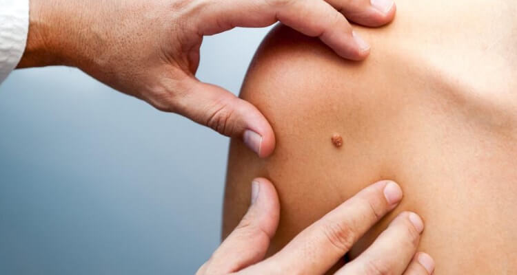
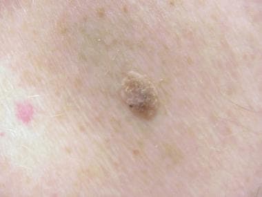
BACKGROUND: Malignant melanoma, the most deadly form of skin cancer, has a good prognosis if treated in the curable early stages. Colour provides critical discriminating information for the diagnosis of malignant melanoma. METHODS: This research introduces a three-dimensional relative colour histogram analysis technique to identify colours characteristic of melanomas and then applies these 'melanoma colours' to differentiate benign skin lesions from melanomas. The relative colour of a skin lesion is determined based on subtracting a representative colour of the surrounding skin from each lesion pixel. A colour mapping for 'melanoma colours' is determined using a training set of images. A percent melanoma colour feature, defined as the percentage of the lesion pixels that are melanoma colours, is used for discriminating melanomas from benign lesions. The technique is evaluated using a clinical image data set of 129 malignant melanomas and 129 benign lesions consisting of 40 seborrheic keratoses and 89 nevocellular nevi. RESULTS: Using the percent melanoma colour feature for discrimination, experimental results yield correct melanoma and benign lesion discrimination rates of 84.3 and 83.0%, respectively. CONCLUSIONS: The results presented in this work suggest that lesion colour in clinical images is strongly related to the presence of melanoma in that lesion. However, colour information should be combined with other information in order to further reduce the false negative and false positive rates. (+info)Seborrheic Keratosis is a common, benign skin condition that typically presents as rough, scaly, tan-to-darkly pigmented growths on the surface of the skin. These lesions can appear anywhere on the body, but they are most commonly found on the face, chest, back, and extremities. Seborrheic Keratoses are caused by an overproduction of keratin, a protein that makes up the outer layer of the skin.
The exact cause of Seborrheic Keratosis is not known, but it is thought to be related to genetic factors and sun exposure. The condition is more common in older adults and is not contagious. While Seborrheic Keratoses are generally harmless, they can be removed for cosmetic reasons or if they become irritated or inflamed. Treatment options include cryotherapy (freezing the lesions with liquid nitrogen), curettage (scraping the lesions off), and laser surgery.
Seborrheic dermatitis is a common, inflammatory skin condition that mainly affects the scalp, face, and upper part of the body. It causes skin irritation, flaking, and redness, often in areas where the skin is oily or greasy. The exact cause of seborrheic dermatitis is not fully understood, but it appears to be related to a combination of genetic, environmental, and microbial factors.
The symptoms of seborrheic dermatitis can vary in severity and may include:
* Greasy or flaky scales on the scalp, eyebrows, eyelashes, ears, or beard
* Redness and inflammation of the skin
* Itching, burning, or stinging sensations
* Yellow or white crusty patches on the scalp or other affected areas
* Hair loss (in severe cases)
Seborrheic dermatitis is a chronic condition that tends to flare up and then subside over time. While there is no cure for seborrheic dermatitis, various treatments can help manage the symptoms and prevent complications. These may include medicated shampoos, topical creams or ointments, and lifestyle changes such as stress reduction and avoiding triggers that worsen symptoms.
It is important to note that seborrheic dermatitis should not be confused with other skin conditions, such as psoriasis or eczema, which may have similar symptoms. A healthcare professional can provide a proper diagnosis and recommend appropriate treatment options based on the individual's specific needs.
Keratosis, in general, refers to a skin condition characterized by the abnormal growth or development of keratin, a protein that forms part of the outer layer of the skin (epidermis). There are several types of keratosis, including:
1. Seborrheic Keratosis: benign, often pigmented, rough, and scaly growths that can appear anywhere on the body. They tend to increase in number with age.
2. Actinic Keratosis: rough, scaly patches or spots on the skin that are caused by long-term exposure to sunlight or artificial UV light. These have the potential to develop into squamous cell carcinoma, a type of skin cancer.
3. Solar Keratosis: another term for actinic keratosis, as it is primarily caused by sun damage.
4. Keratosis Pilaris: a common condition where small, rough bumps appear on the skin, often on the arms, thighs, or cheeks. These are caused by excess keratin blocking hair follicles.
5. Follicular Keratosis: a disorder characterized by the formation of horny plugs within the hair follicles, leading to rough, sandpaper-like bumps on the skin.
6. Intraepidermal Keratosis: a term used to describe the abnormal accumulation of keratin in the epidermis, which can lead to various skin conditions.
It's important to consult with a healthcare professional or dermatologist for proper diagnosis and treatment if you suspect having any form of keratosis.
Actinic keratosis, also known as solar keratosis, is a precancerous skin condition that typically develops in areas exposed to excessive sun damage over the years. It presents as rough, scaly, or crusty patches of skin, often with a pink, red, or brownish tint. These lesions usually appear on the face, ears, scalp, neck, back of the hands, and forearms.
Actinic keratosis is caused by the prolonged exposure to ultraviolet (UV) radiation from sunlight or artificial sources like tanning beds. The UV rays damage the skin's DNA, leading to abnormal skin cell growth and the formation of these precancerous lesions.
While most actinic keratoses remain benign, a small percentage can progress into squamous cell carcinoma, a type of skin cancer. Therefore, it is essential to have any suspicious or changing lesions evaluated by a healthcare professional for proper diagnosis and treatment. Prevention measures include protecting the skin from excessive sun exposure, wearing protective clothing, using broad-spectrum sunscreen with an SPF of at least 30, and avoiding tanning beds.
Malassezia is a genus of fungi (specifically, yeasts) that are commonly found on the skin surfaces of humans and other animals. They are part of the normal flora of the skin, but under certain conditions, they can cause various skin disorders such as dandruff, seborrheic dermatitis, pityriasis versicolor, and atopic dermatitis.
Malassezia species require lipids for growth, and they are able to break down the lipids present in human sebum into fatty acids, which can cause irritation and inflammation of the skin. Malassezia is also associated with fungal infections in people with weakened immune systems.
The genus Malassezia includes several species, such as M. furfur, M. globosa, M. restricta, M. sympodialis, and others. These species can be identified using various laboratory methods, including microscopy, culture, and molecular techniques.
Facial dermatoses refer to various skin conditions that affect the face. These can include a wide range of disorders, such as:
1. Acne vulgaris: A common skin condition characterized by the formation of comedones (blackheads and whiteheads) and inflammatory papules, pustules, and nodules. It primarily affects the face, neck, chest, and back.
2. Rosacea: A chronic skin condition that causes redness, flushing, and visible blood vessels on the face, along with bumps or pimples and sometimes eye irritation.
3. Seborrheic dermatitis: A common inflammatory skin disorder that causes a red, itchy, and flaky rash, often on the scalp, face, and eyebrows. It can also affect other oily areas of the body, like the sides of the nose and behind the ears.
4. Atopic dermatitis (eczema): A chronic inflammatory skin condition that causes red, itchy, and scaly patches on the skin. While it can occur anywhere on the body, it frequently affects the face, especially in infants and young children.
5. Psoriasis: An autoimmune disorder that results in thick, scaly, silvery, or red patches on the skin. It can affect any part of the body, including the face.
6. Contact dermatitis: A skin reaction caused by direct contact with an allergen or irritant, resulting in redness, itching, and inflammation. The face can be affected when allergens or irritants come into contact with the skin through cosmetics, skincare products, or other substances.
7. Lupus erythematosus: An autoimmune disorder that can cause a butterfly-shaped rash on the cheeks and nose, along with other symptoms like joint pain, fatigue, and photosensitivity.
8. Perioral dermatitis: A inflammatory skin condition that causes redness, small bumps, and dryness around the mouth, often mistaken for acne. It can also affect the skin around the nose and eyes.
9. Vitiligo: An autoimmune disorder that results in the loss of pigmentation in patches of skin, which can occur on the face and other parts of the body.
10. Tinea faciei: A fungal infection that affects the facial skin, causing red, scaly, or itchy patches. It is also known as ringworm of the face.
These are just a few examples of skin conditions that can affect the face. If you experience any unusual symptoms or changes in your skin, it's essential to consult a dermatologist for proper diagnosis and treatment.
Scalp dermatoses refer to various skin conditions that affect the scalp. These can include inflammatory conditions such as seborrheic dermatitis (dandruff, cradle cap), psoriasis, atopic dermatitis (eczema), and lichen planus; infectious processes like bacterial folliculitis, tinea capitis (ringworm of the scalp), and viral infections; as well as autoimmune conditions such as alopecia areata. Symptoms can range from mild scaling and itching to severe redness, pain, and hair loss. The specific diagnosis and treatment of scalp dermatoses depend on the underlying cause.
Darier Disease is a genetic skin disorder, also known as Keratosis Follicularis. It is characterized by the formation of greasy, crusted, keratotic papules and plaques that typically appear on the upper arms, torso, and scalp. The lesions may also affect the nasolabial folds, central face, and mucous membranes. Darier Disease is caused by mutations in the ATP2A2 gene, which encodes a calcium pump protein involved in keratinization. It is an autosomal dominant disorder, meaning that a person has a 50% chance of inheriting the disease if one of their parents is affected. The onset of symptoms typically occurs during adolescence or early adulthood. Treatment options include topical medications, oral retinoids, and photodynamic therapy.
A skin cream is not a medical term per se, but it generally refers to a topical emollient preparation intended for application to the skin. It contains a mixture of water, oil, and active ingredients, which are formulated to provide various benefits such as moisturizing, protecting, soothing, or treating specific skin conditions. The exact definition and composition may vary depending on the product's intended use and formulation.
Examples of active ingredients in skin creams include:
1. Moisturizers (e.g., glycerin, hyaluronic acid) - help to retain water in the skin, making it feel softer and smoother.
2. Emollients (e.g., shea butter, coconut oil, petrolatum) - provide a protective barrier that helps prevent moisture loss and soften the skin.
3. Humectants (e.g., urea, lactic acid, alpha-hydroxy acids) - attract water from the environment or deeper layers of the skin to hydrate the surface.
4. Anti-inflammatory agents (e.g., hydrocortisone, aloe vera) - help reduce redness, swelling, and itching associated with various skin conditions.
5. Antioxidants (e.g., vitamin C, vitamin E, green tea extract) - protect the skin from free radical damage and environmental stressors that can lead to premature aging.
6. Sunscreen agents (e.g., zinc oxide, titanium dioxide, chemical filters) - provide broad-spectrum protection against UVA and UVB rays.
7. Skin lighteners (e.g., hydroquinone, kojic acid, arbutin) - help reduce the appearance of hyperpigmentation and even out skin tone.
8. Acne treatments (e.g., benzoyl peroxide, salicylic acid, retinoids) - target acne-causing bacteria, unclog pores, and regulate cell turnover to prevent breakouts.
It is essential to choose a skin cream based on your specific skin type and concerns, as well as any medical conditions or allergies you may have. Always consult with a dermatologist or healthcare provider before starting a new skincare regimen.
Dermatomycoses are a group of fungal infections that affect the skin, hair, and nails. These infections are caused by various types of fungi, including dermatophytes, yeasts, and molds. Dermatophyte infections, also known as tinea, are the most common type of dermatomycoses and can affect different areas of the body, such as the scalp (tinea capitis), beard (tinea barbae), body (tinea corporis), feet (tinea pedis or athlete's foot), hands (tinea manuum), and nails (tinea unguium or onychomycosis). Yeast infections, such as those caused by Candida albicans, can lead to conditions like candidal intertrigo, vulvovaginitis, and balanitis. Mold infections are less common but can cause skin disorders like scalded skin syndrome and phaeohyphomycosis. Dermatomycoses are typically treated with topical or oral antifungal medications.
Skin neoplasms refer to abnormal growths or tumors in the skin that can be benign (non-cancerous) or malignant (cancerous). They result from uncontrolled multiplication of skin cells, which can form various types of lesions. These growths may appear as lumps, bumps, sores, patches, or discolored areas on the skin.
Benign skin neoplasms include conditions such as moles, warts, and seborrheic keratoses, while malignant skin neoplasms are primarily classified into melanoma, squamous cell carcinoma, and basal cell carcinoma. These three types of cancerous skin growths are collectively known as non-melanoma skin cancers (NMSCs). Melanoma is the most aggressive and dangerous form of skin cancer, while NMSCs tend to be less invasive but more common.
It's essential to monitor any changes in existing skin lesions or the appearance of new growths and consult a healthcare professional for proper evaluation and treatment if needed.
Sebum is an oily, waxy substance that is produced by the sebaceous glands in the skin of mammals. It is composed mainly of triglycerides, wax esters, squalene, and free fatty acids, as well as smaller amounts of metabolites and other substances. Sebum plays an important role in the maintenance of the skin's barrier function and in the regulation of its moisture levels. It also has antimicrobial properties that help to protect the skin from infection. Excessive sebum production can contribute to the development of acne and other skin conditions.
Keratolytic agents are substances that cause the softening and sloughing off of excess keratin, the protein that makes up the outermost layer of the skin (stratum corneum). These agents help to break down and remove dead skin cells, increase moisture retention, and promote the growth of new skin cells. They are commonly used in the treatment of various dermatological conditions such as psoriasis, eczema, warts, calluses, and ichthyosis. Examples of keratolytic agents include salicylic acid, urea, lactic acid, and retinoic acid.
Dermoscopy, also known as dermatoscopy or epiluminescence microscopy, is a non-invasive diagnostic technique used in dermatology to evaluate skin lesions, such as moles and pigmented skin tumors. This method involves the use of a handheld device called a dermoscope, which consists of a magnifying lens, a light source, and a transparent plate or immersion fluid that allows for better visualization of the skin's surface structures.
Dermoscopy enables dermatologists to examine the pigmented patterns, vascular structures, and other morphological features hidden beneath the skin's surface that are not visible to the naked eye. By observing these details, dermatologists can improve their ability to differentiate between benign and malignant lesions, leading to more accurate diagnoses and appropriate treatment decisions.
The primary uses of dermoscopy include:
1. Early detection and diagnosis of melanoma and other skin cancers, such as basal cell carcinoma and squamous cell carcinoma.
2. Monitoring the evolution of suspicious moles or lesions over time.
3. Assisting in the identification of various benign skin growths, like seborrheic keratoses, dermatofibromas, and nevi (moles).
4. Improving the diagnostic accuracy for infectious skin conditions, inflammatory processes, and other dermatological disorders.
Overall, dermoscopy is a valuable tool in the field of dermatology that enhances the clinician's ability to diagnose and manage various skin conditions accurately and effectively.
Hair preparations refer to cosmetic or grooming products that are specifically formulated to be applied to the hair or scalp for various purposes such as cleansing, conditioning, styling, coloring, or promoting hair growth. These preparations can come in different forms, including shampoos, conditioners, hair masks, serums, gels, mousses, sprays, and dyes. They may contain a wide range of ingredients, such as detergents, moisturizers, proteins, vitamins, minerals, and other nutrients that can help improve the health, appearance, and manageability of the hair. Some hair preparations may also contain medications or natural extracts that have therapeutic properties for treating specific hair or scalp conditions, such as dandruff, dryness, oiliness, thinning, or hair loss.
Parakeratosis is a medical term that refers to a skin condition where the outermost layer of the skin (the stratum corneum) contains nucleated keratinocytes, which are cells that have not fully matured and still contain their nuclei. This is in contrast to normal stratum corneum, which consists of flat, dead keratinocytes without nuclei.
Parakeratosis can occur in various skin disorders, such as psoriasis, eczema, warts, and certain types of dermatitis. It can also be seen in some benign or malignant skin tumors. The presence of parakeratosis may indicate abnormal differentiation or proliferation of the skin cells, which can contribute to the development of skin lesions or diseases.
In addition to its role in skin disorders, parakeratosis has been implicated in the pathogenesis of certain gastrointestinal diseases, such as Barrett's esophagus and colon cancer, where it is associated with abnormal cell growth and increased risk of malignancy.
Conjunctival diseases refer to a group of medical conditions that affect the conjunctiva, which is the thin, clear mucous membrane that covers the inner surface of the eyelids and the white part of the eye (known as the sclera). The conjunctiva helps to keep the eye moist and protected from irritants.
Conjunctival diseases can cause a range of symptoms, including redness, itching, burning, discharge, grittiness, and pain. Some common conjunctival diseases include:
1. Conjunctivitis (pink eye): This is an inflammation or infection of the conjunctiva that can be caused by viruses, bacteria, or allergies. Symptoms may include redness, itching, discharge, and watery eyes.
2. Pinguecula: This is a yellowish, raised bump that forms on the conjunctiva, usually near the corner of the eye. It is caused by an overgrowth of connective tissue and may be related to sun exposure or dry eye.
3. Pterygium: This is a fleshy growth that extends from the conjunctiva onto the cornea (the clear front part of the eye). It can cause redness, irritation, and vision problems if it grows large enough to cover the pupil.
4. Allergic conjunctivitis: This is an inflammation of the conjunctiva caused by an allergic reaction to substances such as pollen, dust mites, or pet dander. Symptoms may include redness, itching, watery eyes, and swelling.
5. Chemical conjunctivitis: This is an irritation or inflammation of the conjunctiva caused by exposure to chemicals such as chlorine, smoke, or fumes. Symptoms may include redness, burning, and tearing.
6. Giant papillary conjunctivitis (GPC): This is a type of allergic reaction that occurs in response to the presence of a foreign body in the eye, such as a contact lens. Symptoms may include itching, mucus discharge, and a gritty feeling in the eye.
Treatment for conjunctival diseases depends on the underlying cause. In some cases, over-the-counter medications or home remedies may be sufficient to relieve symptoms. However, more severe cases may require prescription medication or medical intervention. It is important to consult with a healthcare provider if you experience persistent or worsening symptoms of conjunctival disease.
Skin diseases, also known as dermatological conditions, refer to any medical condition that affects the skin, which is the largest organ of the human body. These diseases can affect the skin's function, appearance, or overall health. They can be caused by various factors, including genetics, infections, allergies, environmental factors, and aging.
Skin diseases can present in many different forms, such as rashes, blisters, sores, discolorations, growths, or changes in texture. Some common examples of skin diseases include acne, eczema, psoriasis, dermatitis, fungal infections, viral infections, bacterial infections, and skin cancer.
The symptoms and severity of skin diseases can vary widely depending on the specific condition and individual factors. Some skin diseases are mild and can be treated with over-the-counter medications or topical creams, while others may require more intensive treatments such as prescription medications, light therapy, or even surgery.
It is important to seek medical attention if you experience any unusual or persistent changes in your skin, as some skin diseases can be serious or indicative of other underlying health conditions. A dermatologist is a medical doctor who specializes in the diagnosis and treatment of skin diseases.
Bowen's disease is a skin condition that is characterized by the growth of abnormal cells on the outermost layer of the skin (the epidermis). It is also known as squamous cell carcinoma in situ. The affected area often appears as a red, scaly patch or plaque, and it can develop anywhere on the body, but it is most commonly found on sun-exposed areas such as the face, hands, arms, and legs.
Bowen's disease is considered a precancerous condition because there is a risk that the abnormal cells could eventually develop into invasive squamous cell carcinoma, a type of skin cancer. However, not all cases of Bowen's disease will progress to cancer, and some may remain stable or even regress on their own.
The exact cause of Bowen's disease is not known, but it is thought to be associated with exposure to certain chemicals, radiation, and human papillomavirus (HPV) infection. Treatment options for Bowen's disease include cryotherapy, topical chemotherapy, photodynamic therapy, curettage and electrodessication, and surgical excision. Regular follow-up with a healthcare provider is recommended to monitor the condition and ensure that it does not progress to cancer.
Keratoderma, palmoplantar is a medical term that refers to a group of skin conditions characterized by thickening and hardening (hyperkeratosis) of the skin on the palms of the hands and soles of the feet. This condition can affect people of all ages, but it's most commonly seen in children.
The thickening of the skin is caused by an overproduction of keratin, a protein that helps to form the tough, outer layer of the skin. In palmoplantar keratoderma, this excess keratin accumulates in the stratum corneum, the outermost layer of the epidermis, leading to the formation of rough, scaly, and thickened patches on the palms and soles.
There are several different types of palmoplantar keratoderma, each with its own specific symptoms and causes. Some forms of the condition are inherited and present at birth or develop in early childhood, while others may be acquired later in life as a result of an underlying medical condition, such as atopic dermatitis, lichen planus, or psoriasis.
Treatment for palmoplantar keratoderma typically involves the use of emollients and keratolytic agents to help soften and remove the thickened skin. In some cases, oral retinoids or other systemic medications may be necessary to manage more severe symptoms. It's important to consult with a healthcare provider for an accurate diagnosis and treatment plan.
Tinea versicolor is a superficial fungal infection of the skin, caused by the pathogen Malassezia furfur (previously known as Pityrosporum ovale). It is characterized by the appearance of multiple round or oval patches that are hypopigmented (lighter than the surrounding skin) or hyperpigmented (darker than the surrounding skin), scaly, and can be pruritic (itchy). The lesions typically appear on the trunk and proximal extremities, often in a symmetrical pattern. Tinea versicolor is more common in warm, humid climates and in individuals with oily skin or weakened immune systems. It is usually diagnosed based on the clinical presentation and can be confirmed by microscopic examination of skin scrapings or fungal cultures. Treatment typically involves topical antifungal medications, such as clotrimazole, miconazole, or selenium sulfide, but oral medication may be necessary for severe or widespread infections.
Keratoacanthoma is a rapidly growing, dome-shaped, skin tumor that typically arises on sun-exposed areas such as the face, arms, and legs. It is considered a low-grade squamous cell carcinoma (a type of skin cancer) because it shares some characteristics with both benign and malignant tumors.
Keratoacanthomas usually develop over a period of several weeks to months, growing rapidly in size before eventually stabilizing and then gradually regressing on their own within a few months to a year. However, the regression process can take years, and some lesions may not regress completely, leading to cosmetic concerns or even local invasion.
Histologically, keratoacanthomas are characterized by a central keratin-filled crater surrounded by a well-differentiated layer of squamous epithelial cells. The tumor's growth pattern and histological features can make it difficult to distinguish from other types of skin cancer, such as squamous cell carcinoma.
Treatment options for keratoacanthomas include surgical excision, cryosurgery, curettage and electrodesiccation, and topical therapies like imiquimod or 5-fluorouracil. The choice of treatment depends on various factors such as the size, location, and number of lesions, as well as patient preferences and overall health status.
Photosensitivity disorders refer to conditions that cause an abnormal reaction to sunlight or artificial light. This reaction can take the form of various skin changes, such as rashes, inflammation, or pigmentation, and in some cases, it can also lead to systemic symptoms like fatigue, fever, or joint pain.
The two main types of photosensitivity disorders are:
1. Phototoxic reactions: These occur when a substance (such as certain medications, chemicals, or plants) absorbs light energy and transfers it to skin cells, causing damage and inflammation. The reaction typically appears within 24 hours of exposure to the light source and can resemble a sunburn.
2. Photoallergic reactions: These occur when the immune system responds to the combination of light and a particular substance, leading to an allergic response. The reaction may not appear until several days after initial exposure and can cause redness, itching, and blistering.
It is important for individuals with photosensitivity disorders to avoid excessive sun exposure, wear protective clothing, and use broad-spectrum sunscreens with a high SPF rating to minimize the risk of phototoxic or photoallergic reactions.
The eyebrows are a set of hairs that grow above the eyes on the forehead. They are an important feature of human facial anatomy, and play several roles in non-verbal communication and self-expression. Eyebrows help to prevent sweat and other moisture from dripping into the eyes, and also serve as a protective barrier against dirt, dust, and other foreign particles that might otherwise irritate or damage the eyes.
In addition, eyebrows play an important role in human social interaction and communication. They can convey a range of emotions and facial expressions, such as surprise, anger, fear, happiness, and sadness. Eyebrows can also help to frame the eyes and enhance their appearance, making them an important aspect of personal grooming and beauty.
The eyebrows are made up of several components, including hair follicles, sebaceous glands, and muscles that control their movement. The hairs themselves are composed of a protein called keratin, which also makes up the hair on the head, as well as nails and skin. The color and thickness of eyebrow hair can vary widely from person to person, and may be influenced by factors such as age, genetics, and hormonal changes.
In medical terms, changes in the appearance or condition of the eyebrows can sometimes be a sign of underlying health issues. For example, thinning or loss of eyebrows can be associated with conditions such as alopecia, thyroid disorders, or nutritional deficiencies. Changes in eyebrow shape or position can also be a symptom of certain neurological conditions, such as Bell's palsy or stroke. As such, any significant changes in the appearance or condition of the eyebrows should be evaluated by a healthcare professional to rule out any underlying medical causes.
Warts are small, rough growths on the skin or mucous membranes caused by one of several types of human papillomavirus (HPV). They can appear anywhere on the body but most often occur on the hands, fingers, and feet. Warts are benign, non-cancerous growths, but they can be unsightly, uncomfortable, or painful, depending on their location and size.
Warts are caused by HPV infecting the top layer of skin, usually through a small cut or scratch. The virus triggers an overproduction of keratin, a protein in the skin, leading to the formation of a hard, rough growth. Warts can vary in appearance depending on their location and type, but they are generally round or irregularly shaped, with a rough surface that may be flat or slightly raised. They may also contain small black dots, which are actually tiny blood vessels that have clotted.
Warts are contagious and can spread from person to person through direct skin-to-skin contact or by sharing personal items such as towels or razors. They can also be spread by touching a wart and then touching another part of the body. Warts may take several months to develop after exposure to HPV, so it may not always be clear when or how they were contracted.
There are several types of warts, including common warts, plantar warts (which occur on the soles of the feet), flat warts (which are smaller and smoother than other types of warts), and genital warts (which are sexually transmitted). While most warts are harmless and will eventually go away on their own, some may require medical treatment if they are causing discomfort or are unsightly. Treatment options for warts include topical medications, cryotherapy (freezing the wart with liquid nitrogen), and surgical removal.
The scalp is the anatomical region located at the upper part of the human head, covering the skull except for the face and the ears. It is made up of several layers: the skin, the connective tissue, the galea aponeurotica (a strong, flat, tendinous sheet), loose areolar tissue, and the periosteum (the highly vascularized innermost layer that attaches directly to the skull bones). The scalp has a rich blood supply and is home to numerous sensory receptors, including those for touch, pain, and temperature. It also contains hair follicles, sebaceous glands, and sweat glands.
Blepharitis is a common inflammatory condition that affects the eyelids, specifically the eyelash follicles and the edges of the eyelids (called the "eyelid margins"). It can cause symptoms such as redness, swelling, itching, burning, and a crusty or flaky buildup on the lashes. Blepharitis can be caused by a variety of factors, including bacterial infection, skin disorders like seborrheic dermatitis or rosacea, and meibomian gland dysfunction. It is often a chronic condition that requires ongoing treatment to manage symptoms and prevent recurrence.
Carcinoma, basal cell is a type of skin cancer that arises from the basal cells, which are located in the lower part of the epidermis (the outermost layer of the skin). It is also known as basal cell carcinoma (BCC) and is the most common form of skin cancer.
BCC typically appears as a small, shiny, pearly bump or nodule on the skin, often in sun-exposed areas such as the face, ears, neck, hands, and arms. It may also appear as a scar-like area that is white, yellow, or waxy. BCCs are usually slow growing and rarely spread (metastasize) to other parts of the body. However, they can be locally invasive and destroy surrounding tissue if left untreated.
The exact cause of BCC is not known, but it is thought to be related to a combination of genetic and environmental factors, including exposure to ultraviolet (UV) radiation from the sun or tanning beds. People with fair skin, light hair, and blue or green eyes are at increased risk of developing BCC.
Treatment for BCC typically involves surgical removal of the tumor, along with a margin of healthy tissue. Other treatment options may include radiation therapy, topical chemotherapy, or photodynamic therapy. Prevention measures include protecting your skin from UV radiation by wearing protective clothing, using sunscreen, and avoiding tanning beds.
Lichenoid eruptions are skin reactions that resemble the appearance of lichen, a type of slow-growing fungus. These eruptions are characterized by flat, scaly bumps (papules) and rough, discolored patches (plaques) on the skin. They can be caused by various factors, including medications, medical conditions, or as a reaction to certain chemicals or substances that come into contact with the skin.
The term "lichenoid" refers to the resemblance of these eruptions to lichen, which is characterized by its distinctive appearance and growth pattern. Lichenoid eruptions can occur anywhere on the body but are most commonly found on sun-exposed areas such as the arms, legs, and trunk.
The exact cause of lichenoid eruptions can vary, but they are often associated with an autoimmune response in which the body's immune system mistakenly attacks healthy skin cells. This can lead to inflammation, redness, itching, and other symptoms associated with these eruptions. Treatment for lichenoid eruptions typically involves identifying and addressing the underlying cause, as well as managing symptoms with topical medications or other therapies.
A nevus, also known as a mole, is a benign growth or mark on the skin that is usually brown or black. It can be raised or flat and can appear anywhere on the body. Nevi are made up of cells called melanocytes, which produce the pigment melanin. Most nevi develop in childhood or adolescence, but they can also appear later in life. Some people have many nevi, while others have few or none.
There are several types of nevi, including:
* Common nevi: These are the most common type of mole and are usually small, round, and brown or black. They can be flat or raised and can appear anywhere on the body.
* Atypical nevi: These moles are larger than common nevi and have irregular borders and color. They may be flat or raised and can appear anywhere on the body, but are most commonly found on the trunk and extremities. Atypical nevi are more likely to develop into melanoma, a type of skin cancer, than common nevi.
* Congenital nevi: These moles are present at birth and can vary in size from small to large. They are more likely to develop into melanoma than moles that develop later in life.
* Spitz nevi: These are rare, benign growths that typically appear in children and adolescents. They are usually pink or red and dome-shaped.
It is important to monitor nevi for changes in size, shape, color, and texture, as these can be signs of melanoma. If you notice any changes in a mole, or if you have a new mole that is unusual or bleeding, it is important to see a healthcare provider for further evaluation.
Lichen Planus is a chronic, autoimmune skin condition that can also affect the mucous membranes inside the mouth, genitals, and eyes. It is characterized by the appearance of purplish, flat-topped bumps or lesions on the skin, which may be itchy. The exact cause of Lichen Planus is unknown, but it is believed to occur when the immune system mistakenly attacks cells in the skin or mucous membranes. Certain medications, viral infections, and genetic factors may increase the risk of developing this condition. Treatment typically focuses on managing symptoms and may include topical corticosteroids, oral medications, or light therapy.
Fibroblast Growth Factor Receptor 3 (FGFR3) is a type of cell surface receptor that binds to fibroblast growth factors (FGFs), which are signaling proteins involved in various biological processes such as cell division, growth, and wound healing.
FGFR3 is a transmembrane protein with an extracellular domain that contains the binding site for FGFs, a transmembrane domain, and an intracellular tyrosine kinase domain that activates downstream signaling pathways upon FGF binding.
Mutations in the FGFR3 gene have been associated with several human genetic disorders, including thanatophoric dysplasia, achondroplasia, and hypochondroplasia, which are characterized by abnormal bone growth and development. In these conditions, gain-of-function mutations in FGFR3 lead to increased receptor activity and activation of downstream signaling pathways, resulting in impaired endochondral ossification and short-limbed dwarfism.
In addition to its role in bone growth and development, FGFR3 has been implicated in the regulation of cell proliferation, differentiation, and survival in various tissues, including the brain, lung, and kidney. Dysregulation of FGFR3 signaling has also been associated with cancer, including bladder, breast, and cervical cancers.
Acoustic microscopy is a non-invasive imaging technique that uses sound waves to visualize and analyze the structure and properties of various materials, including biological samples. In the context of medical diagnostics and research, acoustic microscopy can be used to examine tissues, cells, and cellular components with high resolution, providing valuable information about their mechanical and physical properties.
In acoustic microscopy, high-frequency sound waves are focused onto a sample using a transducer. The interaction between the sound waves and the sample generates echoes, which contain information about the sample's internal structure and properties. These echoes are then recorded and processed to create an image of the sample.
Acoustic microscopy offers several advantages over other imaging techniques, such as optical microscopy or electron microscopy. For example, it does not require staining or labeling of samples, which can be time-consuming and potentially damaging. Additionally, acoustic microscopy can provide high-resolution images of samples in their native state, allowing researchers to study the effects of various treatments or interventions on living cells and tissues.
In summary, acoustic microscopy is a non-invasive imaging technique that uses sound waves to visualize and analyze the structure and properties of biological samples with high resolution, providing valuable information for medical diagnostics and research.
Cytoplasmic structures refer to the various organelles and inclusions present within the cytoplasm of a eukaryotic cell, excluding the nucleus. These structures are involved in different cellular functions, such as energy production, protein synthesis, waste management, and intracellular transport.
Some examples of cytoplasmic structures include:
1. Mitochondria - organelles that generate energy for the cell through cellular respiration.
2. Ribosomes - complexes composed of ribosomal RNA (rRNA) and proteins that facilitate protein synthesis.
3. Endoplasmic reticulum (ER) - a network of membranous tubules involved in lipid and protein synthesis, folding, and transport.
4. Golgi apparatus - a series of stacked membrane sacs responsible for modifying, sorting, and packaging proteins and lipids for transport to their destinations.
5. Lysosomes - membrane-bound organelles that contain enzymes for breaking down waste materials, cellular debris, and foreign substances.
6. Peroxisomes - single-membrane bound organelles involved in various metabolic processes, including the breakdown of fatty acids and hydrogen peroxide detoxification.
7. Vacuoles - membrane-bound compartments that store water, nutrients, waste products, or enzymes. In plant cells, vacuoles also help maintain turgor pressure.
8. Cytoskeleton - a network of protein filaments (actin microfilaments, intermediate filaments, and microtubules) responsible for maintaining cell shape, providing structural support, and enabling intracellular transport and movement.
9. Inclusions - various membrane-less structures composed of aggregated proteins or other molecules, such as lipid droplets, glycogen granules, and pigment granules (e.g., melanosomes in melanocytes).
These cytoplasmic structures contribute to the overall functioning and maintenance of a eukaryotic cell.
Sebaceous glands are microscopic, exocrine glands that are found in the dermis of mammalian skin. They are attached to hair follicles and produce an oily substance called sebum, which is composed of triglycerides, wax esters, squalene, and metabolites of fat-producing cells (fatty acids, cholesterol). Sebum is released through a duct onto the surface of the skin, where it forms a protective barrier that helps to prevent water loss, keeps the skin and hair moisturized, and has antibacterial properties.
Sebaceous glands are distributed throughout the body, but they are most numerous on the face, scalp, and upper trunk. They can also be found in other areas of the body such as the eyelids (where they are known as meibomian glands), the external ear canal, and the genital area.
Abnormalities in sebaceous gland function can lead to various skin conditions, including acne, seborrheic dermatitis, and certain types of skin cancer.
Pityriasis is a general term used to describe a group of skin conditions characterized by scaling. It includes several specific types, the most common being Pityriasis rosea and Pityriasis simplex capillitii (also known as dandruff).
1. Pityriasis rosea: This is a temporary skin rash that often begins with a single, round, scaly patch on the chest, abdomen, or back. A few days to weeks later, more patches appear. These patches are oval and scaly, and they may be pink, red, or tan. The rash usually lasts about 6-8 weeks.
2. Pityriasis simplex capillitii: This is a very common condition characterized by flaking or scaling of the scalp, which is often referred to as dandruff.
The term "pityriasis" comes from the Greek word "pitýrios," which means "bran."
In medical terms, the skin is the largest organ of the human body. It consists of two main layers: the epidermis (outer layer) and dermis (inner layer), as well as accessory structures like hair follicles, sweat glands, and oil glands. The skin plays a crucial role in protecting us from external factors such as bacteria, viruses, and environmental hazards, while also regulating body temperature and enabling the sense of touch.
Ointment bases refer to the vehicle or foundation in which active pharmaceutical ingredients are dispersed to form a semi-solid medication. These bases provide the necessary consistency for ointments, allowing easy application to the skin or other body surfaces. They can be composed of various materials such as fats, waxes, oils, and emulsifying agents.
The choice of an ointment base depends on several factors, including:
1. The desired physical properties (e.g., spreadability, absorption rate)
2. The route of administration (e.g., dermal, mucosal)
3. The compatibility with the active ingredient(s)
4. The intended therapeutic effect (e.g., occlusive, non-occlusive)
Some common types of ointment bases include:
1. Hydrocarbon bases: Consist of hydrophobic materials like petrolatum, white soft paraffin, and microcrystalline wax. They are generally inert, odorless, and resistant to oxidation.
2. Absorption bases: Contain a mixture of hydrocarbons and higher molecular weight esters or fatty alcohols. These bases have better penetrating properties than hydrocarbon bases and are suitable for drugs with low oil solubility.
3. Emulsifying bases: Comprise of water-in-oil (W/O) or oil-in-water (O/W) emulsions, which allow the dispersion of both hydrophilic and lipophilic drugs. Common examples include cetomacrogol and anhydrous lanette.
4. Water-soluble bases: Primarily consist of polyethylene glycols (PEGs) or other water-soluble materials. They are useful for drugs with high water solubility and provide a cooling sensation upon application.
It is essential to select an appropriate ointment base to ensure the optimal delivery, stability, and efficacy of the active ingredient(s).
Topical administration refers to a route of administering a medication or treatment directly to a specific area of the body, such as the skin, mucous membranes, or eyes. This method allows the drug to be applied directly to the site where it is needed, which can increase its effectiveness and reduce potential side effects compared to systemic administration (taking the medication by mouth or injecting it into a vein or muscle).
Topical medications come in various forms, including creams, ointments, gels, lotions, solutions, sprays, and patches. They may be used to treat localized conditions such as skin infections, rashes, inflammation, or pain, or to deliver medication to the eyes or mucous membranes for local or systemic effects.
When applying topical medications, it is important to follow the instructions carefully to ensure proper absorption and avoid irritation or other adverse reactions. This may include cleaning the area before application, covering the treated area with a dressing, or avoiding exposure to sunlight or water after application, depending on the specific medication and its intended use.
Leukoplakia is a medical term used to describe a white or gray patch that develops on the mucous membranes lining the inside of the mouth. These patches are typically caused by excessive cell growth and cannot be easily scraped off. Leukoplakia is often associated with long-term tobacco use, including smoking and chewing tobacco, as well as alcohol consumption. While most cases of leukoplakia are benign, a small percentage can develop into oral cancer, so it's essential to have any suspicious patches evaluated by a healthcare professional.
Squamous cell carcinoma is a type of skin cancer that begins in the squamous cells, which are flat, thin cells that form the outer layer of the skin (epidermis). It commonly occurs on sun-exposed areas such as the face, ears, lips, and backs of the hands. Squamous cell carcinoma can also develop in other areas of the body including the mouth, lungs, and cervix.
This type of cancer usually develops slowly and may appear as a rough or scaly patch of skin, a red, firm nodule, or a sore or ulcer that doesn't heal. While squamous cell carcinoma is not as aggressive as some other types of cancer, it can metastasize (spread) to other parts of the body if left untreated, making early detection and treatment important.
Risk factors for developing squamous cell carcinoma include prolonged exposure to ultraviolet (UV) radiation from the sun or tanning beds, fair skin, a history of sunburns, a weakened immune system, and older age. Prevention measures include protecting your skin from the sun by wearing protective clothing, using a broad-spectrum sunscreen with an SPF of at least 30, avoiding tanning beds, and getting regular skin examinations.
I'm sorry for any confusion, but "sunlight" is not a term with a specific medical definition. However, sunlight does have various implications in the field of medicine. It is the light that comes from the sun, which is a star at the center of our solar system. Sunlight is essential for the production of vitamin D in humans, and it can also have effects on mood and sleep patterns due to its influence on circadian rhythms.
In a medical context, sunlight is often discussed in relation to its potential health benefits and risks. For instance, moderate sun exposure can help increase vitamin D levels, which are important for bone health, immune function, and other bodily processes. However, excessive sun exposure can lead to harmful effects, such as sunburn, premature skin aging, and an increased risk of skin cancer.
It's essential to balance the benefits and risks of sunlight exposure by practicing safe sun habits, such as wearing protective clothing, using a broad-spectrum sunscreen with an SPF of at least 30, seeking shade during peak sunlight hours, and avoiding intentional tanning.
Petrolatum is a semi-solid mixture of hydrocarbons obtained from petroleum. In the medical field, it's often used as an ointment base or protective dressing because of its impermeability to water and bacteria. It's also known as petroleum jelly or soft paraffin.
Leukoplakia, oral is a predominantly white patch or plaque that cannot be characterized clinically or pathologically as any other disease. It is an oral potentially malignant disorder (OPMD) and represents a significant risk for the development of squamous cell carcinoma. The lesions are typically caused by chronic irritation, such as smoking or smokeless tobacco use, and are most commonly found on the tongue, floor of the mouth, and buccal mucosa. The diagnosis is confirmed through a biopsy, and management includes removal of causative factors and close monitoring for any signs of malignant transformation.
The epidermis is the outermost layer of the skin, composed mainly of stratified squamous epithelium. It forms a protective barrier that prevents water loss and inhibits the entry of microorganisms. The epidermis contains no blood vessels, and its cells are nourished by diffusion from the underlying dermis. The bottom-most layer of the epidermis, called the stratum basale, is responsible for generating new skin cells that eventually move up to replace dead cells on the surface. This process of cell turnover takes about 28 days in adults.
The most superficial part of the epidermis consists of dead cells called squames, which are constantly shed and replaced. The exact rate at which this happens varies depending on location; for example, it's faster on the palms and soles than elsewhere. Melanocytes, the pigment-producing cells, are also located in the epidermis, specifically within the stratum basale layer.
In summary, the epidermis is a vital part of our integumentary system, providing not only physical protection but also playing a crucial role in immunity and sensory perception through touch receptors called Pacinian corpuscles.
Pulse therapy, in the context of drug treatment, refers to a therapeutic regimen where a medication is administered in large doses for a short period of time, followed by a break or "drug-free" interval before the next dose. This cycle is then repeated at regular intervals. The goal of pulse therapy is to achieve high concentrations of the drug in the body to maximize its therapeutic effect while minimizing overall exposure and potential side effects.
This approach is often used for drugs that have a long half-life or slow clearance, as it allows for periodic "washing out" of the drug from the body. Pulse therapy can also help reduce the risk of developing drug resistance in certain conditions like rheumatoid arthritis and tuberculosis. Common examples include pulse methotrexate for rheumatoid arthritis and intermittent preventive treatment with anti-malarial drugs.
It is important to note that the use of pulse therapy should be based on a thorough understanding of the drug's pharmacokinetics, therapeutic index, and potential adverse effects. Close monitoring of patients undergoing pulse therapy is essential to ensure safety and efficacy.
Arsenic poisoning is a condition that occurs when a person ingests or comes into contact with a toxic amount of arsenic, a naturally occurring element found in the earth's crust. Arsenic has no smell or taste, making it difficult to detect in food, water, or air.
Acute arsenic poisoning can occur after a single large exposure to arsenic, while chronic arsenic poisoning occurs after repeated or long-term exposure to lower levels of arsenic. The symptoms of acute arsenic poisoning include vomiting, diarrhea, abdominal pain, and muscle cramps. In severe cases, it can lead to death due to heart failure or respiratory failure.
Chronic arsenic poisoning can cause a range of health problems, including skin changes such as pigmentation and hard patches on the palms and soles, weakness, peripheral neuropathy, and an increased risk of cancer, particularly skin, lung, bladder, and kidney cancer. It can also affect cognitive development in children.
Arsenic poisoning is treated by removing the source of exposure and providing supportive care to manage symptoms. Chelation therapy may be used to remove arsenic from the body in cases of severe acute poisoning or chronic poisoning with high levels of arsenic. Prevention measures include monitoring and reducing exposure to arsenic in food, water, and air, as well as proper handling and disposal of arsenic-containing products.
Acitretin is a synthetic form of retinoic acid, which is a type of vitamin A. It is used to treat severe psoriasis and other skin conditions. Acitretin works by slowing down the rapid growth of skin cells that cause the symptoms of psoriasis. It comes in the form of a capsule and is taken orally.
Common side effects of acitretin include dryness of the skin, lips, and mouth, itching, peeling, redness, or stickiness of the palms and soles, hair loss, and changes in nail growth. Less common but more serious side effects can include liver damage, increased levels of lipids in the blood, and birth defects if taken during pregnancy.
It is important to note that acitretin can cause birth defects, so women who are pregnant or planning to become pregnant should not take this medication. Additionally, because acitretin can remain in the body for a long time, it is recommended that women of childbearing age use effective contraception while taking this medication and for at least three years after stopping it.
Genetic skin diseases are a group of disorders caused by mutations or alterations in the genetic material (DNA), which can be inherited from one or both parents. These mutations affect the structure, function, or development of the skin and can lead to various conditions with different symptoms, severity, and prognosis.
Some examples of genetic skin diseases include:
1. Epidermolysis Bullosa (EB): A group of disorders characterized by fragile skin and mucous membranes that blister and tear easily, leading to painful sores and wounds. There are several types of EB, each caused by mutations in different genes involved in anchoring the epidermis to the dermis.
2. Ichthyosis: A family of genetic disorders characterized by dry, thickened, scaly, or rough skin. The severity and symptoms can vary widely, depending on the specific type and underlying genetic cause.
3. Neurofibromatosis: A group of conditions caused by mutations in the NF1 gene, which regulates cell growth and division. The most common types, NF1 and NF2, are characterized by the development of benign tumors called neurofibromas on the skin and nerves, as well as other symptoms affecting various organs and systems.
4. Tuberous Sclerosis Complex (TSC): A genetic disorder caused by mutations in the TSC1 or TSC2 genes, which control cell growth and division. TSC is characterized by the development of benign tumors in multiple organs, including the skin, brain, heart, kidneys, and lungs.
5. Xeroderma Pigmentosum (XP): A rare genetic disorder caused by mutations in genes responsible for repairing DNA damage from ultraviolet (UV) radiation. People with XP are extremely sensitive to sunlight and have a high risk of developing skin cancer and other complications.
6. Incontinentia Pigmenti (IP): A genetic disorder that affects the development and growth of skin, hair, nails, teeth, and eyes. IP is caused by mutations in the IKBKG gene and primarily affects females.
7. Darier's Disease: An inherited skin disorder characterized by greasy, crusted, keratotic papules and plaques, usually located on the trunk, scalp, and seborrheic areas of the body. Darier's disease is caused by mutations in the ATP2A2 gene.
These are just a few examples of genetic skin disorders. There are many more, each with its unique set of symptoms, causes, and treatments. If you or someone you know has a genetic skin disorder, it is essential to consult with a dermatologist or other healthcare professional for proper diagnosis and treatment.
Ichthyosis Vulgaris is a genetic skin disorder, which is characterized by dry, scaly, and rough skin. It is one of the most common forms of ichthyosis and is usually inherited in an autosomal dominant pattern, meaning only one copy of the altered gene in each cell is sufficient to cause the condition.
The term "ichthyosis" comes from the Greek word "ichthys," which means fish, reflecting the scaly appearance of the skin in individuals with this disorder.
In people with Ichthyosis Vulgaris, the skin cells do not shed properly and instead, they accumulate in scales on the surface of the skin. These scales are typically small, white to grayish-brown, and polygonal in shape. The scales are most often found on the legs, arms, and trunk but can affect any part of the body.
The condition usually appears during early childhood and tends to get worse in dry weather. In many cases, it improves during adulthood, although the skin remains rough and scaly.
Ichthyosis Vulgaris is caused by mutations in the gene called filaggrin, which is responsible for maintaining a healthy barrier function in the skin. This leads to dryness and increased susceptibility to skin infections.
Hyperpigmentation is a medical term that refers to the darkening of skin areas due to an increase in melanin, the pigment that provides color to our skin. This condition can affect people of all races and ethnicities, but it's more noticeable in those with lighter skin tones.
Hyperpigmentation can be caused by various factors, including excessive sun exposure, hormonal changes (such as during pregnancy), inflammation, certain medications, and underlying medical conditions like Addison's disease or hemochromatosis. It can also result from skin injuries, such as cuts, burns, or acne, which leave dark spots known as post-inflammatory hyperpigmentation.
There are several types of hyperpigmentation, including:
1. Melasma: This is a common form of hyperpigmentation that typically appears as symmetrical, blotchy patches on the face, particularly the forehead, cheeks, and upper lip. It's often triggered by hormonal changes, such as those experienced during pregnancy or while taking birth control pills.
2. Solar lentigos (age spots or liver spots): These are small, darkened areas of skin that appear due to prolonged sun exposure over time. They typically occur on the face, hands, arms, and decolletage.
3. Post-inflammatory hyperpigmentation: This type of hyperpigmentation occurs when an injury or inflammation heals, leaving behind a darkened area of skin. It's more common in people with darker skin tones.
Treatment for hyperpigmentation depends on the underlying cause and may include topical creams, chemical peels, laser therapy, or microdermabrasion. Preventing further sun damage is crucial to managing hyperpigmentation, so wearing sunscreen with a high SPF and protective clothing is recommended.
Aminolevulinic acid (ALA) is a naturally occurring compound in the human body and is a key precursor in the biosynthesis of heme, which is a component of hemoglobin in red blood cells. It is also used as a photosensitizer in dermatology for the treatment of certain types of skin conditions such as actinic keratosis and basal cell carcinoma.
In medical terms, ALA is classified as an α-keto acid and a porphyrin precursor. It is synthesized in the mitochondria from glycine and succinyl-CoA in a reaction catalyzed by the enzyme aminolevulinic acid synthase. After its synthesis, ALA is transported to the cytosol where it undergoes further metabolism to form porphyrins, which are then used for heme biosynthesis in the mitochondria.
In dermatology, topical application of ALA followed by exposure to a specific wavelength of light can lead to the production of reactive oxygen species that destroy abnormal cells in the skin while leaving healthy cells unharmed. This makes it an effective treatment for precancerous and cancerous lesions on the skin.
It is important to note that ALA can cause photosensitivity, which means that patients who have undergone ALA-based treatments should avoid exposure to sunlight or other sources of bright light for a period of time after the treatment to prevent adverse reactions.
Thiophenes are organic compounds that contain a heterocyclic ring made up of four carbon atoms and one sulfur atom. The structure of thiophene is similar to benzene, with the benzene ring being replaced by a thiophene ring. Thiophenes are aromatic compounds, which means they have a stable, planar ring structure and delocalized electrons.
Thiophenes can be found in various natural sources such as coal tar, crude oil, and some foods like onions and garlic. They also occur in certain medications, dyes, and pesticides. Some thiophene derivatives have been synthesized and studied for their potential therapeutic uses, including anti-inflammatory, antiviral, and antitumor activities.
In the medical field, thiophenes are used in some pharmaceuticals as building blocks to create drugs with various therapeutic effects. For example, tipepidine, a cough suppressant, contains a thiophene ring. Additionally, some anesthetics and antipsychotic medications also contain thiophene moieties.
It is important to note that while thiophenes themselves are not typically considered medical terms, they play a role in the chemistry of various pharmaceuticals and other medical-related compounds.
 Seborrheic keratosis
Rosacea
Basal-cell carcinoma
Acanthoma fissuratum
List of OMIM disorder codes
Dermatosis papulosa nigra
Apocrine nevus
Milium (dermatology)
Epidermodysplasia verruciformis
Melanoma
Dermatofibroma
Fibroblast growth factor receptor 3
List of skin conditions
Nevus lipomatosus superficialis
Hidrocystoma
Leser-Trélat sign
Clear cell acanthoma
Walter Freudenthal
Melanocytic nevus
1926 in science
Lentigo maligna
Lentigo maligna melanoma
Acanthosis nigricans
Florid cutaneous papillomatosis
Electrodesiccation and curettage
Dermatoscopy
Darier's disease
Lichen planus
Vulvar tumors
Puncta pruritica
Seborrheic keratosis - Wikipedia
 Seborrheic keratosis: Symptoms, treatment, and causes
 Seborrheic Keratosis: Background, Pathophysiology, Etiology
Seborrheic Keratosis - HealthLibrary
 Seborrheic keratosis | Sparrow
 Alternative Cure for Seborrheic Keratoses
 Seborrheic keratosis Information | Mount Sinai - New York
 Assistant Diagnosis of Basal Cell Carcinoma and Seborrheic Keratosis in Chinese Population Using Convolutional Neural Network
 Difference Between Actinic Keratosis and Seborrheic Keratosis | Difference Between
 Seborrheic Keratosis: How Large Will They Grow?
Seborrheic Keratosis & Actinic Keratosis: What's The Difference?
 Seborrheic Keratosis (SK's)
 Seborrheic keratosis
Moles Vs Seborrheic Keratosis - Skin Tag - How To Remove Skin Tags
 PDF: Hydrogen Peroxide Topical Solution, 40% for the Treatment of Seborrheic Keratoses
 Home Remedies For Seborrheic Keratosis With Natural Supplements - Kaancy.com
 Seborrheic Keratosis
 Seborrheic Keratosis (SK)
 Seborrheic-Keratosis - Abrams Dermatology
 Correlation of seborrheic keratosis - dermoscopedia
 Seborrheic Keratosis
seborrheic keratoses
banana peel for seborrheic keratosis
 Can Rashes Be a Sign of Cancer? Itchiness and Symptoms
 Seborrheic Keratosis
 Healing Frequency for Seborrheic Keratosis - Rife Therapies
 Seborrheic Keratosis Shrewsbury, MA | Skin Dermatology Worcester
 Seborrheic Keratosis
 Seborrheic Keratosis Removal From Spire Book NOW
 Treatment of Seborrheic Keratosis - Dermatology Associates Wilmington
Lesions17
- Some dermatologists refer to seborrheic keratoses as "seborrheic warts", because they resemble warts, but strictly speaking the term "warts" refers to lesions that are caused by the human papillomavirus. (wikipedia.org)
- citation needed] A study examining over 4,000 biopsied skin lesions identified clinically as seborrheic keratoses showed 3.1% were malignancies. (wikipedia.org)
- Seborrheic keratoses may be divided into the following types: Dermatosis papulosa nigra (DPN) is a condition of many small, benign skin lesions on the face, a condition generally presenting on darker-skinned individuals. (wikipedia.org)
- Seborrheic keratosis lesions are rarely painful but can be annoying, depending on their position. (medicalnewstoday.com)
- It can be difficult to distinguish between seborrheic keratoses and skin cancer lesions. (medicalnewstoday.com)
- Seborrheic Keratoses are common benign skin lesions. (dermaspalaserclinic.com)
- Seborrheic keratosis should be considered in the differential diagnosis of conjunctival lesions. (ekjo.org)
- 3 Irritative symptoms are frequent in seborrheic keratosis, and the swelling of these lesions can be mistaken for basal cell carcinoma or malignant melanoma. (ekjo.org)
- Seborrheic keratoses correspond to small single or multiple lesions which look like crusts of brown ocher color at first then which become dark brown or even blackish over time. (cliniquecurelabelle.ca)
- Symptoms of seborrheic keratosis can vary from person to person, but include the appearance of patches or lesions on the skin, usually light to dark brown in color. (cliniquecurelabelle.ca)
- Seborrheic keratoses are superficial epithelial lesions that are often pigmented, usually verrucous, but which can sometimes take on a smooth papular appearance. (cliniquecurelabelle.ca)
- Seborrheic keratoses are benign pigmented lesions and their cause is unknown. (cliniquecurelabelle.ca)
- Seborrheic keratoses are not precancerous lesions and they do not require treatment unless they are irritated, itchy or unsightly. (cliniquecurelabelle.ca)
- Seborrheic keratoses are superficial, often pigmented, epithelial lesions that are usually verrucous but may occur as smooth papules. (msdmanuals.com)
- Seborrheic keratoses are benign pigmented lesions. (msdmanuals.com)
- This photo shows seborrheic keratoses (hyperpigmented lesions with a stuck-on appearance) on a patient's back. (msdmanuals.com)
- Leser-Trélat sign is the rapid onset of numerous seborrheic keratoses (benign, often pigmented skin lesions with a "stuck-on" appearance). (msdmanuals.com)
Dermatology1
- A topical cosmetic cream with urea and hydroxy acids shows efficacy and tolerability in the treatment of patients with seborrheic keratoses, investigators reported in a study published in the Journal of Cosmetic Dermatology . (dermatologyadvisor.com)
Warts9
- Condylomas and warts can clinically resemble seborrheic keratoses, and dermatoscopy can be helpful to differentiate them. (wikipedia.org)
- Also known as basal cell papilloma or seborrheic warts , seborrheic keratoses can appear anywhere on the skin except the palms, soles, and mucous membranes. (medicalnewstoday.com)
- Seborrheic keratoses can look like warts but are different from warts. (medicalnewstoday.com)
- Seborrheic keratoses may look like warts, moles, or skin cancer . (medicalnewstoday.com)
- Some seborrheic keratoses resemble warts , moles , skin tags , and skin cancer , but they are unrelated. (eastsidedermatology.com)
- Although seborrheic keratoses are often confused with warts, they are actually quite different. (drgreenderm.com)
- Symptoms of seborrheic keratosis include raised, waxy or scaly bumps on the skin that may look like warts. (spireaesthetics.co.uk)
- Seborrheic keratoses, also known as seborrheic warts, are very common in adults, especially from the age of 40/50. (cliniquecurelabelle.ca)
- They are called warts or seborrhoeic warts because they look warty in appearance. (readingdermatology.co.uk)
Treat seborrheic keratosis3
- While it is very easy to make use of any of the home made methods to treat seborrheic keratosis, you may be able to get better result if you pay a visit to the hospital. (stratadtheory.com)
- The antibacterial, antifungal, anti-infectious, and anti-inflammatory compounds of the oil may treat seborrheic keratosis. (naturallydaily.com)
- The CO2 laser is an effective and revolutionary method to treat seborrheic keratosis. (cliniquecurelabelle.ca)
Cause of seborrheic keratosis is not1
- The cause of seborrheic keratosis is not known. (wikipedia.org)
Dermatologist9
- People with multiple seborrheic keratoses may wish to make a yearly appointment with a dermatologist to check for changes that could be cancerous. (medicalnewstoday.com)
- If the dermatologist carries out a biopsy , they will usually remove the seborrheic keratosis at the same time. (medicalnewstoday.com)
- To diagnose seborrheic keratosis, a physician or dermatologist: A lesion with the features of seborrheic keratosis is unlikely to be skin cancer. (stratadtheory.com)
- If they already have a diagnosis, they should request an appointment if the following occur: People with multiple seborrheic keratoses may wish to make a yearly appointment with a dermatologist to check for changes that could be cancerous. (stratadtheory.com)
- A dermatologist or primary care physician can recognize the skin growth if it is a seborrheic keratosis by observation with naked eye. (myacare.com)
- If seborrheic keratoses appear as a skin cancer, the dermatologist may suggest removing it for further investigation, which is done under a microscope. (myacare.com)
- The dermatologist or primary care physician will numb the skin and then use a targeted electrocurrent to burn the seborrheic keratosis. (myacare.com)
- Usually, a dermatologist can diagnose seborrheic keratoses by looking at your skin. (gardenstatedermatology.com)
- While there is no way to prevent their occurrence, your dermatologist can remove the seborrheic keratoses if desired. (gardenstatedermatology.com)
Symptoms4
- Contact your provider if you have symptoms of seborrheic keratosis. (mountsinai.org)
- This helps to differentiate seborrheic keratosis from other skin conditions that may have similar features or symptoms. (spireaesthetics.co.uk)
- This article will cover the causes and symptoms of seborrheic keratosis, plus some effective essential oils for seborrheic keratosis natural cure. (naturallydaily.com)
- What are the Symptoms of Seborrheic Keratoses? (gardenstatedermatology.com)
Family history of seborrheic2
- A family history of seborrheic keratosis may increase the risk of developing this condition. (cliniquecurelabelle.ca)
- People with family history of seborrheic keratoses may have a higher chance of developing them. (myacare.com)
Exact cause of seborrheic1
- Though the exact cause of seborrheic keratoses remains unknown, according to WebMD , genetics and pregnancy can play a role in their development. (gardenstatedermatology.com)
Variant of seborrheic keratosis2
- The sign of Leser-Trélat Inverted follicular keratosis is generally thought to be a rare variant of seborrheic keratosis, but this position is not universally accepted. (wikipedia.org)
- There have been only three cases reported until now, and all previous cases are thought to be a pigmented variant of seborrheic keratosis which resembled malignant melanoma. (ekjo.org)
Dermatosis2
- There are many types and subtypes of seborrheic keratoses, including stucco keratoses and dermatosis papulosa nigra. (medicalnewstoday.com)
- After treatment, you might develop a new seborrheic dermatosis elsewhere on your body. (sparrow.org)
Diagnosis10
- To date, the gold standard in the diagnosis of seborrheic keratosis is represented by the histolopathologic analysis of a skin biopsy. (wikipedia.org)
- So administering a diagnosis can be the only way to distinguish Seborrheic and the skin cancer. (ag3derm.com)
- With the above findings, a diagnosis of conjunctival seborrheic keratosis was established. (ekjo.org)
- The patient's history indicated that he had undergone conjunctival mass excision on the nasal side of his right eye one year and nine months prior at another hospital, and the pathological diagnosis of the mass at that time was a seborrheic keratosis. (ekjo.org)
- Seborrheic keratosis Differential Diagnosis - Epocrates Online. (medicalopedia.org)
- Available at: https://online.epocrates.com/diseases/61735/Seborrheic-keratosis/Differential-Diagnosis. (medicalopedia.org)
- If you have any concerns regarding your seborrheic keratoses, it is recommended that you consult with us for proper diagnosis and treatment. (cliniquecurelabelle.ca)
- The first step in diagnosis of seborrheic keratosis is examination. (myacare.com)
- Diagnosis of seborrheic keratosis is clinical. (msdmanuals.com)
- Inclusion criteria were age 18 years of age or older and a clinical diagnosis of seborrheic keratoses. (dermatologyadvisor.com)
Actinic5
- Seborrheic Keratosis & Actinic Keratosis: What's The Difference? (ag3derm.com)
- Seborrheic Keratosis is a harmless skin growth that grows on the epidermis layer of the skin, whereas actinic keratosis refers to a dangerous malignant growth that develops in the skin. (ag3derm.com)
- Actinic keratosis is referred to by dermatologists as "pre-cancerous" because of its tendency to develop into skin cancer. (ag3derm.com)
- Reported cases of both Seborrheic & Actinic keratosis are on the increase in contrast to the past. (ag3derm.com)
- If you don't get actinic keratosis treated, it can turn into skin cancer. (healthline.com)
Multiple seborrheic keratoses1
- Closer view of multiple seborrheic keratoses in an autosomally dominant mode of inheritance. (medscape.com)
Biopsy3
- On the penis and genital skin, condylomas and seborrheic keratoses can be difficult to differentiate, even on biopsy. (wikipedia.org)
- Histological Features (Histology) (For professionals) Seborrhoeic keratosis=SK In case of diagnostic doubt, a shave biopsy ought to be done. (globale-dermatologie.com)
- Dermatologists are normally able to diagnose a seborrheic keratosis through simple physical examination, though sometimes a biopsy is necessary to differentiate the growth from a skin cancer. (plazaparkderm.com)
Type of seborrheic2
- Reticulated seborrheic keratoses are usually found on sun-exposed skin, and the reticulated type of seborrheic keratoses may develop from solar lentigines. (medscape.com)
- Our report is the first case of the acanthotic type of seborrheic keratosis without pigmentation that returned after conjunctival mass excision. (ekjo.org)
Pathogenesis of seborrheic1
- This was the first clue into the genetic basis for the pathogenesis of seborrheic keratoses. (medscape.com)
Removal7
- When correctly performed, removal of seborrheic keratoses will not cause much visible scarring. (wikipedia.org)
- Amazon.com: seborrheic keratosis removal top www.amazon.com. (stratadtheory.com)
- If you are concerned about seborrheic keratosis or are interested in seborrheic keratosis removal, contact our office today. (skinderm.com)
- They are removed using a type of procedure called electrosurgery, in which a type of laser burns off the seborrheic keratosis sometimes followed by an instrument known as a curette for removal. (drgreenderm.com)
- Rest in assurance that they are completely harmless, however if you still wish seborrheic keratosis removal is a rather simple, easy, fast and regular process in current times. (newspeaks.net)
- Various treatment options are available for the removal of seborrheic keratoses. (myacare.com)
- Please contact our office for more information on cosmetic versus medical removal of seborrheic keratoses. (dermatologyandskincare.com)
Contagious4
- Seborrheic keratosis is not contagious. (medicalnewstoday.com)
- Seborrheic keratoses are harmless and not contagious. (sparrow.org)
- A seborrheic keratosis isn't contagious or cancerous. (sparrow.org)
- Seborrheic keratoses are not contagious and they are not viral or bacterial. (myacare.com)
Occurrence2
- The occurrence of seborrheic keratosis on the conjunctiva is rare. (ekjo.org)
- Sun exposure might augment the occurrence of seborrheic keratosis. (myacare.com)
Remove seborrheic1
- There are several ways to remove seborrheic keratoses. (medicalnewstoday.com)
Treatment21
- In most cases of seborrheic keratosis, no further treatment is necessary. (medicalnewstoday.com)
- A seborrheic keratosis typically doesn't go away on its own, but treatment isn't needed. (sparrow.org)
- If the dermatologists' results show the skin growth is Seborrheic keratosis then the condition can be resolved by a simple home treatment. (ag3derm.com)
- Is there an over the counter treatment for seborrheic keratosis? (reflectionscenter.com)
- Is treatment for seborrheic keratosis covered by insurance? (reflectionscenter.com)
- Why Choose Reflections For Seborric Keratosis Treatment? (reflectionscenter.com)
- Treatment (For Professionals) How is seborrheic keratosis amenable to treatment? (globale-dermatologie.com)
- Given our current options for the treatment of Seborrheic Keratoses include only more invasive, non-topical therapies, Hydrogen Peroxide Topical Solution (40%) fills a void in our therapeutic repertoire as the first FDA-approved topical therapy for SKs. (skintherapyletter.com)
- The best way to search Seborrheic Keratosis Natural Treatment on the scalp is to gather evidence. (kaancy.com)
- Seborrheic keratosis is non-infectious and harmless, and usually does not need any medical treatment. (rifetherapies.com)
- Though it is not medically necessary, some may choose to undergo treatment to remove the seborrheic keratosis. (skinderm.com)
- Laser Treatment for Seborrheic Keratosis includes an intense beam of light that burns and destroys the growth. (dermatologyassociateswilmington.com)
- This article explores the definition of seborrheic keratosis, its clinical presentation, as well as the treatment options available at LABELLE Clinic, including CO2 laser and cryotherapy. (cliniquecurelabelle.ca)
- As seborrheic keratoses are benign, treatment is usually not required. (myacare.com)
- As they are highly prevalent, it is imperative to understand available treatment options for the management of seborrheic keratosis. (myacare.com)
- Seborrheic keratoses are benign and usually do not require any treatment. (myacare.com)
- Seborrheic keratoses are normally painless and require no treatment. (dermatologyandskincare.com)
- Treatment of a seborrheic keratosis may include the use of special shampoos, lotions or creams which may reduce redness and scaling. (plazaparkderm.com)
- The primary endpoint was the cosmetic efficacy of the device, measuring the percentage of clinical disappearance of seborrheic keratoses and the changes in dermatoscopic features pre- and post-treatment. (dermatologyadvisor.com)
- The patients had a significant reduction in the number of seborrheic keratoses and thickness after treatment. (dermatologyadvisor.com)
- The reported data suggest that this cosmetic device is effective, safe, and well tolerated in the treatment of seborrheic keratoses, due to the synergistic action of its compounds, which reduce local inflammation, itching, and hyperkeratosis while improving skin hydration. (dermatologyadvisor.com)
Dermatologists2
- To evaluate CNN models' performance of identifying the clinical images of basal cell carcinoma (BCC) and seborrheic keratosis (SK) and to compare their performance with that of dermatologists. (hindawi.com)
- Seborrheic (seborrhoeic) keratosis (SK) is seen almost daily by dermatologists and is the most common benign tumour (whether it be in the skin or not). (globale-dermatologie.com)
Skin Tumors1
- Basal cell carcinoma (BCC) and seborrheic keratosis (SK) are benign and malignant skin tumors, respectively [ 1 ]. (hindawi.com)
Growths on the skin2
- Seborrheic keratoses are noncancerous growths on the skin. (medicalnewstoday.com)
- Seborrheic keratosis is a condition that causes wart-like growths on the skin. (mountsinai.org)
Benign growths1
- Seborrheic keratoses are benign growths that occur when cells in the outer layer of skin grow abnormally. (spireaesthetics.co.uk)
Typically3
- Seborrheic keratosis is typically diagnosed by physical examination, during which the doctor will inspect the skin lesion. (spireaesthetics.co.uk)
- Seborrheic keratoses can develop anywhere on the skin, but more typically on so-called seborrheic areas: the face, chest and back. (cliniquecurelabelle.ca)
- Typically, a seborrheic keratosis is small, usually with a maximum diameter of under an inch, and looks as if it has been pasted onto the surface of the skin. (plazaparkderm.com)
Moles1
- You can use this oil to remove skin tags , moles , as well as seborrheic keratosis. (naturallydaily.com)
Scaly3
- Seborrheic keratosis usually appears as small, brown or black, wart-like spots that may feel scaly to the touch. (spireaesthetics.co.uk)
- Answer: Seborrheic keratosis appears as small, waxy, scaly bumps that are tan, brown or black in color. (spireaesthetics.co.uk)
- A seborrheic keratosis usually appears as a raised round or oval scaly spot on the face, arms, chest or back. (plazaparkderm.com)
Basal2
- Rarely seborrheic keratosis and basal cell carcinoma occur at the same location. (wikipedia.org)
- The expression of BCL2, an apoptosis-suppressing oncogene, is low in seborrheic keratosis in contrast to the high values in basal cell and squamous cell carcinoma. (medscape.com)
Noncancerous skin3
- Seborrheic keratosis is a common noncancerous skin growth that is prevalent in older adults. (reflectionscenter.com)
- Seborrheic keratosis (seb-o-REE-ik ker-uh-TOE-sis) is one of the most common types of noncancerous skin growths in older adults. (dermatologyandskincare.com)
- Seborrheic keratoses are noncancerous skin growths that commonly affect individuals over 50 years of age. (plazaparkderm.com)
Difficult to distinguish1
- To the eye of a lay person, however, it may be difficult to distinguish a seborrheic keratosis from other skin conditions, so it is always wise for patients to consult with their physician. (plazaparkderm.com)
Epidermal4
- Clinically, epidermal nevi are similar to seborrheic keratoses in appearance. (wikipedia.org)
- Seborrheic keratoses have a variety of clinical appearances, as seen in the images below, and they develop from the proliferation of epidermal cells. (medscape.com)
- Seborrheic keratoses are thought to result from a clonal expansion of a mutated epidermal keratinocyte. (medscape.com)
- Epidermal growth factors and their receptors have been studied in the development of seborrheic keratoses. (medscape.com)
Liquid nitrogen2
- Freezing a growth with liquid nitrogen (cryotherapy) can be an effective way to remove a seborrheic keratosis. (sparrow.org)
- You can also use liquid nitrogen to freeze seborrheic keratoses. (home-remedies-for-you.com)
Often pigmented1
- Seborrheic keratosis is a superficial epithelial lesion that is often pigmented, usually verrucous, but can sometimes take on a smooth papular appearance. (cliniquecurelabelle.ca)
Irritation1
- Medical reasons for removing seborrheic keratoses include irritation and bleeding. (wikipedia.org)
Epidermis2
- A seborrheic keratosis is a non-cancerous (benign) skin tumour that originates from cells, namely keratinocytes, in the outer layer of the skin called the epidermis. (wikipedia.org)
- Because only the top layers of the epidermis are involved, seborrheic keratoses are often described as having a "pasted on" appearance. (wikipedia.org)
Round or oval-shaped2
- The Seborrheic keratosis, to begin with, is round or oval shaped with defined borders that tempt one to try and scrape them off. (ag3derm.com)
- Seborrheic keratosis is round or oval-shaped growth that can appear on the surface of the skin. (skinderm.com)
Rarely1
- Children rarely develop seborrheic keratoses. (myacare.com)
20221
- estudio documental, realizado de mayo a julio de 2022, en páginas web de instituciones de educación superior que ofrecieron estos cursos. (bvsalud.org)
Common15
- Seborrheic keratosis is the most common benign skin tumor. (wikipedia.org)
- Onset is usually in middle age, although they are common in younger patients too, as they are found in 12% of 15-year-olds to 25-year-olds, which makes the term "senile keratosis" a misnomer. (wikipedia.org)
- Seborrheic keratoses are the most common benign tumor in older individuals. (medscape.com)
- Seborrheic keratosis is more common in people aged 40 years and older. (epnet.com)
- A seborrheic keratosis (seb-o-REE-ik ker-uh-TOE-sis) is a common noncancerous (benign) skin growth. (sparrow.org)
- Seborrheic Keratosis (SK) is a very common benign growth often call the "barnacles of aging" they usually occur in middle aged individuals or older. (dermatologycolumbus.com)
- In Seborrheic keratosis is a very common skin condition. (stratadtheory.com)
- Seborrheic keratoses become more common with age. (eastsidedermatology.com)
- Seborrheic keratosis is a common benign skin growth. (rifetherapies.com)
- Seborrheic keratoses are non-cancerous growths of the skin that are very common. (drgreenderm.com)
- Seborrheic Keratosis is an extremely common skin condition that can be bothersome and unsightly. (natural-health-news.com)
- Seborrheic keratosis is a common lesion found on the eyelids and face of middle-aged and elderly persons. (ekjo.org)
- Seborrheic keratosis is a rather common kind of a skin growth that is non cancerous in nature. (newspeaks.net)
- Seborrheic keratosis is a common skin condition that can cause discomfort and embarrassment. (cliniquecurelabelle.ca)
- Seborrheic keratoses are very common skin growths. (gardenstatedermatology.com)
Acanthotic1
- Activating mutations in FGFR3 have been found in approximately 40% of hyperkeratotic seborrheic keratoses, 40% of acanthotic seborrheic keratoses, and 85% of adenoid seborrheic keratoses. (medscape.com)
Occur1
- Seborrheic Keratosis outgrowths are known to mostly occur on individuals around their thirties and increase growth numbers and size with age. (ag3derm.com)
Pigmentation1
- Seborrheic keratoses have a varying degree of pigmentation. (medscape.com)
Harmless skin1
- In general, seborrheic keratoses are harmless skin growths. (cliniquecurelabelle.ca)
Triggers1
- What triggers seborrheic keratosis? (reflectionscenter.com)
Precancerous1
- Seborrheic keratoses are not precancerous. (myacare.com)
Bothersome1
- Although the growths are not concerning, seborrheic keratoses can be removed if they are bothersome, irritating, or unsightly to the patient. (gardenstatedermatology.com)
Sores1
- The anti-inflammatory and antimicrobial properties of patchouli oil may cure eczema , sores , acne , and seborrheic keratosis. (naturallydaily.com)
20191
- 2019. https://evidence.unboundmedicine.com/evidence/view/EBMG/454958/all/Seborrheic_keratosis_near_the_inner_canthus___Image. (unboundmedicine.com)