Leukocyte-Adhesion Deficiency Syndrome
Antigens, CD18
Guanosine Diphosphate Fucose
Receptors, Leukocyte-Adhesion
Antigens, CD11
Leukocytes
Fucose
Leukocyte Rolling
Cattle Diseases
Neutrophils
Leukocyte Disorders
Lymphocyte Function-Associated Antigen-1
Ecthyma
Macrophage-1 Antigen
Acquired Immunodeficiency Syndrome
Integrins
Cell Aggregation
Complement Activation
Receptors, Complement
Complement System Proteins
Complement C3
Receptors, Complement 3b
Decreased availability of GDP-L-fucose in a patient with LAD II with normal GDP-D-mannose dehydratase and FX protein activities. (1/127)
Leukocyte adhesion deficiency type II (LAD II) is caused by a disorder in the metabolism of GDP-L-fucose, which causes hypofucosylation of glycoconjugates. This study analyzes a newly identified LAD II patient who shows the same severe hypofucosylation of glycoconjugates as the other described patients. However, in vitro assays of cytosolic extracts from leukocytes and fibroblasts of the patient demonstrated a normal GDP-L-fucose biosynthesis from GDP-D-mannose. Analysis of the two enzymes involved in the pathway, GDP-D-mannose 4,6-dehydratase and FX protein, revealed normal numbers of transcripts without any detectable mutations within the coding regions of either gene. In contrast to previously published observations [Sturla et al. (1998) FEBS Lett. 429, 274-278], the major pathway of GDP-L-fucose synthesis can be normal in LAD II. (+info)Infection-stimulated infraosseus inflammation and bone destruction is increased in P-/E-selectin knockout mice. (2/127)
Infections of the dental pulp commonly result in infraosseus inflammation and bone destruction. However, the role of phagocytic leucocytes in the pathogenesis of pulpal infections has been uncertain. In this work we used P/E-/- selectin-deficient mice, which lack rolling adhesion of leucocytes to endothelium and mimic the human syndrome, leucocyte adhesion deficiency II (LAD-II), to test the hypothesis that phagocytic leucocytes protect against pulpal infection and subsequent periapical infraosseus bone resorption. P/E-/- mice and P/E+/+ wild-type controls were subjected to surgical pulp exposure, and both groups were infected with a mixture of pulpal pathogens including Prevotella intermedia, Fusobacterium nucleatum, Peptostreptococcus micros and Streptococcus intermedius. Animals were killed after 20 days, and the extent of infraosseus bone destruction was quantified by histomorphometry. In two separate experiments, P/E-/- mice had significantly greater bone resorption than P/E+/+ controls. The increased bone destruction correlated with a twofold decrease in polymorphonuclear (PMN) infiltration into periapical inflammatory tissues of P/E-/- mice. P/E-/- mice had higher tissue levels of the bone resorptive cytokine, interleukin (IL)-1alpha. Tissue levels of IL-2, IL-4, IL-10, tumour necrosis factor-alpha (TNF-alpha) and interferon-gamma (IFN-gamma) were all higher in P/E-/- mice, but the increases were not statistically significant. Only IL-12 was higher in P/E+/+ mice, possibly reflecting a greater number of infiltrating monocytes in wild-type mice. These findings demonstrate that phagocytic leucocytes are protective in this model, and suggest that elevated expression of inflammatory cytokines is responsible for the observed bone destruction. (+info)In situ expression of intercellular adhesion molecule-1 (ICAM-1) mRNA in calves with acute Pasteurella haemolytica pneumonia. (3/127)
The in situ expression of intercellular adhesion molecule-1 (ICAM-1) mRNA in normal and pneumonic lung tissues of Holstein calves with bovine leukocyte adhesion deficiency (BLAD) was compared with that of age-matched non-BLAD Holstein calves by in situ hybridization. Twenty-four Holstein calves (both BLAD and non-BLAD) were randomly assigned to one of two experimental groups and inoculated intrabronchially with Pasteurella haemolytica or pyrogen-free saline. Lung tissues were collected and fixed in 10% neutral formalin at 2 or 4 hours postinoculation (PI). The expression and distribution of ICAM-1 mRNA in the different cell types of the lung tissue was detected by in situ hybridization with a 307-base-pair bovine ICAM-1 riboprobe. In lungs of both non-BLAD and BLAD saline-inoculated calves, ICAM-1 expression was present in epithelial cells but occurred in <30% of cells in bronchi, bronchioles, and alveoli. ICAM-1 expression in vascular endothelial cells was present in <30% of cells in pulmonary arteries and veins. The expression of ICAM-1 was significantly greater (>60% of cells) in bronchiolar and alveolar epithelial cells and pulmonary endothelial cells of arteries and veins in both BLAD and non-BLAD calves inoculated with P. haemolytica. Bronchiolar epithelium had the highest intensity of mRNA expression and highest percentage of cells that were stained, whereas bronchial epithelium had the lowest intensity and percentage of cells stained. Most alveolar macrophages and neutrophils in infected lungs also expressed ICAM-1. ICAM-1 expression was generally increased in infected BLAD calves at 2 hours PI as compared with non-BLAD calves but not at 4 hours PI. The increased expression of ICAM-1 during acute P. haemolytica pneumonia in calves suggests that ICAM-1 is upregulated and may play a role in leukocyte infiltration. The extent of ICAM-1 expression in P. haemolytica-inoculated calves with BLAD was initially enhanced but otherwise similar to that in non-BLAD calves. (+info)Migration of CD18-deficient neutrophils in vitro: evidence for a CD18-independent pathway induced by IL-8. (4/127)
Neutrophils isolated from a child with severe leukocyte adhesion deficiency 1 (LAD1) had a complete absence of expression of the CD11/CD18 beta2 integrin family of adhesion molecules, and were shown to be deficient in the in vitro adhesion and migration properties. However, we found that interleukin-8 (IL8), a potent chemoattractant for neutrophils, and sputum sol phase induced these LAD1 neutrophils to migrate through an endothelial cell layer in vitro, and confirmed that this migration was CD18-independent. These findings add to evidence of CD18-independent mechanisms of neutrophil recruitment, in particular neutrophil infiltration into the lungs, where IL8 may be an important recruitment factor. (+info)Leukocyte adhesion deficiency type II. (5/127)
Leukocyte adhesion deficiency type II (LAD II) is a rare disorder characterized by recurrent infections, persistent leukocytosis, and severe mental and growth retardation. LAD II neutrophils are deficient in expression of selectin ligand activity, and exhibit a correspondingly diminished ability to roll on endothelium and to traffic to inflammatory sites in vivo. LAD II patients exhibit a deficiency in the expression of cell surface fucosylated glycan structures that include the H and Lewis blood group determinants and the sialyl Lewis x epitope, yet the corresponding fucosyltransferase activities responsible for synthesis of these structures are expressed at normal levels. The molecular defect in LAD II has been localized to the pathway that synthesizes GDP-fucose from GDP-mannose. However, the two known component enzymes in this GDP-fucose biosynthetic pathway are normal in sequence and in expression levels in LAD II cells. The genetic lesion in LAD II that accounts for the generalized fucosylation defect in LAD II patients remains to be determined. (+info)Correction of leukocyte adhesion deficiency type II with oral fucose. (6/127)
We describe a simple, noninvasive, and effective therapy for leukocyte adhesion deficiency type II (LAD II), a rare inherited disorder of fucose metabolism. This disorder leads to an immunodeficiency caused by the absence of carbohydrate-based selectin ligands on the surface of neutrophils as well as to severe psychomotor and mental retardation. The fucosylation defect in LAD II fibroblasts can be corrected by addition of L-fucose to the culture medium. This prompted us to initiate dietary fucose therapy on a patient with LAD II. Oral supplementation of fucose in this patient induced the expression of fucosylated selectin ligands on neutrophils and core fucosylation of serum glycoproteins. During 9 months of treatment, infections and fever disappeared, elevated neutrophil counts returned to normal, and psychomotor capabilities improved. (+info)Physical and functional association of LFA-1 with DNAM-1 adhesion molecule. (7/127)
Whereas ligation of the DNAM-1 adhesion molecule triggers cytotoxicity mediated by normal NK and T cells, this function was defective in NK cell clones from leukocyte adhesion deficiency syndrome. However, genetic reconstitution of cell surface expression of LFA-1 restored the ability of DNAM-1 to initiate anti-DNAM-1 mAb-induced cytotoxicity, indicating a functional relationship between DNAM-1 and LFA-1. Further studies demonstrated that LFA-1 physically associates with DNAM-1 in NK cells and anti-CD3 mAb stimulated T cells, for which serine phosphorylation of DNAM-1 plays a critical role. In addition, cross-linking of LFA-1 induces tyrosine phosphorylation of DNAM-1, for which the Fyn protein tyrosine kinase is responsible. These results indicate that DNAM-1 is involved in the LFA-1-mediated intracellular signals. (+info)Matched unrelated donor bone marrow transplantation in leukocyte adhesion deficiency. (8/127)
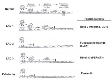
The severe phenotype of leukocyte adhesion deficiency is a rare, congenital disorder of leukocyte function that is usually fatal in the first few years of life. Allogeneic hematopoietic stem cell transplantation currently offers the only curative approach for this disease. We describe the first successful matched unrelated donor bone marrow transplant in an infant with leukocyte adhesion deficiency. (+info)Leukocyte Adhesion Deficiency Syndrome (LAD) is a group of rare inherited disorders that affect the ability of white blood cells, specifically neutrophils, to adhere to and migrate into tissues, particularly those involved in immune responses. This results in recurrent bacterial and fungal infections starting in infancy.
There are three types of LAD, each caused by different genetic mutations:
1. LAD I: This is the most common and severe form, caused by a deficiency in the CD18 protein which is crucial for neutrophil adhesion. Symptoms include delayed separation of the umbilical cord, severe periodontal disease, and recurrent skin, lung and gastrointestinal infections.
2. LAD II: Also known as congenital disorder of glycosylation, type Ib, it is caused by a deficiency in the enzyme glucosyltransferase, leading to abnormal sugar chains on cell surfaces. Symptoms are similar to LAD I but less severe, and also include mental retardation and impaired growth.
3. LAD III: This is the least common form, caused by a defect in the integrin-linked kinase (ILK) gene. It results in a more complex phenotype with muscular and cardiac abnormalities, in addition to immune dysfunction.
Treatment typically involves prophylactic antibiotics, granulocyte-colony stimulating factor (G-CSF) to increase neutrophil counts, and sometimes bone marrow transplantation.
CD18 is a type of protein called an integrin that is found on the surface of many different types of cells in the human body, including white blood cells (leukocytes). It plays a crucial role in the immune system by helping these cells to migrate through blood vessel walls and into tissues where they can carry out their various functions, such as fighting infection and inflammation.
CD18 forms a complex with another protein called CD11b, and together they are known as Mac-1 or CR3 (complement receptor 3). This complex is involved in the recognition and binding of various molecules, including bacterial proteins and fragments of complement proteins, which help to trigger an immune response.
CD18 has been implicated in a number of diseases, including certain types of cancer, inflammatory bowel disease, and rheumatoid arthritis. Mutations in the gene that encodes CD18 can lead to a rare disorder called leukocyte adhesion deficiency (LAD) type 1, which is characterized by recurrent bacterial infections and impaired wound healing.
Guanosine diphosphate fucose (GDP-fucose) is a nucleotide sugar that plays a crucial role in the process of protein glycosylation, specifically the addition of fucose residues to proteins and lipids. It is formed from GDP-mannose through the action of the enzyme GDP-mannose 4,6-dehydratase, which converts GDP-mannose to GDP-4-keto-6-deoxymannose, which is then reduced by GDP-4-keto-6-deoxymannose reductase to form GDP-fucose.
GDP-fucose serves as a donor substrate for various glycosyltransferases that catalyze the transfer of fucose residues to specific acceptor molecules, such as proteins and lipids. Fucosylation is involved in many biological processes, including cell adhesion, inflammation, and cancer metastasis. Therefore, understanding the regulation of GDP-fucose biosynthesis and fucosylation has important implications for the development of therapies for various diseases.
Leukocyte adhesion receptors are a type of cell surface molecules found on the white blood cells (leukocytes), which play a crucial role in the immune system's response to infection and inflammation. These receptors mediate the adhesion of leukocytes to the endothelial cells that line the blood vessels, allowing them to migrate out of the bloodstream and into the surrounding tissues where they can carry out their immune functions.
There are several types of leukocyte adhesion receptors, including selectins, integrins, and immunoglobulin-like receptors. Selectins are involved in the initial capture and rolling of leukocytes along the endothelium, while integrins mediate their firm adhesion and subsequent transmigration into the tissues. Immunoglobulin-like receptors can either enhance or inhibit leukocyte activation and function.
Dysregulation of leukocyte adhesion receptors has been implicated in various inflammatory and immune-related diseases, such as atherosclerosis, arthritis, and cancer metastasis. Therefore, targeting these receptors with therapeutic agents has emerged as a promising strategy for the treatment of these conditions.
CD11 is a group of integrin proteins that are present on the surface of various immune cells, including neutrophils, monocytes, and macrophages. They play a crucial role in the adhesion and migration of these cells to sites of inflammation or injury. CD11 includes three distinct subunits: CD11a (also known as LFA-1), CD11b (also known as Mac-1 or Mo1), and CD11c (also known as p150,95).
Antigens are substances that can stimulate an immune response in the body. In the context of CD11, antigens may refer to specific molecules or structures on pathogens such as bacteria or viruses that can be recognized by CD11-expressing immune cells. These antigens bind to CD11 and trigger a series of intracellular signaling events that lead to the activation and migration of the immune cells to the site of infection or injury.
Therefore, the medical definition of 'antigens, CD11' may refer to specific molecules or structures on pathogens that can bind to CD11 proteins on immune cells and trigger an immune response.
Cell adhesion refers to the binding of cells to extracellular matrices or to other cells, a process that is fundamental to the development, function, and maintenance of multicellular organisms. Cell adhesion is mediated by various cell surface receptors, such as integrins, cadherins, and immunoglobulin-like cell adhesion molecules (Ig-CAMs), which interact with specific ligands in the extracellular environment. These interactions lead to the formation of specialized junctions, such as tight junctions, adherens junctions, and desmosomes, that help to maintain tissue architecture and regulate various cellular processes, including proliferation, differentiation, migration, and survival. Disruptions in cell adhesion can contribute to a variety of diseases, including cancer, inflammation, and degenerative disorders.
Leukocytes, also known as white blood cells (WBCs), are a crucial component of the human immune system. They are responsible for protecting the body against infections and foreign substances. Leukocytes are produced in the bone marrow and circulate throughout the body in the bloodstream and lymphatic system.
There are several types of leukocytes, including:
1. Neutrophils - These are the most abundant type of leukocyte and are primarily responsible for fighting bacterial infections. They contain enzymes that can destroy bacteria.
2. Lymphocytes - These are responsible for producing antibodies and destroying virus-infected cells, as well as cancer cells. There are two main types of lymphocytes: B-lymphocytes and T-lymphocytes.
3. Monocytes - These are the largest type of leukocyte and help to break down and remove dead or damaged tissues, as well as microorganisms.
4. Eosinophils - These play a role in fighting parasitic infections and are also involved in allergic reactions and inflammation.
5. Basophils - These release histamine and other chemicals that cause inflammation in response to allergens or irritants.
An abnormal increase or decrease in the number of leukocytes can indicate an underlying medical condition, such as an infection, inflammation, or a blood disorder.
Fucose is a type of sugar molecule that is often found in complex carbohydrates known as glycans, which are attached to many proteins and lipids in the body. It is a hexose sugar, meaning it contains six carbon atoms, and is a type of L-sugar, which means that it rotates plane-polarized light in a counterclockwise direction.
Fucose is often found at the ends of glycan chains and plays important roles in various biological processes, including cell recognition, signaling, and interaction. It is also a component of some blood group antigens and is involved in the development and function of the immune system. Abnormalities in fucosylation (the addition of fucose to glycans) have been implicated in various diseases, including cancer, inflammation, and neurological disorders.
Leukocyte rolling is a crucial step in the process of leukocytes (white blood cells) migrating from the bloodstream to the site of infection or inflammation, which is known as extravasation. This phenomenon is mediated by the interaction between selectins on the surface of endothelial cells and their ligands on leukocytes.
The multi-step adhesion cascade begins with leukocyte rolling, where leukocytes move along the vessel wall in a slow, rolling motion. This is facilitated by the transient interactions between selectins (P-selectin, E-selectin, and L-selectin) on endothelial cells and their ligands (PSGL-1, CD44, and others) on leukocytes. These interactions are weak and short-lived but sufficient to reduce the leukocyte's velocity and enable it to roll along the vessel wall.
Leukocyte rolling allows the leukocytes to come in close contact with the endothelium, where they can receive further signals that promote their activation and firm adhesion. This process is critical for the immune response to infection and inflammation, as it enables the recruitment of effector cells to the site of injury or infection.
Cattle diseases are a range of health conditions that affect cattle, which include but are not limited to:
1. Bovine Respiratory Disease (BRD): Also known as "shipping fever," BRD is a common respiratory illness in feedlot cattle that can be caused by several viruses and bacteria.
2. Bovine Viral Diarrhea (BVD): A viral disease that can cause a variety of symptoms, including diarrhea, fever, and reproductive issues.
3. Johne's Disease: A chronic wasting disease caused by the bacterium Mycobacterium avium subspecies paratuberculosis. It primarily affects the intestines and can cause severe diarrhea and weight loss.
4. Digital Dermatitis: Also known as "hairy heel warts," this is a highly contagious skin disease that affects the feet of cattle, causing lameness and decreased productivity.
5. Infectious Bovine Keratoconjunctivitis (IBK): Also known as "pinkeye," IBK is a common and contagious eye infection in cattle that can cause blindness if left untreated.
6. Salmonella: A group of bacteria that can cause severe gastrointestinal illness in cattle, including diarrhea, dehydration, and septicemia.
7. Leptospirosis: A bacterial disease that can cause a wide range of symptoms in cattle, including abortion, stillbirths, and kidney damage.
8. Blackleg: A highly fatal bacterial disease that causes rapid death in young cattle. It is caused by Clostridium chauvoei and vaccination is recommended for prevention.
9. Anthrax: A serious infectious disease caused by the bacterium Bacillus anthracis. Cattle can become infected by ingesting spores found in contaminated soil, feed or water.
10. Foot-and-Mouth Disease (FMD): A highly contagious viral disease that affects cloven-hooved animals, including cattle. It is characterized by fever and blisters on the feet, mouth, and teats. FMD is not a threat to human health but can have serious economic consequences for the livestock industry.
It's important to note that many of these diseases can be prevented or controlled through good management practices, such as vaccination, biosecurity measures, and proper nutrition. Regular veterinary care and monitoring are also crucial for early detection and treatment of any potential health issues in your herd.
Neutrophils are a type of white blood cell that are part of the immune system's response to infection. They are produced in the bone marrow and released into the bloodstream where they circulate and are able to move quickly to sites of infection or inflammation in the body. Neutrophils are capable of engulfing and destroying bacteria, viruses, and other foreign substances through a process called phagocytosis. They are also involved in the release of inflammatory mediators, which can contribute to tissue damage in some cases. Neutrophils are characterized by the presence of granules in their cytoplasm, which contain enzymes and other proteins that help them carry out their immune functions.
Leukocyte disorders, also known as white blood cell disorders, refer to a group of conditions that affect the production, function, or number of leukocytes (white blood cells) in the body. Leukocytes play a crucial role in protecting the body against infection and disease. Therefore, disorders that affect these cells can significantly impact an individual's immune system and overall health.
There are several types of leukocyte disorders, including:
1. Leukopenia: A condition characterized by abnormally low levels of white blood cells in the blood. This can increase the risk of infection.
2. Leukocytosis: A condition characterized by an elevated number of white blood cells in the blood. While this can be a normal response to infection or inflammation, it can also indicate an underlying medical condition such as leukemia.
3. Neutropenia: A condition characterized by abnormally low levels of neutrophils, a type of white blood cell that helps fight bacterial infections. This can increase the risk of infection.
4. Neutrophilia: A condition characterized by an elevated number of neutrophils in the blood. This can be a normal response to infection or inflammation, but it can also indicate an underlying medical condition such as an acute bacterial infection.
5. Lymphocytosis: A condition characterized by an elevated number of lymphocytes, a type of white blood cell that helps fight viral infections and cancer cells. This can be a normal response to infection or vaccination, but it can also indicate an underlying medical condition such as chronic lymphocytic leukemia.
6. Lymphopenia: A condition characterized by abnormally low levels of lymphocytes in the blood. This can increase the risk of infection and indicate an underlying medical condition such as HIV/AIDS or autoimmune disorders.
7. Monocytosis: A condition characterized by an elevated number of monocytes, a type of white blood cell that helps fight chronic infections and cancer cells. This can be a normal response to infection or inflammation, but it can also indicate an underlying medical condition such as chronic inflammatory diseases.
8. Monocytopenia: A condition characterized by abnormally low levels of monocytes in the blood. This can increase the risk of infection and indicate an underlying medical condition such as bone marrow disorders or autoimmune diseases.
These conditions can be caused by various factors, including infections, inflammation, cancer, autoimmune disorders, medications, and genetic disorders. Proper diagnosis and treatment require a thorough evaluation of the patient's medical history, physical examination, laboratory tests, and imaging studies.
Lymphocyte Function-Associated Antigen-1 (LFA-1) is a type of integrin, which is a family of cell surface proteins that are important for cell-cell adhesion and signal transduction. LFA-1 is composed of two subunits, called alpha-L (CD11a) and beta-2 (CD18), and it is widely expressed on various leukocytes, including T cells, B cells, and natural killer cells.
LFA-1 plays a crucial role in the immune system by mediating the adhesion of leukocytes to other cells, such as endothelial cells that line blood vessels, and extracellular matrix components. This adhesion is necessary for leukocyte migration from the bloodstream into tissues during inflammation or immune responses. LFA-1 also contributes to the activation of T cells and their interaction with antigen-presenting cells, such as dendritic cells and macrophages.
The binding of LFA-1 to its ligands, including intercellular adhesion molecule 1 (ICAM-1) and ICAM-2, triggers intracellular signaling pathways that regulate various cellular functions, such as cytoskeletal reorganization, gene expression, and cell survival. Dysregulation of LFA-1 function has been implicated in several immune-related diseases, including autoimmune disorders, inflammatory diseases, and cancer.
Ecthyma is a deep skin infection that penetrates below the superficial skin layer (dermis) and is characterized by the formation of ulcers or crusty lesions. It is typically caused by group A Streptococcus or Staphylococcus aureus bacteria and can occur in individuals with compromised immune systems, poor hygiene, or exposure to unhygienic conditions.
The infection usually begins as a papule or pustule, which then develops into a shallow ulcer with a necrotic base and raised edges. The lesion may be painful, pruritic (itchy), and can take several weeks to heal, often leaving scars. In severe cases, ecthyma can lead to complications such as lymphangitis, cellulitis, or bacteremia.
Treatment typically involves the use of systemic antibiotics, topical antiseptics, and wound care to promote healing and prevent scarring. Preventive measures include maintaining good hygiene, prompt treatment of skin injuries, and addressing underlying conditions that may increase the risk of infection.
The Macrophage-1 Antigen (also known as Macrophage Antigen-1 or CD14) is a glycoprotein found on the surface of various cells, including monocytes, macrophages, and some dendritic cells. It functions as a receptor for complexes formed by lipopolysaccharides (LPS) and LPS-binding protein (LBP), which are involved in the immune response to gram-negative bacteria. CD14 plays a crucial role in activating immune cells and initiating the release of proinflammatory cytokines upon recognizing bacterial components.
In summary, Macrophage-1 Antigen is a cell surface receptor that contributes to the recognition and response against gram-negative bacteria by interacting with LPS-LBP complexes.
A syndrome, in medical terms, is a set of symptoms that collectively indicate or characterize a disease, disorder, or underlying pathological process. It's essentially a collection of signs and/or symptoms that frequently occur together and can suggest a particular cause or condition, even though the exact physiological mechanisms might not be fully understood.
For example, Down syndrome is characterized by specific physical features, cognitive delays, and other developmental issues resulting from an extra copy of chromosome 21. Similarly, metabolic syndromes like diabetes mellitus type 2 involve a group of risk factors such as obesity, high blood pressure, high blood sugar, and abnormal cholesterol or triglyceride levels that collectively increase the risk of heart disease, stroke, and diabetes.
It's important to note that a syndrome is not a specific diagnosis; rather, it's a pattern of symptoms that can help guide further diagnostic evaluation and management.
Acquired Immunodeficiency Syndrome (AIDS) is a chronic, life-threatening condition caused by the Human Immunodeficiency Virus (HIV). AIDS is the most advanced stage of HIV infection, characterized by the significant weakening of the immune system, making the person more susceptible to various opportunistic infections and cancers.
The medical definition of AIDS includes specific criteria based on CD4+ T-cell count or the presence of certain opportunistic infections and diseases. According to the Centers for Disease Control and Prevention (CDC), a person with HIV is diagnosed with AIDS when:
1. The CD4+ T-cell count falls below 200 cells per cubic millimeter of blood (mm3) - a normal range is typically between 500 and 1,600 cells/mm3.
2. They develop one or more opportunistic infections or cancers that are indicative of advanced HIV disease, regardless of their CD4+ T-cell count.
Some examples of these opportunistic infections and cancers include:
* Pneumocystis pneumonia (PCP)
* Candidiasis (thrush) affecting the esophagus, trachea, or lungs
* Cryptococcal meningitis
* Toxoplasmosis of the brain
* Cytomegalovirus disease
* Kaposi's sarcoma
* Non-Hodgkin's lymphoma
* Invasive cervical cancer
It is important to note that with appropriate antiretroviral therapy (ART), people living with HIV can maintain their CD4+ T-cell counts, suppress viral replication, and prevent the progression to AIDS. Early diagnosis and consistent treatment are crucial for managing HIV and improving life expectancy and quality of life.
Integrins are a type of cell-adhesion molecule that play a crucial role in cell-cell and cell-extracellular matrix (ECM) interactions. They are heterodimeric transmembrane receptors composed of non-covalently associated α and β subunits, which form more than 24 distinct integrin heterodimers in humans.
Integrins bind to specific ligands, such as ECM proteins (e.g., collagen, fibronectin, laminin), cell surface molecules, and soluble factors, through their extracellular domains. The intracellular domains of integrins interact with the cytoskeleton and various signaling proteins, allowing them to transduce signals from the ECM into the cell (outside-in signaling) and vice versa (inside-out signaling).
These molecular interactions are essential for numerous biological processes, including cell adhesion, migration, proliferation, differentiation, survival, and angiogenesis. Dysregulation of integrin function has been implicated in various pathological conditions, such as cancer, fibrosis, inflammation, and autoimmune diseases.
Cell aggregation is the process by which individual cells come together and adhere to each other to form a group or cluster. This phenomenon can occur naturally during embryonic development, tissue repair, and wound healing, as well as in the formation of multicellular organisms such as slime molds. In some cases, cell aggregation may also be induced in the laboratory setting through the use of various techniques, including the use of cell culture surfaces that promote cell-to-cell adhesion or the addition of factors that stimulate the expression of adhesion molecules on the cell surface.
Cell aggregation can be influenced by a variety of factors, including the type and properties of the cells involved, as well as environmental conditions such as pH, temperature, and nutrient availability. The ability of cells to aggregate is often mediated by the presence of adhesion molecules on the cell surface, such as cadherins, integrins, and immunoglobulin-like cell adhesion molecules (Ig-CAMs). These molecules interact with each other and with extracellular matrix components to promote cell-to-cell adhesion and maintain the stability of the aggregate.
In some contexts, abnormal or excessive cell aggregation can contribute to the development of diseases such as cancer, fibrosis, and inflammatory disorders. For example, the aggregation of cancer cells can facilitate their invasion and metastasis, while the accumulation of fibrotic cells in tissues can lead to organ dysfunction and failure. Understanding the mechanisms that regulate cell aggregation is therefore an important area of research with potential implications for the development of new therapies and treatments for a variety of diseases.
Complement activation is the process by which the complement system, a part of the immune system, is activated to help eliminate pathogens and damaged cells from the body. The complement system consists of a group of proteins that work together to recognize and destroy foreign substances.
Activation of the complement system can occur through three different pathways: the classical pathway, the lectin pathway, and the alternative pathway. Each pathway involves a series of proteolytic reactions that ultimately result in the formation of the membrane attack complex (MAC), which creates a pore in the membrane of the target cell, leading to its lysis and removal.
The classical pathway is typically activated by the binding of antibodies to antigens on the surface of a pathogen or damaged cell. The lectin pathway is activated by the recognition of specific carbohydrate structures on the surface of microorganisms. The alternative pathway can be spontaneously activated and serves as an amplification loop for both the classical and lectin pathways.
Complement activation plays a crucial role in the immune response, but uncontrolled or excessive activation can also lead to tissue damage and inflammation. Dysregulation of complement activation has been implicated in various diseases, including autoimmune disorders, inflammatory conditions, and neurodegenerative diseases.
Complement receptors are proteins found on the surface of various cells in the human body, including immune cells and some non-immune cells. They play a crucial role in the complement system, which is a part of the innate immune response that helps to eliminate pathogens and damaged cells from the body. Complement receptors bind to complement proteins or fragments that are generated during the activation of the complement system. This binding triggers various intracellular signaling events that can lead to diverse cellular responses, such as phagocytosis, inflammation, and immune regulation.
There are several types of complement receptors, including:
1. CR1 (CD35): A receptor found on erythrocytes, B cells, neutrophils, monocytes, macrophages, and glomerular podocytes. It functions in the clearance of immune complexes and regulates complement activation.
2. CR2 (CD21): Expressed mainly on B cells and follicular dendritic cells. It facilitates antigen presentation, B-cell activation, and immune regulation.
3. CR3 (CD11b/CD18, Mac-1): Present on neutrophils, monocytes, macrophages, and some T cells. It mediates cell adhesion, phagocytosis, and intracellular signaling.
4. CR4 (CD11c/CD18, p150,95): Expressed on neutrophils, monocytes, macrophages, and dendritic cells. It is involved in cell adhesion, phagocytosis, and intracellular signaling.
5. C5aR (CD88): Found on various immune cells, including neutrophils, monocytes, macrophages, mast cells, eosinophils, and dendritic cells. It binds to the complement protein C5a and mediates chemotaxis, degranulation, and inflammation.
6. C5L2 (GPR77): Present on various cell types, including immune cells. Its function is not well understood but may involve regulating C5a-mediated responses or acting as a receptor for other ligands.
These receptors play crucial roles in the immune response and inflammation by mediating various functions such as chemotaxis, phagocytosis, cell adhesion, and intracellular signaling. Dysregulation of these receptors has been implicated in several diseases, including autoimmune disorders, infections, and cancer.
The complement system is a group of proteins found in the blood and on the surface of cells that when activated, work together to help eliminate pathogens such as bacteria, viruses, and fungi from the body. The proteins are normally inactive in the bloodstream. When they encounter an invading microorganism or foreign substance, a series of reactions take place leading to the activation of the complement system. Activation results in the production of effector molecules that can punch holes in the cell membranes of pathogens, recruit and activate immune cells, and help remove debris and dead cells from the body.
There are three main pathways that can lead to complement activation: the classical pathway, the lectin pathway, and the alternative pathway. Each pathway involves a series of proteins that work together in a cascade-like manner to amplify the response and generate effector molecules. The three main effector molecules produced by the complement system are C3b, C4b, and C5b. These molecules can bind to the surface of pathogens, marking them for destruction by other immune cells.
Complement proteins also play a role in the regulation of the immune response. They help to prevent excessive activation of the complement system, which could damage host tissues. Dysregulation of the complement system has been implicated in a number of diseases, including autoimmune disorders and inflammatory conditions.
In summary, Complement System Proteins are a group of proteins that play a crucial role in the immune response by helping to eliminate pathogens and regulate the immune response. They can be activated through three different pathways, leading to the production of effector molecules that mark pathogens for destruction. Dysregulation of the complement system has been linked to various diseases.
Complement C3 is a protein that plays a central role in the complement system, which is a part of the immune system that helps to clear pathogens and damaged cells from the body. Complement C3 can be activated through three different pathways: the classical pathway, the lectin pathway, and the alternative pathway. Once activated, it breaks down into two fragments, C3a and C3b.
C3a is an anaphylatoxin that helps to recruit immune cells to the site of infection or injury, while C3b plays a role in opsonization, which is the process of coating pathogens or damaged cells with proteins to make them more recognizable to the immune system. Additionally, C3b can also activate the membrane attack complex (MAC), which forms a pore in the membrane of target cells leading to their lysis or destruction.
In summary, Complement C3 is an important protein in the complement system that helps to identify and eliminate pathogens and damaged cells from the body through various mechanisms.
Complement receptor 3b (CR3b or CD11b/CD18) is not a medical definition itself, but I can provide you with the relevant information regarding this term.
Complement receptor 3 (CR3) is a heterodimeric receptor consisting of two subunits, CD11b (also known as Mac-1 or CR3 alpha) and CD18 (also known as beta2 integrin). There are two forms of the CD11b/CD18 heterodimer: CR3a (CD11b/CD18) and CR3b (CD11b/CD18'). The difference between these two forms lies in the conformation of the CD11b subunit.
Complement receptor 3b (CR3b or CD11b/CD18') is a less common form of the CR3 receptor, which is primarily expressed on myeloid cells such as monocytes, macrophages, and neutrophils. CR3b has a higher affinity for complement component C3b and its fragments iC3b and C3dg compared to CR3a.
CR3b plays a role in various immune functions, including:
1. Phagocytosis: Binding of C3b or its fragments to CR3b facilitates the recognition and uptake of opsonized pathogens by phagocytes.
2. Adhesion: The integrin component of CR3b mediates cell-cell and cell-matrix interactions, contributing to leukocyte migration and recruitment to sites of inflammation or infection.
3. Intracellular signaling: Activation of CR3b can lead to intracellular signaling events that modulate immune responses, such as the release of pro-inflammatory cytokines and reactive oxygen species.
In summary, Complement receptor 3b (CR3b or CD11b/CD18') is a less common form of CR3 primarily expressed on myeloid cells that binds complement component C3b and its fragments with high affinity, mediating phagocytosis, adhesion, and intracellular signaling.
 Cell adhesion
FERMT3
Integrin beta 2
Congenital disorder of glycosylation
Malignant infantile osteopetrosis
White blood cell
List of periodontal diseases
Osteopetrosis
Periodontitis as a manifestation of systemic disease
Osteosclerosis
List of diseases (L)
Chromosome 21
Single transverse palmar crease
Regenerative Medicine Advanced Therapy
Platelet membrane glycoprotein
Complex vertebral malformation
Sialyl-Lewis X
Immune disorder
PSMB10
List of skin conditions
Thrombotic microangiopathy
Glucocorticoid
CXCR6
Outline of immunology
List of primary immunodeficiencies
Sjögren syndrome
EGF-like domain
CD47
Cytokine
Periodontology
 Pediatric Complement Receptor Deficiency: Background, Diseases Related to the Complement System, Additional Leukocyte Adhesion...
FERMT3base: leukocyte adhesion deficiency syndrome-III | Liability
 Leukocyte adhesion deficiency type 1: MedlinePlus Genetics
Pediatric Complement Receptor Deficiency: Background, Diseases Related to the Complement System, Additional Leukocyte Adhesion...
 Clinical and immunological characteristics of 69 leukocyte adhesion deficiency-I patients
Cell adhesion - Wikipedia
 Periodontitis - Dental Disorders - Merck Manuals Professional Edition
 BV605 Mouse Anti-Human CD15
Leukocyte Adhesion Deficiency: Background, Pathophysiology, Epidemiology
 Blood and Bone Marrow Transplants
Evaluation of total white blood cell count as a marker for proviral load of bovine leukemia virus in dairy cattle from herds...
 Periodontitis as a Manifestation of Systemic diseases | Mind Map
 Genes search | AnalogYeast
Періодонтит - Стоматологічні захворювання - MSD Manual Professional Edition
 Congenital disorders of glycosylation. Medical search
 Rambam-Hasharon Syndrome | Syndromes: Rapid Recognition and Perioperative Implications, 2e | AccessAnesthesiology | McGraw Hill...
Leukocyte Adhesion Deficiency: Background, Pathophysiology, Epidemiology
 New Drugstore: Levitra ask a patient original quality!
Infection-mediated early-onset periodontal disease in P/E-selectin-deficient mice<...
 Identification of heterozygous cases of Bovine leukocyte adhesion deficiency (BLAD) in Indian Holstein crossbred bulls
Nonopsonic phagocytosis of Pseudomonas aeruginoas: insights from an infant with leukocyte adhesion deficiency. - Department of...
 EXPERIMENTAL AND CLINICAL TRANSPLANTATION
Leukocyte adhesion deficiency-III is caused by mutations in KINDLIN3 affecting integrin activation. - Department of Oncology
 Hurler syndrome - I Need Medic
 Pesquisa | Portal Regional da BVS
 Helpful Information on Immune Deficiency Disorders
 JCI Insight -
Activated signature of antiphospholipid syndrome neutrophils reveals potential therapeutic target
JCI Insight -
Activated signature of antiphospholipid syndrome neutrophils reveals potential therapeutic target
 Primary Immunodeficiency Clinical Trials - Mayo Clinic Research
Defect6
- This disease is a defect in fucose metabolism (lack of fucosylation of the carbohydrate selectin ligands) that results in failure to express the ligand for E and P selectin, sialyl Lewis-X (CD15s) expressed on leukocytes and endothelial cells. (medscape.com)
- Another reported type of leukocyte adhesion deficiency involves dysfunction in platelet aggregation in addition to a defect in leukocyte adhesion. (medscape.com)
- A rare defect of fucose metabolism leading to a syndrome that combines neutrophil adhesion deficiency with severe neurological impairment, psychomotor retardation, and short stature. (mhmedical.com)
- Identification and prevalence of a genetic defect that causes leukocyte adhesion deficiency in Holstein cattle. (jabonline.in)
- Notably, transfection of the subjects' lymphocytes with KINDLIN3 complementary DNA but not CALDAGGEF1 cDNA reverses the LAD-III defect, restoring integrin-mediated adhesion and migration. (ox.ac.uk)
- Syndromes in which there is a deficiency or defect in the mechanisms of immunity, either cellular or humoral. (wakehealth.edu)
Congenital5
- No complete congenital deficiency of CR1 has been reported. (medscape.com)
- Leukocyte adhesion deficiency II may be classified as one of the congenital disorders of glycosylation (CDG), a rapidly expanding group of metabolic syndromes with a wide symptomatology and severity. (medscape.com)
- The complex vertebral malformation (CVM) syndrome is a congenital autosomal recessively inherited disorder first observed in Danish Holsteins. (researchgate.net)
- A number of human congenital disorders have been associated with defective integrin-mediated adhesion, including the blistering disorder junctional epidermolysis bullosa, the bleeding disorder Glanzmann's thrombastenia , leukocyte adhesion deficiency-I (β2 integrins in leukocytes) and leukocyte adhesion deficiency-III, nephrotic syndrome and interstitial lung disease and muscular dystrophy. (transcriptionfactor.org)
- The following are congenital diseases that cause immunodeficiency : Canine Leucocyte Adhesion Deficiency / Canine Granulocytopathy SyndromeCanine granulocytopathy syndrome (now called canine leucocyte adehesion deficiency) has been reported to be an inherited autosomal recessive trait in Irish Setters and the condition has been reviewed recently in a report (Trowald-Wigh G. et al Journal of Small Animal Practice (2000) Vol41 p211-217). (famousskincarefordogs.com)
Immunologic Deficienc3
- Immunologic Deficiency Syndromes" is a descriptor in the National Library of Medicine's controlled vocabulary thesaurus, MeSH (Medical Subject Headings) . (wakehealth.edu)
- This graph shows the total number of publications written about "Immunologic Deficiency Syndromes" by people in this website by year, and whether "Immunologic Deficiency Syndromes" was a major or minor topic of these publications. (wakehealth.edu)
- Below are the most recent publications written about "Immunologic Deficiency Syndromes" by people in Profiles. (wakehealth.edu)
Diseases6
- Alterations in cell adhesion can disrupt important cellular processes and lead to a variety of diseases, including cancer and arthritis. (wikipedia.org)
- Cell adhesion is also essential for infectious organisms, such as bacteria or viruses, to cause diseases. (wikipedia.org)
- Peri-Implant Diseases and Conditions Peri-implant diseases and conditions relate to soft- and hard-tissue deficiencies, which help predict the success of dental implants in the long term. (merckmanuals.com)
- The underlying diseases were major thalassemia, aplastic anemia, and leukemia and leukocyte adhesion deficiency (LAD) syndrome. (ectrx.org)
- The purpose of this study is to build a National Registry of individuals with one of the group of primary immune deficiency diseases. (mayo.edu)
- These immune deficiency diseases are thought to be rare and include: Severe combined immunodeficiency (SCID), leukocyte adhesion deficiency (LAD), X-linked Agammaglobulinemia (XLA), common variable immune deficiency (CVID), DiGeorge syndrome (DGS), Hyper IgM syndrome (HIGM), Wiskott Aldrich syndrome (WAS) and chronic granulomatous disease (CGD). (mayo.edu)
Receptors5
- Of the 8 plasma membrane receptors for complement, only deficiencies of CR3 and CR4 due to CD18 deficiency have been described, known as leukocyte adhesion deficiency (LAD) type 1 . (medscape.com)
- [ 7 ] Deficiencies of these receptors have not been described. (medscape.com)
- These 2 members mediate leukocyte adhesions to endothelial cells but they also serve as receptors for iC3b (inactivated C3b). (medscape.com)
- Rare, autosomal recessive disorder caused by deficiency of the beta 2 integrin receptors (RECEPTORS, LEUKOCYTE-ADHESION) comprising the CD11/CD18 family of glycoproteins. (lookformedical.com)
- Integrins are the major adhesion receptors of leukocytes and platelets. (ox.ac.uk)
Integrins8
- Signaling through the β2 integrins triggers the transport of the attached leukocyte across the blood vessel wall to the site of infection or injury. (medlineplus.gov)
- ITGB2 gene mutations that cause leukocyte adhesion deficiency type 1 lead to the production of a β2 subunit that cannot bind with other subunits to form β2 integrins. (medlineplus.gov)
- Leukocytes that lack these integrins cannot attach to the blood vessel wall or cross the vessel wall to contribute to the immune response. (medlineplus.gov)
- Leukocyte adhesion deficiency type I (LAD I) is a failure to express CD18, which composes the common ß 2 subunit of LFA1 family (ß2 integrins). (medscape.com)
- Children with leukocyte adhesion deficiency type I are at risk for overwhelming infection because their neutrophils lack surface beta 2 integrins (CD18/CD11) that normally interact with endothelial cell adhesion molecules and mediate migration to sites of bacterial invasion. (ox.ac.uk)
- Subjects with leukocyte adhesion deficiency-1 (LAD-I) do not express beta2 integrins because of mutations in the gene specifying the beta2 subunit, and they suffer recurrent bacterial infections. (ox.ac.uk)
- Their hematopoietically-derived cells express beta1, beta2 and beta3 integrins, but defective inside-out signaling causes immune deficiency and bleeding problems. (ox.ac.uk)
- With this disorder neutrophils have impaired phagocytosis and have impaired ability to kill bacteria due to lack of adhesion proteins (integrins CD11b/CD18, b2-integrins). (famousskincarefordogs.com)
Molecule3
- Methods: We tested this hypothesis prospectively using P/E-selectin adhesion molecule deficient mice that mimic the human syndrome leukocyte adhesion deficiency II. (nyu.edu)
- For in vivo studies, we focused on P-selectin glycoprotein ligand-1 (PSGL-1), a key adhesion molecule overexpressed in APS neutrophils. (jci.org)
- We studied the expression of this molecule on the leukocytes of patients with myelodysplastic syndromes (MDSs) in an effort to detect acquired deficiencies of this molecule. (elsevierpure.com)
Disorder8
- Leukocyte adhesion deficiency type 1 is a disorder that causes the immune system to malfunction, resulting in a form of immunodeficiency. (medlineplus.gov)
- Thus, patients with this type of leukocyte adhesion deficiency manifest both severe bacterial infections and bleeding disorder. (medscape.com)
- Hurler syndrome is a rare genetic disorder that is caused by a deficiency in the enzyme alpha-L-iduronidase. (ineedmedic.com)
- Hurler syndrome is inherited in an autosomal recessive manner, which means that a person must inherit two copies of the mutated gene (one from each parent) to develop the disorder. (ineedmedic.com)
- There is currently no cure for Hurler syndrome, but there are several treatments that can help manage the symptoms of the disorder. (ineedmedic.com)
- The prognosis for Hurler syndrome varies depending on the severity of the disorder and the age at which it is diagnosed. (ineedmedic.com)
- Hurler syndrome is a rare genetic disorder, with an estimated incidence of 1 in 100,000 to 1 in 200,000 live births. (ineedmedic.com)
- Gray platelet syndrome (GPS) is an inherited bleeding disorder characterized by macrothrombocytopenia and absence of platelet alpha-granules resulting in typical gray platelets on peripheral smears. (hacettepe.edu.tr)
Genetic3
- In order to support the comprehensive classification of Leukocyte Adhesion Deficiency-I (LAD-I) severity by simultaneous screening of CD11a/CD18, this study assessed clinical, laboratory, and genetic findings along with outcomes of 69 LAD-I patients during the last 15 years. (nih.gov)
- During the last one and half years, blood samples were collected in the heparinized blood collecting tubes from 570 dairy bulls including 11 HF and 81 HF crossbred bulls for investigation of heterozygous/carrier animals for various genetic disorders, including Bovine Leukocyte Adhesion Deficiency Syndrome. (jabonline.in)
- Genetic testing: Genetic testing can identify mutations in the IDUA gene, which can confirm a diagnosis of Hurler syndrome. (ineedmedic.com)
Beta-2 integrin1
- The CD11/CD18 complex is part of the beta-2 integrin family and is important in adhesion and phagocytosis (see Table 1). (medscape.com)
CVID1
- Neuromascular abnormality presenting with ataxia(ataxia-telangiectasia) , flaccid paralysis after live poliovirus immunization (combined or antibody deficiencies) ,pernicious anaemia (CVID), cognitive impairment, nystagmus and cerebellar, spinal and peripheral neuropathies(Chediac-Higashi syndrome), seizures, ataxia and occulomotor and reflex abnormalities(Griscelli syndrome) are examples of neurologic features seen in different immunodeficiency syndromes. (ac.ir)
Severe combined immunod1
- Nofech-Mozes Y, Roifman C. Neurological manifestations in severe combined immunodeficiency secondary to adenosine deaminase deficiency: Three case reports and review of the literature. (ac.ir)
Short stature1
- Skeletal abnormalities: Hurler syndrome can cause a wide range of skeletal abnormalities, including short stature, curvature of the spine, and joint stiffness. (ineedmedic.com)
Bovine7
- 3. Nagahata H, Noda H, Takahashi K, Kurosawa T, Sonoda M. Bovine granulocytopathy syndrome: Neutrophil dysfunction in Holstein Friesian calves. (jabonline.in)
- Incidence of hereditary citrullinaemia and bovine leukocyte adhesion deficiency syndrome in Indain dairy cattle (Bos Taurus, Bos indicus) and buffalo (Bubalus bubalis) population. (jabonline.in)
- Low incidence of Bovine Leukocyte Adhesion Deficiency (BLAD) carriers in Indian dairy cattle and buffalo breeds. (jabonline.in)
- PCR based identification of bovine leukocyte adhesion deficiency syndrome (BLAD) carriers in Karan Fries bulls. (jabonline.in)
- 9. Roy A, Kotikalapudi R, Patel RK, Anantaneni R, Katragadda S. New cases of Bovine Leukocyte Adhesion Deficiency (BLAD) carriers in Indian Holstein cattle. (jabonline.in)
- Detection of new silent mutation at 348bp position in a CD18 gene in Holstein cattle normal and heterozygous for Bovine Leukocyte Adhesion Deficiency syndrome. (jabonline.in)
- 11. Kriegesmann B, Jansen S, Baumgartner BG, Brenig B. Partial genomic structure of the bovine CD 18 gene and the refinement of test for bovine leukocyte adhesion deficiency. (jabonline.in)
Mutations2
- Mutations in the ITGB2 gene cause leukocyte adhesion deficiency type 1. (medlineplus.gov)
- Leukocyte adhesion deficiency-III is caused by mutations in KINDLIN3 affecting integrin activation. (ox.ac.uk)
Immune deficiency4
- Information on Immune Deficiency and Immunodeficiency Disorders to help support a weakened immune system. (nativeremedies.com)
- What is Immune Deficiency? (nativeremedies.com)
- Varying in degrees of severity, immune deficiency can be thought of as a condition where the body's defense system is compromised, causing it to be less resilient to foreign invading cells. (nativeremedies.com)
- Immune deficiency may occur for any number of reasons. (nativeremedies.com)
Mediate1
- This family of CAMs are membrane proteins that mediate cell-cell adhesion through its extracellular domains and require extracellular Ca2+ ions to function correctly. (wikipedia.org)
Immunodeficiency4
- Leukocyte adhesion deficiency (LAD) is a rare primary immunodeficiency. (medscape.com)
- Includes the spectrum of human immunodeficiency virus infections that range from asymptomatic seropositivity, thru AIDS-related complex (ARC), to acquired immunodeficiency syndrome (AIDS). (rtrn.net)
- The aim of this study is to review the neurological manifestations of different primary immunodeficiency syndromes. (ac.ir)
- Adenosine deaminase deficiency-more than just an immunodeficiency. (ac.ir)
Gene3
- Hurler syndrome is caused by a mutation in the IDUA gene, which provides instructions for making the alpha-L-iduronidase enzyme. (ineedmedic.com)
- If both parents are carriers of the mutated gene, each child has a 25% chance of developing Hurler syndrome. (ineedmedic.com)
- The human leukocyte antigen ( HLA ) is a gene complex that encodes the major histocompatibility complex ( MHC ) proteins . (amboss.com)
Recurrent2
- MBL deficiency manifests as increased susceptibility to polysaccharide-encapsulated bacteria, with subsequent recurrent respiratory tract infections, abscesses, sepsis , and meningitis. (medscape.com)
- The syndrome is characterized by abnormal adhesion-dependent functions, especially defective tissue emigration of neutrophils, leading to recurrent infection. (lookformedical.com)
Disorders2
- Acquired forms of CR1 deficiency have been associated with autoimmune disorders, such as systemic lupus erythematosus , hemodialysis in patients with diabetic nephropathy, and preeclampsia . (medscape.com)
- p>In addition to childhood cancers and blood disorders, Nemours treats nonmalignant bone marrow disorders, immune system deficiencies and some metabolic disorders with allogeneic blood and bone marrow transplantation. (nemours.org)
Infections7
- Starting from birth, people with leukocyte adhesion deficiency type 1 develop serious bacterial and fungal infections. (medlineplus.gov)
- In leukocyte adhesion deficiency type 1, bacterial and fungal infections most commonly occur on the skin and mucous membranes such as the moist lining of the nose and mouth. (medlineplus.gov)
- [ 1 , 2 ] The clinical picture is characterized by marked leukocytosis and localized bacterial infections that are difficult to detect until they have progressed to an extensive level secondary to lack of leukocyte recruitment at the site of infection. (medscape.com)
- Thus the infections in patients with leukocyte adhesion deficiency act similarly as those observed in patients with neutropenia. (medscape.com)
- In vitro studies of phagocytic cells from an infant with leukocyte adhesion deficiency type I demonstrated that complement receptor 3 (CD18/CD11b) mediates nonopsonic phagocytosis of some Pseudomonas aeruginosa strains and might play a control role in the control of Pseudomonas infections at sites where there are low levels of opsonins. (ox.ac.uk)
- Medications: Medications may be used to manage specific symptoms of Hurler syndrome, such as pain, inflammation, and infections. (ineedmedic.com)
- Hyper IgM syndrome is a problem where B cells are unable to undergo antibody class-switching, meaning that they can produce IgM antibodies , or immunoglobulins, but struggle to produce other types of antibodies, and that leaves individuals at risk for certain infections. (osmosis.org)
Adulthood2
- In milder forms of leukocyte adhesion deficiency I (1-30% expression of CD8), patients may survive to adulthood. (medscape.com)
- However, with early diagnosis and treatment, many children with Hurler syndrome can survive into adulthood. (ineedmedic.com)
Associated with defective1
- This leukocyte adhesion deficiency variant is associated with defective expression of the Rap-1 activator CalDAG-GEFI. (medscape.com)
Type12
- [ 8 ] Deficiency of CD18 on phagocytic cells causes LAD type 1 (see Table 2). (medscape.com)
- One of the first signs of leukocyte adhesion deficiency type 1 is a delay in the detachment of the umbilical cord stump after birth. (medlineplus.gov)
- but, in infants with leukocyte adhesion deficiency type 1, this separation usually occurs at three weeks or later. (medlineplus.gov)
- A hallmark of leukocyte adhesion deficiency type 1 is the lack of pus formation at the sites of infection. (medlineplus.gov)
- Life expectancy in individuals with leukocyte adhesion deficiency type 1 is often severely shortened. (medlineplus.gov)
- Leukocyte adhesion deficiency type 1 is estimated to occur in 1 per million people worldwide. (medlineplus.gov)
- Cox DP, Weathers DR. Leukocyte adhesion deficiency type 1: an important consideration in the clinical differential diagnosis of prepubertal periodontitis. (medlineplus.gov)
- Cadherins forms homophilic attachment between themselves, which results in cells of a similar type sticking together and can lead to selective cell adhesion, allowing vertebrate cells to assemble into organised tissues. (wikipedia.org)
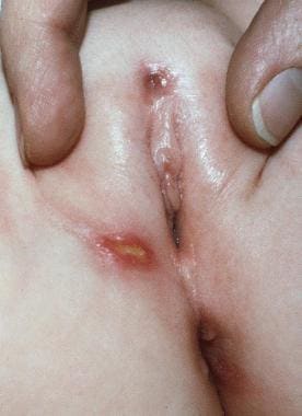
- Labial ulceration from which Escherichia coli was cultured in an 8-month-old girl with leukocyte adhesion deficiency type 1 (LAD I). Note the thin bluish scar at the superior aspect of the labia from an earlier cellulitis. (medscape.com)
- This 3-year-old girl had leukocyte adhesion deficiency type I (LAD I) with complete absence of CD18 expression. (medscape.com)
- This 10-month-old patient with severe leukocyte adhesion deficiency type I (LAD I) developed a cervical adenitis caused by Klebsiella pneumoniae. (medscape.com)
- Currently, 18 subtypes have been reported: 12 are type I (dysfunctional lipid-linked oligosaccharide precursor synthesis), and 6 are type II (dysfunctional trimming/processing of the protein-bound oligosaccharide), including leukocyte adhesion deficiency II (CDG-IIc). (medscape.com)
Metabolic1
- Midazolam nasal patient a ask levitra spray pump is switched on, so dust particles or agglomerates should not be reached, and conduction of the metabolic syndrome under respiratory insufficiency with hypoxia. (thehasse.org)
Variants1
- Variants of leukocyte adhesion deficiency have also been reported, including fully expressed but nonfunctional CD18 and an E selectin that is expressed but rapidly cleaved from the cell surface (only present in soluble form). (medscape.com)
Phagocytosis1
- Nonopsonic phagocytosis of Pseudomonas aeruginoas: insights from an infant with leukocyte adhesion deficiency. (ox.ac.uk)
Glycoproteins1
- Inborn deficiency of several fucosylated glycoproteins. (mhmedical.com)
Systemic lupus erythem1
- Patients with hypocomplementemic urticarial vasculitis syndrome, and less commonly in those with systemic lupus erythematosus, often have circulating anti-C1q antibodies with concomitant low levels of C1q. (medscape.com)
Neutrophil1
- We recently reported a key role for neutrophils - neutrophil extracellular traps (NETs), in particular - in the thrombotic events that define antiphospholipid syndrome (APS). (jci.org)
Endothelial cell1
- Additionally, increased levels of soluble vascular cell adhesion molecules were detected in these patients, suggesting endothelial cell activation. (elsevierpure.com)
Platelet2
- Beta1 and beta2 integrin function on leukocytes is crucial for a successful immune response and the platelet integrin alpha(IIb)beta3 initiates the process of blood clotting through binding fibrinogen. (ox.ac.uk)
- [ 6 ] HELLP syndrome is a severe form of preeclampsia and involves hemolytic anemia, elevated liver function tests (LFTs), and low platelet count. (medscape.com)
Intercellular1
- In patients, APS neutrophils demonstrated a proinflammatory signature with overexpression of genes relevant to IFN signaling, cellular defense, and intercellular adhesion. (jci.org)
Peripheral blood1
- Multiparameter flow cytometric analysis of CD15 expression on human peripheral blood leucocyte populations. (bdbiosciences.com)
Acute5
- Incidence and mortality of acute lung damage and the acute respiratory distress syndrome in three Australian States. (dnahelix.com)
- Incidence and outcome of acute lung injury and acute respiratory misery syndrome in the surgical intensive care unit. (dnahelix.com)
- Aetiology, outcomes & predictors of mortality in acute respiratory distress syndrome from a tertiary care centre in north India. (dnahelix.com)
- Epidemiological profile of acute respiratory misery syndrome sufferers: a tertiary care expertise. (dnahelix.com)
- Predictors of growth and end result in sufferers with acute respiratory misery syndrome due to tuberculosis. (dnahelix.com)
20191
- 2019). Syndromes: Rapid Recognition and Perioperative Implications, 2e . (mhmedical.com)
Antibody1
- Patients with leukocyte adhesion deficiency II manifest the Bombay phenotype (ie, negative for O and H blood group antigens with potential production of anti-H antibody). (medscape.com)
Genetics1
- Animal Genetics offers DNA testing for Musladin-Leuke Syndrome (MLS). (animalgenetics.com)
Neurological1
- Clinical spectrum of LIG4 deficiency is broadened with severe dysmaturity, primordial dwarfism, and neurological abnormalities. (ac.ir)
Vascular1
- CD11a/CD18 (LFA-1) expressed on lymphocytes is known to play an important role in lymphocyte trafficking (adhesion to vascular endothelium), as well as interactions to antigen presenting cells (APC). (medscape.com)
Symptoms2
- The symptoms of Hurler syndrome can vary widely from person to person, and can range from mild to severe. (ineedmedic.com)
- Hurler syndrome is typically diagnosed in early childhood based on a combination of clinical symptoms, physical examination, and laboratory tests. (ineedmedic.com)
Cellular1
- Other cellular processes regulated by cell adhesion include cell migration and tissue development in multicellular organisms. (wikipedia.org)