Leukoencephalitis, Acute Hemorrhagic
Detection of an autoantibody from Pug dogs with necrotizing encephalitis (Pug dog encephalitis). (1/26)
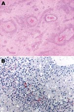
An autoantibody against canine brain tissue was detected in the cerebrospinal fluid (CSF) and serum of two Pug dogs (Nos. 1 and 2) by indirect immunofluorescence assay (IFA). Dog No. 1, a 2-year-old male, exhibited severe depression, ataxia, and generalized seizures and died 2 months after the onset of symptoms. Dog No. 2, a 9-month-old male, exhibited severe generalized seizures and died 17 months after the onset of symptoms. Histopathologic examination revealed a moderate to severe multifocal accumulation of lymphocytes, plasma cells, and a few neutrophils in both the gray and white matter of the cerebrum in dog No. 1. In dog No. 2, the cellular infiltrates were mild, but there was a severe, diffuse, and multifocal necrosis in the cerebral cortex with prominent astrocytosis. With the aid of IFA using fluorescein isothiocyanate-labeled antidog IgG goat serum and a confocal imaging system, specific reactions for glial cells were detected in the CSF of these Pug dogs but not in six canine control CSF samples. Double-labeling IFA using CSF from these Pug dogs and a rabbit antiserum against glial fibrillary acidic protein (GFAP) revealed that the autoantibody recognized GFAP-positive astrocytes and their cytoplasmic projections. By immunoblot analysis, the autoantibody from CSF of these Pug dogs recognized two common positive bands at 58 and 54 kd, which corresponded to the molecular mass of human GFAP. The role of this autoantibody for astrocytes is not yet clear. However, if the presence of the autoantibody is a specific feature of Pug dog encephalitis, it will be a useful clinical diagnostic marker and a key to the pathogenesis of this unique canine neurologic disease. (+info)Neuropsychology: music of the hemispheres. (2/26)
Music may be the food of love but it is also good fodder for cognitive scientists. Here we highlight a recent study of a neuropsychological patient who has lost her ability to read music, but not text, in the absence of any other musical deficit. (+info)Two episodes of leukoencephalitis associated with recombinant hepatitis B vaccination in a single patient. (3/26)
Cases of central nervous system demyelination have been reported after recombinant hepatitis B vaccination, but no causal link has been clearly demonstrated. We present the first case report involving the occurrence of 2 episodes of leukoencephalitis in a previously healthy patient after vaccination and rechallenge with hepatitis B vaccine. (+info)Pediatric acute hemorrhagic leukoencephalitis: report of a surviving patient and review. (4/26)
Acute hemorrhagic leukoencephalitis (AHLE) is a rare, fulminant CNS demyelinating condition usually diagnosed at autopsy. We report the clinical, laboratory, radiographic, and pathologic features of the first nonfatal case of pediatric AHLE confirmed by brain biopsy. Pathologic diagnosis of this condition may be critical to exclude more-common processes and to expedite the decision to administer high-dose corticosteroid therapy, which is potentially lifesaving. (+info)Possible acute hemorrhagic leukoencephalitis manifesting as intracerebral hemorrhage on computed tomography--case report. (5/26)
A 15-year-old girl presented with meningeal irritation and bilateral cerebral signs after contracting influenza. A lumbar puncture revealed bloody cerebrospinal fluid and polymorphonuclear predominant pleocytosis with an elevated protein level and normal glucose level. Computed tomography showed a hematoma in the right basal ganglia and lateral ventricles. Symmetrical low density areas were also noted in the bilateral white matter. The preliminary diagnosis was hemorrhagic cerebrovascular disease of unknown cause. However, her neurological condition deteriorated. Magnetic resonance (MR) imaging showed diffuse high intensity signals in the bilateral white matter and small spotty lesions, indicating hemorrhages in various stages. The final diagnosis was acute hemorrhagic leukoencephalitis (AHL). However, high-dose steroid administration and plasmapheresis failed to improve her condition. Hypothermia could not control her intracranial pressure and she died 12 days after admission. The neuroimaging findings indicated the histological characteristics of AHL, but the hematoma formation is rare. AHL is a fulminant form of brain demyelination and can be fatal, so early diagnosis and aggressive treatment are important for successful recovery. Therefore, early investigation by MR imaging is necessary. (+info)Acute necrotizing encephalopathy: diffusion MR imaging and localized proton MR spectroscopic findings in two infants. (6/26)
In this report, we describe the findings of diffusion MR imaging and proton MR spectroscopy in two infants with acute necrotizing encephalopathy in which there was characteristic symmetrical involvement of the thalami. Diffusion MR images of the lesions showed that the observed apparent diffusion coefficient (ADC) decrease was more prominent in the first patient, who had more severe brain damage and a poorer clinical outcome, than in the second. Proton MR spectroscopy detected an increase in the glutamate/glutamine complex and mobile lipids in the first case but only a small increase of lactate in the second. Diffusion MR imaging and proton MR spectroscopy may provide useful information not only for diagnosis but also for estimating the severity and clinical outcome of acute necrotizing encephalopathy. (+info)Acute hemorrhagic leukoencephalitis mimicking herpes simplex encephalitis: case report. (7/26)
Acute hemorrhagic leukoencephalitis (AHLE) is a more severe form of acute disseminated encephalomyelitis (ADEM) characterized by a fulminant clinical course and the presence of hemorrhagic necrosis of the white matter. We report the case of a 57-year-old woman who developed delirium following a respiratory infection. Magnetic resonance imaging of the brain disclosed signal abnormalities in the frontal and temporal lobes, usually found in herpes simplex encephalitis (HSE). Gram stain, India ink and acid-fast bacilli staining were all negative in CSF as was a polymerase chain reaction (PCR) for herpes simplex virus. A diagnosis of AHLE was made and the patient was treated with i.v. methylprednisolone 1g/day for 5 days. Despite treatment, the patient developed several neurological sequelae compatible with the severity of her illness. (+info)Diffusion-weighted MR imaging findings of acute necrotizing encephalopathy. (8/26)
Multiple, symmetrical brain lesions affecting the bilateral thalami and cerebral white matter, which often show a concentric structure on CT and MR images, characterize acute necrotizing encephalopathy (ANE) of childhood. We describe the imaging findings of a 2-year-old child with ANE obtained with diffusion-weighted MR imaging. We discuss the significance of these findings, as well as the pathophysiology of ANE lesions, with reference to the appearance of the disease as revealed by diffusion-weighted MR imaging. (+info)Acute hemorrhagic leukoencephalitis (AHLE) is a rare and severe inflammatory disease of the central nervous system, characterized by extensive hemorrhage (bleeding) and destruction of the white matter in the brain. It is considered a hyperacute form of necrotizing vasculitis, which affects small blood vessels in the brain, leading to their rupture and subsequent bleeding into the surrounding white matter.
AHLE typically presents with sudden onset of symptoms, including fever, headache, altered mental status, seizures, focal neurological deficits, and signs of increased intracranial pressure. The condition can rapidly progress to coma and death within a few days if not promptly diagnosed and treated.
The exact cause of AHLE remains unclear; however, it is often associated with or preceded by an upper respiratory tract infection, suggesting a possible post-infectious immune-mediated etiology. Some cases have been linked to specific pathogens, such as influenza A virus and Mycoplasma pneumoniae.
Treatment typically involves high-dose corticosteroids, immunoglobulins, plasma exchange, and sometimes additional immunosuppressive therapies to control the inflammatory response. Supportive care, including management of increased intracranial pressure and prevention of complications, is also crucial for patient survival. Despite treatment, AHLE has a high mortality rate, and survivors often experience significant neurological sequelae.
Encephalomyelitis6
- Acute hemorrhagic leukoencephalitis (AHLE) is a rare form of acute disseminated encephalomyelitis that is characterized by a brief yet intense inflammation in the brain and spinal cord. (news-medical.net)
- Acute disseminated encephalomyelitis (ADEM), or acute demyelinating encephalomyelitis, is a rare autoimmune disease marked by a sudden, widespread attack of inflammation in the brain and spinal cord. (wikipedia.org)
- Background: Acute disseminated encephalomyelitis is an autoimmune and demyelinating disease. (bvsalud.org)
- Conclusion: Acute disseminated encephalomyelitis and its variants are rare entities, with an important range of differential diagnosis, which must be identified and quickly treated to avoid their lethal or disabling outcome. (bvsalud.org)
- AHLE is currently considered as a rare, most severe variant of acute disseminated encephalomyelitis. (bvsalud.org)
- Some researchers think that there could exist an overlapping between Anti-NMDA receptor encephalitis cases and neuromyelitis optica or acute disseminated encephalomyelitis . (mdwiki.org)
Encephalitis1
- Acute hemorrhagic leukoencephalitis (AHLE), also called Hurst's encephalitis, is a rare demyelinating disease of the central nervous system characterized by rapid progression and acute inflammation of the white matter of the brain and spinal cord. (bvsalud.org)
ADEM1
- Additionally, hemorrhagic white matter lesions, clusters of macrophages related to axonal injury and ADEM-like appearance were also found in subcortical white matter. (wikipedia.org)
AHLE1
- Background: Acute hemorrhagic leukoencephalitis (AHLE) is an inflammatory disease of the brain, with a fulminant course that leads to a hemorrhagic demyelination of the central nervous system, having a poor prognosis and high mortality. (bvsalud.org)
Poor prognosis1
- There is poor prognosis for acute hemorrhagic leukoencephalitis because the onset of symptoms rapidly deteriorates the body, causing death in a span of a few days to one week. (news-medical.net)
Demyelination3
- The experiment saw the activation of CD8+T cells in C57BL/6 mice, leading to the development of hemorrhagic demyelination within 24 hours. (news-medical.net)
- The finding of bilateral periventricular relatively asymmetrical lesions allied with deep white matter involvement, that may also be present in cortical gray-white matter junction, thalami, basal ganglia, cerebellum, and brainstem suggests an acute demyelination process. (wikipedia.org)
- Other acute disseminated demyelination based on the number of shared cases. (finregistry.fi)
Diseases1
- Inflammatory demyelinating diseases (IDDs), sometimes called Idiopathic (IIDDs) due to the unknown etiology of some of them, are a heterogenous group of demyelinating diseases - conditions that cause damage to myelin , the protective sheath of nerve fibers - that occur against the background of an acute or chronic inflammatory process. (mdwiki.org)
Lesions1
- Clinical case: 27-year-old woman, with a viral respiratory infection 2 weeks prior to the development of a neurological syndrome characterized by paresthesia, motor deficit, status epilepticus and acute encephalopathy, progressing rapidly to coma, with evidence in MRI of diffuse hemorrhagic lesions in cerebral white matter with demyelination and peripheral edema. (bvsalud.org)
Intracranial1
- Through the years, intracerebral hemorrhage has also been termed "cerebral hemorrhage," "intracranial hemorrhage," "hemorrhagic stroke," and "cerebral bleed. (medlink.com)
Coronavirus2
- een zeldzame complicatie van griep (influenza) en andere virale infecties (zoals het coronavirus) is een acute necrotiserende encefalopathie . (hersenletsel-uitleg.nl)
- BACKGROUND AND AIMS: To investigate the prognostic value of blood neurofilament light chain protein (NfL) levels in the acute phase of coronavirus disease 2019 (COVID-19). (bvsalud.org)
Cerebral1
- Caso clínico: mujer de 27 años con cuadro de infección respiratoria viral 2 semanas previas al desarrollo de síndrome neurológico caracterizado por parestesias, déficit motor, estatus epiléptico y encefalopatía aguda, el cual progresó a estado de coma y evidenció en resonancia magnética lesiones difusas hemorrágicas en sustancia blanca cerebral con desmielinización y edema periférico. (bvsalud.org)
Symptoms1
- The levels of several glial and neuronal plasma biomarkers have been found to increase during the acute phase in COVID-19 patients with neurological symptoms. (bvsalud.org)
Disease3
- Clinical case: : We present a case report of a previously healthy young woman with an acute and multifocal clinical course, preceded by a viral respiratory tract infection, followed by a rapid disease progression and a delay in the diagnosis. (bvsalud.org)
- You already know that children, especially young boys, can get myocarditis from the vaccines but you should add to that list the serious possibility of them getting: a brain stem embolism, acute kidney injury, cardiac failure, frontal lobe epilepsy, Hashimoto's encephalopathy, herpes, interstitial lung disease, or Type 1 diabetes mellitus - just to pick a few very serious side effects from a very sobering list. (deplorablesocial.com)
- You already recognize that vaccines can cause myocarditis in children, particularly young boys, but you must also consider the serious risks of a brain stem embolism, acute kidney injury, cardiac failure, frontal lobe epilepsy, Hashimoto's encephalopathy, herpes, interstitial lung disease, or Type 1 diabetes mellitus - just to name a few of the many serious side effects. (jewelryon.com)
Syndrome1
- One of them is Weston-Hurst syndrome, also called acute hemorrhagic leukoencephalitis. (bvsalud.org)
Infection1
- CSF analysis was not indicative of an infectious process, neurological impairment was not present in the acute phase of the infection, and neuroimaging findings were not typical of classical toxic and metabolic disorders. (wikipedia.org)
Clinical2
- We included studies with hospitalized adult COVID-19 patients without major COVID-19-associated central nervous system (CNS) manifestations and with a measurement of blood NfL in the acute phase as well as data regarding at least one clinical outcome including intensive care unit (ICU) admission, need of mechanical ventilation (MV) and death. (bvsalud.org)
- CONCLUSIONS: Blood NfL levels were elevated in the acute phase of COVID-19 patients without major CNS manifestations and associated with clinical severity and poor outcome. (bvsalud.org)
Major1
- This graph shows the total number of publications written about "Leukoencephalitis, Acute Hemorrhagic" by people in this website by year, and whether "Leukoencephalitis, Acute Hemorrhagic" was a major or minor topic of these publications. (sdsu.edu)
Consistent with acute hemorrhagic leuko2
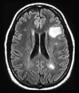
- Brain imaging revealed multifocal T2 hyperintense lesions, edema, and hemorrhages consistent with acute hemorrhagic leukoencephalitis (AHL) and restricted diffusion in the basal ganglia consistent with hypoxic brain injury. (nih.gov)
- Disease progression was consistent with acute hemorrhagic leukoencephalitis with rapid deterioration of consciousness and seizures. (cindyandwendy.com)
Subacute1
- Inflammation of a transverse portion of the spinal cord characterized by acute or subacute segmental demyelination or necrosis. (childrensmercy.org)
Respiratory9
- In less than 6 months, the severe acute respiratory syndrome-coronavirus type 2 (SARS-CoV-2) has spread worldwide infecting nearly 6 million people and killing over 350,000. (nih.gov)
- Clinical case: : We present a case report of a previously healthy young woman with an acute and multifocal clinical course, preceded by a viral respiratory tract infection, followed by a rapid disease progression and a delay in the diagnosis. (bvsalud.org)
- In December 2019, coronavirus disease 2019 spread worldwide, causing acute respiratory distress syndrome. (biomedcentral.com)
- Coronavirus disease 2019, like other acute respiratory syndromes, can invade the central nervous system through hematogenous and neuronal dissemination or it can be an immune response to the cytokine storm. (biomedcentral.com)
- Severe acute respiratory syndrome-coronavirus-2 (SARS-CoV-2) caused coronavirus disease 2019 (COVID-19) in Wuhan, China in December 2019. (biomedcentral.com)
- COVID-19 can appear from an asymptomatic situation to acute respiratory distress syndrome (ARDS) and eventually multiorgan damage [ 2 ]. (biomedcentral.com)
- acute respiratory infections in felines, rabbit hemorrhagic disease, and some cases of gastroenteritis in humans. (doctorinternet.com)
- Rates of cerebrovascular injury from acute respiratory distress syndrome may not be elevated in the setting of COVID-19 infection, but the latter may be associated with a unique pathophysiology deserving further research. (clevelandclinic.org)
- Acute respiratory distress syndrome (ARDS) is a common complication of COVID-19, and cerebrovascular and neurologic complications have been reported with severe COVID-19 infection. (clevelandclinic.org)
Encephalopathy2
Ischemic1
- Brain MRI findings comprised evidence of acute leukoencephalitis (n = 3, of whom one with a hemorrhagic form), cytotoxic edema mimicking ischemic stroke (n = 1), or normal results (n = 2). (bvsalud.org)
Myeloid1
- Acute myeloid and lymphoid leukemia and non-Hodgkin lymphoma have the highest mortality rates. (nih.gov)
Neurological1
- Acute and late neurological complications of COVID19: the quest for evidence. (medscape.com)