Lichenoid Eruptions
Tooth Eruption
Volcanic Eruptions
Prospects for ultraviolet A1 phototherapy as a treatment for chronic cutaneous graft-versus-host disease. (1/39)
BACKGROUND AND OBJECTIVES: Standard or investigative immunosuppressive therapies for cutaneous chronic graft-versus-host disease (GVHD) may prove not only ineffective but also cause serious adverse effects. Repeated exposure of the skin to ultraviolet radiation in the wavelength range 340-400 nm (so-called ultraviolet A1) was recently reported to have a strong local (intracutaneous) immunomodulatory activity. This study was undertaken to evaluate efficacy and safety of this phototherapy. DESIGN AND METHODS: Nine patients with cutaneous (4 lichenoid and 5 sclerodermoid) GVHD and mild or no other organ involvement were enrolled. All patients had developed serious drug toxicity and/or opportunistic infections. Phototherapy was administered three times a week. RESULTS: Complete remission was seen in 5 (2 lichenoid and 3 sclerodermoid) cases and a partial improvement in 4 (2 lichenoid and 2 sclerodermoid) after having received 15.8+/-3.8 (lichenoid GVHD) or 21.6+/-8.0 (sclerodermoid GVHD) sessions of phototherapy. Adverse effects were not registered. At follow-up (range: 6-25 months), two patients with sclerodermoid lesions relapsed after 5 months but responded to another treatment cycle. Patients with lichenoid GVHD showed relapses within one month and prolonged maintenance phototherapy was needed. Problems of drug toxicity and opportunistic infections improved as phototherapy allowed the reduction or interruption of systemic drug therapies. INTERPRETATION AND CONCLUSIONS: Ultraviolet A1 phototherapy may be considered as an appropriate therapeutic approach for sclerodermoid GVHD with no or mild involvement of internal organs. Patients with lichenoid GVHD should be treated only if they develop serious adverse effects to immunosuppressive therapies and opportunistic infections because of the carcinogenic hazard of high cumulative doses of ultraviolet A1 radiation. (+info)The immunopathology of regression in benign lichenoid keratosis, keratoacanthoma and halo nevus. (2/39)
BACKGROUND: Regression is a phenomenon present in a variety of cutaneous lesions. It is likely that similar immunologic mechanisms explain the phenomenon of spontaneous regression occurring in the various lesions. METHODS: Twenty-seven specimens, nine each of halo nevus, keratoacanthoma, and benign lichenoid keratosis, including three examples each of predominantly early, mid, and late regression were examined with antibodies to HLA-II, CD1a, CD3, CD4, CD8, CD20, CD34, CD56, and CD68. RESULTS: Epidermotropism of inflammatory cells, including CD1a positive, CD68 positive, CD3 positive, and CD8 positive cells, was present in benign lichenoid keratosis and keratoacanthoma, but not in halo nevus. In halo nevus, the nests of halo nevus cells tended to be infiltrated by CD1a positive, CD68 positive, CD3 positive, and CD8 positive cells. The blood vessels exhibited endothelial cell swelling with luminal narrowing and disruption within the dermis of all lesions. The CD1a positive cells were increased in number in lesional epidermis except in keratoacanthoma lesions where the density of CD1a positive cells was increased in the epithelial lip, but decreased within the epithelial portion of the keratoacanthoma proper. Conversely, the CD8 positive cells were scarce in the dermis below the epithelial lip of the keratoacanthoma, but increased in the dermis of the neoplastic epithelium. CD1a positive cells were also seen throughout the dermal portion of the lesion, particularly at the lesion base. In halo nevus, the CD1a positive cells and CD68 positive cells within the lesions were larger than those in non-lesional skin, indicating activation. The composition of the inflammatory infiltrate varied within each lesion type according to stage of regression, but T-lymphocytes predominated. CONCLUSION: Cytotoxic T-cells may be the final common denominator of regression in benign lichenoid keratosis, keratoacanthoma, and halo nevus. In halo nevus, cytotoxic T-cells may play the predominant role in regression. In keratoacanthoma and benign lichenoid keratosis, cytotoxic T-cells play a pivotal role, but additional mechanisms may also be involved in the phenomenon of regression. Benign lichenoid keratoses progress through stages of regression accompanied by varying proportions of inflammatory cells, including CD3, CD4, and CD8 positive T-lymphocytes, natural killer cells, macrophages and Langerhans cells. (+info)Progressive familial intrahepatic cholestasis with normal GGT level appearing with lichenification and enlargement of hands and feet. (3/39)
Progressive familial intrahepatic cholestasis is a serious disease of the liver, known as Byler disease, characterized by hepatocellular cholestasis. Severe pruritus and high serum bile acid concentrations are the most important diagnostic criteria of this autosomal recessive inherited disease. Here, we present a five-year-old boy with lichenification and enlargement of hands and feet as a sign of progressive familial intrahepatic cholestasis due to severe pruritus. (+info)Lichen amyloidosus: a study of clinical, histopathologic and immunofluorescence findings in 30 cases. (4/39)
BACKGROUND: Lichen amyloidosus (LA) is a primary localized cutaneous amyloidosis characterized clinically by discrete hyperkeratotic hyperpigmented papules and histologically by deposition of amyloid material in previously normal skin without any evidence of visceral involvement. AIMS AND OBJECTIVES: The aim of this work was to study the etiology, clinical features, histopathology and direct immunofluorescence findings in LA. METHODS: A prospective study of 30 patients with clinical, histological and immunofluorescence findings suggestive of LA was undertaken. After a detailed history and clinical examination, two punch biopsies for histopathology and immunofluorescence were taken. RESULTS: Of the 30 patients, 19 (63.3%) were males and 11 (36.7%) were females with duration of LA ranging from 6-20 months. Pruritus was the presenting symptom in 27 (90%) patients. Shin was involved in 26 (86.7%) followed by arms in three (10%) and back in one (3.3%). Seventeen patients (56%) had used scrubs for more than 2 years. Histopathology, direct immunofluorescence and Congo red staining detected amyloid in all cases. CONCLUSIONS: LA commonly presents over the shins as pruritic discrete hyperpigmented papules. Familial predisposition and friction may have a pathogenic role. Histopathological examination is very useful in the detection of amyloid which may be supplemented with direct immunofluorescence and Congo red staining. (+info)Cutaneous tuberculosis and phlyctenular keratoconjunctivitis: a forgotten association. (5/39)
Cutaneous tuberculosis may be associated with concurrent systemic foci in the body such as lung, lymph node, bone or CNS. Phlyctenular keratoconjunctivitis (PKC) is a manifestation of immunological response to a variety of antigens in the eye, tubercular focus (evident or occult) being the commonest in India. Reports in the existing literature have shown lungs and lymph nodes to be the predominant underlying focus associated with PKC, whereas cutaneous tuberculosis has seldom been found in this situation. We report this forgotten association in two children with cutaneous tuberculosis, one each with lupus vulgaris and scrofuloderma, who also had PKC. Interestingly, one of the cases also had simultaneous lichen scrofulosorum, which is also an immunological response to tubercular antigen and manifests in the skin, thus showing immunological manifestation in two different organ systems along with cutaneous focus of tuberculosis. (+info)Histochemical analysis of pathological alterations in oral lichen planus and oral lichenoid lesions. (6/39)
Lichen planus is a dermatologic disease of unknown etiology characterized by keratotic plaques on the skin. Many patients also harbor white lesions of the oral mucosa. The literature contains numerous reports of lichen planus-like lesions evolving in conjunction with the administration of a variety of pharmacologic agents. It is difficult, if not impossible, to distinguish such lesions from one another. The present study evaluated the epithelial and basement membrane thickness, mast cells (intact cells and degranulated cells subepithelially) and the presence or absence of blood vessels in oral lichen planus and oral lichenoid lesions. The evaluation was done using the periodic acid-schiff (PAS) and toluidine blue staining techniques on 20 cases each of oral lichen planus and oral lichenoid lesions and 5 control specimens of normal buccal mucosa. The results showed an increased number of degranulated mast cells in areas of basement membrane degeneration, increased vascularity and increased PAS-positive basement membrane thickness in oral lichen planus as compared with oral lichenoid lesions. Reduced epithelial thickness was found in oral lichen planus. The present study emphasizes the importance of these parameters in differentiating oral lichen planus from oral lichenoid lesions using special staining techniques. (+info)Lichen striatus in a child after immunization. A case report. (7/39)
Lichen striatus is a self-limited, lichenoid eruption particularly common in children. The lesions are located on extremities and less commonly on the trunk, and they follow the developmental lines of Blaschko. The etiology of lichen striatus is as yet unknown. It has been observed after infection or immunization in atopic patients and in siblings. The authors report on a 15-month-old girl that developed lichen striatus along the Blaschko lines on the trunk and one extremity after receiving the combined vaccine against measles, mumps, and rubella. Six months later, complete resolution of the skin lesions occurred without any treatment, leaving only slightly hypopigmented macules on the extremity. (+info)Bullous mycosis fungoides: a case report. (8/39)
Mycosis fungoides (MF) on extremely rare occasions is associated with vesiculobullous eruptions. We describe a 74-year-old man with previous documented histopathologic diagnosis of poikilodermic-type MF who recently developed flaccid acral bullae on erythematous MF plaques and on normal appearing skin. Histopathology and direct immunofluorescent studies revealed extensive lichenoid changes with intraepidermal bullae. Atypical lymphocyte infiltration was present at the dermoepidermal junction, in bulla fluid, and on the peripheral blood smear, but in lymph node and bone marrow tissue specimens atypical lymphocytes were absent. The plausible cause of these blister formations in cutaneous lymphoma bullosa may be from excessive epidermotropism or toxicity of the tumoral infiltrate. (+info)Lichenoid eruptions are skin reactions that resemble the appearance of lichen, a type of slow-growing fungus. These eruptions are characterized by flat, scaly bumps (papules) and rough, discolored patches (plaques) on the skin. They can be caused by various factors, including medications, medical conditions, or as a reaction to certain chemicals or substances that come into contact with the skin.
The term "lichenoid" refers to the resemblance of these eruptions to lichen, which is characterized by its distinctive appearance and growth pattern. Lichenoid eruptions can occur anywhere on the body but are most commonly found on sun-exposed areas such as the arms, legs, and trunk.
The exact cause of lichenoid eruptions can vary, but they are often associated with an autoimmune response in which the body's immune system mistakenly attacks healthy skin cells. This can lead to inflammation, redness, itching, and other symptoms associated with these eruptions. Treatment for lichenoid eruptions typically involves identifying and addressing the underlying cause, as well as managing symptoms with topical medications or other therapies.
Tooth eruption is the process by which a tooth emerges from the gums and becomes visible in the oral cavity. It is a normal part of dental development that occurs in a predictable sequence and timeframe. Primary or deciduous teeth, also known as baby teeth, begin to erupt around 6 months of age and continue to emerge until approximately 2-3 years of age. Permanent or adult teeth start to erupt around 6 years of age and can continue to emerge until the early twenties.
The process of tooth eruption involves several stages, including the formation of the tooth within the jawbone, the movement of the tooth through the bone and surrounding tissues, and the final emergence of the tooth into the mouth. Proper tooth eruption is essential for normal oral function, including chewing, speaking, and smiling. Any abnormalities in the tooth eruption process, such as delayed or premature eruption, can indicate underlying dental or medical conditions that require further evaluation and treatment.
I'm sorry for any confusion, but "Volcanic Eruptions" are not a medical term or concept. Volcanic eruptions refer to the release of molten rock, ash, and gases from a volcano's opening, or vent, onto the Earth's surface. This is a geological event that occurs due to the movement of tectonic plates and the build-up of pressure within the Earth's crust.
If you have any medical questions or terms you would like me to define, please feel free to ask!
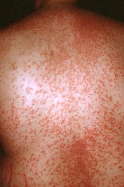
A "drug eruption" is a general term used to describe an adverse skin reaction that occurs as a result of taking a medication. These reactions can vary in severity and appearance, and may include symptoms such as rash, hives, itching, redness, blistering, or peeling of the skin. In some cases, drug eruptions can also cause systemic symptoms such as fever, fatigue, or joint pain.
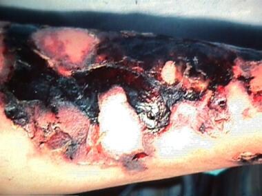
The exact mechanism by which drugs cause eruptions is not fully understood, but it is thought to involve an abnormal immune response to the medication. There are many different types of drug eruptions, including morphilliform rashes, urticaria (hives), fixed drug eruptions, and Stevens-Johnson syndrome/toxic epidermal necrolysis (SJS/TEN), which is a severe and potentially life-threatening reaction.
If you suspect that you are experiencing a drug eruption, it is important to seek medical attention promptly. Your healthcare provider can help determine the cause of the reaction and recommend appropriate treatment. In some cases, it may be necessary to discontinue the medication causing the reaction and switch to an alternative therapy.
 Lichenoid eruption