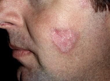
Lupus Erythematosus, Discoid
Lupus Erythematosus, Systemic
Lupus Erythematosus, Cutaneous
Lupus Nephritis
Antibodies, Antinuclear
Autoantibodies
Hydroxychloroquine
Lupus Vasculitis, Central Nervous System
Photosensitivity Disorders
Schizoid Personality Disorder
Antibodies, Anticardiolipin
Facial Dermatoses
Mice, Inbred NZB
Antibodies, Antiphospholipid
Autoantigens
Antiphospholipid Syndrome
Autoimmune Diseases
Immunoglobulin G
Mice, Inbred MRL lpr
Complement C4
Lupus Coagulation Inhibitor
snRNP Core Proteins
Antigen-Antibody Complex
Panniculitis, Lupus Erythematosus
Rheumatic Diseases
Autoimmunity
Arthritis, Rheumatoid
Sjogren's Syndrome
Complement C1q
Blood Platelets
Severity of Illness Index
Prednisolone
Complement C3
RNA, Small Cytoplasmic
B-Lymphocytes
DNA
Immunosuppressive Agents
Connective Tissue Diseases
Joint Diseases
Immunoglobulin M
Case-Control Studies
Enzyme-Linked Immunosorbent Assay
Genetic Predisposition to Disease
B-Cell Activating Factor
Cardiolipins
Collagen Diseases
Mixed Connective Tissue Disease
Complement C4a
Scleroderma, Systemic
Pityriasis
Lupus Vulgaris
Glomerulonephritis
Ribonucleoproteins, Small Nuclear
beta 2-Glycoprotein I
Biological Markers
Complement System Proteins
Vasculitis
Antibody Specificity
T-Lymphocytes
Cryoglobulins
Erythrocytes, Abnormal
Adrenal Cortex Hormones
Receptors, IgG
Plasmapheresis
Azathioprine
Retrospective Studies
Treatment Outcome
Microtubules
Follow-Up Studies
Interferon Regulatory Factors
B-Lymphocyte Subsets
Polymorphism, Single Nucleotide
Complement C4b
Erythrocytes
Prednisone
Antibodies
Complement C2
Lymphocyte Activation
Glucocorticoids
Myelitis, Transverse
Kidney
Antibodies, Anti-Idiotypic
Microscopy, Electron
Skin Diseases
Risk Factors
Genotype
Arthrography
Kidney Glomerulus
Anemia, Hemolytic, Autoimmune
Interferon-alpha
Disease Models, Animal
Platelet Aggregation
Cohort Studies
Flow Cytometry
Tumor Necrosis Factor Ligand Superfamily Member 13
Platelet Activation
Alleles
Polymorphism, Genetic
Receptors, Complement 3b
European Continental Ancestry Group
Lymphocytes
Leukocytes, Mononuclear
Biopsy
Gene Frequency
Immunoglobulin Idiotypes
Fluorescent Antibody Technique
Complement C3c
Rheumatoid Factor
Erythrocyte Membrane
Immunoglobulins
Haplotypes
Complement Activating Enzymes
Cyclophosphamide
Mycophenolic Acid
Magnetic Resonance Imaging
Cytoskeleton
OX40 Ligand
C3 metabolism in a patient with deficiency of the second component of complement (C2) and discoid lupus erythematosus. (1/80)
A patient with a hereditary deficiency of the second component of complement and discoid lupus erythematosus with features of systemic lupus erythematosus was studied. The propositus had a 9-year history of rash and arthralgia. Transient renal disease had completely resolved; there was a history of seizures. Examination of his serum disclosed antinuclear antibodies but no total haemolytic complement activity. C2 was absent. Serum concentrations of C1s, C3, C5 and C9 were elevated; other complement components were present in normal concentration, including C3 pro-activator. The patient's C3 pro-activator was electrophoretically converted by inulin and four of five lipopolysaccharides, but was poorly converted by aggregated human IgG. Two separate turnover studies with radiolabelled C3 showed fractional catabolic rates of 3-03 and 2-48% of the remaining plasma pool/hr (range of three normals: 1-62-2-18%/hr); and estimated C3 synthetic rates of 2-74 and 2-31 mg/kg/hr (range of three normals: 0-89-1-40 mg/kg/hr). Serum complement profiles of the patient's family demonstrated that the C2 deficiency was inherited as an autosomal codominant. One sibling, homozygous for C2 deficiency, and three other siblings, both parents and one daughter, all heterozygous for C2 deficiency, are in good health. Immunofluorescent studies of the patient's diseased skin exhibited substantial deposits of IgG, IgM, C1q, and C4 but not of later acting complement components, properdin, or C3 proactivator. These studies do not support the notion that inflammation in C3-deficient individuals with lupus erythematosus is mediated by the alternative complement pathway. (+info)Dermal fibroblasts sustain proliferation of activated T cells via membrane-bound interleukin-15 upon long-term stimulation with tumor necrosis factor-alpha. (2/80)
In chronic inflammatory conditions, mononuclear cells infiltrate the connective tissue attracted by fibroblast-secreted chemokines. The role of fibroblasts in sustaining the lymphocyte immune response upon cellular infiltration is so far unresolved. We here report that, upon prolonged stimulation with tumor necrosis factor-alpha, dermal fibroblasts enhance proliferation of activated T cells whereas unstimulated fibroblasts do not. T cell growth stimulation requires cell contact of tumor necrosis factor-alpha stimulated fibroblasts to T cells and is not due to soluble factors. Growth stimulation is substantially blocked by neutralizing antibodies to interleukin-15. Fluorescence-activated cell sorter analyses revealed that tumor necrosis factor alpha stimulated fibroblasts expose interleukin-15 in a membrane-bound form on the cell surface whereas nonstimulated fibroblasts and interferon-gamma treated fibroblasts do not. The amount of membrane interleukin-15 increases with the duration of tumor necrosis factor-alpha stimulation for at least 3 d. Unstimulated fibroblasts, however, accumulate interleukin-15 in the cytoplasm. No interleukin-15 could be detected in the culture supernatant. Immunohistochemical analyses confirmed membrane interleukin-15 on dermal fibroblasts in discoid lupus erythematosus skin lesions whereas no membrane interleukin-15 was found on the surface of fibroblasts in healthy skin. We conclude that dermal fibroblasts upon long-term tumor necrosis factor-alpha stimulation during chronic inflammation are involved via membrane-bound interleukin-15 in stimulating proliferation of accumulated, activated T cells. (+info)SET-related cell division autoantigen-1 (CDA1) arrests cell growth. (3/80)
We used an autoimmune serum from a patient with discoid lupus erythematosus to clone a cDNA of 2808 base pairs. Its open reading frame of 2079 base pairs encodes a predicted polypeptide of 693 amino acids named CDA1 (cell division autoantigen-1). CDA1 has a predicted molecular mass of 79,430 Daltons and a pI of 4.26. The size of the cDNA is consistent with its estimated mRNA size. CDA1 comprises an N-terminal proline-rich domain, a central basic domain, and a C-terminal bipartite acidic domain. It has four putative nuclear localization signals and potential sites for phosphorylation by cAMP and cGMP-dependent kinases, protein kinase C, thymidine kinase, casein kinase II, and cyclin-dependent kinases (CDKs). CDA1 is phosphorylated in HeLa cells and by cyclin D1/CDK4, cyclin A/CDK2, and cyclin B/CDK1 in vitro. Its basic and acidic domains contain regions homologous to almost the entire human leukemia-associated SET protein. The same basic region is also homologous to nucleosome assembly proteins, testis TSPY protein, and an uncharacterized brain protein. CDA1 is present in the nuclear fraction of HeLa cells and localizes to the nucleus and nucleolus in HeLa cells transfected with CDA1 or its N terminus containing all four nuclear localization signals. Its acidic C terminus localizes mainly to the cytoplasm. CDA1 levels are low in serum-starved cells, increasing dramatically with serum stimulation. Expression of the CDA1 transgene, but not its N terminus, arrests HeLa cell growth, colony numbers, cell density, and bromodeoxyuridine uptake in a dose-dependent manner. The ability of CDA1 to arrest cell growth is abolished by mutation of the two CDK consensus phosphorylation sites. We propose that CDA1 is a negative regulator of cell growth and that its activity is regulated by its expression level and phosphorylation. (+info)Plasmacytoid dendritic cells (natural interferon- alpha/beta-producing cells) accumulate in cutaneous lupus erythematosus lesions. (4/80)
Plasmacytoid dendritic cell (P-DC) precursors in peripheral blood produce large amounts of interferon (IFN)-alpha/beta when triggered by viruses. However, when incubated with interleukin-3 and CD40 ligand, the same precursors differentiate into mature DCs that stimulate naive CD4(+) T cells to produce Th2 cytokines. We recently reported that P-DCs accumulate in nasal mucosa of experimentally induced allergic rhinitis, supporting a role for this DC subset in Th2-dominated inflammation. Here we examined whether P-DCs accumulate in cutaneous lesions of lupus erythematosus (LE), a disorder associated with increased IFN-alpha/beta production. Our results showed that P-DCs were present in 14 out of 15 tissue specimens of cutaneous LE lesions, but not in normal skin. Importantly, the density of P-DCs in affected skin correlated well (r(s) = 0.79,P < 0.0005) with the high number of cells expressing the IFN-alpha/beta-inducible protein MxA, suggesting that P-DCs produce IFN-alpha/beta locally. Accumulation of P-DCs coincided also with the expression of L-selectin ligand peripheral lymph node addressin on dermal vascular endothelium, adding further support to the notion that these adhesion molecules are important in P-DC extravasation to peripheral tissue sites. Together, our findings suggested that P-DCs are an important source of IFN-alpha/beta in cutaneous LE lesions and may therefore be of pathogenic importance. (+info)Clinical studies with topical fluocinolone acetonide in the treatment of various dermatoses. (5/80)
Fluocinolone acetonide cream is a new, potent topical corticosteroid. When used in conjunction with an occlusive plastic film dressing, herein described, it is highly effective in the treatment of psoriasis of the glabrous skin, pustular and paronychial psoriasis, neurodermatitis, and lichen planus. Psoriasis of the intertriginous areas responds to the local use of the fluocinolone cream alone. Relapses on cessation of treatment respond as a rule to retreatment.Indications, limitations, reactions and contraindications to this form of treatment are discussed. (+info)SEROMUCOID IN LUPUS ERYTHRMATOSUS AND SCLERODERMA. (6/80)
Seromucoid levels in blood of 50 normal men and in 63 cases of cutaneous collagen diseases were studied. There was a marked rise in the seromucoid level in cases of systemic lupus erythematosus and also a significant rise in scleroderma. This rise in seromucoid level may be a reflection of the degenerative connective tissue. (+info)A candidate region on 11p13 for systemic lupus erythematosus: a linkage identified in African-American families. (7/80)
Systemic Lupus Erythematosus (SLE), a chronic, complex disease, is the prototype for systemic human autoimmune diseases. Although environmental factors are crucial in triggering the condition, twin and family studies, as well as genetic linkage and association studies, have established its strong genetic predisposition. During the past few years, there has been considerable interest in identifying genomic segments linked to SLE through either a whole genome scan or a candidate gene approach. The discoid lupus erythematosus (DLE) skin lesion is one of the major, and discriminating, manifestations in SLE, especially in African American patients. In this study we have identified 58 multiplex families--27 African American, 26 European American, and 5 others--where at least one SLE patient is also reported to be afflicted with DLE. These families were chosen from the collection of families that are part of our ongoing linkage study for SLE. A genome-wide parametric and nonparametric linkage analysis was conducted with 320 markers. Significant evidence of linkage was identified in only one chromosomal location, 11p13, in the African American families. The maximum 2-point linkage was 5.6 in these pedigrees, obtained at a marker located 47 CM away from the pterminal end of chromosome 11. The peak multipoint LOD score of 4.6 was obtained very nearby. The segregation of this gene suggests dominant inheritance. These results reveal an important genetic effect related to discoid rash at 11p13 in African Americans with SLE, and demonstrate, through increasing genetic homogeneity, the power of pedigree stratification to detect linkage in complex diseases. (+info)Topical tacrolimus therapy of resistant cutaneous lesions in lupus erythematosus: a possible alternative. (8/80)
OBJECTIVE: To determine the efficacy of tacrolimus ointment 0.1% on resistant cutaneous lesions in patients with lupus erythematosus. METHODS: Twelve patients with skin manifestations were studied. Six had discoid lupus (DL), four subacute cutaneous lupus erythematosus (SCLE) and two systemic lupus erythematosus (SLE). All patients had extensive skin lesions refractory to previous treatment. Patients received topical tacrolimus 0.1% for a minimum of 6 weeks and response was evaluated by physicians' and patients' assessment and documented with photographs at baseline and at the end of the treatment. RESULTS: Eleven of 12 patients completed the therapy. One patient with DL discontinued because of side--effects-peeling and a burning sensation. Six patients were clearly improved, one patient had a minor remission of his face lesion while in four the rashes remained the same. Two patients with SCLE had significant regression of their lesions while the other two had no improvement. In DL, two had certain improvement, one minor improvement and two were without response. The patients with SLE had significant amelioration of their extensive photosensitive rash. CONCLUSION: Tacrolimus ointment 0.1% may be an effective alternative in patients with severe resistant cutaneous manifestations in lupus erythematosus. (+info)Discoid Lupus Erythematosus (DLE) is a chronic autoimmune disease that primarily affects the skin. It is a subtype of Cutaneous Lupus Erythematosus (CLE). DLE is characterized by coin-shaped, disc-like rashes on the face, scalp, and other sun-exposed areas of the body. These lesions are often red, scaly, and may cause scarring and pigmentation changes. Unlike Systemic Lupus Erythematosus (SLE), DLE typically does not affect internal organs, but in some cases, it can progress to SLE. The exact cause of DLE is unknown, but it is believed to be related to a combination of genetic, environmental, and hormonal factors that trigger an abnormal immune response. Treatment for DLE may include topical creams, oral medications, and avoidance of sun exposure.
Systemic Lupus Erythematosus (SLE) is a complex autoimmune disease that can affect almost any organ or system in the body. In SLE, the immune system produces an exaggerated response, leading to the production of autoantibodies that attack the body's own cells and tissues, causing inflammation and damage. The symptoms and severity of SLE can vary widely from person to person, but common features include fatigue, joint pain, skin rashes (particularly a "butterfly" rash across the nose and cheeks), fever, hair loss, and sensitivity to sunlight.
Systemic lupus erythematosus can also affect the kidneys, heart, lungs, brain, blood vessels, and other organs, leading to a wide range of symptoms such as kidney dysfunction, chest pain, shortness of breath, seizures, and anemia. The exact cause of SLE is not fully understood, but it is believed to involve a combination of genetic, environmental, and hormonal factors. Treatment typically involves medications to suppress the immune system and manage symptoms, and may require long-term management by a team of healthcare professionals.
Cutaneous Lupus Erythematosus (CLE) is a skin manifestation of Systemic Lupus Erythematosus (SLE), an autoimmune disease, but it can also occur without systemic involvement. It is characterized by various skin lesions that differ in appearance and distribution. The three main subtypes of CLE are:
1. Acute Cutaneous Lupus Erythematosus (ACLE): This form is typically associated with SLE and is characterized by a classic malar or "butterfly" rash on the face, which is often photosensitive and can be accompanied by discoid lesions. The rash may also appear on other sun-exposed areas of the body.
2. Chronic Cutaneous Lupus Erythematosus (CCLE): This subtype includes Discoid Lupus Erythematosus (DLE) and other less common forms such as lupus panniculitis and chilblain lupus. DLE is characterized by well-circumscribed, erythematous, scaly plaques that can cause scarring and pigmentation changes, often found on the face, scalp, and ears. Lupus panniculitis presents as deep subcutaneous nodules or indurated plaques, typically located on the trunk and proximal extremities. Chilblain lupus is characterized by violaceous, tender, and swollen lesions on acral areas, often triggered by cold exposure.
3. Subacute Cutaneous Lupus Erythematosus (SCLE): This form of CLE presents as non-scarring, papulosquamous or annular polycyclic rashes, often located on the trunk and proximal extremities. The lesions are typically photosensitive and may appear in patients with SLE or those with isolated cutaneous disease.
The diagnosis of Cutaneous Lupus Erythematosus is based on clinical presentation, histopathological findings, and sometimes direct immunofluorescence. Treatment depends on the severity and extent of skin involvement and may include topical therapies, antimalarials, corticosteroids, immunomodulatory agents, or photoprotection measures.
Lupus nephritis is a type of kidney inflammation (nephritis) that can occur in people with systemic lupus erythematosus (SLE), an autoimmune disease. In lupus nephritis, the immune system produces abnormal antibodies that attack the tissues of the kidneys, leading to inflammation and damage. The condition can cause a range of symptoms, including proteinuria (protein in the urine), hematuria (blood in the urine), hypertension (high blood pressure), and eventually kidney failure if left untreated. Lupus nephritis is typically diagnosed through a combination of medical history, physical examination, laboratory tests, and imaging studies. Treatment may include medications to suppress the immune system and control inflammation, such as corticosteroids and immunosuppressive drugs.
Antinuclear antibodies (ANA) are a type of autoantibody that target structures found in the nucleus of a cell. These antibodies are produced by the immune system and attack the body's own cells and tissues, leading to inflammation and damage. The presence of ANA is often used as a marker for certain autoimmune diseases, such as systemic lupus erythematosus (SLE), Sjogren's syndrome, rheumatoid arthritis, scleroderma, and polymyositis.
ANA can be detected through a blood test called the antinuclear antibody test. A positive result indicates the presence of ANA in the blood, but it does not necessarily mean that a person has an autoimmune disease. Further testing is usually needed to confirm a diagnosis and determine the specific type of autoantibodies present.
It's important to note that ANA can also be found in healthy individuals, particularly as they age. Therefore, the test results should be interpreted in conjunction with other clinical findings and symptoms.
Autoantibodies are defined as antibodies that are produced by the immune system and target the body's own cells, tissues, or organs. These antibodies mistakenly identify certain proteins or molecules in the body as foreign invaders and attack them, leading to an autoimmune response. Autoantibodies can be found in various autoimmune diseases such as rheumatoid arthritis, lupus, and thyroiditis. The presence of autoantibodies can also be used as a diagnostic marker for certain conditions.
Hydroxychloroquine is an antimalarial and autoimmune disease medication. It's primarily used to prevent or treat malaria, a disease caused by parasites that enter the body through the bites of infected mosquitoes. It works by killing the malaria parasite in the red blood cells of the human body.
In addition, hydroxychloroquine is also used to treat autoimmune diseases such as rheumatoid arthritis and lupus. In these conditions, the body's immune system mistakenly attacks healthy tissues, causing inflammation and damage. Hydroxychloroquine helps to regulate the immune system and reduce inflammation.
It is important to note that while hydroxychloroquine has been studied as a potential treatment for COVID-19, current evidence does not support its use outside of a clinical trial setting due to lack of efficacy and potential for harm.
Lupus vasculitis in the central nervous system (CNS) is a specific type of inflammation that occurs in the blood vessels of the brain and/or spinal cord due to systemic lupus erythematosus (SLE), an autoimmune disease. In this condition, the body's immune system mistakenly attacks healthy tissue, including blood vessel walls, leading to their inflammation and damage.
CNS vasculitis can cause various neurological symptoms such as headaches, seizures, cognitive impairment, mood changes, stroke-like episodes, and even loss of consciousness. The diagnosis typically involves a combination of clinical evaluation, imaging studies (such as MRI or angiography), and laboratory tests to detect the presence of autoantibodies associated with SLE. Treatment usually includes immunosuppressive therapy to control the inflammation and prevent further damage to the blood vessels in the CNS.
The menisci are crescent-shaped fibrocartilaginous structures located in the knee joint. There are two menisci in each knee: the medial meniscus and the lateral meniscus. The tibial menisci, also known as the medial and lateral menisci, are named according to their location in the knee joint. They lie on the top surface of the tibia (shin bone) and provide shock absorption, stability, and lubrication to the knee joint.
The tibial menisci have a complex shape, with a wider outer portion called the peripheral rim and a narrower inner portion called the central portion or root attachment. The menisci are attached to the bones of the knee joint by ligaments and have a rich blood supply in their outer portions, which helps in healing after injury. However, the inner two-thirds of the menisci have a poor blood supply, making them more prone to degeneration and less likely to heal after injury.
Damage to the tibial menisci can occur due to trauma or degenerative changes, leading to symptoms such as pain, swelling, stiffness, and limited mobility of the knee joint. Treatment for meniscal injuries may include physical therapy, bracing, or surgery, depending on the severity and location of the injury.
Photosensitivity disorders refer to conditions that cause an abnormal reaction to sunlight or artificial light. This reaction can take the form of various skin changes, such as rashes, inflammation, or pigmentation, and in some cases, it can also lead to systemic symptoms like fatigue, fever, or joint pain.
The two main types of photosensitivity disorders are:
1. Phototoxic reactions: These occur when a substance (such as certain medications, chemicals, or plants) absorbs light energy and transfers it to skin cells, causing damage and inflammation. The reaction typically appears within 24 hours of exposure to the light source and can resemble a sunburn.
2. Photoallergic reactions: These occur when the immune system responds to the combination of light and a particular substance, leading to an allergic response. The reaction may not appear until several days after initial exposure and can cause redness, itching, and blistering.
It is important for individuals with photosensitivity disorders to avoid excessive sun exposure, wear protective clothing, and use broad-spectrum sunscreens with a high SPF rating to minimize the risk of phototoxic or photoallergic reactions.
Schizoid Personality Disorder is defined by the American Psychiatric Association's Diagnostic and Statistical Manual, Fifth Edition (DSM-5) as a long-standing pattern of detachment from social relationships, a reduced capacity for emotional expression, and an unusual degree of introversion. This disorder is characterized by:
1. A lack of desire for close relationships,
2. Difficulty expressing emotions and finding enjoyment in most activities,
3. Limited range of emotional expression,
4. Inattention to social norms and conventions,
5. Preference for being alone,
6. Indifference to praise or criticism from others.
These symptoms must be stable and of long duration, typically present for at least a year. The individual's lifestyle, attitudes, and behavior are often seen as eccentric and distant by others. It is important to note that this disorder is different from Schizophrenia and does not include psychotic symptoms such as hallucinations or delusions.
Anticardiolipin antibodies are a type of autoantibody that targets and binds to cardiolipin, a phospholipid component found in the inner mitochondrial membrane of cells. These antibodies are clinically significant because they have been associated with a variety of autoimmune disorders, including antiphospholipid syndrome (APS).
APS is a condition characterized by recurrent blood clots, pregnancy losses, and thrombocytopenia (low platelet count). Anticardiolipin antibodies are one of the three main types of autoantibodies found in APS, along with lupus anticoagulant and anti-β2 glycoprotein I antibodies.
The presence of high levels of anticardiolipin antibodies in the blood can lead to abnormal blood clotting, which can cause serious complications such as deep vein thrombosis, pulmonary embolism, and stroke. Anticardiolipin antibodies can also contribute to pregnancy losses by causing placental insufficiency or abnormal blood clotting in the placenta.
Anticardiolipin antibodies are typically detected through a blood test that measures their levels in the serum. A positive result is usually confirmed with a second test performed at least 12 weeks later to establish persistence. Treatment for anticardiolipin antibody-related disorders typically involves anticoagulation therapy to prevent blood clots and other complications.
Facial dermatoses refer to various skin conditions that affect the face. These can include a wide range of disorders, such as:
1. Acne vulgaris: A common skin condition characterized by the formation of comedones (blackheads and whiteheads) and inflammatory papules, pustules, and nodules. It primarily affects the face, neck, chest, and back.
2. Rosacea: A chronic skin condition that causes redness, flushing, and visible blood vessels on the face, along with bumps or pimples and sometimes eye irritation.
3. Seborrheic dermatitis: A common inflammatory skin disorder that causes a red, itchy, and flaky rash, often on the scalp, face, and eyebrows. It can also affect other oily areas of the body, like the sides of the nose and behind the ears.
4. Atopic dermatitis (eczema): A chronic inflammatory skin condition that causes red, itchy, and scaly patches on the skin. While it can occur anywhere on the body, it frequently affects the face, especially in infants and young children.
5. Psoriasis: An autoimmune disorder that results in thick, scaly, silvery, or red patches on the skin. It can affect any part of the body, including the face.
6. Contact dermatitis: A skin reaction caused by direct contact with an allergen or irritant, resulting in redness, itching, and inflammation. The face can be affected when allergens or irritants come into contact with the skin through cosmetics, skincare products, or other substances.
7. Lupus erythematosus: An autoimmune disorder that can cause a butterfly-shaped rash on the cheeks and nose, along with other symptoms like joint pain, fatigue, and photosensitivity.
8. Perioral dermatitis: A inflammatory skin condition that causes redness, small bumps, and dryness around the mouth, often mistaken for acne. It can also affect the skin around the nose and eyes.
9. Vitiligo: An autoimmune disorder that results in the loss of pigmentation in patches of skin, which can occur on the face and other parts of the body.
10. Tinea faciei: A fungal infection that affects the facial skin, causing red, scaly, or itchy patches. It is also known as ringworm of the face.
These are just a few examples of skin conditions that can affect the face. If you experience any unusual symptoms or changes in your skin, it's essential to consult a dermatologist for proper diagnosis and treatment.
'NZB mice' is a term used to refer to an inbred strain of laboratory mice that are genetically identical to each other and have been used extensively in biomedical research. The 'NZB' designation stands for "New Zealand Black," which refers to the coat color of these mice.
NZB mice are known to spontaneously develop an autoimmune disease that is similar to human systemic lupus erythematosus (SLE), a chronic inflammatory disorder caused by an overactive immune system. This makes them a valuable model for studying the genetic and environmental factors that contribute to the development of SLE, as well as for testing new therapies and treatments.
It's important to note that while NZB mice are an inbred strain, they may still exhibit some variability in their disease phenotype due to genetic modifiers or environmental influences. Therefore, researchers often use large cohorts of mice and standardized experimental conditions to ensure the reproducibility and reliability of their findings.
Arthroscopy is a minimally invasive surgical procedure where an orthopedic surgeon uses an arthroscope (a thin tube with a light and camera on the end) to diagnose and treat problems inside a joint. The surgeon makes a small incision, inserts the arthroscope into the joint, and then uses the attached camera to view the inside of the joint on a monitor. They can then insert other small instruments through additional incisions to repair or remove damaged tissue.
Arthroscopy is most commonly used for joints such as the knee, shoulder, hip, ankle, and wrist. It offers several advantages over traditional open surgery, including smaller incisions, less pain and bleeding, faster recovery time, and reduced risk of infection. The procedure can be used to diagnose and treat a wide range of conditions, including torn ligaments or cartilage, inflamed synovial tissue, loose bone or cartilage fragments, and joint damage caused by arthritis.
Antiphospholipid antibodies are a type of autoantibody that targets and binds to certain proteins found in the blood that attach to phospholipids (a type of fat molecule). These antibodies are associated with an increased risk of developing antiphospholipid syndrome, a disorder characterized by abnormal blood clotting.
There are several types of antiphospholipid antibodies, including:
1. Lupus anticoagulant: This type of antiphospholipid antibody can interfere with blood clotting tests and may increase the risk of thrombosis (blood clots) in both arteries and veins.
2. Anticardiolipin antibodies: These antibodies target a specific phospholipid called cardiolipin, which is found in the inner membrane of mitochondria. High levels of anticardiolipin antibodies are associated with an increased risk of thrombosis and pregnancy complications such as recurrent miscarriage.
3. Anti-β2 glycoprotein I antibodies: These antibodies target a protein called β2 glycoprotein I, which binds to negatively charged phospholipids on the surface of cells. High levels of anti-β2 glycoprotein I antibodies are associated with an increased risk of thrombosis and pregnancy complications.
The exact mechanism by which antiphospholipid antibodies cause blood clotting is not fully understood, but it is thought to involve the activation of platelets, the inhibition of natural anticoagulants, and the promotion of inflammation. Antiphospholipid syndrome can be treated with medications that thin the blood or prevent clots from forming, such as aspirin, warfarin, or heparin.
Autoantigens are substances that are typically found in an individual's own body, but can stimulate an immune response because they are recognized as foreign by the body's own immune system. In autoimmune diseases, the immune system mistakenly attacks and damages healthy tissues and organs because it recognizes some of their components as autoantigens. These autoantigens can be proteins, DNA, or other molecules that are normally present in the body but have become altered or exposed due to various factors such as infection, genetics, or environmental triggers. The immune system then produces antibodies and activates immune cells to attack these autoantigens, leading to tissue damage and inflammation.
Antiphospholipid syndrome (APS) is an autoimmune disorder characterized by the presence of antiphospholipid antibodies in the blood. These antibodies are directed against phospholipids, a type of fat molecule found in cell membranes and plasma lipoproteins. The presence of these antibodies can lead to abnormal blood clotting, which can cause serious complications such as stroke, heart attack, deep vein thrombosis, and pulmonary embolism.
APS can occur either on its own (primary APS) or in conjunction with other autoimmune disorders, such as systemic lupus erythematosus (secondary APS). The exact cause of APS is not fully understood, but it is believed to involve a combination of genetic and environmental factors.
Symptoms of APS can vary widely depending on the location and severity of the blood clots. They may include:
* Recurrent miscarriages or stillbirths
* Blood clots in the legs, lungs, or other parts of the body
* Skin ulcers or lesions
* Headaches, seizures, or stroke-like symptoms
* Kidney problems
* Heart valve abnormalities
Diagnosis of APS typically involves blood tests to detect the presence of antiphospholipid antibodies. Treatment may include medications to prevent blood clots, such as anticoagulants and antiplatelet agents, as well as management of any underlying autoimmune disorders.
Autoimmune diseases are a group of disorders in which the immune system, which normally protects the body from foreign invaders like bacteria and viruses, mistakenly attacks the body's own cells and tissues. This results in inflammation and damage to various organs and tissues in the body.
In autoimmune diseases, the body produces autoantibodies that target its own proteins or cell receptors, leading to their destruction or malfunction. The exact cause of autoimmune diseases is not fully understood, but it is believed that a combination of genetic and environmental factors contribute to their development.
There are over 80 different types of autoimmune diseases, including rheumatoid arthritis, lupus, multiple sclerosis, type 1 diabetes, Hashimoto's thyroiditis, Graves' disease, psoriasis, and inflammatory bowel disease. Symptoms can vary widely depending on the specific autoimmune disease and the organs or tissues affected. Treatment typically involves managing symptoms and suppressing the immune system to prevent further damage.
Immunoglobulin G (IgG) is a type of antibody, which is a protective protein produced by the immune system in response to foreign substances like bacteria or viruses. IgG is the most abundant type of antibody in human blood, making up about 75-80% of all antibodies. It is found in all body fluids and plays a crucial role in fighting infections caused by bacteria, viruses, and toxins.
IgG has several important functions:
1. Neutralization: IgG can bind to the surface of bacteria or viruses, preventing them from attaching to and infecting human cells.
2. Opsonization: IgG coats the surface of pathogens, making them more recognizable and easier for immune cells like neutrophils and macrophages to phagocytose (engulf and destroy) them.
3. Complement activation: IgG can activate the complement system, a group of proteins that work together to help eliminate pathogens from the body. Activation of the complement system leads to the formation of the membrane attack complex, which creates holes in the cell membranes of bacteria, leading to their lysis (destruction).
4. Antibody-dependent cellular cytotoxicity (ADCC): IgG can bind to immune cells like natural killer (NK) cells and trigger them to release substances that cause target cells (such as virus-infected or cancerous cells) to undergo apoptosis (programmed cell death).
5. Immune complex formation: IgG can form immune complexes with antigens, which can then be removed from the body through various mechanisms, such as phagocytosis by immune cells or excretion in urine.
IgG is a critical component of adaptive immunity and provides long-lasting protection against reinfection with many pathogens. It has four subclasses (IgG1, IgG2, IgG3, and IgG4) that differ in their structure, function, and distribution in the body.
'Mice, Inbred MRL-lpr' refers to a specific strain of laboratory mice that are used in biomedical research. The 'MRL' part of the name stands for the breeding colony where they were originally developed, which is the Mouse Repository at the Jackson Laboratory in Bar Harbor, Maine. The 'lpr' designation indicates that these mice carry a mutation in the Fas gene, also known as lpr (lymphoproliferation) gene, which leads to an autoimmune disorder characterized by lymphadenopathy (enlarged lymph nodes), splenomegaly (enlarged spleen), and production of autoantibodies.
The MRL-lpr mice are known for their accelerated aging phenotype, which includes the development of a variety of age-related diseases such as atherosclerosis, osteoporosis, and cancer. They also develop a severe form of systemic lupus erythematosus (SLE), an autoimmune disease that affects many organs in the body. The MRL-lpr mice are widely used as a model to study the pathogenesis of SLE and other autoimmune diseases, as well as to test potential therapies for these conditions.
It is important to note that while inbred mouse strains like MRL-lpr provide valuable insights into human disease mechanisms, they do not perfectly replicate all aspects of human disease, and results obtained in mice may not always translate directly to humans. Therefore, findings from mouse studies should be interpreted with caution and validated in human studies before being applied in clinical practice.
Complement C4 is a protein that plays a crucial role in the complement system, which is a part of the immune system that helps to clear pathogens and damaged cells from the body. Complement C4 is involved in the early stages of the complement activation cascade, where it helps to identify and tag foreign or abnormal cells for destruction by other components of the immune system.
Specifically, Complement C4 can be cleaved into two smaller proteins, C4a and C4b, during the complement activation process. C4b then binds to the surface of the target cell and helps to initiate the formation of the membrane attack complex (MAC), which creates a pore in the cell membrane and leads to lysis or destruction of the target cell.
Deficiencies or mutations in the Complement C4 gene can lead to various immune disorders, including certain forms of autoimmune diseases and susceptibility to certain infections.
I'm sorry for any confusion, but "Lupus Coagulation Inhibitor" is not a recognized medical term or concept in hematology or immunology.
Systemic Lupus Erythematosus (SLE), often referred to simply as lupus, is an autoimmune disease where the body's immune system mistakenly attacks healthy tissue. However, this does not result in a specific coagulation inhibitor.
If you're asking about lupus anticoagulants, these are antibodies that can interfere with clotting tests but paradoxically increase the risk of blood clots in vivo. They are sometimes seen in patients with SLE and other autoimmune diseases.
Please provide more context if you meant something else, so I can give a more accurate response.
SnRNP (small nuclear ribonucleoprotein) core proteins are a group of proteins that are associated with small nuclear RNAs (snRNAs) to form small nuclear ribonucleoprotein particles. These particles play crucial roles in various aspects of RNA processing, such as splicing, 3' end formation, and degradation.
The snRNP core proteins include seven Sm proteins (B, D1, D2, D3, E, F, and G) that form a heptameric ring-like structure called the Sm core, which binds to a conserved sequence motif in the snRNAs called the Sm site. In addition to the Sm proteins, there are also other core proteins such as Sm like (L) proteins and various other protein factors that associate with specific snRNP particles.
Together, these snRNP core proteins help to stabilize the snRNA, facilitate its assembly into functional ribonucleoprotein complexes, and participate in the recognition and processing of target RNAs during post-transcriptional regulation.
An antigen-antibody complex is a type of immune complex that forms when an antibody binds to a specific antigen. An antigen is any substance that triggers an immune response, while an antibody is a protein produced by the immune system to neutralize or destroy foreign substances like antigens.
When an antibody binds to an antigen, it forms a complex that can be either soluble or insoluble. Soluble complexes are formed when the antigen is small and can move freely through the bloodstream. Insoluble complexes, on the other hand, are formed when the antigen is too large to move freely, such as when it is part of a bacterium or virus.
The formation of antigen-antibody complexes plays an important role in the immune response. Once formed, these complexes can be recognized and cleared by other components of the immune system, such as phagocytes, which help to prevent further damage to the body. However, in some cases, the formation of large numbers of antigen-antibody complexes can lead to inflammation and tissue damage, contributing to the development of certain autoimmune diseases.
Lupus erythematosus panniculitis (LEP), also known as lupus profundus, is a type of cutaneous lupus erythematosus (CLE) that affects the fatty layer under the skin (subcutaneous tissue). It is characterized by deep inflammation of the fatty tissue, leading to nodular or indurated (hardened) plaques and subcutaneous ulcerations. The lesions are typically tender and can be painful. LEP most commonly affects the face, trunk, and proximal extremities.
LEP is associated with systemic lupus erythematosus (SLE), but it can also occur in isolation without evidence of systemic involvement. It is estimated that approximately 1-10% of patients with SLE develop LEP. The exact pathogenesis of LEP remains unclear, but it is thought to involve an autoimmune response directed against the fatty tissue.
Histologically, LEP is characterized by lobular panniculitis with lymphocytic infiltration and fat necrosis. Direct immunofluorescence may show deposition of immune complexes along the blood vessels in the affected area.
Treatment of LEP can be challenging, and often requires a multidisciplinary approach involving dermatologists, rheumatologists, and other specialists. Systemic therapies such as corticosteroids, antimalarials, immunosuppressive agents, and biologics may be used to control the disease activity. In addition, local treatments such as intralesional steroid injections or surgical excision of lesions may be considered for refractory cases.
Rheumatic diseases are a group of disorders that cause pain, stiffness, and swelling in the joints, muscles, tendons, ligaments, or bones. They include conditions such as rheumatoid arthritis, osteoarthritis, systemic lupus erythematosus (SLE), gout, ankylosing spondylitis, psoriatic arthritis, and many others. These diseases can also affect other body systems including the skin, eyes, lungs, heart, kidneys, and nervous system. Rheumatic diseases are often chronic and may be progressive, meaning they can worsen over time. They can cause significant pain, disability, and reduced quality of life if not properly diagnosed and managed. The exact causes of rheumatic diseases are not fully understood, but genetics, environmental factors, and immune system dysfunction are believed to play a role in their development.
Autoimmunity is a medical condition in which the body's immune system mistakenly attacks and destroys healthy tissues within the body. In normal function, the immune system recognizes and fights off foreign substances such as bacteria, viruses, and toxins. However, when autoimmunity occurs, the immune system identifies self-molecules or tissues as foreign and produces an immune response against them.
This misguided response can lead to chronic inflammation, tissue damage, and impaired organ function. Autoimmune diseases can affect various parts of the body, including the joints, skin, glands, muscles, and blood vessels. Some common examples of autoimmune diseases are rheumatoid arthritis, lupus, multiple sclerosis, type 1 diabetes, Hashimoto's thyroiditis, and Graves' disease.
The exact cause of autoimmunity is not fully understood, but it is believed to involve a combination of genetic, environmental, and lifestyle factors that trigger an abnormal immune response in susceptible individuals. Treatment for autoimmune diseases typically involves managing symptoms, reducing inflammation, and suppressing the immune system's overactive response using medications such as corticosteroids, immunosuppressants, and biologics.
Rheumatoid arthritis (RA) is a systemic autoimmune disease that primarily affects the joints. It is characterized by persistent inflammation, synovial hyperplasia, and subsequent damage to the articular cartilage and bone. The immune system mistakenly attacks the body's own tissues, specifically targeting the synovial membrane lining the joint capsule. This results in swelling, pain, warmth, and stiffness in affected joints, often most severely in the hands and feet.
RA can also have extra-articular manifestations, affecting other organs such as the lungs, heart, skin, eyes, and blood vessels. The exact cause of RA remains unknown, but it is believed to involve a complex interplay between genetic susceptibility and environmental triggers. Early diagnosis and treatment are crucial in managing rheumatoid arthritis to prevent joint damage, disability, and systemic complications.
Cartilage diseases refer to conditions that affect the cartilaginous tissues in the body. Cartilage is a firm, flexible connective tissue found in many areas of the body, including the joints, ribcage, ears, and nose. It provides structure and support, allows for smooth movement between bones, and protects the ends of bones from friction.
There are several types of cartilage diseases, including:
1. Osteoarthritis (OA): This is a degenerative joint disease that occurs when the protective cartilage that cushions the ends of your bones wears down over time. It can cause pain, stiffness, and loss of mobility in the affected joints.
2. Rheumatoid arthritis (RA): This is an autoimmune disorder that causes inflammation in the lining of the joints, leading to cartilage damage and bone erosion.
3. Traumatic arthritis: This occurs when a joint is injured, causing damage to the cartilage and resulting in pain, stiffness, and loss of mobility.
4. Infectious arthritis: This occurs when a joint becomes infected, leading to inflammation and potential damage to the cartilage.
5. Chondromalacia patellae: This is a condition that affects the cartilage on the back of the kneecap, causing pain and stiffness in the knee.
6. Costochondritis: This is an inflammation of the cartilage in the ribcage, causing chest pain and discomfort.
7. Nasal septal deviation: This is a condition where the cartilage that separates the nostrils is crooked or off-center, causing difficulty breathing through the nose.
8. Osteochondritis dissecans (OCD): This is a joint condition that occurs when a piece of cartilage and bone in a joint becomes detached, causing pain and stiffness.
9. Synovial chondromatosis: This is a rare condition where nodules made up of cartilage form in the lining of a joint, causing pain, swelling, and limited mobility.
Treatment for cartilage diseases varies depending on the specific condition and severity, but may include medication, physical therapy, surgery, or a combination of these.
Sjögren's syndrome is a chronic autoimmune disorder in which the body's immune system mistakenly attacks its own moisture-producing glands, particularly the tear and salivary glands. This can lead to symptoms such as dry eyes, dry mouth, and dryness in other areas of the body. In some cases, it may also affect other organs, leading to a variety of complications.
There are two types of Sjögren's syndrome: primary and secondary. Primary Sjögren's syndrome occurs when the condition develops on its own, while secondary Sjögren's syndrome occurs when it develops in conjunction with another autoimmune disease, such as rheumatoid arthritis or lupus.
The exact cause of Sjögren's syndrome is not fully understood, but it is believed to involve a combination of genetic and environmental factors. Treatment typically focuses on relieving symptoms and may include artificial tears, saliva substitutes, medications to stimulate saliva production, and immunosuppressive drugs in more severe cases.
Complement C1q is a protein that is part of the complement system, which is a group of proteins in the blood that help to eliminate pathogens and damaged cells from the body. C1q is the first component of the classical complement pathway, which is activated by the binding of C1q to antibodies that are attached to the surface of a pathogen or damaged cell.
C1q is composed of six identical polypeptide chains, each containing a collagen-like region and a globular head region. The globular heads can bind to various structures, including the Fc regions of certain antibodies, immune complexes, and some types of cells. When C1q binds to an activating surface, it triggers a series of proteolytic reactions that lead to the activation of other complement components and the formation of the membrane attack complex (MAC), which can punch holes in the membranes of pathogens or damaged cells, leading to their destruction.
In addition to its role in the immune system, C1q has also been found to have roles in various physiological processes, including tissue remodeling, angiogenesis, and the clearance of apoptotic cells. Dysregulation of the complement system, including abnormalities in C1q function, has been implicated in a variety of diseases, including autoimmune disorders, inflammatory diseases, and neurodegenerative conditions.
Nephritis is a medical term that refers to inflammation of the kidneys, specifically affecting the glomeruli - the tiny filtering units inside the kidneys. The condition can cause damage to the glomeruli, leading to impaired kidney function and the leakage of protein and blood into the urine.
Nephritis can result from a variety of causes, including infections, autoimmune disorders, and exposure to certain medications or toxins. Depending on the severity and underlying cause, nephritis may be treated with medications, dietary modifications, or other therapies aimed at reducing inflammation and preserving kidney function. In severe cases, hospitalization and more intensive treatments may be necessary.
Blood platelets, also known as thrombocytes, are small, colorless cell fragments in our blood that play an essential role in normal blood clotting. They are formed in the bone marrow from large cells called megakaryocytes and circulate in the blood in an inactive state until they are needed to help stop bleeding. When a blood vessel is damaged, platelets become activated and change shape, releasing chemicals that attract more platelets to the site of injury. These activated platelets then stick together to form a plug, or clot, that seals the wound and prevents further blood loss. In addition to their role in clotting, platelets also help to promote healing by releasing growth factors that stimulate the growth of new tissue.
A Severity of Illness Index is a measurement tool used in healthcare to assess the severity of a patient's condition and the risk of mortality or other adverse outcomes. These indices typically take into account various physiological and clinical variables, such as vital signs, laboratory values, and co-morbidities, to generate a score that reflects the patient's overall illness severity.
Examples of Severity of Illness Indices include the Acute Physiology and Chronic Health Evaluation (APACHE) system, the Simplified Acute Physiology Score (SAPS), and the Mortality Probability Model (MPM). These indices are often used in critical care settings to guide clinical decision-making, inform prognosis, and compare outcomes across different patient populations.
It is important to note that while these indices can provide valuable information about a patient's condition, they should not be used as the sole basis for clinical decision-making. Rather, they should be considered in conjunction with other factors, such as the patient's overall clinical presentation, treatment preferences, and goals of care.
Prednisolone is a synthetic glucocorticoid drug, which is a class of steroid hormones. It is commonly used in the treatment of various inflammatory and autoimmune conditions due to its potent anti-inflammatory and immunosuppressive effects. Prednisolone works by binding to specific receptors in cells, leading to changes in gene expression that reduce the production of substances involved in inflammation, such as cytokines and prostaglandins.
Prednisolone is available in various forms, including tablets, syrups, and injectable solutions. It can be used to treat a wide range of medical conditions, including asthma, rheumatoid arthritis, inflammatory bowel disease, allergies, skin conditions, and certain types of cancer.
Like other steroid medications, prednisolone can have significant side effects if used in high doses or for long periods of time. These may include weight gain, mood changes, increased risk of infections, osteoporosis, diabetes, and adrenal suppression. As a result, the use of prednisolone should be closely monitored by a healthcare professional to ensure that its benefits outweigh its risks.
Complement C3 is a protein that plays a central role in the complement system, which is a part of the immune system that helps to clear pathogens and damaged cells from the body. Complement C3 can be activated through three different pathways: the classical pathway, the lectin pathway, and the alternative pathway. Once activated, it breaks down into two fragments, C3a and C3b.
C3a is an anaphylatoxin that helps to recruit immune cells to the site of infection or injury, while C3b plays a role in opsonization, which is the process of coating pathogens or damaged cells with proteins to make them more recognizable to the immune system. Additionally, C3b can also activate the membrane attack complex (MAC), which forms a pore in the membrane of target cells leading to their lysis or destruction.
In summary, Complement C3 is an important protein in the complement system that helps to identify and eliminate pathogens and damaged cells from the body through various mechanisms.
"Small cytoplasmic RNAs" (scRNAs) are a heterogeneous group of non-coding RNA molecules that are typically 100-300 nucleotides in length and are located within the cytoplasm of cells. They play various roles in post-transcriptional regulation of gene expression, including serving as components of ribonucleoprotein complexes involved in mRNA splicing, stability, and translation.
Some specific types of scRNAs include small nuclear RNAs (snRNAs), which are involved in spliceosomal complexes that remove introns from pre-mRNA; small nucleolar RNAs (snoRNAs), which guide chemical modifications of other RNA molecules, such as ribosomal RNAs (rRNAs); and microRNAs (miRNAs), which bind to target mRNAs and inhibit their translation or promote their degradation.
It's worth noting that the term "small cytoplasmic RNA" is a broad category, and individual scRNAs can have distinct functions and characteristics.
B-lymphocytes, also known as B-cells, are a type of white blood cell that plays a key role in the immune system's response to infection. They are responsible for producing antibodies, which are proteins that help to neutralize or destroy pathogens such as bacteria and viruses.
When a B-lymphocyte encounters a pathogen, it becomes activated and begins to divide and differentiate into plasma cells, which produce and secrete large amounts of antibodies specific to the antigens on the surface of the pathogen. These antibodies bind to the pathogen, marking it for destruction by other immune cells such as neutrophils and macrophages.
B-lymphocytes also have a role in presenting antigens to T-lymphocytes, another type of white blood cell involved in the immune response. This helps to stimulate the activation and proliferation of T-lymphocytes, which can then go on to destroy infected cells or help to coordinate the overall immune response.
Overall, B-lymphocytes are an essential part of the adaptive immune system, providing long-lasting immunity to previously encountered pathogens and helping to protect against future infections.
Deoxyribonucleic acid (DNA) is the genetic material present in the cells of organisms where it is responsible for the storage and transmission of hereditary information. DNA is a long molecule that consists of two strands coiled together to form a double helix. Each strand is made up of a series of four nucleotide bases - adenine (A), guanine (G), cytosine (C), and thymine (T) - that are linked together by phosphate and sugar groups. The sequence of these bases along the length of the molecule encodes genetic information, with A always pairing with T and C always pairing with G. This base-pairing allows for the replication and transcription of DNA, which are essential processes in the functioning and reproduction of all living organisms.
Immunosuppressive agents are medications that decrease the activity of the immune system. They are often used to prevent the rejection of transplanted organs and to treat autoimmune diseases, where the immune system mistakenly attacks the body's own tissues. These drugs work by interfering with the immune system's normal responses, which helps to reduce inflammation and damage to tissues. However, because they suppress the immune system, people who take immunosuppressive agents are at increased risk for infections and other complications. Examples of immunosuppressive agents include corticosteroids, azathioprine, cyclophosphamide, mycophenolate mofetil, tacrolimus, and sirolimus.
Connective tissue diseases (CTDs) are a group of disorders that involve the abnormal production and accumulation of abnormal connective tissues in various parts of the body. Connective tissues are the structural materials that support and bind other tissues and organs together. They include tendons, ligaments, cartilage, fat, and the material that fills the spaces between cells, called the extracellular matrix.
Connective tissue diseases can affect many different systems in the body, including the skin, joints, muscles, lungs, kidneys, gastrointestinal tract, and blood vessels. Some CTDs are autoimmune disorders, meaning that the immune system mistakenly attacks healthy connective tissues. Others may be caused by genetic mutations or environmental factors.
Some examples of connective tissue diseases include:
* Systemic lupus erythematosus (SLE)
* Rheumatoid arthritis (RA)
* Scleroderma
* Dermatomyositis/Polymyositis
* Mixed Connective Tissue Disease (MCTD)
* Sjogren's syndrome
* Ehlers-Danlos syndrome
* Marfan syndrome
* Osteogenesis imperfecta
The specific symptoms and treatment of connective tissue diseases vary depending on the type and severity of the condition. Treatment may include medications to reduce inflammation, suppress the immune system, or manage pain. In some cases, surgery may be necessary to repair or replace damaged tissues or organs.
Joint diseases is a broad term that refers to various conditions affecting the joints, including but not limited to:
1. Osteoarthritis (OA): A degenerative joint disease characterized by the breakdown of cartilage and underlying bone, leading to pain, stiffness, and potential loss of function.
2. Rheumatoid Arthritis (RA): An autoimmune disorder causing inflammation in the synovial membrane lining the joints, resulting in swelling, pain, and joint damage if left untreated.
3. Infectious Arthritis: Joint inflammation caused by bacterial, viral, or fungal infections that spread through the bloodstream or directly enter the joint space.
4. Gout: A type of arthritis resulting from the buildup of uric acid crystals in the joints, typically affecting the big toe and characterized by sudden attacks of severe pain, redness, and swelling.
5. Psoriatic Arthritis (PsA): An inflammatory joint disease associated with psoriasis, causing symptoms such as pain, stiffness, and swelling in the joints and surrounding tissues.
6. Juvenile Idiopathic Arthritis (JIA): A group of chronic arthritis conditions affecting children, characterized by joint inflammation, pain, and stiffness.
7. Ankylosing Spondylitis: A form of arthritis primarily affecting the spine, causing inflammation, pain, and potential fusion of spinal vertebrae.
8. Bursitis: Inflammation of the fluid-filled sacs (bursae) that cushion joints, leading to pain and swelling.
9. Tendinitis: Inflammation or degeneration of tendons, which connect muscles to bones, often resulting in pain and stiffness near joints.
These conditions can impact the function and mobility of affected joints, causing discomfort and limiting daily activities. Proper diagnosis and treatment are essential for managing joint diseases and preserving joint health.
Immunoglobulin M (IgM) is a type of antibody that is primarily found in the blood and lymph fluid. It is the first antibody to be produced in response to an initial exposure to an antigen, making it an important part of the body's primary immune response. IgM antibodies are large molecules that are composed of five basic units, giving them a pentameric structure. They are primarily found on the surface of B cells as membrane-bound immunoglobulins (mlgM), where they function as receptors for antigens. Once an mlgM receptor binds to an antigen, it triggers the activation and differentiation of the B cell into a plasma cell that produces and secretes large amounts of soluble IgM antibodies.
IgM antibodies are particularly effective at agglutination (clumping) and complement activation, which makes them important in the early stages of an immune response to help clear pathogens from the bloodstream. However, they are not as stable or long-lived as other types of antibodies, such as IgG, and their levels tend to decline after the initial immune response has occurred.
In summary, Immunoglobulin M (IgM) is a type of antibody that plays a crucial role in the primary immune response to antigens by agglutination and complement activation. It is primarily found in the blood and lymph fluid, and it is produced by B cells after they are activated by an antigen.
A case-control study is an observational research design used to identify risk factors or causes of a disease or health outcome. In this type of study, individuals with the disease or condition (cases) are compared with similar individuals who do not have the disease or condition (controls). The exposure history or other characteristics of interest are then compared between the two groups to determine if there is an association between the exposure and the disease.
Case-control studies are often used when it is not feasible or ethical to conduct a randomized controlled trial, as they can provide valuable insights into potential causes of diseases or health outcomes in a relatively short period of time and at a lower cost than other study designs. However, because case-control studies rely on retrospective data collection, they are subject to biases such as recall bias and selection bias, which can affect the validity of the results. Therefore, it is important to carefully design and conduct case-control studies to minimize these potential sources of bias.
An Enzyme-Linked Immunosorbent Assay (ELISA) is a type of analytical biochemistry assay used to detect and quantify the presence of a substance, typically a protein or peptide, in a liquid sample. It takes its name from the enzyme-linked antibodies used in the assay.
In an ELISA, the sample is added to a well containing a surface that has been treated to capture the target substance. If the target substance is present in the sample, it will bind to the surface. Next, an enzyme-linked antibody specific to the target substance is added. This antibody will bind to the captured target substance if it is present. After washing away any unbound material, a substrate for the enzyme is added. If the enzyme is present due to its linkage to the antibody, it will catalyze a reaction that produces a detectable signal, such as a color change or fluorescence. The intensity of this signal is proportional to the amount of target substance present in the sample, allowing for quantification.
ELISAs are widely used in research and clinical settings to detect and measure various substances, including hormones, viruses, and bacteria. They offer high sensitivity, specificity, and reproducibility, making them a reliable choice for many applications.
Genetic predisposition to disease refers to an increased susceptibility or vulnerability to develop a particular illness or condition due to inheriting specific genetic variations or mutations from one's parents. These genetic factors can make it more likely for an individual to develop a certain disease, but it does not guarantee that the person will definitely get the disease. Environmental factors, lifestyle choices, and interactions between genes also play crucial roles in determining if a genetically predisposed person will actually develop the disease. It is essential to understand that having a genetic predisposition only implies a higher risk, not an inevitable outcome.
B-cell activating factor (BAFF) is a type of protein belonging to the tumor necrosis factor (TNF) family. Its primary function is to stimulate and activate B cells, which are a type of white blood cell that plays a crucial role in the immune system by producing antibodies. BAFF helps to promote the survival, differentiation, and activation of B cells, thereby contributing to the adaptive immune response.
BAFF binds to its receptor, known as BAFF receptor (BAFF-R), which is expressed on the surface of B cells. This interaction leads to the activation of various signaling pathways that promote B cell survival and proliferation. Overexpression or excessive production of BAFF has been implicated in several autoimmune diseases, such as rheumatoid arthritis, systemic lupus erythematosus (SLE), and Sjogren's syndrome, due to the abnormal activation and expansion of B cells.
In summary, B-cell activating factor is a protein that plays an essential role in the activation and survival of B cells, which are crucial for the immune response. However, its overexpression or dysregulation can contribute to the development of autoimmune diseases.
Cardiolipins are a type of phospholipid that are primarily found in the inner mitochondrial membrane of cells. They play a crucial role in several important cellular processes, including energy production, apoptosis (programmed cell death), and maintenance of the structural integrity of the mitochondria.
Cardiolipins are unique because they contain four fatty acid chains, whereas most other phospholipids contain only two. This gives cardiolipins a distinctive conical shape that is important for their function in maintaining the curvature and stability of the inner mitochondrial membrane.
Cardiolipins have also been implicated in various diseases, including neurodegenerative disorders, cancer, and bacterial infections. For example, changes in cardiolipin composition or distribution have been linked to mitochondrial dysfunction in Parkinson's disease and other neurological conditions. Additionally, certain bacteria, such as Neisseria gonorrhoeae and Chlamydia trachomatis, can manipulate host cell cardiolipins to facilitate their own survival and replication.
In summary, cardiolipins are essential phospholipids found in the inner mitochondrial membrane that play a critical role in several cellular processes, and have been implicated in various diseases.
Collagen diseases, also known as collagen disorders or connective tissue diseases, refer to a group of medical conditions that affect the body's connective tissues. These tissues provide support and structure for various organs and systems in the body, including the skin, joints, muscles, and blood vessels.
Collagen is a major component of connective tissues, and it plays a crucial role in maintaining their strength and elasticity. In collagen diseases, the body's immune system mistakenly attacks healthy collagen, leading to inflammation, pain, and damage to the affected tissues.
There are several types of collagen diseases, including:
1. Systemic Lupus Erythematosus (SLE): This is a chronic autoimmune disease that can affect various organs and systems in the body, including the skin, joints, kidneys, heart, and lungs.
2. Rheumatoid Arthritis (RA): This is a chronic inflammatory disease that primarily affects the joints, causing pain, swelling, and stiffness.
3. Scleroderma: This is a rare autoimmune disorder that causes thickening and hardening of the skin and connective tissues, leading to restricted movement and organ damage.
4. Dermatomyositis: This is an inflammatory muscle disease that can also affect the skin, causing rashes and weakness.
5. Mixed Connective Tissue Disease (MCTD): This is a rare autoimmune disorder that combines symptoms of several collagen diseases, including SLE, RA, scleroderma, and dermatomyositis.
The exact cause of collagen diseases is not fully understood, but they are believed to be related to genetic, environmental, and hormonal factors. Treatment typically involves a combination of medications, lifestyle changes, and physical therapy to manage symptoms and prevent complications.
Mixed Connective Tissue Disease (MCTD) is a rare overlapping condition of the connective tissues, characterized by the presence of specific autoantibodies against a protein called "U1-snRNP" or "U1-small nuclear ribonucleoprotein." This disorder has features of various connective tissue diseases such as systemic lupus erythematosus (SLE), scleroderma, polymyositis, and rheumatoid arthritis. Symptoms may include swollen hands, joint pain and swelling, muscle weakness, skin thickening, lung involvement, and Raynaud's phenomenon. The exact cause of MCTD is unknown, but it is believed to involve both genetic and environmental factors leading to an autoimmune response. Early diagnosis and treatment are essential for better disease management and preventing severe complications.
Complement C4a is a protein fragment or cleavage product generated during the activation of the complement system, which is a part of the immune system. The complement system helps to eliminate pathogens and damaged cells by marking them for destruction and direct lysis. Complement component 4 (C4) is one of the key proteins in this cascade, and it gets cleaved into C4a and C4b during the activation process.
C4a is a small anaphylatoxin with a molecular weight of approximately 9 kDa. It has chemotactic properties, meaning it can attract immune cells like neutrophils to the site of complement activation. Additionally, C4a can induce histamine release from mast cells and basophils, contributing to local inflammation. However, its precise physiological role in the immune response is not entirely clear, and dysregulation of C4a production has been implicated in several pathological conditions, such as autoimmune diseases and allergies.
Serositis is a medical term that refers to inflammation of the serous membranes, which are thin layers of tissue that line the inner surfaces of body cavities and surround organs such as the heart, lungs, and abdomen. The serous membranes produce a lubricating fluid called serous fluid that helps reduce friction between internal organs and enables them to move smoothly against each other.
Inflammation of these membranes can result in excessive production of serous fluid, leading to the accumulation of fluid in the surrounding body cavities. This accumulation can cause symptoms such as chest pain, coughing, difficulty breathing, or abdominal swelling and discomfort.
Serositis is often associated with various medical conditions, including autoimmune diseases like rheumatoid arthritis, lupus, and Sjogren's syndrome. Infections, cancers, and certain medications may also cause serositis. Treatment typically involves addressing the underlying condition causing the inflammation and managing symptoms with medications such as nonsteroidal anti-inflammatory drugs (NSAIDs), corticosteroids, or immunosuppressive agents.
Systemic Scleroderma, also known as Systemic Sclerosis (SSc), is a rare, chronic autoimmune disease that involves the abnormal growth and accumulation of collagen in various connective tissues, blood vessels, and organs throughout the body. This excessive collagen production leads to fibrosis or scarring, which can cause thickening, hardening, and tightening of the skin and damage to internal organs such as the heart, lungs, kidneys, and gastrointestinal tract.
Systemic Scleroderma is characterized by two main features: small blood vessel abnormalities (Raynaud's phenomenon) and fibrosis. The disease can be further classified into two subsets based on the extent of skin involvement: limited cutaneous systemic sclerosis (lcSSc) and diffuse cutaneous systemic sclerosis (dcSSc).
Limited cutaneous systemic sclerosis affects the skin distally, typically involving fingers, hands, forearms, feet, lower legs, and face. It is often associated with Raynaud's phenomenon, calcinosis, telangiectasias, and pulmonary arterial hypertension.
Diffuse cutaneous systemic sclerosis involves more extensive skin thickening and fibrosis that spreads proximally to affect the trunk, upper arms, thighs, and face. It is commonly associated with internal organ involvement, such as interstitial lung disease, heart disease, and kidney problems.
The exact cause of Systemic Scleroderma remains unknown; however, it is believed that genetic, environmental, and immunological factors contribute to its development. There is currently no cure for Systemic Scleroderma, but various treatments can help manage symptoms, slow disease progression, and improve quality of life.
Pityriasis is a general term used to describe a group of skin conditions characterized by scaling. It includes several specific types, the most common being Pityriasis rosea and Pityriasis simplex capillitii (also known as dandruff).
1. Pityriasis rosea: This is a temporary skin rash that often begins with a single, round, scaly patch on the chest, abdomen, or back. A few days to weeks later, more patches appear. These patches are oval and scaly, and they may be pink, red, or tan. The rash usually lasts about 6-8 weeks.
2. Pityriasis simplex capillitii: This is a very common condition characterized by flaking or scaling of the scalp, which is often referred to as dandruff.
The term "pityriasis" comes from the Greek word "pitýrios," which means "bran."
Lupus vulgaris is not related to systemic lupus erythematosus, which is an autoimmune disease. Instead, it's a specific form of cutaneous tuberculosis, a bacterial infection that affects the skin. It's caused by the Mycobacterium tuberculosis bacteria, the same organism responsible for pulmonary tuberculosis and other forms of tuberculosis.
Lupus vulgaris typically occurs in people who have had prior tuberculous infection or those with a weakened immune system. The condition is characterized by slowly growing, reddish-brown or violaceous papules, nodules, and plaques that may ulcerate and form scars. Lesions often have an apple jelly appearance when a glass slide is pressed against them and examined under a dermatoscope.
Lupus vulgaris lesions usually occur on the face, especially the nose, cheeks, and ears, but they can appear on other parts of the body as well. The condition can lead to significant disfigurement if left untreated. Diagnosis typically involves skin biopsy and culture or PCR for Mycobacterium tuberculosis. Treatment usually consists of a combination of multiple antituberculous drugs, such as isoniazid, rifampin, ethambutol, and pyrazinamide, along with local therapies like surgical excision or laser treatment.
Glomerulonephritis is a medical condition that involves inflammation of the glomeruli, which are the tiny blood vessel clusters in the kidneys that filter waste and excess fluids from the blood. This inflammation can impair the kidney's ability to filter blood properly, leading to symptoms such as proteinuria (protein in the urine), hematuria (blood in the urine), edema (swelling), hypertension (high blood pressure), and eventually kidney failure.
Glomerulonephritis can be acute or chronic, and it may occur as a primary kidney disease or secondary to other medical conditions such as infections, autoimmune disorders, or vasculitis. The diagnosis of glomerulonephritis typically involves a combination of medical history, physical examination, urinalysis, blood tests, and imaging studies, with confirmation often requiring a kidney biopsy. Treatment depends on the underlying cause and severity of the disease but may include medications to suppress inflammation, control blood pressure, and manage symptoms.
Ribonucleoproteins (RNPs) are complexes composed of ribonucleic acid (RNA) and proteins. They play crucial roles in various cellular processes, including gene expression, RNA processing, transport, stability, and degradation. Different types of RNPs exist, such as ribosomes, spliceosomes, and signal recognition particles, each having specific functions in the cell.
Ribosomes are large RNP complexes responsible for protein synthesis, where messenger RNA (mRNA) is translated into proteins. They consist of two subunits: a smaller subunit containing ribosomal RNA (rRNA) and proteins that recognize the start codon on mRNA, and a larger subunit with rRNA and proteins that facilitate peptide bond formation during translation.
Spliceosomes are dynamic RNP complexes involved in pre-messenger RNA (pre-mRNA) splicing, where introns (non-coding sequences) are removed, and exons (coding sequences) are joined together to form mature mRNA. Spliceosomes consist of five small nuclear ribonucleoproteins (snRNPs), each containing a specific small nuclear RNA (snRNA) and several proteins, as well as numerous additional proteins.
Other RNP complexes include signal recognition particles (SRPs), which are responsible for targeting secretory and membrane proteins to the endoplasmic reticulum during translation, and telomerase, an enzyme that maintains the length of telomeres (the protective ends of chromosomes) by adding repetitive DNA sequences using its built-in RNA component.
In summary, ribonucleoproteins are essential complexes in the cell that participate in various aspects of RNA metabolism and protein synthesis.
Small nuclear ribonucleoproteins (snRNPs) are a type of ribonucleoprotein (RNP) found within the nucleus of eukaryotic cells. They are composed of small nuclear RNA (snRNA) molecules and associated proteins, which are involved in various aspects of RNA processing, particularly in the modification and splicing of messenger RNA (mRNA).
The snRNPs play a crucial role in the formation of spliceosomes, large ribonucleoprotein complexes that remove introns (non-coding sequences) from pre-mRNA and join exons (coding sequences) together to form mature mRNA. Each snRNP contains a specific snRNA molecule, such as U1, U2, U4, U5, or U6, which recognizes and binds to specific sequences within the pre-mRNA during splicing. The associated proteins help stabilize the snRNP structure and facilitate its interactions with other components of the spliceosome.
In addition to their role in splicing, some snRNPs are also involved in other cellular processes, such as transcription regulation, RNA export, and DNA damage response. Dysregulation or mutations in snRNP components have been implicated in various human diseases, including cancer, neurological disorders, and autoimmune diseases.
Beta 2-glycoprotein I, also known as apolipoprotein H, is a plasma protein that belongs to the family of proteins called immunoglobulin-binding proteins. It has a molecular weight of approximately 44 kDa and is composed of five domains with similar structures.
Beta 2-glycoprotein I is primarily produced in the liver and circulates in the bloodstream, where it plays a role in several physiological processes, including coagulation, complement activation, and lipid metabolism. It has been identified as an autoantigen in certain autoimmune disorders, such as antiphospholipid syndrome (APS), where autoantibodies against beta 2-glycoprotein I can cause blood clots, miscarriages, and other complications.
In medical terminology, the definition of "beta 2-glycoprotein I" is as follows:
A plasma protein that belongs to the family of immunoglobulin-binding proteins and has a molecular weight of approximately 44 kDa. It is primarily produced in the liver and circulates in the bloodstream, where it plays a role in several physiological processes, including coagulation, complement activation, and lipid metabolism. Autoantibodies against beta 2-glycoprotein I are associated with certain autoimmune disorders, such as antiphospholipid syndrome (APS), where they can cause blood clots, miscarriages, and other complications.
A biological marker, often referred to as a biomarker, is a measurable indicator that reflects the presence or severity of a disease state, or a response to a therapeutic intervention. Biomarkers can be found in various materials such as blood, tissues, or bodily fluids, and they can take many forms, including molecular, histologic, radiographic, or physiological measurements.
In the context of medical research and clinical practice, biomarkers are used for a variety of purposes, such as:
1. Diagnosis: Biomarkers can help diagnose a disease by indicating the presence or absence of a particular condition. For example, prostate-specific antigen (PSA) is a biomarker used to detect prostate cancer.
2. Monitoring: Biomarkers can be used to monitor the progression or regression of a disease over time. For instance, hemoglobin A1c (HbA1c) levels are monitored in diabetes patients to assess long-term blood glucose control.
3. Predicting: Biomarkers can help predict the likelihood of developing a particular disease or the risk of a negative outcome. For example, the presence of certain genetic mutations can indicate an increased risk for breast cancer.
4. Response to treatment: Biomarkers can be used to evaluate the effectiveness of a specific treatment by measuring changes in the biomarker levels before and after the intervention. This is particularly useful in personalized medicine, where treatments are tailored to individual patients based on their unique biomarker profiles.
It's important to note that for a biomarker to be considered clinically valid and useful, it must undergo rigorous validation through well-designed studies, including demonstrating sensitivity, specificity, reproducibility, and clinical relevance.
The complement system is a group of proteins found in the blood and on the surface of cells that when activated, work together to help eliminate pathogens such as bacteria, viruses, and fungi from the body. The proteins are normally inactive in the bloodstream. When they encounter an invading microorganism or foreign substance, a series of reactions take place leading to the activation of the complement system. Activation results in the production of effector molecules that can punch holes in the cell membranes of pathogens, recruit and activate immune cells, and help remove debris and dead cells from the body.
There are three main pathways that can lead to complement activation: the classical pathway, the lectin pathway, and the alternative pathway. Each pathway involves a series of proteins that work together in a cascade-like manner to amplify the response and generate effector molecules. The three main effector molecules produced by the complement system are C3b, C4b, and C5b. These molecules can bind to the surface of pathogens, marking them for destruction by other immune cells.
Complement proteins also play a role in the regulation of the immune response. They help to prevent excessive activation of the complement system, which could damage host tissues. Dysregulation of the complement system has been implicated in a number of diseases, including autoimmune disorders and inflammatory conditions.
In summary, Complement System Proteins are a group of proteins that play a crucial role in the immune response by helping to eliminate pathogens and regulate the immune response. They can be activated through three different pathways, leading to the production of effector molecules that mark pathogens for destruction. Dysregulation of the complement system has been linked to various diseases.
Vasculitis is a group of disorders characterized by inflammation of the blood vessels, which can cause changes in the vessel walls including thickening, narrowing, or weakening. These changes can restrict blood flow, leading to organ and tissue damage. The specific symptoms and severity of vasculitis depend on the size and location of the affected blood vessels and the extent of inflammation. Vasculitis can affect any organ system in the body, and its causes can vary, including infections, autoimmune disorders, or exposure to certain medications or chemicals.
Antibody specificity refers to the ability of an antibody to bind to a specific epitope or antigenic determinant on an antigen. Each antibody has a unique structure that allows it to recognize and bind to a specific region of an antigen, typically a small portion of the antigen's surface made up of amino acids or sugar residues. This highly specific binding is mediated by the variable regions of the antibody's heavy and light chains, which form a pocket that recognizes and binds to the epitope.
The specificity of an antibody is determined by its unique complementarity-determining regions (CDRs), which are loops of amino acids located in the variable domains of both the heavy and light chains. The CDRs form a binding site that recognizes and interacts with the epitope on the antigen. The precise fit between the antibody's binding site and the epitope is critical for specificity, as even small changes in the structure of either can prevent binding.
Antibody specificity is important in immune responses because it allows the immune system to distinguish between self and non-self antigens. This helps to prevent autoimmune reactions where the immune system attacks the body's own cells and tissues. Antibody specificity also plays a crucial role in diagnostic tests, such as ELISA assays, where antibodies are used to detect the presence of specific antigens in biological samples.
T-lymphocytes, also known as T-cells, are a type of white blood cell that plays a key role in the adaptive immune system's response to infection. They are produced in the bone marrow and mature in the thymus gland. There are several different types of T-cells, including CD4+ helper T-cells, CD8+ cytotoxic T-cells, and regulatory T-cells (Tregs).
CD4+ helper T-cells assist in activating other immune cells, such as B-lymphocytes and macrophages. They also produce cytokines, which are signaling molecules that help coordinate the immune response. CD8+ cytotoxic T-cells directly kill infected cells by releasing toxic substances. Regulatory T-cells help maintain immune tolerance and prevent autoimmune diseases by suppressing the activity of other immune cells.
T-lymphocytes are important in the immune response to viral infections, cancer, and other diseases. Dysfunction or depletion of T-cells can lead to immunodeficiency and increased susceptibility to infections. On the other hand, an overactive T-cell response can contribute to autoimmune diseases and chronic inflammation.
Cryoglobulins are immunoglobulins (a type of antibody) that precipitate or become insoluble at reduced temperatures, typically below 37°C (98.6°F), and re-dissolve when rewarmed. They can be found in various clinical conditions such as infections, inflammatory diseases, and lymphoproliferative disorders.
The presence of cryoglobulins in the blood can lead to a variety of symptoms, including purpura (a type of skin rash), arthralgias (joint pain), neuropathy (nerve damage), and glomerulonephritis (kidney inflammation). The diagnosis of cryoglobulinemia is made by detecting the presence of cryoglobulins in the serum, which requires special handling and processing of the blood sample. Treatment of cryoglobulinemia depends on the underlying cause and may include medications such as corticosteroids, immunosuppressive agents, or targeted therapies.
Proteinuria is a medical term that refers to the presence of excess proteins, particularly albumin, in the urine. Under normal circumstances, only small amounts of proteins should be found in the urine because the majority of proteins are too large to pass through the glomeruli, which are the filtering units of the kidneys.
However, when the glomeruli become damaged or diseased, they may allow larger molecules such as proteins to leak into the urine. Persistent proteinuria is often a sign of kidney disease and can indicate damage to the glomeruli. It is usually detected through a routine urinalysis and may be confirmed with further testing.
The severity of proteinuria can vary, and it can be a symptom of various underlying conditions such as diabetes, hypertension, glomerulonephritis, and other kidney diseases. Treatment for proteinuria depends on the underlying cause and may include medications to control blood pressure, manage diabetes, or reduce protein loss in the urine.
Abnormal erythrocytes refer to red blood cells that have an abnormal shape, size, or other characteristics. This can include various types of abnormalities such as:
1. Anisocytosis: Variation in the size of erythrocytes.
2. Poikilocytosis: Variation in the shape of erythrocytes, including but not limited to teardrop-shaped cells (dacrocytes), crescent-shaped cells (sickle cells), and spherical cells (spherocytes).
3. Anemia: A decrease in the total number of erythrocytes or a reduction in hemoglobin concentration, which can result from various underlying conditions such as iron deficiency, chronic disease, or blood loss.
4. Hemoglobinopathies: Abnormalities in the structure or function of hemoglobin, the protein responsible for carrying oxygen in erythrocytes, such as sickle cell anemia and thalassemia.
5. Inclusion bodies: Abnormal structures within erythrocytes, such as Heinz bodies (denatured hemoglobin) or Howell-Jolly bodies (nuclear remnants).
These abnormalities can be detected through a complete blood count (CBC) and peripheral blood smear examination. The presence of abnormal erythrocytes may indicate an underlying medical condition, and further evaluation is often necessary to determine the cause and appropriate treatment.
The adrenal cortex hormones are a group of steroid hormones produced and released by the outer portion (cortex) of the adrenal glands, which are located on top of each kidney. These hormones play crucial roles in regulating various physiological processes, including:
1. Glucose metabolism: Cortisol helps control blood sugar levels by increasing glucose production in the liver and reducing its uptake in peripheral tissues.
2. Protein and fat metabolism: Cortisol promotes protein breakdown and fatty acid mobilization, providing essential building blocks for energy production during stressful situations.
3. Immune response regulation: Cortisol suppresses immune function to prevent overactivation and potential damage to the body during stress.
4. Cardiovascular function: Aldosterone regulates electrolyte balance and blood pressure by promoting sodium reabsorption and potassium excretion in the kidneys.
5. Sex hormone production: The adrenal cortex produces small amounts of sex hormones, such as androgens and estrogens, which contribute to sexual development and function.
6. Growth and development: Cortisol plays a role in normal growth and development by influencing the activity of growth-promoting hormones like insulin-like growth factor 1 (IGF-1).
The main adrenal cortex hormones include:
1. Glucocorticoids: Cortisol is the primary glucocorticoid, responsible for regulating metabolism and stress response.
2. Mineralocorticoids: Aldosterone is the primary mineralocorticoid, involved in electrolyte balance and blood pressure regulation.
3. Androgens: Dehydroepiandrosterone (DHEA) and its sulfate derivative (DHEAS) are the most abundant adrenal androgens, contributing to sexual development and function.
4. Estrogens: Small amounts of estrogens are produced by the adrenal cortex, mainly in women.
Disorders related to impaired adrenal cortex hormone production or regulation can lead to various clinical manifestations, such as Addison's disease (adrenal insufficiency), Cushing's syndrome (hypercortisolism), and congenital adrenal hyperplasia (CAH).
IgG receptors, also known as Fcγ receptors (Fc gamma receptors), are specialized protein molecules found on the surface of various immune cells, such as neutrophils, monocytes, macrophages, and some lymphocytes. These receptors recognize and bind to the Fc region of IgG antibodies, one of the five classes of immunoglobulins in the human body.
IgG receptors play a crucial role in immune responses by mediating different effector functions, including:
1. Antibody-dependent cellular cytotoxicity (ADCC): IgG receptors on natural killer (NK) cells and other immune cells bind to IgG antibodies coated on the surface of virus-infected or cancer cells, leading to their destruction.
2. Phagocytosis: When IgG antibodies tag pathogens or foreign particles, phagocytes like neutrophils and macrophages recognize and bind to these immune complexes via IgG receptors, facilitating the engulfment and removal of the targeted particles.
3. Antigen presentation: IgG receptors on antigen-presenting cells (APCs) can internalize immune complexes, process the antigens, and present them to T cells, thereby initiating adaptive immune responses.
4. Inflammatory response regulation: IgG receptors can modulate inflammation by activating or inhibiting downstream signaling pathways in immune cells, depending on the specific type of Fcγ receptor and its activation state.
There are several types of IgG receptors (FcγRI, FcγRII, FcγRIII, and FcγRIV) with varying affinities for different subclasses of IgG antibodies (IgG1, IgG2, IgG3, and IgG4). The distinct functions and expression patterns of these receptors contribute to the complexity and fine-tuning of immune responses in the human body.
Plasmapheresis is a medical procedure where the liquid portion of the blood (plasma) is separated from the blood cells. The plasma, which may contain harmful substances such as antibodies or toxins, is then removed and replaced with fresh plasma or a plasma substitute. The remaining blood cells are mixed with the new plasma and returned to the body. This process is also known as therapeutic plasma exchange (TPE). It's used to treat various medical conditions including certain autoimmune diseases, poisonings, and neurological disorders.
Azathioprine is an immunosuppressive medication that is used to prevent the rejection of transplanted organs and to treat autoimmune diseases such as rheumatoid arthritis, lupus, and inflammatory bowel disease. It works by suppressing the activity of the immune system, which helps to reduce inflammation and prevent the body from attacking its own tissues.
Azathioprine is a prodrug that is converted into its active form, 6-mercaptopurine, in the body. This medication can have significant side effects, including decreased white blood cell count, increased risk of infection, and liver damage. It may also increase the risk of certain types of cancer, particularly skin cancer and lymphoma.
Healthcare professionals must carefully monitor patients taking azathioprine for these potential side effects. They may need to adjust the dosage or stop the medication altogether if serious side effects occur. Patients should also take steps to reduce their risk of infection and skin cancer, such as practicing good hygiene, avoiding sun exposure, and using sunscreen.
Retrospective studies, also known as retrospective research or looking back studies, are a type of observational study that examines data from the past to draw conclusions about possible causal relationships between risk factors and outcomes. In these studies, researchers analyze existing records, medical charts, or previously collected data to test a hypothesis or answer a specific research question.
Retrospective studies can be useful for generating hypotheses and identifying trends, but they have limitations compared to prospective studies, which follow participants forward in time from exposure to outcome. Retrospective studies are subject to biases such as recall bias, selection bias, and information bias, which can affect the validity of the results. Therefore, retrospective studies should be interpreted with caution and used primarily to generate hypotheses for further testing in prospective studies.
Treatment outcome is a term used to describe the result or effect of medical treatment on a patient's health status. It can be measured in various ways, such as through symptoms improvement, disease remission, reduced disability, improved quality of life, or survival rates. The treatment outcome helps healthcare providers evaluate the effectiveness of a particular treatment plan and make informed decisions about future care. It is also used in clinical research to compare the efficacy of different treatments and improve patient care.
Methylprednisolone is a synthetic glucocorticoid drug, which is a class of hormones that naturally occur in the body and are produced by the adrenal gland. It is often used to treat various medical conditions such as inflammation, allergies, and autoimmune disorders. Methylprednisolone works by reducing the activity of the immune system, which helps to reduce symptoms such as swelling, pain, and redness.
Methylprednisolone is available in several forms, including tablets, oral suspension, and injectable solutions. It may be used for short-term or long-term treatment, depending on the condition being treated. Common side effects of methylprednisolone include increased appetite, weight gain, insomnia, mood changes, and increased susceptibility to infections. Long-term use of methylprednisolone can lead to more serious side effects such as osteoporosis, cataracts, and adrenal suppression.
It is important to note that methylprednisolone should be used under the close supervision of a healthcare provider, as it can cause serious side effects if not used properly. The dosage and duration of treatment will depend on various factors such as the patient's age, weight, medical history, and the condition being treated.
Microtubules are hollow, cylindrical structures composed of tubulin proteins in the cytoskeleton of eukaryotic cells. They play crucial roles in various cellular processes such as maintaining cell shape, intracellular transport, and cell division (mitosis and meiosis). Microtubules are dynamic, undergoing continuous assembly and disassembly, which allows them to rapidly reorganize in response to cellular needs. They also form part of important cellular structures like centrioles, basal bodies, and cilia/flagella.
Follow-up studies are a type of longitudinal research that involve repeated observations or measurements of the same variables over a period of time, in order to understand their long-term effects or outcomes. In medical context, follow-up studies are often used to evaluate the safety and efficacy of medical treatments, interventions, or procedures.
In a typical follow-up study, a group of individuals (called a cohort) who have received a particular treatment or intervention are identified and then followed over time through periodic assessments or data collection. The data collected may include information on clinical outcomes, adverse events, changes in symptoms or functional status, and other relevant measures.
The results of follow-up studies can provide important insights into the long-term benefits and risks of medical interventions, as well as help to identify factors that may influence treatment effectiveness or patient outcomes. However, it is important to note that follow-up studies can be subject to various biases and limitations, such as loss to follow-up, recall bias, and changes in clinical practice over time, which must be carefully considered when interpreting the results.
Interferon Regulatory Factors (IRFs) are a family of transcription factors that play crucial roles in the regulation of immune responses, particularly in the expression of interferons (IFNs) and other genes involved in innate immunity and inflammation. In humans, there are nine known IRF proteins (IRF1-9), each with distinct functions and patterns of expression.
The primary function of IRFs is to regulate the transcription of type I IFNs (IFN-α and IFN-β) and other immune response genes in response to various stimuli, such as viral infections, bacterial components, and proinflammatory cytokines. IRFs can either activate or repress gene expression by binding to specific DNA sequences called interferon-stimulated response elements (ISREs) and/or IFN consensus sequences (ICSs) in the promoter regions of target genes.
IRF1, IRF3, and IRF7 are primarily involved in type I IFN regulation, with IRF1 acting as a transcriptional activator for IFN-β and various ISRE-containing genes, while IRF3 and IRF7 function as master regulators of the type I IFN response to viral infections. Upon viral recognition by pattern recognition receptors (PRRs), IRF3 and IRF7 are activated through phosphorylation and translocate to the nucleus, where they induce the expression of type I IFNs and other antiviral genes.
IRF2, IRF4, IRF5, and IRF8 have more diverse roles in immune regulation, including the control of T-cell differentiation, B-cell development, and myeloid cell function. For example, IRF4 is essential for the development and function of Th2 cells, while IRF5 and IRF8 are involved in the differentiation of dendritic cells and macrophages.
IRF6 and IRF9 have unique functions compared to other IRFs. IRF6 is primarily involved in epithelial cell development and differentiation, while IRF9 forms a complex with STAT1 and STAT2 to regulate the transcription of IFN-stimulated genes (ISGs) during the type I IFN response.
In summary, IRFs are a family of transcription factors that play crucial roles in various aspects of immune regulation, including antiviral responses, T-cell and B-cell development, and myeloid cell function. Dysregulation of IRF activity can lead to the development of autoimmune diseases, chronic inflammation, and cancer.
B-lymphocytes, also known as B-cells, are a type of white blood cell that plays a central role in the humoral immune response. They are responsible for producing antibodies, which are proteins that help to neutralize or destroy pathogens such as viruses and bacteria.
B-lymphocyte subsets refer to distinct populations of B-cells that can be identified based on their surface receptors and functional characteristics. Some common B-lymphocyte subsets include:
1. Naive B-cells: These are mature B-cells that have not yet been exposed to an antigen. They express surface receptors called immunoglobulin M (IgM) and immunoglobulin D (IgD).
2. Memory B-cells: These are B-cells that have previously encountered an antigen and mounted an immune response. They express high levels of surface immunoglobulins and can quickly differentiate into antibody-secreting plasma cells upon re-exposure to the same antigen.
3. Plasma cells: These are fully differentiated B-cells that secrete large amounts of antibodies in response to an antigen. They lack surface immunoglobulins and do not undergo further division.
4. Regulatory B-cells: These are a subset of B-cells that modulate the immune response by producing anti-inflammatory cytokines and suppressing the activation of other immune cells.
5. B-1 cells: These are a population of B-cells that are primarily found in the peripheral blood and mucosal tissues. They produce natural antibodies that provide early protection against pathogens and help to maintain tissue homeostasis.
Understanding the different B-lymphocyte subsets and their functions is important for diagnosing and treating immune-related disorders, including autoimmune diseases, infections, and cancer.
Single Nucleotide Polymorphism (SNP) is a type of genetic variation that occurs when a single nucleotide (A, T, C, or G) in the DNA sequence is altered. This alteration must occur in at least 1% of the population to be considered a SNP. These variations can help explain why some people are more susceptible to certain diseases than others and can also influence how an individual responds to certain medications. SNPs can serve as biological markers, helping scientists locate genes that are associated with disease. They can also provide information about an individual's ancestry and ethnic background.
Complement C4b is a protein fragment that is formed during the activation of the complement system, which is a part of the immune system. The complement system helps to eliminate pathogens and damaged cells from the body by tagging them for destruction and attracting immune cells to the site of infection or injury.
C4b is generated when the C4 protein is cleaved into two smaller fragments, C4a and C4b, during the activation of the classical or lectin pathways of the complement system. C4b then binds covalently to the surface of the target cell or pathogen, forming a complex with other complement proteins that can create a membrane attack complex (MAC) and cause cell lysis.
C4b can also act as an opsonin, coating the surface of the target cell or pathogen and making it easier for immune cells to recognize and phagocytose them. Additionally, C4b can activate the alternative pathway of the complement system, leading to further amplification of the complement response.
Erythrocytes, also known as red blood cells (RBCs), are the most common type of blood cell in circulating blood in mammals. They are responsible for transporting oxygen from the lungs to the body's tissues and carbon dioxide from the tissues to the lungs.
Erythrocytes are formed in the bone marrow and have a biconcave shape, which allows them to fold and bend easily as they pass through narrow blood vessels. They do not have a nucleus or mitochondria, which makes them more flexible but also limits their ability to reproduce or repair themselves.
In humans, erythrocytes are typically disc-shaped and measure about 7 micrometers in diameter. They contain the protein hemoglobin, which binds to oxygen and gives blood its red color. The lifespan of an erythrocyte is approximately 120 days, after which it is broken down in the liver and spleen.
Abnormalities in erythrocyte count or function can lead to various medical conditions, such as anemia, polycythemia, and sickle cell disease.
Prednisone is a synthetic glucocorticoid, which is a type of corticosteroid hormone. It is primarily used to reduce inflammation in various conditions such as asthma, allergies, arthritis, and autoimmune disorders. Prednisone works by mimicking the effects of natural hormones produced by the adrenal glands, suppressing the immune system's response and reducing the release of substances that cause inflammation.
It is available in oral tablet form and is typically prescribed to be taken at specific times during the day, depending on the condition being treated. Common side effects of prednisone include increased appetite, weight gain, mood changes, insomnia, and easy bruising. Long-term use or high doses can lead to more serious side effects such as osteoporosis, diabetes, cataracts, and increased susceptibility to infections.
Healthcare providers closely monitor patients taking prednisone for extended periods to minimize the risk of adverse effects. It is essential to follow the prescribed dosage regimen and not discontinue the medication abruptly without medical supervision, as this can lead to withdrawal symptoms or a rebound of the underlying condition.
Antibodies are proteins produced by the immune system in response to the presence of a foreign substance, such as a bacterium or virus. They are capable of identifying and binding to specific antigens (foreign substances) on the surface of these invaders, marking them for destruction by other immune cells. Antibodies are also known as immunoglobulins and come in several different types, including IgA, IgD, IgE, IgG, and IgM, each with a unique function in the immune response. They are composed of four polypeptide chains, two heavy chains and two light chains, that are held together by disulfide bonds. The variable regions of the heavy and light chains form the antigen-binding site, which is specific to a particular antigen.
Complement C2 is a protein that plays a crucial role in the complement system, which is a part of the immune system that helps to eliminate pathogens and damaged cells from the body. Specifically, C2 is a component of the classical complement pathway, which is activated by the binding of antibodies to antigens on the surface of foreign particles or cells.
When the classical pathway is activated, C2 is cleaved into two fragments: C2a and C2b. C2a then binds to C4b to form the C3 convertase (C4b2a), which cleaves C3 into C3a and C3b. C3b can then go on to form the membrane attack complex, which creates a pore in the membrane of the target cell, leading to its lysis.
In summary, Complement C2 is a protein that helps to activate the complement system and destroy foreign particles or cells through the formation of the C3 convertase and the membrane attack complex.
Lymphocyte activation is the process by which B-cells and T-cells (types of lymphocytes) become activated to perform effector functions in an immune response. This process involves the recognition of specific antigens presented on the surface of antigen-presenting cells, such as dendritic cells or macrophages.
The activation of B-cells leads to their differentiation into plasma cells that produce antibodies, while the activation of T-cells results in the production of cytotoxic T-cells (CD8+ T-cells) that can directly kill infected cells or helper T-cells (CD4+ T-cells) that assist other immune cells.
Lymphocyte activation involves a series of intracellular signaling events, including the binding of co-stimulatory molecules and the release of cytokines, which ultimately result in the expression of genes involved in cell proliferation, differentiation, and effector functions. The activation process is tightly regulated to prevent excessive or inappropriate immune responses that can lead to autoimmunity or chronic inflammation.
Glucocorticoids are a class of steroid hormones that are naturally produced in the adrenal gland, or can be synthetically manufactured. They play an essential role in the metabolism of carbohydrates, proteins, and fats, and have significant anti-inflammatory effects. Glucocorticoids suppress immune responses and inflammation by inhibiting the release of inflammatory mediators from various cells, such as mast cells, eosinophils, and lymphocytes. They are frequently used in medical treatment for a wide range of conditions, including allergies, asthma, rheumatoid arthritis, dermatological disorders, and certain cancers. Prolonged use or high doses of glucocorticoids can lead to several side effects, such as weight gain, mood changes, osteoporosis, and increased susceptibility to infections.
Monoclonal antibodies are a type of antibody that are identical because they are produced by a single clone of cells. They are laboratory-produced molecules that act like human antibodies in the immune system. They can be designed to attach to specific proteins found on the surface of cancer cells, making them useful for targeting and treating cancer. Monoclonal antibodies can also be used as a therapy for other diseases, such as autoimmune disorders and inflammatory conditions.
Monoclonal antibodies are produced by fusing a single type of immune cell, called a B cell, with a tumor cell to create a hybrid cell, or hybridoma. This hybrid cell is then able to replicate indefinitely, producing a large number of identical copies of the original antibody. These antibodies can be further modified and engineered to enhance their ability to bind to specific targets, increase their stability, and improve their effectiveness as therapeutic agents.
Monoclonal antibodies have several mechanisms of action in cancer therapy. They can directly kill cancer cells by binding to them and triggering an immune response. They can also block the signals that promote cancer growth and survival. Additionally, monoclonal antibodies can be used to deliver drugs or radiation directly to cancer cells, increasing the effectiveness of these treatments while minimizing their side effects on healthy tissues.
Monoclonal antibodies have become an important tool in modern medicine, with several approved for use in cancer therapy and other diseases. They are continuing to be studied and developed as a promising approach to treating a wide range of medical conditions.
Transverse Myelitis is a neurological disorder that involves inflammation of the spinal cord, leading to damage in both sides of the cord. This results in varying degrees of motor, sensory, and autonomic dysfunction, typically defined by the level of the spine that's affected. Symptoms may include a sudden onset of lower back pain, muscle weakness, paraesthesia or loss of sensation, and bowel/bladder dysfunction. The exact cause is often unknown but can be associated with infections, autoimmune disorders, or other underlying conditions.
A kidney, in medical terms, is one of two bean-shaped organs located in the lower back region of the body. They are essential for maintaining homeostasis within the body by performing several crucial functions such as:
1. Regulation of water and electrolyte balance: Kidneys help regulate the amount of water and various electrolytes like sodium, potassium, and calcium in the bloodstream to maintain a stable internal environment.
2. Excretion of waste products: They filter waste products from the blood, including urea (a byproduct of protein metabolism), creatinine (a breakdown product of muscle tissue), and other harmful substances that result from normal cellular functions or external sources like medications and toxins.
3. Endocrine function: Kidneys produce several hormones with important roles in the body, such as erythropoietin (stimulates red blood cell production), renin (regulates blood pressure), and calcitriol (activated form of vitamin D that helps regulate calcium homeostasis).
4. pH balance regulation: Kidneys maintain the proper acid-base balance in the body by excreting either hydrogen ions or bicarbonate ions, depending on whether the blood is too acidic or too alkaline.
5. Blood pressure control: The kidneys play a significant role in regulating blood pressure through the renin-angiotensin-aldosterone system (RAAS), which constricts blood vessels and promotes sodium and water retention to increase blood volume and, consequently, blood pressure.
Anatomically, each kidney is approximately 10-12 cm long, 5-7 cm wide, and 3 cm thick, with a weight of about 120-170 grams. They are surrounded by a protective layer of fat and connected to the urinary system through the renal pelvis, ureters, bladder, and urethra.
Anti-idiotypic antibodies are a type of immune protein that recognizes and binds to the unique identifying region (idiotype) of another antibody. These antibodies are produced by the immune system as part of a regulatory feedback mechanism, where they can modulate or inhibit the activity of the original antibody. They have been studied for their potential use in immunotherapy and vaccine development.
Electron microscopy (EM) is a type of microscopy that uses a beam of electrons to create an image of the sample being examined, resulting in much higher magnification and resolution than light microscopy. There are several types of electron microscopy, including transmission electron microscopy (TEM), scanning electron microscopy (SEM), and reflection electron microscopy (REM).
In TEM, a beam of electrons is transmitted through a thin slice of the sample, and the electrons that pass through the sample are focused to form an image. This technique can provide detailed information about the internal structure of cells, viruses, and other biological specimens, as well as the composition and structure of materials at the atomic level.
In SEM, a beam of electrons is scanned across the surface of the sample, and the electrons that are scattered back from the surface are detected to create an image. This technique can provide information about the topography and composition of surfaces, as well as the structure of materials at the microscopic level.
REM is a variation of SEM in which the beam of electrons is reflected off the surface of the sample, rather than scattered back from it. This technique can provide information about the surface chemistry and composition of materials.
Electron microscopy has a wide range of applications in biology, medicine, and materials science, including the study of cellular structure and function, disease diagnosis, and the development of new materials and technologies.
Skin diseases, also known as dermatological conditions, refer to any medical condition that affects the skin, which is the largest organ of the human body. These diseases can affect the skin's function, appearance, or overall health. They can be caused by various factors, including genetics, infections, allergies, environmental factors, and aging.
Skin diseases can present in many different forms, such as rashes, blisters, sores, discolorations, growths, or changes in texture. Some common examples of skin diseases include acne, eczema, psoriasis, dermatitis, fungal infections, viral infections, bacterial infections, and skin cancer.
The symptoms and severity of skin diseases can vary widely depending on the specific condition and individual factors. Some skin diseases are mild and can be treated with over-the-counter medications or topical creams, while others may require more intensive treatments such as prescription medications, light therapy, or even surgery.
It is important to seek medical attention if you experience any unusual or persistent changes in your skin, as some skin diseases can be serious or indicative of other underlying health conditions. A dermatologist is a medical doctor who specializes in the diagnosis and treatment of skin diseases.
Medical Definition:
"Risk factors" are any attribute, characteristic or exposure of an individual that increases the likelihood of developing a disease or injury. They can be divided into modifiable and non-modifiable risk factors. Modifiable risk factors are those that can be changed through lifestyle choices or medical treatment, while non-modifiable risk factors are inherent traits such as age, gender, or genetic predisposition. Examples of modifiable risk factors include smoking, alcohol consumption, physical inactivity, and unhealthy diet, while non-modifiable risk factors include age, sex, and family history. It is important to note that having a risk factor does not guarantee that a person will develop the disease, but rather indicates an increased susceptibility.
Genotype, in genetics, refers to the complete heritable genetic makeup of an individual organism, including all of its genes. It is the set of instructions contained in an organism's DNA for the development and function of that organism. The genotype is the basis for an individual's inherited traits, and it can be contrasted with an individual's phenotype, which refers to the observable physical or biochemical characteristics of an organism that result from the expression of its genes in combination with environmental influences.
It is important to note that an individual's genotype is not necessarily identical to their genetic sequence. Some genes have multiple forms called alleles, and an individual may inherit different alleles for a given gene from each parent. The combination of alleles that an individual inherits for a particular gene is known as their genotype for that gene.
Understanding an individual's genotype can provide important information about their susceptibility to certain diseases, their response to drugs and other treatments, and their risk of passing on inherited genetic disorders to their offspring.
In the field of medicine, "time factors" refer to the duration of symptoms or time elapsed since the onset of a medical condition, which can have significant implications for diagnosis and treatment. Understanding time factors is crucial in determining the progression of a disease, evaluating the effectiveness of treatments, and making critical decisions regarding patient care.
For example, in stroke management, "time is brain," meaning that rapid intervention within a specific time frame (usually within 4.5 hours) is essential to administering tissue plasminogen activator (tPA), a clot-busting drug that can minimize brain damage and improve patient outcomes. Similarly, in trauma care, the "golden hour" concept emphasizes the importance of providing definitive care within the first 60 minutes after injury to increase survival rates and reduce morbidity.
Time factors also play a role in monitoring the progression of chronic conditions like diabetes or heart disease, where regular follow-ups and assessments help determine appropriate treatment adjustments and prevent complications. In infectious diseases, time factors are crucial for initiating antibiotic therapy and identifying potential outbreaks to control their spread.
Overall, "time factors" encompass the significance of recognizing and acting promptly in various medical scenarios to optimize patient outcomes and provide effective care.
Arthrography is a medical imaging technique used to diagnose problems within joints. It involves the injection of a contrast agent, such as a radiopaque dye or air, into the joint space, followed by the use of fluoroscopy or X-ray imaging to visualize the internal structures of the joint. This can help to identify injuries, tears, or other abnormalities in the cartilage, ligaments, tendons, or bones within the joint.
The procedure is typically performed on an outpatient basis and may be used to diagnose conditions such as shoulder dislocations, rotator cuff tears, meniscal tears in the knee, or hip labral injuries. It is a relatively safe and minimally invasive procedure, although there may be some temporary discomfort or swelling at the injection site. Patients are usually advised to avoid strenuous activity for a day or two following the procedure to allow the contrast agent to fully dissipate from the joint.
A kidney glomerulus is a functional unit in the nephron of the kidney. It is a tuft of capillaries enclosed within a structure called Bowman's capsule, which filters waste and excess fluids from the blood. The glomerulus receives blood from an afferent arteriole and drains into an efferent arteriole.
The process of filtration in the glomerulus is called ultrafiltration, where the pressure within the glomerular capillaries drives plasma fluid and small molecules (such as ions, glucose, amino acids, and waste products) through the filtration membrane into the Bowman's space. Larger molecules, like proteins and blood cells, are retained in the blood due to their larger size. The filtrate then continues down the nephron for further processing, eventually forming urine.
Hemolytic anemia, autoimmune is a type of anemia characterized by the premature destruction of red blood cells (RBCs) in which the immune system mistakenly attacks and destroys its own RBCs. This occurs when the body produces autoantibodies that bind to the surface of RBCs, leading to their rupture (hemolysis). The symptoms may include fatigue, weakness, shortness of breath, and dark colored urine. The diagnosis is made through blood tests that measure the number and size of RBCs, reticulocyte count, and the presence of autoantibodies. Treatment typically involves suppressing the immune system with medications such as corticosteroids or immunosuppressive drugs, and sometimes removal of the spleen (splenectomy) may be necessary.
Interferon-alpha (IFN-α) is a type I interferon, which is a group of signaling proteins made and released by host cells in response to the presence of viruses, parasites, and tumor cells. It plays a crucial role in the immune response against viral infections. IFN-α has antiviral, immunomodulatory, and anti-proliferative effects.
IFN-α is produced naturally by various cell types, including leukocytes (white blood cells), fibroblasts, and epithelial cells, in response to viral or bacterial stimulation. It binds to specific receptors on the surface of nearby cells, triggering a signaling cascade that leads to the activation of genes involved in the antiviral response. This results in the production of proteins that inhibit viral replication and promote the presentation of viral antigens to the immune system, enhancing its ability to recognize and eliminate infected cells.
In addition to its role in the immune response, IFN-α has been used as a therapeutic agent for various medical conditions, including certain types of cancer, chronic hepatitis B and C, and multiple sclerosis. However, its use is often limited by side effects such as flu-like symptoms, depression, and neuropsychiatric disorders.
Animal disease models are specialized animals, typically rodents such as mice or rats, that have been genetically engineered or exposed to certain conditions to develop symptoms and physiological changes similar to those seen in human diseases. These models are used in medical research to study the pathophysiology of diseases, identify potential therapeutic targets, test drug efficacy and safety, and understand disease mechanisms.
The genetic modifications can include knockout or knock-in mutations, transgenic expression of specific genes, or RNA interference techniques. The animals may also be exposed to environmental factors such as chemicals, radiation, or infectious agents to induce the disease state.
Examples of animal disease models include:
1. Mouse models of cancer: Genetically engineered mice that develop various types of tumors, allowing researchers to study cancer initiation, progression, and metastasis.
2. Alzheimer's disease models: Transgenic mice expressing mutant human genes associated with Alzheimer's disease, which exhibit amyloid plaque formation and cognitive decline.
3. Diabetes models: Obese and diabetic mouse strains like the NOD (non-obese diabetic) or db/db mice, used to study the development of type 1 and type 2 diabetes, respectively.
4. Cardiovascular disease models: Atherosclerosis-prone mice, such as ApoE-deficient or LDLR-deficient mice, that develop plaque buildup in their arteries when fed a high-fat diet.
5. Inflammatory bowel disease models: Mice with genetic mutations affecting intestinal barrier function and immune response, such as IL-10 knockout or SAMP1/YitFc mice, which develop colitis.
Animal disease models are essential tools in preclinical research, but it is important to recognize their limitations. Differences between species can affect the translatability of results from animal studies to human patients. Therefore, researchers must carefully consider the choice of model and interpret findings cautiously when applying them to human diseases.
Platelet aggregation is the clumping together of platelets (thrombocytes) in the blood, which is an essential step in the process of hemostasis (the stopping of bleeding) after injury to a blood vessel. When the inner lining of a blood vessel is damaged, exposure of subendothelial collagen and tissue factor triggers platelet activation. Activated platelets change shape, become sticky, and release the contents of their granules, which include ADP (adenosine diphosphate).
ADP then acts as a chemical mediator to attract and bind additional platelets to the site of injury, leading to platelet aggregation. This forms a plug that seals the damaged vessel and prevents further blood loss. Platelet aggregation is also a crucial component in the formation of blood clots (thrombosis) within blood vessels, which can have pathological consequences such as heart attacks and strokes if they obstruct blood flow to vital organs.
A cohort study is a type of observational study in which a group of individuals who share a common characteristic or exposure are followed up over time to determine the incidence of a specific outcome or outcomes. The cohort, or group, is defined based on the exposure status (e.g., exposed vs. unexposed) and then monitored prospectively to assess for the development of new health events or conditions.
Cohort studies can be either prospective or retrospective in design. In a prospective cohort study, participants are enrolled and followed forward in time from the beginning of the study. In contrast, in a retrospective cohort study, researchers identify a cohort that has already been assembled through medical records, insurance claims, or other sources and then look back in time to assess exposure status and health outcomes.
Cohort studies are useful for establishing causality between an exposure and an outcome because they allow researchers to observe the temporal relationship between the two. They can also provide information on the incidence of a disease or condition in different populations, which can be used to inform public health policy and interventions. However, cohort studies can be expensive and time-consuming to conduct, and they may be subject to bias if participants are not representative of the population or if there is loss to follow-up.
Flow cytometry is a medical and research technique used to measure physical and chemical characteristics of cells or particles, one cell at a time, as they flow in a fluid stream through a beam of light. The properties measured include:
* Cell size (light scatter)
* Cell internal complexity (granularity, also light scatter)
* Presence or absence of specific proteins or other molecules on the cell surface or inside the cell (using fluorescent antibodies or other fluorescent probes)
The technique is widely used in cell counting, cell sorting, protein engineering, biomarker discovery and monitoring disease progression, particularly in hematology, immunology, and cancer research.
Tumor Necrosis Factor Ligand Superfamily Member 13 (TNFSF13), also known as APRIL (A Proliferation-Inducing Ligand), is a type II transmembrane protein and a member of the tumor necrosis factor (TNF) ligand superfamily. It plays a crucial role in the immune system, particularly in the activation, proliferation, and differentiation of B cells, which are key players in the humoral immune response.
TNFSF13 is expressed by various cell types, including macrophages, dendritic cells, and neutrophils. It binds to two receptors: TACI (Transmembrane Activator and Calcium Modulator and Cyclophilin Ligand Interactor) and BCMA (B-cell Maturation Antigen), which are primarily found on the surface of B cells. The interaction between TNFSF13 and its receptors promotes the survival, proliferation, and differentiation of B cells into plasma cells, ultimately leading to increased antibody production.
Dysregulation of TNFSF13 has been implicated in several autoimmune and inflammatory diseases, such as rheumatoid arthritis, systemic lupus erythematosus (SLE), and multiple sclerosis (MS). Therefore, targeting this molecule or its signaling pathways has been a focus of research for the development of novel therapeutic strategies in these conditions.
Platelet activation is the process by which platelets (also known as thrombocytes) become biologically active and change from their inactive discoid shape to a spherical shape with pseudopodia, resulting in the release of chemical mediators that are involved in hemostasis and thrombosis. This process is initiated by various stimuli such as exposure to subendothelial collagen, von Willebrand factor, or thrombin during vascular injury, leading to platelet aggregation and the formation of a platelet plug to stop bleeding. Platelet activation also plays a role in inflammation, immune response, and wound healing.
Thrombocytopenia is a medical condition characterized by an abnormally low platelet count (thrombocytes) in the blood. Platelets are small cell fragments that play a crucial role in blood clotting, helping to stop bleeding when a blood vessel is damaged. A healthy adult typically has a platelet count between 150,000 and 450,000 platelets per microliter of blood. Thrombocytopenia is usually diagnosed when the platelet count falls below 150,000 platelets/µL.
Thrombocytopenia can be classified into three main categories based on its underlying cause:
1. Immune thrombocytopenia (ITP): An autoimmune disorder where the immune system mistakenly attacks and destroys its own platelets, leading to a decreased platelet count. ITP can be further divided into primary or secondary forms, depending on whether it occurs alone or as a result of another medical condition or medication.
2. Decreased production: Thrombocytopenia can occur when there is insufficient production of platelets in the bone marrow due to various causes, such as viral infections, chemotherapy, radiation therapy, leukemia, aplastic anemia, or vitamin B12 or folate deficiency.
3. Increased destruction or consumption: Thrombocytopenia can also result from increased platelet destruction or consumption due to conditions like disseminated intravascular coagulation (DIC), thrombotic thrombocytopenic purpura (TTP), hemolytic uremic syndrome (HUS), or severe bacterial infections.
Symptoms of thrombocytopenia may include easy bruising, prolonged bleeding from cuts, spontaneous nosebleeds, bleeding gums, blood in urine or stools, and skin rashes like petechiae (small red or purple spots) or purpura (larger patches). The severity of symptoms can vary depending on the degree of thrombocytopenia and the presence of any underlying conditions. Treatment for thrombocytopenia depends on the cause and may include medications, transfusions, or addressing the underlying condition.
An allele is a variant form of a gene that is located at a specific position on a specific chromosome. Alleles are alternative forms of the same gene that arise by mutation and are found at the same locus or position on homologous chromosomes.
Each person typically inherits two copies of each gene, one from each parent. If the two alleles are identical, a person is said to be homozygous for that trait. If the alleles are different, the person is heterozygous.
For example, the ABO blood group system has three alleles, A, B, and O, which determine a person's blood type. If a person inherits two A alleles, they will have type A blood; if they inherit one A and one B allele, they will have type AB blood; if they inherit two B alleles, they will have type B blood; and if they inherit two O alleles, they will have type O blood.
Alleles can also influence traits such as eye color, hair color, height, and other physical characteristics. Some alleles are dominant, meaning that only one copy of the allele is needed to express the trait, while others are recessive, meaning that two copies of the allele are needed to express the trait.
Genetic polymorphism refers to the occurrence of multiple forms (called alleles) of a particular gene within a population. These variations in the DNA sequence do not generally affect the function or survival of the organism, but they can contribute to differences in traits among individuals. Genetic polymorphisms can be caused by single nucleotide changes (SNPs), insertions or deletions of DNA segments, or other types of genetic rearrangements. They are important for understanding genetic diversity and evolution, as well as for identifying genetic factors that may contribute to disease susceptibility in humans.
Complement receptor 3b (CR3b or CD11b/CD18) is not a medical definition itself, but I can provide you with the relevant information regarding this term.
Complement receptor 3 (CR3) is a heterodimeric receptor consisting of two subunits, CD11b (also known as Mac-1 or CR3 alpha) and CD18 (also known as beta2 integrin). There are two forms of the CD11b/CD18 heterodimer: CR3a (CD11b/CD18) and CR3b (CD11b/CD18'). The difference between these two forms lies in the conformation of the CD11b subunit.
Complement receptor 3b (CR3b or CD11b/CD18') is a less common form of the CR3 receptor, which is primarily expressed on myeloid cells such as monocytes, macrophages, and neutrophils. CR3b has a higher affinity for complement component C3b and its fragments iC3b and C3dg compared to CR3a.
CR3b plays a role in various immune functions, including:
1. Phagocytosis: Binding of C3b or its fragments to CR3b facilitates the recognition and uptake of opsonized pathogens by phagocytes.
2. Adhesion: The integrin component of CR3b mediates cell-cell and cell-matrix interactions, contributing to leukocyte migration and recruitment to sites of inflammation or infection.
3. Intracellular signaling: Activation of CR3b can lead to intracellular signaling events that modulate immune responses, such as the release of pro-inflammatory cytokines and reactive oxygen species.
In summary, Complement receptor 3b (CR3b or CD11b/CD18') is a less common form of CR3 primarily expressed on myeloid cells that binds complement component C3b and its fragments with high affinity, mediating phagocytosis, adhesion, and intracellular signaling.
The term "European Continental Ancestry Group" is a medical/ethnic classification that refers to individuals who trace their genetic ancestry to the continent of Europe. This group includes people from various ethnic backgrounds and nationalities, such as Northern, Southern, Eastern, and Western European descent. It is often used in research and medical settings for population studies or to identify genetic patterns and predispositions to certain diseases that may be more common in specific ancestral groups. However, it's important to note that this classification can oversimplify the complex genetic diversity within and between populations, and should be used with caution.
Blood sedimentation, also known as erythrocyte sedimentation rate (ESR), is a medical test that measures the rate at which red blood cells settle at the bottom of a tube of unclotted blood over a specific period of time. The test is used to detect and monitor inflammation in the body.
During an acute inflammatory response, certain proteins in the blood, such as fibrinogen, increase in concentration. These proteins cause red blood cells to stick together and form rouleaux (stacks of disc-shaped cells). As a result, the red blood cells settle more quickly, leading to a higher ESR.
The ESR test is a non-specific test, meaning that it does not identify the specific cause of inflammation. However, it can be used as an indicator of underlying conditions such as infections, autoimmune diseases, and cancer. The test is also used to monitor the effectiveness of treatment for these conditions.
The ESR test is usually performed by drawing a sample of blood into a special tube and allowing it to sit undisturbed for one hour. The distance that the red blood cells have settled is then measured and recorded as the ESR. Normal values for ESR vary depending on age and gender, with higher values indicating greater inflammation.
Lymphocytes are a type of white blood cell that is an essential part of the immune system. They are responsible for recognizing and responding to potentially harmful substances such as viruses, bacteria, and other foreign invaders. There are two main types of lymphocytes: B-lymphocytes (B-cells) and T-lymphocytes (T-cells).
B-lymphocytes produce antibodies, which are proteins that help to neutralize or destroy foreign substances. When a B-cell encounters a foreign substance, it becomes activated and begins to divide and differentiate into plasma cells, which produce and secrete large amounts of antibodies. These antibodies bind to the foreign substance, marking it for destruction by other immune cells.
T-lymphocytes, on the other hand, are involved in cell-mediated immunity. They directly attack and destroy infected cells or cancerous cells. T-cells can also help to regulate the immune response by producing chemical signals that activate or inhibit other immune cells.
Lymphocytes are produced in the bone marrow and mature in either the bone marrow (B-cells) or the thymus gland (T-cells). They circulate throughout the body in the blood and lymphatic system, where they can be found in high concentrations in lymph nodes, the spleen, and other lymphoid organs.
Abnormalities in the number or function of lymphocytes can lead to a variety of immune-related disorders, including immunodeficiency diseases, autoimmune disorders, and cancer.
Mononuclear leukocytes are a type of white blood cells (leukocytes) that have a single, large nucleus. They include lymphocytes (B-cells, T-cells, and natural killer cells), monocytes, and dendritic cells. These cells play important roles in the body's immune system, including defending against infection and disease, and participating in immune responses and surveillance. Mononuclear leukocytes can be found in the bloodstream as well as in tissues throughout the body. They are involved in both innate and adaptive immunity, providing specific and nonspecific defense mechanisms to protect the body from harmful pathogens and other threats.
A biopsy is a medical procedure in which a small sample of tissue is taken from the body to be examined under a microscope for the presence of disease. This can help doctors diagnose and monitor various medical conditions, such as cancer, infections, or autoimmune disorders. The type of biopsy performed will depend on the location and nature of the suspected condition. Some common types of biopsies include:
1. Incisional biopsy: In this procedure, a surgeon removes a piece of tissue from an abnormal area using a scalpel or other surgical instrument. This type of biopsy is often used when the lesion is too large to be removed entirely during the initial biopsy.
2. Excisional biopsy: An excisional biopsy involves removing the entire abnormal area, along with a margin of healthy tissue surrounding it. This technique is typically employed for smaller lesions or when cancer is suspected.
3. Needle biopsy: A needle biopsy uses a thin, hollow needle to extract cells or fluid from the body. There are two main types of needle biopsies: fine-needle aspiration (FNA) and core needle biopsy. FNA extracts loose cells, while a core needle biopsy removes a small piece of tissue.
4. Punch biopsy: In a punch biopsy, a round, sharp tool is used to remove a small cylindrical sample of skin tissue. This type of biopsy is often used for evaluating rashes or other skin abnormalities.
5. Shave biopsy: During a shave biopsy, a thin slice of tissue is removed from the surface of the skin using a sharp razor-like instrument. This technique is typically used for superficial lesions or growths on the skin.
After the biopsy sample has been collected, it is sent to a laboratory where a pathologist will examine the tissue under a microscope and provide a diagnosis based on their findings. The results of the biopsy can help guide further treatment decisions and determine the best course of action for managing the patient's condition.
Gene frequency, also known as allele frequency, is a measure in population genetics that reflects the proportion of a particular gene or allele (variant of a gene) in a given population. It is calculated as the number of copies of a specific allele divided by the total number of all alleles at that genetic locus in the population.
For example, if we consider a gene with two possible alleles, A and a, the gene frequency of allele A (denoted as p) can be calculated as follows:
p = (number of copies of allele A) / (total number of all alleles at that locus)
Similarly, the gene frequency of allele a (denoted as q) would be:
q = (number of copies of allele a) / (total number of all alleles at that locus)
Since there are only two possible alleles for this gene in this example, p + q = 1. These frequencies can help researchers understand genetic diversity and evolutionary processes within populations.
Immunoglobulin idiotypes refer to the unique antigenic determinants found on the variable regions of an immunoglobulin (antibody) molecule. These determinants are specific to each individual antibody and can be used to distinguish between different antibodies produced by a single individual or between antibodies produced by different individuals.
The variable region of an antibody is responsible for recognizing and binding to a specific antigen. The amino acid sequence in this region varies between different antibodies, and it is these variations that give rise to the unique idiotypes. Idiotypes can be used as markers to study the immune response, including the clonal selection and affinity maturation of B cells during an immune response.
Immunoglobulin idiotypes are also important in the development of monoclonal antibodies for therapeutic use. By identifying and isolating a specific antibody with the desired idiotype, it is possible to produce large quantities of identical antibodies that can be used to treat various diseases, including cancer and autoimmune disorders.
The Fluorescent Antibody Technique (FAT) is a type of immunofluorescence assay used in laboratory medicine and pathology for the detection and localization of specific antigens or antibodies in tissues, cells, or microorganisms. In this technique, a fluorescein-labeled antibody is used to selectively bind to the target antigen or antibody, forming an immune complex. When excited by light of a specific wavelength, the fluorescein label emits light at a longer wavelength, typically visualized as green fluorescence under a fluorescence microscope.
The FAT is widely used in diagnostic microbiology for the identification and characterization of various bacteria, viruses, fungi, and parasites. It has also been applied in the diagnosis of autoimmune diseases and certain cancers by detecting specific antibodies or antigens in patient samples. The main advantage of FAT is its high sensitivity and specificity, allowing for accurate detection and differentiation of various pathogens and disease markers. However, it requires specialized equipment and trained personnel to perform and interpret the results.
Complement C3c is a protein component of the complement system, which is a part of the immune system that helps to clear pathogens and damaged cells from the body. Complement C3c is formed when the third component of the complement system (C3) is cleaved into two smaller proteins, C3a and C3b, during the complement activation process.
C3b can then be further cleaved into C3c and C3dg. C3c is a stable fragment that remains in the circulation and can be measured in blood tests as a marker of complement activation. It plays a role in the opsonization of pathogens, which means it coats them to make them more recognizable to immune cells, and helps to initiate the membrane attack complex (MAC), which forms a pore in the cell membrane of pathogens leading to their lysis or destruction.
Abnormal levels of C3c may indicate an underlying inflammatory or immune-mediated condition, such as infection, autoimmune disease, or cancer.
Rheumatoid factor (RF) is an autoantibody, specifically an immunoglobulin M (IgM) antibody, that can be detected in the blood serum of some people with rheumatoid arthritis (RA), other inflammatory conditions, and infectious diseases. RF targets the Fc portion of IgG, leading to immune complex formation and subsequent inflammation, which contributes to the pathogenesis of RA. However, not all patients with RA test positive for RF, and its presence does not necessarily confirm a diagnosis of RA. Other conditions can also lead to elevated RF levels, such as infections, liver diseases, and certain malignancies. Therefore, the interpretation of RF results should be considered alongside other clinical, laboratory, and imaging findings for an accurate diagnosis and appropriate management.
An erythrocyte, also known as a red blood cell, is a type of cell that circulates in the blood and is responsible for transporting oxygen throughout the body. The erythrocyte membrane refers to the thin, flexible barrier that surrounds the erythrocyte and helps to maintain its shape and stability.
The erythrocyte membrane is composed of a lipid bilayer, which contains various proteins and carbohydrates. These components help to regulate the movement of molecules into and out of the erythrocyte, as well as provide structural support and protection for the cell.
The main lipids found in the erythrocyte membrane are phospholipids and cholesterol, which are arranged in a bilayer structure with the hydrophilic (water-loving) heads facing outward and the hydrophobic (water-fearing) tails facing inward. This arrangement helps to maintain the integrity of the membrane and prevent the leakage of cellular components.
The proteins found in the erythrocyte membrane include integral proteins, which span the entire width of the membrane, and peripheral proteins, which are attached to the inner or outer surface of the membrane. These proteins play a variety of roles, such as transporting molecules across the membrane, maintaining the shape of the erythrocyte, and interacting with other cells and proteins in the body.
The carbohydrates found in the erythrocyte membrane are attached to the outer surface of the membrane and help to identify the cell as part of the body's own immune system. They also play a role in cell-cell recognition and adhesion.
Overall, the erythrocyte membrane is a complex and dynamic structure that plays a critical role in maintaining the function and integrity of red blood cells.
Immunoglobulins (Igs), also known as antibodies, are glycoprotein molecules produced by the immune system's B cells in response to the presence of foreign substances, such as bacteria, viruses, and toxins. These Y-shaped proteins play a crucial role in identifying and neutralizing pathogens and other antigens, thereby protecting the body against infection and disease.
Immunoglobulins are composed of four polypeptide chains: two identical heavy chains and two identical light chains, held together by disulfide bonds. The variable regions of these chains form the antigen-binding sites, which recognize and bind to specific epitopes on antigens. Based on their heavy chain type, immunoglobulins are classified into five main isotypes or classes: IgA, IgD, IgE, IgG, and IgM. Each class has distinct functions in the immune response, such as providing protection in different body fluids and tissues, mediating hypersensitivity reactions, and aiding in the development of immunological memory.
In medical settings, immunoglobulins can be administered therapeutically to provide passive immunity against certain diseases or to treat immune deficiencies, autoimmune disorders, and other conditions that may benefit from immunomodulation.
A haplotype is a group of genes or DNA sequences that are inherited together from a single parent. It refers to a combination of alleles (variant forms of a gene) that are located on the same chromosome and are usually transmitted as a unit. Haplotypes can be useful in tracing genetic ancestry, understanding the genetic basis of diseases, and developing personalized medical treatments.
In population genetics, haplotypes are often used to study patterns of genetic variation within and between populations. By comparing haplotype frequencies across populations, researchers can infer historical events such as migrations, population expansions, and bottlenecks. Additionally, haplotypes can provide information about the evolutionary history of genes and genomic regions.
In clinical genetics, haplotypes can be used to identify genetic risk factors for diseases or to predict an individual's response to certain medications. For example, specific haplotypes in the HLA gene region have been associated with increased susceptibility to certain autoimmune diseases, while other haplotypes in the CYP450 gene family can affect how individuals metabolize drugs.
Overall, haplotypes provide a powerful tool for understanding the genetic basis of complex traits and diseases, as well as for developing personalized medical treatments based on an individual's genetic makeup.
Thrombosis is the formation of a blood clot (thrombus) inside a blood vessel, obstructing the flow of blood through the circulatory system. When a clot forms in an artery, it can cut off the supply of oxygen and nutrients to the tissues served by that artery, leading to damage or tissue death. If a thrombus forms in the heart, it can cause a heart attack. If a thrombus breaks off and travels through the bloodstream, it can lodge in a smaller vessel, causing blockage and potentially leading to damage in the organ that the vessel supplies. This is known as an embolism.
Thrombosis can occur due to various factors such as injury to the blood vessel wall, abnormalities in blood flow, or changes in the composition of the blood. Certain medical conditions, medications, and lifestyle factors can increase the risk of thrombosis. Treatment typically involves anticoagulant or thrombolytic therapy to dissolve or prevent further growth of the clot, as well as addressing any underlying causes.
Complement activating enzymes are proteins that play a crucial role in the activation of the complement system, which is a part of the immune system. The complement system is a complex series of biochemical reactions that help to eliminate pathogens and damaged cells from the body.
There are several types of complement activating enzymes, including:
1. Classical pathway activators: These include the C1, C4, and C2 components of the complement system. When activated, they trigger a series of reactions that lead to the formation of the membrane attack complex (MAC), which creates a pore in the membrane of the target cell, leading to its lysis.
2. Alternative pathway activators: These include factors B, D, and P. They are constantly active at low levels and can be activated by surfaces that are not normally found in the body, such as bacterial cell walls. Once activated, they also trigger the formation of the MAC.
3. Lectin pathway activators: These include mannose-binding lectin (MBL) and ficolins. They bind to carbohydrates on the surface of microbes and activate the complement system through the MBL-associated serine proteases (MASPs).
Overall, complement activating enzymes play a critical role in the immune response by helping to identify and eliminate pathogens and damaged cells from the body.
Cyclophosphamide is an alkylating agent, which is a type of chemotherapy medication. It works by interfering with the DNA of cancer cells, preventing them from dividing and growing. This helps to stop the spread of cancer in the body. Cyclophosphamide is used to treat various types of cancer, including lymphoma, leukemia, multiple myeloma, and breast cancer. It can be given orally as a tablet or intravenously as an injection.
Cyclophosphamide can also have immunosuppressive effects, which means it can suppress the activity of the immune system. This makes it useful in treating certain autoimmune diseases, such as rheumatoid arthritis and lupus. However, this immunosuppression can also increase the risk of infections and other side effects.
Like all chemotherapy medications, cyclophosphamide can cause a range of side effects, including nausea, vomiting, hair loss, fatigue, and increased susceptibility to infections. It is important for patients receiving cyclophosphamide to be closely monitored by their healthcare team to manage these side effects and ensure the medication is working effectively.
Mycophenolic Acid (MPA) is an immunosuppressive drug that is primarily used to prevent rejection in organ transplantation. It works by inhibiting the enzyme inosine monophosphate dehydrogenase, which is a key enzyme for the de novo synthesis of guanosine nucleotides, an essential component for the proliferation of T and B lymphocytes. By doing this, MPA reduces the activity of the immune system, thereby preventing it from attacking the transplanted organ.
Mycophenolic Acid is available in two forms: as the sodium salt (Mycophenolate Sodium) and as the morpholinoethyl ester (Mycophenolate Mofetil), which is rapidly hydrolyzed to Mycophenolic Acid after oral administration. Common side effects of MPA include gastrointestinal symptoms such as diarrhea, nausea, and vomiting, as well as an increased risk of infections due to its immunosuppressive effects.
Medical Definition:
Magnetic Resonance Imaging (MRI) is a non-invasive diagnostic imaging technique that uses a strong magnetic field and radio waves to create detailed cross-sectional or three-dimensional images of the internal structures of the body. The patient lies within a large, cylindrical magnet, and the scanner detects changes in the direction of the magnetic field caused by protons in the body. These changes are then converted into detailed images that help medical professionals to diagnose and monitor various medical conditions, such as tumors, injuries, or diseases affecting the brain, spinal cord, heart, blood vessels, joints, and other internal organs. MRI does not use radiation like computed tomography (CT) scans.
The cytoskeleton is a complex network of various protein filaments that provides structural support, shape, and stability to the cell. It plays a crucial role in maintaining cellular integrity, intracellular organization, and enabling cell movement. The cytoskeleton is composed of three major types of protein fibers: microfilaments (actin filaments), intermediate filaments, and microtubules. These filaments work together to provide mechanical support, participate in cell division, intracellular transport, and help maintain the cell's architecture. The dynamic nature of the cytoskeleton allows cells to adapt to changing environmental conditions and respond to various stimuli.
OX40 ligand, also known as CD134L or TNFSF4, is a type II transmembrane protein belonging to the tumor necrosis factor (TNF) superfamily. It is a homotrimeric glycoprotein that plays an essential role in the activation and survival of T cells during immune responses.
The OX40 ligand binds to its receptor, OX40 (also known as CD134 or TNFRSF4), which is expressed on activated CD4+ and CD8+ T cells. The interaction between OX40L and OX40 provides a costimulatory signal that enhances T cell proliferation, survival, and effector functions.
OX40 ligand is primarily expressed on antigen-presenting cells such as dendritic cells, B cells, and macrophages, but it can also be induced on non-hematopoietic cells like endothelial cells and fibroblasts in response to inflammation.
In addition to its role in T cell activation, OX40 ligand has been implicated in the pathogenesis of various autoimmune diseases, making it a potential target for immunotherapy.
STAT4 (Signal Transducer and Activator of Transcription 4) is a transcription factor protein that plays a crucial role in the immune response. When activated, STAT4 translocates to the nucleus and binds to specific DNA sequences, regulating the expression of target genes involved in various cellular processes such as differentiation, proliferation, and activation of immune cells like T-cells.
Activation of STAT4 occurs through phosphorylation by receptor associated kinases, following cytokine stimulation, particularly interleukin (IL)-12 and type I interferons. Once activated, STAT4 forms homodimers or heterodimers with other STAT proteins, which then translocate to the nucleus and bind to specific DNA sequences called gamma-activated sites (GAS) in the promoter regions of target genes.
Mutations in the STAT4 gene have been associated with various autoimmune diseases, including rheumatoid arthritis, systemic lupus erythematosus, and multiple sclerosis, highlighting its importance in maintaining immune homeostasis.
Subacute11
- Cutaneous lupus erythematosus (CLE) can be divided into three main subtypes: acute, subacute, and chronic, all of which demonstrate photosensitivity. (medscape.com)
- Subacute cutaneous lupus erythematosus (SCLE) characteristically presents as annular or psoriasiform plaques in a photodistribution. (medscape.com)
- This syndrome has been described in patients with DLE, subacute cutaneous lupus erythematosus (SCLE), and SLE. (logicalimages.com)
- Subacute lupus erythematosus: moderate internal involvement with cutaneous manifestations. (globale-dermatologie.com)
- Early lesions in discoid lupus erythematosus (DLE) may be difficult to distinguish from those of subacute cutaneous lupus erythematosus (SCLE). (medscape.com)
- CLE can be classified based on clinical and histopathologic findings: typical morphological subsets are acute cutaneous (ACLE), subacute cutaneous (SCLE), intermittent cutaneous [ICLE, also termed lupus erythematosus tumidus (LET)], and chronic cutaneous (CCLE) lupus erythematosus ( 2 , 3 ). (frontiersin.org)
- Subacute cutaneous lupus erythematosus (SCLE) is a nonscarring, non-atrophy-producing, photosensitive dermatosis. (medscape.com)
- Early lesions of subacute cutaneous lupus erythematosus may simulate polymorphous light eruption. (medscape.com)
- Subacute cutaneous lupus erythematosus (SCLE) occurs in genetically predisposed individuals, most often in patients with human leukocyte antigen B8 (HLA-B8), human leukocyte antigen DR3 (HLA-DR3), human leukocyte antigen DRw52 (HLA-DRw52), and human leukocyte antigen DQ1 (HLA-DQ1). (medscape.com)
- Anti-SSA(Ro)/SSB(La) antibodies are detected in some patients with Sjögren syndrome , systemic lupus erythematosus (SLE), subacute cutaneous lupus erythematosus (SCLE), rheumatoid arthritis , or other connective tissue disorders. (dermnetnz.org)
- It has been classified into three forms - acute (= LUPUS ERYTHEMATOSUS, SYSTEMIC with skin lesions), subacute, and chronic (= LUPUS ERYTHEMATOSUS, DISCOID). (bvsalud.org)
Lichen planus5
- Herein, we present a patient with an overlap of discoid lupus erythematosus and lichen planus who was successfully treated with thalidomide for over 19 years without significant side effects. (escholarship.org)
- Several unusual variants of chronic cutaneous lupus erythematosus (CCLE), other than DLE, have been reported, and mucosal surfaces may be affected by lesions that appear identical to DLE of the skin or by lesions that may simulate lichen planus. (medscape.com)
- Lesions such as cinnamon-induced stomatitis (RDH May 1993), erosive lichen planus (RDH November 1990), erythema multiforme (RDH August 1989) and discoid lupus erythematosus (RDH February 1993) should all be considered in the differential diagnosis. (rdhmag.com)
- In this review, the dermoscopic patterns of psoriasis, lichen planus (LP), pityriasis rosea (PR), prurigo nodularis (PN), eczema, pityriasis lichenoides et varioliformis acuta (PLEVA), pityriasis lichenoides chronica (PLC), and discoid lupus erythematosus (DLE) are described and their importance is highlighted. (emjreviews.com)
- Local (intra-articular or intralesional) injection of [CV004 trade name] may be given as part of the short-term management of inflammatory joint and tendon disorders, and localised inflammatory and hypertrophic skin lesions including those of lichen simplex, lichen planus, granuloma annulare, discoid lupus erythematosus, and keloids. (who.int)
CCLE7
- Discoid lupus erythematosus is the most common type of chronic cutaneous lupus (CCLE), an autoimmune skin condition on the lupus erythematosus spectrum of illnesses. (wikipedia.org)
- Chronic cutaneous lupus erythematosus (CCLE) can be further divided into three main types: discoid lupus erythematosus (DLE), tumid lupus, and lupus panniculitis. (medscape.com)
- CCLE can be further subdivided into chronic discoid lupus erythematosus (CDLE), lupus erythematosus profundus (LEP) and chilblain lupus erythematosus (ChLE), of which CDLE represents the most frequent CCLE subtype ( 4 ). (frontiersin.org)
- Relative to SLE, epidemiologic studies on chronic cutaneous lupus erythematosus (CCLE) are rare and limited to populations without racial diversity. (cdc.gov)
- Age-standardized incidence rates stratified by sex and race were calculated for discoid lupus erythematosus (DLE) in particular and for CCLE in general. (cdc.gov)
- CCLE comprises discoid lupus erythematosus (DLE), lupus erythematosus profundus (LEP), lupus erythematous tumidus (LET) and chilblain lupus erythematosus (CHLE). (cdc.gov)
- Lupus erythematosus profundus (LEP) is a rare subset of chronic cutaneous lupus erythematosus (CCLE), with a reported incidence of 1-3% in all LE cases. (bvsalud.org)
Cutaneous lupus17
- Acute cutaneous lupus erythematosus (ACLE) most commonly presents as symmetric erythema overlying the malar cheeks and nasal bridge with sparing of the nasolabial folds (butterfly rash). (medscape.com)
- In Europe and the United States, the incidence of isolated cutaneous lupus erythematosus (CLE) has ranged from 4 to 4.3 cases per 100,000 population, slightly higher than the incidence of SLE (approximately 3 cases per 100,000 population). (medscape.com)
- Discoid lupus erythematosus (DLE), also known as cutaneous lupus erythematosus, is an autoimmune disease that affects the skin. (vcahospitals.com)
- There are two other types of lupus that affect the skin: vesicular cutaneous lupus erythematosus and exfoliative cutaneous lupus, both of which are relatively uncommon. (vcahospitals.com)
- Discoid lupus erythematosus (DLE) is a disfiguring autoimmune skin disease and the most common form of chronic cutaneous lupus erythematosus. (logicalimages.com)
- Grönhagen CM, Fored CM, Granath F, Nyberg F. Cutaneous lupus erythematosus and the association with systemic lupus erythematosus: a population-based cohort of 1088 patients in Sweden. (medscape.com)
- Incidence of cutaneous lupus erythematosus, 1965-2005: a population-based study. (medscape.com)
- Cutaneous lupus erythematosus: a personal approach to management. (medscape.com)
- Emergency Central , emergency.unboundmedicine.com/emergency/view/Diagnosaurus/114176/all/Discoid__chronic_cutaneous__lupus_erythematosus. (unboundmedicine.com)
- They include discoid or cutaneous lupus, drug-induced systemic lupus, and neonatal lupus. (bcm.edu)
- Cutaneous lupus erythematosus (CLE) is an interferon (IFN)-driven autoimmune disease that may be limited to the skin or can be associated with systemic lupus erythematosus (SLE). (frontiersin.org)
- Cutaneous lupus erythematosus (CLE) is a heterogeneous autoimmune skin disease that can occur isolated to the skin or with additional systemic manifestation in several organs [systemic lupus erythematosus (SLE)] ( 1 ). (frontiersin.org)
- other subtypes include acute cutaneous lupus erythematosus (ACLE) and chronic cutaneous lupus erythematosus. (medscape.com)
- Chronic cutaneous lupus erythematosus includes discoid lupus erythematosus (DLE), lupus erythematous panniculitis/profundus, lupus tumidus, and chilblain lupus. (medscape.com)
- Furthermore, the levels were correlated with the patients' cutaneous disease activity severity levels as measured by the Cutaneous Lupus Area and Severity Index (CLASI). (medscape.com)
- What is the differential diagnosis for neonatal cutaneous lupus erythematosus? (dermnetnz.org)
- Cutaneous lupus erythematosus (CLE) comprises multiple dermatological disorders, which may be skin-limited or found in association with underlying systemic lupus erythematosus (SLE). (cdc.gov)
Lesions17
- Patients with systemic lupus erythematous develop discoid lupus lesions with some frequency. (wikipedia.org)
- Discoid lupus erythematosus (DLE) skin lesions first present as dull or purplish red, disc-shaped flat or raised and firm areas of skin. (wikipedia.org)
- Sun exposure triggers lesions in people with discoid lupus erythematous (DLE). (wikipedia.org)
- However, scientists do not understand the connection between these antibodies and the lesions seen in discoid lupus. (wikipedia.org)
- Discoid rash is one of the 11 diagnostic criteria for systemic lupus erythematosus (SLE), and 20% of patients with SLE will manifest discoid lesions. (logicalimages.com)
- The presence of erythema multiforme -like lesions in a patient with lupus, along with a speckled pattern of antinuclear antibody (ANA), positive anti-Ro/SSA or anti-La/SSB, and positive rheumatoid factor (RF) is known as Rowell syndrome. (logicalimages.com)
- Lehmann P, Hölzle E, Kind P, Goerz G, Plewig G. Experimental reproduction of skin lesions in lupus erythematosus by UVA and UVB radiation. (medscape.com)
- Parish LC, Kennedy RJ, Hurley J. Palmar lesions in lupus erythematosus. (medscape.com)
- Clinical, serologic and immunogenetic studies in patients with chronic cutaneous (discoid) lupus erythematosus who have verrucous and/or hypertrophic skin lesions. (medscape.com)
- Treatment of the cutaneous lesions of systemic lupus erythematosus with thalidomide. (medscape.com)
- Long-term thalidomide use in refractory cutaneous lesions of lupus erythematosus: a 65 series of Brazilian patients. (medscape.com)
- Discoid lupus lesions , which are thick and disk-shaped. (medicalnewstoday.com)
- The sun can also trigger the development of skin lesions, such as a butterfly rash or discoid lupus. (medicalnewstoday.com)
- Lupus erythematosus (LE) skin lesions typically feature a histopathologic pattern termed interface dermatitis, defined by the presence of necroptotic keratinocytes and an epitheliotropic cytotoxic lymphocytic infiltrate at the dermo-epidermal junction ( 7 , 8 ). (frontiersin.org)
- Conversion to systemic lupus erythematosus (SLE) is uncommon (5%), but in some cases discoid lupus-like lesions are the initial cutaneous sign of SLE. (mhmedical.com)
- 1 Approximately 25% of patients with SLE will have discoid lupus lesions at some point in their disease. (mhmedical.com)
- There are different patterns of oral mucosal disease that may occur with the ingestion of a systemic drug: anaphylactic stomatitis, intraoral fixed drug eruptions, lichenoid drug reactions, lupus erythematosus-like eruptions, pemphigus-like drug reactions, and non-specific vesiculoulcerative lesions. (rdhmag.com)
SCLE2
- SCLE may occur in patients with systemic lupus erythematosus (SLE), Sjögren syndrome , deficiency of the second component of complement (C2d), or it may be drug-induced. (medscape.com)
- One study demonstrated that patients with SCLE, as well as those with discoid lupus erythematosus, but not those with lupus tumidus, have elevated levels of type I interferon-regulated genes in their blood. (medscape.com)
Systemic lupus24
- However, patients who present initially with discoid lupus infrequently develop systemic lupus. (wikipedia.org)
- When a patient initially presents with discoid lupus, the doctor should ensure that the patient does not have systemic lupus erythematosus. (wikipedia.org)
- DLE may occur in the absence of systemic disease, or it may occur in association with systemic lupus erythematosus (SLE). (medscape.com)
- [ 4 ] Overall, patients with DLE rarely fulfill four or more of the 17 Systemic Lupus International Collaborating Clinics (SLICC) criteria used to classify SLE. (medscape.com)
- Worldwide, the prevalence of systemic lupus erythematosus (SLE) ranges from 17-48 cases per 100,000 population. (medscape.com)
- The 1982 revised criteria for the classification of systemic lupus erythematosus. (medscape.com)
- The protective effect of antimalarial drugs on thrombovascular events in systemic lupus erythematosus. (medscape.com)
- Petri M. Use of hydroxychloroquine to prevent thrombosis in systemic lupus erythematosus and in antiphospholipid antibody-positive patients. (medscape.com)
- Hydroxychloroquine sulfate treatment is associated with later onset of systemic lupus erythematosus. (medscape.com)
- Systemic lupus erythematosus (SLE) is an autoimmune disease . (medlineplus.gov)
- Systemic lupus erythematosus (eh-RITH-eh-muh-TOE-sus) is the most common form. (webmd.com)
- Treatment of systemic lupus erythematosus in adults. (nih.gov)
- Overview of Systemic Lupus. (merckmanuals.com)
- Systemic lupus erythematosus is a chronic, multisystem, inflammatory disorder of autoimmune etiology, occurring predominantly in young women. (merckmanuals.com)
- Systemic lupus erythematosus (SLE) is more common and severe among Black and Asian patients than among White patients. (merckmanuals.com)
- Systemic lupus erythematosus (SLE) is a complex connective tissue disorder that involves multiple organs. (bmj.com)
- Systemic lupus erythematosus is the most common and most serious type of lupus. (bcm.edu)
- CDC previously funded five population-based registries to study US populations diagnosed with systemic lupus erythematosus (SLE) or discoid lupus (skin lupus). (cdc.gov)
- It treats symptoms of illness like rheumatic arthritis, discoid or systemic lupus erythematosus. (apsense.com)
- Other autoimmune diseases, including hemolytic anemia and thrombocytopenia can be secondary to systemic lupus. (ashgi.org)
- Systemic lupus can cause a wide variety of symptoms because of the many different organ systems it can affect. (ashgi.org)
- Some discoid cases advance into systemic lupus erythematosus which can be fatal. (ashgi.org)
- The aim of this study was to evaluate the efficacy of MTX in a particular group of patients with systemic lupus erythematosus (SLE). (bmj.com)
- 1 The first data concerning successful use of MTX in systemic lupus erythematosus (SLE) were published as early as 1965 by Miescher et al 2 but it is only recently that interest has grown. (bmj.com)
Acute1
- There are three types: Systemic (or acute) lupus erythematosus: important involvement of the internal organs with or without skin manifestations. (globale-dermatologie.com)
Hydroxychloroquine2
- Clinical and pharmacogenetic influences on response to hydroxychloroquine in discoid lupus erythematosus: a retrospective cohort study. (medscape.com)
- However, a subsequent large case series reported a reduced risk of cutaneous neonatal lupus with a later onset of the rash when hydroxychloroquine was taken for SLE. (dermnetnz.org)
Types of lupus1
- Are there different types of lupus? (webmd.com)
Scalp3
- When discoid lupus is on the scalp, it starts as a red flat or raised area of skin that then loses hair and develops extensive scarring. (wikipedia.org)
- Discoid lupus (DLE) of the scalp is one of the scarring alopecias. (scarringalopecia.org)
- Discoid lupus sores on the scalp or other areas can cause hair to fall out temporarily. (medicalnewstoday.com)
People with lupus5
- Nearly all people with lupus have a positive test for antinuclear antibody (ANA). (medlineplus.gov)
- Many people with lupus experience a red or purplish rash that extends from the bridge of the nose over to the cheeks in a shape that resembles that of a butterfly. (medicalnewstoday.com)
- Some people with lupus experience Raynaud's phenomenon , which affects the blood vessels in the fingers, toes, hands, or feet. (medicalnewstoday.com)
- Some people with lupus have only mild symptoms. (bcm.edu)
- CDC currently funds five longitudinal studies designed to follow people with lupus. (cdc.gov)
Subtypes1
- Lupus erythematosus is a polygenic autoimmune disease linked to various HLA subtypes, immune signaling, and environmental factors, which ultimately leads to autoantibody production and T-cell dysfunction. (medscape.com)
Clinical5
- Correlation of clinical features with laboratory findings in lupus erythematosus. (medscape.com)
- Martens PB, Moder KG, Ahmed I. Lupus panniculitis: clinical perspectives from a case series. (medscape.com)
- 15 The key role of aberrant B cell autoreactivity in SLE was revealed in a landmark murine study using a knockout gene mutation to prevent lupus mice from developing B cells, which resulted in a lack of autoantibody formation and clinical manifestations (nephritis or vasculitis). (bmj.com)
- Support state-of-the-art clinical, patient-reported, and sample lupus repositories. (cdc.gov)
- What are the clinical features of neonatal lupus erythematosus? (dermnetnz.org)
Follicular1
- In our case there was also infiltration in the dermis, the epidermis was normal and there was no evidence of discoid lupus erythematosus including atrophy, follicular plaguing or basal layer vacuolation. (who.int)
Etiology1
- However, the exact etiology of discoid lupus erythematosus (DLE) is not well understood. (medscape.com)
Treated with thalidomide1
- We present the data of a single-center retrospective studied among 18 patients with chronic lupus erythematosus, treated with thalidomide from 1998 to 2003. (nih.gov)
Panniculitis1
- Tumid lupus typically presents with juicy papules and plaques that lack scale and heal without scarring, whereas lupus panniculitis involves the subcutaneous tissue, leading to painful subcutaneous nodules that heal with depression and atrophy. (medscape.com)
Diagnosis3
- Thalidomide had been initiated a mean of 10.6 years after diagnosis of chronic lupus erythematosus. (nih.gov)
- The American and European Rheumatology societies have published classification criteria to assist the diagnosis of lupus. (medlineplus.gov)
- Joint pain and fevers are common, but symptoms are not specific to lupus and may be attributed to other more common diseases before proper diagnosis is made. (ashgi.org)
Photosensitivity1
- A person with lupus may have photosensitivity, which is a sensitivity to ultraviolet (UV) light. (medicalnewstoday.com)
Lesion2
- Chronic scarred lesion of discoid lupus erythematosus. (medscape.com)
- A skin biopsy of a lesion for direct immunofluorescence shows a positive lupus band (IgG, IgM, and C3 in a band-like pattern along the dermal-epidermal junction) in most patients. (mhmedical.com)
Neonatal lupus9
- Who gets neonatal lupus erythematosus? (dermnetnz.org)
- Neonatal lupus erythematosus affects 10-15% of babies born to mothers who are anti-SSA(Ro), anti-SSB(La), or anti-U1RNP antibody -positive. (dermnetnz.org)
- Neonatal lupus erythematosus can affect all ethnicities. (dermnetnz.org)
- Ro autoantibodies are particularly associated with the development of cardiac manifestations of neonatal lupus erythematosus. (dermnetnz.org)
- How is neonatal lupus erythematosus diagnosed? (dermnetnz.org)
- Neonatal lupus erythematosus should be suspected clinically in a baby with congenital heart block and/or typical rash, and confirmed on testing of both mother and baby for ANA, Ro and La (ENA) autoantibodies. (dermnetnz.org)
- Initial studies did not seem to show any benefit for non-cardiac manifestations of neonatal lupus. (dermnetnz.org)
- What is the outcome for neonatal lupus erythematosus? (dermnetnz.org)
- The cutaneous features of neonatal lupus erythematosus slowly resolve over 6-12 months as the maternal antibodies clear from the baby's circulation. (dermnetnz.org)
Squamous Cell Carc1
- Squamous cell carcinoma complicating discoid lupus erythematosus in Chinese patients: review of the literature, 1964-2010. (medscape.com)
Affects4
- Discoid lupus erythematosus mainly affects the skin . (webmd.com)
- Lupus is an autoimmune disease that affects about 5 million people around the world . (medicalnewstoday.com)
- The less serious is discoid lupus which affects the skin. (ashgi.org)
- Discoid lupus, which affects the skin, really isn't "better. (ashgi.org)
Autoantibodies2
- Inflammation in lupus is caused by the formation of autoantibodies and immune complexes and can cause inflammatory responses and activate the complement system. (bmj.com)
- However, B cells are increasingly considered to have additional functions, supported by studies finding them to occur in highest numbers in chronic discoid lupus erythematosus (CDLE), a subtype in which autoantibodies are often absent. (frontiersin.org)
Macules1
- 2 : characterized by macules discoid lupus erythematosus. (moviecultists.com)
Skin10
- When discoid lupus is on the lip, it often has a grey or red colour with a thickened top layer of skin (known as hyperkeratosis), areas where the top layer has worn away (known as erosion), and a surrounding rim of redness. (wikipedia.org)
- Discoid lupus erythematosus is the most common type of cutaneous (skin) lupus in dogs. (vcahospitals.com)
- Discoid (or chronic) lupus erythematosus: skin involvement only. (globale-dermatologie.com)
- How does lupus erythematosus appear on the skin? (globale-dermatologie.com)
- Discoid lupus rashes often leave scars or light-colored patches of skin after it heals. (webmd.com)
- National Institute of Arthritis and Musculoskeletal and Skin Diseases (NIAMS) Information Clearinghouse, National Institutes of Health (NIH) Health Topics: "The Many Shades of Lupus. (webmd.com)
- Severe lupus can cause temporary hair loss if there is inflammation of the skin. (medicalnewstoday.com)
- Dogs with discoid lupus will have bald, crusty, irritated areas of skin, usually on the face and head. (ashgi.org)
- Discoid lupus erythematosus (DLE) is a less serious type, affecting exposed areas of the skin and sometimes the joints. (ecopolitan.com)
- A form of lupus erythematosus in which the skin may be the only organ involved or in which skin involvement precedes the spread into other body systems. (bvsalud.org)
Arthritis1
- Although lupus is not a type of arthritis , the inflammation that it causes can result in symptoms of arthritis. (medicalnewstoday.com)
Photosensitive dermatosis1
- Discoid lupus erythematosus (DLE) is a chronic photosensitive dermatosis characterized by scarring and atrophy. (unict.it)
Connective1
- Individuals of African and Hispanic descent are at increased risk, and there may be a positive family history of lupus or connective tissue disease. (logicalimages.com)
Rash1
- Other conditions can cause a malar rash, however, so this symptom alone is not enough to indicate lupus. (medicalnewstoday.com)
Disparities1
- Perform high-impact research investigations to advance understanding of racial, ethnic, and socioeconomic disparities in lupus. (cdc.gov)
Drug-induced2
Vulgaris1
- Lupus erythematosus was first described and distinguished from lupus vulgaris by Cazenave and Schedel in 1833. (bmj.com)
Erythema1
- Although thalidomide is a U.S. Food and Drug Admistration (FDA) approved medication for erythema nodosum leprosum and multiple myeloma, it has many off-label uses, including for discoid lupus erythematosus (DLE), Behçet's disease, apththous ulcers in HIV patients, and prurigo nodularis. (escholarship.org)
Symptoms of lupus3
- What are the signs and symptoms of lupus? (webmd.com)
- The symptoms of lupus may appear or get worse during flares. (medicalnewstoday.com)
- One of the most common symptoms of lupus is joint problems. (medicalnewstoday.com)
Person with lupus3
- Remember that each person with lupus has different symptoms. (webmd.com)
- In a person with lupus, the immune system mistakes healthy body tissues for harmful substances. (medicalnewstoday.com)
- A person with lupus may notice some of the following symptoms. (medicalnewstoday.com)
Patients4
- If the patients do indeed have discoid lupus, the doctor may see tiny spines of keratin that look like carpet tacks and are called langue au chat. (wikipedia.org)
- Microbiological characteristics of the ocular surface in the patients with discoid lupus erythematosus. (unict.it)
- We studied 40patients: 20 (group A) affected by discoid lupus erythematosus (DLE) (13 F - 7 M) with signs of distress and/or dry eyes (burning,foreign body sensation, dryness and itching) and 20 (group B 11 F - 9 M) patients with signs of distress and/or dry eyes (burning, foreignbody sensation, dryness and itching). (unict.it)
- Follow the treatment history, health care access, and natural history (i.e., severity, morbidity, and mortality related to lupus) of patients participating in the studies. (cdc.gov)
Organs1
- These medicines are used if you have severe lupus that is affecting the nervous system, kidney, or other organs. (medlineplus.gov)