Hemic and Lymphatic Diseases
Endothelium, Lymphatic
Ki-1 (CD30)-positive anaplastic large cell lymphoma, sarcomatoid variant accompanied by spontaneously regressing lymphadenopathy. (1/851)
Although it has been reported that primary Ki-1 (CD30)-positive anaplastic large cell lymphoma (ALCL) of the skin may undergo spontaneous regression, it is rare for ALCL without cutaneous involvement to have spontaneously regressing lymphadenopathy. We report a case of sarcomatoid variant of ALCL accompanied by spontaneously regressing lymphadenopathy. The patient had gastric and pulmonary involvement of ALCL in addition to systemic lymphadenopathy, but with no cutaneous involvement. The lymphadenopathy spontaneously improved gradually during a period of one month without any treatment. At the same time, multiple small nodules in both lung fields decreased on chest computed tomography and multiple elevated gastric tumors with dimples were endoscopically recognized to have improved. He has since been treated with combination chemotherapy because of recurrence of the lymphadenopathy. (+info)Angiogenesis extent and macrophage density increase simultaneously with pathological progression in B-cell non-Hodgkin's lymphomas. (2/851)
Node biopsies of 30 benign lymphadenopathies and 71 B-cell non-Hodgkin's lymphomas (B-NHLs) were investigated for microvessel and macrophage counts using immunohistochemistry and morphometric analysis. Both counts were significantly higher in B-NHL. Moreover, when these were grouped into low-grade and high-grade lymphomas, according to the Kiel classification and Working Formulation (WF), statistically significant higher counts were found in the high-grade tumours. Immunohistochemistry and electron microscopy revealed a close spatial association between microvessels and macrophages. Overall, the results suggest that, in analogy to what has already been shown in solid tumours, angiogenesis occurring in B-NHLs increases with tumour progression, and that macrophages promote the induction of angiogenesis via the release of their angiogenic factors. (+info)Adrenergic innervation in reactive human lymph nodes. (3/851)
Several experimental models have demonstrated that the central nervous system is functionally linked to the immune system by means of the autonomic nervous system. Samples of 36 lymph nodes of patients whose ages ranged from 16 to 69 y were studied. In order to demonstrate the existence and distribution of sympathetic nerve fibres, a polyclonal antibody antityrosine hydroxylase (TH), with the streptavidin-biotin system of detection, was used. TH-positive nerve fibres appeared in all reactive patterns of the lymph nodes studied. Thin nerve fascicles ramified at the hilar region and also in the connective tissue septae. Adventitial adrenergic nerve fibres were found following afferent, and to a lesser extent, efferent blood vessels. Another source of incoming nerve fibres was found at capsular level, accompanying blood vessels. On the arterial side, the innervation ceased before reaching the follicular arterioles. Our demonstration of innervation in postcapillary venules could support a regulatory role of adrenergic neurotransmitters in lymphocyte traffic. Occasional nerve fibres were also seen in T areas among parenchymatous cells. These findings confirm the existence of sympathetic innervation in human lymph nodes, and provide indirect evidence that the psychoneuroimmune axis could also exist in humans. (+info)The prevalence and CT appearance of the levator claviculae muscle: a normal variant not to be mistaken for an abnormality. (4/851)
BACKGROUND AND PURPOSE: The levator claviculae muscle is an infrequently recognized variant in humans, occurring in 2% to 3% of the population, and has rarely been reported in the radiologic or anatomic literature. The importance of this muscle to radiologists is in distinguishing it from an abnormality; most commonly, cervical adenopathy. After discovering this muscle on the CT scans of two patients during routine clinical examinations, we conducted a study to determine the prevalence and appearance of the muscle on CT studies. METHODS: We evaluated 300 CT scans that adequately depicted the expected location of the muscle. The most superior level in which the muscle could be identified and the apparent location of insertion on the clavicle were recorded for all subjects in whom the muscle was detected. RESULTS: Seven levator claviculae muscles were identified in six subjects (2%). It was bilateral in one, on the left in four, and on the right in one. It was identified up to the level of the transverse process of C3 in all cases. The insertion was the middle third of the clavicle for two muscles and the lateral third of the clavicle for the remaining five muscles. CONCLUSION: Because the levator claviculae muscle will most likely be encountered during a radiologist's career, it is important to recognize this muscle as a variant and not as an abnormality. (+info)Primary Sjogren's syndrome in the North East of England: a long-term follow-up study. (5/851)
OBJECTIVE: Although primary Sjogren's syndrome is often a benign condition, characterized by lymphocytic infiltration of salivary and lacrimal glands, some patients develop systemic features. We have previously found that anti-Ro antibodies identified patients with more systemic disease, with increased incidence of parotid swelling, lymphadenopathy and lymphoma. METHODS: We have followed up a cohort of 100 patients over 10 yr, to establish whether the phenotypic expression of disease changed, and whether the different autoantibody patterns expressed at presentation could be used to predict outcome. RESULTS: While seronegative patients (ANA, RF, Ro and La negative) remained polysymptomatic, they did not develop systemic complications or serological changes. Thirty-nine per cent of ANA- or RF-positive patients who were negative for Ro and La were given revised diagnoses over the follow-up period, including rheumatoid arthritis, systemic lupus erythematosus, mixed connective tissue disease and scleroderma. Parotid swelling and lymphadenopathy were more common in Ro/La-positive patients, where the relative risk of developing non-Hodgkin's lymphoma was 49.7. CONCLUSION: Both HLA B8 and DR3 were present in 79% of Ro/La-positive patients, but were found together in only 4% of seronegative patients, supporting the view that these clinical subgroups of primary Sjogren's syndrome are both serologically and immunogenetically distinct. Patients who are initially autoantibody (including Ro and La) negative do not evolve into 'systemic' Sjogren's syndrome or other connective tissue diseases. (+info)A central role for alpha beta T cells in the pathogenesis of murine lupus. (6/851)
We have previously shown that female transgenic mice expressing IFN-gamma in the epidermis, under the control of the involucrin promoter, develop inflammatory skin disease and a form of murine lupus. To investigate the pathogenesis of this syndrome, we generated female IFN-gamma transgenic mice congenitally deficient in either alpha beta or gamma delta T cells. TCR delta-/- transgenics continued to produce antinuclear autoantibodies and to develop severe kidney lesions. In contrast, TCR beta-/- IFN-gamma transgenic mice failed to produce antinucleosome, anti-dsDNA, or antihistone autoantibodies, and kidney disease was abolished. Both alpha beta- and gamma delta-deficient transgenics continued to develop IFN-gamma-associated skin disease, lymphadenopathy, and splenomegaly. The data show that the autoantibody-mediated pathology of murine lupus in IFN-gamma transgenic mice is completely alpha beta T cell dependent and that gamma delta T cells cannot drive autoantibody production. These results imply that production of antinuclear autoantibodies in IFN-gamma transgenic animals is Ag driven, and we identified clusters of apoptotic cells in the epidermis of the mice as a possible source of self Ags. Our findings emphasize the relevance of this murine lupus model to the human disease. (+info)Cladribine activity in adult langerhans-cell histiocytosis. (7/851)
Langerhans-cell histiocytosis (LCH) results from the accumulation of tissue histiocytes derived from the same progenitor cells as monocytes. Because cladribine is potently toxic to monocytes, we conducted a phase II trial of cladribine. Cladribine was administered to 13 LCH patients at 0.14 mg/kg per day by 2-hour intravenous infusion for 5 consecutive days, every 4 weeks for a maximum of six courses. Median age was 42 years (range, 19 to 72) and median pretreatment disease duration was 99 months (range, 6 to 252). One patient was untreated, one had received prior prednisone only, one prior radiation only, six prior radiation and chemotherapy, and four prior surgery, radiation, and chemotherapy. Seven patients had cutaneous involvement, six multifocal osseous, six pulmonary, two each with soft tissue and nodal involvement, and four had diabetes insipidus. Of 13 patients, 12 were evaluable for response and all for toxicity. After a median of three courses (range, 1 to 6), seven (58%) patients achieved complete responses (two pathologic and five clinical) and two (17%) patients achieved partial responses; overall response rate, 75%. Median response follow-up duration was 33 months (range, 1 to 65). Seven patients experienced grade 3 to 4 neutropenia. Only one patient had a documented infection, dermatomal herpes zoster. At a median follow-up of 42 months (range, 5 to 76), 12 patients remain alive and one patient has died. Thus, cladribine has major activity in adult LCH and warrants further investigation in both pediatric and adult LCH as a single agent and in combination with other drugs. (+info)Lymphatic and haematopoietic cancer mortality in a population attending school adjacent to styrene-butadiene facilities, 1963-1993. (8/851)
STUDY OBJECTIVE: To evaluate the risk of mortality from lymphatic and haematopoietic cancers and other causes among students. DESIGN: The study used school records, yearbooks, and Texas Department of Health records for the school years 1963-64 to 1992-93 to construct a cohort of 15,403 students. Three mortality databases were searched to identify deaths, and mortality rates in the cohort were compared with mortality rates from the United States and Texas. Computed standardised mortality ratios and 95% confidence intervals were used. SETTING: Eastern Texas high school adjacent to facilities that have been producing synthetic styrene-butadiene since 1943. MAIN RESULTS: 338 deaths were identified. The all causes standardised mortality ratio was 0.84 (95% confidence intervals 0.74, 0.95) for men and 0.89 (0.73, 1.09) for women. The standardised mortality ratio for all lymphatic and haematopoietic cancers was 1.64 (95% confidence intervals 0.85, 2.87) for men and 0.47 (0.06, 1.70) for women. The slight male excess in lymphatic and haematopoietic cancers was stronger among men who attended school for two years or less. CONCLUSIONS: The overall mortality from lymphatic and haematopoietic cancer among the students was little different from that of the United States as a whole. A moderate excess for men, predominantly among the shorter-term students, was offset by a deficit among women. These variations are compatible with random fluctuations; the overall pattern is not indicative of an effect of environmental exposure sustained while attending the high school. (+info)Hemic diseases typically refer to conditions that affect the blood, bone marrow, or lymphatic system. Here are some examples:
1. Hematologic disorders: These are diseases related to the blood and include anemia (deficiency of red blood cells), leukemia (cancer of white blood cells), hemophilia (a genetic disorder that affects the blood's ability to clot), and thalassemia (a genetic disorder affecting red blood cell production).
2. Bone marrow disorders: These are conditions that affect the bone marrow, which is the spongy tissue inside bones where blood cells are produced. Examples include aplastic anemia (where the bone marrow doesn't produce enough new cells), myelodysplastic syndromes (conditions where the bone marrow produces abnormal cells), and leukemia.
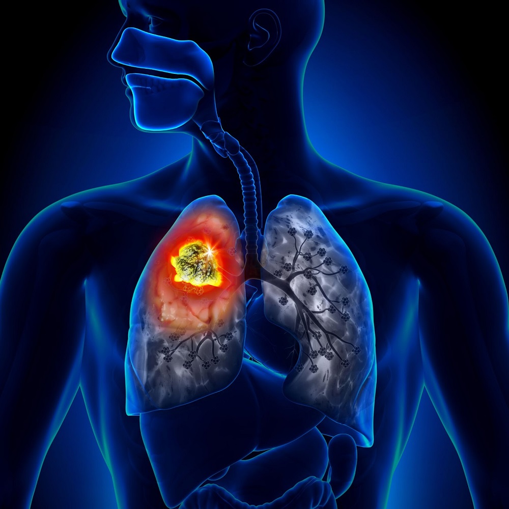
3. Lymphatic disorders: These are conditions that affect the lymphatic system, which is a network of tissues and organs that help rid the body of waste and toxins. Examples include lymphoma (cancer of the lymphatic system), Hodgkin's disease, and non-Hodgkin's lymphoma.
It's important to note that this is not an exhaustive list, and there are many other hemic and lymphatic diseases. If you have concerns about any symptoms or conditions, it's best to consult with a healthcare professional for accurate information and treatment options.
Lymphatic diseases refer to a group of conditions that affect the lymphatic system, which is an important part of the immune and circulatory systems. The lymphatic system consists of a network of vessels, organs, and tissues that help to transport lymph fluid throughout the body, fight infection, and remove waste products.
Lymphatic diseases can be caused by various factors, including genetics, infections, cancer, and autoimmune disorders. Some common types of lymphatic diseases include:
1. Lymphedema: A condition that causes swelling in the arms or legs due to a blockage or damage in the lymphatic vessels.
2. Lymphoma: A type of cancer that affects the lymphatic system, including Hodgkin's and non-Hodgkin's lymphoma.
3. Infections: Certain bacterial and viral infections can affect the lymphatic system, such as tuberculosis, cat-scratch disease, and HIV/AIDS.
4. Autoimmune disorders: Conditions such as rheumatoid arthritis, lupus, and scleroderma can cause inflammation and damage to the lymphatic system.
5. Congenital abnormalities: Some people are born with abnormalities in their lymphatic system, such as malformations or missing lymph nodes.
Symptoms of lymphatic diseases may vary depending on the specific condition and its severity. Treatment options may include medication, physical therapy, surgery, or radiation therapy. It is important to seek medical attention if you experience symptoms of a lymphatic disease, as early diagnosis and treatment can improve outcomes.
Lymphatic vessels are thin-walled, valved structures that collect and transport lymph, a fluid derived from the interstitial fluid surrounding the cells, throughout the lymphatic system. They play a crucial role in immune function and maintaining fluid balance in the body. The primary function of lymphatic vessels is to return excess interstitial fluid, proteins, waste products, and immune cells to the bloodstream via the subclavian veins near the heart.
There are two types of lymphatic vessels:
1. Lymphatic capillaries: These are the smallest lymphatic vessels, found in most body tissues except for the central nervous system (CNS). They have blind ends and are highly permeable to allow the entry of interstitial fluid, proteins, and other large molecules.
2. Larger lymphatic vessels: These include precollecting vessels, collecting vessels, and lymphatic trunks. Precollecting vessels have valves that prevent backflow of lymph and merge to form larger collecting vessels. Collecting vessels contain smooth muscle in their walls, which helps to propel the lymph forward. They also have valves at regular intervals to ensure unidirectional flow towards the heart. Lymphatic trunks are large vessels that collect lymph from various regions of the body and eventually drain into the two main lymphatic ducts: the thoracic duct and the right lymphatic duct.
Overall, lymphatic vessels play a vital role in maintaining fluid balance, immune surveillance, and waste removal in the human body.
The lymphatic system is a complex network of organs, tissues, vessels, and cells that work together to defend the body against infectious diseases and also play a crucial role in the immune system. It is made up of:
1. Lymphoid Organs: These include the spleen, thymus, lymph nodes, tonsils, adenoids, and Peyer's patches (in the intestines). They produce and mature immune cells.
2. Lymphatic Vessels: These are thin tubes that carry clear fluid called lymph towards the heart.
3. Lymph: This is a clear-to-white fluid that contains white blood cells, mainly lymphocytes, which help fight infections.
4. Other tissues and cells: These include bone marrow where immune cells are produced, and lymphocytes (T cells and B cells) which are types of white blood cells that help protect the body from infection and disease.
The primary function of the lymphatic system is to transport lymph throughout the body, collecting waste products, bacteria, viruses, and other foreign substances from the tissues, and filtering them out through the lymph nodes. The lymphatic system also helps in the absorption of fats and fat-soluble vitamins from food in the digestive tract.
The endothelium is a thin layer of cells that lines the interior surface of blood vessels and lymphatic vessels. The lymphatic endothelium, specifically, is the type of endothelial cell that forms the walls of lymphatic vessels. These vessels are an important part of the immune system and play a crucial role in transporting fluid, waste products, and immune cells throughout the body.
The lymphatic endothelium helps to regulate the movement of fluids and cells between the tissues and the bloodstream. It also contains specialized structures called valves that help to ensure the unidirectional flow of lymph fluid towards the heart. Dysfunction of the lymphatic endothelium has been implicated in a variety of diseases, including lymphedema, inflammation, and cancer metastasis.
Lymphangiogenesis is the formation of new lymphatic vessels from pre-existing ones. It is a complex biological process that involves the growth, differentiation, and remodeling of lymphatic endothelial cells, which line the interior surface of lymphatic vessels. Lymphangiogenesis plays crucial roles in various physiological processes, including tissue drainage, immune surveillance, and lipid absorption. However, it can also contribute to pathological conditions such as cancer metastasis, inflammation, and fibrosis when it is dysregulated.
The process of lymphangiogenesis is regulated by a variety of growth factors, receptors, and signaling molecules, including vascular endothelial growth factor (VEGF)-C, VEGF-D, and their receptor VEGFR-3, as well as other factors such as angiopoietins, integrins, and matrix metalloproteinases. Understanding the mechanisms of lymphangiogenesis has important implications for developing novel therapies for a range of diseases associated with abnormal lymphatic vessel growth and function.
 Lymphatic disease
 Lymphatic Diseases | Lymph Nodes | Swollen Glands | MedlinePlus
 Review of Malpractice Litigation in the Diagnosis and Treatment of Venous & Lymphatic Disease
 Template:Lymphatic disease - wikidoc
 NIH VideoCast - Yet to Be Charted: Lymphatic System in Health and Disease (Day 2)
 The Lymphatics in Kidney Disease (Discoveries & Impact August 2023) | Internal Medicine
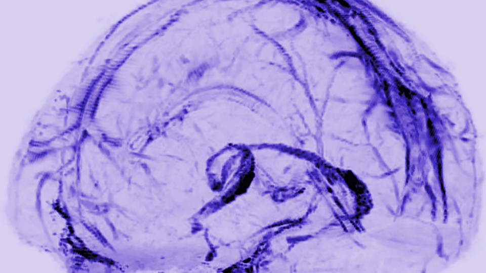
 Impaired meningeal lymphatic drainage in patients with idiopathic Parkinson's disease | Lund University Publications
 What kind of disease is lymphatic filariasis - The Medical Questions
 Cystic Hygroma (Macrocystic Lymphatic Malformation) Imaging and Diagnosis: Practice Essentials, Magnetic Resonance Imaging,...
 The Role of Hyperactive KRAS Signaling in Complex Lymphatic Anomalies : 2023 CLA Conference : Videos : Researchers & Clinicians...
 Elimination of lymphatic filariasis in South East Asia | The BMJ
 Modulating Vascular Lymphatic Growth in Disease: Current and Potential Pharmacological Approaches for Prevention and Treatment
 CDC - Lymphatic Filariasis
 Significant improvement in quality of life following surgery for hydrocoele caused by lymphatic filariasis in Malawi: A...
 Cardiovascular, Lymphatic and Metabolic Disease
 Structure and Function of the Lymphatic System in Dogs
 Cardiovascular & Lymphatic Systems | Carolina Biological Supply
 LETR1 is a lymphatic endothelial-specific lncRNA governing cell proliferation and migration through KLF4 and SEMA3C | Nature...
 Infectious Diseases Affecting the Cardiovascular and Lymphatic Systems
 2020-2021 BCSC Basic and Clinical Science Course™
 Clinical Experts Meeting on Lymphatic Filariasis | DNDi
 Bancroftian and Brugian Lymphatic Filariasis - Infectious Diseases - MSD Manual Professional Edition
 Targeted Lymphatic and Microvessel Treatments in metabolic-DISease HFpEF - Science Teams
 Developing a community-led SMS reporting tool for the rapid assessment of lymphatic filariasis morbidity burden: case studies...