Lymphocytic choriomeningitis virus
Lymphocytic Choriomeningitis
Arenavirus
Lassa virus
CD8-Positive T-Lymphocytes
Mice, Inbred C57BL
T-Lymphocytes, Cytotoxic
Arenaviruses, Old World
Pichinde virus
Immunologic Memory
Epitopes, T-Lymphocyte
Cytotoxicity, Immunologic
Histocompatibility Antigen H-2D
Dystroglycans
Mice, Inbred BALB C
Glycoproteins
Mice, Transgenic
Mice, Knockout
Arenaviruses, New World
Cricetinae
Hemorrhagic Fevers, Viral
Complement Fixation Tests
Viral Vaccines
Lymphocyte Activation
CD4-Positive T-Lymphocytes
Vaccinia
Pore Forming Cytotoxic Proteins
Rhabdoviridae Infections
Immunization, Passive
Arenaviridae
L Cells (Cell Line)
Perforin
T-Lymphocytes
Viral Interference
Virus Replication
Adoptive Transfer
Interferon-gamma
Callimico
Mice, Inbred C3H
Cytotoxicity Tests, Immunologic
Vero Cells
Rodent Diseases
Immunity, Cellular
Hemorrhagic Fever, American
Vesicular stomatitis Indiana virus
Immunodominant Epitopes
Interferon Type I
Chromium Radioisotopes
Mice, Inbred CBA
Vaccinia virus
Cercopithecus aethiops
Viral Plaque Assay
Meninges
Interferons
Receptors, Antigen, T-Cell
Receptors, Interferon
Peptide Fragments
Chronic Disease
T-Lymphocyte Subsets
Antigens, CD8
Nucleocapsid Proteins
Mice, Inbred Strains
Immune Tolerance
Molecular Sequence Data
Neutralization Tests
Antigen Presentation
Receptor, Interferon alpha-beta
Clone Cells
Granzymes
Flow Cytometry
Carrier State
Amino Acid Sequence
Fluorescent Antibody Technique
Receptors, Virus
Interferon-beta
Immunization
Dendritic Cells
Lymphocyte Depletion
Killer Cells, Natural
Hypersensitivity, Delayed
Antibody Formation
Immunity, Innate
Immunosuppression
Interferon Regulatory Factor-7
Disease Models, Animal
Histocompatibility Antigens Class I
Virus Internalization
Animals, Laboratory
Cytopathogenic Effect, Viral
Brain
Cross Reactions
Lymphoid Tissue
Antigens, Ly
Viruses
Vaccines, DNA
Cells, Cultured
Liver
Immunity
Major Histocompatibility Complex
Protection against lymphocytic choriomeningitis virus infection induced by a reduced peptide bond analogue of the H-2Db-restricted CD8(+) T cell epitope GP33. (1/698)
Recent investigations have suggested that pseudopeptides containing modified peptide bonds might advantageously replace natural peptides in therapeutic strategies. We have generated eight reduced peptide bond Psi(CH2-NH) analogues corresponding to the H-2Db-restricted CD8(+) T cell epitope (called GP33) of the glycoprotein of the lymphocytic choriomeningitis virus. One of these pseudopeptides, containing a reduced peptide bond between residues 6 and 7 (Psi(6-7)), displayed very similar properties of binding to major histocompatibility complex (MHC) and recognition by T cell receptor transgenic T cells specific for GP33 when compared with the parent peptide. We assessed in vitro and in vivo the proteolytic resistance of GP33 and Psi(6-7) and analyzed its contribution to the priming properties of these peptides. The Psi(6-7) analogue exhibited a dramatically increased proteolytic resistance when compared with GP33, and we show for the first time that MHC-peptide complexes formed in vivo with a pseudopeptide display a sustained half-life compared with the complexes formed with the natural peptide. Furthermore, in contrast to immunizations with GP33, three injections of Psi(6-7) in saline induced significant antiviral protection in mice. The enhanced ability of Psi(6-7) to induce antiviral protection may result from the higher stability of the analogue and/or of the MHC-analogue complexes. (+info)TRANCE, a tumor necrosis factor family member critical for CD40 ligand-independent T helper cell activation. (2/698)
CD40 ligand (CD40L), a tumor necrosis factor (TNF) family member, plays a critical role in antigen-specific T cell responses in vivo. CD40L expressed on activated CD4(+) T cells stimulates antigen-presenting cells such as dendritic cells, resulting in the upregulation of costimulatory molecules and the production of various inflammatory cytokines required for CD4(+) T cell priming in vivo. However, CD40L- or CD40-deficient mice challenged with viruses mount protective CD4(+) T cell responses that produce normal levels of interferon gamma, suggesting a CD40L/CD40-independent mechanism of CD4(+) T cell priming that to date has not been elucidated. Here we show that CD4(+) T cell responses to viral infection were greatly diminished in CD40-deficient mice by administration of a soluble form of TNF-related activation-induced cytokine receptor (TRANCE-R) to inhibit the function of another TNF family member, TRANCE. Thus, the TRANCE/TRANCE-R interaction provides costimulation required for efficient CD4(+) T cell priming during viral infection in the absence of CD40L/CD40. These results also indicate that not even the potent inflammatory microenvironment induced by viral infections is sufficient to elicit efficient CD4(+) T cell priming without proper costimulation provided by the TNF family (CD40L or TRANCE). Moreover, the data suggest that TRANCE/TRANCE-R may be a novel and important target for immune intervention. (+info)Use of a high-affinity peptide that aborts MHC-restricted cytotoxic T lymphocyte activity against multiple viruses in vitro and virus-induced immunopathologic disease in vivo. (3/698)
Binding of a specific peptide(s) from a viral protein to major histocompatibility complex (MHC) class I molecules is a critical step in the activation of CD8(+) cytotoxic T lymphocytes (CTLs). Once activated, CTLs can cause lethal disease in an infected host, for example, by killing virus-containing ependymal and ventricular cells in the central nervous system or viral protein-expressing beta cells in the pancreatic islets of Langerhans. Here we describe the usage of a designed (not natural) high-affinity peptide to compete with viral peptide(s)-MHC binding. This peptide blocks virus-induced CTL-mediated disease both in the CNS and in the pancreatic islets in vivo. Further, the blocking peptide aborts MHC-restricted killing of target cells by CTLs generated to three separate viruses: lymphocytic choriomeningitis virus, influenza virus, and simian virus 40. (+info)CpG-containing oligonucleotides are efficient adjuvants for induction of protective antiviral immune responses with T-cell peptide vaccines. (4/698)
Synthetic nonmethylated oligonucleotides containing CpG dinucleotides (CpG-ODNs) have been shown to exhibit immunostimulatory activity. CpG-ODNs have the capacity to directly activate B cells, macrophages, and dendritic cells, and we show here that this is reflected by cell surface binding of oligonucleotides to these cell subsets. However, T cells are not directly activated by CpG-ODNs, which correlates with the failure to bind to the T-cell surface. Efficient competition for CpG-induced B-cell activation by non-CpG-containing oligonucleotides suggests that oligonucleotides might bind to an as yet undefined sequence-nonspecific receptor prior to cellular activation. Induction of protective T-cell responses against challenge infection with lymphocytic choriomeningitis virus (LCMV) or with recombinant vaccinia virus expressing the LCMV glycoprotein was achieved by immunizing mice with the immunodominant major histocompatibility complex class I-binding LCMV glycoprotein-derived peptide gp33 together with CpG-ODNs. In these experiments, B cells, potentially serving as CpG-ODN-activated antigen-presenting cells (APCs), were not required for induction of protective immunity since CpG-ODN-gp33-immunized B-cell-deficient mice were equally protected against challenge infection with both viruses. This finding suggested that macrophages and/or dendritic cells were sufficiently activated in vivo by CpG-ODNs to serve as potent APCs for the induction of naive T cells. Furthermore, treatment with CpG-ODN alone induced protection against infection with Listeria monocytogenes via antigen-independent activation of macrophages. These data suggest that CpG activation of macrophages and dendritic cells may provide a critical step in CpG-ODN adjuvant activity. (+info)In vivo selection of neutralization-resistant virus variants but no evidence of B cell tolerance in lymphocytic choriomeningitis virus carrier mice expressing a transgenic virus-neutralizing antibody. (5/698)
B cell tolerance is maintained by active deletion and functional anergy of self-reactive B cells depending on the time, amount, and site of the self-antigen expression. To study B cell tolerance toward a transplacentally transmitted viral Ag, we crossed transgenic mice expressing the mu heavy and the kappa light chain of the lymphocytic choriomeningitis virus (LCMV)-neutralizing mAb KL25 (HL25-transgenic mice) with persistently infected LCMV carrier mice. Although HL25-transgenic LCMV carrier mice exhibited the same high virus titers as nontransgenic LCMV carrier mice, no evidence for B cell tolerance was found. In contrast, enhanced LCMV-neutralizing Ab titers were measured that, however, did not clear the virus. Instead, LCMV isolates from different tissues turned out to be neutralization resistant Ab escape variants expressing different substitutions of amino acid Asn119 of the LCMV-glycoprotein 1 that displays the neutralizing B cell epitope. Virus variants with the same mutations were also selected in vitro in the presence of the transgenic mAb KL25 confirming that substitutions of Asn119 have been selected by LCMV-neutralizing Abs. Thus, despite abundant expression of viral neo-self-antigen in HL25-transgenic LCMV carrier mice, transgenic B cells expressing LCMV-neutralizing Abs were rather stimulated than tolerized and neutralization resistant Ab escape variants were selected in vivo. (+info)Identification of MaTu-MX agent as a new strain of lymphocytic choriomeningitis virus (LCMV) and serological indication of horizontal spread of LCMV in human population. (6/698)
In this study we elucidated the molecular character of MaTu-MX, previously described as an unusual transmissible agent. Amino acid sequencing of peptides generated from a 58-kDa MX-related protein purified from MaTu human carcinoma cells allowed us to identify it as a nucleoprotein (NP) of lymphocytic choriomeningitis virus (LCMV). Northern blot analysis detected LCMV-specific RNAs in MaTu cells. Comparative immunoprecipitations showed cross-reactivity between NP of LCMV strain WE and MX NP. Using RT-PCR, we have cloned MX NP cDNA. According to sequence comparison, MX LCMV is as closely related to both LCMV strains WE and Armstrong as these strains are to one another. Based on this finding we propose that MX is a new strain of LCMV. We also showed that the stability of MX NP in MaTu cells is very high and that the virus is transmissible by cell-to-cell contact or by cell-free extract to human HeLa and monkey Vero cells, but not to human AGS, canine MDCK, mouse NIH 3T3, and hamster CHO cells. Finally, employing MX LCMV NP in immunoprecipitation and solid-phase radioimmunoassay, we found 37.5% prevalence of anti-LCMV antibodies in human sera, suggesting possible horizontal spread of the virus in the human population. (+info)Two roads diverged: interferon alpha/beta- and interleukin 12-mediated pathways in promoting T cell interferon gamma responses during viral infection. (7/698)
Viral infections induce CD8 T cell expansion and interferon (IFN)-gamma production for defense, but the innate cytokines shaping these responses have not been identified. Although interleukin (IL)-12 has the potential to contribute, IL-12-dependent T cell IFN-gamma has not been detected during viral infections. Moreover, certain viruses fail to induce IL-12, and elicit high levels of IFN-alpha/beta to negatively regulate it. The endogenous factors promoting virus-induced T cell IFN-gamma production were defined in studies evaluating CD8 T cell responses during lymphocytic choriomeningitis virus infections of mice. Two divergent supporting pathways were characterized. Under normal conditions of infections, the CD8 T cell IFN-gamma response was dependent on endogenous IFN-alpha/beta effects, but was IL-12 independent. In contrast, in the absence of IFN-alpha/beta functions, an IL-12 response was revealed and substituted an alternative pathway to IFN-gamma. IFN-alpha/beta-mediated effects resulted in enhanced, but the alternative pathway also promoted, resistance to infection. These observations define uniquely important IFN-alpha/beta-controlled pathways shaping T cell responses during viral infections, and demonstrate plasticity of immune responses in accessing divergent innate mechanisms to achieve similar ultimate goals. (+info)A novel approach to visualize polyclonal virus-specific CD8 T cells in vivo. (8/698)
Recent technical breakthroughs in generating soluble MHC class I-peptide tetramers now allow the direct visualization of virus-specific CD8 T cells after infection in vivo. However, this technique requires the knowledge of the immunodominant viral epitopes recognized by T cells. Here, we describe an alternative approach to visualize polyclonal virus-specific CD8 T cells in vivo using a simple adoptive transfer system. In our approach, C57BL/6 (Thy1.2) mice were infected with lymphocytic choriomeningitis virus, vesicular stomatitis virus, or vaccinia virus to induce virus-specific memory T cells. Tracer T cells (2 x 10⁶) from these virus-immune mice were adoptively transferred into nonirradiated (C57BL/6 x B6.PL-Thy-1a)F1 mice. After infection of the F1-recipient mice with the appropriate virus, the transferred cells expanded vigorously, and on day 8 postinfection 60-80% of total CD8 T cells were of donor T cell origin. Under the same conditions memory CD4 T cells gave rise to at least 10 times less cell numbers than memory CD8 T cells. The transfer system described here not only allows to visualize effector and memory CD8 T cells in vivo but also to isolate them for further in vitro characterization without knowing the epitopes recognized by these Ag-specific CD8 T cells. (+info)Lymphocytic choriomeningitis virus (LCMV) is an Old World arenavirus that primarily infects rodents, particularly the house mouse (Mus musculus). The virus is harbored in these mice without causing any apparent disease, but they can shed the virus in their urine, droppings, and saliva.
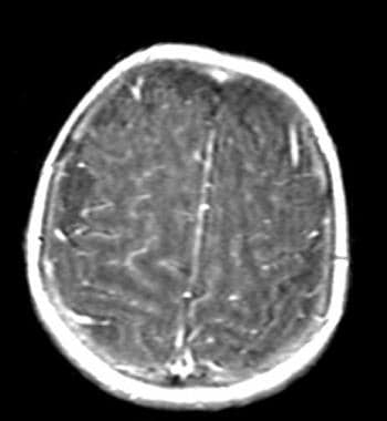
Humans can contract LCMV through close contact with infected rodents or their excreta, inhalation of aerosolized virus, or ingestion of contaminated food or water. In humans, LCMV infection can cause a mild to severe illness called lymphocytic choriomeningitis (LCM), which primarily affects the meninges (the membranes surrounding the brain and spinal cord) and, less frequently, the brain and spinal cord itself.
The incubation period for LCMV infection is typically 1-2 weeks, after which symptoms may appear. Initial symptoms include fever, malaise, headache, muscle aches, and nausea. In some cases, the illness may progress to involve the meninges (meningitis), resulting in neck stiffness, light sensitivity, and altered mental status. In rare instances, LCMV infection can lead to encephalitis (inflammation of the brain) or myelitis (inflammation of the spinal cord), causing more severe neurological symptoms such as seizures, paralysis, or long-term neurological damage.
Most individuals who contract LCMV recover completely within a few weeks to months; however, immunocompromised individuals are at risk for developing severe and potentially fatal complications from the infection. Pregnant women infected with LCMV may also face an increased risk of miscarriage or fetal abnormalities.
Prevention measures include avoiding contact with rodents, especially house mice, and their excreta, maintaining good hygiene, and using appropriate personal protective equipment when handling potentially contaminated materials. There is no specific treatment for LCMV infection; management typically involves supportive care to alleviate symptoms and address complications as they arise.
Lymphocytic choriomeningitis (LCM) is a viral infectious disease caused by the lymphocytic choriomeningitis virus (LCMV). The infection primarily affects the membranes surrounding the brain and spinal cord (meninges), as well as the cerebrospinal fluid, brain, and spinal cord tissue. It is transmitted to humans through close contact with infected rodents, particularly the house mouse (Mus musculus) or its urine, feces, saliva, or nesting materials.
The symptoms of LCM can vary widely but often include fever, severe headache, stiff neck, sensitivity to light, and sometimes vomiting. In some cases, it may also cause muscle aches, joint pain, and rash. A more severe form of the disease can affect the brain and spinal cord, causing confusion, seizures, or even long-term neurological damage.
LCM is typically diagnosed based on symptoms, laboratory tests, and detection of LCMV in cerebrospinal fluid or blood. Treatment usually involves supportive care to manage symptoms, as there is no specific antiviral therapy available for this infection. Most people with LCM recover completely within a few weeks, but severe cases may require hospitalization and intensive care support.
Preventive measures include avoiding contact with rodents, especially their urine, feces, and saliva, and maintaining good hygiene practices such as washing hands thoroughly after handling animals or being in areas where rodents might be present.
Arenaviridae infections are viral illnesses caused by members of the Arenaviridae family of viruses, which include several Old World and New World arenaviruses. These viruses are primarily transmitted to humans through contact with infected rodents or their excreta.
Old World arenaviruses include Lassa fever virus, Lymphocytic choriomeningitis virus (LCMV), and Lujo virus, among others. They are endemic in Africa and can cause severe hemorrhagic fever with high mortality rates.
New World arenaviruses, found mainly in the Americas, include Junin virus, Machupo virus, Guanarito virus, and Sabia virus. These viruses can cause hemorrhagic fever as well, although their severity varies.
In general, Arenaviridae infections can present with a wide range of symptoms, from mild flu-like illness to severe hemorrhagic fever, depending on the specific virus and the individual's immune status. Treatment typically involves supportive care, while some viruses have specific antiviral therapies available. Prevention measures include avoiding contact with rodents and their excreta, as well as implementing public health interventions to control rodent populations in endemic areas.
Arenavirus is a type of virus that belongs to the family Arenaviridae. These viruses are enveloped and have a single-stranded, bi-segmented RNA genome. They are named after the Latin word "arena" which means "sand" because their virions contain ribosomes which resemble sand granules when viewed under an electron microscope.
Arenaviruses are primarily associated with rodents and can cause chronic infection in their natural hosts. Some arenaviruses can also infect humans and other animals, causing severe hemorrhagic fevers. Examples of human diseases caused by arenaviruses include Lassa fever, Argentine hemorrhagic fever, Bolivian hemorrhagic fever, and Venezuelan hemorrhagic fever.
These viruses are typically transmitted to humans through contact with infected rodents or their excreta, but some can also be spread from person to person through close contact with an infected individual's blood or other bodily fluids. There are currently no vaccines available for most arenaviruses, and treatment is primarily supportive, focusing on managing symptoms and complications.
I'm sorry for any confusion, but "Viruses, Unclassified" is not a recognized medical or scientific category. Generally, viruses are classified based on various characteristics such as genome structure, mode of replication, host range, and symptoms they cause. The International Committee on Taxonomy of Viruses (ICTV) is the organization responsible for the formal classification of viruses.
If you have any specific questions about certain unclassified viral entities or phenomena, I'd be happy to help if I can! Please provide more context so I can give a more accurate and helpful response.
Lassa virus is an arenavirus that causes Lassa fever, a type of hemorrhagic fever. It is primarily transmitted to humans through contact with infected rodents or their urine and droppings. The virus can also be spread through person-to-person transmission via direct contact with the blood, urine, feces, or other bodily fluids of an infected person.
The virus was first discovered in 1969 in the town of Lassa in Nigeria, hence its name. It is endemic to West Africa and is a significant public health concern in countries such as Sierra Leone, Liberia, Guinea, and Nigeria. The symptoms of Lassa fever can range from mild to severe and may include fever, sore throat, muscle pain, chest pain, and vomiting. In severe cases, the virus can cause bleeding, organ failure, and death.
Prevention measures for Lassa fever include avoiding contact with rodents, storing food in rodent-proof containers, and practicing good hygiene. There is no vaccine available to prevent Lassa fever, but ribavirin, an antiviral drug, has been shown to be effective in treating the disease if administered early in the course of illness.
CD8-positive T-lymphocytes, also known as CD8+ T cells or cytotoxic T cells, are a type of white blood cell that plays a crucial role in the adaptive immune system. They are named after the CD8 molecule found on their surface, which is a protein involved in cell signaling and recognition.
CD8+ T cells are primarily responsible for identifying and destroying virus-infected cells or cancerous cells. When activated, they release cytotoxic granules that contain enzymes capable of inducing apoptosis (programmed cell death) in the target cells. They also produce cytokines such as interferon-gamma, which can help coordinate the immune response and activate other immune cells.
CD8+ T cells are generated in the thymus gland and are a type of T cell, which is a lymphocyte that matures in the thymus and plays a central role in cell-mediated immunity. They recognize and respond to specific antigens presented on the surface of infected or cancerous cells in conjunction with major histocompatibility complex (MHC) class I molecules.
Overall, CD8+ T cells are an essential component of the immune system's defense against viral infections and cancer.
C57BL/6 (C57 Black 6) is an inbred strain of laboratory mouse that is widely used in biomedical research. The term "inbred" refers to a strain of animals where matings have been carried out between siblings or other closely related individuals for many generations, resulting in a population that is highly homozygous at most genetic loci.
The C57BL/6 strain was established in 1920 by crossing a female mouse from the dilute brown (DBA) strain with a male mouse from the black strain. The resulting offspring were then interbred for many generations to create the inbred C57BL/6 strain.
C57BL/6 mice are known for their robust health, longevity, and ease of handling, making them a popular choice for researchers. They have been used in a wide range of biomedical research areas, including studies of cancer, immunology, neuroscience, cardiovascular disease, and metabolism.
One of the most notable features of the C57BL/6 strain is its sensitivity to certain genetic modifications, such as the introduction of mutations that lead to obesity or impaired glucose tolerance. This has made it a valuable tool for studying the genetic basis of complex diseases and traits.
Overall, the C57BL/6 inbred mouse strain is an important model organism in biomedical research, providing a valuable resource for understanding the genetic and molecular mechanisms underlying human health and disease.
Cytotoxic T-lymphocytes, also known as CD8+ T cells, are a type of white blood cell that plays a central role in the cell-mediated immune system. They are responsible for identifying and destroying virus-infected cells and cancer cells. When a cytotoxic T-lymphocyte recognizes a specific antigen presented on the surface of an infected or malignant cell, it becomes activated and releases toxic substances such as perforins and granzymes, which can create pores in the target cell's membrane and induce apoptosis (programmed cell death). This process helps to eliminate the infected or malignant cells and prevent the spread of infection or cancer.
Arenaviruses, Old World, are a group of viruses within the Arenaviridae family that primarily cause disease in humans and animals in Africa and Europe. These viruses are enveloped and have a bi-segmented single-stranded RNA genome. The name "Old World" is used to distinguish them from the New World arenaviruses, which are found in the Americas.
Some of the most well-known Old World arenaviruses include Lassa fever virus, Lymphocytic choriomeningitis virus (LCMV), and Lujo virus. These viruses can cause a range of symptoms in humans, from mild febrile illness to severe hemorrhagic fever.
Lassa fever virus is endemic in West Africa and can cause a severe and often fatal hemorrhagic fever. LCMV is found worldwide and typically causes a mild illness in humans, although it can lead to more severe disease in immunocompromised individuals. Lujo virus was first identified in 2008 in South Africa and has caused a small number of severe and often fatal hemorrhagic fever cases.
Old World arenaviruses are primarily transmitted to humans through contact with infected rodents or their excreta, although human-to-human transmission can also occur through close contact with an infected person's blood or bodily fluids. Prevention and control measures include avoiding contact with rodents, practicing good hygiene, and using personal protective equipment when caring for sick individuals.
Nucleoproteins are complexes formed by the association of proteins with nucleic acids (DNA or RNA). These complexes play crucial roles in various biological processes, such as packaging and protecting genetic material, regulating gene expression, and replication and repair of DNA. In these complexes, proteins interact with nucleic acids through electrostatic, hydrogen bonding, and other non-covalent interactions, leading to the formation of stable structures that help maintain the integrity and function of the genetic material. Some well-known examples of nucleoproteins include histones, which are involved in DNA packaging in eukaryotic cells, and reverse transcriptase, an enzyme found in retroviruses that transcribes RNA into DNA.
Pichinde virus (PICV) is an enveloped, negative-sense, single-stranded RNA virus that belongs to the family Arenaviridae. It is primarily found in rodents, specifically the Pichinde deer mouse (Oligoryzomys fulvescens), which are endemic to South America, particularly Colombia.
PICV is not known to cause disease in humans and is often used as a model organism for studying arenaviral pathogenesis and immunity. However, accidental laboratory infections have been reported, resulting in mild febrile illness or seroconversion without symptoms. Therefore, it is recommended that appropriate biosafety measures be taken when handling this virus.
An antigen is any substance that can stimulate an immune response, particularly the production of antibodies. Viral antigens are antigens that are found on or produced by viruses. They can be proteins, glycoproteins, or carbohydrates present on the surface or inside the viral particle.
Viral antigens play a crucial role in the immune system's recognition and response to viral infections. When a virus infects a host cell, it may display its antigens on the surface of the infected cell. This allows the immune system to recognize and target the infected cells for destruction, thereby limiting the spread of the virus.
Viral antigens are also important targets for vaccines. Vaccines typically work by introducing a harmless form of a viral antigen to the body, which then stimulates the production of antibodies and memory T-cells that can recognize and respond quickly and effectively to future infections with the actual virus.
It's worth noting that different types of viruses have different antigens, and these antigens can vary between strains of the same virus. This is why there are often different vaccines available for different viral diseases, and why flu vaccines need to be updated every year to account for changes in the circulating influenza virus strains.
Immunologic memory, also known as adaptive immunity, refers to the ability of the immune system to recognize and mount a more rapid and effective response upon subsequent exposure to a pathogen or antigen that it has encountered before. This is a key feature of the vertebrate immune system and allows for long-term protection against infectious diseases.
Immunologic memory is mediated by specialized cells called memory T cells and B cells, which are produced during the initial response to an infection or immunization. These cells persist in the body after the pathogen has been cleared and can quickly respond to future encounters with the same or similar antigens. This rapid response leads to a more effective and efficient elimination of the pathogen, resulting in fewer symptoms and reduced severity of disease.
Immunologic memory is the basis for vaccines, which work by exposing the immune system to a harmless form of a pathogen or its components, inducing an initial response and generating memory cells that provide long-term protection against future infections.
The spleen is an organ in the upper left side of the abdomen, next to the stomach and behind the ribs. It plays multiple supporting roles in the body:
1. It fights infection by acting as a filter for the blood. Old red blood cells are recycled in the spleen, and platelets and white blood cells are stored there.
2. The spleen also helps to control the amount of blood in the body by removing excess red blood cells and storing platelets.
3. It has an important role in immune function, producing antibodies and removing microorganisms and damaged red blood cells from the bloodstream.
The spleen can be removed without causing any significant problems, as other organs take over its functions. This is known as a splenectomy and may be necessary if the spleen is damaged or diseased.
An epitope is a specific region on an antigen (a substance that triggers an immune response) that is recognized and bound by an antibody or a T-cell receptor. In the case of T-lymphocytes, which are a type of white blood cell that plays a central role in cell-mediated immunity, epitopes are typically presented on the surface of infected cells in association with major histocompatibility complex (MHC) molecules.
T-lymphocytes recognize and respond to epitopes through their T-cell receptors (TCRs), which are membrane-bound proteins that can bind to specific epitopes presented on the surface of infected cells. There are two main types of T-lymphocytes: CD4+ T-cells, also known as helper T-cells, and CD8+ T-cells, also known as cytotoxic T-cells.
CD4+ T-cells recognize epitopes presented in the context of MHC class II molecules, which are typically expressed on the surface of professional antigen-presenting cells such as dendritic cells, macrophages, and B-cells. CD4+ T-cells help to coordinate the immune response by producing cytokines that activate other immune cells.
CD8+ T-cells recognize epitopes presented in the context of MHC class I molecules, which are expressed on the surface of almost all nucleated cells. CD8+ T-cells are able to directly kill infected cells by releasing cytotoxic granules that contain enzymes that can induce apoptosis (programmed cell death) in the target cell.
In summary, epitopes are specific regions on antigens that are recognized and bound by T-lymphocytes through their T-cell receptors. CD4+ T-cells recognize epitopes presented in the context of MHC class II molecules, while CD8+ T-cells recognize epitopes presented in the context of MHC class I molecules.
Immunologic cytotoxicity refers to the damage or destruction of cells that occurs as a result of an immune response. This process involves the activation of immune cells, such as cytotoxic T cells and natural killer (NK) cells, which release toxic substances, such as perforins and granzymes, that can kill target cells.
In addition, antibodies produced by B cells can also contribute to immunologic cytotoxicity by binding to antigens on the surface of target cells and triggering complement-mediated lysis or antibody-dependent cellular cytotoxicity (ADCC) by activating immune effector cells.
Immunologic cytotoxicity plays an important role in the body's defense against viral infections, cancer cells, and other foreign substances. However, it can also contribute to tissue damage and autoimmune diseases if the immune system mistakenly targets healthy cells or tissues.
H-2 antigens are a group of cell surface proteins found in mice that play a critical role in the immune system. They are similar to the human leukocyte antigen (HLA) complex in humans and are involved in the presentation of peptide antigens to T cells, which is a crucial step in the adaptive immune response.
The H-2 antigens are encoded by a cluster of genes located on chromosome 17 in mice. They are highly polymorphic, meaning that there are many different variations of these proteins circulating in the population. This genetic diversity allows for a wide range of potential peptide antigens to be presented to T cells, thereby enhancing the ability of the immune system to recognize and respond to a variety of pathogens.
The H-2 antigens are divided into two classes based on their function and structure. Class I H-2 antigens are found on almost all nucleated cells and consist of a heavy chain, a light chain, and a peptide fragment. They present endogenous peptides, such as those derived from viruses that infect the cell, to CD8+ T cells.
Class II H-2 antigens, on the other hand, are found primarily on professional antigen-presenting cells, such as dendritic cells and macrophages. They consist of an alpha chain and a beta chain and present exogenous peptides, such as those derived from bacteria that have been engulfed by the cell, to CD4+ T cells.
Overall, H-2 antigens are essential components of the mouse immune system, allowing for the recognition and elimination of pathogens and infected cells.
Histocompatibility antigen H-2D is a type of major histocompatibility complex (MHC) class I molecule found in mice. It is a transmembrane protein located on the surface of nucleated cells, which plays a crucial role in the adaptive immune system. The primary function of H-2D is to present endogenous peptide antigens to CD8+ T cells, also known as cytotoxic T lymphocytes (CTLs).
H-2D molecules are encoded by genes within the H-2D region of the MHC on chromosome 17. These genes have multiple alleles, resulting in a high degree of polymorphism, which contributes to the diversity of the immune response among different mouse strains. The peptide-binding groove of H-2D molecules is formed by two alpha helices and eight beta pleats, creating a specific binding site for antigenic peptides.
The peptides presented by H-2D molecules are derived from intracellular proteins that undergo degradation in the proteasome. These peptides are then transported into the endoplasmic reticulum, where they bind to H-2D molecules with the assistance of chaperone proteins like tapasin and calreticulin. The H-2D-peptide complex is then transported to the cell surface for presentation to CD8+ T cells.
Recognition of H-2D-peptide complexes by CD8+ T cells leads to their activation, proliferation, and differentiation into effector CTLs. Activated CTLs can recognize and eliminate virus-infected or malignant cells displaying specific H-2D-peptide complexes, thereby playing a critical role in the cell-mediated immune response.
In summary, histocompatibility antigen H-2D is a polymorphic MHC class I molecule in mice that presents endogenous peptide antigens to CD8+ T cells, contributing significantly to the adaptive immune response and the elimination of infected or malignant cells.
Dystroglycans are a type of protein that play a crucial role in the structure and function of the muscle membrane (sarcolemma). They are an essential component of the dystrophin-glycoprotein complex, which helps maintain the stability and integrity of the sarcolemma during muscle contraction and relaxation.
Dystroglycans consist of two subunits: alpha-dystroglycan and beta-dystroglycan. Alpha-dystroglycan is a large, heavily glycosylated protein that extends from the intracellular space to the extracellular matrix, where it interacts with various extracellular matrix proteins such as laminin and agrin. Beta-dystroglycan, on the other hand, spans the muscle membrane and binds to dystrophin, a cytoskeletal protein that helps maintain the structural integrity of the sarcolemma.
Mutations in genes encoding for proteins involved in the glycosylation of alpha-dystroglycan can lead to a group of genetic disorders known as congenital muscular dystrophies, which are characterized by muscle weakness, hypotonia, and developmental delays. These disorders include Walker-Warburg syndrome, Fukuyama congenital muscular dystrophy, and Muscle-Eye-Brain disease, among others.
BALB/c is an inbred strain of laboratory mouse that is widely used in biomedical research. The strain was developed at the Institute of Cancer Research in London by Henry Baldwin and his colleagues in the 1920s, and it has since become one of the most commonly used inbred strains in the world.
BALB/c mice are characterized by their black coat color, which is determined by a recessive allele at the tyrosinase locus. They are also known for their docile and friendly temperament, making them easy to handle and work with in the laboratory.
One of the key features of BALB/c mice that makes them useful for research is their susceptibility to certain types of tumors and immune responses. For example, they are highly susceptible to developing mammary tumors, which can be induced by chemical carcinogens or viral infection. They also have a strong Th2-biased immune response, which makes them useful models for studying allergic diseases and asthma.
BALB/c mice are also commonly used in studies of genetics, neuroscience, behavior, and infectious diseases. Because they are an inbred strain, they have a uniform genetic background, which makes it easier to control for genetic factors in experiments. Additionally, because they have been bred in the laboratory for many generations, they are highly standardized and reproducible, making them ideal subjects for scientific research.
Viral proteins are the proteins that are encoded by the viral genome and are essential for the viral life cycle. These proteins can be structural or non-structural and play various roles in the virus's replication, infection, and assembly process. Structural proteins make up the physical structure of the virus, including the capsid (the protein shell that surrounds the viral genome) and any envelope proteins (that may be present on enveloped viruses). Non-structural proteins are involved in the replication of the viral genome and modulation of the host cell environment to favor viral replication. Overall, a thorough understanding of viral proteins is crucial for developing antiviral therapies and vaccines.
Glycoproteins are complex proteins that contain oligosaccharide chains (glycans) covalently attached to their polypeptide backbone. These glycans are linked to the protein through asparagine residues (N-linked) or serine/threonine residues (O-linked). Glycoproteins play crucial roles in various biological processes, including cell recognition, cell-cell interactions, cell adhesion, and signal transduction. They are widely distributed in nature and can be found on the outer surface of cell membranes, in extracellular fluids, and as components of the extracellular matrix. The structure and composition of glycoproteins can vary significantly depending on their function and location within an organism.
Transgenic mice are genetically modified rodents that have incorporated foreign DNA (exogenous DNA) into their own genome. This is typically done through the use of recombinant DNA technology, where a specific gene or genetic sequence of interest is isolated and then introduced into the mouse embryo. The resulting transgenic mice can then express the protein encoded by the foreign gene, allowing researchers to study its function in a living organism.
The process of creating transgenic mice usually involves microinjecting the exogenous DNA into the pronucleus of a fertilized egg, which is then implanted into a surrogate mother. The offspring that result from this procedure are screened for the presence of the foreign DNA, and those that carry the desired genetic modification are used to establish a transgenic mouse line.
Transgenic mice have been widely used in biomedical research to model human diseases, study gene function, and test new therapies. They provide a valuable tool for understanding complex biological processes and developing new treatments for a variety of medical conditions.
A "knockout" mouse is a genetically engineered mouse in which one or more genes have been deleted or "knocked out" using molecular biology techniques. This allows researchers to study the function of specific genes and their role in various biological processes, as well as potential associations with human diseases. The mice are generated by introducing targeted DNA modifications into embryonic stem cells, which are then used to create a live animal. Knockout mice have been widely used in biomedical research to investigate gene function, disease mechanisms, and potential therapeutic targets.
Lassa fever is an acute viral hemorrhagic fever caused by the Lassa virus. It is primarily transmitted to humans through contact with infected rodents or their excreta, and it can also spread from person to person via bodily fluids. The symptoms of Lassa fever typically include fever, sore throat, muscle pain, chest pain, headache, and vomiting. In severe cases, the disease can cause bleeding from the mouth and nose, as well as complications such as deafness and encephalitis. Lassa fever is endemic to West Africa, particularly in Nigeria, Guinea, Liberia, and Sierra Leone.
Arenaviruses, New World, are a group of viruses in the Arenaviridae family that primarily infect rodents and can cause disease in humans. They are named after the Latin word "arena" which means "sand" because of the sandy-like appearance of their virions when viewed under an electron microscope.
New World arenaviruses include several different species, such as Junín virus, Machupo virus, Guanarito virus, and Sabia virus, among others. These viruses are endemic to certain regions in the Americas, particularly in South America. They are transmitted to humans through close contact with infected rodents or their excreta, and can cause severe hemorrhagic fever with high fatality rates if left untreated.
Some New World arenaviruses, such as Junín virus and Machupo virus, have been associated with outbreaks of human disease in the past, while others, like Guanarito virus and Sabia virus, have caused sporadic cases of illness. There are currently no vaccines available for most New World arenaviruses, although research is ongoing to develop effective countermeasures against these viruses.
Cricetinae is a subfamily of rodents that includes hamsters, gerbils, and relatives. These small mammals are characterized by having short limbs, compact bodies, and cheek pouches for storing food. They are native to various parts of the world, particularly in Europe, Asia, and Africa. Some species are popular pets due to their small size, easy care, and friendly nature. In a medical context, understanding the biology and behavior of Cricetinae species can be important for individuals who keep them as pets or for researchers studying their physiology.
**Hemorrhagic fevers, viral** are a group of severe, potentially fatal illnesses caused by viruses that affect the body's ability to regulate its blood vessels and clotting abilities. These viruses belong to several different families including *Filoviridae* (e.g., Ebola, Marburg), *Arenaviridae* (e.g., Lassa, Machupo), *Bunyaviridae* (e.g., Hantavirus, Crimean-Congo hemorrhagic fever virus) and *Flaviviridae* (e.g., Dengue, Yellow Fever).
The initial symptoms are non-specific and include sudden onset of fever, fatigue, muscle aches, joint pains, headache, and vomiting. As the disease progresses, it may lead to capillary leakage, internal and external bleeding, and multi-organ failure resulting in shock and death in severe cases.
The transmission of these viruses can occur through various means depending on the specific virus. For example, some are transmitted via contact with infected animals or their urine/feces (e.g., Hantavirus), others through insect vectors like ticks (Crimean-Congo hemorrhagic fever) or mosquitoes (Dengue, Yellow Fever), and yet others through direct contact with infected body fluids (Ebola, Marburg).
There are no specific treatments for most viral hemorrhagic fevers. However, some experimental antiviral drugs have shown promise in treating certain types of the disease. Supportive care, such as maintaining blood pressure, replacing lost fluids and electrolytes, and managing pain, is critical to improving outcomes. Prevention measures include avoiding areas where the viruses are common, using personal protective equipment when caring for infected individuals or handling potentially contaminated materials, and controlling insect vectors.
Sources: Centers for Disease Control and Prevention (CDC), World Health Organization (WHO).
Antibodies, viral are proteins produced by the immune system in response to an infection with a virus. These antibodies are capable of recognizing and binding to specific antigens on the surface of the virus, which helps to neutralize or destroy the virus and prevent its replication. Once produced, these antibodies can provide immunity against future infections with the same virus.
Viral antibodies are typically composed of four polypeptide chains - two heavy chains and two light chains - that are held together by disulfide bonds. The binding site for the antigen is located at the tip of the Y-shaped structure, formed by the variable regions of the heavy and light chains.
There are five classes of antibodies in humans: IgA, IgD, IgE, IgG, and IgM. Each class has a different function and is distributed differently throughout the body. For example, IgG is the most common type of antibody found in the bloodstream and provides long-term immunity against viruses, while IgA is found primarily in mucous membranes and helps to protect against respiratory and gastrointestinal infections.
In addition to their role in the immune response, viral antibodies can also be used as diagnostic tools to detect the presence of a specific virus in a patient's blood or other bodily fluids.
Complement fixation tests are a type of laboratory test used in immunology and serology to detect the presence of antibodies in a patient's serum. These tests are based on the principle of complement activation, which is a part of the immune response. The complement system consists of a group of proteins that work together to help eliminate pathogens from the body.
In a complement fixation test, the patient's serum is mixed with a known antigen and complement proteins. If the patient has antibodies against the antigen, they will bind to it and activate the complement system. This results in the consumption or "fixation" of the complement proteins, which are no longer available to participate in a secondary reaction.
A second step involves adding a fresh source of complement proteins and a dye-labeled antibody that recognizes a specific component of the complement system. If complement was fixed during the first step, it will not be available for this secondary reaction, and the dye-labeled antibody will remain unbound. Conversely, if no antibodies were present in the patient's serum, the complement proteins would still be available for the second reaction, leading to the binding of the dye-labeled antibody.
The mixture is then examined under a microscope or using a spectrophotometer to determine whether the dye-labeled antibody has bound. If it has not, this indicates that the patient's serum contains antibodies specific to the antigen used in the test, and a positive result is recorded.
Complement fixation tests have been widely used for the diagnosis of various infectious diseases, such as syphilis, measles, and influenza. However, they have largely been replaced by more modern serological techniques, like enzyme-linked immunosorbent assays (ELISAs) and nucleic acid amplification tests (NAATs), due to their increased sensitivity, specificity, and ease of use.
A viral vaccine is a biological preparation that introduces your body to a specific virus in a way that helps your immune system build up protection against the virus without causing the illness. Viral vaccines can be made from weakened or inactivated forms of the virus, or parts of the virus such as proteins or sugars. Once introduced to the body, the immune system recognizes the virus as foreign and produces an immune response, including the production of antibodies. These antibodies remain in the body and provide immunity against future infection with that specific virus.
Viral vaccines are important tools for preventing infectious diseases caused by viruses, such as influenza, measles, mumps, rubella, polio, hepatitis A and B, rabies, rotavirus, chickenpox, shingles, and some types of cancer. Vaccination programs have led to the control or elimination of many infectious diseases that were once common.
It's important to note that viral vaccines are not effective against bacterial infections, and separate vaccines must be developed for each type of virus. Additionally, because viruses can mutate over time, it is necessary to update some viral vaccines periodically to ensure continued protection.
Lymphocyte activation is the process by which B-cells and T-cells (types of lymphocytes) become activated to perform effector functions in an immune response. This process involves the recognition of specific antigens presented on the surface of antigen-presenting cells, such as dendritic cells or macrophages.
The activation of B-cells leads to their differentiation into plasma cells that produce antibodies, while the activation of T-cells results in the production of cytotoxic T-cells (CD8+ T-cells) that can directly kill infected cells or helper T-cells (CD4+ T-cells) that assist other immune cells.
Lymphocyte activation involves a series of intracellular signaling events, including the binding of co-stimulatory molecules and the release of cytokines, which ultimately result in the expression of genes involved in cell proliferation, differentiation, and effector functions. The activation process is tightly regulated to prevent excessive or inappropriate immune responses that can lead to autoimmunity or chronic inflammation.
CD4-positive T-lymphocytes, also known as CD4+ T cells or helper T cells, are a type of white blood cell that plays a crucial role in the immune response. They express the CD4 receptor on their surface and help coordinate the immune system's response to infectious agents such as viruses and bacteria.
CD4+ T cells recognize and bind to specific antigens presented by antigen-presenting cells, such as dendritic cells or macrophages. Once activated, they can differentiate into various subsets of effector cells, including Th1, Th2, Th17, and Treg cells, each with distinct functions in the immune response.
CD4+ T cells are particularly important in the immune response to HIV (human immunodeficiency virus), which targets and destroys these cells, leading to a weakened immune system and increased susceptibility to opportunistic infections. The number of CD4+ T cells is often used as a marker of disease progression in HIV infection, with lower counts indicating more advanced disease.
A Laboratory Infection, also known as a laboratory-acquired infection (LAI), is an infection that occurs in individuals who are exposed to pathogens or other harmful microorganisms while working in a laboratory setting. These infections can occur through various routes of exposure, including inhalation, skin contact, or ingestion of contaminated materials.
Laboratory infections pose significant risks to laboratory workers, researchers, and even visitors who may come into contact with infectious agents during their work or visit. To minimize these risks, laboratories follow strict biosafety protocols, including the use of personal protective equipment (PPE), proper handling and disposal of contaminated materials, and adherence to established safety guidelines.
Examples of laboratory infections include tuberculosis, salmonella, hepatitis B and C, and various other bacterial, viral, fungal, and parasitic infections. Prompt diagnosis, treatment, and implementation of appropriate infection control measures are crucial to prevent the spread of these infections within the laboratory setting and beyond.
Vaccinia is actually not a medical term with a specific definition, but it refers to the virus used in the smallpox vaccine. The vaccinia virus is related to, but less harmful than, the variola virus that causes smallpox. When vaccinia virus is introduced into the skin, it leads to an immune response that protects against smallpox.
The term "vaccinia" also refers to the characteristic pockmark-like lesion that forms on the skin as part of the body's reaction to the vaccine. This lesion is a result of the infection and replication of the vaccinia virus in the skin cells, which triggers an immune response that helps protect against smallpox.
It's worth noting that while the smallpox vaccine is no longer routinely administered due to the eradication of smallpox, it may still be used in certain circumstances, such as in laboratory workers who handle the virus or in the event of a bioterrorism threat involving smallpox.
Viral diseases are illnesses caused by the infection and replication of viruses in host organisms. These infectious agents are obligate parasites, meaning they rely on the cells of other living organisms to survive and reproduce. Viruses can infect various types of hosts, including animals, plants, and microorganisms, causing a wide range of diseases with varying symptoms and severity.
Once a virus enters a host cell, it takes over the cell's machinery to produce new viral particles, often leading to cell damage or death. The immune system recognizes the viral components as foreign and mounts an immune response to eliminate the infection. This response can result in inflammation, fever, and other symptoms associated with viral diseases.
Examples of well-known viral diseases include:
1. Influenza (flu) - caused by influenza A, B, or C viruses
2. Common cold - usually caused by rhinoviruses or coronaviruses
3. HIV/AIDS - caused by human immunodeficiency virus (HIV)
4. Measles - caused by measles morbillivirus
5. Hepatitis B and C - caused by hepatitis B virus (HBV) and hepatitis C virus (HCV), respectively
6. Herpes simplex - caused by herpes simplex virus type 1 (HSV-1) or type 2 (HSV-2)
7. Chickenpox and shingles - both caused by varicella-zoster virus (VZV)
8. Rabies - caused by rabies lyssavirus
9. Ebola - caused by ebolaviruses
10. COVID-19 - caused by severe acute respiratory syndrome coronavirus 2 (SARS-CoV-2)
Prevention and treatment strategies for viral diseases may include vaccination, antiviral medications, and supportive care to manage symptoms while the immune system fights off the infection.
Pore-forming cytotoxic proteins are a group of toxins that can create pores or holes in the membranes of cells, leading to cell damage or death. These toxins are produced by various organisms, including bacteria, fungi, and plants, as a defense mechanism or to help establish an infection.
The pore-forming cytotoxic proteins can be divided into two main categories:
1. Membrane attack complex/perforin (MACPF) domain-containing proteins: These are found in many organisms, including humans. They form pores by oligomerizing, or clustering together, in the target cell membrane. An example of this type of toxin is the perforin protein, which is released by cytotoxic T cells and natural killer cells to destroy virus-infected or cancerous cells.
2. Cholesterol-dependent cytolysins (CDCs): These are mainly produced by gram-positive bacteria. They bind to cholesterol in the target cell membrane, forming a prepore structure that then undergoes conformational changes to create a pore. An example of a CDC is alpha-hemolysin from Staphylococcus aureus, which can lyse red blood cells and damage various other cell types.
These pore-forming cytotoxic proteins play a significant role in host-pathogen interactions and have implications for the development of novel therapeutic strategies.
Rhabdoviruses are negative-sense, single-stranded RNA viruses that belong to the family Rhabdoviridae. They have a wide host range, including humans, and can cause various diseases.
Rhabdoviridae infections refer to the infectious diseases caused by rhabdoviruses. The most well-known member of this family is the rabies virus, which causes rabies, a fatal zoonotic disease that affects warm-blooded animals, including humans. Rabies is transmitted through the saliva of infected animals, usually via bites or scratches.
Other rhabdoviruses can also cause human diseases, such as:
1. Vesicular stomatitis virus (VSV): It primarily affects livestock, causing vesicular lesions in the mouth and on the feet. However, it can also infect humans, causing flu-like symptoms or a rash around the mouth and hands.
2. Chandipura virus: This rhabdovirus is associated with acute encephalitis, particularly in children. It is transmitted through mosquitoes and has been identified in several countries, including India and Nigeria.
3. Human basalotid fibroblast growth factor (bFGF) receptor-binding virus: This recently discovered rhabdovirus was found to be associated with a case of acute respiratory illness. More research is needed to understand its epidemiology, transmission, and clinical significance.
Prevention and control measures for Rhabdoviridae infections include vaccination against rabies, public education on avoiding contact with potentially infected animals, and personal protective measures such as wearing gloves when handling animals or their tissues.
Passive immunization is a type of temporary immunity that is transferred to an individual through the injection of antibodies produced outside of the body, rather than through the active production of antibodies in the body in response to vaccination or infection. This can be done through the administration of preformed antibodies, such as immune globulins, which contain a mixture of antibodies that provide immediate protection against specific diseases.
Passive immunization is often used in situations where individuals have been exposed to a disease and do not have time to develop their own active immune response, or in cases where individuals are unable to produce an adequate immune response due to certain medical conditions. It can also be used as a short-term measure to provide protection until an individual can receive a vaccination that will confer long-term immunity.
Passive immunization provides immediate protection against disease, but the protection is typically short-lived, lasting only a few weeks or months. This is because the transferred antibodies are gradually broken down and eliminated by the body over time. In contrast, active immunization confers long-term immunity through the production of memory cells that can mount a rapid and effective immune response upon re-exposure to the same pathogen in the future.
Arenaviridae is a family of viruses that includes several species known to cause disease in humans and animals. The name "Arenaviridae" comes from the Latin word "arena," meaning "sand," due to the sandy appearance of these viruses when viewed under an electron microscope.
The virions (complete virus particles) of Arenaviridae are typically enveloped, spherical or pleomorphic in shape, and measure between 50-300 nanometers in diameter. The genome of Arenaviridae viruses is composed of two single-stranded, negative-sense RNA segments called the L (large) segment and the S (small) segment. These segments encode for several viral proteins, including the glycoprotein (GP), nucleoprotein (NP), and the RNA-dependent RNA polymerase (L).
Arenaviridae viruses are primarily transmitted to humans through contact with infected rodents or their excreta. Some of the most well-known human pathogens in this family include Lassa fever virus, Junín virus, Machupo virus, and Guanarito virus, which can cause severe hemorrhagic fevers. Other Arenaviridae viruses, such as lymphocytic choriomeningitis virus (LCMV), can cause milder illnesses in humans, including fever, rash, and meningitis.
Prevention and control of Arenaviridae infections typically involve reducing exposure to infected rodents and their excreta, as well as the development of vaccines and antiviral therapies for specific viruses in this family.
Perforin is a protein that plays a crucial role in the immune system's response to virally infected or cancerous cells. It is primarily produced and released by cytotoxic T-cells and natural killer (NK) cells, two types of white blood cells involved in defending the body against infection and disease.
Perforin functions by creating pores or holes in the membrane of target cells, leading to their lysis or destruction. This process allows for the release of cellular contents and the exposure of intracellular antigens, which can then be processed and presented to other immune cells, thereby enhancing the immune response against the pathogen or abnormal cells.
In summary, perforin is a vital component of the immune system's cytotoxic activity, contributing to the elimination of infected or malignant cells and maintaining overall health and homeostasis in the body.
T-lymphocytes, also known as T-cells, are a type of white blood cell that plays a key role in the adaptive immune system's response to infection. They are produced in the bone marrow and mature in the thymus gland. There are several different types of T-cells, including CD4+ helper T-cells, CD8+ cytotoxic T-cells, and regulatory T-cells (Tregs).
CD4+ helper T-cells assist in activating other immune cells, such as B-lymphocytes and macrophages. They also produce cytokines, which are signaling molecules that help coordinate the immune response. CD8+ cytotoxic T-cells directly kill infected cells by releasing toxic substances. Regulatory T-cells help maintain immune tolerance and prevent autoimmune diseases by suppressing the activity of other immune cells.
T-lymphocytes are important in the immune response to viral infections, cancer, and other diseases. Dysfunction or depletion of T-cells can lead to immunodeficiency and increased susceptibility to infections. On the other hand, an overactive T-cell response can contribute to autoimmune diseases and chronic inflammation.
Viral interference is a phenomenon where the replication of one virus is inhibited or blocked by the presence of another virus. This can occur when two different viruses infect the same cell and compete for the cell's resources, such as nucleotides, energy, and replication machinery. As a result, the replication of one virus may be suppressed, allowing the other virus to predominate.
This phenomenon has been observed in both in vitro (laboratory) studies and in vivo (in the body) studies. It has been suggested that viral interference may play a role in the outcome of viral coinfections, where an individual is infected with more than one virus at the same time. Viral interference can also be exploited as a potential strategy for antiviral therapy, where one virus is used to inhibit the replication of another virus.
It's important to note that not all viruses interfere with each other, and the outcome of viral coinfections can depend on various factors such as the specific viruses involved, the timing and sequence of infection, and the host's immune response.
Virus replication is the process by which a virus produces copies or reproduces itself inside a host cell. This involves several steps:
1. Attachment: The virus attaches to a specific receptor on the surface of the host cell.
2. Penetration: The viral genetic material enters the host cell, either by invagination of the cell membrane or endocytosis.
3. Uncoating: The viral genetic material is released from its protective coat (capsid) inside the host cell.
4. Replication: The viral genetic material uses the host cell's machinery to produce new viral components, such as proteins and nucleic acids.
5. Assembly: The newly synthesized viral components are assembled into new virus particles.
6. Release: The newly formed viruses are released from the host cell, often through lysis (breaking) of the cell membrane or by budding off the cell membrane.
The specific mechanisms and details of virus replication can vary depending on the type of virus. Some viruses, such as DNA viruses, use the host cell's DNA polymerase to replicate their genetic material, while others, such as RNA viruses, use their own RNA-dependent RNA polymerase or reverse transcriptase enzymes. Understanding the process of virus replication is important for developing antiviral therapies and vaccines.
Adoptive transfer is a medical procedure in which immune cells are transferred from a donor to a recipient with the aim of providing immunity or treating a disease, such as cancer. This technique is often used in the field of immunotherapy and involves isolating specific immune cells (like T-cells) from the donor, expanding their numbers in the laboratory, and then infusing them into the patient. The transferred cells are expected to recognize and attack the target cells, such as malignant or infected cells, leading to a therapeutic effect. This process requires careful matching of donor and recipient to minimize the risk of rejection and graft-versus-host disease.
A cell line is a culture of cells that are grown in a laboratory for use in research. These cells are usually taken from a single cell or group of cells, and they are able to divide and grow continuously in the lab. Cell lines can come from many different sources, including animals, plants, and humans. They are often used in scientific research to study cellular processes, disease mechanisms, and to test new drugs or treatments. Some common types of human cell lines include HeLa cells (which come from a cancer patient named Henrietta Lacks), HEK293 cells (which come from embryonic kidney cells), and HUVEC cells (which come from umbilical vein endothelial cells). It is important to note that cell lines are not the same as primary cells, which are cells that are taken directly from a living organism and have not been grown in the lab.
Interferon-gamma (IFN-γ) is a soluble cytokine that is primarily produced by the activation of natural killer (NK) cells and T lymphocytes, especially CD4+ Th1 cells and CD8+ cytotoxic T cells. It plays a crucial role in the regulation of the immune response against viral and intracellular bacterial infections, as well as tumor cells. IFN-γ has several functions, including activating macrophages to enhance their microbicidal activity, increasing the presentation of major histocompatibility complex (MHC) class I and II molecules on antigen-presenting cells, stimulating the proliferation and differentiation of T cells and NK cells, and inducing the production of other cytokines and chemokines. Additionally, IFN-γ has direct antiproliferative effects on certain types of tumor cells and can enhance the cytotoxic activity of immune cells against infected or malignant cells.
"Callimico" is a genus name in the family Callitrichidae, which includes various species of small, New World monkeys. The specific species that falls under this genus is "Callimico goeldii," also known as Goeldi's marmoset or Goeldi's monkey.
Goeldi's marmosets are native to the western Amazon Basin in South America, particularly in regions of Brazil, Colombia, Ecuador, and Peru. They have a distinctive appearance with a body size of about 8-12 inches long and weighing around 10-14 ounces. They have a brownish or grayish fur coat, with a naked face that has a white stripe running from the corner of their eyes to the corners of their mouths.
Goeldi's marmosets are omnivorous and feed on fruits, insects, small vertebrates, and exudates from trees. They are known for their highly social behavior, living in groups of 2-15 individuals. They have a complex communication system that includes vocalizations, facial expressions, and body postures.
In summary, "Callimico" is the genus name for Goeldi's marmoset, a small New World monkey species native to the western Amazon Basin in South America.
'C3H' is the name of an inbred strain of laboratory mice that was developed at the Jackson Laboratory in Bar Harbor, Maine. The mice are characterized by their uniform genetic background and have been widely used in biomedical research for many decades.
The C3H strain is particularly notable for its susceptibility to certain types of cancer, including mammary tumors and lymphomas. It also has a high incidence of age-related macular degeneration and other eye diseases. The strain is often used in studies of immunology, genetics, and carcinogenesis.
Like all inbred strains, the C3H mice are the result of many generations of brother-sister matings, which leads to a high degree of genetic uniformity within the strain. This makes them useful for studying the effects of specific genes or environmental factors on disease susceptibility and other traits. However, it also means that they may not always be representative of the genetic diversity found in outbred populations, including humans.
Cytotoxicity tests, immunologic are a group of laboratory assays used to measure the immune-mediated damage or destruction (cytotoxicity) of cells. These tests are often used in medical research and clinical settings to evaluate the potential toxicity of drugs, biological agents, or environmental factors on specific types of cells.
Immunologic cytotoxicity tests typically involve the use of immune effector cells, such as cytotoxic T lymphocytes (CTLs) or natural killer (NK) cells, which can recognize and kill target cells that express specific antigens on their surface. The tests may also involve the use of antibodies or other immune molecules that can bind to target cells and trigger complement-mediated cytotoxicity.
There are several types of immunologic cytotoxicity tests, including:
1. Cytotoxic T lymphocyte (CTL) assays: These tests measure the ability of CTLs to recognize and kill target cells that express specific antigens. The test involves incubating target cells with CTLs and then measuring the amount of cell death or damage.
2. Natural killer (NK) cell assays: These tests measure the ability of NK cells to recognize and kill target cells that lack self-antigens or express stress-induced antigens. The test involves incubating target cells with NK cells and then measuring the amount of cell death or damage.
3. Antibody-dependent cellular cytotoxicity (ADCC) assays: These tests measure the ability of antibodies to bind to target cells and recruit immune effector cells, such as NK cells or macrophages, to mediate cell lysis. The test involves incubating target cells with antibodies and then measuring the amount of cell death or damage.
4. Complement-dependent cytotoxicity (CDC) assays: These tests measure the ability of complement proteins to bind to target cells and form a membrane attack complex that leads to cell lysis. The test involves incubating target cells with complement proteins and then measuring the amount of cell death or damage.
Immunologic cytotoxicity tests are important tools in immunology, cancer research, and drug development. They can help researchers understand how immune cells recognize and kill infected or damaged cells, as well as how to develop new therapies that enhance or inhibit these processes.
Vero cells are a line of cultured kidney epithelial cells that were isolated from an African green monkey (Cercopithecus aethiops) in the 1960s. They are named after the location where they were initially developed, the Vervet Research Institute in Japan.
Vero cells have the ability to divide indefinitely under certain laboratory conditions and are often used in scientific research, including virology, as a host cell for viruses to replicate. This allows researchers to study the characteristics of various viruses, such as their growth patterns and interactions with host cells. Vero cells are also used in the production of some vaccines, including those for rabies, polio, and Japanese encephalitis.
It is important to note that while Vero cells have been widely used in research and vaccine production, they can still have variations between different cell lines due to factors like passage number or culture conditions. Therefore, it's essential to specify the exact source and condition of Vero cells when reporting experimental results.
An epitope is a specific region on the surface of an antigen (a molecule that can trigger an immune response) that is recognized by an antibody, B-cell receptor, or T-cell receptor. It is also commonly referred to as an antigenic determinant. Epitopes are typically composed of linear amino acid sequences or conformational structures made up of discontinuous amino acids in the antigen. They play a crucial role in the immune system's ability to differentiate between self and non-self molecules, leading to the targeted destruction of foreign substances like viruses and bacteria. Understanding epitopes is essential for developing vaccines, diagnostic tests, and immunotherapies.
Rodent-borne diseases are infectious diseases transmitted to humans (and other animals) by rodents, their parasites or by contact with rodent urine, feces, or saliva. These diseases can be caused by viruses, bacteria, fungi, or parasites. Some examples of rodent-borne diseases include Hantavirus Pulmonary Syndrome, Leptospirosis, Salmonellosis, Rat-bite fever, and Plague. It's important to note that rodents can also cause allergic reactions in some people through their dander, urine, or saliva. Proper sanitation, rodent control measures, and protective equipment when handling rodents can help prevent the spread of these diseases.
Cellular immunity, also known as cell-mediated immunity, is a type of immune response that involves the activation of immune cells, such as T lymphocytes (T cells), to protect the body against infected or damaged cells. This form of immunity is important for fighting off infections caused by viruses and intracellular bacteria, as well as for recognizing and destroying cancer cells.
Cellular immunity involves a complex series of interactions between various immune cells and molecules. When a pathogen infects a cell, the infected cell displays pieces of the pathogen on its surface in a process called antigen presentation. This attracts T cells, which recognize the antigens and become activated. Activated T cells then release cytokines, chemicals that help coordinate the immune response, and can directly attack and kill infected cells or help activate other immune cells to do so.
Cellular immunity is an important component of the adaptive immune system, which is able to learn and remember specific pathogens in order to mount a faster and more effective response upon subsequent exposure. This form of immunity is also critical for the rejection of transplanted organs, as the immune system recognizes the transplanted tissue as foreign and attacks it.
Hemorrhagic fever, American is a group of viral diseases that are transmitted to humans by infected ticks, mosquitoes or rodents. The most common types of American hemorrhagic fevers include:
1. Hantavirus Pulmonary Syndrome (HPS): It is caused by Sin Nombre virus and is transmitted to humans through inhalation of aerosolized urine, droppings or saliva of infected rodents.
2. Colorado Tick Fever (CTF): It is caused by a Coltivirus and is transmitted to humans through the bite of an infected tick.
3. Venezuelan Equine Encephalitis (VEE): It is caused by an Alphavirus and is transmitted to humans through the bite of an infected mosquito.
4. Eastern Equine Encephalitis (EEE) and Western Equine Encephalitis (WEE): They are also caused by Alphaviruses and are transmitted to humans through the bite of an infected mosquito.
These diseases are called hemorrhagic fevers because they are characterized by bleeding disorders, high fever, muscle and joint pain, headache, and fatigue. In severe cases, they can lead to shock, organ failure, and death. There are no specific treatments for these diseases, but early detection and supportive care can improve outcomes. Prevention measures include avoiding contact with rodents, using insect repellent, and wearing protective clothing in areas where the diseases are common.
Vesicular stomatitis Indiana virus (VSIV) is a single-stranded, negative-sense RNA virus that belongs to the family Rhabdoviridae and genus Vesiculovirus. It is the causative agent of vesicular stomatitis (VS), a viral disease that primarily affects horses and cattle, but can also infect other species including swine, sheep, goats, and humans.
The virus is transmitted through direct contact with infected animals or their saliva, as well as through insect vectors such as black flies and sandflies. The incubation period for VS ranges from 2 to 8 days, after which infected animals develop fever, lethargy, and vesicular lesions in the mouth, nose, and feet. These lesions can be painful and may cause difficulty eating or walking.
In humans, VSIV infection is typically asymptomatic or causes mild flu-like symptoms such as fever, muscle aches, and headache. Occasionally, individuals may develop vesicular lesions on their skin or mucous membranes, particularly if they have had contact with infected animals.
Diagnosis of VSIV infection is typically made through virus isolation from lesion exudates or blood, as well as through serological testing. Treatment is generally supportive and aimed at relieving symptoms, as there are no specific antiviral therapies available for VS. Prevention measures include vaccination of susceptible animals, vector control, and biosecurity measures to prevent the spread of infection between animals.
Immunodominant epitopes refer to specific regions or segments on an antigen (a molecule that can trigger an immune response) that are particularly effective at stimulating an immune response. These epitopes are often the parts of the antigen that are most recognized by the immune system, and as a result, they elicit a strong response from immune cells such as T-cells or B-cells.
In the context of T-cell responses, immunodominant epitopes are typically short peptide sequences (usually 8-15 amino acids long) that are presented to T-cells by major histocompatibility complex (MHC) molecules on the surface of antigen-presenting cells. The T-cell receptor recognizes and binds to these epitopes, triggering a cascade of immune responses aimed at eliminating the pathogen or foreign substance that contains the antigen.
In some cases, immunodominant epitopes may be the primary targets of vaccines or other immunotherapies, as they can elicit strong and protective immune responses. However, in other cases, immunodominant epitopes may also be associated with immune evasion or tolerance, where the immune system fails to mount an effective response against a pathogen or cancer cell. Understanding the properties and behavior of immunodominant epitopes is therefore crucial for developing effective vaccines and immunotherapies.
Interferon type I is a class of signaling proteins, also known as cytokines, that are produced and released by cells in response to the presence of pathogens such as viruses, bacteria, and parasites. These interferons play a crucial role in the body's innate immune system and help to establish an antiviral state in surrounding cells to prevent the spread of infection.
Interferon type I includes several subtypes, such as interferon-alpha (IFN-α), interferon-beta (IFN-β), and interferon-omega (IFN-ω). When produced, these interferons bind to specific receptors on the surface of nearby cells, triggering a cascade of intracellular signaling events that lead to the activation of genes involved in the antiviral response.
The activation of these genes results in the production of enzymes that inhibit viral replication and promote the destruction of infected cells. Interferon type I also enhances the adaptive immune response by promoting the activation and proliferation of immune cells such as T-cells and natural killer (NK) cells, which can directly target and eliminate infected cells.
Overall, interferon type I plays a critical role in the body's defense against viral infections and is an important component of the immune response to many different types of pathogens.
Chromium radioisotopes are unstable isotopes or variants of the chemical element chromium that emit radiation as they decay into more stable forms. These isotopes have an excess of energy and particles, making them unstable and capable of emitting ionizing radiation in the form of gamma rays or subatomic particles such as alpha or beta particles.
Chromium has several radioisotopes, including chromium-50, chromium-51, and chromium-53, among others. Chromium-51 is one of the most commonly used radioisotopes in medical applications, particularly in diagnostic procedures such as red blood cell labeling and imaging studies.
It's important to note that handling and using radioisotopes require proper training and safety measures due to their potential radiation hazards.
A viral RNA (ribonucleic acid) is the genetic material found in certain types of viruses, as opposed to viruses that contain DNA (deoxyribonucleic acid). These viruses are known as RNA viruses. The RNA can be single-stranded or double-stranded and can exist as several different forms, such as positive-sense, negative-sense, or ambisense RNA. Upon infecting a host cell, the viral RNA uses the host's cellular machinery to translate the genetic information into proteins, leading to the production of new virus particles and the continuation of the viral life cycle. Examples of human diseases caused by RNA viruses include influenza, COVID-19 (SARS-CoV-2), hepatitis C, and polio.
"CBA" is an abbreviation for a specific strain of inbred mice that were developed at the Cancer Research Institute in London. The "Inbred CBA" mice are genetically identical individuals within the same strain, due to many generations of brother-sister matings. This results in a homozygous population, making them valuable tools for research because they reduce variability and increase reproducibility in experimental outcomes.
The CBA strain is known for its susceptibility to certain diseases, such as autoimmune disorders and cancer, which makes it a popular choice for researchers studying those conditions. Additionally, the CBA strain has been widely used in studies related to transplantation immunology, infectious diseases, and genetic research.
It's important to note that while "Inbred CBA" mice are a well-established and useful tool in biomedical research, they represent only one of many inbred strains available for scientific investigation. Each strain has its own unique characteristics and advantages, depending on the specific research question being asked.
Vaccinia virus is a large, complex DNA virus that belongs to the Poxviridae family. It is the virus used in the production of the smallpox vaccine. The vaccinia virus is not identical to the variola virus, which causes smallpox, but it is closely related and provides cross-protection against smallpox infection.
The vaccinia virus has a unique replication cycle that occurs entirely in the cytoplasm of infected cells, rather than in the nucleus like many other DNA viruses. This allows the virus to evade host cell defenses and efficiently produce new virions. The virus causes the formation of pocks or lesions on the skin, which contain large numbers of virus particles that can be transmitted to others through close contact.
Vaccinia virus has also been used as a vector for the delivery of genes encoding therapeutic proteins, vaccines against other infectious diseases, and cancer therapies. However, the use of vaccinia virus as a vector is limited by its potential to cause adverse reactions in some individuals, particularly those with weakened immune systems or certain skin conditions.
Virus cultivation, also known as virus isolation or viral culture, is a laboratory method used to propagate and detect viruses by introducing them to host cells and allowing them to replicate. This process helps in identifying the specific virus causing an infection and studying its characteristics, such as morphology, growth pattern, and sensitivity to antiviral agents.
The steps involved in virus cultivation typically include:
1. Collection of a clinical sample (e.g., throat swab, blood, sputum) from the patient.
2. Preparation of the sample by centrifugation or filtration to remove cellular debris and other contaminants.
3. Inoculation of the prepared sample into susceptible host cells, which can be primary cell cultures, continuous cell lines, or embryonated eggs, depending on the type of virus.
4. Incubation of the inoculated cells under appropriate conditions to allow viral replication.
5. Observation for cytopathic effects (CPE), which are changes in the host cells caused by viral replication, such as cell rounding, shrinkage, or lysis.
6. Confirmation of viral presence through additional tests, like immunofluorescence assays, polymerase chain reaction (PCR), or electron microscopy.
Virus cultivation is a valuable tool in diagnostic virology, vaccine development, and research on viral pathogenesis and host-virus interactions. However, it requires specialized equipment, trained personnel, and biosafety measures due to the potential infectivity of the viruses being cultured.
"Rodentia" is not a medical term, but a taxonomic category in biology. It refers to the largest order of mammals, comprising over 40% of all mammal species. Commonly known as rodents, this group includes mice, rats, hamsters, gerbils, guinea pigs, squirrels, prairie dogs, capybaras, beavers, and many others.
While "Rodentia" itself is not a medical term, certain conditions or issues related to rodents can have medical implications. For instance, rodents are known to carry and transmit various diseases that can affect humans, such as hantavirus, leptospirosis, salmonellosis, and lymphocytic choriomeningitis (LCMV). Therefore, understanding the biology and behavior of rodents is important in the context of public health and preventive medicine.
I'm sorry for any confusion, but "Viral Hepatitis, Animal" is not a standard medical classification or definition. Hepatitis refers to inflammation of the liver, and viral hepatitis refers to inflammation caused by a virus. The term "animal" in this context doesn't provide a clear meaning.
However, it's worth noting that some animals can contract viral hepatitis, similar to humans. For instance, there are hepatitis A, B, and C-like viruses that have been identified in various animal species. These are typically not transmissible to humans.
If you're referring to a specific medical condition or context, could you please provide more details? I'd be happy to help further with more information.
'Cercopithecus aethiops' is the scientific name for the monkey species more commonly known as the green monkey. It belongs to the family Cercopithecidae and is native to western Africa. The green monkey is omnivorous, with a diet that includes fruits, nuts, seeds, insects, and small vertebrates. They are known for their distinctive greenish-brown fur and long tail. Green monkeys are also important animal models in biomedical research due to their susceptibility to certain diseases, such as SIV (simian immunodeficiency virus), which is closely related to HIV.
A viral plaque assay is a laboratory technique used to measure the infectivity and concentration of viruses in a sample. This method involves infecting a monolayer of cells (usually in a petri dish or multi-well plate) with a known volume of a virus-containing sample, followed by overlaying the cells with a nutrient-agar medium to restrict viral spread and enable individual plaques to form.
After an incubation period that allows for viral replication and cell death, the cells are stained, and clear areas or "plaques" become visible in the monolayer. Each plaque represents a localized region of infected and lysed cells, caused by the progeny of a single infectious virus particle. The number of plaques is then counted, and the viral titer (infectious units per milliliter or PFU/mL) is calculated based on the dilution factor and volume of the original inoculum.
Viral plaque assays are essential for determining viral titers, assessing virus-host interactions, evaluating antiviral agents, and studying viral pathogenesis.
An acute disease is a medical condition that has a rapid onset, develops quickly, and tends to be short in duration. Acute diseases can range from minor illnesses such as a common cold or flu, to more severe conditions such as pneumonia, meningitis, or a heart attack. These types of diseases often have clear symptoms that are easy to identify, and they may require immediate medical attention or treatment.
Acute diseases are typically caused by an external agent or factor, such as a bacterial or viral infection, a toxin, or an injury. They can also be the result of a sudden worsening of an existing chronic condition. In general, acute diseases are distinct from chronic diseases, which are long-term medical conditions that develop slowly over time and may require ongoing management and treatment.
Examples of acute diseases include:
* Acute bronchitis: a sudden inflammation of the airways in the lungs, often caused by a viral infection.
* Appendicitis: an inflammation of the appendix that can cause severe pain and requires surgical removal.
* Gastroenteritis: an inflammation of the stomach and intestines, often caused by a viral or bacterial infection.
* Migraine headaches: intense headaches that can last for hours or days, and are often accompanied by nausea, vomiting, and sensitivity to light and sound.
* Myocardial infarction (heart attack): a sudden blockage of blood flow to the heart muscle, often caused by a buildup of plaque in the coronary arteries.
* Pneumonia: an infection of the lungs that can cause coughing, chest pain, and difficulty breathing.
* Sinusitis: an inflammation of the sinuses, often caused by a viral or bacterial infection.
It's important to note that while some acute diseases may resolve on their own with rest and supportive care, others may require medical intervention or treatment to prevent complications and promote recovery. If you are experiencing symptoms of an acute disease, it is always best to seek medical attention to ensure proper diagnosis and treatment.
The meninges are the protective membranes that cover the brain and spinal cord. They consist of three layers: the dura mater (the outermost, toughest layer), the arachnoid mater (middle layer), and the pia mater (the innermost, delicate layer). These membranes provide protection and support to the central nervous system, and contain blood vessels that supply nutrients and remove waste products. Inflammation or infection of the meninges is called meningitis, which can be a serious medical condition requiring prompt treatment.
Interferons (IFNs) are a group of signaling proteins made and released by host cells in response to the presence of pathogens such as viruses, bacteria, parasites, or tumor cells. They belong to the larger family of cytokines and are crucial for the innate immune system's defense against infections. Interferons exist in multiple forms, classified into three types: type I (alpha and beta), type II (gamma), and type III (lambda). These proteins play a significant role in modulating the immune response, inhibiting viral replication, regulating cell growth, and promoting apoptosis of infected cells. Interferons are used as therapeutic agents for various medical conditions, including certain viral infections, cancers, and autoimmune diseases.
1. Receptors: In the context of physiology and medicine, receptors are specialized proteins found on the surface of cells or inside cells that detect and respond to specific molecules, known as ligands. These interactions can trigger a range of responses within the cell, such as starting a signaling pathway or changing the cell's behavior. There are various types of receptors, including ion channels, G protein-coupled receptors, and enzyme-linked receptors.
2. Antigen: An antigen is any substance (usually a protein) that can be recognized by the immune system, specifically by antibodies or T-cells, as foreign and potentially harmful. Antigens can be derived from various sources, such as bacteria, viruses, fungi, parasites, or even non-living substances like pollen, chemicals, or toxins. An antigen typically contains epitopes, which are the specific regions that antibodies or T-cell receptors recognize and bind to.
3. T-Cell: Also known as T lymphocytes, T-cells are a type of white blood cell that plays a crucial role in cell-mediated immunity, a part of the adaptive immune system. They are produced in the bone marrow and mature in the thymus gland. There are several types of T-cells, including CD4+ helper T-cells, CD8+ cytotoxic T-cells, and regulatory T-cells (Tregs). T-cells recognize antigens presented to them by antigen-presenting cells (APCs) via their surface receptors called the T-cell receptor (TCR). Once activated, T-cells can proliferate and differentiate into various effector cells that help eliminate infected or damaged cells.
Membrane glycoproteins are proteins that contain oligosaccharide chains (glycans) covalently attached to their polypeptide backbone. They are integral components of biological membranes, spanning the lipid bilayer and playing crucial roles in various cellular processes.
The glycosylation of these proteins occurs in the endoplasmic reticulum (ER) and Golgi apparatus during protein folding and trafficking. The attached glycans can vary in structure, length, and composition, which contributes to the diversity of membrane glycoproteins.
Membrane glycoproteins can be classified into two main types based on their orientation within the lipid bilayer:
1. Type I (N-linked): These glycoproteins have a single transmembrane domain and an extracellular N-terminus, where the oligosaccharides are predominantly attached via asparagine residues (Asn-X-Ser/Thr sequon).
2. Type II (C-linked): These glycoproteins possess two transmembrane domains and an intracellular C-terminus, with the oligosaccharides linked to tryptophan residues via a mannose moiety.
Membrane glycoproteins are involved in various cellular functions, such as:
* Cell adhesion and recognition
* Receptor-mediated signal transduction
* Enzymatic catalysis
* Transport of molecules across membranes
* Cell-cell communication
* Immunological responses
Some examples of membrane glycoproteins include cell surface receptors (e.g., growth factor receptors, cytokine receptors), adhesion molecules (e.g., integrins, cadherins), and transporters (e.g., ion channels, ABC transporters).
Interferon receptors are cell surface proteins that bind to interferons, which are a group of signaling proteins made and released by host cells in response to the presence of viruses, parasites, or tumor cells. These receptors belong to the class II cytokine receptor family and are found on the membranes of many cell types, including leukocytes, fibroblasts, and endothelial cells.
There are two main types of interferon receptors: type I and type II. Type I interferon receptors (IFNAR) bind to type I interferons (IFN-α, IFN-β, and IFN-ω), while type II interferon receptors (IFNGR) bind to type II interferon (IFN-γ).
Once interferons bind to their respective receptors, they activate a signaling cascade that leads to the expression of genes involved in the immune response, such as those encoding antiviral proteins and cytokines. This helps to protect cells from viral infection and modulate the immune system's response to threats.
Interferon receptors play an essential role in the body's defense against infectious diseases and cancer. Dysregulation of interferon signaling has been implicated in various pathological conditions, including autoimmune disorders and viral infections that evade the immune system.
A lymphocyte count is a laboratory test that measures the number of white blood cells called lymphocytes in a sample of blood. Lymphocytes are a vital part of the immune system and help fight off infections and diseases. A normal lymphocyte count ranges from 1,000 to 4,800 cells per microliter (µL) of blood for adults.
An abnormal lymphocyte count can indicate an infection, immune disorder, or blood cancer. A low lymphocyte count is called lymphopenia, while a high lymphocyte count is called lymphocytosis. The cause of an abnormal lymphocyte count should be investigated through further testing and clinical evaluation.
A peptide fragment is a short chain of amino acids that is derived from a larger peptide or protein through various biological or chemical processes. These fragments can result from the natural breakdown of proteins in the body during regular physiological processes, such as digestion, or they can be produced experimentally in a laboratory setting for research or therapeutic purposes.
Peptide fragments are often used in research to map the structure and function of larger peptides and proteins, as well as to study their interactions with other molecules. In some cases, peptide fragments may also have biological activity of their own and can be developed into drugs or diagnostic tools. For example, certain peptide fragments derived from hormones or neurotransmitters may bind to receptors in the body and mimic or block the effects of the full-length molecule.
A chronic disease is a long-term medical condition that often progresses slowly over a period of years and requires ongoing management and care. These diseases are typically not fully curable, but symptoms can be managed to improve quality of life. Common chronic diseases include heart disease, stroke, cancer, diabetes, arthritis, and COPD (chronic obstructive pulmonary disease). They are often associated with advanced age, although they can also affect children and younger adults. Chronic diseases can have significant impacts on individuals' physical, emotional, and social well-being, as well as on healthcare systems and society at large.
T-lymphocyte subsets refer to distinct populations of T-cells, which are a type of white blood cell that plays a central role in cell-mediated immunity. The two main types of T-lymphocytes are CD4+ and CD8+ cells, which are defined by the presence or absence of specific proteins called cluster differentiation (CD) molecules on their surface.
CD4+ T-cells, also known as helper T-cells, play a crucial role in activating other immune cells, such as B-lymphocytes and macrophages, to mount an immune response against pathogens. They also produce cytokines that help regulate the immune response.
CD8+ T-cells, also known as cytotoxic T-cells, directly kill infected cells or tumor cells by releasing toxic substances such as perforins and granzymes.
The balance between these two subsets of T-cells is critical for maintaining immune homeostasis and mounting effective immune responses against pathogens while avoiding excessive inflammation and autoimmunity. Therefore, the measurement of T-lymphocyte subsets is essential in diagnosing and monitoring various immunological disorders, including HIV infection, cancer, and autoimmune diseases.
CD8 antigens are a type of protein found on the surface of certain immune cells called cytotoxic T lymphocytes or cytotoxic T cells. These cells play a critical role in the adaptive immune response, which is the specific and targeted response of the immune system to foreign substances (antigens) that invade the body.
CD8 antigens help cytotoxic T cells recognize and respond to infected or abnormal cells, such as those that have been infected by a virus or have become cancerous. When a cytotoxic T cell encounters a cell displaying a specific antigen bound to a CD8 molecule, it becomes activated and releases toxic substances that can kill the target cell.
CD8 antigens are also known as cluster of differentiation 8 antigens or CD8 receptors. They belong to a larger family of proteins called major histocompatibility complex class I (MHC class I) molecules, which present antigens to T cells and play a crucial role in the immune system's ability to distinguish between self and non-self.
Nucleocapsid proteins are structural proteins that are associated with the viral genome in many viruses. They play a crucial role in the formation and stability of the viral particle, also known as the virion. In particular, nucleocapsid proteins bind to the viral RNA or DNA genome and help to protect it from degradation by host cell enzymes. They also participate in the assembly and disassembly of the virion during the viral replication cycle.
In some viruses, such as coronaviruses, the nucleocapsid protein is also involved in regulating the transcription and replication of the viral genome. The nucleocapsid protein of SARS-CoV-2, for example, has been shown to interact with host cell proteins that are involved in the regulation of gene expression, which may contribute to the virus's ability to manipulate the host cell environment and evade the immune response.
Overall, nucleocapsid proteins are important components of many viruses and are often targeted by antiviral therapies due to their essential role in the viral replication cycle.
Inbred strains of mice are defined as lines of mice that have been brother-sister mated for at least 20 consecutive generations. This results in a high degree of homozygosity, where the mice of an inbred strain are genetically identical to one another, with the exception of spontaneous mutations.
Inbred strains of mice are widely used in biomedical research due to their genetic uniformity and stability, which makes them useful for studying the genetic basis of various traits, diseases, and biological processes. They also provide a consistent and reproducible experimental system, as compared to outbred or genetically heterogeneous populations.
Some commonly used inbred strains of mice include C57BL/6J, BALB/cByJ, DBA/2J, and 129SvEv. Each strain has its own unique genetic background and phenotypic characteristics, which can influence the results of experiments. Therefore, it is important to choose the appropriate inbred strain for a given research question.
Immune tolerance, also known as immunological tolerance or specific immune tolerance, is a state of unresponsiveness or non-reactivity of the immune system towards a particular substance (antigen) that has the potential to elicit an immune response. This occurs when the immune system learns to distinguish "self" from "non-self" and does not attack the body's own cells, tissues, and organs.
In the context of transplantation, immune tolerance refers to the absence of a destructive immune response towards the transplanted organ or tissue, allowing for long-term graft survival without the need for immunosuppressive therapy. Immune tolerance can be achieved through various strategies, including hematopoietic stem cell transplantation, costimulation blockade, and regulatory T cell induction.
In summary, immune tolerance is a critical mechanism that prevents the immune system from attacking the body's own structures while maintaining the ability to respond appropriately to foreign pathogens and antigens.
Molecular sequence data refers to the specific arrangement of molecules, most commonly nucleotides in DNA or RNA, or amino acids in proteins, that make up a biological macromolecule. This data is generated through laboratory techniques such as sequencing, and provides information about the exact order of the constituent molecules. This data is crucial in various fields of biology, including genetics, evolution, and molecular biology, allowing for comparisons between different organisms, identification of genetic variations, and studies of gene function and regulation.
Neutralization tests are a type of laboratory assay used in microbiology and immunology to measure the ability of a substance, such as an antibody or antitoxin, to neutralize the activity of a toxin or infectious agent. In these tests, the substance to be tested is mixed with a known quantity of the toxin or infectious agent, and the mixture is then incubated under controlled conditions. After incubation, the mixture is tested for residual toxicity or infectivity using a variety of methods, such as cell culture assays, animal models, or biochemical assays.
The neutralization titer is then calculated based on the highest dilution of the test substance that completely neutralizes the toxin or infectious agent. Neutralization tests are commonly used in the diagnosis and evaluation of immune responses to vaccines, as well as in the detection and quantification of toxins and other harmful substances.
Examples of neutralization tests include the serum neutralization test for measles antibodies, the plaque reduction neutralization test (PRNT) for dengue virus antibodies, and the cytotoxicity neutralization assay for botulinum neurotoxins.
Antigen presentation is the process by which certain cells in the immune system, known as antigen presenting cells (APCs), display foreign or abnormal proteins (antigens) on their surface to other immune cells, such as T-cells. This process allows the immune system to recognize and mount a response against harmful pathogens, infected or damaged cells.
There are two main types of antigen presentation: major histocompatibility complex (MHC) class I and MHC class II presentation.
1. MHC class I presentation: APCs, such as dendritic cells, macrophages, and B-cells, process and load antigens onto MHC class I molecules, which are expressed on the surface of almost all nucleated cells in the body. The MHC class I-antigen complex is then recognized by CD8+ T-cells (cytotoxic T-cells), leading to the destruction of infected or damaged cells.
2. MHC class II presentation: APCs, particularly dendritic cells and B-cells, process and load antigens onto MHC class II molecules, which are mainly expressed on the surface of professional APCs. The MHC class II-antigen complex is then recognized by CD4+ T-cells (helper T-cells), leading to the activation of other immune cells, such as B-cells and macrophages, to eliminate the pathogen or damaged cells.
In summary, antigen presentation is a crucial step in the adaptive immune response, allowing for the recognition and elimination of foreign or abnormal substances that could potentially harm the body.
A interferon alpha-beta receptor (IFNAR) is a cell surface receptor that binds to and mediates the effects of interferon-alpha (IFN-α) and interferon-beta (IFN-β), which are types of cytokines involved in the immune response. The IFNAR is a heterodimeric protein complex consisting of two subunits, IFNAR1 and IFNAR2, which are both transmembrane proteins.
The binding of IFN-α or IFN-β to the IFNAR leads to the activation of several intracellular signaling pathways, including the JAK-STAT (Janus kinase-signal transducer and activator of transcription) pathway. This results in the regulation of gene expression and the induction of various cellular responses such as antiviral activity, cell growth inhibition, and immune cell activation.
Abnormalities in the IFNAR signaling pathway have been implicated in several diseases, including viral infections, autoimmune disorders, and cancer.
A clone is a group of cells that are genetically identical to each other because they are derived from a common ancestor cell through processes such as mitosis or asexual reproduction. Therefore, the term "clone cells" refers to a population of cells that are genetic copies of a single parent cell.
In the context of laboratory research, cells can be cloned by isolating a single cell and allowing it to divide in culture, creating a population of genetically identical cells. This is useful for studying the behavior and characteristics of individual cell types, as well as for generating large quantities of cells for use in experiments.
It's important to note that while clone cells are genetically identical, they may still exhibit differences in their phenotype (physical traits) due to epigenetic factors or environmental influences.
Granzymes are a group of proteases (enzymes that break down other proteins) that are stored in the granules of cytotoxic T cells and natural killer (NK) cells. They play an important role in the immune response by inducing apoptosis (programmed cell death) in target cells, such as virus-infected or cancer cells. Granzymes are released into the immunological synapse between the effector and target cells, where they can enter the target cell and cleave specific substrates, leading to the activation of caspases and ultimately apoptosis. There are several different types of granzymes, each with distinct substrate specificities and functions.
RNA viruses are a type of virus that contain ribonucleic acid (RNA) as their genetic material, as opposed to deoxyribonucleic acid (DNA). RNA viruses replicate by using an enzyme called RNA-dependent RNA polymerase to transcribe and replicate their RNA genome.
There are several different groups of RNA viruses, including:
1. Negative-sense single-stranded RNA viruses: These viruses have a genome that is complementary to the mRNA and must undergo transcription to produce mRNA before translation can occur. Examples include influenza virus, measles virus, and rabies virus.
2. Positive-sense single-stranded RNA viruses: These viruses have a genome that can serve as mRNA and can be directly translated into protein after entry into the host cell. Examples include poliovirus, rhinoviruses, and coronaviruses.
3. Double-stranded RNA viruses: These viruses have a genome consisting of double-stranded RNA and use a complex replication strategy involving both transcription and reverse transcription. Examples include rotaviruses and reoviruses.
RNA viruses are known to cause a wide range of human diseases, ranging from the common cold to more severe illnesses such as hepatitis C, polio, and COVID-19. Due to their high mutation rates and ability to adapt quickly to new environments, RNA viruses can be difficult to control and treat with antiviral drugs or vaccines.
Flow cytometry is a medical and research technique used to measure physical and chemical characteristics of cells or particles, one cell at a time, as they flow in a fluid stream through a beam of light. The properties measured include:
* Cell size (light scatter)
* Cell internal complexity (granularity, also light scatter)
* Presence or absence of specific proteins or other molecules on the cell surface or inside the cell (using fluorescent antibodies or other fluorescent probes)
The technique is widely used in cell counting, cell sorting, protein engineering, biomarker discovery and monitoring disease progression, particularly in hematology, immunology, and cancer research.
I'm sorry for any confusion, but "Animal Hepatitis" is not a medical term used to describe a specific disease. Hepatitis refers to inflammation of the liver, and it can be caused by various factors, including viruses, alcohol, drugs, and certain medical conditions.
However, there are several viral hepatitis types that can infect animals, such as Hepatitis A, B, and C, which primarily affect humans. But there are also other hepatitis viruses that are species-specific and primarily infect animals, such as:
1. Canine Hepatitis (Adenovirus Type 1): This is a viral infection that affects dogs and causes liver damage, respiratory signs, and occasionally death.
2. Feline Infectious Peritonitis (FIP) Virus: While not strictly a hepatitis virus, this feline coronavirus can cause severe inflammation of the liver and other organs in cats.
3. Equine Infectious Anemia Virus (EIAV): This retrovirus affects horses and causes cyclic fever, anemia, and occasionally liver disease.
4. Avian Hepatitis E Virus: A recently discovered virus that infects birds and can cause hepatitis and other systemic signs in chickens and other avian species.
If you're looking for information on a specific animal hepatitis virus or a different medical term, please provide more context so I can give you a more accurate answer.
A carrier state is a condition in which a person carries and may be able to transmit a genetic disorder or infectious disease, but does not show any symptoms of the disease themselves. This occurs when an individual has a recessive allele for a genetic disorder or is infected with a pathogen, but does not have the necessary combination of genes or other factors required to develop the full-blown disease.
For example, in the case of cystic fibrosis, which is caused by mutations in the CFTR gene, a person who carries one normal allele and one mutated allele for the disease is considered a carrier. They do not have symptoms of cystic fibrosis themselves, but they can pass the mutated allele on to their offspring, who may then develop the disease if they inherit the mutation from both parents.
Similarly, in the case of infectious diseases, a person who is infected with a pathogen but does not show any symptoms may still be able to transmit the infection to others. This is known as being an asymptomatic carrier or a healthy carrier. For example, some people who are infected with hepatitis B virus (HBV) may not develop any symptoms of liver disease, but they can still transmit the virus to others through contact with their blood or other bodily fluids.
It's important to note that in some cases, carriers of certain genetic disorders or infectious diseases may have mild or atypical symptoms that do not meet the full criteria for a diagnosis of the disease. In these cases, they may be considered to have a "reduced penetrance" or "incomplete expression" of the disorder or infection.
An amino acid sequence is the specific order of amino acids in a protein or peptide molecule, formed by the linking of the amino group (-NH2) of one amino acid to the carboxyl group (-COOH) of another amino acid through a peptide bond. The sequence is determined by the genetic code and is unique to each type of protein or peptide. It plays a crucial role in determining the three-dimensional structure and function of proteins.
The Fluorescent Antibody Technique (FAT) is a type of immunofluorescence assay used in laboratory medicine and pathology for the detection and localization of specific antigens or antibodies in tissues, cells, or microorganisms. In this technique, a fluorescein-labeled antibody is used to selectively bind to the target antigen or antibody, forming an immune complex. When excited by light of a specific wavelength, the fluorescein label emits light at a longer wavelength, typically visualized as green fluorescence under a fluorescence microscope.
The FAT is widely used in diagnostic microbiology for the identification and characterization of various bacteria, viruses, fungi, and parasites. It has also been applied in the diagnosis of autoimmune diseases and certain cancers by detecting specific antibodies or antigens in patient samples. The main advantage of FAT is its high sensitivity and specificity, allowing for accurate detection and differentiation of various pathogens and disease markers. However, it requires specialized equipment and trained personnel to perform and interpret the results.
Virus receptors are specific molecules (commonly proteins) on the surface of host cells that viruses bind to in order to enter and infect those cells. This interaction between the virus and its receptor is a critical step in the infection process. Different types of viruses have different receptor requirements, and identifying these receptors can provide important insights into the biology of the virus and potential targets for antiviral therapies.
Interferon-beta (IFN-β) is a type of cytokine - specifically, it's a protein that is produced and released by cells in response to stimulation by a virus or other foreign substance. It belongs to the interferon family of cytokines, which play important roles in the body's immune response to infection.
IFN-β has antiviral properties and helps to regulate the immune system. It works by binding to specific receptors on the surface of cells, which triggers a signaling cascade that leads to the activation of genes involved in the antiviral response. This results in the production of proteins that inhibit viral replication and promote the death of infected cells.
IFN-β is used as a medication for the treatment of certain autoimmune diseases, such as multiple sclerosis (MS). In MS, the immune system mistakenly attacks the protective coating around nerve fibers in the brain and spinal cord, causing inflammation and damage to the nerves. IFN-β has been shown to reduce the frequency and severity of relapses in people with MS, possibly by modulating the immune response and reducing inflammation.
It's important to note that while IFN-β is an important component of the body's natural defense system, it can also have side effects when used as a medication. Common side effects of IFN-β therapy include flu-like symptoms such as fever, chills, and muscle aches, as well as injection site reactions. More serious side effects are rare but can occur, so it's important to discuss the risks and benefits of this treatment with a healthcare provider.
Immunization is defined medically as the process where an individual is made immune or resistant to an infectious disease, typically through the administration of a vaccine. The vaccine stimulates the body's own immune system to recognize and fight off the specific disease-causing organism, thereby preventing or reducing the severity of future infections with that organism.
Immunization can be achieved actively, where the person is given a vaccine to trigger an immune response, or passively, where antibodies are transferred to the person through immunoglobulin therapy. Immunizations are an important part of preventive healthcare and have been successful in controlling and eliminating many infectious diseases worldwide.
Dendritic cells (DCs) are a type of immune cell that play a critical role in the body's defense against infection and cancer. They are named for their dendrite-like projections, which they use to interact with and sample their environment. DCs are responsible for processing antigens (foreign substances that trigger an immune response) and presenting them to T cells, a type of white blood cell that plays a central role in the immune system's response to infection and cancer.
DCs can be found throughout the body, including in the skin, mucous membranes, and lymphoid organs. They are able to recognize and respond to a wide variety of antigens, including those from bacteria, viruses, fungi, and parasites. Once they have processed an antigen, DCs migrate to the lymph nodes, where they present the antigen to T cells. This interaction activates the T cells, which then go on to mount a targeted immune response against the invading pathogen or cancerous cells.
DCs are a diverse group of cells that can be divided into several subsets based on their surface markers and function. Some DCs, such as Langerhans cells and dermal DCs, are found in the skin and mucous membranes, where they serve as sentinels for invading pathogens. Other DCs, such as plasmacytoid DCs and conventional DCs, are found in the lymphoid organs, where they play a role in activating T cells and initiating an immune response.
Overall, dendritic cells are essential for the proper functioning of the immune system, and dysregulation of these cells has been implicated in a variety of diseases, including autoimmune disorders and cancer.
Lymphocyte depletion is a medical term that refers to the reduction in the number of lymphocytes (a type of white blood cell) in the body. Lymphocytes play a crucial role in the immune system, as they help to fight off infections and diseases.
Lymphocyte depletion can occur due to various reasons, including certain medical treatments such as chemotherapy or radiation therapy, immune disorders, viral infections, or bone marrow transplantation. This reduction in lymphocytes can make a person more susceptible to infections and diseases, as their immune system is weakened.
There are different types of lymphocytes, including T cells, B cells, and natural killer (NK) cells, and lymphocyte depletion can affect one or all of these types. In some cases, lymphocyte depletion may be temporary and resolve on its own or with treatment. However, in other cases, it may be more prolonged and require medical intervention to manage the associated risks and complications.
Natural Killer (NK) cells are a type of lymphocyte, which are large granular innate immune cells that play a crucial role in the host's defense against viral infections and malignant transformations. They do not require prior sensitization to target and destroy abnormal cells, such as virus-infected cells or tumor cells. NK cells recognize their targets through an array of germline-encoded activating and inhibitory receptors that detect the alterations in the cell surface molecules of potential targets. Upon activation, NK cells release cytotoxic granules containing perforins and granzymes to induce target cell apoptosis, and they also produce a variety of cytokines and chemokines to modulate immune responses. Overall, natural killer cells serve as a critical component of the innate immune system, providing rapid and effective responses against infected or malignant cells.
Delayed hypersensitivity, also known as type IV hypersensitivity, is a type of immune response that takes place several hours to days after exposure to an antigen. It is characterized by the activation of T cells (a type of white blood cell) and the release of various chemical mediators, leading to inflammation and tissue damage. This reaction is typically associated with chronic inflammatory diseases, such as contact dermatitis, granulomatous disorders (e.g. tuberculosis), and certain autoimmune diseases.
The reaction process involves the following steps:
1. Sensitization: The first time an individual is exposed to an antigen, T cells are activated and become sensitized to it. This process can take several days.
2. Memory: Some of the activated T cells differentiate into memory T cells, which remain in the body and are ready to respond quickly if the same antigen is encountered again.
3. Effector phase: Upon subsequent exposure to the antigen, the memory T cells become activated and release cytokines, which recruit other immune cells (e.g. macrophages) to the site of inflammation. These cells cause tissue damage through various mechanisms, such as phagocytosis, degranulation, and the release of reactive oxygen species.
4. Chronic inflammation: The ongoing immune response can lead to chronic inflammation, which may result in tissue destruction and fibrosis (scarring).
Examples of conditions associated with delayed hypersensitivity include:
* Contact dermatitis (e.g. poison ivy, nickel allergy)
* Tuberculosis
* Leprosy
* Sarcoidosis
* Rheumatoid arthritis
* Type 1 diabetes mellitus
* Multiple sclerosis
* Inflammatory bowel disease (e.g. Crohn's disease, ulcerative colitis)
Antibody formation, also known as humoral immune response, is the process by which the immune system produces proteins called antibodies in response to the presence of a foreign substance (antigen) in the body. This process involves several steps:
1. Recognition: The antigen is recognized and bound by a type of white blood cell called a B lymphocyte or B cell, which then becomes activated.
2. Differentiation: The activated B cell undergoes differentiation to become a plasma cell, which is a type of cell that produces and secretes large amounts of antibodies.
3. Antibody production: The plasma cells produce and release antibodies, which are proteins made up of four polypeptide chains (two heavy chains and two light chains) arranged in a Y-shape. Each antibody has two binding sites that can recognize and bind to specific regions on the antigen called epitopes.
4. Neutralization or elimination: The antibodies bind to the antigens, neutralizing them or marking them for destruction by other immune cells. This helps to prevent the spread of infection and protect the body from harmful substances.
Antibody formation is an important part of the adaptive immune response, which allows the body to specifically recognize and respond to a wide variety of pathogens and foreign substances.
In the field of medicine, "time factors" refer to the duration of symptoms or time elapsed since the onset of a medical condition, which can have significant implications for diagnosis and treatment. Understanding time factors is crucial in determining the progression of a disease, evaluating the effectiveness of treatments, and making critical decisions regarding patient care.
For example, in stroke management, "time is brain," meaning that rapid intervention within a specific time frame (usually within 4.5 hours) is essential to administering tissue plasminogen activator (tPA), a clot-busting drug that can minimize brain damage and improve patient outcomes. Similarly, in trauma care, the "golden hour" concept emphasizes the importance of providing definitive care within the first 60 minutes after injury to increase survival rates and reduce morbidity.
Time factors also play a role in monitoring the progression of chronic conditions like diabetes or heart disease, where regular follow-ups and assessments help determine appropriate treatment adjustments and prevent complications. In infectious diseases, time factors are crucial for initiating antibiotic therapy and identifying potential outbreaks to control their spread.
Overall, "time factors" encompass the significance of recognizing and acting promptly in various medical scenarios to optimize patient outcomes and provide effective care.
Innate immunity, also known as non-specific immunity or natural immunity, is the inherent defense mechanism that provides immediate protection against potentially harmful pathogens (like bacteria, viruses, fungi, and parasites) without the need for prior exposure. This type of immunity is present from birth and does not adapt to specific threats over time.
Innate immune responses involve various mechanisms such as:
1. Physical barriers: Skin and mucous membranes prevent pathogens from entering the body.
2. Chemical barriers: Enzymes, stomach acid, and lysozyme in tears, saliva, and sweat help to destroy or inhibit the growth of microorganisms.
3. Cellular responses: Phagocytic cells (neutrophils, monocytes, macrophages) recognize and engulf foreign particles and pathogens, while natural killer (NK) cells target and eliminate virus-infected or cancerous cells.
4. Inflammatory response: When an infection occurs, the innate immune system triggers inflammation to increase blood flow, recruit immune cells, and remove damaged tissue.
5. Complement system: A group of proteins that work together to recognize and destroy pathogens directly or enhance phagocytosis by coating them with complement components (opsonization).
Innate immunity plays a crucial role in initiating the adaptive immune response, which is specific to particular pathogens and provides long-term protection through memory cells. Both innate and adaptive immunity work together to maintain overall immune homeostasis and protect the body from infections and diseases.
Immunosuppression is a state in which the immune system's ability to mount an immune response is reduced, compromised or inhibited. This can be caused by certain medications (such as those used to prevent rejection of transplanted organs), diseases (like HIV/AIDS), or genetic disorders. As a result, the body becomes more susceptible to infections and cancer development. It's important to note that immunosuppression should not be confused with immunity, which refers to the body's ability to resist and fight off infections and diseases.
Interferon Regulatory Factor-7 (IRF-7) is a transcription factor that plays a crucial role in the induction of type I interferons (IFNs) and proinflammatory cytokines in response to viral infections. It belongs to the Interferon Regulatory Factor family, which consists of nine members (IRF-1 to IRF-9) that regulate various biological processes, including immune responses, cell growth, and differentiation.
IRF-7 is primarily expressed at low levels in most cells but can be strongly induced during viral infections. Once activated, IRF-7 forms a complex with other transcription factors, such as phosphorylated interferon response factors 3 (IRF-3) and activating transcription factor 2 (ATF-2/c-Jun), to bind to the promoter regions of type I IFN genes, including IFN-α and IFN-β. This binding leads to the transcriptional activation of these genes, resulting in the production of type I IFNs.
Type I IFNs are critical components of the innate immune response against viral infections, as they can induce an antiviral state in infected and neighboring cells by upregulating various interferon-stimulated genes (ISGs). These ISGs encode proteins that inhibit different stages of the viral life cycle, thereby preventing viral replication and spread.
In summary, Interferon Regulatory Factor-7 is a key transcription factor involved in the induction of type I interferons during viral infections, playing a critical role in the early innate immune response against pathogens.
Animal disease models are specialized animals, typically rodents such as mice or rats, that have been genetically engineered or exposed to certain conditions to develop symptoms and physiological changes similar to those seen in human diseases. These models are used in medical research to study the pathophysiology of diseases, identify potential therapeutic targets, test drug efficacy and safety, and understand disease mechanisms.
The genetic modifications can include knockout or knock-in mutations, transgenic expression of specific genes, or RNA interference techniques. The animals may also be exposed to environmental factors such as chemicals, radiation, or infectious agents to induce the disease state.
Examples of animal disease models include:
1. Mouse models of cancer: Genetically engineered mice that develop various types of tumors, allowing researchers to study cancer initiation, progression, and metastasis.
2. Alzheimer's disease models: Transgenic mice expressing mutant human genes associated with Alzheimer's disease, which exhibit amyloid plaque formation and cognitive decline.
3. Diabetes models: Obese and diabetic mouse strains like the NOD (non-obese diabetic) or db/db mice, used to study the development of type 1 and type 2 diabetes, respectively.
4. Cardiovascular disease models: Atherosclerosis-prone mice, such as ApoE-deficient or LDLR-deficient mice, that develop plaque buildup in their arteries when fed a high-fat diet.
5. Inflammatory bowel disease models: Mice with genetic mutations affecting intestinal barrier function and immune response, such as IL-10 knockout or SAMP1/YitFc mice, which develop colitis.
Animal disease models are essential tools in preclinical research, but it is important to recognize their limitations. Differences between species can affect the translatability of results from animal studies to human patients. Therefore, researchers must carefully consider the choice of model and interpret findings cautiously when applying them to human diseases.
Histocompatibility antigens, class I are proteins found on the surface of most cells in the body. They play a critical role in the immune system's ability to differentiate between "self" and "non-self." These antigens are composed of three polypeptides - two heavy chains and one light chain - and are encoded by genes in the major histocompatibility complex (MHC) on chromosome 6 in humans.
Class I MHC molecules present peptide fragments from inside the cell to CD8+ T cells, also known as cytotoxic T cells. This presentation allows the immune system to detect and destroy cells that have been infected by viruses or other intracellular pathogens, or that have become cancerous.
There are three main types of class I MHC molecules in humans: HLA-A, HLA-B, and HLA-C. The term "HLA" stands for human leukocyte antigen, which reflects the original identification of these proteins on white blood cells (leukocytes). The genes encoding these molecules are highly polymorphic, meaning there are many different variants in the population, and matching HLA types is essential for successful organ transplantation to minimize the risk of rejection.
Listeriosis is an infection caused by the bacterium Listeria monocytogenes. It primarily affects older adults, individuals with weakened immune systems, pregnant women, and newborns. The bacteria can be found in contaminated food, water, or soil. Symptoms of listeriosis may include fever, muscle aches, headache, stiff neck, confusion, loss of balance, and convulsions. In severe cases, it can lead to meningitis (inflammation of the membranes surrounding the brain and spinal cord) or bacteremia (bacterial infection in the bloodstream). Pregnant women may experience only mild flu-like symptoms, but listeriosis can lead to miscarriage, stillbirth, premature delivery, or serious illness in newborns.
It's important to note that listeriosis is a foodborne illness, and proper food handling, cooking, and storage practices can help prevent infection. High-risk individuals should avoid consuming unpasteurized dairy products, raw or undercooked meat, poultry, and seafood, as well as soft cheeses made from unpasteurized milk.
Virus internalization, also known as viral entry, is the process by which a virus enters a host cell to infect it and replicate its genetic material. This process typically involves several steps:
1. Attachment: The viral envelope proteins bind to specific receptors on the surface of the host cell.
2. Entry: The virus then enters the host cell through endocytosis or membrane fusion, depending on the type of virus.
3. Uncoating: Once inside the host cell, the viral capsid is removed, releasing the viral genome into the cytoplasm.
4. Replication: The viral genome then uses the host cell's machinery to replicate itself and produce new viral particles.
It's important to note that the specific mechanisms of virus internalization can vary widely between different types of viruses, and are an active area of research in virology and infectious disease.
'Laboratory animals' are defined as non-human creatures that are used in scientific research and experiments to study various biological phenomena, develop new medical treatments and therapies, test the safety and efficacy of drugs, medical devices, and other products. These animals are kept under controlled conditions in laboratory settings and are typically purpose-bred for research purposes.
The use of laboratory animals is subject to strict regulations and guidelines to ensure their humane treatment and welfare. The most commonly used species include mice, rats, rabbits, guinea pigs, hamsters, dogs, cats, non-human primates, and fish. Other less common species may also be used depending on the specific research question being studied.
The primary goal of using laboratory animals in research is to advance our understanding of basic biological processes and develop new medical treatments that can improve human and animal health. However, it is important to note that the use of animals in research remains a controversial topic due to ethical concerns regarding their welfare and potential for suffering.
A Cytopathic Effect (CPE) is a visible change in the cell or group of cells due to infection by a pathogen, such as a virus. When the cytopathic effect is caused specifically by a viral infection, it is referred to as a "Viral Cytopathic Effect" (VCPE).
The VCPE can include various changes in the cell's morphology, size, and structure, such as rounding, shrinkage, multinucleation, inclusion bodies, and formation of syncytia (multinucleated giant cells). These changes are often used to identify and characterize viruses in laboratory settings.
The VCPE is typically observed under a microscope after the virus has infected cell cultures, and it can help researchers determine the type of virus, the degree of infection, and the effectiveness of antiviral treatments. The severity and timing of the VCPE can vary depending on the specific virus and the type of cells that are infected.
The brain is the central organ of the nervous system, responsible for receiving and processing sensory information, regulating vital functions, and controlling behavior, movement, and cognition. It is divided into several distinct regions, each with specific functions:
1. Cerebrum: The largest part of the brain, responsible for higher cognitive functions such as thinking, learning, memory, language, and perception. It is divided into two hemispheres, each controlling the opposite side of the body.
2. Cerebellum: Located at the back of the brain, it is responsible for coordinating muscle movements, maintaining balance, and fine-tuning motor skills.
3. Brainstem: Connects the cerebrum and cerebellum to the spinal cord, controlling vital functions such as breathing, heart rate, and blood pressure. It also serves as a relay center for sensory information and motor commands between the brain and the rest of the body.
4. Diencephalon: A region that includes the thalamus (a major sensory relay station) and hypothalamus (regulates hormones, temperature, hunger, thirst, and sleep).
5. Limbic system: A group of structures involved in emotional processing, memory formation, and motivation, including the hippocampus, amygdala, and cingulate gyrus.
The brain is composed of billions of interconnected neurons that communicate through electrical and chemical signals. It is protected by the skull and surrounded by three layers of membranes called meninges, as well as cerebrospinal fluid that provides cushioning and nutrients.
Lymphopenia is a term used in medicine to describe an abnormally low count of lymphocytes, which are a type of white blood cell that plays a crucial role in the body's immune system. Lymphocytes help fight off infections and diseases by producing antibodies and attacking infected cells.
A normal lymphocyte count ranges from 1,000 to 4,800 cells per microliter (cells/μL) of blood in adults. A lymphocyte count lower than 1,000 cells/μL is generally considered lymphopenia.
Several factors can cause lymphopenia, including viral infections, certain medications, autoimmune disorders, and cancer. It's important to note that a low lymphocyte count alone may not indicate a specific medical condition, and further testing may be necessary to determine the underlying cause. If left untreated, lymphopenia can increase the risk of infections and other complications.
Cross reactions, in the context of medical diagnostics and immunology, refer to a situation where an antibody or a immune response directed against one antigen also reacts with a different antigen due to similarities in their molecular structure. This can occur in allergy testing, where a person who is allergic to a particular substance may have a positive test result for a different but related substance because of cross-reactivity between them. For example, some individuals who are allergic to birch pollen may also have symptoms when eating certain fruits, such as apples, due to cross-reactive proteins present in both.
A viral genome is the genetic material (DNA or RNA) that is present in a virus. It contains all the genetic information that a virus needs to replicate itself and infect its host. The size and complexity of viral genomes can vary greatly, ranging from a few thousand bases to hundreds of thousands of bases. Some viruses have linear genomes, while others have circular genomes. The genome of a virus also contains the information necessary for the virus to hijack the host cell's machinery and use it to produce new copies of the virus. Understanding the genetic makeup of viruses is important for developing vaccines and antiviral treatments.
Lymphoid tissue is a specialized type of connective tissue that is involved in the immune function of the body. It is composed of lymphocytes (a type of white blood cell), which are responsible for producing antibodies and destroying infected or cancerous cells. Lymphoid tissue can be found throughout the body, but it is particularly concentrated in certain areas such as the lymph nodes, spleen, tonsils, and Peyer's patches in the small intestine.
Lymphoid tissue provides a site for the activation, proliferation, and differentiation of lymphocytes, which are critical components of the adaptive immune response. It also serves as a filter for foreign particles, such as bacteria and viruses, that may enter the body through various routes. The lymphatic system, which includes lymphoid tissue, helps to maintain the health and integrity of the body by protecting it from infection and disease.
I'm assuming you are asking for information about "Ly" antigens in the context of human immune system and immunology.
Ly (Lymphocyte) antigens are a group of cell surface markers found on human leukocytes, including T cells, NK cells, and some B cells. These antigens were originally identified through serological analysis and were historically used to distinguish different subsets of lymphocytes based on their surface phenotype.
The "Ly" nomenclature has been largely replaced by the CD (Cluster of Differentiation) system, which is a more standardized and internationally recognized classification system for cell surface markers. However, some Ly antigens are still commonly referred to by their historical names, such as:
* Ly-1 or CD5: A marker found on mature T cells, including both CD4+ and CD8+ subsets.
* Ly-2 or CD8: A marker found on cytotoxic T cells, which are a subset of CD8+ T cells that can directly kill infected or damaged cells.
* Ly-3 or CD56: A marker found on natural killer (NK) cells, which are a type of immune cell that can recognize and destroy virus-infected or cancerous cells without the need for prior activation.
It's worth noting that while these antigens were originally identified through serological analysis, they are now more commonly detected using flow cytometry, which allows for the simultaneous measurement of multiple surface markers on individual cells. This has greatly expanded our ability to identify and characterize different subsets of immune cells and has led to a better understanding of their roles in health and disease.
"Newborn animals" refers to the very young offspring of animals that have recently been born. In medical terminology, newborns are often referred to as "neonates," and they are classified as such from birth until about 28 days of age. During this time period, newborn animals are particularly vulnerable and require close monitoring and care to ensure their survival and healthy development.
The specific needs of newborn animals can vary widely depending on the species, but generally, they require warmth, nutrition, hydration, and protection from harm. In many cases, newborns are unable to regulate their own body temperature or feed themselves, so they rely heavily on their mothers for care and support.
In medical settings, newborn animals may be examined and treated by veterinarians to ensure that they are healthy and receiving the care they need. This can include providing medical interventions such as feeding tubes, antibiotics, or other treatments as needed to address any health issues that arise. Overall, the care and support of newborn animals is an important aspect of animal medicine and conservation efforts.
A virus is a small infectious agent that replicates inside the living cells of an organism. It is not considered to be a living organism itself, as it lacks the necessary components to independently maintain its own metabolic functions. Viruses are typically composed of genetic material, either DNA or RNA, surrounded by a protein coat called a capsid. Some viruses also have an outer lipid membrane known as an envelope.
Viruses can infect all types of organisms, from animals and plants to bacteria and archaea. They cause various diseases by invading the host cell, hijacking its machinery, and using it to produce numerous copies of themselves, which can then infect other cells. The resulting infection and the immune response it triggers can lead to a range of symptoms, depending on the virus and the host organism.
Viruses are transmitted through various means, such as respiratory droplets, bodily fluids, contaminated food or water, and vectors like insects. Prevention methods include vaccination, practicing good hygiene, using personal protective equipment, and implementing public health measures to control their spread.
I could not find a specific medical definition for "Vaccines, DNA." However, I can provide you with some information about DNA vaccines.
DNA vaccines are a type of vaccine that uses genetically engineered DNA to stimulate an immune response in the body. They work by introducing a small piece of DNA into the body that contains the genetic code for a specific antigen (a substance that triggers an immune response). The cells of the body then use this DNA to produce the antigen, which prompts the immune system to recognize and attack it.
DNA vaccines have several advantages over traditional vaccines. They are relatively easy to produce, can be stored at room temperature, and can be designed to protect against a wide range of diseases. Additionally, because they use DNA to stimulate an immune response, DNA vaccines do not require the growth and culture of viruses or bacteria, which can make them safer than traditional vaccines.
DNA vaccines are still in the experimental stages, and more research is needed to determine their safety and effectiveness. However, they have shown promise in animal studies and are being investigated as a potential tool for preventing a variety of infectious diseases, including influenza, HIV, and cancer.
"Cells, cultured" is a medical term that refers to cells that have been removed from an organism and grown in controlled laboratory conditions outside of the body. This process is called cell culture and it allows scientists to study cells in a more controlled and accessible environment than they would have inside the body. Cultured cells can be derived from a variety of sources, including tissues, organs, or fluids from humans, animals, or cell lines that have been previously established in the laboratory.
Cell culture involves several steps, including isolation of the cells from the tissue, purification and characterization of the cells, and maintenance of the cells in appropriate growth conditions. The cells are typically grown in specialized media that contain nutrients, growth factors, and other components necessary for their survival and proliferation. Cultured cells can be used for a variety of purposes, including basic research, drug development and testing, and production of biological products such as vaccines and gene therapies.
It is important to note that cultured cells may behave differently than they do in the body, and results obtained from cell culture studies may not always translate directly to human physiology or disease. Therefore, it is essential to validate findings from cell culture experiments using additional models and ultimately in clinical trials involving human subjects.
The liver is a large, solid organ located in the upper right portion of the abdomen, beneath the diaphragm and above the stomach. It plays a vital role in several bodily functions, including:
1. Metabolism: The liver helps to metabolize carbohydrates, fats, and proteins from the food we eat into energy and nutrients that our bodies can use.
2. Detoxification: The liver detoxifies harmful substances in the body by breaking them down into less toxic forms or excreting them through bile.
3. Synthesis: The liver synthesizes important proteins, such as albumin and clotting factors, that are necessary for proper bodily function.
4. Storage: The liver stores glucose, vitamins, and minerals that can be released when the body needs them.
5. Bile production: The liver produces bile, a digestive juice that helps to break down fats in the small intestine.
6. Immune function: The liver plays a role in the immune system by filtering out bacteria and other harmful substances from the blood.
Overall, the liver is an essential organ that plays a critical role in maintaining overall health and well-being.
Immunity, in medical terms, refers to the body's ability to resist or fight against harmful foreign substances or organisms such as bacteria, viruses, parasites, and fungi. This resistance is achieved through various mechanisms, including the production of antibodies, the activation of immune cells like T-cells and B-cells, and the release of cytokines and other chemical messengers that help coordinate the immune response.
There are two main types of immunity: innate immunity and adaptive immunity. Innate immunity is the body's first line of defense against infection and involves nonspecific mechanisms such as physical barriers (e.g., skin and mucous membranes), chemical barriers (e.g., stomach acid and enzymes), and inflammatory responses. Adaptive immunity, on the other hand, is specific to particular pathogens and involves the activation of T-cells and B-cells, which recognize and remember specific antigens (foreign substances that trigger an immune response). This allows the body to mount a more rapid and effective response to subsequent exposures to the same pathogen.
Immunity can be acquired through natural means, such as when a person recovers from an infection and develops immunity to that particular pathogen, or artificially, through vaccination. Vaccines contain weakened or inactivated forms of a pathogen or its components, which stimulate the immune system to produce a response without causing the disease. This response provides protection against future infections with that same pathogen.
The Major Histocompatibility Complex (MHC) is a group of cell surface proteins in vertebrates that play a central role in the adaptive immune system. They are responsible for presenting peptide antigens to T-cells, which helps the immune system distinguish between self and non-self. The MHC is divided into two classes:
1. MHC Class I: These proteins present endogenous (intracellular) peptides to CD8+ T-cells (cytotoxic T-cells). The MHC class I molecule consists of a heavy chain and a light chain, together with an antigenic peptide.
2. MHC Class II: These proteins present exogenous (extracellular) peptides to CD4+ T-cells (helper T-cells). The MHC class II molecule is composed of two heavy chains and two light chains, together with an antigenic peptide.
MHC genes are highly polymorphic, meaning there are many different alleles within a population. This diversity allows for better recognition and presentation of various pathogens, leading to a more robust immune response. The term "histocompatibility" refers to the compatibility between donor and recipient MHC molecules in tissue transplantation. Incompatible MHC molecules can lead to rejection of the transplanted tissue due to an activated immune response against the foreign MHC antigens.
Poly(I):C is a synthetic double-stranded RNA (dsRNA) molecule made up of polycytidylic acid (poly C) and polyinosinic acid (poly I), joined by a 1:1 ratio of their phosphodiester linkages. It is used in research as an immunostimulant, particularly to induce the production of interferons and other cytokines, and to activate immune cells such as natural killer (NK) cells, dendritic cells, and macrophages. Poly(I):C has been studied for its potential use in cancer immunotherapy and as a vaccine adjuvant. It can also induce innate antiviral responses and has been explored as an antiviral agent itself.
 Lymphocytic choriomeningitis - Wikipedia
 Lymphocytic Choriomeningitis Virus (LCMV) Infection: Background, Pathophysiology, Epidemiology
 Notes from the Field: A Cluster of Lymphocytic Choriomeningitis Virus Infections Transmitted Through Organ Transplantation -...
 Lymphocytic Choriomeningitis - Infections - MSD Manual Consumer Version
 Lymphocytic Choriomeningitis Virus in Person Living with HIV, Connecticut, USA, 2021
 Lymphocytic choriomeningitis virus | Profiles RNS
 Pathogens | Free Full-Text | Emerging and Neglected Viruses of Zoonotic Importance in Croatia
 Arenaviridae - Lassa Fever Encephalitis and Lymphocytic Choriomeningitis Virus - Physeo
 Lymphocytic choriomeningitis virus (LCMV) (Arenaviridae, Mammarenavirus) - Molecular diagnosis (PCR). - IVAMI
 Zoonotic Diseases: List of Health Threats Posed by Pets
 Echoencephalography of encephalopathy due to congenital lymphocytic choriomeningitis virus. | J Neuroimaging;32(3): 412-419,...
 New Perspective on the Geographic Distribution and Evolution of Lymphocytic Choriomeningitis Virus, Central Europe - NFL Alumni...
 Restriction of in vitro T cell-mediated cytotoxicity in lymphocytic choriomeningitis within a syngeneic or semiallogeneic...
 Viruses as Quasispecies: Biological Implications | SpringerLink
 The transcription factor FoxO1 sustains expression of the inhibitory receptor PD-1 and survival of antiviral CD8(+) T cells...
 Diseases and conditions - Canada.ca
 Macrophage phagocytosis of SARS-CoV-2-infected cells mediates potent plasmacytoid dendritic cell activation | Cellular &...
 Frontiers | A novel automated morphological analysis of Iba1+ microglia using a deep learning assisted model
 Catalog