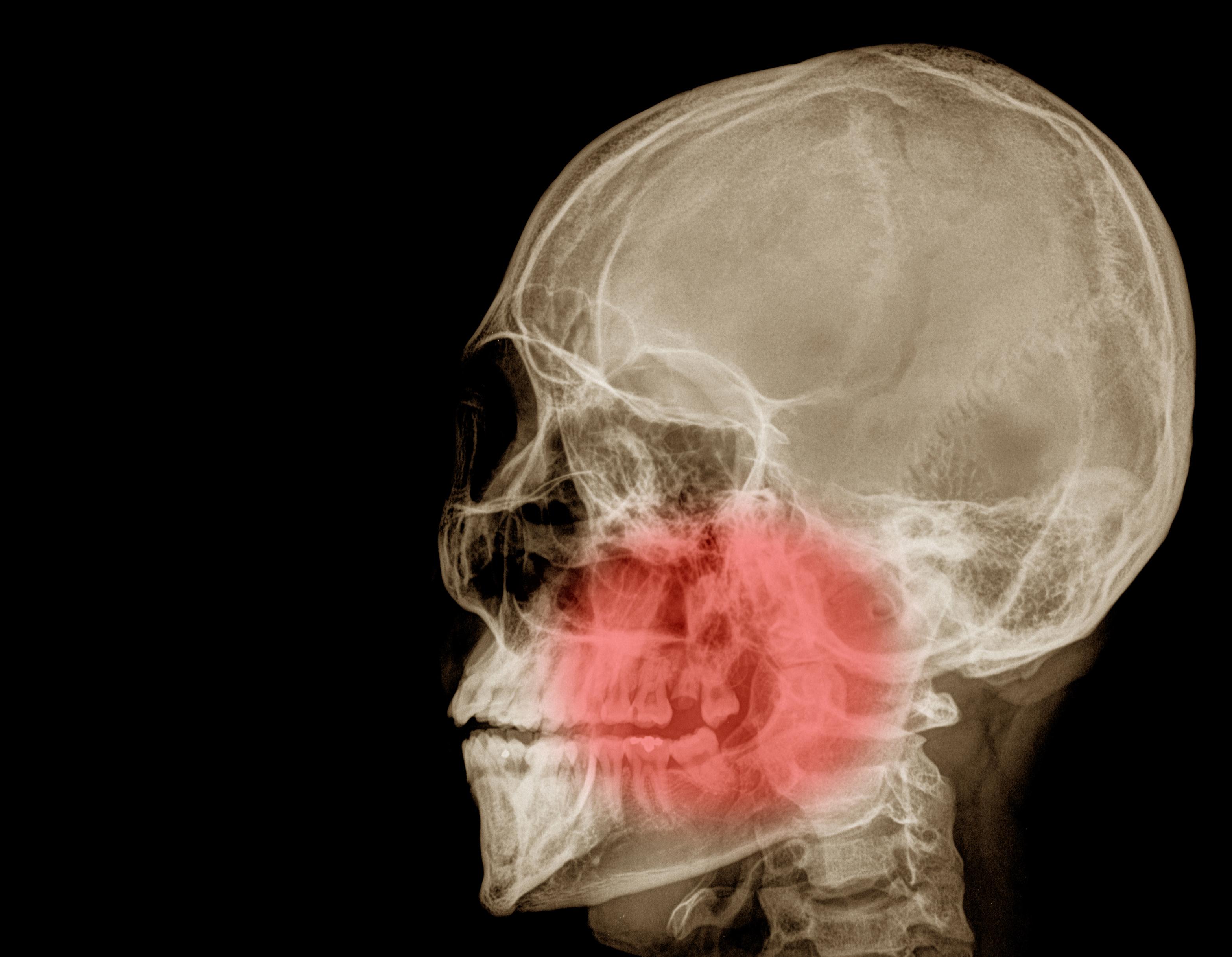
Mandibular Diseases
Radiography, Panoramic
Jaw, Edentulous
Mandibular Condyle
Chin
Maxilla
Retrognathia
Facial Bones
Osteogenesis, Distraction
Temporomandibular Joint
Dental Implantation, Endosseous
Alveolar Process
Dental Implants
Jaw Fixation Techniques
Molar
Jaw Neoplasms
Masticatory Muscles
Dental Prosthesis, Implant-Supported
Ameloblastoma
Maxillary Diseases
Oral Surgical Procedures
Mandibular Nerve
Malocclusion, Angle Class III
Dental Occlusion
Incisor
Mandibular Advancement
Cone-Beam Computed Tomography
Odontogenic Cysts
Hyoid Bone
Vertical Dimension
Malocclusion, Angle Class II
Dental Arch
Dentition
Jaw Cysts
Bicuspid
Alveolar Ridge Augmentation
Extraoral Traction Appliances
Skull Base
Orthodontic Appliances, Functional
Pterygoid Muscles
Occlusal Splints
Pierre Robin Syndrome
Alveolar Bone Loss
Cuspid
Bone Regeneration
Odontoma
Orthodontic Appliance Design
Osteoma
Orthognathic Surgical Procedures
Osteolysis, Essential
Stomatognathic System
Malocclusion
Jaw Diseases
Models, Anatomic
Anatomic Landmarks
Tooth, Impacted
Cementoma
Temporal Muscle
Activator Appliances
Palate
Nasal Bone
Mandibular Osteotomy
Zygoma
Malocclusion, Angle Class I
Overbite
Gingival Neoplasms
Fossils
Dental Prosthesis Design
Granuloma, Giant Cell
Tooth Socket
Craniofacial Abnormalities
Finite Element Analysis
Imaging, Three-Dimensional
Palatal Expansion Technique
Dentition, Mixed
Centric Relation
Bone Remodeling
Tomography, X-Ray Computed
Tongue
Cherubism
Radiographic Magnification
Free Tissue Flaps
Odontogenesis
Orthodontic Retainers
Anatomic Variation
Tooth Eruption
Orthodontics, Interceptive
Hominidae
Serial Extraction
Fibroma, Desmoplastic
Orthodontics, Corrective
Temporomandibular Joint Disorders
Tooth Calcification
Tooth Attrition
Blade Implantation
Sella Turcica
Microradiography
Reconstructive Surgical Procedures
Biomechanical Phenomena
Haversian System
Dental Prosthesis Retention
Denture, Complete
Anatomy, Comparative
Tooth, Supernumerary
Dental Stress Analysis
Periodontal Ligament
Temporal Bone
Orthodontic Anchorage Procedures
Bone Matrix
Osteotomy, Le Fort
Stress, Mechanical
Face
Oral Hemorrhage
Felidae
Fibula
Surgery, Oral
Tooth Crown
Beetles
Radiometric Dating
Orthodontic Appliances
Cartilage
Anodontia
Chondrosarcoma, Mesenchymal
Image Processing, Computer-Assisted
Trigeminal Nerve Injuries
Jaw Relation Record
Osteomyelitis
Bone Substitutes
Tooth Root
Patient Care Planning
Euthanasia, Animal
Hyperostosis, Cortical, Congenital
Branchial Region
Tooth Apex
Myofibroma
Periapical Diseases
Frontal Bone
Bone Density
Head
Periosteum
Humpback Whale
Tomography Scanners, X-Ray Computed
Maxillofacial Injuries
Open Bite
Bone Cysts, Aneurysmal
Biological Evolution
Mouth Floor
Osseointegration
Anatomy, Cross-Sectional
Temporomandibular Joint Disc
Bone and Bones
Fibrous Dysplasia of Bone
Sotos Syndrome
Tooth, Unerupted
Gingival Hyperplasia
Tooth, Deciduous
Diphosphonates
A variety of conditions, collectively known as mandibular diseases, can affect the jawbone including infections, tumors, congenital abnormalities, and temporomandibular joint disorders, often presenting with pain, limited movement, or changes in facial structure.
Mandibular diseases encompass a wide range of conditions affecting the jawbone, or mandible. Understanding these conditions involves recognizing their diverse origins, from developmental issues to acquired problems like infections and tumors. Here’s a breakdown of key information:
- Congenital Conditions: These are present at birth.
- Cleft Chin/Mandibular Cleft: A relatively common variation, often harmless, involving incomplete fusion of the mandible during development.
- Treacher Collins Syndrome: A genetic disorder affecting facial bone and tissue development, often resulting in a significantly underdeveloped mandible.
- Pierre Robin Sequence: Characterized by a small mandible, tongue displacement, and breathing difficulties.
- Developmental Disorders: These manifest during growth.
- Condylar Hypoplasia: Underdevelopment of the mandibular condyle (the part of the jaw that articulates with the skull), potentially leading to malocclusion.
- Hemifacial Microsomia: Uneven development of facial structures on one side, frequently involving the mandible.
- Infections: The mandible is susceptible to various infections.
- Osteomyelitis: A bone infection, often caused by bacteria, that can affect the mandible, particularly after trauma or dental procedures. Symptoms include pain, swelling, fever, and potentially pus formation.
- Ludwig's Angina: A severe, rapidly spreading infection of the floor of the mouth, often involving the mandible, and potentially compromising the airway.
- Tumors: Both benign and malignant tumors can arise in the mandible.
- Ameloblastoma: A benign, slow-growing tumor originating from tooth-forming cells; it’s the most common odontogenic (tooth-related) tumor of the mandible.
- Odontogenic Keratocyst: Another benign cyst-like tumor, also originating from tooth-forming cells, with a tendency to recur.
- Osteosarcoma: A malignant bone tumor that can occur in the mandible, though less common than in other bones.
- Metastatic Cancer: Cancer originating from other parts of the body can spread to the mandible.
- Other Conditions:
- Temporomandibular Joint (TMJ) Disorders: While not strictly a disease of the mandible, TMJ disorders often involve pain and dysfunction related to the mandible’s movement.
- Fractures: The mandible is the most commonly fractured facial bone, usually due to trauma.
- Osteonecrosis of the Jaw (ONJ): A condition where the jawbone doesn't heal properly, often associated with certain medications (like bisphosphonates) or radiation therapy.
- Fibrous Dysplasia: A bone disorder where normal bone is replaced with fibrous tissue, potentially affecting the mandible’s structure and function.
Mandibular diseases require careful consideration due to the jaw’s critical role in functions like chewing, speech, and facial aesthetics. Recognizing the diverse range of potential issues is the first step in understanding their impact.
Panoramic radiography is a two-dimensional dental radiographic technique which provides a broad view of the maxillary and mandibular dental structures, including the teeth, supporting bone, and temporomandibular joints, in a single image.
Panoramic radiography is a widely utilized diagnostic imaging technique in dentistry and maxillofacial radiology, offering a broad view of the dental structures and surrounding tissues. It’s a fascinating process that provides a wealth of information.
- What it is: A panoramic radiograph (often called a “panorex”) is a two-dimensional image showing the entire dentition (teeth), supporting structures of the upper and lower jaws, and portions of the temporal bones and sinuses.
- How it works: The x-ray source and film/digital sensor move simultaneously around the patient’s head, capturing a single, flattened image. This differs from traditional bitewing or periapical radiographs which focus on individual teeth or areas.
- What it shows: A panoramic radiograph can reveal:
- Dental caries (cavities) – though not always as precisely as bitewings.
- Impacted teeth – particularly wisdom teeth.
- Bone loss associated with periodontal (gum) disease.
- Cysts and tumors within the jaws.
- Fractures of the mandible (lower jaw) or maxilla (upper jaw).
- Sinus abnormalities.
- Temporomandibular joint (TMJ) issues – though dedicated TMJ imaging is often preferred.
- Root morphology and abnormalities.
- Procedure details: During the procedure, the patient typically stands or sits with their chin resting in a designated support. A bite block is used to ensure proper positioning. The machine then rotates around the head, taking the x-ray. The entire process usually takes only a few seconds.
- Radiation exposure: While radiography involves radiation, the dose from a panoramic radiograph is generally considered low. Modern digital systems further reduce radiation exposure compared to traditional film-based systems.
- Limitations: Because it’s a two-dimensional image, panoramic radiographs can sometimes distort the image, making precise measurements difficult. Small fractures or early-stage caries may also be missed. It is not ideal for evaluating individual tooth root details.
- Digital vs. Film: Digital panoramic radiography is now the standard of care. It offers several advantages over film, including reduced radiation, faster image acquisition, and the ability to enhance and manipulate the image digitally.
- Artifacts: Various artifacts can appear on a panoramic radiograph, such as those from jewelry, dentures, or patient movement. These artifacts can sometimes obscure important structures.
Panoramic radiography remains a valuable tool for initial assessment and comprehensive overview of the maxillofacial region, providing crucial information for diagnosis and treatment planning.
An edentulous jaw—a jaw lacking teeth—can undergo bone resorption over time, leading to changes in facial structure and potential difficulties with speech and nutrition.
Understanding the edentulous jaw is crucial for maintaining overall health and quality of life, especially as we age. The complete loss of teeth, or edentulism, significantly impacts the jawbone and surrounding structures. Here's a breakdown of key information:
- Bone Resorption: Following tooth loss, the alveolar bone – the part of the jawbone that holds teeth in place – begins to resorb (shrink). This happens because the bone no longer receives the stimulation from chewing forces provided by teeth. The rate of resorption varies between individuals.
- Facial Changes: As the jawbone diminishes, it can lead to a collapse of facial structures. This manifests as a decreased vertical dimension (space between the upper and lower jaw), a sunken-in appearance of the cheeks, and more prominent wrinkles around the mouth.
- Soft Tissue Changes: The supporting soft tissues, like the gums and muscles, also undergo changes. Gum tissue can become thin and fragile. Muscle tone may decrease, affecting speech and chewing efficiency.
- Digestive Issues: Complete tooth loss severely compromises the ability to properly chew food. This can lead to digestive problems, as larger food particles are harder to break down, potentially causing discomfort and nutrient malabsorption.
- Speech Alterations: Teeth play a role in articulation. Losing them can result in slurred speech or difficulty pronouncing certain sounds. The extent of speech changes depends on how many teeth are missing and individual speech patterns.
- Temporomandibular Joint (TMJ) Effects: While not a direct cause, edentulism can exacerbate existing TMJ disorders or contribute to discomfort. Changes in bite and chewing patterns can place stress on the TMJ.
- Neuromuscular Adaptations: The brain attempts to compensate for the loss of teeth by adapting neuromuscular pathways. This can lead to altered chewing patterns and potentially uneven muscle development.
- Prosthetic Considerations: Dentures are the most common solution for restoring function and aesthetics in edentulous individuals. Different types of dentures exist, including conventional, immediate, and implant-supported options, each with its own advantages and disadvantages.
- Nutritional Impact: Difficulty chewing can lead to dietary restrictions, potentially resulting in nutritional deficiencies. Individuals may avoid certain foods (like fruits, vegetables, and lean meats) due to the effort required to consume them.
- Psychological Effects: Tooth loss can significantly impact self-esteem and social interactions. Individuals may feel self-conscious about their appearance and avoid smiling or speaking in public.
Managing an edentulous jaw requires a comprehensive understanding of the changes that occur and the available options for restoring function and quality of life.
The mandibular condyle, a rounded projection of the mandible, articulates with the temporal bone to form the temporomandibular joint, enabling jaw movements like chewing and speaking.
Understanding the mandibular condyle is crucial for grasping jaw function and potential issues related to the temporomandibular joint (TMJ). It’s a fascinating and often overlooked part of our anatomy.
- The mandibular condyle is the rounded projection at the upper end of the mandible (lower jaw bone).
- It’s a key component of the TMJ, articulating with the temporal bone of the skull. This joint allows for a wide range of jaw movements – opening, closing, chewing, and speaking.
- The condyle is covered with cartilage, similar to other joints in the body, which allows for smooth, low-friction movement. This cartilage is vital for absorbing forces during function.
- The shape of the mandibular condyle isn’t perfectly round; it’s more oval or elliptical. This shape contributes to the complex movements of the jaw.
- During jaw opening and closing, the condyle glides forward and backward within the mandibular fossa of the temporal bone, and also rotates.
- The condyle develops through a process called endochondral ossification, meaning it starts as cartilage and gradually turns into bone. This process continues into early adulthood.
- Variations in condylar shape and size are common and can influence jaw function.
- The mandibular condyle is susceptible to fractures, particularly in cases of trauma to the face or jaw.
- Conditions affecting the cartilage of the condyle can lead to degenerative joint disease, impacting jaw function and causing discomfort.
- The position and movement of the condyle are influenced by the muscles of mastication (chewing), which attach to the mandible and control jaw movements.
- Imaging techniques can be used to visualize the mandibular condyle and assess its structure and function.
- The condyle plays a role in maintaining proper bite alignment and overall facial harmony.
In essence, the mandibular condyle is a critical structure for everyday functions, and its health is integral to comfortable and effective jaw mechanics.
The chin—composed of the mental protuberance and contributing to facial aesthetics and speech—is formed by the anterior portions of the mandible and plays a role in chewing, swallowing, and maintaining airway patency.
The chin, often underestimated, plays a significant role in facial structure, function, and even how we perceive ourselves and others. Understanding its anatomy and potential variations can offer insight into its importance.
- The chin, medically known as the mental protuberance, is formed by the anterior projection of the mandible, or lower jawbone.
- Bone structure is the primary determinant of chin shape and size. Variations in bone growth, influenced by genetics and development, lead to diverse chin profiles.
- Muscles attaching to the chin, such as the mentalis, contribute to facial expressions like pouting and frowning. The depressor menti muscle pulls the chin downwards.
- Skin and soft tissues covering the chin influence its appearance. Factors like fat distribution and skin elasticity affect how prominent the chin appears.
- A receding chin, where the chin is set back relative to the rest of the face, can sometimes be associated with a weaker jawline.
- A prominent chin can create a stronger facial profile and is often considered a sign of determination.
- Chin dimples, a common feature, are caused by variations in the underlying muscle and connective tissue. They are generally considered a cosmetic variation.
- The chin serves as an attachment point for several facial muscles crucial for chewing and facial expression.
- Sensory nerves supply the chin, providing sensation to the skin and lips. Damage to these nerves can result in numbness or altered sensation.
- The chin’s position influences occlusion, or how the upper and lower teeth come together. Significant deviations can affect bite and chewing function.
- Developmental variations can occur, leading to conditions like micrognathia (a small chin) or macrognathia (an abnormally large chin).
- The chin is susceptible to injury from trauma, potentially resulting in fractures or soft tissue damage.
Ultimately, the chin is a complex anatomical feature that contributes significantly to facial aesthetics and functionality, and its variations are a normal part of human diversity.
Cephalometry, a standardized radiographic technique, involves taking precise measurements of the skull to assess skeletal and dental relationships, crucial for orthodontic treatment planning and diagnosing craniofacial abnormalities.
Cephalometry, the measurement of the human skull, is a crucial technique primarily utilized in orthodontics, oral and maxillofacial surgery, and anthropology. It provides a standardized way to assess the skeletal and soft tissue relationships of the face and head, aiding in diagnosis and treatment planning.
- Radiographic Cephalometry is the most common form, employing a standardized X-ray called a cephalogram. This lateral skull X-ray captures a side view of the head, from the nose to the chin and the top of the skull.
- Landmarks are specific, anatomically defined points on the skull and face that are identified on the cephalogram. These landmarks are consistent and reproducible, allowing for accurate measurements. Examples include the nasion (bridge of the nose), porion (outermost point of the ear), and menton (lowest point of the chin).
- Measurements are taken between these landmarks to analyze skeletal patterns. These measurements are categorized into:
- Skeletal measurements: Assess the size and relationship of the maxilla (upper jaw) and mandible (lower jaw) to the cranial base. Examples include ANB (angle between A-point, Nasion, and B-point), and SNB (angle between Sella, Nasion, and B-point).
- Dental measurements: Evaluate the position of the teeth within the jaws.
- Soft tissue measurements: Analyze the profile and soft tissue relationships.
- Analysis of these measurements helps classify malocclusions (misalignment of teeth) into different skeletal patterns:
- Class I: Normal relationship between the upper and lower jaws.
- Class II: Upper jaw is positioned forward relative to the lower jaw (overbite).
- Class III: Lower jaw is positioned forward relative to the upper jaw (underbite).
- Tracing involves manually or digitally outlining the contours of the skull and soft tissues on the cephalogram. This tracing is then used to perform the measurements.
- Computerized Cephalometry utilizes software to automate the tracing and measurement process, increasing accuracy and efficiency.
- Growth Prediction is possible through cephalometric analysis, particularly in growing children. By analyzing the growth patterns, potential future skeletal changes can be estimated.
- 3D Cephalometry is an emerging technique using cone-beam computed tomography (CBCT) to create a three-dimensional image of the skull, providing more comprehensive and accurate measurements.
- Cranial Vault Measurements are used in anthropology to study population variations and evolutionary trends. These measurements focus on the overall shape and size of the cranial vault.
Cephalometry remains a cornerstone of diagnostic evaluation in fields dealing with facial structures, providing a detailed and objective assessment of skeletal and dental relationships.
The maxilla comprises two bones that fuse during infancy to form the upper jaw, contributing to the formation of the hard palate, nasal cavity, and orbits.
The maxilla is a fascinating and crucial component of facial structure and function, often overlooked until issues arise. Understanding its anatomy and potential concerns can provide valuable insight into overall health.
- The maxilla refers to both the upper jawbone and, collectively, the two bones that form the upper jaw. It’s a paired bone, meaning there are two, one on each side of the face.
- Anatomical Divisions: The maxilla isn’t a single, uniform structure. It’s divided into several parts: the body, the processes (frontal, zygomatic, alveolar, palatine, and maxillary sinus processes), and the orbital portion. Each contributes to different functions.
- Key Functions: The maxilla performs several vital roles. It provides structural support for the face, forms the upper portion of the mouth, contains the upper teeth (via the alveolar process), contributes to the floor of the orbit (eye socket), and houses the maxillary sinus.
- Maxillary Sinus: The maxillary sinus is the largest of the paranasal sinuses, air-filled spaces within the maxillary bone. These sinuses lighten the skull, contribute to voice resonance, and help humidify and warm inhaled air.
- Alveolar Process: This is the part of the maxilla that holds the upper teeth. Bone loss in this area can occur due to periodontal disease or tooth extraction, leading to changes in facial structure.
- Development & Growth: The maxilla develops through intramembranous ossification, meaning bone forms directly from mesenchymal tissue. Its growth is closely linked to dental development and is influenced by factors like genetics and nutrition.
- Common Concerns: Issues affecting the maxilla can include sinus infections (sinusitis), fractures (often from facial trauma), cysts or tumors, and malocclusion (misalignment of teeth).
- Bone Composition: The maxilla is composed of cortical bone (dense outer layer) and cancellous bone (spongy inner layer). This structure provides both strength and lightness.
- Vascular & Nerve Supply: The maxilla receives blood supply from the maxillary artery and is innervated by branches of the trigeminal nerve, crucial for sensation in the face and teeth.
- Articulation: The maxilla articulates (forms joints) with numerous other bones, including the frontal, nasal, zygomatic, palatine, inferior nasal conchae, and vomer bones. This complex network contributes to facial stability.
The maxilla, therefore, is a complex and integral part of the human anatomy, playing a critical role in facial structure, dental health, and overall well-being.
Sustaining a mandibular injury, commonly known as a broken jaw, can result from significant force to the face and often presents with pain, swelling, difficulty chewing, and malocclusion—a misalignment of the teeth.
Mandibular injuries, commonly known as jaw fractures, represent a significant area of concern in trauma management. Understanding the nature of these injuries and potential associated complications is crucial for comprehensive care.
- Causes: Mandibular fractures typically result from blunt force trauma, such as assaults, motor vehicle accidents, or falls. Less commonly, they can occur due to pathological fractures related to underlying bone weakening conditions like osteoporosis or metastatic cancer.
- Common Fracture Locations: The mandible is most susceptible to fracture at the angle, body, and condyle. The symphysis menti (chin area) is also a frequent site. The specific location often correlates with the mechanism of injury.
- Signs and Symptoms: Pain is the most prominent symptom, often exacerbated by chewing or talking. Other indicators include swelling, bruising, malocclusion (difficulty aligning the teeth), numbness in the lower lip or chin (indicating nerve damage), and instability of the jaw. Difficulty swallowing or speech disturbances may also be present.
- Associated Injuries: Mandibular fractures are rarely isolated. They frequently occur alongside other injuries, including facial lacerations, dental trauma (fractured or displaced teeth), head injuries (concussions or intracranial hemorrhage), and cervical spine injuries.
- Neurological Complications: The inferior alveolar nerve runs through the mandible and can be damaged during a fracture, leading to lower lip and chin numbness or tingling. Damage to the lingual nerve can cause numbness on the tongue.
- Vascular Injuries: Although less common, fractures can compromise blood vessels in the area, leading to bleeding and potentially hematoma formation.
- Fracture Patterns: Fractures can be simple, comminuted (multiple fragments), or open (bone protruding through the skin). The classification influences treatment considerations.
- Initial Management: Stabilization of the airway, breathing, and circulation is paramount. Immobilization of the jaw is often achieved using temporary fixation methods.
- Long-Term Considerations: Potential long-term complications include malunion (improper healing), nonunion (failure to heal), chronic pain, and temporomandibular joint (TMJ) dysfunction.
Understanding these aspects of mandibular injuries provides a foundational knowledge base for recognizing and addressing these complex conditions.
Retrognathia, a condition characterized by a receding chin and jaw, can contribute to difficulties with chewing, speech, and breathing due to the altered alignment of the dental arches and airway.
Retrognathia, a condition where the jaw (typically the lower jaw, or mandible) is set back relative to the upper jaw, can present in varying degrees of severity and impact facial aesthetics and function. Understanding the nuances of this condition is important for recognizing its potential effects.
- Definition: Retrognathia literally means “backward jaw.” It’s a deviation in skeletal development, not simply a cosmetic issue.
- Causes: Several factors can contribute to retrognathia. These include genetic predisposition, meaning it can run in families. Developmental issues during childhood, such as problems with jaw growth, can also play a role. In some cases, acquired retrognathia can occur due to trauma or surgery affecting the jaw.
- Appearance: Individuals with retrognathia often exhibit a receding chin, a flat facial profile, and a noticeable concavity in the lower face. The upper teeth may protrude over the lower teeth, creating an overbite.
- Functional Impacts: Beyond aesthetics, retrognathia can lead to difficulty chewing, problems with speech, and temporomandibular joint (TMJ) disorders. The altered jaw position can strain the jaw muscles and joints.
- Breathing Concerns: In more severe cases, retrognathia can contribute to obstructive sleep apnea by narrowing the airway. This is because the tongue may fall back into the throat during sleep, blocking airflow.
- Dental Implications: Malocclusion (misalignment of teeth) is common with retrognathia. This can lead to uneven wear on the teeth, difficulty cleaning, and an increased risk of cavities and gum disease.
- Severity Levels: Retrognathia is classified by its severity. Mild retrognathia may only be noticeable aesthetically, while severe retrognathia can significantly impact function and quality of life.
- Associated Conditions: Sometimes, retrognathia occurs alongside other craniofacial differences, such as Pierre Robin sequence or Treacher Collins syndrome.
- Diagnosis: Diagnosis typically involves a clinical examination of the face and jaw, along with radiographic imaging such as X-rays and cephalometric analyses to assess the skeletal relationship.
- Variations: Retrognathia can affect either the upper or lower jaw, though it’s more commonly associated with a receding lower jaw. Upper jaw retrognathia is less frequent but can also cause functional and aesthetic concerns.
Ultimately, retrognathia is a complex condition with a range of potential impacts, and recognizing its characteristics is key to understanding its implications.
Distraction osteogenesis is a biological process where new bone is formed between the surfaces of bone that have been gradually separated, typically achieved through the use of an external or internal fixation device to create and maintain space for regeneration.
Osteogenesis and distraction are fascinating processes within the realm of bone healing and reconstruction, representing the body’s remarkable capacity to regenerate tissue. Understanding these mechanisms is crucial for appreciating how skeletal injuries are addressed and even how bone can be intentionally lengthened or reshaped.
- Osteogenesis refers to the formation of new bone. It’s not simply “repair” but the actual creation of bone tissue. This happens through two primary pathways: intramembranous and endochondral ossification.
- Intramembranous ossification directly converts mesenchymal tissue into bone, seen in flat bones like the skull.
- Endochondral ossification uses a cartilage template which is gradually replaced by bone, the process responsible for long bone development.
- Several factors influence osteogenesis, including growth factors (like Bone Morphogenetic Proteins – BMPs), adequate blood supply, mechanical stress, and the presence of osteogenic cells (osteoblasts).
- Distraction osteogenesis is a specific biological process of new bone formation as a direct response to sustained, controlled traction. It’s the principle behind bone lengthening and reconstruction procedures.
- The process typically involves a corticotomy – a precise cut through the bone.
- Following a latency period (usually several days) allowing a fibrous union to form, a device (an external or internal fixator) is used to slowly and gradually distract (pull apart) the bone segments.
- This distraction creates space, and the body responds by depositing new bone within that space. This new bone is initially woven bone, which matures into lamellar bone over time.
- The rate of distraction is carefully controlled, typically around 1mm per day, to allow for organized bone formation. Too fast, and fibrous non-union can occur; too slow, and the process stalls.
- Mechanical forces are critical in distraction osteogenesis. The tension stimulates osteogenesis, and the gradual separation guides the new bone formation.
- Distraction osteogenesis isn’t limited to limb lengthening. It’s also used for correcting bone deformities, filling bone defects (e.g., after tumor resection or trauma), and even in craniofacial surgery.
- Complications can include pin site infections, nerve damage, joint contractures, and non-union (failure of bone to heal).
- The quality of the newly formed bone is generally good, though it may initially be less dense than original bone. Continued weight-bearing and remodeling help to improve bone density over time.
- Bone transport is a related technique where a segment of bone is gradually moved from one location to another using distraction principles, useful for filling large bone defects.
Osteogenesis and distraction represent powerful tools in skeletal repair and reconstruction, demonstrating the body’s inherent ability to adapt and regenerate when provided with the appropriate biological and mechanical stimuli.
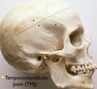
The temporomandibular joint, connecting the jawbone to the skull, can be affected by a variety of biomechanical, psychological, and systemic factors leading to pain and dysfunction in the jaw, face, and neck.
Understanding the temporomandibular joint (TMJ) and associated discomfort is crucial for overall well-being, as it impacts many daily functions. Here's a breakdown of key information regarding this complex joint:
- The TMJ connects your jawbone (mandible) to your skull (temporal bone). It’s a bilateral joint, meaning you have one on each side of your face, just in front of your ears.
- Functionally, the TMJ allows for essential movements like talking, chewing, yawning, and facial expressions. These movements involve a coordinated effort of muscles, ligaments, and the joint itself.
- Anatomy includes the mandibular condyle (the rounded end of the jawbone), the temporal fossa (a depression in the skull), an articular disc (cartilage cushioning the joint), and surrounding ligaments and muscles.
- Common symptoms associated with TMJ disorders can include pain or tenderness in the jaw joint and surrounding muscles, limited jaw movement or locking, clicking, popping, or grating sounds when opening or closing the mouth, difficulty chewing, and facial pain.
- Potential contributing factors to TMJ issues are numerous. These can include teeth grinding (bruxism), clenching, stress, arthritis, jaw injury, and poor posture.
- Muscle involvement is significant. Muscles like the masseter, temporalis, and pterygoids play a vital role in jaw movement and can become strained or fatigued, contributing to discomfort.
- The articular disc is a key component. Displacement or degeneration of this disc can lead to clicking, popping, and locking sensations.
- Bruxism, or teeth grinding, often occurs during sleep and can place significant stress on the TMJ, leading to muscle fatigue and joint pain.
- Self-care strategies may involve applying warm or cold compresses to the jaw, eating soft foods, avoiding excessive chewing, and practicing relaxation techniques to reduce stress.
- Proper posture can also play a role. Maintaining good head and neck alignment can help reduce strain on the TMJ.
The temporomandibular joint is a fascinating and often overlooked aspect of our anatomy, and understanding its complexities can empower individuals to manage their comfort and function.
Endosseous dental implantation is a surgical procedure involving the placement of biocompatible titanium posts directly into the jawbone to provide a stable foundation for replacement teeth, effectively mimicking the structure and function of natural tooth roots.
Dental implantation, specifically endosseous implants, represents a significant advancement in restorative dentistry, offering a durable and aesthetically pleasing solution for missing teeth. This process involves surgically placing a titanium post into the jawbone, which then serves as an artificial root for a replacement tooth.
- Titanium’s biocompatibility is a cornerstone of implant success. The material integrates with the jawbone through a process called osseointegration, forming a strong and lasting bond.
- Osseointegration typically requires several months, during which the implant site is left undisturbed to allow the bone to grow around the titanium post. This phase is crucial for long-term stability.
- Implant components consist of three main parts: the implant itself (the titanium post), an abutment (which connects the implant to the crown), and the crown (the visible part of the tooth).
- Candidates for endosseous implants generally need sufficient bone density and healthy gums. Bone grafting procedures can be employed to augment bone volume if necessary.
- Surgical placement is usually performed under local anesthesia, though sedation options are available. The procedure involves creating a flap in the gum tissue to expose the jawbone, drilling a precise hole for the implant, and carefully inserting the titanium post.
- Post-operative care is vital for successful healing. This includes maintaining excellent oral hygiene, following a soft food diet, and attending follow-up appointments to monitor osseointegration.
- Different implant systems exist, varying in size, shape, and surface characteristics. The choice of system depends on individual anatomical factors and the location of the missing tooth.
- Potential complications, while relatively uncommon, can include infection, nerve damage, sinus problems (for upper implants), and implant failure.
- Long-term success rates for endosseous implants are high, often exceeding 95% with proper care and maintenance. Regular dental check-ups and diligent oral hygiene are essential.
- Alternatives to implants include dentures and bridges, but implants offer superior stability, function, and preservation of surrounding bone structure.
Ultimately, endosseous dental implants provide a robust and natural-feeling solution for tooth loss, improving both oral health and quality of life.
The alveolar process is the part of the maxilla and mandible that forms the bony socket for teeth, providing their support and enabling mastication.
Understanding the alveolar process is fundamental to grasping dental and facial structure. It’s a complex area, and knowing its components is key to understanding overall oral health.
- The alveolar process is the part of the maxilla (upper jaw) and mandible (lower jaw) that surrounds the roots of the teeth. It’s essentially the bony socket that holds teeth in place.
- It’s composed of cortical bone on the outer and inner surfaces, and cancellous bone (spongy bone) in between. This cancellous bone contains bone marrow.
- Alveolar bone proper is the bundle bone directly lining the tooth socket. This bone is highly specialized and responds to forces applied by the teeth.
- Supporting alveolar crest is the bone forming the crest of the alveolar process, located between the tooth roots. It’s a crucial area for maintaining tooth stability.
- Interdental septa are bony partitions that extend between the teeth in the alveolar process. They aren’t continuous, allowing for some flexibility.
- The periodontal ligament connects the tooth root to the alveolar bone proper. This ligament isn’t part of the alveolar process itself, but is intimately linked to its health and function.
- The alveolar process isn’t static; it constantly remodels in response to chewing forces, tooth movement, and inflammation.
- Loss of teeth leads to alveolar resorption, where the bone gradually shrinks over time. This is because the bone needs the stimulation from the tooth roots to maintain its density.
- The shape and size of the alveolar process vary significantly between individuals and depend on factors like genetics, age, and dental habits.
- Developmentally, the alveolar process forms alongside the teeth, and its growth is closely tied to tooth eruption.
- The alveolar process provides attachment for the muscles of facial expression and mastication (chewing).
In essence, the alveolar process is a dynamic and vital component of the jaw, providing the foundation for teeth and contributing to facial structure and function.
Mastication, or chewing, is the initial and crucial mechanical breakdown of food that increases surface area for enzymatic digestion and facilitates swallowing.
Mastication, commonly known as chewing, is a fundamental process in the digestive system and overall health. It’s far more complex than simply breaking down food; it initiates digestion and impacts nutrient absorption.
- Anatomy Involved: Mastication relies on a coordinated effort between several structures. These include the teeth (incisors, canines, premolars, and molars), the jaw muscles (masseter, temporalis, medial and lateral pterygoids), the temporomandibular joint (TMJ), the tongue, and the salivary glands.
- Stages of Mastication: The process isn’t uniform. It begins with initial biting and cutting by the incisors and canines. This is followed by grinding and crushing by the premolars and molars, reducing food particle size. The tongue constantly repositions food between the teeth for efficient breakdown.
- Saliva’s Role: Saliva is crucial. It moistens food, making it easier to form a bolus (a rounded mass of chewed food). It contains amylase, an enzyme that begins the breakdown of carbohydrates. Saliva also contains lysozyme, which has antibacterial properties.
- Physiological Benefits: Proper mastication offers several advantages. It increases surface area for digestive enzymes to act upon, improving nutrient extraction. It stimulates saliva production, aiding digestion and protecting teeth. It can also promote satiety, potentially assisting with weight management.
- Impact on Digestion: Thoroughly chewed food is easier for the stomach to process, reducing the workload and potentially minimizing digestive discomfort. Incomplete mastication can lead to larger food particles reaching the intestines, potentially causing issues with absorption and gut health.
- Muscle Strength & Development: Regular chewing exercises the jaw muscles, contributing to their strength and development. This can be particularly important for individuals with age-related muscle loss or those recovering from facial injuries.
- Sensory Input: Mastication provides sensory feedback to the brain regarding food texture and consistency, contributing to the enjoyment of eating and influencing appetite.
- Potential Issues: Problems with any component of the mastication process – teeth, jaw muscles, TMJ, or saliva production – can lead to difficulties with eating, pain, and digestive issues.
Understanding mastication highlights its importance beyond simply preparing food for swallowing; it’s a vital component of a healthy digestive system and overall well-being.
Dental implants are titanium posts surgically placed into the jawbone to serve as a stable foundation for replacement teeth, offering a long-term solution for tooth loss and restoring both function and aesthetics.
Dental implants represent a significant advancement in restorative dentistry, offering a durable and aesthetically pleasing solution for missing teeth. Understanding the process and considerations involved can be beneficial for anyone exploring tooth replacement options.
- What are Dental Implants? They are essentially artificial tooth roots, typically made of biocompatible titanium, surgically placed into the jawbone. These act as a foundation for replacement teeth that look, feel, and function like natural teeth.
- The Implant Process: It’s generally a multi-stage process. First, an evaluation, including X-rays and potentially a CT scan, is performed to assess bone density and determine suitability.
- Surgical Placement: The implant itself is surgically inserted into the jawbone. A healing period, often several months, follows to allow osseointegration – the process where the implant fuses with the bone.
- Abutment Placement: Once osseointegration is complete, an abutment is attached to the implant. This serves as a connector between the implant and the final restoration.
- Crown Placement: Finally, a custom-made crown, designed to match the surrounding teeth in shape, size, and color, is attached to the abutment.
- Types of Implants: While titanium is most common, zirconia implants are also available, offering a metal-free alternative. Different implant systems exist, varying in design and surface treatment.
- Candidates for Implants: Good candidates generally have sufficient bone density, healthy gums, and are in good overall health. Certain medical conditions and habits, like smoking, can affect implant success.
- Benefits of Implants: They offer several advantages, including improved speech, easier chewing, enhanced aesthetics, and prevention of bone loss in the jaw. They also don’t require altering adjacent teeth, unlike traditional bridges.
- Implant Maintenance: Proper oral hygiene, including regular brushing, flossing, and professional cleanings, is crucial for long-term implant success.
- Potential Complications: While generally safe, potential complications can include infection, nerve damage, sinus problems (for upper implants), and implant failure.
- Cost Considerations: Dental implants are typically more expensive upfront than other tooth replacement options, but their longevity and benefits can make them a cost-effective solution in the long run.
Dental implants continue to evolve with advancements in materials and techniques, providing increasingly predictable and successful outcomes for individuals seeking to restore their smiles and oral function.
Odontogenic tumors are a diverse group of neoplasms arising from the developmental tissues of the teeth and surrounding structures, often presenting as painless swellings and occasionally impacting adjacent anatomical landmarks.
Odontogenic tumors represent a fascinating, yet complex, area within oral pathology, arising from the tissues involved in tooth development – the ectomesenchyme, ectoderm, and mesenchyme. Understanding these growths requires a grasp of normal tooth formation, as disruptions in these processes can lead to a variety of benign and, rarely, malignant neoplasms.
- Classification is broadly divided into epithelial, mesenchymal, and mixed tumors. Epithelial tumors originate from the enamel organ or its remnants, like the ameloblastoma, the most common odontogenic tumor, often presenting as a slow-growing, locally aggressive lesion.
- Mesenchymal tumors stem from the dental papilla or follicle, including odontoma, which is essentially a malformation resembling a tooth or teeth, and fibroma, a benign connective tissue tumor.
- Mixed tumors, as the name suggests, contain both epithelial and mesenchymal components. Odontogenic carcinomas are malignant mixed tumors, thankfully quite rare.
- Ameloblastomas typically appear in adults, most frequently in the mandible, and are characterized by their microscopic appearance – islands of enamel-producing cells. They aren’t cancerous but can cause significant local destruction due to their infiltrative growth pattern.
- Odontomas are often discovered incidentally on radiographs, appearing as dense, tooth-like structures. They are usually asymptomatic and are more common in younger individuals.
- Cementoblastomas are benign tumors producing cementum-like tissue, often attached to a tooth root. They can be difficult to differentiate from other lesions radiographically.
- Calcifying epithelial odontogenic cyst (CEOC), previously considered a cyst, is now classified as a benign odontogenic tumor. It often presents as a well-defined radiolucency and can be associated with a developing tooth.
- Pindborg tumor (solid ameloblasticoma) is a less common variant of ameloblastoma, characterized by a more solid, less cystic appearance.
- Radiographic features are crucial for initial assessment. Lesions can appear as radiolucencies (dark areas) or radiopacities (white areas) on X-rays, and their location, size, and shape can provide clues to the type of tumor.
- Histopathological examination – microscopic analysis of tissue samples – is essential for definitive diagnosis. This allows pathologists to identify the specific cellular characteristics of the tumor.
- Clinical presentation can vary widely. Some tumors are asymptomatic, while others may cause pain, swelling, or tooth mobility. The symptoms depend on the size, location, and growth rate of the tumor.
- Growth patterns differ significantly between tumor types. Some are slow-growing and well-defined, while others are aggressive and infiltrative.
Odontogenic tumors, while often benign, require careful evaluation due to their potential for local destruction and, in rare cases, malignant transformation. Understanding their diverse characteristics is vital for appropriate management.
The jaw, comprised of the mandible and maxilla, facilitates essential functions like chewing, speech, and facial structure through a complex interplay of muscles, ligaments, and teeth.
Understanding the intricacies of the jaw is crucial for appreciating its role in essential bodily functions. It’s far more than just a structure for chewing; it’s a complex system impacting speech, breathing, and even facial expression.
- The jaw, or mandible, is the only movable bone in the skull. This mobility is enabled by the temporomandibular joint (TMJ), connecting the jaw to the temporal bones of the skull.
- Muscles of mastication – including the masseter, temporalis, medial and lateral pterygoids – are responsible for jaw movement. These muscles work in coordination to allow for actions like opening, closing, and side-to-side movements.
- The jaw houses 32 teeth in a complete adult set, arranged in four quadrants. These teeth are categorized as incisors, canines, premolars, and molars, each serving a specific function in breaking down food.
- Salivary glands located near the jaw (parotid, submandibular, and sublingual) produce saliva, initiating the digestive process and keeping the mouth moist.
- Nerves such as the trigeminal nerve provide sensation to the jaw and control the muscles involved in chewing. Damage to these nerves can result in pain or loss of function.
- Bone density in the jaw is influenced by factors like age, diet, and tooth loss. Reduced bone density can lead to difficulties with dental implants or dentures.
- Fractures of the jaw can occur due to trauma, and often require immobilization for healing. The location and severity of the fracture dictate the treatment approach.
- The jaw’s shape and size are influenced by genetics and developmental factors. Variations in jaw structure can contribute to malocclusion (misalignment of teeth).
- Bruxism, or teeth grinding, can place excessive stress on the jaw muscles and TMJ, potentially leading to pain and dysfunction.
- The jaw plays a vital role in speech articulation, assisting in the formation of sounds.
The jaw is a remarkable structure, demonstrating the body’s intricate design and the interconnectedness of its systems.
The human skull is a bony structure that supports the face and protects the brain, comprising 22 bones—eight cranial and 14 facial—connected by sutures.
The skull is a remarkably complex and vital structure, serving as the body’s primary protective casing for the brain and the foundation for the face. Understanding its components and functions is crucial for appreciating its significance.
- The skull is comprised of 22 bones, not including the three tiny bones within each middle ear (malleus, incus, and stapes). These bones are divided into two main groups: the cranium and the facial bones.
- The cranium (or braincase) consists of eight bones that enclose and protect the brain. These include the frontal bone, two parietal bones, two temporal bones, the occipital bone, the sphenoid bone, and the ethmoid bone.
- Sutures are fibrous joints that connect the cranial bones. These aren’t fully fused at birth, allowing for brain growth, and gradually close with age. Examples include the sagittal suture (between the parietal bones), the coronal suture (between the frontal and parietal bones), and the lambdoid suture (between the parietal and occipital bones).
- Foramina are openings in the skull bones that allow for the passage of nerves and blood vessels. The foramen magnum, located in the occipital bone, is the largest and allows the spinal cord to connect to the brain.
- The facial bones form the framework of the face, providing attachment points for facial muscles and supporting the organs of sight, smell, taste, and hearing. These include the nasal bones, maxillae (upper jaw), zygomatic bones (cheekbones), mandible (lower jaw – the only movable bone of the skull), lacrimal bones, palatine bones, and the vomer.
- Sinuses are air-filled spaces within some of the skull bones (frontal, ethmoid, sphenoid, and maxillae). They lighten the skull, contribute to voice resonance, and help to humidify and warm inhaled air.
- The mandible is unique as it’s the only movable bone in the skull, enabling chewing, speech, and facial expressions. It articulates with the temporal bones at the temporomandibular joint (TMJ).
- The skull provides attachment points for numerous muscles involved in head movement, facial expression, and chewing. These muscles are crucial for a wide range of daily functions.
- The shape and features of the skull can vary significantly based on factors like genetics, age, and sex. These variations can be observed in the size and prominence of certain bony landmarks.
- Ossicles are the three smallest bones in the human body, located within the middle ear. They are crucial for transmitting sound vibrations to the inner ear.
The skull is a testament to the intricate design of the human body, providing essential protection and support for some of our most vital organs and functions.
Modern jaw fixation techniques commonly utilize titanium plates and screws to stabilize fractured mandible segments, allowing for proper healing and restoration of oral function.
Jaw fixation techniques are crucial procedures employed when the jawbone (mandible) is fractured or needs to be stabilized for reconstructive surgery. Understanding these methods provides insight into the recovery process following facial trauma or complex dental procedures.
- Maxillomandibular Fixation (MMF), often referred to as wiring the jaw shut, is a traditional technique. It involves securing the upper and lower jaws together using stainless steel wires or elastic bands.
- Arch Bars: These are metal frames fitted with eyelets, placed along the outer surfaces of the teeth. Wires or elastics are then attached to these bars to hold the jaws in a fixed position. Arch bars are commonly used for more complex fractures or when significant stability is required.
- Intermaxillary Fixation (IMF): This is a broader term encompassing any method of stabilizing the maxilla (upper jaw) and mandible (lower jaw) in relation to each other. MMF is a type of IMF.
- Elastics: Elastic bands can be used as a less rigid form of IMF, particularly for minor fractures or after surgical correction of jaw discrepancies. They allow for some degree of movement while still providing stabilization.
- Rigid Fixation: This involves using titanium plates and screws to directly stabilize the fractured bone segments. This method often eliminates the need for intermaxillary fixation with wires or elastics, allowing for earlier jaw function. It’s considered a more modern and stable approach.
- Temporary Fixation: In some cases, a temporary fixation may be used initially, such as a circummandibular wire, to stabilize the jaw until definitive treatment (like rigid fixation) can be performed. This involves a wire placed around the outside of the lower jaw.
- Duration of Fixation: The length of time the jaw remains fixated varies greatly depending on the severity and location of the fracture, the patient’s age and health, and the chosen fixation method. It can range from a few weeks to several months.
- Post-Fixation Care: Maintaining good oral hygiene during fixation is paramount, often involving specialized mouth rinses and careful cleaning around the fixation devices. A liquid or pureed diet is essential during the period of jaw immobilization.
- Complications: Potential complications, though relatively uncommon, can include infection, malocclusion (misalignment of the teeth), and damage to the teeth or surrounding structures.
Jaw fixation techniques have evolved significantly, offering a range of options to address various clinical scenarios and optimize patient outcomes following injury or surgery.
The molar teeth, located at the back of the mouth, are larger and flatter than other teeth, possessing a broad surface area designed for grinding food during chewing to aid in digestion.
Understanding the molar is crucial for maintaining good oral health, as these teeth play a significant role in chewing and digestion. Here's a breakdown of important information regarding molars:
- Location: Molars are the largest teeth and are located at the back of the mouth – both upper and lower jaws. They are positioned behind the premolars.
- Types: There are three sets of molars that typically develop:
- First Molars: These usually erupt around age 6, and are often the first permanent teeth to appear.
- Second Molars: These erupt around age 12.
- Third Molars (Wisdom Teeth): These are the last to erupt, typically between ages 17 and 25, though they may not erupt at all for some individuals.
- Structure: Like all teeth, molars have two main parts: the crown (the visible part) and the root (embedded in the jawbone). Molars have a large, flat chewing surface with multiple cusps (pointed or rounded projections).
- Function: Molars are primarily responsible for grinding food. Their broad, flat surfaces are ideal for breaking down food particles into smaller pieces, aiding in digestion.
- Cusps: The number of cusps varies depending on the molar’s location. Maxillary (upper) molars typically have three cusps, while mandibular (lower) molars usually have four.
- Nerve Supply: Molars receive nerve supply from the trigeminal nerve, which provides sensation to the face and jaw.
- Common Issues: Molars are prone to cavities due to their complex shape and location at the back of the mouth, where they can be harder to clean effectively. They are also susceptible to cracking or fracturing under pressure.
- Developmental Variations: Sometimes, molars can be impacted (unable to erupt fully) or may develop abnormalities in their shape or size.
- Root Canal Anatomy: Molars often have multiple roots (typically two or three), which can complicate root canal treatment if needed. The anatomy of these roots can be quite variable.
- Eruption Sequence: The eruption of molars follows a predictable pattern, though individual timing can vary. Monitoring this sequence is important during childhood.
Maintaining good oral hygiene practices, such as regular brushing and flossing, is essential for protecting your molars and ensuring their long-term health and function.
Jaw neoplasms encompass a diverse group of growths, both benign and malignant, that can arise from the various tissues composing the mandible and maxilla, potentially causing symptoms like swelling, pain, or difficulty with oral function.
Jaw neoplasms, encompassing both benign and malignant growths within the jawbones (mandible and maxilla), represent a complex area of oral and maxillofacial pathology. Understanding their characteristics is crucial for recognizing potential issues.
- Types of Neoplasms: These growths are broadly categorized as either odontogenic (originating from tooth-forming tissues) or non-odontogenic (arising from other tissues within the jaw).
- Odontogenic Tumors: Common benign odontogenic tumors include ameloblastomas (often slow-growing and locally aggressive), odontomas (tooth-like structures), and cemento-ossifying fibromas. Malignant odontogenic tumors, though rarer, include ameloblastic carcinoma.
- Non-Odontogenic Tumors: These can be further divided into soft tissue sarcomas (like fibrosarcoma and liposarcoma) and bone sarcomas (such as osteosarcoma and chondrosarcoma). Metastatic tumors from other primary sites (lung, breast, prostate) are also frequently found in the jaw.
- Benign vs. Malignant Characteristics: Benign tumors generally grow slowly, are well-defined, and do not spread to distant sites. Malignant tumors tend to grow rapidly, are often poorly defined, and have the potential to metastasize.
- Symptoms: Symptoms can vary widely depending on the size and location of the neoplasm. Common signs include swelling, pain (which may be intermittent or constant), difficulty chewing or swallowing, numbness or tingling in the jaw or lip, and loosening of teeth. Sometimes, neoplasms are asymptomatic and discovered during routine imaging.
- Radiographic Features: Imaging, particularly radiographs (like panoramic x-rays and cone-beam computed tomography - CBCT), plays a vital role in initial assessment. Radiographic features can suggest the type of neoplasm, including its shape, density, and effect on surrounding structures. Radiolucency (dark areas on x-rays) and radioopacity (bright areas) are key descriptors.
- Histopathology: A definitive diagnosis always requires histopathological examination – microscopic analysis of a tissue sample obtained through biopsy. This determines the specific type of neoplasm and its grade (aggressiveness).
- Growth Patterns: Neoplasms can exhibit different growth patterns, such as expansile (causing the jaw to bulge outwards), destructive (eroding surrounding bone), or infiltrative (spreading into adjacent tissues).
- Age Distribution: Certain neoplasms are more common in specific age groups. For example, ameloblastomas are frequently seen in young adults, while osteosarcomas are more prevalent in adolescents and young adults.
- Treatment Considerations: Treatment approaches depend on the type, size, location, and grade of the neoplasm. Options may include surgical resection, radiation therapy, and chemotherapy.
Understanding the nuances of jaw neoplasms is essential for recognizing potential health concerns and seeking appropriate evaluation when changes in the jaw are observed.
Often subtle, facial asymmetry is a common variation in the human face where the left and right sides are not perfectly identical due to factors ranging from genetics and muscle function to developmental processes and prior injuries.
Facial asymmetry is a common phenomenon, and understanding its potential causes and variations can be helpful. It’s important to remember that perfect symmetry is rare in the human body, and a degree of asymmetry is entirely normal.
- Developmental factors are frequently responsible. During growth, the skull and facial bones develop, and slight variations in this process can lead to asymmetry. This is often noticeable from childhood.
- Dental issues can contribute. Misalignment of teeth, jaw development problems (like an uneven bite), or previous dental work can affect facial structure and create asymmetry.
- Muscle strength imbalances play a role. Differences in the strength of facial muscles on either side can cause one side of the face to appear different. This can be due to habits like consistently chewing on one side or conditions affecting muscle function.
- Nerve conditions can be a cause. Conditions affecting the facial nerves, such as Bell’s palsy, can cause temporary or permanent weakness on one side of the face, leading to noticeable asymmetry.
- Trauma or injury to the face, including fractures or soft tissue damage, can result in asymmetry. The extent of asymmetry depends on the severity and location of the injury.
- Habitual movements like sleeping on one side consistently, or frequently tilting the head, can subtly influence facial structure over time.
- Aging naturally leads to changes in facial structure. Loss of collagen and fat, along with changes in muscle tone, can accentuate existing asymmetries or create new ones.
- Bone structure itself can be asymmetrical. The underlying bony structure of the skull isn’t always perfectly symmetrical, and this can be reflected in the face.
- Soft tissue variations such as differences in fat distribution or skin thickness can contribute to the appearance of asymmetry.
- Postural habits can influence how the facial muscles are used and contribute to subtle asymmetries over time.
Facial asymmetry exists on a spectrum, ranging from barely noticeable to quite prominent, and understanding the potential origins can provide context.
Implant-supported dental prostheses offer a robust and long-lasting solution for tooth replacement by utilizing surgically placed titanium implants to anchor crowns, bridges, or dentures, providing enhanced stability and function compared to traditional options.
Implant-supported dental prostheses represent a significant advancement in restorative dentistry, offering a stable and functional solution for individuals with missing teeth. These prostheses differ from traditional dentures as they anchor directly to dental implants surgically placed into the jawbone, providing superior retention and stability.
- Dental Implants: These are typically made of biocompatible titanium and act as artificial tooth roots. The jawbone fuses with the implant through a process called osseointegration, creating a strong foundation.
- Abutments: Once osseointegration is complete, an abutment is attached to the implant. This serves as a connector between the implant and the final prosthesis. Abutments can be stock or custom-made.
- Prosthesis Types: Several types of prostheses can be supported by implants:
- Single Crowns: Replace a single missing tooth. They are cemented or screwed onto the abutment.
- Fixed Partial Dentures (Bridges): Replace multiple adjacent missing teeth. They consist of artificial teeth anchored to abutments at either end.
- Overdentures: These are complete dentures that snap onto or are attached to implants, providing increased stability and retention compared to conventional dentures. They can be removable or fixed.
- Full-Arch Prostheses (All-on-4/All-on-6): Replace all teeth in an arch using typically 4 or 6 implants. These are often fixed and offer a permanent solution.
- Benefits:
- Improved Chewing Function: Implants restore the ability to chew a wider variety of foods.
- Enhanced Speech: Missing teeth can affect speech; implants can improve articulation.
- Bone Preservation: Implants stimulate the jawbone, preventing bone loss that typically occurs with tooth loss.
- Increased Confidence: Restoring a complete smile can significantly boost self-esteem.
- Long-Term Stability: With proper care, implant-supported prostheses can last for many years.
- Materials: Prostheses are commonly made from materials like:
- Zirconia: A strong, tooth-colored ceramic.
- Porcelain Fused to Metal (PFM): A combination of metal and porcelain for strength and aesthetics.
- Acrylic Resin: Often used for temporary prostheses or overdenture teeth.
- Maintenance:
- Regular Brushing & Flossing: Essential for maintaining oral hygiene around the prosthesis and implants.
- Professional Cleanings: Regular visits to a dental professional for cleaning and evaluation are important.
- Avoiding Excessive Force: Avoid habits like clenching or grinding teeth, which can put stress on the implants.
- Complications: While generally successful, potential complications can include:
- Peri-implantitis: Inflammation around the implant, potentially leading to bone loss.
- Mechanical Issues: Loosening of screws or fractures of the prosthesis.
- Nerve Damage: Rare, but possible during implant placement.
Implant-supported dental prostheses have revolutionized tooth replacement, offering a durable and aesthetically pleasing option for restoring smiles and improving quality of life.
Ameloblastoma is a rare, benign odontogenic tumor originating from cells forming dental enamel and dentin, typically presenting as a slow-growing, painless swelling of the jaw.
Ameloblastoma is a rare, benign odontogenic tumor – meaning it originates from cells that form tooth structures. Understanding its characteristics is important for recognizing potential signs and knowing what it generally entails.
- Prevalence: It represents approximately 1-1.5% of all jaw cysts and tumors. It’s most commonly found in adults between the ages of 30 and 40, though it can occur at any age.
- Location: The vast majority of ameloblastomas (around 80%) occur in the mandible (lower jaw), specifically in the posterior region (molars and ramus). They can occur in the maxilla (upper jaw) but are less frequent there.
- Growth Pattern: Ameloblastomas are typically slow-growing and locally aggressive. This means they expand and infiltrate surrounding tissues, but they rarely metastasize (spread to distant sites). Their growth can be quite extensive before causing noticeable symptoms.
- Symptoms: Many ameloblastomas are asymptomatic, meaning they don't cause any pain or discomfort, especially in the early stages. When symptoms do appear, they can include:
- Swelling: A gradual, painless swelling of the jaw is the most common sign.
- Pain: Pain is not usually a primary symptom, but can occur if the tumor becomes large enough to cause pressure on nerves or if it becomes infected.
- Teeth Mobility: Teeth near the tumor may become loose or shift position.
- Difficulty Chewing: Large tumors can interfere with chewing.
- Numbness: In rare cases, numbness in the lower lip or chin can occur if the tumor affects nerves.
- Histopathology: Under a microscope, ameloblastomas exhibit a characteristic microscopic appearance. They are composed of cells resembling odontoblasts (cells that form dentin) arranged in various patterns, including nests, columns, and sheets. Different histological subtypes exist, influencing the tumor’s behavior.
- Radiographic Features: On dental X-rays, ameloblastomas typically appear as:
- Radiolucency: A dark area on the X-ray, indicating bone loss.
- Well-defined or Ill-defined Borders: The borders can vary, sometimes appearing clearly demarcated and other times being more irregular.
- Multilocular Appearance: Often, the radiolucency contains multiple compartments or cysts within it.
- Root Resorption: The roots of nearby teeth may be resorbed (dissolved).
- Types: Several histological variants exist, including:
- Solid Ameloblastoma: The most common type, characterized by a solid mass of tumor cells.
- Cystic Ameloblastoma: Features a cystic component, often larger and more aggressive.
- Desmoplastic Ameloblastoma: Contains a fibrous stroma and can be more invasive.
- Granular Cell Ameloblastoma: Contains granular cells.
- Recurrence: Due to their infiltrative nature, ameloblastomas have a relatively high rate of recurrence after surgical removal, even with complete resection. Careful surgical technique and long-term follow-up are important.
Ameloblastoma requires careful consideration due to its unique characteristics and potential for local recurrence, making understanding its nature crucial for anyone encountering related symptoms.
A variety of conditions, collectively known as maxillary diseases, can affect the maxillary sinuses—air-filled spaces within the maxillary bones—leading to symptoms like facial pain, pressure, nasal congestion, and sometimes dental issues.
Maxillary diseases encompass a wide range of conditions affecting the maxillary sinuses, which are air-filled spaces located within the maxillary bones – forming the upper jaw. Understanding these conditions involves recognizing their diverse causes, symptoms, and potential complications.
- Sinusitis is arguably the most common maxillary disease. It involves inflammation of the sinus lining, often due to viral, bacterial, or fungal infections. Symptoms include facial pain, pressure, nasal congestion, and potentially fever.
- Nasal polyps are soft, painless growths that can develop in the sinus lining, including the maxillary sinuses. They can obstruct airflow and contribute to chronic sinusitis.
- Maxillary cysts are fluid-filled sacs that can form within the maxillary sinus. These are often asymptomatic but can cause pain or pressure if they grow large. Different types exist, including mucoceles and odontogenic cysts (related to dental issues).
- Maxillary tumors are less common but can be benign or malignant. Symptoms depend on the tumor’s size and location, potentially including facial swelling, pain, or nasal obstruction.
- Dental infections can spread to the maxillary sinus, leading to sinusitis or even abscess formation. This is particularly common with upper posterior teeth.
- Trauma to the face can cause fractures of the maxillary sinus, leading to pain, swelling, and potential complications like cerebrospinal fluid leaks.
- Granulomatosis with polyangiitis (GPA), formerly known as Wegener's granulomatosis, is a rare autoimmune disease that can affect the maxillary sinuses, causing inflammation and tissue damage.
- Fungal infections, such as aspergillosis, can occur in the maxillary sinuses, particularly in individuals with weakened immune systems.
- Mucocele is a pseudocyst caused by blockage of a minor salivary gland duct, often appearing as a painless swelling.
- Fibrocystic disease is a rare condition where the maxillary sinus is filled with a fibrous tissue mass.
The presentation of maxillary diseases can vary significantly, and accurate diagnosis often requires a comprehensive evaluation of symptoms and imaging studies. Recognizing the potential range of conditions is crucial for understanding the complexities of this anatomical region.
Osteoradionecrosis, a serious complication following radiation therapy to the head and neck, is characterized by progressive destruction of the jawbone due to compromised blood supply and impaired healing.
Osteoradionecrosis is a serious late effect following radiation therapy to the head and neck, and understanding its complexities is crucial for those who have undergone such treatment.
- Definition: Osteoradionecrosis (ORN) is a progressive deterioration of bone in the irradiated area, ultimately leading to necrosis (tissue death). It’s not an infection, though secondary infection is a common complication.
- Etiology: The primary cause is radiation damage to the bone’s blood supply. Radiation causes endarteritis obliterans – a narrowing and hardening of the small blood vessels within the bone. This reduces blood flow, making the bone vulnerable to injury and unable to heal.
- Risk Factors: Several factors increase the risk of developing ORN:
- High Radiation Dose: Higher cumulative radiation doses are strongly correlated with increased risk.
- Large Volume Irradiation: Irradiation of a larger area of bone increases susceptibility.
- Chemotherapy: Concurrent chemotherapy can exacerbate radiation-induced damage.
- Dental Extractions: Tooth extractions, particularly after radiation, are a major trigger.
- Poor Oral Hygiene: Existing dental disease and poor hygiene contribute to the risk.
- Underlying Medical Conditions: Conditions like diabetes and osteoporosis can impair healing.
- Hyperbaric Oxygen Therapy (HBOT): While sometimes used as a treatment, prior HBOT can potentially increase risk in some cases.
- Stages: ORN is typically classified into stages based on severity:
- Stage 0: Pre-clinical changes, such as bone marrow edema, detected on imaging.
- Stage 1: Bone exposed but without signs of infection.
- Stage 2: Exposed bone with secondary infection.
- Stage 3: Extensive bone necrosis with significant functional impairment.
- Symptoms: Symptoms can be subtle initially, but may include:
- Pain: Often a dull, aching pain that may worsen over time.
- Swelling: Localized swelling in the irradiated area.
- Non-healing Ulcers: Areas of exposed bone that don’t heal.
- Loose Teeth: Teeth may become loose or mobile.
- Pathologic Fracture: In severe cases, the bone may fracture easily.
- Pathophysiology: The initial radiation damage leads to hypoxia (low oxygen levels) in the bone. This makes the bone cells more susceptible to damage and impairs their ability to repair themselves. Reduced blood flow further compromises the bone’s health.
- Diagnosis: Diagnosis typically involves a combination of:
- Clinical Examination: Assessing symptoms and examining the oral cavity.
- Radiographic Imaging: X-rays, CT scans, and MRI can reveal bone changes.
- Bone Scan: Can detect areas of increased bone turnover.
- Management: Management focuses on preventing progression and managing symptoms. This can include meticulous oral hygiene, antibiotics for secondary infections, and pain control.
Understanding the potential for osteoradionecrosis is vital for long-term care following radiation therapy to the head and neck, and proactive oral health maintenance is paramount.
Oral surgical procedures encompass a diverse range of interventions, from routine extractions and impacted tooth removal to more complex reconstructive surgeries and the placement of dental implants, all aimed at addressing conditions within the mouth and jaw.
Oral surgical procedures encompass a wide range of interventions performed on the mouth, jaws, and facial structures. Understanding these procedures can be helpful for anyone considering or learning about them.
- Tooth Extraction: This is perhaps the most common oral surgical procedure, involving the removal of a tooth from its socket. Extractions are performed for various reasons, including severe decay, infection, crowding, or impacted teeth.
- Impacted Tooth Removal: Specifically refers to teeth, often wisdom teeth, that haven’t fully erupted through the gums. This often requires a surgical approach to access and remove the tooth.
- Dental Implants: These are artificial tooth roots, typically made of titanium, surgically placed into the jawbone to provide a foundation for replacement teeth. The process involves multiple stages, including implant placement, osseointegration (bone fusion), and crown attachment.
- Bone Grafting: Often performed in conjunction with dental implants, bone grafting involves adding bone material to the jaw to increase bone density and volume, providing a stable base for the implant. Sources of bone can be from the patient (autograft), a donor (allograft), or synthetic materials.
- Socket Preservation: After a tooth extraction, a bone graft material can be placed into the socket to preserve the bone volume and prevent collapse, which is beneficial if a future implant is planned.
- Biopsy: A surgical procedure to remove a small tissue sample for microscopic examination, used to diagnose oral lesions, cysts, or potentially cancerous growths.
- Cyst and Tumor Removal: Surgical removal of cysts or tumors found in the oral cavity, jaws, or surrounding tissues. The complexity of the procedure depends on the size and location of the growth.
- Temporomandibular Joint (TMJ) Surgery: Procedures addressing disorders of the TMJ, which connects the jaw to the skull. These can range from arthroscopy (minimally invasive) to open joint surgery.
- Corrective Jaw Surgery (Orthognathic Surgery): This involves repositioning the jaws to correct bite problems, improve facial aesthetics, and alleviate TMJ disorders. It's often performed in conjunction with orthodontic treatment.
- Frenectomy: Removal of the frenulum, a small fold of tissue connecting the lip or tongue to the gums. This can be done to address speech impediments or restricted tongue movement.
- Apicoectomy: A surgical procedure performed to remove the tip of a tooth root and surrounding infected tissue, often used when root canal treatment has failed.
- Sinus Lift: A procedure to increase the amount of bone in the upper jaw, often necessary before placing dental implants in the back of the mouth, as this area is close to the sinus cavity.
Oral surgical procedures are constantly evolving with advancements in technology and techniques, offering a variety of solutions for maintaining oral health and improving quality of life.
The mandibular nerve is the largest branch of the trigeminal nerve, responsible for providing sensory and motor functions to the lower face, including sensation to the chin, lower lip, and parts of the tongue, as well as controlling the muscles of mastication.
Understanding the mandibular nerve is crucial for comprehending facial sensation and motor function. It’s a complex structure with significant responsibilities, and a grasp of its anatomy and function can illuminate a range of potential issues.
- The mandibular nerve (V3) is the largest branch of the trigeminal nerve (CN V), the fifth cranial nerve.
- It’s primarily responsible for sensory innervation to the lower face, including the lower jaw, part of the tongue, and the floor of the mouth.
- Motor innervation is also a key function, controlling the muscles of mastication – those involved in chewing.
- The nerve exits the skull via the foramen ovale, a large opening in the sphenoid bone.
- Its main branches include:
- Masseteric nerve: Innervates the masseter muscle (elevates the mandible).
- Temporal nerve: Innervates the temporalis muscle (elevates and retracts the mandible).
- Buccal nerve: Provides sensory innervation to the cheek and motor innervation to the buccinator muscle.
- Lingual nerve: Supplies sensation to the anterior two-thirds of the tongue, and receives taste sensation from there. It’s often at risk during wisdom tooth extraction.
- Inferior alveolar nerve: Travels through the mandibular canal, providing sensory innervation to the lower teeth and chin, and motor innervation to the mylohyoid muscle. It eventually becomes the mental nerve.
- Mental nerve: Emerges from the mental foramen and provides sensory innervation to the chin and lower lip.
- Damage to the mandibular nerve can result in a variety of symptoms, including:
- Numbness or tingling in the lower face.
- Weakness in the muscles of mastication, leading to difficulty chewing.
- Pain, which can be sharp, burning, or aching.
- Loss of sensation to touch, pain, and temperature.
- The lingual nerve is particularly susceptible to injury during dental procedures, potentially causing altered taste or numbness of the tongue.
- The inferior alveolar nerve block is a common dental procedure used to anesthetize the lower teeth and surrounding tissues.
- The mandibular nerve’s complex pathway means that injuries can occur at various points along its course, leading to different patterns of sensory and motor deficits.
A thorough understanding of the mandibular nerve is essential for appreciating the intricate network of nerves that govern facial function and sensation.
Angle Class III malocclusion is characterized by a relationship where the lower teeth protrude beyond the upper teeth when biting down, often resulting from a relatively protrusive mandible or a retrusive maxilla.
Malocclusion, specifically Angle Class III, refers to a specific type of misalignment of the teeth and jaws. Understanding the characteristics and potential implications of this condition can be helpful for general knowledge.
- Angle Class III malocclusion is defined by the relationship between the upper and lower jaws. In this case, the lower jaw protrudes forward relative to the upper jaw. This is often described as an “underbite.”
- The classification was originally defined by Dr. Edward Angle, a pioneer in orthodontics, based on the mesiobuccal cusp of the upper first molar relating to the buccal groove of the lower first molar. In Class III, the upper molar is positioned behind the lower molar.
- Causes can be multifaceted. Genetic predisposition plays a significant role, meaning it often runs in families. However, environmental factors can also contribute, such as prolonged habits like thumb-sucking or tongue thrusting during childhood. Skeletal growth patterns during development are also crucial.
- Characteristics beyond the underbite include a reverse overjet (where the upper teeth are behind the lower teeth when biting together), and potentially crowding or spacing of teeth. Individuals may experience difficulty with chewing, speech impediments in some cases, and aesthetic concerns.
- Severity varies greatly. Some cases are mild, with minimal functional impact, while others are severe, causing significant difficulties with biting and jaw function.
- Associated features can include a flat facial profile, a prominent chin, and sometimes, difficulty closing the lips comfortably.
- The condition can affect both deciduous (baby) teeth and permanent teeth. Early identification is often possible during childhood dental check-ups.
- Dental wear can be a consequence of Class III malocclusion, as the teeth may contact in abnormal ways during chewing. This can lead to chipping, fractures, or excessive wear over time.
- Temporomandibular joint (TMJ) issues are sometimes associated with Class III malocclusion, although not always directly caused by it. The altered bite can place stress on the jaw joints.
- Growth modification is sometimes considered during adolescence when growth is still occurring, but the approach depends on the underlying cause and severity.
Angle Class III malocclusion is a complex dental condition with a range of presentations and potential effects, and understanding its core features is important for general awareness.
Dental occlusion, or the way upper and lower teeth come together, significantly impacts chewing efficiency, speech, and can contribute to temporomandibular joint disorders if misaligned.
Dental occlusion, often simply called “bite,” is a crucial aspect of oral health and overall well-being, encompassing how your upper and lower teeth come together. Understanding its complexities can provide insight into potential issues and maintenance strategies.
- Definition: Occlusion refers to the relationship between your maxillary (upper) and mandibular (lower) teeth when the jaws are closed. It’s not just about teeth touching; it’s about the way they touch.
- Ideal Occlusion: A “normal” or ideal occlusion, known as Class I occlusion, features the upper teeth slightly overlapping the lower teeth. This allows for efficient chewing and even distribution of forces.
- Malocclusion Types: Deviations from ideal occlusion are called malocclusions. These are categorized by the relationship of the first molars:
- Class II Occlusion: The upper teeth protrude significantly over the lower teeth (overbite). Often described as “buck teeth.”
- Class III Occlusion: The lower teeth protrude beyond the upper teeth (underbite).
- Crossbite: Some upper teeth bite inside the lower teeth. Can be anterior (front) or posterior (back).
- Open Bite: The upper and lower teeth do not meet when the mouth is closed.
- Overjet: Horizontal distance between the upper and lower incisors when the teeth are in occlusion. Excessive overjet can be a concern.
- Causes of Malocclusion: Several factors contribute to malocclusion:
- Genetics: Tooth size, jaw size, and tooth shape are often inherited.
- Habits: Thumb-sucking, tongue thrusting, and prolonged pacifier use can influence tooth alignment.
- Dental Development: Missing or extra teeth, or teeth that erupt abnormally, can disrupt occlusion.
- Trauma: Injuries to the jaw or teeth can alter bite.
- Consequences of Poor Occlusion: Malocclusion can lead to a variety of issues:
- Difficulty Chewing: Uneven bite forces can make chewing less efficient and potentially painful.
- Speech Impediments: Certain malocclusions can affect speech clarity.
- Temporomandibular Joint (TMJ) Disorders: Improper bite can strain the TMJ, leading to pain, clicking, and limited jaw movement.
- Tooth Wear: Uneven forces can cause excessive wear on certain teeth.
- Gum Disease & Tooth Decay: Crowded teeth are harder to clean, increasing the risk of these issues.
- Maintaining Occlusion: Good oral hygiene practices are essential.
- Brushing & Flossing: Remove plaque and food particles to prevent gum disease and tooth decay.
- Diet: Limit sugary and acidic foods that contribute to tooth erosion.
- Awareness of Habits: Be mindful of habits like clenching or grinding teeth (bruxism).
- Occlusal Analysis: Dentists assess occlusion by examining tooth contact patterns, jaw movements, and overall bite stability. This is often done using articulating paper to visualize where teeth are making contact.
Understanding dental occlusion is vital for appreciating the interconnectedness of oral structures and their impact on overall health. It’s a dynamic system that requires ongoing attention to maintain proper function and prevent potential problems.
The incisor teeth, typically eight in total for adults, are the sharp, chisel-shaped front teeth used primarily for biting into food and aiding in articulation.
Incisors are a crucial part of the human dentition, playing a significant role in both function and aesthetics. Understanding their anatomy, development, and potential issues is important for overall oral health awareness.
- Number and Position: Typically, adults have eight incisors – four central incisors and four lateral incisors – located in the front of the mouth, one on each side of both the upper and lower jaws.
- Anatomy: Incisors have a flat, blade-like shape with a single cusp (pointed edge). This shape is specifically designed for shearing or cutting food. The crown (visible part) is wider than the root.
- Development: Incisors are usually the first teeth to erupt in infants, typically around 6-12 months of age. The central incisors erupt first, followed by the lateral incisors. This process continues with the permanent incisors erupting around ages 6-8.
- Enamel: The outer layer of the incisor is enamel, the hardest substance in the human body. It protects the underlying dentin from damage.
- Root Structure: Incisors generally have one root, providing stability within the jawbone.
- Common Issues:
- Chipping: Due to their prominent position, incisors are susceptible to chipping from trauma or biting on hard objects.
- Fractures: More severe impacts can cause complete or partial fractures of the tooth.
- Discoloration: Incisors can become discolored due to staining from food, drinks, or tobacco use.
- Spacing: Gaps between incisors (diastemas) are common and can be addressed with various treatments.
- Crowding: Insufficient space in the jaw can cause incisors to become crowded or overlap.
- Function: Beyond cutting food, incisors play a role in speech articulation and contribute to facial aesthetics. They help form sounds like "f" and "v".
- Pulp: Inside the incisor, beneath the dentin, is the pulp, containing nerves and blood vessels that keep the tooth alive and healthy.
- Cementum: Covering the root of the incisor is cementum, which helps anchor the tooth to the jawbone via the periodontal ligament.
Ultimately, the incisors are foundational to many oral functions and maintaining their health is a key component of a functional and aesthetically pleasing smile.
Mandibular advancement devices work by gently repositioning the lower jaw forward during sleep to increase the space in the upper airway, thereby reducing snoring and obstructive sleep apnea events.
Mandibular advancement is a therapeutic approach primarily associated with the treatment of obstructive sleep apnea (OSA), but its applications extend beyond that single condition. It involves repositioning the lower jaw (mandible) forward during sleep. Here's a breakdown of key information regarding this technique:
- Mechanism of Action: The core principle is increasing the pharyngeal airway space. By moving the mandible forward, the tongue is also pulled forward, preventing it from collapsing backward into the airway during sleep. This reduces airway obstruction and improves breathing.
- Devices Used: Mandibular advancement devices (MADs) are the most common tools. These are custom-fitted or “boil-and-bite” oral appliances that resemble mouthguards. They work by gently holding the lower jaw in a forward position.
- Types of MADs: There are numerous designs. Some are single-piece, while others are two-piece, allowing for independent adjustment of the upper and lower jaw components. Titratable MADs allow for gradual adjustment of the advancement, optimizing comfort and effectiveness.
- Indications: While primarily used for mild to moderate obstructive sleep apnea, MADs can also be considered for individuals who experience snoring, upper airway resistance syndrome, and those who cannot tolerate continuous positive airway pressure (CPAP) therapy.
- Effectiveness: The effectiveness varies depending on the severity of the condition and individual anatomy. Studies show MADs can reduce apnea-hypopnea index (AHI) – a measure of sleep apnea severity – in many patients.
- Potential Side Effects: Common side effects include jaw discomfort, tooth soreness, excessive salivation, and dry mouth. Less frequent side effects can include changes in bite, temporomandibular joint (TMJ) pain, and gum irritation.
- Patient Selection: Not everyone is a suitable candidate. Individuals with significant TMJ disorders, loose teeth, or severe sleep apnea may not benefit from or tolerate MAD therapy.
- Compliance: Consistent use is crucial for effectiveness. Proper fitting and regular follow-up (with the provider who fitted the device) are important to ensure optimal results and address any issues.
- Long-Term Use: MADs are often intended for long-term use, potentially years, to maintain airway patency during sleep. Regular dental check-ups are recommended to monitor for any dental changes.
- Alternatives: Other treatment options for OSA include lifestyle modifications (weight loss, positional therapy), nasal decongestants, and surgical interventions.
Mandibular advancement represents a significant option for managing sleep-disordered breathing, offering a less invasive alternative to other therapies for many individuals.
Micrognathism, a condition characterized by an abnormally small lower jaw, can lead to difficulties with breathing, eating, and dental alignment due to the restricted space for tongue placement and tooth eruption.
Micrognathism, a condition characterized by an abnormally small lower jaw, can present in varying degrees of severity and impact an individual’s overall health and development. Understanding the potential implications is crucial for recognizing its effects.
- Definition: Micrognathism literally means "small jaw." It refers to a condition where the mandible (lower jaw) is significantly underdeveloped compared to the maxilla (upper jaw) and the rest of the facial structure.
- Causes: The etiology is diverse. It can be congenital, meaning present at birth, often linked to genetic syndromes like Treacher Collins syndrome, Pierre Robin sequence, or Hemifacial Microsomia. Acquired micrognathism can develop later in life due to trauma, infection, or certain medications.
- Respiratory Issues: A small lower jaw can cause the tongue to fall back into the airway, particularly during sleep, leading to obstructive sleep apnea. This results in interrupted breathing, snoring, and daytime sleepiness.
- Feeding Difficulties: In infants, micrognathism can make suckling and feeding challenging. The reduced jaw size may hinder the ability to create a proper seal around the nipple or bottle.
- Dental Problems: Malocclusion (misalignment of teeth) is a common consequence. Teeth may become crowded, impacted, or have difficulty erupting properly. This can lead to difficulties with chewing and speech.
- Speech Impairments: The positioning of the tongue and teeth significantly impacts speech. Micrognathism can contribute to difficulty articulating certain sounds, resulting in speech impediments.
- Facial Aesthetics: The condition can affect facial balance and symmetry. A receding chin can be a noticeable feature.
- Temporomandibular Joint (TMJ) Dysfunction: The altered jaw relationship can place stress on the TMJ, potentially leading to pain, clicking, or limited jaw movement.
- Airway Compromise: Severe cases can lead to significant airway obstruction, impacting oxygen intake and potentially requiring intervention.
- Associated Syndromes: It’s frequently observed in conjunction with other syndromes, meaning the presentation can be complex and involve multiple systems. Recognizing these associated features is important for a comprehensive understanding.
Ultimately, recognizing the multifaceted nature of micrognathism is key to understanding its potential impact on an individual’s life.
The tooth is a complex, mineralized structure primarily composed of enamel, dentin, and cementum, anchored in the jawbone and crucial for both mechanical food breakdown and clear speech.
Understanding the intricacies of a tooth is fundamental to appreciating overall health. Here's a breakdown of its components and functions:
- Enamel: This is the outermost layer, and the hardest substance in the human body. It’s primarily composed of minerals, mainly hydroxyapatite, and protects the more sensitive inner layers.
- Dentin: Beneath the enamel lies dentin, a slightly softer tissue. It makes up the bulk of the tooth and contains microscopic tubules that can transmit sensations like temperature changes.
- Pulp: The innermost part of the tooth, the pulp contains nerves, blood vessels, and connective tissue. It’s responsible for providing nourishment and sensation to the tooth.
- Cementum: This covers the root of the tooth, helping to anchor it to the jawbone via the periodontal ligament. It’s softer than enamel and dentin.
- Periodontal Ligament: These fibers connect the cementum to the alveolar bone of the jaw, providing support and allowing for slight tooth movement during chewing.
- Root: The portion of the tooth embedded in the jawbone, providing stability. Teeth can have one or multiple roots.
- Crown: The visible part of the tooth above the gum line, used for chewing and breaking down food.
- Types of Teeth: Humans typically have four types: incisors (for biting), canines (for tearing), premolars (for grinding), and molars (for crushing).
- Amelogenesis & Dentinogenesis: These are the processes by which enamel and dentin are formed, respectively, beginning in fetal development and continuing until the tooth erupts.
- Tooth Eruption: The process by which teeth emerge from the gums. This typically begins around six months of age and continues through adolescence with the emergence of wisdom teeth.
- Saliva's Role: Saliva plays a crucial role in protecting teeth by neutralizing acids, remineralizing enamel, and washing away food particles.
- Fluoride's Benefit: Fluoride strengthens enamel, making it more resistant to acid attacks from bacteria.
The remarkable structure of a tooth allows it to withstand immense forces while performing essential functions in digestion and speech.
A dental overlay denture is a removable prosthetic device used to replace missing teeth, fitting over existing natural teeth or dental implants to provide support and improve aesthetics and function.
Dentures and overlays represent significant advancements in restorative dentistry, offering solutions for individuals experiencing tooth loss. Understanding the nuances of each can help appreciate the options available for regaining both function and aesthetics.
- Dentures are removable prosthetic appliances designed to replace missing teeth and surrounding tissues. They come in two primary types: complete and partial.
- Complete dentures are used when all natural teeth are missing. They consist of a base (usually acrylic) that rests on the gums and artificial teeth positioned to resemble natural dentition.
- Partial dentures are employed when some natural teeth remain. They utilize clasps or attachments to anchor to existing teeth, providing support and preventing further shifting.
- Denture materials have evolved. While acrylic remains common, options like flexible dentures offer increased comfort and adaptability.
- Overlay dentures, sometimes called implant-supported dentures, represent a hybrid approach. They combine the stability of dental implants with the removability of a traditional denture.
- An overlay denture typically snaps onto dental implants strategically placed in the jawbone. This provides significantly improved retention and chewing efficiency compared to conventional dentures.
- Implant-supported overlays minimize bone loss in the jaw, a common consequence of long-term denture wear.
- The number of implants required for an overlay denture varies, typically ranging from two to six, depending on the denture’s size and the patient’s bone density.
- Ball-retained and bar-attached are two common attachment mechanisms used in overlay dentures. Ball-retained uses ball-shaped attachments, while bar-attached utilizes a custom-made bar connecting multiple implants.
- Proper denture care is crucial for longevity. This includes daily cleaning with a denture brush and cleanser, as well as regular professional adjustments to ensure a comfortable and secure fit.
- Overlay dentures generally require less adhesive than traditional dentures due to their enhanced stability.
- Both dentures and overlays require a period of adjustment as the mouth adapts to the new appliance. Initial discomfort or speech changes are common but usually subside with time.
- Material advancements continue to improve the aesthetics and functionality of both denture types, with options for more natural-looking teeth and gum tissues.
Ultimately, dentures and overlay dentures offer viable pathways to restoring a functional and confident smile for those experiencing tooth loss, each with its own set of advantages and considerations.
Cone-beam computed tomography (CBCT) utilizes X-rays to generate three-dimensional images of hard tissues, including teeth, bone, and sinuses, with significantly lower radiation exposure compared to conventional computed tomography.
Cone-Beam Computed Tomography (CBCT) is a fascinating imaging modality increasingly utilized in various medical fields, particularly dentistry and otolaryngology. It provides detailed three-dimensional images of hard tissues, offering advantages over traditional two-dimensional radiography.
- CBCT utilizes a cone-shaped X-ray beam that rotates around the patient. This differs from conventional CT scans which use a fan-shaped beam.
- The resulting data is reconstructed using complex algorithms to create a volumetric dataset. This allows for visualization from any plane.
- Radiation dose is generally lower than that of a conventional medical CT scan, though it is still higher than traditional dental X-rays. This is a key consideration.
- Image resolution is typically lower than medical CT, but often sufficient for the intended applications, such as implant planning or sinus evaluation.
- CBCT is particularly useful for assessing bony structures – the jaw, sinuses, temporomandibular joint, and facial bones are commonly imaged.
- Applications in dentistry include: implant planning, assessment of impacted teeth, evaluation of periodontal disease, and detection of periapical lesions.
- In otolaryngology (ENT), CBCT aids in evaluating sinus disease, temporal bone pathology, and facial trauma.
- The technology allows for accurate measurements of distances, angles, and volumes, crucial for surgical planning.
- Artifacts can occur due to metal restorations (fillings, crowns, implants) which can scatter the X-ray beam and degrade image quality.
- Image acquisition time is relatively fast, often taking only a few seconds to a minute to complete a scan.
- CBCT images are viewed using specialized software that allows for manipulation and analysis of the 3D dataset.
- The technology is continually evolving, with improvements in image quality and reductions in radiation dose being ongoing areas of research.
- Soft tissue visualization is limited with CBCT; it excels at imaging dense tissues like bone.
Cone-Beam Computed Tomography has become an invaluable tool for detailed anatomical assessment, enabling more precise diagnoses and treatment planning in a range of medical specialties.
Odontogenic cysts are epithelium-lined sacs typically developing in the jaws from remnants of dental development or inflammatory processes related to teeth, and can be asymptomatic but may cause swelling or pain as they expand.
Odontogenic cysts are a fascinating, though sometimes concerning, area within oral pathology. These cysts originate from the remnants of dental development or from inflammatory processes related to teeth, and understanding their characteristics is key to appreciating their clinical behavior.
- Definition: An odontogenic cyst is a closed sac-like structure, typically filled with fluid, that develops in the jaws and is associated with teeth or dental tissues. They are not cancerous, but can cause significant problems if left untreated due to their potential for expansion and bone resorption.
- Developmental Odontogenic Cysts: These arise from remnants of dental lamina or dental papilla, structures involved in tooth formation. Examples include:
- Dentigerous Cyst: Forms around the crown of an unerupted tooth, most commonly seen with impacted wisdom teeth. It’s often asymptomatic but can cause swelling or pain.
- Eruption Cyst: A localized swelling over an erupting tooth, often seen in children. It represents a dentigerous cyst that has become fluctuant and is about to rupture.
- Odontogenic Keratocyst (OKC): Now classified as a keratocystic odontogenic tumor (KOT), this cyst is aggressive and has a high recurrence rate. It contains keratin, a protein found in skin and nails.
- Inflammatory Odontogenic Cysts: These develop in response to infection or inflammation within a tooth.
- Periapical Cyst (Radicular Cyst): The most common type, forming at the apex (tip) of a non-vital (dead) tooth due to chronic inflammation. It appears as a radiolucency (dark area) on dental X-rays.
- Lateral Periodontal Cyst: Arises from the lateral (side) of a periodontal ligament space, often associated with a non-vital tooth.
- Gingival Cyst (Adult Paradental Cyst): Found in the gingiva (gum tissue) near a tooth, usually arising from remnants of Hertwig’s epithelial root sheath.
- Clinical Features: Many odontogenic cysts are asymptomatic and discovered incidentally during routine dental X-rays. When symptoms do occur, they can include:
- Swelling (may be slow-growing)
- Pain (usually mild, but can become significant if infected)
- Displacement of teeth
- Root resorption (breakdown of tooth roots)
- Paraesthesia (numbness) – if the cyst is large and affects nerves.
- Radiographic Appearance: Dental X-rays are crucial for diagnosis. Cysts typically appear as radiolucent (dark) areas surrounded by a radiopaque (white) border, called a radiopaque line. The appearance can vary depending on the type and size of the cyst.
- Histopathology: A definitive diagnosis requires a biopsy of the cyst lining, examined under a microscope by a pathologist. This confirms the type of cyst and rules out other conditions.
- Treatment: The primary goal of treatment is complete removal of the cyst and addressing the underlying cause (e.g., root canal treatment for a periapical cyst). Various surgical techniques can be employed, including enucleation (scooping out the cyst) and marsupialization (creating a window in the cyst to allow drainage).
Understanding odontogenic cysts is vital for anyone interested in the complexities of dental and maxillofacial pathology, as their varied presentations and potential for growth necessitate careful evaluation and management.
The hyoid bone, a U-shaped structure in the neck, does not articulate with any other bone but provides attachment points for muscles related to tongue movement and swallowing.
The hyoid bone is a fascinating and often overlooked component of the human anatomy, playing a crucial role in speech and swallowing. It’s a unique structure because it doesn’t articulate with any other bone – it’s essentially a ‘floating’ bone in the anterior neck.
- The hyoid bone is U-shaped or horseshoe-shaped.
- Its primary function is to serve as an attachment point for muscles associated with the tongue and larynx.
- Muscles of the tongue attach to the hyoid, enabling speech articulation and food manipulation.
- Muscles of the larynx (voice box) also connect to the hyoid, contributing to vocalization.
- The hyoid bone assists in elevating the larynx during swallowing, preventing food from entering the trachea.
- It consists of a body and two pairs of projections called the greater horns (or major cornua) and the lesser horns (or minor cornua).
- The greater horns project laterally and posteriorly, while the lesser horns are shorter and project more superiorly.
- The bone is composed of dense, irregular bone tissue.
- Its position in the neck varies slightly between individuals and is influenced by age and body habitus.
- The hyoid bone’s position can be affected by certain conditions, though these are relatively rare.
- Developmentally, the hyoid bone originates from the second and third pharyngeal arches.
- It’s a key landmark for anatomical study and surgical procedures in the neck region.
- The bone is relatively fragile and can be susceptible to fracture, though this is uncommon.
- The hyoid bone is often considered in forensic investigations, particularly in cases involving strangulation, as it can show evidence of trauma.
Understanding the hyoid bone and its function provides insight into the complex mechanics of human communication and essential physiological processes.
The vertical dimension—referring to the distance between the maxillary and mandibular teeth—is a crucial measurement in dentistry impacting chewing efficiency, temporomandibular joint health, and facial aesthetics.
Understanding vertical dimension is crucial when considering the overall health and function of the mouth and jaw. It refers to the distance between two defined points, typically the most occlusal (biting) point of the upper teeth and a corresponding point on the lower teeth when the jaws are in a relaxed, closed position. This measurement isn’t static; it changes throughout life and is influenced by various factors.
- Developmental Stages: In children, vertical dimension is directly related to growth. As the face and jaws develop, the vertical dimension naturally increases.
- Tooth Loss: Losing teeth, particularly posterior (back) teeth, can lead to a reduction in vertical dimension. This happens because the support for the lips and cheeks is diminished, and the muscles can pull the jaw upwards.
- Muscle Activity: The tone and activity of the muscles of mastication (chewing) and facial muscles significantly impact vertical dimension. Strong muscle activity can maintain or even increase it, while weakness can lead to a decrease.
- Temporomandibular Joint (TMJ): The health and function of the TMJ are intrinsically linked to vertical dimension. Dysfunction in the TMJ can alter the way the jaws close, affecting this measurement.
- Wear and Abrasion: Tooth wear, caused by bruxism (teeth grinding) or dietary habits, gradually reduces the clinical crown height of teeth, effectively decreasing vertical dimension over time.
- Removable Dentures: Ill-fitting or poorly designed dentures can either increase or decrease vertical dimension, leading to discomfort and functional problems.
- Neuromuscular Control: The brain’s control over the muscles involved in chewing and swallowing plays a role in establishing and maintaining vertical dimension.
- Age-Related Changes: As people age, there's a natural tendency for the vertical dimension to decrease due to bone resorption, muscle weakness, and tooth wear.
- Occlusion: The way the upper and lower teeth come together (occlusion) is a key determinant of vertical dimension. Changes in occlusion can alter the measurement.
- Parafunctional Habits: Habits like tongue thrusting or lip biting can also influence vertical dimension over time.
Maintaining a balanced vertical dimension is important for proper chewing, speech, swallowing, and facial aesthetics. Significant alterations can lead to a range of issues, impacting overall oral health and quality of life.
The masseter muscle, a primary contributor to chewing, is a thick, quadrilateral muscle located on the side of the face that elevates the mandible, closing the jaw and enabling powerful biting forces.
Understanding the masseter muscle is crucial for comprehending jaw function and related discomforts. This powerful muscle plays a significant role in chewing and facial expression, and its intricacies are often overlooked.
- The masseter muscle is one of the three muscles of mastication, meaning it’s primarily responsible for chewing. The other two are the temporalis and the medial/lateral pterygoids.
- It’s a thick, rectangular muscle located on the side of the face, running from the zygomatic arch (cheekbone) to the mandible (lower jaw).
- Its primary actions include elevation of the mandible (closing the jaw), and protraction (moving the jaw forward). It also contributes to lateral excursion (side-to-side movement).
- The muscle has two main portions: a superficial portion, which is more prominent and responsible for jaw closure, and a deep portion, which assists with jaw stabilization and finer movements.
- Innervation is provided by the mandibular branch of the trigeminal nerve (CN V3). This nerve also provides sensation to the lower face.
- Blood supply comes primarily from the masseteric artery, a branch of the maxillary artery.
- Overactivity of the masseter muscle can occur due to habits like teeth grinding (bruxism) or clenching, leading to jaw pain, headaches, and even muscle hypertrophy (enlargement).
- The muscle’s size and strength can vary significantly between individuals, influenced by genetics, diet, and chewing habits.
- Palpation (feeling with your fingers) can often identify areas of tension or tenderness within the masseter muscle, particularly during jaw movement or clenching.
- The masseter muscle works in coordination with other muscles of mastication to achieve efficient and controlled chewing.
In essence, the masseter muscle is a fundamental component of facial and oral functionality, and recognizing its role is important for understanding the mechanics of the jaw.
Angle Class II malocclusion is characterized by a retrognathic mandible or a protrusive maxilla, resulting in a distoclusion where the first molar of the maxilla occludes distal to the groove between the first and second molars of the mandible.
Malocclusion, specifically Angle Class II, refers to a misalignment of teeth and jaws, a common dental condition with varying degrees of severity. Understanding its characteristics can provide insight into its potential effects and associated features.
- Angle Class II is defined by the mesiobuccal cusp of the maxillary first molar occluding distally to the buccal groove of the mandibular first molar. This is the foundational classification point.
- There are two primary subdivisions: Class II Division 1 and Class II Division 2.
- Class II Division 1 is characterized by a visibly protruding upper incisor teeth, often with a significant overjet (horizontal overlap of the upper teeth over the lower teeth). There's typically a deep bite, meaning the upper teeth cover a large portion of the lower teeth.
- Class II Division 2 presents with a retroclined upper incisor – the upper front teeth are tilted backward. This often results in an increased overbite (vertical overlap of the upper teeth over the lower teeth) and a less pronounced protrusion of the upper lip.
- Genetic factors play a significant role in the development of Class II malocclusion, meaning it often runs in families.
- Habits during childhood, such as prolonged thumb-sucking or pacifier use, can contribute to the development or worsening of the condition.
- Jaw growth patterns are also crucial; a smaller or retruded mandible (lower jaw) relative to the maxilla (upper jaw) is a common underlying cause.
- Individuals with Class II malocclusion may experience difficulty in chewing efficiently, potentially leading to digestive issues.
- Speech impediments, particularly with sounds requiring precise tongue placement, can sometimes occur.
- Temporomandibular joint (TMJ) disorders are sometimes associated with Class II malocclusion, though the relationship is complex and not always direct.
- The appearance of the smile and facial profile can be affected, leading to aesthetic concerns for some individuals.
- Crowding or spacing of teeth can be present alongside the Class II classification, further complicating the bite.
- The condition can contribute to uneven wear on the teeth over time.
- Open bite (lack of vertical overlap between upper and lower teeth in certain areas) can sometimes be a secondary feature.
Angle Class II malocclusion is a complex dental condition with a variety of presentations and potential consequences, and recognizing its characteristics is important for understanding its impact.
The dental arch is the curved structure formed by the teeth, encompassing both the maxillary (upper) and mandibular (lower) arches, and serves as the foundational framework for chewing, speech, and facial aesthetics.
Understanding the dental arch is fundamental to comprehending oral anatomy and function. It’s a curved structure formed by the teeth and the alveolar bone that supports them, essentially creating the framework for chewing, speaking, and facial aesthetics.
- The dental arch isn’t a single, static structure; it exists in two arches: the maxillary arch (upper jaw) and the mandibular arch (lower jaw).
- Arch form describes the overall curvature. Common classifications include:
- Elliptical: A relatively broad arch shape.
- Square: A more rectangular arch form.
- Tapering: An arch that narrows towards the posterior (back) teeth.
- Ovoid: A rounded, oval shape.
- Arch length refers to the mesiodistal (front-to-back) dimension of the arch. Discrepancies in arch length compared to tooth size are common and contribute to crowding or spacing.
- Arch width is the buccolingual (cheek-to-tongue) dimension. It varies significantly between individuals and between the maxillary and mandibular arches.
- Occlusion – how the upper and lower teeth come together – is intimately linked to the dental arch. Ideal occlusion involves proper alignment of the arches and teeth.
- The alveolar ridge is the bony part of the jaw that holds the teeth. Its shape and integrity directly influence the dental arch form.
- Tooth eruption plays a crucial role in arch development. The timing and pattern of tooth emergence contribute to the final arch shape.
- Variations in arch form and size are normal and influenced by genetics, growth patterns, and functional habits (like tongue posture and swallowing).
- The dental arch provides support for the soft tissues of the mouth, including the lips, cheeks, and tongue, contributing to facial structure.
- Arch collapse can occur when teeth are lost and not replaced, leading to bone loss and changes in facial appearance.
In essence, the dental arch is a complex and dynamic structure that is central to oral health and overall well-being.
The development of dentition, encompassing the formation and arrangement of teeth, typically follows a predictable sequence beginning with primary teeth eruption in infancy and continuing with the permanent dentition throughout childhood and adolescence.
Dentition, the development and arrangement of teeth, is a fascinating and crucial aspect of overall health. Understanding the different stages and types of teeth can provide insight into normal oral function and potential issues.
- Primary Dentition: Also known as baby teeth, these typically begin to erupt around 6 months of age and continue until around 3 years old. A full set consists of 20 teeth: 8 incisors, 4 canines, and 8 molars. These teeth are smaller and fewer in number than permanent teeth.
- Mixed Dentition: This stage occurs as permanent teeth begin to replace primary teeth, usually starting around age 6 and lasting until around 12. Both primary and permanent teeth are present in the mouth simultaneously.
- Permanent Dentition: The complete set of adult teeth, usually totaling 32 teeth, including wisdom teeth. Eruption typically finishes in early adulthood.
- Tooth Types: There are four main types of permanent teeth:
- Incisors: The eight front teeth (four upper, four lower) used for biting into food.
- Canines: The four pointed teeth (two upper, two lower) used for tearing food.
- Premolars: The eight teeth located behind the canines (four upper, four lower) used for grinding and chewing.
- Molars: The twelve teeth located at the back of the mouth (six upper, six lower) – including wisdom teeth – used for grinding and chewing.
- Tooth Structure: Each tooth has several layers:
- Enamel: The hard, outer layer protecting the tooth. It’s the most mineralized tissue in the body.
- Dentin: The layer beneath the enamel, making up the bulk of the tooth.
- Pulp: The innermost layer containing nerves and blood vessels.
- Cementum: A bone-like tissue covering the root of the tooth.
- Dental Arches: Teeth are arranged in two arches: the maxillary arch (upper jaw) and the mandibular arch (lower jaw).
- Occlusion: Refers to the way upper and lower teeth come together when biting. Proper occlusion is important for efficient chewing and preventing issues like temporomandibular joint (TMJ) disorders.
- Eruption Patterns: While there are typical eruption timelines, variations are common. Factors like genetics and overall health can influence when teeth emerge.
- Root Development: Teeth have roots that anchor them in the jawbone. Root development continues even after the crown of the tooth has erupted.
- Wisdom Teeth: The third molars, often erupting in late adolescence or early adulthood. They may become impacted (unable to erupt properly) and sometimes require removal.
Understanding the intricacies of dentition provides a foundation for appreciating the importance of oral hygiene and the complex processes involved in maintaining a healthy smile.
Jaw cysts are fluid-filled sacs that can develop in the jawbone, often causing swelling and potentially displacing teeth, and are typically diagnosed through radiographic imaging.
Jaw cysts are a relatively common finding in dental and maxillofacial imaging, and understanding their characteristics is important for anyone interested in oral health. These fluid-filled sacs can develop in the jawbone and, while often benign, can cause a range of symptoms depending on their size and location.
- Development: Most jaw cysts originate from remnants of dental development, specifically from tissues involved in tooth formation. This is why they are frequently found near the roots of teeth, though they can occur in other areas of the jaw.
- Types: There are several different types of jaw cysts, with the most common being:
- Odontogenic cysts: These arise from tooth-related tissues. Examples include dentigerous cysts (associated with impacted teeth), radicular cysts (formed at the root of a dead tooth), and ameloblastoma cysts (rare, potentially aggressive).
- Non-odontogenic cysts: These are less common and don't originate from tooth tissues. Examples include nasolabial cysts and dermoid cysts.
- Symptoms: Many jaw cysts are asymptomatic, meaning they don't cause any noticeable problems and are discovered during routine dental X-rays. However, larger cysts can cause:
- Swelling: A visible lump in the jaw.
- Pain: May be present, especially if the cyst becomes infected.
- Displacement of teeth: The cyst can push on adjacent teeth, causing them to move.
- Root resorption: The cyst can erode the roots of nearby teeth.
- Jaw fracture: In rare cases, a large cyst can weaken the jawbone and increase the risk of fracture.
- Diagnosis: Diagnosis typically involves a combination of:
- Radiographic imaging: X-rays, CT scans, or cone-beam computed tomography (CBCT) are used to visualize the cyst and assess its size and location.
- Clinical examination: A physical examination of the jaw and surrounding tissues.
- Aspiration: A sample of fluid may be drawn from the cyst to help determine its type.
- Progression: Cysts can grow slowly over time. Some remain stable, while others may expand and cause more significant problems. The rate of growth and potential for complications vary depending on the type of cyst.
- Appearance on Imaging: Radiographically, cysts typically appear as radiolucent (darker) areas within the jawbone. Their shape and borders can vary depending on the type of cyst. Some cysts have well-defined borders, while others are more irregular.
Understanding the nature of jaw cysts can empower individuals to be more informed about their oral health and the potential findings that may be observed during dental evaluations.
A bicuspid aortic valve, present in approximately 1-2% of the population, is a congenital heart defect where the aortic valve only has two leaflets instead of the usual three, potentially leading to stenosis or regurgitation over time.
Understanding the intricacies of the bicuspid valve, also known as the mitral valve, is crucial for comprehending its role in cardiovascular health. This valve regulates blood flow between the left atrium and the left ventricle of the heart, ensuring unidirectional movement and preventing backflow.
- A normal mitral valve has two leaflets, or cusps – the anterior and posterior leaflets – that open and close with each heartbeat.
- The term “bicuspid” itself refers to this normal two-leaflet structure; it isn’t inherently a problem. However, variations in the structure can occur.
- Mitral valve prolapse (MVP) is a condition where the leaflets bulge (prolapse) into the left atrium during ventricular contraction. This is a common finding, and many individuals with MVP are asymptomatic.
- MVP can range in severity. Mild cases may not require any intervention, while more significant prolapse can lead to mitral regurgitation.
- Mitral regurgitation happens when the valve doesn’t close properly, allowing blood to leak backward into the left atrium.
- Symptoms of mitral regurgitation, if they develop, can include shortness of breath, fatigue, palpitations, and swelling in the ankles and feet.
- The causes of mitral valve problems are diverse. They can be congenital (present at birth), resulting from rheumatic fever, or develop due to age-related changes or other heart conditions.
- The anatomy of the mitral valve is complex, involving the leaflets, the annulus (the ring that supports the valve), the chordae tendineae (the “heart strings” that connect the leaflets to the papillary muscles), and the papillary muscles themselves.
- Issues with any of these components can contribute to valve dysfunction.
- Mitral stenosis, a narrowing of the valve opening, is less common but can also occur, obstructing blood flow.
- Diagnostic tools used to assess the mitral valve include echocardiography, which provides images of the heart’s structure and function.
- The severity of mitral valve issues is often graded based on the amount of regurgitation or the degree of stenosis.
The bicuspid valve, while typically a normal structure, can be subject to a range of conditions impacting heart function, and understanding these variations is important for a broader comprehension of cardiovascular physiology.
Alveolar ridge augmentation is a surgical procedure used to restore lost bone in the jaw where teeth are missing, often utilizing bone grafts to create a stable foundation for dental implants.
Alveolar ridge augmentation is a surgical procedure focused on restoring lost bone in the jaw where teeth once were, creating a solid foundation for potential dental implants or improving the fit of dentures. Here’s a breakdown of key information regarding this process:
- Bone Loss Causes: The most common reason for needing alveolar ridge augmentation is bone resorption following tooth extraction. When a tooth is removed, the bone that supported it no longer receives stimulation from chewing and gradually diminishes over time. Periodontal (gum) disease can also significantly contribute to bone loss.
- Graft Materials: Several materials can be used to augment the alveolar ridge. These include:
- Autografts: Bone taken from another site in the patient’s own body, typically the hip, chin, or distal radius. This is considered the “gold standard” due to its biocompatibility and ability to promote bone growth.
- Allografts: Bone harvested from a deceased donor. These are thoroughly processed to ensure safety and are readily available.
- Xenografts: Bone derived from another species, most commonly bovine (cow) bone. These materials are also processed to remove any potential for disease transmission.
- Synthetic Bone Grafts: Man-made materials designed to mimic the structure of natural bone.
- Surgical Techniques: The specific technique used depends on the extent of bone loss and the patient’s individual anatomy. Common approaches include:
- Socket Preservation: Performed immediately after tooth extraction, this involves placing a bone graft material into the socket to minimize bone resorption.
- Lateral Bone Grafting: Bone is harvested from another site and placed alongside the alveolar ridge.
- Alveolar Ridge Split Grafting: A saw is used to create a split in the existing alveolar ridge, and bone graft material is inserted into the space.
- Sinus Lift: Used for upper posterior teeth, this involves lifting the sinus membrane to create space for bone grafting.
- Post-Operative Care: Following the procedure, it’s crucial to follow specific instructions to promote healing and prevent complications. This typically includes:
- Soft Diet: Avoiding hard or chewy foods for a specified period.
- Oral Hygiene: Maintaining meticulous oral hygiene, but being gentle around the surgical site.
- Medications: Taking prescribed pain medication and antibiotics as directed.
- Activity Restrictions: Avoiding strenuous activity that could disrupt the healing process.
- Healing Time: The healing process can vary depending on the technique used and the individual patient. It generally takes several months for the bone graft to integrate with the existing bone. Full integration and sufficient bone density for implant placement can take 6-9 months or longer.
- Success Rates: Alveolar ridge augmentation generally has a high success rate, but it’s not guaranteed. Factors that can affect success include smoking, diabetes, and poor oral hygiene.
Alveolar ridge augmentation represents a significant advancement in restorative dentistry, offering solutions for patients experiencing bone loss and seeking to improve their oral health and quality of life.
Maxillary neoplasms, encompassing a diverse range of cancerous and non-cancerous growths within the maxillary sinus, often present with symptoms like facial pain, nasal obstruction, and swelling, and are typically diagnosed through imaging and biopsy to determine the specific histological type and guide appropriate management strategies.
Maxillary neoplasms, encompassing tumors arising in the maxillary sinus and surrounding bone, represent a complex area of head and neck oncology. Understanding their characteristics is crucial for comprehending their potential impact and management approaches.
- Etiology: The causes are often multifactorial, with a combination of genetic predisposition, environmental factors (like tobacco and alcohol use), and chronic inflammation potentially playing roles. Some are linked to specific viral infections.
- Histopathology: These tumors exhibit a wide range of histological types. Squamous cell carcinoma is by far the most common, accounting for over 80% of cases. Other, less frequent types include adenocarcinoma, adenoid cystic carcinoma, melanoma, and sarcomas. The specific type significantly influences prognosis and treatment.
- Symptoms: Early-stage maxillary neoplasms can be insidious, presenting with vague symptoms. Common presentations include:
- Nasal obstruction: A feeling of blockage in the nose.
- Facial pain or pressure: Often localized to the cheek or upper teeth.
- Nasal discharge: May be bloody or purulent.
- Epistaxis: Nosebleeds.
- Dental symptoms: Pain or loosening of upper teeth.
- Proptosis: Bulging of the eye.
- Paresthesia: Numbness or tingling in the cheek or upper lip.
- Staging: The TNM staging system (Tumor, Node, Metastasis) is used to classify the extent of the disease. This system considers the size and location of the primary tumor (T), involvement of regional lymph nodes (N), and the presence of distant metastasis (M). Accurate staging is vital for determining appropriate treatment strategies.
- Imaging: Diagnostic imaging plays a critical role.
- Computed Tomography (CT) scans: Provide detailed images of the bony structures and soft tissues of the maxilla.
- Magnetic Resonance Imaging (MRI): Offers superior soft tissue contrast, useful for assessing tumor extension into surrounding structures like the brain or orbit.
- Positron Emission Tomography (PET) scans: Can help identify areas of increased metabolic activity, suggesting tumor involvement.
- Prognosis: Prognosis is highly variable and depends on several factors, including:
- Histological grade: How aggressive the tumor cells appear under a microscope.
- Stage at diagnosis: Earlier stages generally have a better prognosis.
- Tumor location: Certain locations are more difficult to access and treat.
- Patient’s overall health: Coexisting medical conditions can impact treatment tolerance and outcomes.
- Treatment Modalities: Management typically involves a multidisciplinary approach.
- Surgery: Often the primary treatment, aiming for complete tumor resection with adequate margins.
- Radiation therapy: Used as an adjunct to surgery to eliminate residual disease or as a primary treatment for tumors that are not surgically resectable.
- Chemotherapy: May be used in combination with surgery and/or radiation therapy, particularly for advanced or metastatic disease.
- Targeted therapy: Emerging therapies that target specific molecular pathways involved in tumor growth.
Understanding the nuances of maxillary neoplasms is essential for navigating the complexities of this disease and optimizing patient care.
Extraoral traction appliances, commonly used in orthodontic treatment, apply force to the teeth through headgear or other external devices connected to brackets or other fixed appliances to correct more complex malocclusions like severe overjets or deep bites.
Extraoral traction appliances represent a fascinating, though sometimes daunting, area of orthodontic treatment. These devices utilize external force to guide tooth movement and correct skeletal discrepancies, particularly in growing individuals. Understanding their mechanics and applications is crucial for appreciating the scope of modern orthodontics.
- Principle of Operation: These appliances work by applying a force outside the mouth, typically to the head or chin, connected to the teeth via an intraoral component (like a high-pull headgear or chin cap). This external force is then transmitted through the dentition, influencing bone growth and tooth position.
- Common Types: Several variations exist, each designed for specific clinical scenarios.
- High-Pull Headgear: Primarily used to correct deep overbites and retrusive (set back) upper jaws. It applies a downward and backward pull on the maxilla (upper jaw).
- Reverse-Pull Headgear: Employed to correct underbites (Class III malocclusions) and promote forward growth of the maxilla. It applies a forward pull on the maxilla.
- Chin Caps: Used in conjunction with headgear, particularly reverse-pull, to provide a stable anchor point for the force application. They fit under the chin and help direct the force.
- Facemasks: Commonly used in young children with severe underbites to protract the maxilla. They are worn over the face and connected to headgear.
- Cervical Headgear: Used to correct deep overbites and can also be used to retract molar teeth. It applies force to the neck.
- Patient Compliance: Successful treatment with extraoral traction relies heavily on patient cooperation. Appliances must be worn for the prescribed number of hours each day, typically 12-22 hours, to achieve the desired results.
- Growth Modification: A key benefit of these appliances is their ability to influence skeletal growth in growing patients. By applying force during periods of active growth, orthodontists can modify the direction and amount of jaw development.
- Force Levels & Adjustments: The amount of force applied is carefully calibrated by the orthodontist. Regular adjustments are necessary to maintain optimal force levels and ensure continued progress.
- Potential Side Effects: While generally safe, some side effects can occur. These may include discomfort, skin irritation at the appliance contact points, and temporary changes in facial appearance.
- Age Considerations: Extraoral traction is most effective in growing individuals, as skeletal changes are more readily achievable. Its use in adults is limited, as bone growth has largely ceased.
- Intraoral Components: These appliances always involve an intraoral component, such as bands on the molars or brackets on the teeth, to which the extraoral force is applied.
- Treatment Duration: The length of treatment varies depending on the severity of the malocclusion, the patient's growth potential, and compliance with wear instructions. It can range from several months to a few years.
Extraoral traction appliances, though requiring dedication from the patient, remain a valuable tool in the orthodontic arsenal for achieving comprehensive and lasting corrections, particularly in younger individuals undergoing growth modification.
The skull base, a complex anatomical region, supports the brain and critical structures like cranial nerves and blood vessels, making it susceptible to a variety of challenging tumors and conditions.
Understanding the skull base is crucial for comprehending a complex anatomical region with significant neurological implications. It’s a fascinating and delicate area, and knowing its components can provide insight into potential issues affecting it.
- The skull base is essentially the inferior portion of the cranium, supporting the brain and providing attachment points for numerous structures. It’s not a flat surface, but rather a complex, uneven floor with numerous foramina (holes) and canals.
- Anatomically, it’s divided into three distinct layers: the anterior, middle, and posterior cranial fossae. Each fossa houses different parts of the brain and associated structures.
- The anterior cranial fossa primarily contains the frontal lobes of the brain and the olfactory bulbs. Key structures include the cribriform plate (allowing olfactory nerves to pass through) and the crista galli (attachment point for the falx cerebri).
- The middle cranial fossa is the largest and houses the temporal lobes, sphenoid sinuses, and the pituitary gland. The sphenoid bone is a major component here, with the sella turcica specifically cradling the pituitary.
- The posterior cranial fossa contains the cerebellum, brainstem, and parts of the occipital lobes. The foramen magnum, the largest opening in the skull base, allows the spinal cord to connect to the brain.
- Numerous cranial nerves traverse the skull base through specific foramina. These nerves control vital functions like vision, hearing, facial movement, and swallowing. Damage to these nerves, or compression within the skull base, can lead to a variety of neurological deficits.
- The meninges (dura mater, arachnoid mater, and pia mater) also extend and drape over the skull base, providing protection and support to the brain.
- The blood supply to the brain enters through various openings in the skull base, including the carotid canals and jugular foramina.
- Conditions affecting the skull base can range from benign tumors (like meningiomas) to more aggressive malignancies. These can impact neurological function depending on their location and size.
- The petrous bone, part of the temporal bone, is a particularly dense and important structure within the middle cranial fossa, housing the inner ear and critical vascular and nerve pathways.
The skull base represents a critical intersection of neurological and structural anatomy, and its intricate design is essential for proper brain function.
Functional orthodontic appliances utilize muscle training and growth modification to guide jaw development and correct bite issues, often employed in growing children to address underlying skeletal discrepancies before or during growth spurts.
Functional orthodontic appliances represent a fascinating area within dentistry, focusing on guiding growth and correcting jaw discrepancies, rather than simply straightening teeth. These appliances are typically used in growing individuals, aiming to influence the development of the facial structure alongside dental alignment.
- Purpose: The primary goal of functional appliances isn’t just to move teeth; it’s to modify skeletal relationships. This means addressing issues like an underbite (Class II malocclusion) or an overbite (Class III malocclusion) by encouraging or restricting jaw growth.
- Types: Several designs exist, each with a specific mechanism of action. Common examples include:
- Activators: These appliances are worn full-time and utilize the patient’s muscle activity to reposition the mandible (lower jaw). They often have a shell-like structure that fits over the teeth.
- Headgear: While sometimes perceived as less aesthetically pleasing, headgear applies force to the maxilla (upper jaw) to restrain its growth or encourage forward movement. Different types exist, like cervical headgear and reverse-pull headgear.
- Twinblock: This appliance consists of two separate blocks – one for the upper teeth and one for the lower – that fit together, guiding the jaw into a corrected position. It relies on patient cooperation for optimal results.
- Herbst Appliance: A metal appliance bonded to the upper and lower molars, the Herbst appliance promotes forward growth of the lower jaw. It’s known for its relatively predictable effects.
- Bionator: Similar to an activator, the Bionator is a removable appliance designed to influence jaw position and growth through muscle activity and tooth guidance.
- Mechanism of Action: Functional appliances work through a combination of factors:
- Muscle Training: They encourage the use of specific muscles, influencing jaw posture and growth patterns.
- Bone Remodeling: Applied forces stimulate bone deposition and resorption, gradually altering the skeletal structure.
- Dental Guidance: The appliance’s design guides teeth into more favorable positions, contributing to the overall correction.
- Treatment Duration: The length of treatment varies significantly depending on the severity of the malocclusion, the patient’s growth potential, and the appliance used. It typically ranges from 6 to 18 months, often followed by a retention phase.
- Patient Compliance: Successful treatment hinges on patient cooperation. Many functional appliances are removable and require consistent wear as prescribed. Poor compliance can significantly reduce effectiveness.
- Age Considerations: These appliances are most effective during periods of active growth, typically during adolescence. While some limited correction is possible in adults, the potential for skeletal change is significantly reduced.
- Potential Side Effects: Common side effects can include temporary discomfort, increased salivation, and speech alterations during the initial adaptation period.
Ultimately, functional orthodontic appliances offer a unique approach to correcting malocclusions by addressing the underlying skeletal issues, potentially avoiding or minimizing the need for more invasive procedures later in life.
The pterygoid muscles – comprising the medial and lateral pterygoids – play a crucial role in movements like chewing, protrusive jaw actions, and side-to-side motions of the mandible, alongside contributing to the opening and closing of the mouth.
Understanding the pterygoid muscles is crucial for comprehending jaw movement and overall facial function. These paired muscles, located in the infratemporal fossa (a space deep within the skull), play a significant role in chewing, speech, and even facial expressions.
- The medial pterygoid muscle originates from the medial pterygoid plate of the sphenoid bone, the maxillary tuberosity, and the palatine bone. Its insertion point is the medial surface of the angle and ramus of the mandible.
- Its primary actions include elevation of the mandible (closing the jaw), and lateral excursion (moving the jaw sideways). It also assists in protrusion (moving the jaw forward).
- The lateral pterygoid muscle has a more complex origin, stemming from the lateral pterygoid plate of the sphenoid bone. It inserts into the condylar neck of the mandible and the articular disc of the temporomandibular joint (TMJ).
- This muscle is responsible for opening the jaw, protrusion, and lateral excursion. It’s particularly important in initiating jaw opening.
- Both pterygoid muscles work in coordination with other muscles of mastication (chewing), such as the masseter and temporalis.
- The pterygoid muscles are innervated by the mandibular nerve (V3), a branch of the trigeminal nerve. This nerve also provides sensory innervation to the face.
- The blood supply to these muscles comes primarily from the maxillary artery, a major artery in the face.
- Dysfunction in these muscles can contribute to issues like temporomandibular joint disorders (TMD), causing pain, clicking, or limited jaw movement.
- The pterygoid muscles also contribute to hyoid bone depression during swallowing, working with the suprahyoid muscles.
- Anatomical variations in the pterygoid muscles are relatively common, and can sometimes impact surgical procedures in the area.
In essence, the pterygoid muscles are a dynamic duo, essential for a wide range of functions beyond simply chewing, and their intricate interplay is vital for maintaining proper oral and facial mechanics.
Pierre Robin Syndrome is a rare congenital condition characterized by a combination of a small lower jaw (micrognathia), a tongue that falls back in the throat (glossoptosis), and difficulty breathing, often leading to airway obstruction.
Pierre Robin Syndrome is a complex condition present at birth, characterized by a specific set of anatomical features. Understanding these features and their potential implications is crucial for comprehensive awareness.
- Micrognathia, or a small lower jaw, is a hallmark of the syndrome. This underdeveloped jaw often restricts the tongue’s space, leading to the next key feature.
- Glossoptosis, meaning the tongue falls back into the throat. This can obstruct the airway, causing breathing difficulties, particularly when lying on the back.
- Airway obstruction is a significant concern, ranging from mild difficulty breathing to severe respiratory distress. The degree of obstruction varies greatly between individuals.
- Cleft palate is frequently, but not always, present. It can be a complete or incomplete cleft, impacting feeding and speech development.
- Feeding difficulties are common due to the combination of a small jaw and potential cleft palate. Infants may struggle to create sufficient suction for effective feeding.
- Genetic factors play a role, but the exact cause is often unknown. It can be associated with chromosomal abnormalities or occur sporadically.
- Associated features can include heart defects, skeletal abnormalities, and developmental delays, though these are not present in all cases.
- Neurological involvement can occur, potentially leading to intellectual disability or other neurological issues, but this is variable.
- Facial appearance is often distinctive, with a receding chin and a relatively normal-sized upper jaw compared to the smaller lower jaw.
- The severity of Pierre Robin Syndrome varies widely. Some individuals experience mild symptoms manageable with supportive care, while others require more intensive interventions.
- Growth and development may be affected, requiring ongoing monitoring and potential interventions to address specific needs.
- The syndrome can impact speech development, often requiring speech therapy to address articulation challenges.
Pierre Robin Syndrome presents a unique set of challenges, and recognizing the core characteristics is essential for informed understanding of this condition.
The cuspid tooth, also known as the canine tooth, is characterized by its pointed shape and is typically the first tooth to erupt in both the upper and lower jaws, playing a crucial role in tearing food and guiding the jaw into proper occlusion.
Cuspids, also known as canines, are important teeth with a unique role in both childhood and adult dentition. Understanding their anatomy and function can provide insight into their significance for overall oral health.
- Anatomy: Cuspids are typically the longest and sharpest teeth. They have a single cusp (pointed projection) and a strong, conical root. This shape distinguishes them from incisors and premolars.
- Eruption: They are usually the last of the permanent teeth to erupt, typically appearing between ages 11 and 13. However, eruption timing can vary.
- Function: Cuspids play a crucial role in tearing food. Their pointed shape is ideal for grasping and ripping, initiating the breakdown of food during chewing.
- Guidance: They also contribute to guiding the mandible (lower jaw) during lateral movements. When the jaws move side-to-side, cuspids typically disclude the other teeth, protecting them from excessive force.
- Root Development: Sometimes, the developing root of a cuspid can encounter an obstruction, causing it to erupt ectopically (in an abnormal position). This can lead to impaction, where the tooth is unable to fully emerge.
- Impacted Cuspids: Impacted cuspids are more common in the upper jaw. They can sometimes be guided into their correct position with orthodontic treatment, but in some cases, surgical intervention may be considered.
- Stability: Cuspids are generally considered to be the most stable teeth in the arch, meaning they are less likely to shift position over time. This stability contributes to their role in maintaining the overall bite.
- Developmental Anomalies: Missing cuspids (agenesis) are relatively rare but can occur. This can affect bite stability and may require orthodontic or prosthetic solutions.
- Periodontal Health: Like all teeth, cuspids are susceptible to periodontal disease (gum disease). Maintaining good oral hygiene is essential for preventing inflammation and bone loss around the cuspids.
- Occlusion: The proper alignment of cuspids is vital for a healthy occlusion (bite). Misaligned cuspids can contribute to temporomandibular joint (TMJ) disorders and other dental problems.
Understanding the characteristics of cuspids is fundamental to appreciating their contribution to a functional and aesthetically pleasing dentition.
The remarkable process of bone regeneration involves a complex interplay of cellular events, including the recruitment of mesenchymal stem cells and osteoblasts, alongside the deposition of a new extracellular matrix to restore damaged skeletal tissue.
Bone regeneration is a fascinating and complex biological process, representing the body’s natural ability to replace damaged or lost bone tissue with new, functional bone. Understanding this process is crucial for appreciating how fractures heal and how interventions can potentially enhance bone repair.
- The Basic Science: Bone regeneration isn’t simply “filling in” a gap. It’s a carefully orchestrated sequence of events. Initially, a hematoma (blood clot) forms at the fracture site, providing a scaffold and releasing growth factors.
- Inflammation Phase: This is the first stage, lasting days to weeks. Inflammatory cells arrive to clear debris and signal the next phases. While often perceived negatively, inflammation is essential for initiating repair.
- Soft Callus Formation: Fibroblasts and chondroblasts migrate to the site, creating a soft callus composed of cartilage and fibrous tissue. This provides initial stability, but isn’t strong bone.
- Hard Callus Formation: Over weeks to months, the soft callus is gradually replaced by hard callus, which is woven bone. Osteoblasts (bone-forming cells) are key players here, depositing new bone matrix.
- Bone Remodeling: This is the final, longest phase, lasting months to years. The woven bone of the hard callus is remodeled into lamellar bone, the strong, organized bone tissue found in healthy skeletons. Osteoclasts (bone-resorbing cells) remove unnecessary bone, shaping it to withstand stress.
- Growth Factors: Numerous growth factors influence bone regeneration, including Bone Morphogenetic Proteins (BMPs), Transforming Growth Factor beta (TGF-β), and Platelet-Derived Growth Factor (PDGF). These molecules stimulate cell proliferation, differentiation, and matrix production.
- Vascularization: Blood vessel formation (angiogenesis) is critical throughout the process. New blood vessels deliver oxygen and nutrients to the healing site and remove waste products.
- Mechanical Stimulation: Weight-bearing and movement promote bone regeneration. Mechanical stress stimulates osteoblast activity and bone deposition. Immobilization can hinder healing.
- Cell Sources: Bone regeneration relies on several cell sources: periosteal cells (from the outer layer of bone), endosteal cells (from the inner layer), and bone marrow-derived cells (including mesenchymal stem cells).
- Factors Affecting Regeneration: Age, nutrition (particularly calcium, vitamin D, and protein), underlying medical conditions (like diabetes), and smoking can all impact the rate and quality of bone regeneration.
Bone regeneration continues to be a vibrant area of research, with ongoing efforts to develop therapies that accelerate and improve bone healing in various clinical scenarios.
A odontoma is a benign tumor of the teeth, typically discovered during routine dental X-rays, and represents a malformation rather than a true neoplasm composed of dental tissues like enamel, dentin, and cementum.
Odontomas are benign tumors that represent the most common odontogenic (originating from tooth-forming tissues) tumors. Understanding their nature can provide insight into their characteristics and potential impact.
- Definition: An odontoma is a malformation, not a true neoplasm (growth), composed of dental tissues – enamel, dentin, cementum, and pulp – arranged in a disorganized manner. Essentially, it’s a tooth or tooth-like structure that develops abnormally.
- Types: There are two main types:
- Compound Odontoma: This consists of multiple, malformed tooth-like structures with distinct crowns and roots, all encased in a common capsule. It’s the more frequently observed type.
- Complex Odontoma: This presents as a single, hard mass of disorganized dental tissues, lacking the distinct shape of individual teeth. It often appears as a dense radiopacity on dental X-rays.
- Location: Odontomas are most commonly found in the jaws, particularly the maxilla (upper jaw). They can occur in either the anterior (front) or posterior (back) regions.
- Age of Occurrence: They are typically discovered during childhood or adolescence, often between the ages of 7 and 14, but can be found at any age.
- Symptoms: Many odontomas are asymptomatic and discovered incidentally during routine dental X-rays. When symptoms do occur, they can include:
- Delayed eruption of normal teeth.
- Pain or discomfort in the affected area.
- Swelling of the gums.
- Malocclusion (misalignment of teeth).
- Radiographic Appearance: On dental X-rays, odontomas appear as dense, radiopaque masses. Compound odontomas show multiple tooth-like structures, while complex odontomas appear as a single, irregular mass.
- Growth Pattern: Odontomas are generally slow-growing and do not typically invade surrounding tissues. However, larger odontomas can cause expansion of the jaw.
- Composition: The tissues within an odontoma are similar to normal tooth structure, but they are disorganized and malformed. The exact cause of odontoma formation is not fully understood, but genetic factors and trauma during tooth development may play a role.
- Associated Findings: Sometimes, odontomas are associated with the absence or impaction of adjacent teeth. This is because the developing odontoma can interfere with the normal eruption process.
Odontomas, while generally harmless, represent a fascinating anomaly in dental development and understanding their characteristics is important.
Modern orthodontic appliance design increasingly utilizes computer-aided design and manufacturing (CAD/CAM) technology and shape memory alloys to create more precise, efficient, and comfortable tooth-moving mechanisms.
Orthodontic appliance design is a fascinating field, constantly evolving with new materials and techniques to achieve optimal tooth movement and facial aesthetics. Understanding the principles behind these designs is key to appreciating the complexity of orthodontic treatment.
- Fixed Appliances (Braces): These are the most common type, consisting of brackets bonded to teeth, an archwire that guides tooth movement, and often elastic or wire ligatures to hold the archwire in place.
- Bracket Materials: Historically made of stainless steel, brackets are now also available in ceramic (tooth-colored for aesthetics) and titanium alloys (lighter force delivery). Material selection impacts force application and visibility.
- Archwire Alloys: Archwires are crucial for delivering the necessary forces. Common alloys include stainless steel (initial engagement), nickel-titanium (gentle, continuous force), and copper-nickel-titanium (increased force). Archwire composition dictates the type and amount of force applied.
- Slot Size & Bracket Prescription: Brackets come in different slot sizes (typically .018” or .022”) and prescriptions (describing the angulation, torque, and offset built into the bracket). These parameters are customized to each patient’s specific needs.
- Self-Ligating Brackets: These brackets have a built-in mechanism to clip the archwire in place, eliminating the need for ligatures. They can potentially reduce friction and treatment time.
- Functional Appliances: These appliances modify jaw growth, often used in growing patients with malocclusions. Examples include Herbst appliances and Twinrock appliances. They address skeletal discrepancies rather than just tooth position.
- Removable Appliances: These include retainers (to maintain tooth position after treatment) and clear aligners (a series of custom-made aligners that gradually move teeth). Clear aligners offer a more discreet treatment option.
- Palatal Expanders: Used to widen the upper jaw, these appliances can be fixed or removable. They are often used in children to correct crossbites or create space for crowded teeth.
- Headgear: While less common now, headgear delivers extraoral force to correct more severe malocclusions. It’s typically used in conjunction with braces.
- Temporary Anchorage Devices (TADs): These are small titanium screws temporarily placed in the jawbone to provide anchorage for tooth movement. TADs allow for more controlled and efficient tooth movement.
- Digital Design & 3D Printing: Increasingly, orthodontic appliances are designed digitally and fabricated using 3D printing technology. This allows for greater precision and customization.
- Lingual Braces: These are placed on the tongue side of the teeth, making them virtually invisible. They require specialized techniques for placement and adjustment.
The field of orthodontic appliance design continues to advance, offering increasingly sophisticated and personalized treatment options for patients seeking straighter teeth and improved oral health.
A osteoma is a benign, slow-growing bone tumor typically found on the skull, often asymptomatic and discovered incidentally during imaging for other reasons.
Osteomas are benign (non-cancerous) bone growths that typically develop slowly. They are relatively common and often don't cause any noticeable symptoms, meaning many people live with them without even knowing they exist. Here's a breakdown of information regarding these growths:
- Composition: Osteomas are composed of dense, mature bone tissue. This differentiates them from other types of bone tumors.
- Location: They most frequently occur on the skull and facial bones, particularly the frontal and parietal bones. They can also be found in the jaws, hands, and feet, though less commonly.
- Appearance: They usually present as hard, slow-growing lumps under the skin. The size can vary significantly, from a few millimeters to several centimeters.
- Symptoms: As mentioned, many are asymptomatic. However, larger osteomas can cause discomfort or pressure if they press on nerves or other structures. In the jaw, they might interfere with chewing or speech.
- Cause: The exact cause is unknown, but several factors are thought to play a role. These include genetics (there can be a familial tendency), trauma to the area, and potentially inflammation.
- Diagnosis: Diagnosis typically involves imaging techniques such as X-rays, CT scans, or MRI. These scans can help determine the size, shape, and location of the growth, and confirm its bony nature.
- Growth Pattern: Osteomas generally stop growing once they reach a certain size. They don't typically spread to other parts of the body.
- Associated Conditions: In rare cases, multiple osteomas can be associated with certain genetic syndromes, such as Gardner syndrome.
- Treatment: Because they are benign and often asymptomatic, treatment is usually not necessary. If an osteoma is causing symptoms or is cosmetically concerning, surgical removal might be considered.
Osteomas, while sometimes concerning to discover, are generally harmless growths that require monitoring rather than immediate intervention.
Orthognathic surgical procedures are designed to correct significant irregularities of the jaw bones, typically involving moving the mandible or maxilla to improve both facial appearance and functional problems like chewing or breathing.
Orthognathic surgical procedures, commonly known as jaw surgery, are complex medical interventions focused on correcting significant discrepancies in the bones of the face. These procedures aim to improve both the function – like chewing, speaking, and breathing – and the aesthetics of the face.
- Indications for orthognathic surgery typically involve severe malocclusion (bad bite) that cannot be adequately addressed with orthodontics (braces) alone. This can include underbites, overbites, crossbites, and open bites.
- Surgical Techniques vary depending on which jaw(s) require correction.
- Maxillary Osteotomy involves cutting the upper jaw (maxilla) to reposition it forward, backward, up, or down.
- Mandibular Osteotomy involves cutting the lower jaw (mandible) to correct its position. This can be a single osteotomy or a split osteotomy, allowing for more precise movements.
- Chin Genioplasty is often performed in conjunction with mandibular osteotomy to improve chin projection.
- Pre-Surgical Planning is extensive. This includes:
- Cephalometric Analysis: X-rays are used to assess facial structure and plan the surgical movements.
- Virtual Surgical Planning (VSP): 3D models of the patient’s skull are created to simulate the surgery and predict outcomes.
- Orthodontic Preparation: Braces are typically worn for 6-18 months before surgery to align the teeth in preparation for the jaw repositioning.
- The Surgical Procedure itself is usually performed under general anesthesia. The cuts are made inside the mouth to minimize external scarring. Plates and screws are used to stabilize the repositioned jaw segments.
- Post-Operative Care is crucial for successful healing.
- Diet: A liquid or soft food diet is required for several weeks.
- Swelling and Bruising: These are common and gradually subside over weeks to months.
- Oral Hygiene: Maintaining excellent oral hygiene is vital to prevent infection.
- Orthodontic Finishing: After the jaw has healed (typically several months), braces are adjusted to finalize the bite.
- Potential Complications, while relatively uncommon, can include:
- Infection
- Nerve Damage (potentially causing temporary or permanent numbness in the lips, chin, or teeth)
- Bleeding
- Relapse (the jaw shifting back towards its original position)
- Difficulty with Jaw Movement
- Recovery Time varies significantly depending on the complexity of the surgery and individual healing rates. Full recovery can take up to 12 months or longer.
- Long-Term Outcomes generally result in improved facial balance, bite function, and overall quality of life for appropriately selected patients.
Orthognathic surgical procedures represent a significant undertaking, but can offer substantial benefits for individuals with severe jaw discrepancies.
Prognathism, a condition characterized by a forward protrusion of the jaw, can range in severity from mild cosmetic concerns to significant functional issues affecting breathing, chewing, and speech.
Prognathism, a condition characterized by a forward protrusion of the jaw, can manifest in various ways and has a complex underlying etiology. Understanding the nuances of this condition requires a detailed look at its different types, potential causes, and associated features.
- Prognathism is broadly categorized into mandibular prognathism (lower jaw protrusion) and maxillary prognathism (upper jaw protrusion), though the former is far more common.
- Mandibular prognathism often presents with a prominent chin and can affect chewing, speech, and even breathing. It can be further divided into anterior (forward) and posterior (backward) prognathism, depending on which part of the mandible is most affected.
- Maxillary prognathism is less frequent and involves the upper jaw extending forward, potentially leading to an overbite and difficulties with dental alignment.
- Genetic factors play a significant role in the development of prognathism. It can be inherited through familial patterns, suggesting a strong genetic component.
- Developmental factors during fetal development can also contribute. Issues with bone growth or the timing of growth spurts can lead to jaw discrepancies.
- Environmental influences, while less direct, may play a role. Historically, certain dietary deficiencies were linked to skeletal abnormalities, though this is less common in developed nations.
- Associated features can include malocclusion (misalignment of teeth), difficulty closing the mouth completely, and in severe cases, obstructive sleep apnea due to airway obstruction.
- The degree of functional impact varies greatly. Mild prognathism may be purely cosmetic, while more severe cases can significantly affect daily life.
- Skeletal maturity is a crucial factor. Prognathism developing during growth phases presents differently than that appearing after facial growth is complete.
- Cephalometric analysis, a specialized type of X-ray, is often used to precisely measure jaw relationships and assess the severity of prognathism.
- Soft tissue profile is also considered, as the amount of soft tissue covering the jaw can influence the overall appearance of prognathism.
- The condition can sometimes be syndromic, meaning it occurs as part of a broader genetic syndrome affecting multiple body systems.
Ultimately, prognathism is a multifaceted condition with a range of presentations and underlying causes, requiring careful consideration of individual factors.
Essential osteolysis is a rare condition characterized by progressive bone destruction, typically affecting a single limb, and arising from an unknown cause despite extensive investigation, often presenting with pain and eventual functional impairment.
Osteolysis, a condition characterized by the progressive destruction of bone, can manifest in various forms, with essential osteolysis being a particularly intriguing and often perplexing subset. Understanding its nuances is crucial for comprehending its potential impact.
- Essential osteolysis, also known as idiopathic osteolysis, is a rare disorder where bone is gradually resorbed without any apparent underlying cause like trauma, infection, or tumor. The term “essential” signifies that the cause is unknown, making diagnosis and management challenging.
- The condition typically presents with pain, often initially mild and intermittent, but progressively worsening over time. This pain is usually localized to the affected area, commonly the bones of the hands and feet, but can occur in other skeletal locations.
- Radiographic findings are key to diagnosis. X-rays reveal areas of bone loss, often described as “moth-eaten” or “punched-out” lesions. These lesions are typically well-defined but can be extensive as the disease progresses.
- The exact mechanism driving bone resorption in essential osteolysis remains unclear. Several theories have been proposed, including an autoimmune component, a localized inflammatory process, or a defect in bone remodeling.
- Affected individuals often experience a slow, insidious onset of symptoms. The disease can remain stable for years, then experience periods of rapid bone loss.
- While the condition is not typically life-threatening, it can lead to significant disability due to pain, joint instability, and functional limitations. Bone fractures can occur with minimal trauma.
- There is no known cure for essential osteolysis. Management focuses on alleviating symptoms and preventing complications. This may involve pain medication, physical therapy, and supportive measures like bracing or orthotics.
- The condition is relatively uncommon, and its prevalence is difficult to determine due to its rarity and often delayed diagnosis. It can affect individuals of any age, but is most frequently diagnosed in middle-aged adults.
- Genetic factors may play a role in some cases, although the inheritance pattern is not well-defined. Family history of similar bone disorders may be present in a small number of affected individuals.
- Distinguishing essential osteolysis from other causes of bone loss, such as arthritis, infection, or malignancy, is critical for appropriate management. Careful evaluation of clinical presentation, radiographic findings, and laboratory tests is essential.
Essential osteolysis presents a complex clinical picture, demanding careful observation and a focus on symptom management given the currently unknown etiology.
The stomatognathic system, encompassing the structures and functions involved in chewing, swallowing, speech, and related processes, integrates the temporomandibular joints, muscles of mastication, teeth, and supporting tissues to maintain oral and facial harmony.
Understanding the intricacies of the stomatognathic system is crucial for comprehending overall physiological function, as it’s far more than just chewing. It’s a complex, interconnected network responsible for a multitude of processes beyond simply breaking down food.
- The stomatognathic system encompasses all the structures involved in the oral cavity and their relationship to the head, neck, and even the spine. This includes, but isn’t limited to, the temporomandibular joints (TMJs), muscles of mastication, teeth, alveolar bone, maxilla, mandible, and associated neurological components.
- Neuromuscular control is paramount. The system relies on precise coordination between the brain, cranial nerves (particularly the trigeminal and facial nerves), and the muscles responsible for jaw movement, swallowing, and speech. Proprioception – the sense of body position – plays a vital role in this control.
- Occlusion, or how the upper and lower teeth come together, is a key element. Ideal occlusion allows for efficient chewing, even distribution of forces, and stability of the jaw. Malocclusion (misalignment) can contribute to various issues.
- Muscles of mastication – the masseter, temporalis, medial and lateral pterygoids – aren’t just for chewing. They contribute to facial expression, head posture, and even emotional expression. Their tone and function are heavily influenced by systemic factors.
- The temporomandibular joints (TMJs) are complex structures acting as a hinge and sliding mechanism. They allow for a wide range of jaw movements. The articular disc within the joint is crucial for smooth function and shock absorption.
- Saliva is an often-underestimated component. It’s not just for lubrication; it contains enzymes that begin the digestive process, has antibacterial properties, and aids in taste perception. Salivary gland function is integral to the system.
- Sensory input from the oral cavity – touch, pressure, temperature, pain – is constantly relayed to the brain, influencing motor output and contributing to our awareness of food texture and taste.
- Postural influences are significant. The position of the head and neck directly impacts the alignment of the jaw and the function of the TMJs. Poor posture can contribute to strain and dysfunction.
- Breathing patterns can also affect the system. Mouth breathing, for example, can alter jaw position and muscle activity over time.
- Swallowing is a complex coordinated process involving multiple muscle groups and neurological pathways. Dysfunction in swallowing can lead to aspiration and nutritional deficiencies.
In essence, the stomatognathic system is a dynamic, integrated unit where any disruption in one component can have cascading effects throughout the entire system, impacting not only oral function but also overall health and well-being.
Malocclusion, or the improper alignment of teeth when the jaws are closed, can result from genetic factors, developmental habits like prolonged pacifier use, or injury, and may lead to difficulties with chewing, speaking, and maintaining oral hygiene.
Malocclusion, often referred to as a “bad bite,” encompasses a variety of deviations from the normal alignment of teeth when the jaws are closed. Understanding the different types and potential consequences can be helpful for general knowledge.
- Class I Malocclusion: This is the most common type, where the upper teeth slightly overlap the lower teeth. It’s often considered normal, but can still present with crowding or other issues.
- Class II Malocclusion: Characterized by an overbite, where the upper teeth significantly protrude over the lower teeth. This is sometimes called “retrognathic” meaning the lower jaw is set back.
- Class III Malocclusion: An underbite, where the lower teeth protrude beyond the upper teeth. This is sometimes called “prognathic” meaning the lower jaw is forward.
- Open Bite: A space remains between the upper and lower teeth when biting down. This can affect chewing and speech.
- Crossbite: Some upper teeth bite inside the lower teeth. This can occur in the front or back of the mouth.
- Crowding: Insufficient space in the jaw to accommodate all teeth, leading to overlapping or rotated teeth.
- Spacing: Gaps between teeth, often due to missing teeth or jaw size discrepancies.
- Impacted Teeth: Teeth that are blocked from erupting properly, commonly wisdom teeth.
- Causes can be multifaceted, including genetic factors, developmental issues, missing teeth, extra teeth, prolonged thumb-sucking, or tongue thrusting.
- Potential consequences of untreated malocclusion can include difficulty chewing, speech impediments, increased risk of tooth decay and gum disease due to difficulty cleaning, and potential temporomandibular joint (TMJ) disorders.
- Severity varies greatly. Some cases are purely cosmetic, while others significantly impact oral function and overall health.
- Treatment options historically involved braces, but advancements now include clear aligners, retainers, and in some cases, surgical intervention. The approach depends on the type and severity of the malocclusion.
- Early detection is often beneficial, as intervention during growth can sometimes simplify treatment.
Ultimately, malocclusion is a complex condition with a wide range of presentations and potential effects, making a thorough understanding of its various forms important.
A variety of conditions, ranging from infections and injuries to jaw diseases like temporomandibular joint disorders and various cancers, can affect the functionality of the mandible and maxilla, leading to pain, difficulty with chewing, and changes in facial structure.
Jaw diseases encompass a wide range of conditions affecting the temporomandibular joint (TMJ) and surrounding structures, impacting a person’s ability to speak, eat, and even breathe comfortably. Understanding these conditions is crucial for recognizing potential issues and adopting appropriate self-care strategies.
- Temporomandibular Joint Disorders (TMD): This is a collective term for conditions causing pain and dysfunction in the jaw joint and muscles controlling jaw movement. Symptoms can include jaw pain, clicking or popping sounds, difficulty opening or closing the mouth, headaches, and earaches.
- Osteoarthritis of the TMJ: Similar to osteoarthritis in other joints, this involves the breakdown of cartilage in the jaw joint, leading to pain, stiffness, and limited range of motion. It’s often associated with aging and chronic joint stress.
- Rheumatoid Arthritis: This autoimmune disease can affect the TMJ, causing inflammation, pain, and eventual joint damage. It often presents bilaterally, affecting both jaw joints.
- Psoriatic Arthritis: Another form of inflammatory arthritis, psoriatic arthritis can also involve the TMJ, particularly in individuals with skin and nail psoriasis.
- Fibromyalgia: While not strictly a jaw disease, fibromyalgia frequently causes widespread musculoskeletal pain, including jaw pain and tenderness.
- Condyloma: A benign growth on the condyle (the rounded end of the jawbone that fits into the skull). These are usually painless but can cause discomfort if they interfere with jaw function.
- Osteomyelitis: A rare but serious infection of the jawbone, usually caused by bacteria. Symptoms include pain, swelling, fever, and redness.
- Giant Cell Tumor: A relatively uncommon, aggressive tumor that can occur in the jawbone. It often causes pain and swelling.
- Cysts and Tumors: Various benign and malignant cysts and tumors can develop in the jaw, causing pain, swelling, and potentially affecting jaw function. Odontogenic cysts originate from tooth-related tissues.
- Trismus: Often referred to as lockjaw, trismus is a restriction in the ability to open the mouth, frequently caused by inflammation or muscle spasm. It can be a symptom of other jaw conditions or occur after dental procedures.
- Eagle Syndrome: A rare condition caused by an elongated styloid process (a bony projection near the jaw joint) or calcified stylohyoid ligament, leading to facial pain, difficulty swallowing, and a foreign body sensation in the throat.
- Jaw Fracture: A break in the jawbone, usually caused by trauma. Symptoms include pain, swelling, bruising, and difficulty opening or closing the mouth.
Jaw diseases can significantly impact quality of life, and recognizing the diverse range of potential conditions is the first step towards managing symptoms and maintaining oral health.
Anatomic models are invaluable tools for medical education and patient communication, providing a three-dimensional representation of complex biological structures to enhance understanding of anatomy and physiology.
Anatomic models are invaluable tools used across various fields, particularly in medicine, education, and research, to visualize and understand the complex structures of the human body. They serve as a tangible representation of what can often only be seen through imaging or dissection.
- Purpose: Primarily, anatomic models facilitate learning and comprehension. They allow for repeated examination of structures without the ethical or practical constraints of working with cadavers. They’re used by students, patients seeking to understand their conditions, and professionals needing a refresher on anatomy.
- Types of Models: A wide range exists, categorized by material, scale, and level of detail.
- Plastic Models: These are common, durable, and often color-coded to differentiate structures. They range from simple skeletal models to highly detailed organ systems.
- Digital Models: Increasingly prevalent, these utilize computer software to create interactive 3D representations. They allow for manipulation, sectioning, and labeling of structures. Virtual reality and augmented reality applications are expanding this area.
- Clay Models: Useful for demonstrating musculature and soft tissue relationships, allowing for manipulation and shaping.
- Paper Models: Often used for basic anatomical understanding and can be constructed by students.
- Silicone Models: Offer a realistic tactile experience, often used for surgical simulation.
- Specific Applications:
- Skeletal Models: Illustrate bone structure, articulation, and landmarks. Useful for studying fractures, joint replacements, and skeletal disorders.
- Muscular Models: Demonstrate muscle origins, insertions, and actions. Helpful in understanding movement and musculoskeletal injuries.
- Organ Models: Provide detailed views of internal organs, their relationships, and common pathologies. Examples include heart models for understanding cardiac anatomy and brain models for neurological studies.
- Vascular Models: Show the network of arteries, veins, and capillaries. Useful for studying circulation and vascular diseases.
- Nervous System Models: Illustrate the brain, spinal cord, and peripheral nerves. Helpful in understanding neurological pathways and disorders.
- Level of Detail: Models vary in their complexity. Some are simplified for introductory learning, while others are highly detailed, accurately representing even minute anatomical features. The choice of model depends on the intended use and the level of understanding required.
- Scale: Models can be life-size, larger than life (for emphasis of certain features), or smaller than life (for portability or demonstration of multiple structures).
- Materials & Construction: Beyond the materials listed above, models can incorporate metal armatures for support, magnetic connections for assembly, and specialized coatings for realism.
- Limitations: While incredibly useful, it’s important to remember that models are representations, not perfect replicas. They may not fully capture the variability found in living anatomy or the dynamic nature of tissues.
Anatomic models continue to evolve with advancements in technology, offering increasingly sophisticated and realistic tools for understanding the intricacies of the human body.
Understanding anatomic landmarks—such as bony prominences, palpable pulses, and specific surface features—is crucial for healthcare professionals to accurately locate structures, assess physical conditions, and perform procedures with precision.
Understanding anatomic landmarks is fundamental to grasping the structure and function of the human body, serving as reference points for locating other structures and understanding spatial relationships. These landmarks aren’t always deep within the body; many are visible or palpable on the surface, making them crucial for physical examination and describing locations.
- Bony Landmarks: These are prominent bones that can be easily felt. Examples include:
- The clavicle (collarbone): Easily located at the base of the neck, it serves as a landmark for underlying structures like the brachial plexus.
- The sternum (breastbone): The midline bony structure of the chest, used for counting ribs and locating the heart’s approximate position.
- The iliac crest: The superior border of the hip bone, a key landmark for abdominal assessments and injections.
- The greater trochanter: A bony prominence on the femur (thigh bone), palpable on the side of the hip.
- The malleoli (ankle bones): The bony bumps on either side of the ankle, used for locating arteries and tendons.
- Surface Markings: These are visible features on the skin.
- The umbilicus (belly button): A central landmark for dividing the abdomen into quadrants.
- The linea alba: A fibrous band running vertically down the midline of the abdomen, often visible as a slight depression.
- Skin folds and creases: Can indicate underlying muscle attachments or joint movements.
- Muscular Landmarks: While not always directly visible, the contours of muscles can be felt.
- The acromion: The bony point of the shoulder, marking the attachment of several muscles.
- The spinous processes of the vertebrae: Palpable along the spine, providing a reference for spinal levels.
- The belly of a muscle: Feeling the bulk of a muscle can help identify its location and function.
- Vascular Landmarks: Though not always directly visible, the pulsations of arteries can be felt.
- The radial artery: Located on the thumb side of the wrist, commonly used to measure pulse.
- The brachial artery: Found in the bend of the elbow, used for blood pressure measurements.
- The carotid artery: Located in the neck, providing a pulse assessment.
- Joint Landmarks: The points where bones meet.
- The acromioclavicular (AC) joint: Where the clavicle meets the acromion of the scapula.
- The glenohumeral joint (shoulder joint): The main joint of the shoulder.
- The knee joint: Defined by the patella (kneecap) and the surrounding bony structures.
These anatomic landmarks are interconnected and used in combination to accurately describe the location of structures, assess physical conditions, and understand the body’s complex organization. They provide a framework for understanding anatomy and physiology, and are essential for anyone studying or working with the human body.
An impacted tooth, commonly a wisdom tooth, occurs when there isn't enough room in the jaw for it to erupt fully, potentially leading to pain, infection, and damage to adjacent teeth.
Impacted teeth represent a common dental concern, often causing discomfort and requiring attention. Here's a breakdown of information regarding this condition:
- An impacted tooth occurs when a tooth is blocked from fully erupting through the gums. This typically happens because there isn't enough room in the jaw for the tooth to emerge properly.
- Wisdom teeth (third molars) are the most frequently impacted teeth, but any tooth can become impacted.
- Causes include a jaw that is too small to accommodate all teeth, the tooth emerging at an unusual angle, or a blockage from other teeth.
- Symptoms can vary. Some impacted teeth cause no noticeable issues. Others may lead to: pain, swelling of the gums, infection, damage to nearby teeth, and the formation of cysts or tumors.
- Partial impaction means a portion of the tooth has broken through the gums, but not fully. This area is prone to food trapping and bacterial growth.
- Complete impaction means the tooth remains entirely within the jawbone. This may not cause immediate symptoms but can still lead to complications over time.
- Diagnosis usually involves a clinical examination and dental X-rays, which reveal the position of the impacted tooth and its relationship to surrounding structures.
- Treatment options depend on the severity of the impaction and the symptoms it's causing. Options can include monitoring the tooth if it's not causing problems, or removal of the impacted tooth.
- Tooth removal is a common procedure performed by a dental professional. It may involve a simple extraction or a more complex surgical procedure if the tooth is deeply impacted.
- Potential complications following tooth removal can include dry socket, infection, nerve damage (though rare), and sinus problems (with upper wisdom teeth).
- Good oral hygiene practices, such as regular brushing and flossing, are important even with impacted teeth to help prevent infection and other complications.
Understanding the nature of impacted teeth and their potential consequences is crucial for maintaining overall oral health.
Cementoma is a benign odontogenic tumor characterized by the abnormal proliferation of cementum-like tissue, often found attached to the root of a tooth and typically discovered during routine radiographic examinations.
Cementoma is a relatively uncommon benign odontogenic tumor, meaning it originates from the tissues that form teeth. Understanding its characteristics can be helpful in recognizing potential oral health changes.
- Cementomas are typically slow-growing lesions. This means changes are often gradual and may not cause immediate discomfort.
- They are composed of cementum, a hard, bone-like tissue that normally covers the roots of teeth, and varying amounts of fibrous or bony tissue. The proportion of these components dictates the specific type of cementoma.
- There are three main types: true cementoma, cemento-osseous dysplasia, and pericemental fibroma. True cementomas are the least common and are well-defined, encapsulated masses.
- Cemento-osseous dysplasia is more common and often appears as a radiopaque (white) lesion on dental X-rays, frequently found in the jaws. It can be asymptomatic for a long time.
- Pericemental fibromas are also relatively common and are characterized by a proliferation of fibrous tissue around the root of a tooth. They often appear as a swelling.
- These growths are generally not cancerous and do not spread to other parts of the body.
- They are most frequently discovered during routine dental examinations through radiographic imaging (X-rays).
- Symptoms, when present, can include a painless swelling, sometimes causing tooth mobility or displacement. However, many cementomas are asymptomatic and discovered incidentally.
- The exact cause of cementomas is unknown, but genetic factors and trauma have been suggested as possible contributing factors.
- Treatment is not always necessary, especially if the lesion is small, asymptomatic, and not causing any functional problems.
- The age range for diagnosis is broad, but they are more commonly found in adults, particularly in the third and fourth decades of life.
- Radiographic features are crucial for diagnosis, showing varying degrees of radiopacity depending on the composition of the lesion.
Cementoma represents a fascinating area within oral pathology, and recognizing its potential presentation is important for overall oral health awareness.
X-ray microtomography is a non-destructive imaging technique utilizing X-rays to create high-resolution three-dimensional reconstructions of an object's internal structure, revealing details at the microscopic level.
X-ray microtomography is a fascinating imaging technique that allows us to visualize the internal structure of an object with very high resolution, far exceeding what’s possible with conventional X-rays. It’s become a powerful tool across many scientific disciplines, particularly in medicine and materials science.
- Principle: At its core, micro-CT uses X-rays to create cross-sectional images. Unlike traditional X-rays which produce a 2D projection, micro-CT acquires hundreds or even thousands of images as the sample is rotated.
- Image Reconstruction: These 2D projections are then computationally reconstructed to create a 3D representation of the object’s internal structure. This reconstruction process is complex, involving algorithms to account for X-ray attenuation and geometric distortions.
- Resolution: The resolution of micro-CT is typically measured in micrometers (µm), meaning it can resolve features as small as a few microns in size. This is significantly better than the millimeter-level resolution of clinical CT scans.
- Contrast Mechanisms: Contrast in micro-CT images arises from differences in X-ray absorption. Denser materials, like bone or metal, absorb more X-rays and appear brighter, while less dense materials, like soft tissues, absorb fewer X-rays and appear darker.
- Applications in Bone Analysis: Micro-CT is extensively used to study bone microstructure. It allows for detailed assessment of bone density, trabecular architecture, and cortical bone thickness. Researchers can quantify parameters like bone volume fraction, trabecular number, and connectivity.
- Soft Tissue Imaging: While traditionally better suited for dense materials, advancements in micro-CT technology and contrast agents have improved its ability to image soft tissues. Techniques like staining with heavy metals can enhance contrast.
- Contrast Agents: Various contrast agents can be used to improve the visibility of specific structures. Iodine-based compounds are commonly used, but other materials like barium sulfate or gold nanoparticles can also be employed.
- Data Acquisition: The process involves carefully selecting X-ray energy levels and exposure parameters to optimize image quality and minimize artifacts. The sample is mounted on a rotating stage and scanned over a specific angular range.
- Artifacts: Several artifacts can affect micro-CT images, including beam hardening, scatter radiation, and motion blur. These artifacts can distort the reconstructed images and require careful correction.
- Limitations: Micro-CT has limitations, including relatively slow scan times, limited field of view, and potential for radiation damage to sensitive samples.
X-ray microtomography continues to evolve, with ongoing developments in detector technology, reconstruction algorithms, and contrast agents, promising even more detailed and insightful imaging capabilities in the future.
The temporal muscle, a fan-shaped muscle covering much of the temporal bone, is primarily responsible for elevating and retracting the mandible during chewing and clenching.
Understanding the temporal muscle is crucial for grasping basic head and jaw function. It’s a fascinating structure with a significant role in everyday activities like chewing and speaking.
- The temporal muscle is a broad, fan-shaped muscle located on the side of the head, covering much of the temporal bone.
- It’s divided into several parts: anterior, middle, posterior, and deep. Each portion contributes differently to muscle action.
- The primary function of the temporal muscle is to elevate and retract the mandible (lower jaw), which is essential for chewing.
- The anterior fibers are largely responsible for jaw elevation, bringing the teeth together.
- The posterior fibers pull the mandible backward, contributing to jaw retraction.
- The middle fibers assist in both elevation and retraction.
- The deep temporal muscle helps stabilize the temporomandibular joint (TMJ) and assists in chewing.
- The muscle is innervated by the motor branches of the trigeminal nerve (CN V), specifically the mandibular division.
- Blood supply to the temporal muscle comes primarily from the superficial temporal artery and the deep temporal arteries.
- The muscle originates from a large area of the temporal bone and inserts onto the coronoid process and ramus of the mandible.
- Muscle strength and coordination are vital for efficient chewing and preventing TMJ issues.
- The temporal muscle works in conjunction with other muscles of mastication (masseter, medial pterygoid, lateral pterygoid) to achieve complex jaw movements.
- Palpation of the temporal muscle can sometimes reveal tenderness or tightness, which may be associated with tension headaches or TMJ disorders.
- The muscle’s size and development can vary between individuals, influenced by factors like genetics and chewing habits.
In conclusion, the temporal muscle is a key component of the masticatory system, playing a vital role in numerous functions beyond simply chewing.
Activator appliances are removable orthodontic devices used primarily in the early stages of treatment to correct bite issues and encourage jaw growth through neuromuscular stimulation and gentle tooth movement.
Activator appliances represent a fascinating area within dental orthopedics, focusing on the stimulation of growth and the correction of jaw discrepancies. These appliances are typically used in growing children and adolescents, though their application can sometimes extend to adults with specific needs.
- Activator appliances are removable, and unlike many fixed braces, they work primarily by influencing the growth of the jaws themselves, rather than just moving the teeth. This is achieved through a combination of neuromuscular stimulation and physical guidance.
- The appliance consists of two parts – an upper and a lower component – which fit together and encourage the jaw to adopt a new, more favorable position.
- Neuromuscular stimulation is a key principle. The appliance’s design encourages the tongue to rest in a specific position, which in turn influences the muscles of the jaw and face. This can help to correct habits like mouth breathing and tongue thrusting, which often contribute to malocclusion.
- Activators are often used to address Class II malocclusions, where the upper jaw protrudes forward relative to the lower jaw. They aim to encourage the upper jaw to move backward and the lower jaw to grow forward.
- Different designs exist, tailored to specific clinical situations. Some activators, like the Fränkel appliance, incorporate facial shields to provide additional guidance and support.
- Treatment duration varies considerably, typically ranging from several months to a couple of years, depending on the severity of the malocclusion and the patient’s growth potential.
- Compliance is crucial for successful treatment. The appliance needs to be worn for the prescribed amount of time each day, usually a significant portion of the day and night, to achieve the desired results.
- While activators can be effective in correcting jaw discrepancies and improving facial aesthetics, they are not a one-size-fits-all solution. Careful assessment of the individual case is essential.
- Retention is an important phase following active treatment. A retainer may be used to help stabilize the corrected jaw position and prevent relapse.
- The appliance’s construction involves precise impressions and measurements to ensure a comfortable and effective fit. Adjustments are often needed during treatment to accommodate changes in the patient’s growth and response.
Understanding the principles behind Activator appliances provides insight into a unique approach to orthodontic treatment, focusing on growth modification and functional correction.
The palate, comprising the hard and soft tissues forming the roof of the mouth, plays a crucial role in speech, chewing, and swallowing by separating the oral and nasal cavities.
The palate is a fascinating structure within the oral cavity, playing a crucial role in several essential functions. Understanding its anatomy and potential issues can provide valuable insight into overall oral health.
- The palate is divided into two main parts: the hard palate and the soft palate.
- The hard palate forms the bony front portion of the roof of the mouth. It’s created by the maxilla (upper jaw) and palatine bones. Its primary function is to provide a rigid surface against which the tongue can push food during chewing and speech.
- The soft palate, located behind the hard palate, is muscular and flexible.
- Key structures within the soft palate include the uvula (the fleshy appendage hanging down at the back of the throat), and the palatine tonsils (lymphoid tissue on either side of the throat).
- The soft palate’s functions are diverse: it closes off the nasal passage during swallowing to prevent food from entering the nose, and it contributes to speech articulation by controlling airflow.
- Palatal rugae are the ridges you can feel on the hard palate. These are important for manipulating food during chewing and are also unique to each individual, much like fingerprints.
- Variations in palatal shape can occur. A high-arched palate is a common anatomical variation, while a flat palate is another. These variations can sometimes influence speech and dental alignment.
- Certain conditions can affect the palate. Cleft palate is a congenital condition where the roof of the mouth doesn’t completely close during fetal development.
- Palatitis refers to inflammation of the palate, which can be caused by various factors like irritation, infection, or injury.
- The palate also contains numerous nerve endings which contribute to the sense of taste and temperature perception.
- The muscles of the soft palate are innervated by the vagus nerve, controlling movements essential for swallowing and speech.
In conclusion, the palate is a complex and vital component of the mouth, contributing significantly to our ability to eat, speak, and breathe effectively.
The nasal bone forms the bridge of the nose and is crucial for both facial structure and protecting the nasal cavity, consisting of two small rectangular bones.
Understanding the nasal bone is crucial for comprehending facial structure and potential injuries. Here's a breakdown of key information regarding this important facial feature:
- The nasal bone is a small, paired bone forming the bridge of the nose. Most people have two of these bones, one for each side of the nose.
- These bones are located at the top of the nasal cavity and articulate (connect) with several other facial bones, including the frontal bone (forehead), the maxillae (upper jaw), and the nasal cartilage.
- Functionally, the nasal bones provide support to the upper portion of the nose, contributing to its shape and protecting the nasal cavity. They also play a role in the initial direction of airflow into the nasal passages.
- The structure of the nasal bone is relatively simple. Each bone is roughly rectangular, with two surfaces and four borders. They are covered by periosteum, a membrane that aids in bone healing.
- Developmentally, the nasal bones are formed through intramembranous ossification, meaning they develop directly from mesenchymal tissue rather than from cartilage. This process begins during fetal development and continues into early childhood.
- Fractures to the nasal bones are among the most common facial fractures, often resulting from trauma like falls, sports injuries, or assaults. The location and severity of the fracture will influence its presentation.
- Variations in the size and shape of the nasal bones contribute to the diversity of nose shapes observed among individuals. These variations are largely determined by genetics and ethnicity.
- The blood supply to the nasal bones comes primarily from branches of the facial artery. This vascularization is important for bone health and repair.
- Innervation (nerve supply) to the nasal bones is provided by branches of the trigeminal nerve, specifically the ophthalmic and maxillary divisions. This provides sensation to the surrounding tissues.
- The nasal septum, which divides the nasal cavity into two passages, is primarily composed of cartilage, but the anterior portion is anchored to the nasal bones.
In essence, the nasal bone is a foundational element of facial anatomy, providing both structural support and contributing to the unique characteristics of each individual’s face.
Mandibular osteotomy is a surgical procedure involving the cutting and repositioning of the lower jawbone to correct significant discrepancies in jaw size, alignment, or facial balance, often addressing issues like underbite or asymmetry.
Mandibular osteotomy is a surgical procedure focused on reshaping the lower jaw, or mandible. It’s a complex operation undertaken to correct significant discrepancies in jaw size and alignment, impacting both function and aesthetics.
- Indications for mandibular osteotomy typically involve severe malocclusion (bad bite) that doesn’t respond to orthodontic treatment alone. This can include a receding chin, a prominent chin, an open bite, a crossbite, or difficulty chewing or speaking.
- The procedure involves cutting the jawbone in one or more locations, allowing it to be repositioned forward, backward, upward, or downward. Common cut locations include the parasymphysis (front of the chin) and the ramus (angle of the jaw).
- There are several types of osteotomies, including sagittal split osteotomy (SSO), which is the most common, and vertical ramus osteotomy. The choice depends on the specific correction needed.
- Orthodontic preparation is crucial before surgery. Braces are usually placed for 6-18 months to align the teeth as much as possible, creating a stable base for the jaw repositioning.
- Post-operative care involves a period of jaw immobilization, often with maxillomandibular fixation (MMF) – wiring the upper and lower jaws together – or the use of elastic bands. This typically lasts several weeks.
- Diet is initially restricted to liquids, gradually progressing to soft foods as healing progresses.
- Swelling and bruising are common after surgery and usually subside over several weeks. Pain is managed with medication.
- Potential complications, while relatively uncommon, can include nerve damage (leading to temporary or permanent numbness in the lower lip and chin), infection, malunion (improper bone healing), and relapse of the bite.
- Recovery time varies, but full recovery and stabilization of the bite can take several months to a year, with continued orthodontic refinement often necessary.
- The goal of mandibular osteotomy is to improve facial balance, correct bite problems, and enhance chewing, speaking, and overall quality of life.
Mandibular osteotomy represents a significant intervention in craniofacial surgery, offering substantial benefits for individuals with severe jaw discrepancies.
A partially edentulous jaw—characterized by some missing teeth—can lead to altered bite forces, potential bone loss, and difficulties with chewing and speech over time.
Understanding the complexities of the jaw, particularly when dealing with tooth loss – either complete (edentulous) or partial – is crucial for maintaining overall health and quality of life. The jaw, or mandible, is a remarkably strong bone, but its structure and function are intimately tied to the presence of teeth. When teeth are missing, significant changes can occur.
- Edentulous Jaws (Complete Tooth Loss): This refers to a complete lack of teeth in either the upper or lower jaw, or both. Over time, the bone itself begins to resorb, meaning it shrinks and loses density. This happens because the stimulation provided by chewing forces is no longer present.
- Bone resorption leads to a decrease in jaw height and changes in facial structure, potentially causing a sunken-in appearance.
- The remaining alveolar ridge (the bone that once held the teeth) becomes less stable, making it more difficult to support dentures.
- Dentures, while helpful, don’t provide the same level of stimulation as natural teeth, so bone loss continues, albeit at a slower rate.
- Difficulty with speech and chewing can occur, impacting nutrition and overall well-being.
- Partially Edentulous Jaws (Some Tooth Loss): This is a more common scenario where some natural teeth remain.
- Teeth adjacent to the spaces left by missing teeth can shift and tilt, leading to malocclusion (misalignment of the bite).
- This shifting can create difficulties with cleaning and increase the risk of gum disease and further tooth loss.
- Bone loss still occurs in the areas where teeth are missing, but it may be less pronounced than in completely edentulous jaws.
- Uneven bite forces can place excessive stress on remaining teeth, increasing their risk of fracture or wear.
- Dental appliances like partial dentures, bridges, or implants are often used to address these issues and restore function.
- Implant-supported restorations are particularly beneficial as they provide stimulation to the jawbone, helping to prevent further resorption.
- The temporomandibular joint (TMJ), which connects the jaw to the skull, can be affected by both complete and partial tooth loss, potentially leading to TMJ disorders characterized by pain, clicking, or limited jaw movement.
- Maintaining good oral hygiene, even with dentures or partials, is essential to prevent inflammation and infection of the gums.
- Dietary considerations are important; softer foods may be easier to manage with fewer teeth, but a balanced diet is still crucial for overall health.
Successfully navigating life with a jaw affected by tooth loss requires understanding these changes and proactively addressing the challenges they present to maintain oral and systemic health.
An edentulous mouth, characterized by the complete loss of teeth, can significantly impact nutritional intake due to difficulties with chewing and may lead to alterations in facial structure over time.
Understanding the edentulous mouth is crucial for maintaining overall health and quality of life, even without natural teeth. This condition, the complete loss of teeth, presents unique challenges and considerations.
- Changes in Bone Structure: After tooth loss, the alveolar bone – the bone that supports teeth – begins to resorb or shrink. This process happens over time and can significantly alter the shape of the jaw, leading to a decreased lower facial height.
- Soft Tissue Alterations: The gums and other soft tissues in the mouth change. They become flatter and less resilient, potentially impacting denture stability and comfort.
- Nutritional Impacts: Chewing efficiency is dramatically reduced with no teeth. This can lead to difficulties consuming a varied diet, potentially resulting in nutritional deficiencies. Individuals may favor softer foods, limiting nutrient intake.
- Speech Alterations: Teeth play a role in articulation. Complete tooth loss can affect speech clarity, particularly with sounds requiring precise tongue and lip placement.
- Psychological Effects: Losing all teeth can impact self-esteem and social interactions. Concerns about appearance and difficulty eating in public can contribute to feelings of isolation or embarrassment.
- Denture Considerations: Dentures are the most common method of replacing missing teeth. They require regular adjustments and maintenance to ensure proper fit and function. Different types of dentures exist, including conventional, immediate, and implant-supported options.
- Oral Hygiene Needs: Maintaining excellent oral hygiene is even more important in an edentulous mouth. This includes cleaning the dentures thoroughly daily, as well as cleaning the gums and remaining oral tissues to prevent irritation and infection.
- Saliva Changes: Saliva production can sometimes be affected by denture wearing, leading to either increased or decreased saliva flow. Both scenarios can cause discomfort.
- Taste Perception: The palate, which contributes to taste sensation, can be covered by a denture, potentially altering taste perception.
- Temporomandibular Joint (TMJ) Effects: Significant bone loss can indirectly affect the TMJ, potentially leading to discomfort or changes in jaw movement.
Managing an edentulous mouth requires consistent care and attention to maintain oral health and overall well-being.
Angle Class I malocclusion is characterized by a normal overjet and overbite, where the mesiobuccal cusp of the maxillary first molar occludes with the buccal groove of the mandibular first molar, though crowding or spacing may still be present.
Malocclusion, specifically Angle Class I, refers to a common type of misalignment of the teeth and jaws. Understanding its characteristics can provide insight into its nature and potential implications.
- Angle Class I malocclusion is defined by a normal overjet and overbite, but with crowding, spacing, or rotations of the teeth. This means when the upper and lower teeth bite together, the mesiobuccal cusp of the upper first molar occludes with the mesiobuccal groove of the lower first molar. This molar relationship is considered “normal” in the classification system.
- The primary characteristic isn’t a problem with how far forward or backward the jaws relate to each other (anteroposterior relationship), but rather issues within the dental arches themselves.
- Crowding is a frequent finding, where there isn’t enough space in the jaw for all the teeth to erupt properly, leading to overlapping or twisted teeth.
- Spacing (diastemas) can also occur, where gaps exist between teeth due to insufficient tooth size or excessive jaw size.
- Rotations are common, where teeth are twisted on their axis.
- Mild to moderate Class I malocclusion often presents with aesthetic concerns, such as crooked teeth.
- More severe cases can impact chewing efficiency, speech, and potentially contribute to localized areas of increased plaque retention and gum disease.
- The etiology is multifactorial, involving genetic predisposition, habits (like prolonged thumb sucking), and discrepancies between jaw size and tooth size.
- Treatment approaches vary depending on the severity and individual circumstances, and may involve orthodontic appliances to reposition teeth.
- Retention is a crucial phase after active treatment, involving the use of retainers to maintain the achieved alignment.
- Early intervention in growing children can sometimes address underlying issues contributing to malocclusion.
- The condition is highly prevalent within the population, and often doesn’t require intervention if it doesn’t cause functional or aesthetic problems.
In essence, Angle Class I malocclusion represents a common dental variation where the molar relationship is normal, but other dental irregularities exist within the arches.
An overbite occurs when the upper teeth significantly overlap the lower teeth, potentially leading to issues with chewing, speech, and jaw joint stress.
Overbite is a common dental condition characterized by the excessive overlap of the upper teeth over the lower teeth when the mouth is closed. Understanding the nuances of this condition can provide valuable insight into its potential causes and characteristics.
- An overbite is typically measured in millimeters, and a small amount of overlap is considered normal.
- A vertical overbite refers to the amount of upper incisors covering the lower incisors. Generally, 2-3mm is considered normal.
- Causes can be multifaceted, including genetics, prolonged thumb-sucking as a child, tongue thrusting, or even jaw structure.
- Crowding of teeth can contribute to an overbite, as the teeth attempt to find space within the jaw.
- Malocclusion is a broader term encompassing misaligned teeth, and an overbite is a specific type of malocclusion.
- Skeletal overbite arises from a discrepancy in the size or position of the upper and lower jaws.
- Dental overbite is caused by the position of the teeth themselves, rather than the jaw structure.
- Symptoms aren't always obvious, but can include difficulty chewing, speech impediments, and potential jaw pain.
- Gum irritation or damage can occur due to the upper teeth excessively pressing on the lower gums.
- Wear and tear on teeth is possible, as the overbite can cause uneven pressure during biting and chewing.
- Temporomandibular joint (TMJ) disorders have been linked to severe malocclusions, including significant overbites.
- Early intervention during childhood, when the jaw is still developing, can sometimes address the underlying causes.
- Treatment options vary depending on the severity and cause, and may involve orthodontic appliances.
- Retainers are often used after orthodontic treatment to maintain the corrected alignment.
- Proper oral hygiene is crucial, especially with braces or other appliances, to prevent cavities and gum disease.
- Regular dental check-ups are important for monitoring the condition and ensuring optimal oral health.
In conclusion, overbite is a complex dental issue with a range of potential causes and effects, and understanding its characteristics is key to maintaining good oral health.
Gingival neoplasms, encompassing both benign and malignant growths of the gum tissue, can manifest as painless swellings, bleeding, or changes in tissue color and texture, often requiring histological examination for accurate diagnosis and characterization.
Gingival neoplasms, encompassing both benign and malignant growths of the gingival tissues, represent a significant area of oral pathology requiring careful consideration. Understanding their characteristics is crucial for recognizing potential issues.
- Benign Gingival Neoplasms are far more common than their malignant counterparts.
- Fibroma is the most prevalent benign tumor, often appearing as a slow-growing, firm, pink or red nodule. It’s frequently linked to irritation or trauma.
- Giant Cell Fibroma presents similarly to a fibroma but exhibits larger, more prominent giant cells under microscopic examination. It’s typically found on the interdental papilla.
- Papilloma is a surface epithelial growth, often caused by the human papillomavirus (HPV). It appears as a cauliflower-like, pink or red growth.
- Granuloma can be inflammatory or neoplastic. Fibrous epulis, a reactive lesion, is often categorized within this group and appears as a reddish, painless swelling, more common in females.
- Pyogenic Granuloma is a rapidly developing, vascular lesion that bleeds easily, even with minor trauma. It’s not truly neoplastic but is often included in discussions of gingival growths.
- Malignant Gingival Neoplasms are less frequent but considerably more serious.
- Squamous Cell Carcinoma (SCC) is the most common oral cancer, and can occur on the gingiva. It often presents as an ulcerated lesion that doesn’t heal, or as a persistent, firm mass. Risk factors include tobacco and alcohol use.
- Melanoma is a highly aggressive malignancy arising from melanocytes. Gingival melanomas are rare but have a poor prognosis. They often appear as a rapidly growing, dark-pigmented mass.
- Sarcoma encompasses a variety of malignant tumors arising from connective tissues. Kaposi’s sarcoma, associated with human herpesvirus 8 (HHV-8), can manifest on the gingiva, particularly in immunocompromised individuals.
- Clinical Presentation can vary widely. Symptoms may include bleeding, pain, swelling, ulceration, or changes in tissue color and texture.
- Histopathological Examination is essential for definitive diagnosis. A biopsy is required to determine the specific type of neoplasm and guide appropriate management.
- Differential Diagnosis is important, as many other conditions can mimic gingival neoplasms, including inflammatory lesions, reactive hyperplasia, and other benign growths.
Gingival neoplasms require careful observation and accurate identification to ensure appropriate understanding of the condition.
Fossils provide invaluable insights into the history of life on Earth, demonstrating the evolutionary changes of organisms over millions of years through preserved remains or traces of ancient life.
Fossils represent a fascinating window into the history of life on Earth, offering tangible evidence of organisms that lived long ago. Understanding their formation and types provides insight into the planet’s evolving biodiversity.
- Fossilization is a rare process. Most organisms decompose completely after death, leaving no trace. Specific conditions are needed for fossilization to occur.
- Types of Fossilization include:
- Permineralization/Petrification: Minerals fill the pores of organic material, turning it into stone. This is common with bone and wood.
- Casts and Molds: An organism decays, leaving a cavity (mold). This cavity can then be filled with minerals, creating a cast.
- Replacement: The original organic material is replaced by minerals, preserving the shape.
- Compression: Organisms are flattened under pressure, leaving a carbon film. Common with plants.
- Preservation in Amber: Insects and small organisms are trapped in tree resin, which hardens into amber.
- Freezing: Organisms are preserved in ice, like mammoths in Siberia.
- Dating Fossils is crucial for understanding the timeline of life.
- Relative Dating determines the age of a fossil relative to other fossils or rock layers.
- Radiometric Dating uses the decay of radioactive isotopes to determine the absolute age of a fossil. Carbon-14 dating is used for relatively recent fossils, while other isotopes are used for older ones.
- Index Fossils are particularly useful in dating rock layers. These are fossils of organisms that lived for a short period of time and were geographically widespread.
- Fossil Record is incomplete. It represents only a fraction of all organisms that have ever lived. Factors like preservation bias and accessibility influence what fossils are discovered.
- Trace Fossils aren’t the remains of the organism itself, but evidence of its activity. Examples include footprints, burrows, and coprolites (fossilized feces). These provide information about behavior.
- Transitional Fossils exhibit characteristics of both ancestral and descendant groups, providing evidence for evolution. Archaeopteryx, with features of both reptiles and birds, is a classic example.
- Paleontology is the study of fossils and ancient life. Paleontologists analyze fossils to reconstruct ancient ecosystems, understand evolutionary relationships, and learn about past climates.
- Fossils are found in sedimentary rock layers, formed from accumulated sediments over time. The deeper the layer, generally the older the fossils.
The study of fossils continues to reveal new insights into the history of life, constantly refining our understanding of the planet and its inhabitants.
Historically, craniology—the study of skull measurements—was falsely used to support theories of racial difference and intelligence, but modern neuroscience recognizes the brain’s plasticity and complexity far exceeds what can be determined from external skull features.
Craniology, the study of the human skull, has a fascinating and complex history, evolving from early attempts at personality assessment to its modern role in anthropology, medicine, and forensic science. Understanding its development and current applications provides valuable insight into our understanding of the human species.
- Early craniology, often referred to as phrenology, popularized by Franz Joseph Gall in the early 19th century, was based on the now-discredited belief that the shape of the skull could reveal personality traits and mental abilities. Gall proposed that different brain areas were responsible for specific functions, and these areas would be larger if more developed, causing bumps on the skull.
- This initial approach was largely pseudoscience, lacking empirical support and relying on subjective interpretations. However, it did stimulate interest in the relationship between the brain and behavior, paving the way for more rigorous neurological research.
- Modern craniology is a far more scientific discipline. It utilizes precise measurements and imaging techniques to study skull morphology.
- Cranial morphology – the study of skull shape and size – is crucial in biological anthropology for understanding human evolution, population relationships, and genetic diversity. By comparing skulls from different populations and time periods, anthropologists can trace migration patterns and evolutionary changes.
- In forensic science, craniology plays a vital role in identifying individuals. Skeletal remains, including the skull, can be analyzed to estimate age, sex, ancestry, and stature. Unique cranial features can be compared to antemortem records (like dental records or photographs) to establish identity.
- Cranial sutures, the fibrous joints connecting the skull bones, are particularly important in forensic analysis. Their degree of fusion can provide an estimate of age at death.
- Cranial non-metric traits, such as the shape of the nasal aperture or the presence of certain bony projections, are also used in ancestry estimation. These traits vary in frequency among different populations.
- Medical applications of craniology include the diagnosis and treatment of craniofacial disorders. Skull abnormalities can be identified through imaging techniques like CT scans and MRIs, aiding in surgical planning and the management of conditions like craniosynostosis (premature fusion of cranial sutures).
- Paleopathology, the study of disease in ancient populations, often relies on examining skulls for evidence of trauma, infection, or nutritional deficiencies. These findings can provide insights into the health and lifestyles of past peoples.
- Advances in 3D scanning and modeling have revolutionized craniology, allowing for detailed analysis of skull morphology and the creation of virtual reconstructions. This technology is used in both research and clinical settings.
The field of craniology continues to evolve, integrating new technologies and methodologies to deepen our understanding of the human skull and its significance in various scientific disciplines.
Effective dental prosthesis design necessitates a comprehensive understanding of biomechanical principles, material science, and precise anatomical considerations to ensure functional restoration and long-term stability within the oral cavity.
Dental prosthesis design is a complex field aiming to restore function and aesthetics to individuals with missing or damaged teeth. It involves a multitude of considerations, from biocompatibility of materials to the precise anatomical replication required for a successful outcome.
- Complete Dentures: These are removable appliances used when all natural teeth are missing. Design focuses on establishing proper vertical dimension, horizontal relationships between the jaws, and ensuring adequate retention and stability through denture base adaptation to the underlying tissues. Impression accuracy is paramount for a comfortable and functional fit.
- Partial Dentures: Used when some natural teeth remain, partial dentures utilize clasps to attach to existing teeth for support. Design considerations include:
- Stress distribution: Clasps must distribute forces evenly to avoid damaging supporting teeth.
- Mucosal coverage: The denture base needs to cover enough tissue for support without impinging on anatomical landmarks.
- Occlusal harmony: Ensuring the partial denture teeth articulate correctly with the natural teeth is crucial for chewing efficiency.
- Fixed Partial Prostheses (Bridges): These are cemented or bonded to adjacent teeth (abutments) to replace missing teeth. Key design aspects are:
- Abutment preparation: The shape and preparation of the abutment teeth are critical for retention and resistance form. Proper taper and margins are essential.
- Pontic design: The pontic (artificial tooth) must be shaped to facilitate cleaning and avoid impinging on the gingiva.
- Connector design: The connector between the pontic and abutments must be strong enough to withstand chewing forces.
- Implant-Supported Prostheses: These utilize dental implants surgically placed into the jawbone to support a prosthesis. Design options include:
- Single Crowns: A single crown is attached to a single implant.
- Fixed Bridges: Multiple crowns are connected to multiple implants.
- Overdentures: A denture is retained and stabilized by implants, offering improved retention and function compared to conventional dentures. Angulation and position of implants are vital for optimal prosthetic outcomes.
- Materials: A wide range of materials are used, each with specific properties:
- Acrylic Resin: Commonly used for denture bases due to its ease of processing and relatively low cost.
- Porcelain/Ceramics: Used for crowns and veneers due to their aesthetic qualities and biocompatibility. Layering techniques are used to mimic natural tooth appearance.
- Metal Alloys: Used for frameworks in bridges and partial dentures for strength and durability.
- Zirconia: A strong and aesthetic ceramic material increasingly used for crowns and implant abutments.
- Occlusion: The way the teeth come together is a fundamental design principle. Proper occlusal scheme (e.g., canine guidance, even occlusion) is essential for preventing excessive stress on the prosthesis and supporting structures.
- Esthetics: Achieving a natural appearance is a significant goal. This involves careful consideration of tooth shape, size, color, and arrangement. Characterization (adding subtle details) enhances realism.
Ultimately, dental prosthesis design is a highly individualized process, tailored to the specific needs and anatomy of each patient to restore both function and confidence.
Giant cell granulomas are reactive lesions characterized by the presence of multinucleated giant cells, typically forming in response to a foreign body or chronic inflammation, and can occur in both bone and soft tissues.
Giant cell granulomas are fascinating, though sometimes concerning, reactive lesions that occur in various tissues throughout the body. Understanding their characteristics is key to appreciating their nature.
- Giant cell granulomas aren’t actually tumors, but rather a non-neoplastic (non-cancerous) growth. They represent the body’s attempt to wall off a foreign body or chronic inflammatory process.
- The defining feature is the presence of multinucleated giant cells, hence the name. These cells are formed by the fusion of macrophages, and their role is to attempt to engulf and break down substances the body perceives as harmful.
- They are commonly found in the jawbones (particularly the mandible and maxilla), but can also occur in soft tissues and other bones. When found in the jaw, they are often referred to as “giant cell lesions of the jaw”.
- Causes are often unclear, but they are frequently linked to trauma, irritation, or a chronic inflammatory stimulus. Sometimes, they appear without any obvious trigger.
- Clinically, they can present as a slowly enlarging, painless swelling. However, larger lesions can cause pain, discomfort, or even disrupt normal function (like chewing or speaking).
- Radiographically (on X-rays), they typically appear as a well-defined, radiolucent (darker) area within the bone. They can be unilocular (containing one cavity) or multilocular (containing multiple cavities).
- Histologically (under a microscope), the lesion is characterized by abundant multinucleated giant cells scattered within a vascularized fibrous stroma. There may also be areas of hemorrhage and bone resorption.
- There are different subtypes, including reparative granulomas (more common, often associated with trauma) and aggressive granulomas (less common, more likely to be locally destructive).
- The behavior of these granulomas can vary. Some are self-limiting and may resolve on their own, while others are more persistent and require intervention.
- Treatment options, when considered, can range from observation to surgical curettage (scraping out the lesion) or more extensive surgical resection.
Giant cell granulomas, while not malignant, require careful consideration due to their potential for growth and impact on surrounding structures.
Following tooth socket extraction, a blood clot forms to initiate healing and protect the underlying bone and nerves, and disruption of this clot can lead to painful complications like dry socket.
Understanding the intricacies of a tooth socket is crucial for grasping post-extraction healing and potential complications. Here's a breakdown of key information regarding this anatomical feature:
- The tooth socket, also known as the alveolar socket, is the bony cavity in the jawbone (maxilla or mandible) that holds the root of a tooth securely in place.
- It’s formed by compact bone on the outer and inner surfaces, with a spongy inner layer called cancellous bone containing the periodontal ligament.
- The periodontal ligament (PDL) is a vital connective tissue that anchors the tooth to the socket bone. It’s not just for attachment; it also contains nerves and blood vessels providing sensation and nourishment.
- Following tooth extraction, a blood clot naturally forms within the socket. This clot is essential for initiating the healing process. It acts as a scaffold for new tissue growth.
- The initial clot is gradually replaced by granulation tissue, a type of connective tissue rich in blood vessels. This tissue fills the socket over several weeks.
- Over time, the granulation tissue is remodeled into new bone, eventually completing the socket’s closure. This process, known as socket healing, can take several months to a year.
- Socket dimensions vary depending on the tooth’s size and shape. Larger sockets generally take longer to heal.
- Bone remodeling continues even after complete socket closure, adapting to the altered forces in the mouth.
- Certain factors can influence socket healing, including oral hygiene, smoking, and underlying health conditions.
- A dry socket (alveolar osteitis) occurs when the blood clot is dislodged prematurely or dissolves, exposing the underlying bone and nerves. This causes significant pain.
- The socket’s health is also important for future dental procedures, such as dental implants, as sufficient bone volume is needed for successful implant placement.
- The socket contains nerve endings which is why extraction sites can be sensitive for a period of time.
In essence, the tooth socket is a dynamic structure that undergoes significant changes following tooth removal, and understanding its anatomy and healing process is fundamental to oral health.
Dental occlusion, specifically the centric relationship, refers to the fully intercuspated position of the mandible in the temporomandibular joint, representing maximal interdigitation of the teeth and often considered the most stable and repeatable position for jaw closure.
Dental occlusion, specifically focusing on the centric relationship, is a crucial aspect of dental health and function. Understanding how teeth come together impacts everything from chewing efficiency to jaw joint stability.
- Centric Occlusion (CO) refers to the relationship of the maxillary (upper) and mandibular (lower) teeth when the jaws are in complete intercuspation – meaning all teeth are in contact. It’s often described as the most retruded (backwards) position of the mandible.
- Centric Relation (CR) is the most repeatable position of the mandible to the maxilla. It’s a neuromuscular position, meaning it’s guided by the muscles and joints, and isn’t necessarily where the teeth naturally meet. CR is often, but not always, coincident with CO.
- Ideally, CO and CR should align. When they do, it signifies a stable and efficient bite. This minimizes stress on the teeth, temporomandibular joints (TMJs), and surrounding muscles.
- When CO and CR are mismatched – a condition called occlusal disharmony – it can lead to a variety of issues. These can include:
- Wear facets on teeth, indicating abnormal contact points.
- Muscle soreness in the jaw, face, and neck.
- TMJ disorders, characterized by clicking, popping, or pain in the jaw joint.
- Tooth mobility or even fracture in severe cases.
- Assessing centric occlusion involves evaluating how teeth contact during closure and lateral movements. Factors considered include:
- Even distribution of force across all teeth.
- Absence of premature contacts (teeth hitting before others).
- Proper incisal guidance (how the front teeth interact during closure).
- The concept of mutually protected occlusion is often sought. This means the posterior teeth protect the anterior teeth during lateral excursions, and the anterior teeth protect the posterior teeth during centric occlusion.
- Variations in occlusion exist, and not all deviations are problematic. Some individuals function perfectly well with slight discrepancies. However, significant imbalances can contribute to dental and musculoskeletal issues.
- Canine guidance is a desirable feature of occlusion, where the canines (cuspids) bear the brunt of the force during lateral movements, discluding the posterior teeth.
- Maintaining good oral hygiene and avoiding habits like clenching or grinding (bruxism) can help preserve a healthy centric occlusion.
Ultimately, centric occlusion is a complex interplay of tooth morphology, jaw muscle function, and joint positioning, all working together to create a functional and comfortable bite.
Craniofacial abnormalities encompass a diverse range of conditions present at birth that affect the development of the head and face, potentially impacting functions like breathing, vision, hearing, and speech.
Craniofacial abnormalities encompass a diverse range of conditions affecting the bones and tissues of the head and face, present either at birth (congenital) or developing later in life. Understanding these conditions requires recognizing their varied presentations and underlying causes.
- Congenital craniofacial abnormalities are typically the result of disruptions during fetal development. These can range in severity from minor cosmetic differences to life-threatening conditions impacting breathing, feeding, and neurological function.
- Craniosynostosis is a common abnormality where one or more of the fibrous joints between the bones of a baby’s skull close prematurely. This can restrict brain growth and lead to an abnormally shaped head. Different suture closures result in distinct head shapes (e.g., scaphocephaly, plagiocephaly, brachycephaly).
- Cleft lip and cleft palate are among the most frequently encountered craniofacial differences. A cleft lip occurs when the tissue that makes up the lip doesn’t completely join during pregnancy. A cleft palate happens when the roof of the mouth doesn’t close completely. These can affect feeding, speech, and hearing.
- Pierre Robin sequence involves a combination of features: a small lower jaw (micrognathia), a tongue that falls back in the throat (glossoptosis), and difficulty breathing. This can lead to airway obstruction.
- Treacher Collins syndrome is a genetic disorder affecting the development of facial bones and tissues. It often results in prominent cheekbones, a recessed chin, and abnormalities of the ears, eyes, and other facial structures.
- Hemifacial microsomia is characterized by incomplete development of one side of the face, including the jaw, cheek, and ear. The severity can vary widely.
- Acquired craniofacial abnormalities can result from trauma, infection, or certain diseases.
- Fractures to the facial bones are common after injury and can affect appearance and function.
- Certain infections during childhood can affect bone growth and development.
- Progressive conditions like fibrous dysplasia can cause abnormal bone growth and facial asymmetry.
- Genetic factors play a significant role in many craniofacial abnormalities, with some conditions being inherited and others resulting from spontaneous genetic mutations.
- Environmental factors such as exposure to certain substances during pregnancy may also contribute to the development of some abnormalities.
- Diagnosis often involves a physical examination, imaging studies (X-rays, CT scans, MRI), and sometimes genetic testing.
- Management focuses on addressing the specific challenges presented by each condition, which may include surgical intervention, speech therapy, and other supportive care.
Craniofacial abnormalities represent a complex area of medical concern, requiring a comprehensive understanding of developmental biology, genetics, and surgical techniques to address the diverse needs of individuals affected by these conditions.
Finite element analysis is a computational technique used to predict how a physical object will react to real-world forces, vibration, heat, fluid flow, and other physical effects by dividing it into a finite number of elements and solving equations for each element.
Finite Element Analysis is a powerful computational technique used extensively in various engineering and medical fields, and increasingly, within the realm of biomechanics and medical device design. It allows for the simulation of how a physical object behaves under different conditions, providing insights without the need for physical prototyping in many cases.
- Core Principle: At its heart, FEA involves breaking down a complex object into smaller, simpler pieces called “finite elements.” These elements are interconnected at points called “nodes.”
- Mathematical Foundation: Each element is governed by mathematical equations that describe its behavior under stress, strain, temperature changes, or other physical phenomena. These equations are based on principles of physics like elasticity, heat transfer, and fluid dynamics.
- Meshing: The process of dividing the object into these elements is called “meshing.” The density and type of elements (e.g., tetrahedral, hexahedral) significantly impact the accuracy of the results. Finer meshes generally provide more accurate results but require greater computational resources.
- Boundary Conditions: To solve the equations, you need to define “boundary conditions.” These specify how the object is supported (fixed), what forces are applied to it, and any constraints on its movement.
- Material Properties: Accurate material properties are crucial. This includes things like Young’s modulus (stiffness), Poisson’s ratio (how a material deforms in different directions), density, and thermal conductivity. In medical applications, this often involves characterizing the properties of bone, soft tissue, or biomaterials.
- Types of Analysis: FEA encompasses a wide range of analysis types:
- Static Analysis: Determines the behavior of an object under constant loads.
- Dynamic Analysis: Examines the object’s response to time-varying loads, including vibrations and impacts.
- Modal Analysis: Identifies the natural frequencies and mode shapes of an object, which are important for avoiding resonance.
- Thermal Analysis: Simulates heat transfer within the object.
- Fluid-Structure Interaction (FSI): Combines fluid dynamics and structural analysis to model how fluids interact with solid structures.
- Applications in Medicine:
- Implant Design: Optimizing the shape and material of implants (e.g., hip replacements, dental implants) to minimize stress shielding and ensure long-term stability.
- Surgical Planning: Simulating surgical procedures to predict outcomes and optimize techniques.
- Biomechanics Research: Studying the mechanical behavior of tissues and organs to understand disease processes and develop new treatments.
- Medical Device Testing: Evaluating the safety and effectiveness of medical devices.
- Output & Visualization: FEA software generates results that are typically visualized as color-coded plots showing stress distributions, displacement patterns, temperature gradients, or other relevant parameters. Interpreting these results requires a strong understanding of both the FEA methodology and the underlying physics.
- Software Packages: Numerous commercial and open-source FEA software packages are available, including ANSYS, Abaqus, COMSOL, and CalculiX.
Finite Element Analysis continues to evolve with advancements in computing power and numerical methods, making it an indispensable tool for understanding and predicting the behavior of complex systems in the medical field and beyond.
Advancements in three-dimensional imaging techniques, such as computed tomography and magnetic resonance imaging, allow for detailed visualization of anatomical structures and pathological processes, aiding in more accurate diagnoses and treatment planning.
Three-dimensional (3D) imaging has revolutionized many areas of medicine, offering a more comprehensive and detailed view of the human body than traditional two-dimensional (2D) techniques. It’s become an invaluable tool for diagnosis, treatment planning, and even surgical guidance.
- Core Principle: 3D imaging isn’t a single technology, but rather a collection of techniques that reconstruct a 3D representation from a series of 2D images. This is achieved through mathematical algorithms and powerful computing capabilities.
- Computed Tomography (CT) Scans: CT scans utilize X-rays taken from multiple angles to create cross-sectional images. These slices are then stacked and processed to generate a 3D model. Applications include visualizing complex fractures, tumors, and blood vessel abnormalities.
- Magnetic Resonance Imaging (MRI): MRI uses strong magnetic fields and radio waves to produce detailed images of organs and tissues. Similar to CT, 3D MRI involves acquiring multiple slices that are reconstructed into a 3D volume. Advantages include superior soft tissue contrast and the absence of ionizing radiation.
- Ultrasound (US): While traditionally 2D, advancements in ultrasound technology now allow for 3D and even 4D (real-time 3D) imaging. Common uses are in obstetrics to visualize fetal development and in cardiology to assess heart function.
- Positron Emission Tomography (PET) & Single-Photon Emission Computed Tomography (SPECT): These nuclear medicine techniques detect metabolic activity within the body. 3D reconstruction allows for precise localization of areas with increased or decreased metabolic rates, aiding in the diagnosis of cancer and neurological disorders.
- Image Processing & Visualization: Once the 3D volume is created, specialized software is used to manipulate and visualize the data. This includes techniques like volume rendering, which displays the data as a semi-transparent object, and surface rendering, which creates a solid, 3D surface model.
- Applications in Surgery: 3D imaging is increasingly used for surgical planning and intraoperative guidance. Surgeons can use 3D models to visualize the surgical site, plan the approach, and even practice the procedure virtually before operating on the patient.
- Advanced Techniques: Emerging techniques like diffusion tensor imaging (DTI) for visualizing white matter tracts in the brain and optical coherence tomography (OCT) for high-resolution imaging of the retina are also benefiting from 3D reconstruction.
- Data Acquisition: The quality of the 3D image is heavily dependent on the quality of the initial 2D images. Factors like slice thickness, image resolution, and patient motion can all affect the final result.
- Post-Processing: Sophisticated algorithms are used to correct for artifacts, enhance contrast, and segment specific structures within the 3D volume. This allows for more accurate measurements and analysis.
Three-dimensional imaging continues to evolve, with ongoing research focused on improving image quality, reducing scan times, and developing new applications for this powerful technology.
The palatal expansion technique utilizes appliances, typically screw-type expanders, to gradually widen the maxillary bone, addressing issues like crowding and correcting bite problems by increasing the space available for teeth.
Palatal expansion technique is a dental procedure primarily aimed at widening the upper jaw, also known as the maxilla. It’s a fascinating area of orthodontics with a long history and evolving methods.
- The core principle revolves around the midpalatal suture, a fibrous joint running down the center of the palate. In younger, growing individuals, this suture is more pliable, allowing for easier expansion.
- Rapid Palatal Expansion (RPE) is a common method, utilizing a device called a palatal expander. This typically consists of an acrylic splint anchored to molars with bands, and a central screw.
- The screw is activated (turned) by a dental professional, usually in small increments over days or weeks. Each turn applies outward pressure to the molars.
- This pressure is then transmitted to the maxillary bone, stimulating bone remodeling at the midpalatal suture. New bone is deposited, gradually widening the palate.
- Force application is crucial. Too little force won’t achieve expansion, while excessive force can cause discomfort, damage to teeth, or even suture instability.
- Expansion direction isn’t solely horizontal. There’s often a degree of downward and forward movement as well, which can improve facial aesthetics and airway space.
- Retention is a vital phase after expansion. A retainer, often a Hawley retainer or a clear aligner, is worn to prevent relapse – the tendency for the palate to narrow again. Retention periods can be lengthy, sometimes years.
- Age is a significant factor. Expansion is generally easier and more predictable in children and adolescents while the midpalatal suture is still open. Expansion in adults is possible, but often requires a surgical procedure called Surgical Assisted Rapid Palatal Expansion (SARPE).
- SARPE involves surgically dividing the midpalatal suture to allow for expansion in adults where the suture has fused. It’s a more complex procedure with a longer recovery period.
- Indications for palatal expansion include posterior crossbite (where upper back teeth bite inside the lower back teeth), crowding, and impacted teeth. It can also be used to improve breathing in some cases.
- Monitoring during RPE involves regular check-ups to assess the progress of expansion and ensure proper tooth movement. Radiographs may be taken to evaluate bone changes.
- Potential side effects can include temporary discomfort, gum irritation, and, rarely, damage to adjacent teeth or the palate.
Palatal expansion technique remains a cornerstone of orthodontic treatment, offering a valuable solution for a range of dental and skeletal discrepancies.
The mixed dentition stage, typically occurring between ages 6 and 12, is characterized by the simultaneous presence of both primary (baby) teeth and permanent teeth as the jaw accommodates a growing dental arch.
Mixed dentition represents a fascinating and crucial stage in oral development, marking the transition from a fully primary (baby) dentition to a fully permanent (adult) dentition. It’s a period of significant change and adaptation for both the child and their oral structures.
- The mixed dentition stage typically occurs between the ages of 6 and 12 years. This timeframe can vary based on individual development.
- Both primary and permanent teeth are present simultaneously within the mouth during this phase. This coexistence is what defines it as “mixed.”
- Eruption sequence is predictable, but individual timing varies. Generally, the first permanent molars erupt around age 6, behind the last primary molars, and don’t replace any tooth. Incisors and canines follow, replacing their primary predecessors in a fairly consistent order.
- Root resorption of primary teeth is essential for permanent tooth eruption. Permanent teeth developing beneath the primary teeth stimulate cells to break down the roots of the primary teeth, loosening them and eventually leading to exfoliation (falling out).
- Spacing (diastemas) is common and often desirable. As permanent teeth are generally larger than primary teeth, initial spacing is normal and allows for proper alignment as the jaw grows. This space can close naturally.
- Permanent teeth are generally larger and have more complex root structures than primary teeth. This contributes to their greater stability and longevity.
- Enamel and dentin are thicker in permanent teeth, providing increased protection against decay. However, they are still susceptible to cavities.
- The presence of a third set of molars (wisdom teeth) is not part of the mixed dentition. These erupt much later, typically in adolescence or early adulthood.
- Malocclusion (misalignment of teeth) can become apparent during mixed dentition. This is often due to a combination of genetic factors, jaw growth patterns, and habits like thumb-sucking.
- Monitoring tooth eruption and alignment is important. Observing the sequence and timing of tooth emergence can provide insights into overall development.
Understanding the nuances of mixed dentition is key to appreciating the dynamic process of oral growth and development throughout childhood.
A jaw fracture—typically resulting from significant force to the face—can manifest as pain when chewing, bruising, swelling, or difficulty aligning the teeth, and often requires stabilization for proper healing.
Jaw fractures, also known as mandibular fractures, are breaks in the bones of the jaw. These injuries can range in severity from a single, hairline crack to a complete break with displacement of the bone fragments. Understanding the nature of these fractures and potential associated issues is important for recognizing the scope of the injury.
- Causes are typically high-impact trauma, such as:
- Motor vehicle accidents: These are a common cause due to the force involved.
- Assaults: Direct blows to the face can easily fracture the jaw.
- Falls: Especially in elderly individuals with weakened bones.
- Sports injuries: Contact sports like boxing, football, and hockey carry a risk.
- Symptoms can vary depending on the location and severity of the fracture, but often include:
- Pain: Usually significant and worsens with movement.
- Swelling: Around the jaw and face.
- Bruising: Discoloration of the skin.
- Difficulty chewing: Or opening and closing the mouth.
- Numbness: In the chin or lower lip, indicating nerve damage.
- Malocclusion: Teeth not fitting together properly.
- Bleeding: From the mouth or gums.
- Instability of the jaw: A feeling that the jaw is loose.
- Types of Jaw Fractures are categorized by location and pattern:
- Condylar fractures: Occur at the jaw joint, near the ear.
- Angle fractures: Happen at the angle of the jaw, near the back molars.
- Body fractures: Affect the main portion of the jawbone.
- Alveolar fractures: Involve the part of the jaw that holds the teeth.
- Simple fractures: The bone is broken in one place.
- Comminuted fractures: The bone is broken into multiple pieces.
- Open (compound) fractures: The bone breaks through the skin.
- Potential Complications can arise from jaw fractures:
- Infection: Especially with open fractures.
- Nerve damage: Leading to numbness or weakness in the face.
- Damage to teeth: Teeth may be chipped, loosened, or displaced.
- Problems with bite: Malocclusion can lead to long-term dental issues.
- Difficulty breathing: If the fracture affects the airway.
- Temporomandibular joint (TMJ) dysfunction: Pain and limited movement of the jaw joint.
Jaw fractures require careful consideration due to their potential impact on essential functions like eating, speaking, and breathing, and understanding the nature of the injury is crucial.
The third molars, commonly known as wisdom teeth, are often the last teeth to develop and can become impacted or cause crowding if there isn't sufficient space in the jaw for their eruption.
Third molars, commonly known as wisdom teeth, are a fascinating and often troublesome part of dental anatomy. They represent the last teeth to develop and erupt, typically appearing between the ages of 17 and 25, though some individuals never develop them at all. Understanding their potential issues and normal development is key to navigating this stage of dental maturity.
- Development and Eruption: Wisdom teeth develop similarly to other teeth, forming beneath the gums. However, due to the limited space in the jaw, they often become impacted – meaning they are blocked from erupting fully.
- Impaction Types: There are several ways a wisdom tooth can become impacted:
- Mesial Impaction: The most common type, where the wisdom tooth leans forward towards the second molar.
- Vertical Impaction: The tooth is positioned upright but is unable to break through the gum.
- Distal Impaction: The tooth leans backward, away from the second molar.
- Horizontal Impaction: The tooth lies sideways within the jawbone.
- Potential Problems: Impacted wisdom teeth can lead to a variety of issues:
- Pain and Inflammation: Pressure from the erupting tooth can cause discomfort, swelling, and tenderness in the gums.
- Infection: The area around an impacted tooth can become a breeding ground for bacteria, leading to pericoronitis (inflammation of the gum tissue around the crown of a partially erupted tooth).
- Damage to Adjacent Teeth: Impacted wisdom teeth can push against neighboring molars, causing crowding, shifting, or even damage.
- Cyst Formation: In some cases, a cyst can develop around the impacted tooth.
- Normal Eruption: When sufficient space exists, wisdom teeth can erupt fully and function like any other molar. However, even fully erupted wisdom teeth can be difficult to clean, increasing the risk of cavities and gum disease.
- Root Development: The roots of wisdom teeth often aren’t fully formed until early adulthood. This is a factor considered when evaluating their potential for future problems.
- Variations in Number: Some people may have fewer than four wisdom teeth, or none at all. This is a normal variation and doesn’t necessarily indicate a health issue.
- Monitoring: Regular dental check-ups allow for monitoring the development and position of wisdom teeth, even if they aren’t currently causing problems.
Ultimately, third molars represent a unique dental challenge, and their management depends on individual circumstances and potential for complications.
Achieving centric relation—a jaw relationship where the condyles articulate in the most posterior position within the temporomandibular joint—is fundamentally important for stable occlusion and can influence the distribution of forces during chewing and speaking.
Centric relation is a fundamental concept in dentistry, particularly concerning how the teeth relate to one another and to the temporomandibular joints (TMJs). Understanding this relationship is crucial for stable and functional occlusion, or how the upper and lower teeth come together.
- Definition: Centric relation is the most repeatable position of the mandible (lower jaw) to the maxilla (upper jaw). It’s not necessarily the most common biting position, but it’s a reference point for dental procedures.
- How it’s determined: It’s established by the patient making posterior (back teeth) contacts first, then sliding forward until there’s no further backward movement of the mandible. This is often guided by the dentist using tactile and sometimes visual cues.
- Distinction from Centric Occlusion: This is a key difference. Centric occlusion is where the teeth naturally meet during a comfortable bite. Centric relation and centric occlusion can be the same, but frequently they are not. If they differ, it’s called a discrepancy.
- Importance in Treatment Planning: Dentists use centric relation as a baseline for various treatments, including:
- Complete dentures: Establishing a stable base for dentures relies heavily on accurately recording centric relation.
- Fixed prosthodontics (crowns, bridges): Designing restorations that function harmoniously with the TMJs often involves referencing centric relation.
- Orthodontics (braces): While not always the primary goal, centric relation can be a factor in orthodontic treatment planning.
- Musculature and TMJ Involvement: Achieving centric relation involves coordinated function of the muscles of mastication (chewing) and proper joint positioning within the TMJs.
- Recording Centric Relation: Various techniques are used to record this position, including using wax rims, interocclusal records, and specialized registration materials. These records are then used to mount models of the teeth in an articulator, a device that mimics jaw movements.
- Clinical Significance of Discrepancies: A significant discrepancy between centric relation and centric occlusion can potentially contribute to issues like:
- Temporomandibular Joint Disorder (TMD): Although not a direct cause, discrepancies can be a contributing factor.
- Tooth Wear: Uneven forces during chewing can lead to accelerated tooth wear.
- Restorative Issues: Poorly fitting restorations can result from inaccurate records.
- Not a “Perfect” Bite: It’s important to note that centric relation isn’t necessarily the “ideal” bite. It’s a position, a reference point. A comfortable and functional occlusion can exist even if it doesn’t perfectly coincide with centric relation.
Ultimately, centric relation serves as a crucial reference point for dental professionals to understand and manage the complex relationship between teeth, muscles, and joints, aiming for a stable and functional dental system.
Bone remodeling is a continuous, dynamic process involving bone resorption by osteoclasts and bone formation by osteoblasts, maintaining skeletal integrity and regulating calcium homeostasis throughout life.
Bone remodeling is a continuous, lifelong process crucial for maintaining skeletal health, repairing microdamage, and adapting to mechanical stresses. Understanding this dynamic process is fundamental to appreciating bone’s remarkable resilience and its response to various physiological conditions.
- Bone remodeling is a coordinated effort involving four main cell types: osteoblasts, osteoclasts, osteocytes, and bone lining cells.
- Osteoclasts are responsible for bone resorption, breaking down existing bone tissue. They are large, multinucleated cells derived from hematopoietic stem cells. This process releases minerals, primarily calcium, into the bloodstream.
- Following resorption, osteoblasts step in to initiate bone formation. These cells synthesize and secrete the organic matrix of bone, called osteoid, which then mineralizes.
- Osteocytes are mature bone cells embedded within the bone matrix. They are derived from osteoblasts and play a critical role in sensing mechanical strain and signaling to osteoblasts and osteoclasts to regulate remodeling. They reside in small cavities called lacunae and communicate through canaliculi.
- Bone lining cells are flattened osteoblasts that cover the bone surface when it is not actively being remodeled. They are thought to regulate the passage of calcium and phosphate in and out of the bone.
- The remodeling process occurs in distinct phases: activation, resorption, reversal, formation, and quiescence. Each phase is tightly regulated by various systemic hormones and local factors.
- Hormonal regulation is significant. Parathyroid hormone (PTH) stimulates bone resorption, increasing blood calcium levels. Calcitonin has the opposite effect, inhibiting resorption and promoting calcium deposition in bone. Vitamin D is essential for calcium absorption in the gut and plays a role in bone mineralization.
- Mechanical loading is a powerful stimulus for bone remodeling. Weight-bearing exercise and physical activity promote bone formation, increasing bone density and strength. Conversely, prolonged inactivity or immobilization can lead to bone loss.
- Remodeling isn’t uniform throughout the skeleton. Areas subjected to higher stress, like the weight-bearing bones, experience more frequent remodeling.
- The rate of remodeling varies with age. It is highest during growth and puberty, slowing down in adulthood. However, remodeling continues throughout life, albeit at a reduced rate.
- Microdamage accumulation within bone triggers remodeling to repair these defects, preventing fractures. This is a continuous process, even in healthy bone.
- Local factors, such as growth factors (e.g., TGF-β, IGF-1) and cytokines, also influence remodeling by modulating the activity of bone cells.
Bone remodeling is a complex and vital process that ensures the skeleton remains strong, adaptable, and capable of maintaining calcium homeostasis throughout an individual’s lifespan.
X-ray computed tomography utilizes X-rays and computational algorithms to generate detailed cross-sectional images of the body, allowing visualization of internal structures with greater clarity than traditional radiography.
X-ray computed tomography (CT) is a powerful medical imaging technique that provides detailed cross-sectional images of the body. It’s a cornerstone of modern diagnostics, offering insights beyond what traditional X-rays can reveal.
- How it Works: Unlike a standard X-ray which produces a single image, a CT scan uses X-rays taken from many different angles around the body. A computer then processes these images to create cross-sectional “slices.” Think of it like taking many pictures of a loaf of bread and then reconstructing the entire loaf from those slices.
- X-ray Dose: CT scans do involve exposure to ionizing radiation from X-rays. The amount of radiation varies depending on the body part scanned and the specific CT machine. Modern scanners are designed to minimize radiation exposure while maintaining image quality.
- Image Reconstruction: The raw data from the X-ray detectors is converted into images using complex mathematical algorithms. These algorithms essentially “reconstruct” the internal structures based on how the X-rays were absorbed.
- Hounsfield Units: CT images are often displayed using Hounsfield Units (HU). These units quantify the radiodensity of tissues. Water is assigned a value of 0 HU, air is negative, and dense materials like bone are positive. This allows for precise differentiation between different tissues.
- Contrast Agents: Sometimes, a contrast agent (often containing iodine or barium) is administered intravenously or orally to enhance the visibility of certain structures, such as blood vessels or organs. This helps to highlight differences in tissue density.
- Applications: CT scans are used extensively to diagnose a wide range of conditions, including:
- Internal injuries from trauma.
- Cancer detection and staging.
- Vascular diseases (e.g., aneurysms, blood clots).
- Infections.
- Musculoskeletal problems.
- Scan Time: A typical CT scan can take anywhere from a few seconds to several minutes, depending on the area being scanned and whether contrast is used. Faster scanners are continually being developed.
- Image Types: CT scans can produce various image formats, including axial (cross-sectional), coronal (front-to-back), and sagittal (side-to-side) views. These different orientations help visualize anatomy from multiple perspectives.
- Artifacts: Sometimes, images can be affected by artifacts – distortions or errors that can mimic or obscure real anatomy. These can be caused by metal implants, patient motion, or other factors.
X-ray computed tomography remains a vital tool in medical imaging, continually evolving with advancements in technology to provide increasingly detailed and accurate diagnostic information.
Paleodontology, the study of ancient teeth, provides valuable insights into the dietary habits, health, and evolutionary relationships of extinct species through microscopic analysis of enamel and dentin.
Paleodontology, the study of ancient teeth, offers a fascinating window into the lives of our ancestors and extinct species. It’s a surprisingly rich field, providing insights far beyond simply identifying what an organism ate.
- Tooth morphology (shape and structure) is a primary focus. Differences in cusp patterns, enamel thickness, and overall tooth size can reveal evolutionary relationships between species. For example, comparing the teeth of early hominins helps us understand our lineage.
- Dental wear patterns are incredibly informative. Microscopic scratches and facets on teeth show how an animal processed its food – whether it was grinding tough vegetation, shearing meat, or crushing hard seeds. These patterns can reconstruct ancient diets.
- Dental pathologies – evidence of disease or trauma – are frequently found in ancient teeth. Cavities, abscesses, enamel hypoplasia (defects in enamel development due to stress during growth), and even evidence of chewing on specific objects can be identified. These reveal information about health, lifestyle, and potential environmental stressors.
- Cementochronology is a technique similar to tree-ring dating, but using growth layers in cementum (a tissue covering the tooth root). These layers can estimate an individual’s age at death, providing valuable demographic data.
- Isotope analysis of tooth enamel is a powerful tool. The ratio of stable isotopes (like carbon and strontium) incorporated into enamel during tooth formation reflects the animal’s diet and the environment it lived in. This can reveal migration patterns and dietary shifts.
- Dental calculus (tartar) is a treasure trove of information. It can contain preserved DNA from the animal itself, as well as remnants of food particles, bacteria, and even plant microfossils. This allows for direct analysis of ancient diets and oral microbiomes.
- Tooth replacement patterns differ between species. Studying the sequence and timing of tooth eruption and shedding can provide clues about growth rates and life history strategies.
- Enamel microstructure reveals information about growth rates and environmental conditions during tooth development. Variations in enamel prism structure can indicate periods of stress or abundance.
- Attrition rates (the rate at which teeth wear down) can be used to estimate lifespan and dietary abrasiveness. Faster attrition suggests a coarser diet or the presence of grit in food.
- Comparative odontography involves comparing the teeth of different species to identify similarities and differences, aiding in taxonomic classification and understanding evolutionary adaptations.
Ultimately, paleodontology isn’t just about teeth; it’s about reconstructing the lives, environments, and evolutionary histories of creatures long gone, using the durable evidence they left behind in their mouths.
The tongue, a muscular hydrostat in the oral cavity, is crucial for taste, speech, swallowing, and assists in the mechanical digestion of food through manipulation and movement.
The tongue is a fascinating and incredibly important muscular organ, often underestimated in its complexity and role in overall health. It’s far more than just a tool for taste!
- The tongue is primarily composed of skeletal muscle, making it a highly flexible and powerful structure. It’s divided into two parts: the oral tongue (the front two-thirds) and the base of the tongue (the back one-third).
- Taste buds are located on papillae – small bumps – across the tongue’s surface. Contrary to popular belief, different areas of the tongue don’t exclusively detect specific tastes; all areas can perceive all tastes (sweet, sour, salty, bitter, and umami).
- The tongue plays a crucial role in speech articulation. It works with the lips, teeth, and palate to form the sounds necessary for communication.
- Saliva production is significantly influenced by the tongue. It helps mix food with saliva, initiating the digestive process and forming a bolus for swallowing.
- The tongue contains numerous blood vessels and nerves. This makes changes in its appearance, such as color or texture, potentially indicative of underlying health conditions.
- A normal tongue is typically pink and covered in small papillae. Variations in color can occur due to diet, hydration, or other factors.
- Geographic tongue is a benign condition characterized by irregular, map-like patches on the tongue’s surface. It’s usually painless and doesn’t require treatment.
- Hairy tongue occurs when papillae become elongated, trapping debris and bacteria, giving the tongue a hairy appearance. It’s often linked to poor oral hygiene or certain medications.
- The tongue aids in swallowing by pushing the bolus of food towards the pharynx.
- Lymphoid tissue (tonsils and lingual tonsils) at the base of the tongue contributes to the immune system, helping to fight off infections.
- Changes in the tongue, such as persistent sores, lumps, or difficulty moving it, can be associated with various factors and should be noted.
- The tongue’s surface microbiome, like that of the gut, is increasingly recognized as important for overall health.
Understanding the tongue and its functions highlights its vital contribution to our daily lives, from enjoying food to communicating effectively.
Cherubism is a rare, genetic disorder characterized by excessive bone growth in the face, particularly in the jaw and cheekbones, often presenting in early childhood and typically resolving after puberty.
Cherubism is a rare genetic disorder that primarily affects the bones of the face, leading to a characteristic appearance often described as cherubic. Understanding the condition involves recognizing its causes, symptoms, and potential progression.
- Genetic Basis: Cherubism is caused by mutations in the SHH gene. This gene provides instructions for making a protein crucial for embryonic development, particularly in the formation of bones and tissues of the face. The mutation isn't typically inherited; most cases arise from a new, spontaneous mutation.
- Symptom Onset: Symptoms usually become noticeable in early childhood, often between the ages of 2 and 5. However, the condition can sometimes present later.
- Facial Features: The hallmark of cherubism is a swelling of the cheeks, giving the face a rounded, full appearance. This swelling isn't due to fat, but rather to abnormal bone growth.
- Jaw Involvement: The mandible (lower jaw) is frequently affected, becoming enlarged and squared. The maxilla (upper jaw) can also be involved, though less commonly.
- Dental Issues: Teeth may be misaligned or fail to erupt properly due to the altered jaw structure.
- Eyes: The eyes may appear widely spaced, contributing to the cherubic look. Downward slanting palpebral fissures (the opening between the eyelids) can also be present.
- Progression & Regression: The condition typically progresses during childhood and adolescence. Importantly, cherubism often undergoes spontaneous regression after puberty. The bone growth slows and may even normalize over time. However, some degree of facial asymmetry or bone overgrowth can persist into adulthood.
- Bone Density: The newly formed bone in cherubism is often less dense than normal bone, making it more susceptible to fracture.
- Diagnosis: Diagnosis is usually based on clinical examination (the characteristic facial features) and confirmed through imaging studies, such as X-rays or CT scans, which reveal the abnormal bone growth. Genetic testing can identify the SHH gene mutation.
- Associated Complications: While cherubism itself isn't life-threatening, the altered facial structure can sometimes lead to difficulties with chewing, speech, or breathing, though these are uncommon.
Cherubism, while visually striking, is often a self-limiting condition with a tendency to improve with age, though ongoing monitoring of facial development is important.
Free tissue flaps involve the complete transfer of a segment of tissue, with its own blood supply, from one area of the body to another to reconstruct a defect resulting from trauma, cancer, or other conditions.
Free tissue flaps represent a sophisticated reconstructive surgical technique used to restore tissue to areas of the body that have been damaged or lost due to trauma, cancer resection, or infection. This process involves transferring a segment of tissue – skin, muscle, bone, or a combination – from one part of the body (the donor site) to another (the recipient site) with the help of microsurgical techniques.
- Microvascular Anastomosis: The cornerstone of free flap surgery is the reconnection of blood vessels – arteries and veins – at the recipient site. This is performed under magnification using specialized instruments to ensure adequate blood supply to the transferred tissue. Without successful revascularization, the flap will not survive.
- Donor Site Selection: Careful consideration is given to choosing a donor site that provides tissue with characteristics similar to the missing tissue, and that will have minimal functional or cosmetic impact on the donor area. Common donor sites include the thigh (for muscle and skin), the abdomen (for skin and muscle), the forearm (for skin and muscle), and the fibula (for bone).
- Types of Flaps: There are several types of free flaps, categorized by the tissues they contain. These include:
- Skin Flaps: Primarily used for covering skin defects.
- Muscle Flaps: Provide volume and can be used to fill larger defects, often with good blood supply.
- Myocutaneous Flaps: Contain both muscle and skin, offering a combination of volume and coverage.
- Osteomyocutaneous Flaps: Include bone, muscle, and skin, used for complex reconstructions involving bone loss.
- Composite Flaps: Contain multiple tissue types, such as skin, muscle, nerve, and bone, for highly specialized reconstructions.
- Indications for Use: Free flaps are often employed in situations where local tissue is insufficient to cover a defect, or when tissue transfer is required over a significant distance. Common applications include:
- Head and Neck Reconstruction: Following cancer surgery or trauma.
- Breast Reconstruction: After mastectomy.
- Lower Extremity Reconstruction: For coverage of open fractures, burns, or tumor resections.
- Reconstruction of Complex Wounds: Resulting from trauma or infection.
- Postoperative Care: Postoperative care is crucial for flap survival. This includes:
- Monitoring Blood Flow: Frequent assessment of the flap’s color, temperature, and capillary refill. Doppler ultrasound may be used to assess blood flow.
- Elevation of the Limb: To reduce swelling and promote venous drainage.
- Immobilization: Protecting the flap from movement during the initial healing phase.
- Wound Care: Maintaining a clean and protected wound environment.
- Potential Complications: As with any surgical procedure, free flap surgery carries potential risks, including:
- Flap Failure: Due to vascular compromise.
- Infection: At either the donor or recipient site.
- Hematoma: Collection of blood under the skin.
- Seroma: Collection of fluid under the skin.
- Donor Site Morbidity: Pain, wound healing problems, or functional limitations at the donor site.
Free tissue flaps have revolutionized reconstructive surgery, offering patients a means to regain function and improve their quality of life after significant tissue loss.
Osteogenesis, the process of new bone formation, is a complex interplay of osteoblasts, osteocytes, and osteoclasts regulated by growth factors, hormones, and mechanical stress, ultimately leading to skeletal development and repair.
Osteogenesis, the process of bone formation, is a complex and fascinating area of biological study. It’s fundamental to growth, repair, and skeletal maintenance throughout life, and understanding its intricacies can shed light on various conditions affecting the skeletal system.
- Two primary pathways drive osteogenesis: intramembranous and endochondral ossification. Intramembranous ossification directly forms bone from mesenchymal tissue, typically seen in flat bones like those of the skull.
- Endochondral ossification, the more common process, utilizes a cartilage template. This template is gradually replaced by bone tissue, responsible for the formation of most bones in the body, including long bones.
- Mesenchymal stem cells (MSCs) are crucial precursors in both pathways. These multipotent cells differentiate into osteoblasts, the bone-forming cells.
- Osteoblasts synthesize and secrete the organic matrix of bone, called osteoid, which is primarily composed of collagen.
- Mineralization is a critical step where calcium phosphate crystals are deposited within the osteoid, hardening the matrix and forming mature bone. This process is tightly regulated by various factors.
- Bone remodeling is a continuous process involving bone resorption by osteoclasts (cells that break down bone) and bone formation by osteoblasts. This ensures bone adapts to mechanical stress and repairs microdamage.
- Growth factors, such as Bone Morphogenetic Proteins (BMPs), play a significant role in stimulating osteoblast differentiation and bone formation. They are key signaling molecules in osteogenesis.
- Hormonal influences are also vital. Parathyroid hormone (PTH), calcitriol (vitamin D), and growth hormone all impact bone metabolism and osteogenesis.
- Mechanical loading is essential for optimal bone development and maintenance. Weight-bearing exercise stimulates osteoblast activity and increases bone density.
- Genetic factors significantly influence osteogenesis. Mutations in genes involved in collagen synthesis or bone development can lead to skeletal disorders.
- Vascularization is critical, as blood vessels provide nutrients and oxygen to developing bone tissue and remove waste products. Angiogenesis (formation of new blood vessels) is closely linked to osteogenesis.
- Osteocytes, mature bone cells embedded within the bone matrix, regulate bone remodeling and respond to mechanical stimuli. They are essential for maintaining bone health.
Understanding osteogenesis provides a foundation for appreciating the dynamic nature of the skeletal system and the complex interplay of cellular, molecular, and mechanical factors that govern bone formation and maintenance.
Odontogenesis, the complex process of tooth development, begins in the embryonic stage with interactions between the ectoderm and mesenchyme, ultimately leading to the formation of enamel, dentin, cementum, and supporting structures.
Odontogenesis, the fascinating process of tooth development, is a complex interplay of cellular and molecular events. Understanding this process is crucial for comprehending dental anomalies and potential treatments, though this information is for general knowledge only.
- Tooth development occurs in stages: initiation, budding, cap, bell, and root formation/apposition. These stages aren’t rigidly defined but represent a continuum of changes.
- Initiation begins around the sixth week of gestation in humans, involving interactions between the oral epithelium and underlying ectomesenchyme. Signaling pathways like Wnt and BMP are critical here.
- Budding stage sees the formation of tooth buds from the dental lamina. These buds are small, rounded epithelial structures.
- During the cap stage, the enamel organ takes on a cap-like shape over the dental papilla. This is when the first signs of cusp development appear.
- The bell stage is characterized by a more distinct enamel organ, stratifying into outer and inner enamel epithelium, stellate reticulum, and stratum intermedium. This is when amelogenesis (enamel formation) and dentinogenesis (dentin formation) truly begin.
- Ameloblasts, derived from the inner enamel epithelium, are responsible for secreting enamel. Enamel is the hardest substance in the human body, composed primarily of hydroxyapatite.
- Odontoblasts, originating from the dental papilla, produce dentin. Dentin forms the bulk of the tooth and provides support for the enamel.
- Root formation involves the growth of the root sheath derived from the Hertwig’s epithelial root sheath. This guides the formation of the root dentin and cementum.
- Cementoblasts deposit cementum, a bone-like substance covering the root, which anchors the tooth to the periodontal ligament.
- The process is heavily regulated by gene expression. Mutations in genes involved in odontogenesis can lead to various dental defects, such as amelogenesis imperfecta or dentinogenesis imperfecta.
- Ectodermal dysplasia is a group of genetic disorders affecting the development of teeth, hair, nails, and sweat glands, demonstrating the broad impact of disruptions in ectodermal development.
- Tooth eruption is a separate, but related, process involving complex interactions between the developing tooth, the alveolar bone, and the periodontal ligament.
- Succedaneous teeth (permanent teeth that replace primary teeth) develop alongside their predecessors, while permanent teeth without predecessors (like molars) develop de novo.
- Cellular communication via growth factors, signaling molecules, and matrix proteins is essential throughout all stages of odontogenesis.
Odontogenesis is a remarkable example of developmental biology, showcasing the intricate coordination required to form a functional and durable structure like a tooth.
Dental models are precise reproductions of a patient’s teeth and surrounding structures, traditionally created from impressions, and now increasingly through digital scanning, used for diagnostic purposes, treatment planning, and fabrication of appliances like retainers or crowns.
Dental models are crucial tools in dentistry, serving a multitude of purposes beyond simply showing patients their teeth. They are physical representations of a patient’s dentition and surrounding structures, and understanding their creation and use is important for appreciating modern dental care.
- Types of Models: There are several kinds of dental models. Plaster models are traditional, created from impressions using materials like alginate or polyvinyl siloxane. Stone models are harder and more durable, often made by pouring a dental stone material into the impression. Increasingly, digital models are generated from intraoral scans, offering a precise and efficient alternative to traditional impressions.
- Impression Materials: The accuracy of a dental model heavily relies on the impression material used. Alginate is commonly used for preliminary impressions due to its ease of use and cost-effectiveness, but it has limited dimensional stability. Polyvinyl siloxane (PVS), also known as addition silicone, offers superior accuracy and stability, making it ideal for final impressions.
- Model Creation Process: After an impression is taken, it’s disinfected and a dental stone or plaster is carefully poured into it. The material is vibrated to remove air bubbles and allowed to set. Once set, the model is separated from the impression. Digital models bypass this step, directly creating a 3D representation in dental software.
- Diagnostic Uses: Dental models are invaluable for diagnosis. They allow dentists to visualize malocclusion (bad bite), assess tooth wear, and plan restorative treatments like crowns, bridges, and veneers. They are also used in orthodontic treatment planning, showing how teeth will move with braces or aligners.
- Treatment Planning: Wax-ups are frequently performed on dental models to simulate the final result of restorative procedures. This allows for precise evaluation of aesthetics and function before any actual work is done on the patient’s teeth. Surgical guides for implant placement are also often fabricated using models.
- Patient Education: Dental models are excellent tools for patient communication. They help patients understand their dental condition and the proposed treatment plan in a tangible way. It’s easier to grasp the concept of a crown or root canal when you can see it on a model of your own teeth.
- Specialized Models: Study models are basic representations used for general assessment. Working models are more detailed and used for specific procedures like crown and bridge fabrication. Articulated models mimic the jaw movements, allowing dentists to evaluate occlusion and plan complex restorations.
Dental models continue to evolve with advancements in digital dentistry, but their fundamental role in diagnosis, treatment planning, and patient education remains essential to comprehensive dental care.
Dental radiography utilizes ionizing radiation to produce images of teeth and surrounding oral structures, enabling the detection of cavities, impacted teeth, and bone loss that may not be clinically visible.
Dental radiography is a crucial component of modern dental care, allowing for the detection and diagnosis of conditions not visible during a clinical exam. It’s a fascinating field blending physics and biology to provide insights into oral health.
- What is it? Dental radiography utilizes X-rays to produce images of teeth, gums, and surrounding bone structures. These images reveal details like cavities, impacted teeth, bone loss, and the position of developing teeth.
- Types of Radiographs: Several types exist, each serving a specific purpose.
- Intraoral radiographs are taken inside the mouth. These include:
- Bitewing radiographs: Show the crowns of upper and lower teeth in one view, primarily used to detect cavities between teeth.
- Periapical radiographs: Display the entire tooth, from crown to root tip, and surrounding bone. Useful for assessing root infections, abscesses, and bone health.
- Occlusal radiographs: Larger films placed on the biting surface of the teeth, used to detect abnormalities in the floor of the mouth or palate.
- Extraoral radiographs are taken outside the mouth. These include:
- Panoramic radiographs: Provide a wide view of the entire mouth, including teeth, jaws, sinuses, and temporomandibular joints (TMJ). Useful for assessing impacted teeth, cysts, tumors, and overall jaw structure.
- Cephalometric radiographs: Used primarily in orthodontics to assess jaw relationships and plan treatment.
- Cone-Beam Computed Tomography (CBCT): Creates 3D images of the teeth, bone, and soft tissues. Offers detailed views for complex cases like implant planning and evaluation of jaw lesions.
- Intraoral radiographs are taken inside the mouth. These include:
- How it Works: X-rays are a form of electromagnetic radiation. When directed through the mouth, different tissues absorb varying amounts of radiation. Dense tissues like bone absorb more, appearing white on the image, while softer tissues like gums and pulp absorb less, appearing darker.
- Digital vs. Traditional Radiography:
- Traditional radiography uses film that must be chemically processed.
- Digital radiography uses electronic sensors to capture images, offering several advantages:
- Reduced radiation exposure: Digital sensors are more sensitive, requiring less radiation.
- Immediate image viewing: Images appear on a computer screen instantly.
- Image enhancement: Digital images can be manipulated to improve contrast and clarity.
- Environmentally friendly: Eliminates the need for chemical processing.
- Radiation Safety: While X-rays involve radiation, the amount used in dental radiography is very low and considered safe. Lead aprons and thyroid collars are used to minimize exposure to other parts of the body.
- What can be detected? Beyond cavities, dental radiographs can help identify:
- Bone loss associated with periodontal (gum) disease.
- Impacted teeth that are unable to erupt properly.
- Cysts and tumors in the jaw.
- Root and nerve problems.
- Developmental abnormalities.
- The success of endodontic (root canal) treatment.
Dental radiography continues to evolve with technological advancements, providing increasingly accurate and detailed information for comprehensive oral healthcare.
An osteotomy is a surgical procedure that involves cutting and reshaping bone to correct deformities, alleviate pain, or improve joint function by altering the mechanical alignment of the limb.
Osteotomy is a surgical procedure that involves cutting and reshaping bone. It’s a technique used to correct deformities, improve joint alignment, and alleviate pain, particularly in weight-bearing joints like the hip, knee, and ankle. Understanding the nuances of this procedure can be helpful for those seeking information about bone surgery.
- Indications: Osteotomy is frequently considered when non-surgical treatments, such as physical therapy and pain medication, haven’t provided sufficient relief. Common conditions addressed include osteoarthritis affecting only one side of a joint (unicompartmental arthritis), congenital deformities present from birth, and malunions (bones that heal in an incorrect position after a fracture).
- Types of Osteotomy: Several types exist, categorized by how the bone is cut and moved.
- High Tibial Osteotomy (HTO): Performed on the tibia (shinbone) to realign the knee joint, shifting weight-bearing forces away from the damaged compartment.
- Femoral Osteotomy: Involves cutting and reshaping the femur (thighbone), often used for hip dysplasia or to correct angular deformities.
- Acetabular Osteotomy: Performed on the acetabulum (hip socket) to improve coverage of the femoral head, commonly used in younger patients with hip dysplasia.
- Surgical Procedure: The surgery typically involves making an incision to access the bone. Using specialized tools, the bone is precisely cut, and then either a wedge of bone is removed (wedge osteotomy) or a section is added (addition osteotomy) to achieve the desired correction. The bone is then secured in its new position with plates, screws, or rods.
- Post-operative Care: Following osteotomy, a period of protected weight-bearing is crucial. This often involves using crutches or a walker for several weeks to allow the bone to heal. Physical therapy is a vital component of recovery, focusing on regaining range of motion, strength, and stability.
- Potential Complications: As with any surgery, osteotomy carries potential risks. These can include infection, nerve or blood vessel damage, delayed healing, non-union (failure of the bone to heal), and hardware failure (plates or screws breaking or loosening).
- Recovery Timeline: The recovery process varies depending on the type of osteotomy, the individual’s health, and adherence to post-operative instructions. Full recovery can take several months, with gradual return to activities. Pain management is an important aspect of the initial recovery phase.
- Biomechanical Principles: The success of osteotomy relies on altering the biomechanics of the joint. By changing the alignment, the procedure aims to redistribute weight-bearing forces, reducing stress on the damaged cartilage and alleviating pain.
Osteotomy remains a valuable surgical option for specific orthopedic conditions, offering the potential to improve joint function and quality of life.
Variations in tooth abnormalities, encompassing conditions like missing teeth, extra teeth, or unusually shaped teeth, can stem from genetic factors, developmental issues during tooth formation, or environmental influences impacting enamel development.
Tooth abnormalities represent a diverse range of variations from the typical structure, development, or position of teeth. Understanding these differences can provide insight into potential underlying factors and overall dental health.
- Developmental Anomalies: These occur during tooth formation and can be quite varied.
- Amelogenesis Imperfecta: A hereditary defect affecting enamel development, resulting in teeth that are soft, discolored, and prone to rapid decay.
- Dentinal Defects: Issues with dentin formation, potentially leading to structural weaknesses and sensitivity.
- Enamel Hypoplasia/Hypocalcification: Deficiencies in enamel formation, often appearing as pits, fissures, or discoloration on the tooth surface. These can be caused by systemic illnesses, nutritional deficiencies, or trauma during tooth development.
- Gemination: A single tooth germ attempts to divide, resulting in a tooth with a cleft crown.
- Fusion: Two separate tooth germs unite during development, creating a single, larger tooth.
- Concrescence: The roots of two teeth are fused together.
- Structural Abnormalities: These relate to the physical composition of the tooth.
- Dens Invaginatus (Dens in Dente): A developmental anomaly where the enamel organ invaginates into the dentin, creating a pit or groove within the tooth. Commonly found in maxillary lateral incisors.
- Taurodontism: An enlargement of the pulp chamber and root canals, making the tooth appear more barrel-shaped.
- Dilaceration: An abnormal bend in the root of the tooth.
- Number Abnormalities: Variations in the typical number of teeth.
- Hypodontia: Missing one or more teeth.
- Supernumerary Teeth: Having extra teeth beyond the normal complement. These can occur in any part of the dental arch.
- Delayed Eruption: Teeth taking longer than expected to emerge.
- Position Abnormalities: Issues with how teeth are aligned.
- Ectopic Eruption: A tooth erupting in an abnormal location.
- Impaction: A tooth failing to fully erupt, often due to lack of space or an unfavorable position. Wisdom teeth are commonly impacted.
- Transposition: Teeth erupting in the wrong order or position.
- Color Abnormalities: Unusual tooth coloration.
- Fluorosis: Excessive fluoride intake during tooth development, causing white spots or streaks on the enamel.
- Tetracycline Staining: Exposure to tetracycline antibiotics during tooth development can cause permanent discoloration.
- Internal Resorption: Tooth structure is broken down from within, potentially causing discoloration.
Tooth abnormalities are often identified during routine dental examinations and can have a significant impact on oral function, aesthetics, and overall dental health. Recognizing these variations is crucial for understanding potential complications and managing long-term oral care.
Following orthodontic retainer wear, as prescribed after braces or aligner treatment, is crucial for maintaining teeth alignment by preventing natural tendencies for relapse and ensuring long-term stability of the corrected bite.
Orthodontic retainers are crucial appliances used after orthodontic treatment, like braces, to maintain the newly aligned position of teeth. Understanding their purpose, types, and care is essential for long-term success.
- Purpose: The primary goal of a retainer is to prevent relapse – the natural tendency of teeth to shift back towards their original positions. Teeth have a “memory” and will attempt to return to where they were before treatment.
- Types of Retainers: There are several common types:
- Hawley Retainers: These are removable retainers made of acrylic and wire. They offer excellent control and durability, but require diligent wear and can be more noticeable.
- Clear Plastic Retainers (Vivera, etc.): These are also removable, made of a transparent plastic material. They are more aesthetically pleasing and comfortable, but can be less durable than Hawley retainers.
- Bonded (Fixed) Retainers: These are wires bonded to the back of the teeth, typically the lower incisors. They are not removable and provide continuous retention, but require careful oral hygiene.
- Wear Schedule: Initially, retainers are usually worn full-time – 22-24 hours a day – for a period determined by the orthodontist. This period often decreases over time, transitioning to nighttime wear only. Consistent wear, as instructed, is vital.
- Care and Maintenance:
- Cleaning: Retainers should be cleaned daily with a toothbrush and mild soap, or a specialized retainer cleaning solution. Avoid using abrasive toothpaste.
- Storage: When not in use, retainers should be stored in a protective case to prevent damage or loss.
- Handling: Handle retainers with care to avoid bending or breaking the wires or plastic.
- Checking for Fit: Regularly check the retainer for proper fit. Any looseness, cracking, or distortion should be addressed.
- Potential Issues:
- Loss or Damage: Losing or damaging a retainer can lead to tooth shifting.
- Poor Hygiene: Inadequate cleaning can lead to plaque and tartar buildup, increasing the risk of gum disease and cavities.
- Non-Compliance: Not wearing the retainer as prescribed is the most common cause of relapse.
- Long-Term Retention: Some individuals may require long-term retainer wear, even after the initial retention phase, to maintain their smile. This is particularly true for those with a strong tendency for relapse.
- Impact of Lifestyle: Certain habits, like teeth grinding (bruxism), can put extra stress on retainers and may require adjustments or a different type of retainer.
Ultimately, orthodontic retainers are a vital investment in maintaining the results of orthodontic treatment and ensuring a healthy, beautiful smile for years to come.
Human anatomy exhibits remarkable anatomic variation, meaning individuals commonly present with differences in the size, shape, and arrangement of organs and structures, which are generally considered normal deviations rather than pathological conditions.
Anatomic variation is a fascinating aspect of human biology, reflecting the inherent plasticity of our development and the subtle differences that make each individual unique. It’s important to understand that what is considered “normal” in anatomy is often defined by the most common presentation, but significant deviations from that norm occur frequently and aren’t necessarily indicative of disease.
- Prevalence: Anatomic variations are surprisingly common. Studies suggest a substantial percentage of the population exhibits some form of variation in their anatomy, ranging from minor differences in blood vessel branching to complete duplication of organs.
- Developmental Origins: These variations arise during embryonic development. Factors like genetic influences, environmental exposures, and even random chance can affect how tissues and organs form.
- Vascular Variations: Variations in the cardiovascular system are particularly well-documented. This includes differences in the origin and course of arteries (like the subclavian or renal arteries), the presence of accessory vessels, and variations in venous drainage patterns.
- Nervous System Variations: The nervous system also displays considerable variation. This can involve differences in nerve root levels, branching patterns of peripheral nerves, and even variations in the size and shape of brain structures.
- Skeletal Variations: Skeletal anatomy is subject to variation as well. Examples include differences in the number of ribs, variations in the shape of vertebrae, and the presence of accessory bones.
- Organ Variations: Variations can occur in the structure and position of internal organs. This includes things like horseshoe kidney (fusion of the kidneys), variations in liver lobe morphology, and differences in the position of the spleen.
- Clinical Significance: While many variations are asymptomatic, some can have clinical implications. Variations in blood vessel anatomy, for example, can affect surgical approaches or the risk of bleeding. Nerve variations can influence the presentation of neurological symptoms.
- Genetic Component: Many anatomic variations have a genetic component, meaning they tend to run in families. However, the inheritance patterns are often complex and not fully understood.
- Imaging Techniques: Modern imaging techniques like CT scans, MRI, and angiography have greatly improved our ability to identify and characterize anatomic variations.
- Evolutionary Perspective: Anatomic variation is also important from an evolutionary perspective. It represents the raw material upon which natural selection acts, allowing populations to adapt to changing environments.
Understanding anatomic variation is crucial for a complete picture of human biology, highlighting the remarkable diversity within our species and the complex interplay of factors that shape our individual anatomy.
The process of tooth eruption typically follows a predictable pattern, beginning with the first primary teeth around six months of age and continuing with the permanent dentition throughout childhood and adolescence, though variations in timing are common.
Tooth eruption is a fascinating and complex physiological process that occurs throughout childhood and adolescence, marking the appearance of teeth through the gums. Understanding the typical timeline and potential variations can be helpful in monitoring dental development.
- Primary Teeth (Baby Teeth): The first teeth to erupt are typically the lower central incisors, usually appearing around 6-10 months of age.
- This is followed by the upper central incisors (8-12 months).
- Lateral incisors (both upper and lower) generally emerge between 9-13 months.
- First molars appear around 13-19 months, and canines around 16-22 months.
- The second molars are usually the last primary teeth to erupt, appearing between 20-33 months.
- A full set of 20 primary teeth is typically present by age 3.
- Eruption Sequence Variability: It’s important to note that these are average timelines, and significant variation is normal.
- Permanent Teeth: The eruption of permanent teeth begins around age 6, with the appearance of the first molars.
- Lower central incisors typically follow around 6-7 years.
- Upper central incisors erupt around 7-8 years.
- Lateral incisors (both upper and lower) appear between 7-9 years.
- Canines emerge around 9-11 years.
- First premolars erupt around 10-12 years, and second premolars between 10-12 years as well.
- Second molars appear around 11-13 years.
- Third Molars (Wisdom Teeth): These are the last teeth to erupt, typically between 17-25 years, though some individuals may never develop them.
- Eruption Symptoms: Teething can sometimes be accompanied by symptoms like irritability, drooling, gum inflammation, and a mild increase in temperature.
- Factors Influencing Eruption: Genetics, nutrition, and overall health can all influence the timing of tooth eruption.
- Root Formation: Tooth eruption is closely linked to root formation; as the root develops, the tooth moves towards the gum line.
- Eruption Space: Adequate space in the jaw is necessary for proper tooth eruption. Crowding can sometimes cause teeth to erupt at an unusual angle or become impacted.
- Delayed Eruption: Sometimes teeth may take longer to erupt than expected, which can be due to a variety of factors.
Tooth eruption is a continuous process that continues well into young adulthood, and understanding its stages can provide insight into a person’s overall dental health and development.
Interceptive orthodontics focuses on recognizing and addressing malocclusion and jaw growth problems in children while they are still developing, often utilizing appliances to guide teeth and jaws into proper alignment and potentially prevent more complex treatments later in life.
Orthodontics, specifically the interceptive phase, focuses on guiding the growth and development of jaws and teeth in growing children, aiming to prevent more significant problems later in life. It’s a proactive approach, rather than a reactive one, and understanding its principles can be beneficial.
- Early Treatment Window: Interceptive orthodontics is most effective during the mixed dentition stage – when both baby and permanent teeth are present, typically between ages 6 and 10. This is when growth modification is still possible.
- Addressing Bad Habits: A key component involves identifying and correcting detrimental oral habits.
- Thumb Sucking: Prolonged thumb or finger sucking can lead to open bites or protruding front teeth. Appliances can discourage this behavior.
- Tongue Thrusting: An abnormal tongue position during swallowing or speech can affect tooth alignment. Therapy and appliances may be used.
- Mouth Breathing: Chronic mouth breathing can alter facial and dental development. Addressing underlying causes is crucial.
- Space Management: As permanent teeth develop, insufficient space can cause crowding.
- Serial Extractions: Carefully removing certain baby teeth can create room for erupting permanent teeth.
- Palatal Expansion: Using an appliance to widen the upper jaw can alleviate crowding and correct crossbites.
- Guidance of Jaw Growth: Interceptive treatment can influence the direction of jaw growth.
- Functional Appliances: These appliances utilize muscle activity to encourage proper jaw development, often addressing underbites or overbites.
- Headgear: While less common now, headgear can apply gentle force to guide jaw growth.
- Correcting Crossbites: A crossbite occurs when upper teeth bite inside the lower teeth. Early correction can prevent uneven wear and jaw joint problems.
- Reducing Treatment Time Later: By addressing issues early, interceptive orthodontics can often shorten the duration and complexity of comprehensive orthodontic treatment needed in adolescence.
- Focus on Development: It’s important to remember that interceptive orthodontics isn’t about straightening teeth as much as it is about creating a stable and functional foundation for future dental development.
- Phase One Treatment: Interceptive orthodontics is often referred to as “Phase One” treatment, with comprehensive braces or aligners potentially following as “Phase Two” once all permanent teeth have erupted.
Ultimately, interceptive orthodontics represents a valuable strategy for promoting optimal dental and facial development in children, potentially leading to healthier smiles and improved overall oral health.
The Hominidae family, commonly known as great apes, encompasses humans, chimpanzees, gorillas, orangutans, and bonobos, all sharing a common ancestor and exhibiting complex behaviors and cognitive abilities.
Hominidae, the family encompassing great apes, represents a fascinating area of biological study, offering insights into our own evolutionary history and the diversity of primate life. Understanding this family requires delving into their characteristics, classification, and behaviors.
- Classification: Hominidae is broadly divided into two subfamilies: Ponginae (orangutans) and Homininae. Homininae further splits into Gorillinae (gorillas) and Hominini (humans, chimpanzees, and bonobos). This reflects a complex evolutionary branching pattern.
- Physical Characteristics: Members of Hominidae generally share several key traits. These include large body size, relative to other primates; lack of a tail; complex brains; and highly developed cognitive abilities. Specific features vary greatly between species. For example, gorillas are powerfully built with pronounced sexual dimorphism, while orangutans are more arboreal with longer arms.
- Locomotion: While all great apes can climb, their primary modes of locomotion differ. Gorillas and humans are primarily terrestrial, utilizing knuckle-walking and bipedalism respectively. Orangutans are largely arboreal, employing brachiation (arm-swinging). Chimpanzees and bonobos exhibit a mix of terrestrial quadrupedalism and arboreal climbing.
- Diet: The diet of Hominidae is diverse. Gorillas are primarily herbivores, consuming leaves, stems, and fruits. Chimpanzees and bonobos are omnivorous, eating fruits, leaves, insects, and occasionally meat. Orangutans primarily consume fruits, but also eat leaves, bark, and insects. Human diets are exceptionally varied.
- Social Structure: Social organization varies significantly. Gorillas live in relatively stable groups led by a dominant silverback male. Chimpanzees and bonobos live in multi-male, multi-female communities with complex social hierarchies. Orangutans are generally more solitary, with adult males maintaining large territories. Humans exhibit incredibly complex and diverse social structures.
- Cognitive Abilities: Great apes demonstrate remarkable cognitive abilities, including tool use, problem-solving, self-recognition (demonstrated by the mirror test in chimpanzees, bonobos, and orangutans), and complex communication. These abilities are particularly pronounced in chimpanzees and bonobos, who share a high degree of genetic similarity with humans.
- Geographic Distribution: The geographic range of Hominidae is limited. Orangutans are found only in Borneo and Sumatra. Gorillas are restricted to central African forests. Chimpanzees and bonobos inhabit forests and savannas of central Africa. Humans have a global distribution.
- Conservation Status: Many Hominidae species are facing significant threats, primarily due to habitat loss, poaching, and disease. Orangutans, gorillas, chimpanzees, and bonobos are all classified as endangered or critically endangered by conservation organizations.
The study of Hominidae continues to reveal crucial information about primate evolution, behavior, and the challenges facing these magnificent creatures in a rapidly changing world.
Serial extraction is a phased orthodontic treatment involving the strategic removal of primary teeth to create space for erupting permanent teeth, guiding their alignment and potentially avoiding more complex interventions later on.
Serial extraction is a fascinating and often underappreciated orthodontic technique used primarily in children with significant crowding of teeth. It’s a method focused on creating space for permanent teeth to erupt properly, rather than relying solely on expansion or extraction of permanent teeth later in life.
- What it is: Serial extraction involves the strategic removal of primary (baby) teeth before the permanent teeth are ready to take their place. This isn’t done randomly; it’s a carefully planned sequence.
- Why it’s done: The main goal is to relieve crowding. When a child’s jaw is too small to accommodate all their permanent teeth, crowding occurs. Serial extraction aims to guide the existing teeth into a more favorable position as the permanent teeth erupt, preventing impaction and the need for more complex treatments later.
- The process: It’s a phased approach. Typically, the first primary molars are removed. This allows the canines to drift forward, closing the space. Then, the primary canines are removed, allowing the premolars to drift forward. This creates a chain reaction, progressively opening up space.
- Ideal timing: The best age for serial extraction varies, but it generally occurs between ages 8 and 11, when the permanent incisors and canines are developing and beginning to erupt. Careful monitoring of tooth development with radiographs is crucial.
- Benefits: Besides reducing crowding, serial extraction can improve bite alignment, reduce the risk of impacted teeth, and potentially shorten the overall duration of future orthodontic treatment. It can also improve facial aesthetics.
- Considerations: It’s not suitable for every child. The amount of crowding, the stage of tooth development, and the child’s overall growth pattern all play a role in determining if it’s an appropriate treatment option. Lip support and the overall facial profile are also important factors.
- Space maintenance: After a primary tooth is extracted, there's a risk of adjacent teeth shifting and losing the created space. While the natural eruption of teeth often helps maintain space, sometimes a space maintainer is used to prevent unwanted tooth movement.
- Potential drawbacks: Temporary aesthetic concerns can arise due to gaps from extracted primary teeth. There's also a possibility of overcorrection, where too much space is created.
Serial extraction remains a valuable tool in the field of orthodontics, offering a proactive approach to managing crowding and setting the stage for a healthier, more aligned smile.
Desmoplastic fibromas are rare, locally aggressive benign tumors typically occurring in young adults, most commonly affecting the anterior abdomen and proximal extremities, characterized by a distinctive proliferation of fibroblasts and collagen within a fibrous stroma.
Desmoplastic fibromas are relatively rare, benign soft tissue tumors, most commonly found in young adults. Understanding their characteristics is important for anyone encountering information about these growths.
- Desmoplastic fibromas typically arise in the posterior thigh, specifically around the knee, but can occur in other locations like the arms or trunk.
- Histologically, these tumors are characterized by a distinctive pattern. They feature spindle-shaped cells embedded within a dense, collagenous stroma – hence the “desmoplastic” designation, meaning scar-forming.
- The cells themselves are generally well-differentiated, meaning they resemble normal cells, which is a hallmark of benign tumors. However, they can sometimes exhibit a slightly atypical appearance, leading to diagnostic challenges.
- Growth rate is usually slow, and the tumors are often painless, especially when small. Larger lesions may cause discomfort or a palpable mass.
- Imaging studies, such as MRI, typically show a well-defined, lobulated mass with variable signal intensity. The surrounding tissues are usually not significantly involved.
- Differential diagnosis is crucial, as desmoplastic fibromas can sometimes be mistaken for other soft tissue tumors, including low-grade fibrosarcomas. Careful pathological examination is essential for accurate diagnosis.
- Local recurrence is a known possibility after surgical excision, particularly if the tumor is not completely removed with adequate margins.
- These tumors are not considered malignant and do not typically metastasize (spread to distant sites).
- The exact cause of desmoplastic fibromas is unknown, and there are no identified risk factors.
- Treatment generally involves surgical removal of the tumor. The goal is complete excision with clear margins to minimize the risk of recurrence.
In conclusion, desmoplastic fibromas represent a unique type of benign soft tissue tumor with specific histological features and a generally favorable prognosis.
Sustaining facial injuries—ranging from soft tissue lacerations and fractures to nerve damage—requires careful assessment to determine the extent of trauma and potential long-term functional or cosmetic impacts.
Facial injuries represent a significant area of concern due to the complex anatomy of the face and the potential for both functional and cosmetic impairment. Understanding the types of injuries, potential complications, and initial management can be beneficial.
- Fractures: These are breaks in the facial bones. Common sites include the nose, cheekbones (zygomatic bones), jaw (mandible), and forehead (frontal bone). Symptoms can include pain, swelling, bruising, numbness, and malocclusion (misalignment of the teeth).
- Soft Tissue Injuries: These encompass lacerations (cuts), abrasions (scrapes), contusions (bruises), and avulsions (tissue torn away). The severity varies greatly, from minor skin wounds to deep injuries affecting muscles and nerves.
- Orbital Injuries: Injuries to the eye socket (orbit) can involve fractures of the orbital bones, damage to the eye itself, or injury to the muscles controlling eye movement. Symptoms may include vision changes, double vision, pain with eye movement, and swelling around the eye.
- Nerve Damage: The face contains numerous nerves responsible for sensation and movement. Injuries can lead to numbness, tingling, weakness, or paralysis of facial muscles. Damage to the facial nerve is particularly common and can affect blinking, smiling, and tear production.
- Dental Injuries: These can range from chipped or fractured teeth to complete avulsion (tooth knocked out). Prompt attention is crucial for attempting to save avulsed teeth.
- Internal Injuries: While less visible, facial trauma can be associated with underlying injuries to the brain, sinuses, or blood vessels.
- Bleeding: The face is highly vascular, so bleeding can be profuse. Direct pressure is usually effective in controlling most facial bleeding.
- Airway Compromise: Swelling and bleeding can obstruct the airway, making breathing difficult. This is a critical concern requiring immediate attention.
- Initial Management: Applying direct pressure to control bleeding, immobilizing the head and neck if a spinal injury is suspected, and keeping the injured person calm are important first steps. Cold compresses can help reduce swelling.
- Long-Term Complications: These can include chronic pain, scarring, disfigurement, vision loss, and persistent nerve damage.
Facial injuries require careful consideration due to their impact on appearance, function, and overall well-being. Recognizing the potential severity and understanding the various types of injuries is important for appropriate initial response.
Osteonecrosis, often triggered by disrupted blood supply to bone, results in bone tissue death and can lead to joint collapse and chronic pain, particularly affecting the hip, knee, and shoulder.
Osteonecrosis, often referred to as avascular necrosis (AVN) or bone death, is a serious condition that can lead to significant disability. Understanding the underlying causes and potential progression is crucial for anyone experiencing related symptoms.
- Definition: It occurs when blood supply to a bone is disrupted, leading to bone tissue death. Any bone can be affected, but it most commonly impacts the hip, knee, shoulder, and ankle.
- Causes: Several factors can contribute to osteonecrosis.
- Trauma: Fractures or dislocations can directly damage blood vessels supplying the bone.
- Long-term corticosteroid use: Prolonged use of corticosteroids (like prednisone) is a significant risk factor, though the exact mechanism isn’t fully understood.
- Excessive alcohol consumption: Chronic, heavy alcohol use is linked to increased risk.
- Certain medical conditions: These include sickle cell anemia, lupus, HIV/AIDS, and Gaucher's disease. These conditions can affect blood flow or bone health.
- Radiation therapy: Radiation treatment for cancer can damage blood vessels.
- Decompression sickness: Commonly known as "the bends," this can occur in divers.
- Symptoms: Symptoms develop gradually and can vary depending on the stage of the condition and the affected joint.
- Early stages: May present with pain that worsens with weight-bearing activity. Pain might be difficult to pinpoint initially.
- Later stages: Pain becomes more constant and severe, even at rest. Joint stiffness and limited range of motion develop.
- Advanced stages: Joint collapse and arthritis can occur, leading to significant disability.
- Diagnosis: Diagnosis typically involves a combination of:
- Physical examination: Assessing range of motion, pain location, and overall joint function.
- Imaging tests:
- X-rays: Can reveal changes in bone density and joint space, but may not show early signs.
- MRI: The most sensitive imaging technique for detecting early osteonecrosis. It can show changes in bone marrow before they are visible on X-rays.
- Bone scan: Can identify areas of decreased blood flow.
- Progression: The progression of osteonecrosis varies greatly. Some cases remain stable for years, while others progress rapidly to joint collapse. The stage of the disease at diagnosis significantly impacts prognosis.
- Factors influencing progression: These include the size and location of the affected area, the individual’s activity level, and underlying health conditions.
- Potential complications: These can include arthritis, joint collapse, chronic pain, and disability.
Understanding the complexities of osteonecrosis is vital for recognizing potential issues and appreciating the importance of maintaining overall bone health.
Hyperostosis, characterized by excessive bone formation, can manifest in various forms affecting different skeletal sites and potentially leading to pain, nerve compression, or limited range of motion.
Hyperostosis refers to an abnormal increase in bone density, essentially meaning bones become thicker than they should be. It’s a descriptive term, not a disease in itself, and can manifest in various ways depending on the underlying cause and location. Understanding the different types and potential associated features is key to grasping the condition.
- Types of Hyperostosis: Several distinct forms exist, each with unique characteristics.
- Melorheostosis: This is a rare, non-hereditary bone disorder where excessive bone growth occurs along the length of a bone, often resembling melted candle wax on X-rays. It typically affects the limbs.
- Osteopoikilosis: Characterized by numerous small, round or oval-shaped bony nodules scattered throughout the skeleton. It’s often discovered incidentally on imaging done for other reasons.
- Osteosclerosis: A general term for increased bone density, which can be localized or widespread. It can be idiopathic (meaning no known cause) or secondary to other conditions.
- Frontal Hyperostosis: Specifically affects the frontal bone of the skull, leading to thickening. It can be congenital (present at birth) or develop later in life.
- Hyperostotic Sclerosing Osteomyelitis: A chronic inflammatory bone condition causing localized thickening and pain.
- Potential Causes: The reasons behind hyperostosis are diverse.
- Genetic Factors: Some forms, like osteopoikilosis, have a genetic component, often inherited in an autosomal dominant pattern.
- Inflammation: Chronic inflammation, as seen in certain types of osteomyelitis, can stimulate bone growth.
- Vascular Issues: Problems with blood supply to the bone can sometimes contribute.
- Unknown Etiology: In many cases, the cause remains unclear (idiopathic).
- Symptoms: Symptoms vary greatly depending on the type and location of the hyperostosis.
- Pain: May be present, especially with melorheostosis or hyperostotic sclerosing osteomyelitis.
- Limited Range of Motion: If the hyperostosis affects a joint, it can restrict movement.
- Deformity: Significant bone overgrowth can lead to visible changes in shape.
- Asymptomatic: Many individuals with osteopoikilosis, for example, experience no symptoms at all.
- Diagnosis: Typically involves imaging techniques.
- X-rays: Often the first step, revealing increased bone density and characteristic patterns.
- CT Scans: Provide more detailed images of the bone structure.
- MRI: Can help assess soft tissues and identify inflammation.
- Bone Scans: May be used to detect areas of increased bone activity.
- Associated Conditions: Sometimes hyperostosis occurs alongside other medical issues.
- Soft Tissue Tumors: Melorheostosis can be associated with soft tissue growths.
- Skin Abnormalities: Certain genetic syndromes linked to hyperostosis may also cause skin changes.
- Other Skeletal Abnormalities: Depending on the specific type, other bone issues may be present.
Hyperostosis is a complex group of conditions with varied presentations, and recognizing the specific type is crucial for understanding its potential implications.
Corrective orthodontics focuses on the diagnosis, prevention, and treatment of malocclusions – or “bad bites” – which can involve straightening teeth, aligning jaw structures, and improving overall oral function and aesthetics through appliances like braces and aligners.
Orthodontics, specifically corrective treatments, focuses on diagnosing, preventing, and treating malocclusions – problems with the alignment of teeth and jaws. It’s a specialized area of dentistry dedicated to achieving a healthy bite and a beautiful smile.
- Malocclusion Types: There are several classifications of malocclusion. Class I malocclusions involve a normal bite, but teeth may be crowded or misaligned. Class II (overbite) occurs when the upper teeth significantly overlap the lower teeth. Class III (underbite) is when the lower teeth protrude beyond the upper teeth. Open bites involve a space between the upper and lower front teeth when biting down, while crossbites occur when some upper teeth bite inside the lower teeth.
- Treatment Options: Corrective orthodontics employs a variety of methods. Braces are the most traditional, using brackets and wires to gradually shift teeth. Clear aligners (like Invisalign) offer a more discreet alternative, utilizing a series of custom-made, removable plastic trays. Retainers are used after braces or aligners to maintain the new tooth positions.
- Age Considerations: While often associated with adolescence, orthodontic treatment is increasingly common for adults. Growth modification is more effective in younger, growing patients, but significant improvements can be achieved at any age. Early intervention (ages 7-10) can address issues like crowding and guide jaw growth.
- Components of Braces: Modern braces aren’t just metal brackets anymore. Ceramic braces blend with tooth color for a more aesthetic appearance. Self-ligating braces use a built-in mechanism to hold the wire, potentially reducing friction and treatment time. Wires come in various materials (stainless steel, nickel-titanium) and shapes to apply specific forces.
- Aligner Mechanics: Clear aligners work by applying gentle, consistent pressure to teeth. Each aligner is worn for about one to two weeks, gradually moving teeth towards their desired positions. Compliance – wearing the aligners for the recommended 20-22 hours per day – is crucial for success.
- Common Issues Addressed: Beyond aesthetics, corrective orthodontics addresses functional problems. These include difficulty chewing, speech impediments, and temporomandibular joint (TMJ) disorders potentially linked to bite issues. Correcting alignment can also improve oral hygiene and reduce the risk of tooth decay and gum disease.
- Retention Phase: The retention phase is vital. Teeth have a tendency to relapse to their original positions. Retainers, whether fixed or removable, are essential to stabilize the results and maintain the corrected alignment long-term.
Ultimately, corrective orthodontics aims to improve not only the appearance of a smile but also the overall health and function of the mouth.
Monostotic fibrous dysplasia is a benign bone disorder characterized by the replacement of normal bone with fibrous tissue and immature bone, typically affecting a single bone, most commonly the femur, tibia, or ribs.
Fibrous dysplasia, monostotic is a bone disorder where normal bone is replaced with fibrous tissue, resulting in a weakened and often deformed bone. This response will detail aspects of the monostotic form, meaning it affects only one bone.
- Definition: Monostotic fibrous dysplasia (MFD) is a non-cancerous bone lesion characterized by the replacement of normal bone marrow and bone with fibrous tissue and immature, woven bone. It’s considered a developmental abnormality, not a true neoplasm (tumor).
- Affected Bones: The most commonly affected bones include the femur (thigh bone), tibia (shin bone), humerus (upper arm bone), and ribs. It can occur in virtually any bone, however.
- Symptoms: Many individuals with MFD are asymptomatic, meaning they experience no noticeable symptoms. When symptoms do occur, they vary depending on the location and severity of the lesion. Common symptoms include:
- Pain: Often dull and aching, potentially worsening with activity.
- Deformity: The affected bone may appear visibly misshapen.
- Fractures: The weakened bone is more prone to fractures, sometimes occurring with minimal trauma.
- Limping: If a weight-bearing bone is affected, a limp may develop.
- Cause: The exact cause isn’t fully understood, but MFD is believed to arise from a genetic mutation occurring after conception (postzygotic). This mutation affects bone-forming cells. It is not inherited.
- Diagnosis: Diagnosis typically involves imaging studies such as X-rays, which often reveal a characteristic “ground glass” appearance of the affected bone. Further imaging like CT scans or MRI may be used to assess the extent of the lesion.
- Progression: MFD generally stops progressing once skeletal maturity is reached (typically after puberty). However, existing lesions can still be susceptible to fracture.
- Complications: While generally benign, potential complications can include:
- Pathological Fractures: Fractures occurring due to weakened bone.
- Deformity: Progressive bone deformity, particularly if left untreated during growth.
- Nerve Compression: If the lesion expands and presses on nearby nerves.
- Appearance on Imaging: The “ground glass” appearance on X-ray is a key diagnostic feature. It represents a mixture of fibrous tissue and immature bone. The lesion may also appear expanded or sclerotic (dense).
- Histology: Microscopic examination of bone tissue obtained through biopsy reveals a characteristic pattern of fibrous tissue interspersed with irregular bone spicules.
Monostotic fibrous dysplasia is a relatively uncommon bone condition, but understanding its characteristics is important for recognizing potential issues related to bone health and structure.
Temporomandibular joint disorders (TMJ) encompass a range of conditions affecting the jaw joint and surrounding muscles, often resulting in pain in the jaw, face, and neck, alongside difficulty chewing or speaking.
Temporomandibular Joint Disorders (TMD) encompass a range of conditions affecting the jaw joint and the muscles controlling jaw movement. Understanding these disorders involves recognizing their diverse presentation and potential causes.
- Anatomy: The temporomandibular joint (TMJ) connects your jawbone (mandible) to your skull. This complex joint allows for talking, chewing, and yawning.
- Causes: The exact cause of TMD is often difficult to pinpoint, but factors can include:
- Genetics: A predisposition to developing TMD can be inherited.
- Arthritis: Osteoarthritis or rheumatoid arthritis can affect the TMJ.
- Jaw Injury: Trauma to the jaw or TMJ can lead to dysfunction.
- Teeth Grinding/Clenching (Bruxism): This puts excessive stress on the joint.
- Stress: Psychological stress can contribute to muscle tension and jaw clenching.
- Symptoms: Symptoms vary widely in severity and type. Common ones include:
- Jaw Pain: This can be dull, aching, or sharp.
- Clicking or Popping Sounds: These sounds in the jaw joint aren't always problematic, but can accompany pain.
- Limited Jaw Movement: Difficulty opening or closing the mouth fully.
- Headaches: Often mistaken for tension headaches.
- Ear Pain: Pain radiating to the ear, sometimes with a feeling of fullness.
- Facial Pain: Pain in the face, cheek, or temple.
- Muscle Spasms: Tightness and discomfort in the jaw muscles.
- Types of TMD: While often used interchangeably, there are different classifications:
- Myofascial Pain Dysfunction: The most common type, involving muscle pain and tenderness.
- Internal Derangement of the Joint: Problems with the disc within the joint, like displacement.
- Degenerative Joint Disease: Breakdown of the cartilage in the joint.
- Aggravating Factors: Certain habits can worsen TMD symptoms:
- Chewing Gum: Excessive chewing can strain the jaw muscles.
- Biting Nails: This can put stress on the joint.
- Poor Posture: Slouching can affect jaw alignment.
- Hard Foods: Consuming very hard or chewy foods.
- Self-Care Measures: Some approaches individuals may find helpful include:
- Soft Food Diet: Reducing strain on the jaw.
- Heat/Cold Packs: Applying heat or cold to the jaw muscles.
- Relaxation Techniques: Managing stress through meditation or deep breathing.
- Avoiding Extreme Jaw Movements: Limiting wide yawning or prolonged talking.
Understanding the complexities of Temporomandibular Joint Disorders is crucial for recognizing the potential impact on daily life and exploring available strategies for managing symptoms.
Tooth calcification is the complex physiological process by which dental tissues—enamel, dentin, cementum, and bone—are formed through the deposition of calcium phosphate crystals, beginning in utero and continuing until tooth eruption and maturation are complete.
Tooth calcification is a fundamental process in tooth development, essentially the hardening of dental tissues. Understanding this process provides insight into how teeth form and potential issues that can arise during development.
- What is it? Calcification refers to the deposition of calcium phosphate in the form of hydroxyapatite crystals within the dental tissues – enamel, dentin, and cementum. It’s not a single event, but a series of stages.
- Stages of Calcification: It begins with the formation of the dental hard tissues matrix, followed by the mineralization of that matrix. This happens in distinct phases for each tooth type.
- Enamel Calcification: This is the first stage, beginning in utero (during fetal development). It’s a complex process involving ameloblasts, cells responsible for enamel formation. Enamel calcification is generally completed shortly after tooth eruption.
- Dentin Calcification: Begins before birth, but continues throughout life, albeit at a slower rate after the tooth erupts. Dentin formation is a continuous process, adding layers throughout the tooth’s lifespan.
- Cementum Calcification: This occurs after tooth eruption and continues throughout life, contributing to root formation and support.
- Timing is Crucial: The timing of calcification is very specific for each tooth. Variations in this timing can lead to developmental anomalies.
- First Permanent Molars: These are often the first teeth to begin calcification, sometimes as early as birth.
- Central Incisors: Typically begin calcification around 6-8 years of age.
- Canines: Usually start calcification around 6-7 years of age.
- Factors Influencing Calcification: Several factors can influence the process.
- Genetics: Plays a significant role in the timing and quality of calcification.
- Nutrition: Adequate intake of calcium, phosphorus, vitamin D, and fluoride is essential for proper calcification. Deficiencies can lead to enamel hypoplasia (defective enamel formation).
- Systemic Diseases: Certain systemic illnesses during tooth development (e.g., fluorosis from excessive fluoride intake, tetracycline staining) can disrupt calcification.
- Hormonal Influences: Hormonal imbalances can also affect the process.
- Detecting Calcification: Dental radiographs (X-rays) are used to assess the stages of tooth development and calcification. Radiographic analysis can reveal if calcification is progressing normally or if there are any delays or abnormalities.
- Consequences of Abnormal Calcification: Issues can range from mild cosmetic defects to structural weaknesses in the teeth.
- Enamel Hypoplasia: Results in pits, grooves, or discoloration in the enamel.
- Dentinogenesis Imperfecta: A genetic disorder affecting dentin development, leading to brittle and discolored teeth.
- Delayed Eruption: Can occur if calcification is delayed.
Tooth calcification is a complex and vital process, and understanding its intricacies is important for appreciating the development and potential vulnerabilities of our teeth.
Tooth attrition is the physiological wearing down of tooth structure caused by tooth-to-tooth contact during functional or parafunctional movements like chewing or grinding.
Tooth attrition is a common dental phenomenon that affects many individuals throughout their lives, representing the loss of tooth structure due to tooth-to-tooth contact. Understanding the causes, stages, and potential consequences can help individuals be more aware of their oral health.
- Definition: Attrition specifically refers to the wear of teeth caused by mechanical abrasion from functional or parafunctional contact between opposing teeth. This differs from abrasion (caused by external substances) and erosion (caused by chemical dissolution).
- Causes: Several factors contribute to tooth attrition:
- Bruxism: This is the involuntary grinding or clenching of teeth, often occurring during sleep. It’s a significant driver of attrition.
- Malocclusion: Improper alignment of teeth can lead to excessive forces on certain teeth during chewing or jaw movements.
- Diet: Consuming a diet high in coarse or abrasive foods (like those containing sand or grit) can accelerate wear.
- Age: Natural wear and tear accumulates over time as teeth are used for chewing.
- Tooth Position: Teeth that are positioned in a way that they experience excessive contact are more prone to attrition.
- Stages of Attrition: Attrition progresses through stages:
- Initial Stage: Minor enamel loss, often appearing as slight flattening of cusp tips. May not be noticeable without a dental examination.
- Intermediate Stage: More significant enamel loss, with noticeable flattening of occlusal surfaces and potential dentin exposure.
- Advanced Stage: Extensive loss of enamel and dentin, potentially leading to pulp exposure, tooth sensitivity, and structural weakening. Teeth may become significantly shorter.
- Signs and Symptoms:
- Tooth Sensitivity: Exposed dentin can cause sensitivity to hot, cold, or sweet stimuli.
- Changes in Bite: As teeth wear down, the bite relationship can change, leading to discomfort or difficulty chewing.
- Flattened or Shortened Teeth: Visually noticeable changes in tooth shape and length.
- Cracked or Chipped Teeth: Weakened teeth are more susceptible to fractures.
- Jaw Pain or Muscle Soreness: Bruxism, a common cause of attrition, can also cause jaw pain and muscle soreness.
- Consequences:
- Increased Risk of Cavities: Enamel loss exposes the underlying dentin, which is more vulnerable to decay.
- Pulpitis: Exposure of the pulp (the inner part of the tooth containing nerves and blood vessels) can lead to inflammation and pain.
- Tooth Loss: Severe attrition can compromise tooth structure to the point where extraction becomes necessary.
- Temporomandibular Joint (TMJ) Disorders: Bruxism associated with attrition can contribute to TMJ problems.
- Management: While there are no methods to reverse attrition, certain approaches can help manage its progression and consequences.
- Dietary Modifications: Reducing consumption of abrasive foods.
- Awareness of Parafunctional Habits: Attempting to become conscious of and reduce teeth grinding or clenching.
- Occlusal Adjustment: In some cases, reshaping the biting surfaces of teeth to distribute forces more evenly.
Ultimately, tooth attrition is a natural process that can be influenced by various factors, and understanding these factors is key to maintaining long-term oral health.
Anthropology is the holistic study of humankind, encompassing human biology, behavior, and societies—both past and present—through diverse research methods like ethnography, archaeology, and linguistic analysis.
Anthropology, the holistic study of humankind, is a fascinating and incredibly broad field. It seeks to understand the human condition in all its aspects, spanning across time and space. Here’s a breakdown of key areas within the discipline:
- Four Subfields: Traditionally, anthropology is divided into four main subfields: cultural anthropology, archaeology, biological (or physical) anthropology, and linguistic anthropology. These aren’t entirely separate; there’s often overlap and integration.
- Cultural Anthropology: This focuses on the study of living people and their cultures. It employs methods like participant observation – immersing oneself in a culture to understand it from an insider’s perspective – and ethnography, the written account of such research. Topics include kinship, religion, political systems, and economic practices.
- Archaeology: Archaeologists investigate past peoples and cultures by excavating and analyzing material remains – artifacts, features, and ecofacts. This provides insights into past lifeways, technologies, and social structures. Dating techniques like radiocarbon dating are crucial tools.
- Biological Anthropology: This subfield examines the biological and behavioral aspects of humans, their extinct hominin ancestors, and related non-human primates. Areas of study include human evolution, primate behavior, human genetics, and human biological variation. Paleoanthropology, the study of ancient hominins, is a significant component.
- Linguistic Anthropology: This explores the relationship between language and culture. It investigates how language shapes thought, social identity, and cultural beliefs. Historical linguistics traces the development of languages over time, while sociolinguistics examines language use in social contexts.
- Applied Anthropology: Increasingly important, this area uses anthropological knowledge to address practical problems. It can involve working in areas like public health, development, cultural resource management, and forensic anthropology.
- Holism: A core principle of anthropology is holism, the idea that all aspects of a culture or biological system are interconnected and must be considered together.
- Cultural Relativism: Anthropologists strive for cultural relativism, understanding cultures on their own terms, rather than judging them based on the standards of one’s own culture. This doesn’t mean accepting all practices, but rather seeking to understand the logic and context behind them.
- Ethnography: The cornerstone research method in cultural anthropology, ethnography involves detailed, in-depth study of a particular culture or community. It’s a qualitative approach focused on understanding meaning and perspective.
Anthropology continues to evolve, addressing contemporary challenges like globalization, climate change, and social inequality with its unique perspective on the human experience.
The sella turcica, a bony structure at the base of the skull, protects the pituitary gland and is crucial for assessing potential abnormalities through imaging techniques like MRI.
The sella turcica, often called the pituitary fossa, is a crucial bony structure at the base of the skull, playing a vital role in protecting a very important gland. Understanding its anatomy and potential issues can provide insight into various health conditions.
- The sella turcica is a saddle-shaped depression within the sphenoid bone. Its name literally translates to “Turkish saddle” in Latin, describing its appearance.
- Its primary function is to house the pituitary gland, a small but incredibly powerful endocrine gland responsible for regulating many bodily functions.
- The structure is comprised of several key parts: the tuberculum sellae (front wall), dorsum sellae (back wall), and the diaphragma sellae (a dural fold forming the “roof” of the sella).
- The pituitary stalk, connecting the pituitary gland to the hypothalamus, passes through a small opening in the diaphragma sellae. This connection is vital for hormonal control.
- Variations in size and shape of the sella turcica are common and can be normal anatomical differences. However, significant deviations can sometimes indicate underlying issues.
- An enlarged sella turcica can be caused by a pituitary adenoma (a benign tumor of the pituitary gland), increased intracranial pressure, or other less common conditions.
- A smaller sella turcica, while less frequently problematic, can sometimes lead to pituitary hormone deficiencies.
- Imaging techniques, such as MRI and CT scans, are used to visualize the sella turcica and assess the pituitary gland’s size and shape. These scans can help identify abnormalities.
- The bony structure’s integrity is important because damage or fractures to the sella turcica can disrupt pituitary function, leading to hormonal imbalances.
- The cavernous sinuses, containing important nerves and blood vessels, are located on either side of the sella turcica, meaning issues in this area can have widespread effects.
- Blood supply to the pituitary gland is unique, with a specialized network of vessels delivering hormones directly to the general circulation. The sella turcica’s structure supports this intricate vascular system.
In conclusion, the sella turcica is a deceptively simple-sounding structure with a complex and vital role in maintaining overall health through its protection of the pituitary gland and its surrounding neurovascular structures.
Microradiography is a specialized radiographic technique utilizing low-dose X-rays to create magnified images of small specimens, often used in research to visualize the distribution of specific elements or compounds within tissues.
Microradiography is a specialized radiographic technique offering significantly enhanced resolution compared to conventional X-ray imaging. It’s a fascinating field with applications spanning multiple areas of biological and material science.
- Microradiography utilizes extremely fine-grained emulsions or digital detectors to capture images formed by X-rays. This is the core principle behind its superior detail.
- The technique relies on high magnification, often achieved through specialized optics or projection systems. This allows for visualization of structures at the microscopic level.
- Unlike traditional radiography which focuses on bone density or large-scale anatomical features, microradiography excels at revealing internal structures of small objects like insects, plant tissues, or even tiny manufactured components.
- There are two primary types: projection microradiography and transmission electron microscopy (TEM)-based microradiography. Projection microradiography is more akin to a highly magnified X-ray, while TEM-based methods involve using X-rays to create images within a TEM.
- X-ray sources used in microradiography can range from conventional X-ray tubes to synchrotron radiation facilities. Synchrotron sources provide much higher intensity and tunable energy, enabling more detailed and specialized imaging.
- Contrast agents can be employed to enhance the visibility of specific structures. These agents absorb X-rays differently than surrounding tissues, creating a difference in image density.
- Applications include insect morphology studies, allowing researchers to visualize internal organs and skeletal structures without dissection. It’s also used in botany to examine seed development and vascular systems.
- In materials science, microradiography is valuable for non-destructive testing of microstructures in materials, identifying defects, and analyzing grain boundaries.
- Image analysis is a crucial component. Specialized software is often required to process and interpret the high-resolution images generated by microradiography.
- The technique is sensitive to radiation damage, particularly in biological samples. Minimizing exposure time and using appropriate radiation shielding are important considerations.
- Spatial resolution can reach levels of a few micrometers or even nanometers, depending on the equipment and technique used. This makes it a powerful tool for detailed structural analysis.
Microradiography continues to evolve with advancements in detector technology and X-ray sources, solidifying its role as a vital technique for exploring the microscopic world.
Reconstructive surgical procedures aim to restore form and function following trauma, illness, or congenital defects, utilizing techniques like tissue grafting, flap surgery, and prosthetic reconstruction to improve quality of life.
Reconstructive surgical procedures represent a vital field within medicine, focused on restoring form and function to tissues and organs damaged by trauma, disease, congenital defects, or aging. These procedures differ significantly from cosmetic surgery, prioritizing the restoration of normal appearance and, crucially, the ability to function.
- Burn Reconstruction: This often involves skin grafting, tissue expansion, and scar management techniques to improve appearance and range of motion following severe burns. Multiple stages are frequently required.
- Breast Reconstruction: Commonly performed after mastectomy for breast cancer, options include implant-based reconstruction and autologous reconstruction (using tissue from other parts of the body, like the abdomen or back).
- Head and Neck Reconstruction: Addresses defects caused by cancer resection, trauma, or congenital abnormalities. Procedures can involve bone, cartilage, muscle, and skin grafts, sometimes utilizing microvascular techniques.
- Hand Surgery: Reconstructive hand surgery aims to restore function after injury, including tendon repair, nerve grafting, and joint reconstruction. Complex injuries may require multiple operations.
- Lower Extremity Reconstruction: Focuses on restoring limb function after trauma, infection, or tumor removal. This can involve bone grafting, soft tissue coverage, and even amputation with prosthetic fitting.
- Craniofacial Surgery: Corrects congenital and acquired deformities of the skull and face. This is often performed in children but can also be applied to adults.
- Microsurgery: A cornerstone of many reconstructive procedures, microsurgery involves using an operating microscope to connect blood vessels and nerves, allowing for the transfer of tissues from one part of the body to another.
- Scar Revision: Techniques like Z-plasty, W-plasty, and laser therapy can improve the appearance and flexibility of scars, reducing discomfort and improving function.
- Lymphedema Surgery: Addresses chronic swelling caused by lymphatic system dysfunction, often following cancer treatment. Procedures can include lymphovenous anastomosis and lymph node transfer.
- Tissue Expansion: A technique where a balloon-like implant is placed under the skin and gradually inflated to stretch the surrounding tissue, providing extra skin for reconstruction.
Reconstructive surgical procedures continue to evolve with advancements in technology and surgical techniques, offering increasingly sophisticated solutions for individuals seeking to regain quality of life after debilitating injuries or conditions.
Understanding biomechanical phenomena—the study of the mechanical laws relating to the movement and function of living organisms—is crucial for analyzing human motion, injury mechanisms, and the design of effective rehabilitation strategies.
Biomechanical phenomena are central to understanding how the human body functions, moves, and responds to forces. It’s a fascinating intersection of biology and mechanics, explaining everything from a simple step to the complex coordination of athletic performance.
- Force is a fundamental concept. It can be internal, generated by muscles contracting, or external, like gravity or impact. Understanding the magnitude, direction, and point of application of these forces is crucial.
- Leverage plays a significant role in movement. Bones act as levers, joints as fulcrums, and muscles provide the effort to overcome resistance. Different classes of levers exist, each offering advantages in speed, range of motion, or force production.
- Stress and Strain are related but distinct. Stress is the force applied per unit area within a material (like bone), while strain is the deformation resulting from that stress. Bone’s ability to withstand stress is vital for preventing fractures.
- Newton’s Laws of Motion directly apply to the body. Inertia (first law) explains resistance to changes in motion. Acceleration (second law) relates force, mass, and acceleration (F=ma). Action-reaction (third law) means for every force, there’s an equal and opposite reaction.
- Fluid Mechanics impacts circulation and respiration. Blood flow is governed by principles of viscosity, pressure gradients, and resistance within blood vessels. Airflow in the lungs follows similar principles.
- Kinematics describes motion without considering the forces causing it. This includes displacement, velocity, and acceleration. Analyzing kinematics helps understand movement patterns.
- Kinetics does consider the forces causing motion. This involves calculating forces, moments, and power. It’s essential for understanding how muscles generate movement.
- Tissue Properties are critical. Viscoelasticity describes how tissues respond to loading over time – they exhibit both viscous (fluid-like) and elastic (solid-like) characteristics. This impacts how tissues deform and recover.
- Impact forces are significant in injuries. The magnitude and duration of an impact determine the extent of tissue damage. Factors like surface hardness and body orientation influence impact forces.
- Center of Gravity (COG) and Base of Support are key to stability. Maintaining the COG within the base of support prevents falls. Shifting the COG intentionally is essential for controlled movement.
Ultimately, biomechanical phenomena provide a framework for understanding the intricate relationship between the structure and function of the human body, and how it interacts with its environment.
The Haversian system is the fundamental functional unit of compact bone, consisting of a central canal containing blood vessels and nerves surrounded by concentric layers of lamellae, lacunae housing osteocytes, and canaliculi connecting the lacunae to facilitate nutrient exchange.
Understanding the intricate structure of bone is fundamental to grasping its strength and function, and central to this is the Haversian System. It’s a remarkable microscopic arrangement that allows bone to be both robust and dynamic.
- The Haversian System, also known as an osteon, is the basic functional unit of compact bone.
- At the core of each system is the Haversian canal, a central channel containing blood vessels and nerves. These provide nourishment and innervation to the bone tissue.
- Surrounding the Haversian canal are concentric layers of bone matrix called lamellae. These layers are arranged in a circular fashion, like rings around a tree trunk.
- Within the lamellae are small spaces called lacunae. These house osteocytes, mature bone cells.
- Canaliculi are tiny channels that radiate outwards from the lacunae, connecting them to each other and to the Haversian canal. This network allows for nutrient and waste exchange between the osteocytes and the blood supply.
- Volkmann's canals are transverse channels that connect Haversian canals to each other and to the periosteum (the outer covering of the bone) and endosteum (the lining of the medullary cavity). They provide additional pathways for blood vessels and nerves.
- The arrangement of the Haversian systems is not random. They are aligned parallel to the long axis of the bone, providing maximum strength and resistance to stress.
- Bone is constantly being remodeled, and Haversian systems are formed as part of this process. Old systems are resorbed, and new ones are created, allowing bone to adapt to changing mechanical demands.
- The density and arrangement of Haversian systems vary depending on the location in the bone and the stresses it experiences. Areas subjected to greater stress have more densely packed systems.
- The presence of these systems contributes significantly to the overall hardness and rigidity of bone, while still allowing for a degree of flexibility.
In essence, the Haversian System is a beautifully engineered structure that exemplifies the efficiency and adaptability of biological systems, providing both structural integrity and a means for ongoing maintenance and repair within bone tissue.
The stability of a dental prosthesis—such as dentures or implants—is significantly influenced by factors including surface area, fit, neuromuscular control, and the physiological properties of the supporting tissues.
Dental prosthesis retention is a crucial aspect of restorative dentistry, ensuring a replacement tooth or teeth remain stable and functional within the mouth. Understanding the principles behind it is key to appreciating how dentures, bridges, and implants stay in place.
- Mechanical Retention relies on physical interlocking features. This is most prominent in overdentures utilizing attachments like precision attachments or magnets, and in fixed partial dentures (bridges) where the crowns are cemented onto prepared abutment teeth. The shape and fit of these components are paramount.
- Adhesion plays a role, particularly with complete dentures. Saliva creates a thin film between the denture base and the oral tissues, generating cohesive forces. However, this is a relatively weak force and significantly affected by saliva volume and denture surface area.
- Cohesion refers to the intermolecular forces between the denture base material and the saliva. Acrylic resins, commonly used in dentures, exhibit some cohesive properties, contributing to retention.
- Surface Tension contributes minimally, but the saliva film creates surface tension that can aid in holding the denture against the tissues.
- Anatomical Form of the underlying tissues is vital. For dentures, the alveolar ridge (the bone supporting the teeth) must have adequate height and width to provide a stable base. The shape of the palate and the buccal flanges (sides of the denture) also influence retention.
- Neuromuscular Control is significant, especially for lower dentures. The muscles of the cheeks, tongue, and lips provide a dynamic support system, helping to position and retain the denture. This requires a degree of patient coordination and adaptation.
- Proper Denture Design is essential. Factors like denture base extension, tooth arrangement, and occlusal scheme (how the teeth come together) all impact retention and stability.
- Material Properties of the denture base and teeth influence retention. The material must be strong, durable, and have appropriate wettability (ability to be wetted by saliva).
- Fit is arguably the most important factor. A precise fit to the underlying tissues is critical for maximizing retention, regardless of the type of prosthesis. This involves accurate impressions and careful laboratory fabrication.
- Interfacial Film – a layer of organic debris, saliva, and bacteria that forms between the denture and the oral mucosa. While unavoidable, its thickness can affect retention and comfort.
Ultimately, successful dental prosthesis retention is a complex interplay of these factors, requiring careful consideration during the planning, fabrication, and maintenance of any dental restoration.
Complete dentures are removable prosthetic appliances used to replace all teeth in either the upper or lower jaw, and are fabricated from a durable acrylic resin base supporting artificial teeth.
Complete dentures are prosthetic devices designed to replace all natural teeth in either the upper or lower jaw, or both. Understanding their function and care is crucial for maintaining oral health and overall well-being.
- Composition: Complete dentures are typically made of acrylic resin, though some may incorporate porcelain or other materials for increased strength or aesthetics. The baseplate, which rests on the gums, is usually pink acrylic to mimic natural gum tissue.
- Immediate Dentures: These are fabricated before teeth are extracted and inserted immediately after removal. This avoids a period of being toothless, but often require adjustments as the gums heal and shrink.
- Conventional Dentures: These are made after the gums have fully healed, usually several months after teeth are extracted. This allows for a more precise fit.
- Impression Process: Creating dentures involves taking precise impressions of your upper and lower jaws. These impressions are used to create models, on which the dentures are constructed.
- Fit and Stability: Achieving a secure fit is vital. Dentures rely on suction and the shape of the jawbone for retention. Bone resorption (loss of bone density) over time can affect denture fit, potentially requiring relining or replacement.
- Speech Adaptation: It often takes time to adjust to speaking with dentures. Initially, speech may sound different or slurred, but practice usually improves clarity.
- Dietary Considerations: Initially, a softer diet is recommended while you adjust to chewing with dentures. Gradually, you can reintroduce more textured foods. Avoid very hard, sticky, or chewy foods that could dislodge or damage the dentures.
- Cleaning: Daily cleaning is essential. Remove dentures nightly and brush them with a denture brush and denture cleanser (not regular toothpaste, which is abrasive). Soak them in a denture cleaning solution to keep them hydrated and prevent staining.
- Storage: When not in use, store dentures in a container filled with water or denture cleaning solution to prevent warping.
- Common Issues: Sore spots, irritation, and difficulty chewing are common initially. These often resolve with adjustments. Persistent problems should be addressed.
- Oral Hygiene: Even with dentures, maintaining good oral hygiene is important. Brush your gums, tongue, and palate with a soft-bristled brush to remove plaque and stimulate circulation.
Proper care and maintenance of complete dentures are key to ensuring comfort, function, and longevity of these important dental appliances.
The study of comparative anatomy reveals evolutionary relationships between organisms by examining similarities and differences in their physical structures, illustrating how modifications over time have adapted species to diverse environments.
Comparative anatomy is a fascinating field that explores the similarities and differences in the anatomical structures of different species. It provides valuable insights into evolutionary relationships and the functional adaptations of organisms.
- Homologous structures are perhaps the most well-known concept. These are structures in different species that have a similar underlying anatomy due to shared ancestry, even if their function has diverged. A classic example is the pentadactyl limb – the five-fingered (or toed) limb found in humans, bats, whales, and birds. While used for grasping, flying, swimming, and walking respectively, the bone structure remains fundamentally the same.
- Analogous structures, in contrast, are structures that have similar functions but evolved independently and do not share a common ancestor. The wings of insects and birds are a prime example. Both enable flight, but their anatomical construction is vastly different, reflecting separate evolutionary pathways.
- Vestigial structures are remnants of organs or structures that had a function in an ancestral species but are now reduced and non-functional (or have a significantly altered function). The human appendix, the pelvic bones in whales, and the wings of flightless birds are all examples. These structures provide evidence of evolutionary change over time.
- Embryology offers another crucial area for comparison. Early embryonic development often reveals striking similarities between different species, even those that appear very different as adults. For instance, vertebrate embryos all exhibit gill slits and a tail at some point during development, even if these structures are lost or modified later on.
- Skeletal systems demonstrate clear patterns. Mammalian skeletons share a common basic plan, with variations reflecting adaptations to different lifestyles. The number of bones, their shape, and their articulation points all vary, but the fundamental arrangement is conserved. Fish skeletons, being cartilaginous or bony, differ significantly but still exhibit a clear axial skeleton and paired appendages.
- Muscular systems also show comparative features. The arrangement of muscles, their attachment points, and their fiber types vary depending on the animal’s locomotion and behavior. Comparing muscle anatomy can reveal how different species have adapted to their environments.
- Nervous systems exhibit increasing complexity as we move up the evolutionary ladder. Simple invertebrates have nerve nets, while vertebrates possess a centralized nervous system with a brain and spinal cord. The size and complexity of the brain correlate with behavioral sophistication.
- Circulatory systems range from simple diffusion in small organisms to complex, multi-chambered hearts in larger animals. The presence of closed versus open circulatory systems is a key distinction, with closed systems being more efficient for delivering oxygen and nutrients.
- Respiratory systems are adapted to different environments. Gills are used for aquatic respiration, while lungs are used for terrestrial respiration. The structure of lungs varies depending on the animal’s metabolic rate and activity level.
- Digestive systems reflect dietary habits. Herbivores have longer digestive tracts than carnivores to facilitate the breakdown of plant matter. The presence of specialized structures like rumens or cecums further aids in digestion.
Ultimately, comparative anatomy is a powerful tool for understanding the diversity of life and the processes that have shaped it.
The natural process of tooth movement is influenced by constant, albeit subtle, forces from chewing, swallowing, and speech, leading to remodeling of the periodontal ligament and alveolar bone throughout life.
Tooth movement is a fascinating biological process, constantly occurring to varying degrees throughout life, and understanding its mechanisms is key to appreciating dental health.
- Bone remodeling is the fundamental process driving tooth movement. Teeth aren’t rigidly fixed in the jawbone; they reside within the alveolar bone, supported by the periodontal ligament. This ligament isn’t just a passive connector, but a dynamic tissue containing cells responsible for bone formation (osteoblasts) and bone resorption (osteoclasts).
- Pressure and tension are the primary stimuli. When a force is applied to a tooth – like from chewing, or orthodontic appliances – it creates pressure on one side of the tooth and tension on the opposite side.
- Osteoclasts are activated on the pressure side. These cells break down bone, creating space for the tooth to move in that direction.
- Osteoblasts are stimulated on the tension side. These cells deposit new bone, filling in the space left behind as the tooth shifts.
- Periodontal ligament fibers play a crucial role. These fibers aren’t uniform; they have different orientations and connect the tooth to the bone. When a force is applied, these fibers are stretched or compressed, signaling the bone remodeling cells.
- Hyalinization occurs initially. When a force is first applied, the periodontal ligament undergoes hyalinization – a temporary loss of cellularity and increased fluid content. This allows the tooth to move more easily, but prolonged hyalinization can be detrimental.
- Frontal resorption is a common mechanism. Bone is resorbed directly in front of the direction of tooth movement.
- Back-to-back resorption involves simultaneous bone resorption on one side and deposition on the other.
- Undermining resorption occurs when bone is resorbed beneath the tooth, allowing it to tip or rotate.
- Tooth root resorption can occur as an unintended consequence. Excessive or improperly applied forces can sometimes lead to the breakdown of the tooth root itself.
- Age influences the rate of tooth movement. Younger individuals generally exhibit faster tooth movement due to higher bone turnover rates.
- Force magnitude and duration are critical. Light, continuous forces are generally considered more effective and less damaging than heavy, intermittent forces.
- Individual variability exists. Factors like bone density, periodontal health, and systemic conditions can all affect the rate and extent of tooth movement.
Tooth movement is a complex interplay of biological processes, demonstrating the remarkable adaptability of the skeletal system and the dynamic relationship between teeth and their supporting structures.
A supernumerary tooth is an extra tooth that can develop in any part of the jaw, often occurring in the maxillary anterior region and sometimes requiring monitoring or intervention due to potential crowding or misalignment issues.
Supernumerary teeth, often called extra teeth, are a fascinating anomaly in dental development. They represent a deviation from the typical complement of 32 permanent teeth, and understanding their characteristics is important for recognizing potential issues.
- Definition: A supernumerary tooth is any tooth that develops in addition to the regular series of primary or permanent teeth. They can occur in any part of the dental arch, but are most common in the maxillary anterior (front upper) region.
- Prevalence: They are relatively rare, occurring in approximately 1-4% of the general population, but are more common in individuals with certain syndromes.
- Types: Supernumerary teeth are classified based on their morphology and location. Common types include:
- Mesiodens: These are the most frequent type, appearing as a single, cone-shaped tooth between the maxillary central incisors.
- Paramolar: Found distal (behind) to the maxillary molars.
- Lateral Supernumeraries: Located lateral (to the side) of the normal tooth series.
- Supplemental: Resemble normal teeth in shape and size, and erupt into the dental arch.
- Rudimentary: Small and malformed, often remaining impacted.
- Causes: The exact cause isn't fully understood, but several factors are believed to contribute:
- Genetic factors: Supernumerary teeth can run in families.
- Environmental factors: Certain environmental influences during tooth development may play a role.
- Syndromes: They are frequently associated with syndromes like cleidocranial dysplasia, Gardner syndrome, and Ellis-van Creveld syndrome.
- Impacted vs. Erupted: Supernumerary teeth can be either impacted (stuck beneath the gum line or bone) or erupted (visible in the mouth). Impacted teeth are more likely to cause problems.
- Potential Complications:
- Crowding: They can cause crowding of other teeth, leading to misalignment.
- Eruption Interference: Impacted supernumerary teeth can block the eruption of normal teeth.
- Root Resorption: They may cause resorption (dissolution) of the roots of adjacent teeth.
- Cyst Formation: In some cases, cysts can develop around impacted supernumerary teeth.
- Aesthetic Concerns: Erupted supernumerary teeth can sometimes affect the appearance of a smile.
- Diagnosis: They are typically detected during routine dental examinations, often through clinical examination and radiographic (X-ray) imaging.
- Management: The approach to managing supernumerary teeth depends on whether they are causing problems. If asymptomatic, they may be monitored. If causing complications, intervention may be considered.
Understanding the nuances of supernumerary teeth provides valuable insight into the complexities of dental development and potential variations in tooth number.
Using techniques like finite element analysis, dental stress analysis assesses the distribution of forces within teeth, restorations, and surrounding structures to predict potential failure points and optimize treatment planning.
Dental stress analysis is a fascinating field within dentistry focused on understanding the forces acting on teeth and supporting structures. It’s crucial for predicting tooth behavior under various conditions and optimizing dental treatments.
- Finite Element Analysis (FEA) is the most common technique. It involves creating a computer model of the tooth (or jaw) and applying simulated forces – like chewing or clenching – to see how stress is distributed.
- Photoelasticity is an older, but still valuable, method. It uses special plastic models of teeth that become visible under polarized light when stressed, revealing stress patterns.
- Strain Gauges can be directly bonded to teeth (typically extracted teeth used in research) to measure the actual strain experienced under load. This provides in vitro data.
- Stress concentration is a key concept. It refers to areas where stress is significantly higher than the average stress, often occurring at points like the cervical margin (gumline) of a tooth or around fillings.
- Occlusion – how teeth come together when biting – plays a massive role. Malocclusion (bad bite) can lead to uneven stress distribution and potential tooth damage.
- Restorative materials have different mechanical properties. Stress analysis helps determine which material (e.g., composite, amalgam, ceramic) is best suited for a particular restoration to withstand chewing forces.
- Implant dentistry heavily relies on stress analysis. Assessing stress on implants and surrounding bone is vital for long-term success. Bone density and implant placement are critical factors.
- Bruxism (teeth grinding) creates excessive occlusal forces, leading to high stress levels and potential fracture. Analysis can help understand the impact and guide management strategies.
- Orthodontic treatment aims to redistribute forces and correct malocclusion. Stress analysis can be used to predict the outcome of orthodontic movements and minimize unwanted side effects.
- Fracture mechanics principles are applied to understand how cracks initiate and propagate in teeth, helping to predict tooth failure.
Dental stress analysis continues to evolve with advancements in computing power and materials science, offering increasingly precise insights into the biomechanics of the dentition.
The periodontal ligament is a specialized connective tissue that serves to attach the teeth to the alveolar bone, providing both support and a sensory function while also facilitating the transmission of forces during chewing.
The periodontal ligament is a fascinating and crucial component of tooth support, often overlooked but vital for oral health. Understanding its structure and function can provide insight into how teeth remain firmly anchored yet still have a degree of flexibility.
- The periodontal ligament (PDL) is a complex connective tissue that surrounds the root of the tooth and connects it to the alveolar bone. It’s not just a simple space filler; it’s a dynamic tissue.
- Composition: The PDL is primarily composed of collagen fibers – specifically, bundles of principled fibers (Sharpey’s fibers) that embed into both the cementum covering the tooth root and the alveolar bone. It also contains fibroblasts, cementoblasts, osteoblasts, osteoclasts, macrophages, and epithelial rests of Malassez.
- Fiber Types: There are several types of principal fibers, each oriented differently to resist various forces:
- Alveolar crest fibers: Run horizontally between teeth.
- Horizontal fibers: Connect the cementum to the alveolar crest.
- Oblique fibers: The most numerous, running obliquely from the cementum to the alveolar bone. They resist rotational and intrusive forces.
- Apical fibers: Run vertically as the root approaches the apex.
- Interradicular fibers: Found in multi-rooted teeth, connecting the cementum between the roots.
- Functions: The PDL performs several essential functions:
- Supportive: It anchors the tooth in its socket, resisting forces from chewing and other oral functions.
- Nutritive: It provides nutrients to the cementum and alveolar bone via blood vessels within the ligament.
- Formative: Cells within the PDL (fibroblasts, cementoblasts, osteoblasts) are responsible for forming and remodeling cementum and bone, allowing for tooth movement and repair.
- Sensory: The PDL contains nerve fibers that transmit sensations of pressure, pain, and temperature. This allows for proprioception – awareness of tooth position.
- Resorptive: Cells like osteoclasts can resorb bone and cementum, enabling orthodontic tooth movement or responding to inflammatory processes.
- PDL Space: The width of the PDL space is typically between 0.25 and 0.5 mm, but it can vary depending on factors like age, tooth position, and functional forces.
- Epithelial Rests of Malassez: These remnants of Hertwig’s epithelial root sheath can become involved in the formation of periodontal cysts or contribute to inflammation.
- Blood Supply: The PDL is highly vascularized, receiving blood supply from the gingival, supraperiosteal, and dentoalveolar arteries. This rich blood supply is crucial for its metabolic activity and repair capabilities.
- Innervation: Nerves enter the PDL through the apical foramen and along the blood vessels, providing sensory input.
In essence, the periodontal ligament is a dynamic and multifaceted tissue that plays a critical role in maintaining the health, stability, and function of our teeth.
The temporal bone is a complex structure forming part of the skull that houses critical organs for hearing and balance, and protects vital vascular and neurological structures.
The temporal bone is a fascinating and complex structure in the skull, playing a critical role in several essential functions. Understanding its anatomy and potential issues can provide valuable insight into how we hear, maintain balance, and even taste.
- The temporal bone is located on the sides and base of the skull, forming a significant portion of the cranial floor.
- It’s paired, meaning there’s one on each side of the head.
- Five main parts comprise the temporal bone: squamous, petrous, mastoid, tympanic, and styloid processes. Each contributes uniquely to the bone’s overall function.
- The squamous part is the large, flat, plate-like portion forming the upper and lateral skull. It’s a key attachment point for muscles.
- The petrous part is dense and pyramid-shaped, housing the inner ear structures – the cochlea and semicircular canals – vital for hearing and balance. It also contains the internal auditory canal through which cranial nerves VII and VIII pass.
- The mastoid part is the prominent bony projection behind the ear, containing air cells. These cells lighten the bone and contribute to resonance. The mastoid process serves as an attachment point for several neck muscles.
- The tympanic part forms part of the external auditory canal (ear canal) and contributes to the ear drum’s housing.
- The styloid process is a slender, pointed projection inferior to the ear, serving as an attachment point for muscles and ligaments of the tongue and pharynx.
- Several foramina (holes) are present within the temporal bone, allowing passage for cranial nerves and blood vessels. These include the internal acoustic meatus, stylomastoid foramen, and jugular foramen.
- The temporal bone articulates with other skull bones, including the parietal, occipital, and sphenoid bones.
- Fractures of the temporal bone can occur due to trauma and may lead to hearing loss, facial nerve paralysis, cerebrospinal fluid leaks, or dizziness.
- The middle ear ossicles (malleus, incus, and stapes) are housed within a cavity inside the temporal bone, transmitting sound vibrations from the eardrum to the inner ear.
- The jugular bulb, a large vein, lies within the temporal bone and drains blood from the brain.
- Variations in the temporal bone’s anatomy are common and can be important considerations in surgical procedures.
In conclusion, the temporal bone is a remarkably intricate structure, essential for numerous physiological processes, and its complex anatomy reflects the vital functions it supports.
Orthodontic anchorage procedures utilize strategically placed devices, like mini-screws or plates, to provide a fixed point for applying force during tooth movement, enabling more efficient and predictable orthodontic treatment.
Orthodontic anchorage procedures are a cornerstone of modern orthodontic treatment, enabling the controlled movement of teeth. Understanding these techniques is crucial for achieving predictable and effective results.
- Anchorage refers to the resistance to unwanted tooth movement during orthodontic treatment. It’s essentially the foundation upon which tooth movements are based. Without adequate anchorage, desired tooth movements can be compromised.
- Traditional anchorage historically relied on the molars as the primary source of resistance. This is because molars are generally larger and have more root surface area, offering greater stability. However, this isn’t always sufficient, especially in complex cases.
- Absolute anchorage utilizes implants—typically mini-screws—placed temporarily in the jawbone. These provide a rigid point of resistance, independent of tooth movement. This is a significant advancement, allowing for more controlled and efficient tooth movements.
- Mini-screw implants (MSIs) are commonly used for temporary anchorage devices (TADs). They are small titanium screws, usually 6-8mm in length, inserted into the alveolar bone. They offer excellent initial stability and are relatively easy to place and remove.
- Skeletal anchorage involves the use of plates or larger implants anchored to the underlying bone. This provides even greater stability than MSIs, but is typically reserved for more complex cases requiring substantial anchorage.
- En-masse retraction is a common application of anchorage procedures, particularly with MSIs. This involves moving multiple teeth backward simultaneously, often used in cases of severe crowding.
- Vertical anchorage utilizes anchorage to control vertical dimensions, such as intruding (moving upwards) or extruding (moving downwards) teeth. MSIs are frequently used for vertical control.
- Indirect anchorage involves utilizing the resistance of other teeth to control the movement of the target teeth. This is a more traditional approach, but can be effective when combined with other techniques.
- Direct anchorage relies on the direct application of force to the anchorage unit (e.g., a mini-screw) to control tooth movement. This is the hallmark of absolute anchorage.
- Factors influencing anchorage success include bone density, screw placement technique, loading protocols (how force is applied), and patient compliance with any post-placement instructions.
- Biomechanical considerations are vital. The direction, magnitude, and duration of force applied must be carefully planned to achieve the desired tooth movements while maintaining anchorage.
Orthodontic anchorage procedures continue to evolve with advancements in materials and techniques, offering increasingly precise and predictable methods for achieving optimal orthodontic outcomes.
The scientific study of paleontology involves the analysis of prehistoric life, including fossils to understand the evolution of organisms and their ancient environments.
Paleontology, the study of ancient life, is a fascinating field that blends biology, geology, chemistry, and even computer science to understand the history of life on Earth. It’s much more than just dinosaurs, though those certainly capture the public imagination! Here’s a breakdown of key aspects:
- Fossilization is a rare process. Most organisms decompose completely after death. For a fossil to form, specific conditions are needed – rapid burial is crucial, protecting the remains from scavengers and the elements.
- There are several types of fossils: body fossils (actual remains like bones, shells, or leaves), trace fossils (evidence of activity, like footprints, burrows, or coprolites – fossilized feces), and mold and cast fossils (impressions left in sediment).
- Stratigraphy is a fundamental principle. Fossils found in deeper layers of rock are generally older than those found in shallower layers. This allows paleontologists to establish a relative timeline of life.
- Radiometric dating provides absolute ages for fossils and the rocks they are found in. This relies on the decay of radioactive isotopes. Different isotopes are used for different time scales.
- Paleontology isn’t just about finding fossils. It involves detailed analysis of their anatomy, comparing them to living organisms, and reconstructing ancient ecosystems.
- Phylogenetic analysis uses fossil data, alongside genetic information from living species, to build evolutionary trees showing the relationships between different groups of organisms.
- Paleoecology focuses on understanding the environments in which ancient organisms lived, including climate, vegetation, and interactions with other species.
- Mass extinction events have dramatically shaped the history of life. The most famous is the Cretaceous-Paleogene extinction event, which wiped out the non-avian dinosaurs.
- Microfossils – fossils of microscopic organisms – are incredibly important. They are abundant and provide a wealth of information about past environments and the evolution of life.
- Taphonomy is the study of what happens to an organism after death, from the moment it dies to the time its fossil is discovered. Understanding taphonomic processes is crucial for interpreting fossil finds accurately.
Paleontology continues to evolve with new discoveries and technologies, constantly refining our understanding of the incredible story of life on Earth.
The lip is a complex and sensitive anatomical structure composed of muscle, skin, and mucous membrane, crucial for functions like speech, eating, and facial expression, and is susceptible to conditions ranging from dryness and cracking to more serious lesions.
Understanding the lip and its complexities is crucial for overall facial health and recognizing potential issues. Here's a breakdown of important information:
- The lips are a dynamic anatomical structure, primarily composed of muscle, skin, and mucous membrane. They are highly vascular, meaning they contain many blood vessels, which contributes to their pink color.
- Skin on the outer lip (vermilion border) is thinner than skin elsewhere on the body, making it more susceptible to damage from environmental factors like sun and cold.
- The vermilion border itself lacks sweat and sebaceous glands, contributing to its unique texture and vulnerability to dryness.
- Sensory nerves are abundant in the lips, providing a heightened sense of touch, crucial for eating, speaking, and kissing.
- Muscles within the lips (orbicularis oris) are responsible for a wide range of facial expressions, including smiling, frowning, and puckering.
- Hydration is key to maintaining lip health. Dehydration can lead to chapping and cracking.
- Common issues include cheilitis, an inflammation of the lips, which can manifest as redness, scaling, and cracking. Several types exist, including angular cheilitis (affecting the corners of the mouth).
- Cold sores (herpes labialis) are caused by the herpes simplex virus and appear as painful blisters. They are highly contagious.
- Sun exposure can cause sunburn and long-term damage, increasing the risk of skin cancer on the lips. Consistent use of lip balm with SPF is recommended.
- Allergic reactions to lip products (lipsticks, balms, etc.) can cause contact dermatitis, resulting in redness, itching, and swelling.
- Lip biting or picking can lead to chronic irritation, inflammation, and even infection.
- Variations in lip size and shape are normal and influenced by genetics.
- The lips play a vital role in speech articulation and contribute significantly to facial aesthetics.
Maintaining awareness of these aspects of the lip can help in understanding its function and recognizing when changes occur.
The human face exhibits a complex arrangement of muscles, nerves, blood vessels, and skin that facilitates expressions, sensory perception, and crucial functions like eating and breathing.
Understanding the face and its complexities is crucial for appreciating its role in both health and communication. It’s a remarkably intricate structure, far beyond just aesthetic appearance.
- The face is comprised of 22 bones, forming the framework for its shape and features. These bones provide protection for vital organs like the eyes, nose, and brain.
- Skin is the largest organ of the body, and the facial skin is particularly delicate, being thinner and more sensitive than skin elsewhere. It contains numerous nerve endings, contributing to our sense of touch.
- Muscles allow for a wide range of expressions, crucial for non-verbal communication. There are over 43 muscles in the face, enabling everything from smiling and frowning to subtle nuances in emotion.
- The eyes are not just for sight; they are also indicators of overall health. Changes in pupil size, redness, or swelling can sometimes reflect underlying conditions.
- The nose is responsible for filtering, warming, and humidifying the air we breathe. Its structure is composed of both bone and cartilage.
- Lips are highly sensitive due to a dense network of nerves. Their color can provide clues about circulation and oxygen levels.
- Salivary glands located in the face produce saliva, essential for digestion and maintaining oral health.
- The sinuses are air-filled cavities within the facial bones. Inflammation of these sinuses can lead to discomfort and pressure.
- Blood vessels are abundant in the face, contributing to its color and temperature. Visible blood vessels can sometimes indicate underlying vascular issues.
- Nerves transmit signals from the face to the brain and vice versa, enabling sensation and muscle control. Damage to these nerves can result in facial paralysis or numbness.
- The lymph nodes in the face and neck play a role in the immune system, filtering lymph fluid and helping to fight infection.
- Variations in facial features are largely determined by genetics, but can also be influenced by environmental factors and aging.
- Aging affects the face through loss of collagen and elastin, leading to wrinkles and changes in skin elasticity.
The face is a complex and fascinating area of the body, reflecting both our internal health and our external expressions.
Achieving optimal denture retention relies on a combination of factors including accurate denture fit, sufficient denture base coverage, and the physiological properties of the supporting tissues, all working together to create suction and stability during function.
Denture retention, the ability of dentures to stay securely in place during function, is a common concern for denture wearers. Several factors influence how well dentures remain stable, and understanding these can help manage potential issues.
- Anatomy: The underlying bone structure of the jaw plays a crucial role. Bone resorption (loss) occurs after tooth extraction, and this can reduce the support for dentures over time. The shape and size of the jaw also contribute.
- Denture Design: Properly designed dentures distribute forces evenly across the supporting tissues. This includes the shape of the denture base, the arrangement of teeth, and the use of features like flanges that extend onto the gums.
- Saliva: Adequate saliva flow is essential. Saliva creates surface tension, helping the denture adhere to the tissues. Reduced saliva can significantly impact retention.
- Muscle Activity: The muscles of the cheeks, tongue, and lips contribute to denture stability. Their function helps keep the denture in place during speech and chewing.
- Neuromuscular Control: The ability to coordinate these muscles is important. Some individuals naturally have better neuromuscular control, which aids in denture retention.
- Denture Base Material: The material used for the denture base (typically acrylic resin) affects its fit and how well it interacts with the oral tissues.
- Polishing: A smooth, polished denture surface reduces friction and improves comfort, potentially enhancing retention.
- Immediate Dentures: Dentures placed immediately after tooth extraction often have reduced retention initially, as the tissues are still healing and changing.
- Overdentures: These dentures are supported by remaining roots or implants, providing significantly improved retention and stability.
- Adhesives: Denture adhesives can provide additional retention, but they should be considered a temporary aid and not a substitute for a well-fitting denture.
- Diet: Avoiding excessively sticky or hard foods can help prevent denture dislodgement.
- Proper Insertion/Removal: Correct technique for inserting and removing dentures is important to avoid damaging the denture or the tissues.
Ultimately, maintaining good denture retention involves a complex interplay of anatomical, mechanical, and physiological factors. Addressing any changes in fit or comfort is important for continued successful denture wear.
The study of cadavers—preserved human remains—is crucial for medical education, allowing students to develop a comprehensive understanding of anatomy and surgical techniques through direct observation and practice.
Cadavers play a crucial, yet often unseen, role in medical education and advancement. Understanding their significance requires acknowledging the complex history and ethical considerations surrounding their use.
- A cadaver, derived from the Latin word for “corpse,” is a human body preserved for the study of anatomy and surgical practice. Historically, obtaining cadavers was fraught with difficulty and often involved unethical practices like grave robbing.
- Anatomical study is the primary purpose. Medical students, surgeons, and other healthcare professionals utilize cadavers to learn the intricate structures of the human body in a three-dimensional context, something impossible to fully grasp from textbooks or digital models.
- Surgical training benefits immensely. Trainees can practice surgical techniques, refine their skills, and develop spatial awareness on cadavers before operating on living patients. This reduces risk and improves patient outcomes.
- Preservation techniques vary. Formalin (formaldehyde in water) is a common embalming fluid used to prevent decomposition and preserve tissues. Newer methods, like plastination, replace bodily fluids with polymers, creating durable specimens.
- Donation is now the primary source of cadavers for medical education in many regions. Individuals can legally donate their bodies to science through established programs, ensuring respectful and ethical use.
- Variations in anatomy are readily apparent when studying cadavers. No two bodies are exactly alike, allowing students to appreciate the natural range of human anatomical variation. This is critical for understanding potential complications during surgery.
- Research utilizes cadavers for biomechanical studies, testing medical devices, and investigating disease processes. This contributes to advancements in medical technology and treatment strategies.
- Respectful handling is paramount. Medical institutions have strict protocols for the handling, storage, and eventual disposition of cadavers, emphasizing dignity and reverence.
- Pathological conditions can be observed in cadavers, providing valuable insights into the effects of disease on the human body. This aids in the understanding of disease progression and development of diagnostic tools.
- Imaging studies, such as X-rays and CT scans, are often performed on cadavers to correlate anatomical structures with radiological findings, enhancing diagnostic skills.
The continued availability of cadavers remains essential for the education of future healthcare professionals and the ongoing progress of medical science.
Significant oral hemorrhage, often stemming from trauma, underlying medical conditions, or certain medications, requires careful assessment of bleeding severity and potential systemic implications due to the rich vascularity of the oral tissues.
Oral hemorrhage, or bleeding in the mouth, can be a concerning symptom with a variety of potential causes and levels of severity. Understanding the basics can help assess the situation, though it's important to remember that self-diagnosis isn't a substitute for professional evaluation.
- Common Causes: Many instances of oral bleeding stem from relatively minor issues.
- Trauma: This is perhaps the most frequent cause, including cuts from sharp foods, toothbrushing too vigorously, dental work, or accidental impacts.
- Gingivitis & Periodontitis: Gum disease, in its early (gingivitis) or advanced (periodontitis) stages, causes inflammation and bleeding gums, especially during brushing.
- Aphthous Ulcers (Canker Sores): These painful sores can bleed if irritated.
- Dry Mouth (Xerostomia): Lack of saliva can lead to tissue dryness and increased susceptibility to bleeding.
- Vitamin Deficiencies: Vitamin C and K deficiencies can impair blood clotting and increase bleeding risk.
- Medications: Blood thinners (anticoagulants) like warfarin or aspirin significantly increase bleeding tendency.
- Underlying Medical Conditions: Less common, but more serious causes include:
- Bleeding Disorders: Conditions like hemophilia or thrombocytopenia affect blood clotting.
- Leukemia & Other Blood Cancers: These can cause abnormal bleeding.
- Liver Disease: Impaired liver function can affect clotting factor production.
- Oral Cancer: Though less common, bleeding can be a symptom of oral cancer, particularly if it's persistent and unexplained.
- Types of Bleeding: The appearance of the bleeding can offer clues.
- Gingival Bleeding: Bleeding from the gums, often diffuse and occurring during brushing.
- Localized Bleeding: Bleeding from a specific area, like a cut or ulcer.
- Spontaneous Bleeding: Bleeding that occurs without obvious cause.
- Petechiae: Small, pinpoint red spots caused by capillary bleeding.
- Ecchymosis: Larger areas of bruising.
- Initial Management: For minor bleeding:
- Direct Pressure: Apply firm, consistent pressure to the bleeding area with a clean gauze pad for 15-20 minutes.
- Cold Compress: Applying a cold compress can help constrict blood vessels.
- Mouthwash: A saltwater rinse can help keep the area clean.
- Avoid Irritants: Steer clear of spicy, acidic, or hard foods.
- Things to Observe: Pay attention to these factors:
- Duration of Bleeding: How long does the bleeding last?
- Amount of Bleeding: Is it a small trickle or a significant flow?
- Associated Symptoms: Are there other symptoms like pain, swelling, fever, or easy bruising elsewhere?
- Frequency: Is this a one-time occurrence or a recurring problem?
Oral hemorrhage requires careful attention, and recognizing the potential causes and appropriate initial steps can be beneficial in understanding the situation.
The felidae family, encompassing all species of cats, exhibits remarkable physiological adaptations for predatory behavior including specialized dentition, retractable claws, and exceptional night vision facilitated by a tapetum lucidum.
Felidae, encompassing all species in the family Felidae – the cats – represent a fascinating and diverse group of carnivores. Their evolutionary history and biological characteristics are quite remarkable, impacting their behavior, physiology, and overall health.
- Anatomy & Physiology: Cats possess a highly specialized anatomy geared towards predation. This includes:
- Retractable claws: Allowing for silent stalking and secure grip.
- Exceptional night vision: Due to a tapetum lucidum, a reflective layer behind the retina.
- Flexible bodies: Enabling agility and the ability to fit into tight spaces.
- Powerful musculature: Providing bursts of speed and strength.
- Specialized teeth: Designed for tearing meat, with prominent canines and carnassial teeth.
- Diet & Nutrition: As obligate carnivores, cats require a diet primarily consisting of animal protein.
- Taurine: An essential amino acid found only in animal tissues, crucial for heart function, vision, and reproduction. Deficiency can lead to serious health problems.
- Arachidonic acid: Another essential fatty acid obtained from animal sources.
- High protein requirements: Cats have a higher protein need than many other mammals.
- Behavioral Characteristics: Feline behavior is complex and influenced by both instinct and learning.
- Crepuscular activity: Most active during dawn and dusk.
- Territoriality: Marking territory through scent glands, scratching, and vocalizations.
- Hunting instincts: Even well-fed cats retain strong hunting drives.
- Grooming: A vital behavior for hygiene, thermoregulation, and social bonding.
- Common Health Concerns: While generally robust, certain health issues are prevalent in felids.
- Feline Lower Urinary Tract Disease (FLUTD): Affecting the bladder and urethra, causing difficulty urinating.
- Chronic Kidney Disease (CKD): A progressive loss of kidney function, common in older cats.
- Hypertrophic Cardiomyopathy (HCM): A thickening of the heart muscle, potentially leading to heart failure.
- Feline Leukemia Virus (FeLV) & Feline Immunodeficiency Virus (FIV): Viral infections that compromise the immune system.
- Dental disease: Very common, often leading to pain and systemic illness.
- Classification & Diversity: The family Felidae is divided into two subfamilies:
- Pantherinae: Includes the "big cats" – lions, tigers, jaguars, and leopards.
- Felinae: Contains the "small cats" – domestic cats, cheetahs, pumas, lynxes, and many others.
- Sensory Perception: Cats have highly developed senses.
- Hearing: Excellent, capable of detecting a wide range of frequencies.
- Smell: Significantly better than humans, used for communication and prey detection.
- Taste: Limited compared to other mammals; cats cannot taste sweetness.
- Whiskers (vibrissae): Sensory organs used to navigate in tight spaces and detect changes in air currents.
Understanding the unique biology of felids is essential for appreciating their place in the natural world and ensuring their well-being.
A diastema, commonly known as a gap between teeth, typically forms due to a mismatch in size between the jaw and the teeth, or from habits like thumb-sucking, and is generally not a health concern but can be addressed for aesthetic reasons.
Diastema, commonly known as gaps between teeth, is a surprisingly prevalent dental characteristic with a range of underlying causes and potential considerations. Understanding these aspects can provide valuable insight into this common variation in dental anatomy.
- A genetic predisposition is frequently the primary reason for diastemas. If parents or close family members have gaps, there's a higher likelihood of inheriting the trait. This can relate to the size and shape of the jaw, or the size of the teeth themselves.
- Mismatched jaw and tooth size is another common factor. If the jawbone is larger than the teeth, spaces can develop as the teeth erupt. Conversely, if teeth are smaller than the jaw, gaps can also occur.
- Labial frenum size plays a significant role, particularly in the upper front teeth. The labial frenum is the tissue connecting the upper lip to the gum. An unusually large or low frenum can push teeth apart, creating a diastema.
- Habits during childhood can contribute. Thumb-sucking, tongue-thrusting, and prolonged use of pacifiers can exert pressure on the teeth, leading to gaps. These habits, if continued for an extended period, can alter the dental arch.
- Periodontal (gum) disease can cause tooth movement and ultimately, diastemas. Bone loss associated with gum disease weakens the support structures of the teeth, allowing them to shift.
- Missing or undersized teeth can also create spaces. If a tooth is congenitally missing or smaller than normal, adjacent teeth may drift into the empty space.
- Impacted canines can sometimes cause diastemas. When canine teeth don't erupt properly, they can push on adjacent teeth, creating gaps.
- Diastemas are generally not a health concern in themselves. However, in some cases, larger gaps can affect speech or chewing.
- Aesthetic considerations are often the primary reason people seek information about diastemas. Some individuals prefer to close the gaps for cosmetic reasons.
- The size and location of the diastema can vary significantly. Gaps can be small and subtle, or large and noticeable, and can occur between any teeth, though they are most common between the upper central incisors.
Ultimately, diastema is a natural variation in dental anatomy, and its presence doesn't necessarily indicate a problem. Understanding the potential causes can help individuals appreciate the unique characteristics of their smile.
Maxillary fractures, often resulting from high-impact facial trauma, involve breaks in the bones forming the upper jaw and can present with symptoms like malocclusion, numbness in the upper lip, and difficulty breathing.
Maxillary fractures, commonly known as fractures of the upper jaw, represent a significant area of facial trauma with a wide range of complexities. Understanding the nature of these injuries is crucial for comprehending their potential impact and management.
- Anatomy: The maxilla is a complex bone forming the upper jaw, part of the hard palate, parts of the nasal cavity and orbits (eye sockets), and containing the upper teeth. Fractures can occur in any part of this structure.
- Causes: These fractures typically result from high-impact trauma, such as motor vehicle accidents, falls, assaults, or sports injuries. The force can be direct to the maxilla or transmitted through other facial bones.
- Types of Fractures: Maxillary fractures are categorized based on location and pattern. Common types include:
- Le Fort I fractures: Horizontal fractures separating the teeth and alveolar process from the rest of the maxilla.
- Le Fort II fractures: Fractures extending through the maxilla, including the bridge of the nose and the orbits.
- Le Fort III fractures: Fractures extending through the zygomatic bones (cheekbones) and separating the entire facial skeleton from the cranial base.
- Isolated alveolar fractures: Fractures involving only the tooth-bearing portion of the maxilla.
- Signs and Symptoms: Recognizing the signs of a maxillary fracture is important. These can include:
- Pain in the upper jaw, face, or teeth.
- Swelling and bruising around the face and eyes.
- Malocclusion (difficulty biting together).
- Numbness or tingling in the upper lip, nose, or cheeks.
- Nasal obstruction or bleeding.
- Mobility of the upper teeth.
- Deformity of the facial structure.
- Complications: Untreated or improperly managed maxillary fractures can lead to several complications:
- Infection: Open fractures are susceptible to infection.
- Malunion/Nonunion: The fracture may heal incorrectly or not at all.
- Chronic pain: Persistent pain in the face or jaw.
- Dental damage: Loss or damage to teeth.
- Orbital injuries: Damage to the eye socket and its contents.
- Airway compromise: Swelling and displacement of the maxilla can obstruct the airway.
- Diagnostic Imaging: Radiographic imaging is essential for diagnosing and assessing the extent of maxillary fractures. This typically includes:
- Panoramic radiographs: Provide a broad view of the upper and lower jaws.
- Computed tomography (CT) scans: Offer detailed three-dimensional images of the facial bones.
- Plain films: May be used in some cases, but are less detailed than CT scans.
- Initial Management: Immediate care focuses on stabilizing the patient, ensuring a clear airway, controlling bleeding, and preventing further injury.
Understanding these aspects of maxillary fractures provides a foundational knowledge of these complex injuries and their potential consequences.
Certain species of beetles can transmit diseases to humans and animals through bites or by carrying pathogens on their bodies, though this is relatively uncommon.
Beetles, a remarkably diverse group of insects, represent the largest order in the class Insecta – Coleoptera. Understanding their impact on human health, while often indirect, is important.
- Diversity: There are over 400,000 described species of beetles worldwide, occupying nearly every habitat except the deep sea. This vast number means varied interactions with humans.
- Mechanical Vectors: Certain beetles, like carpet beetles and drugstore beetles, aren’t disease carriers themselves, but can act as mechanical vectors for allergens or fungal spores. They pick these up while foraging and deposit them in homes.
- Allergies: Exposure to carpet beetle larvae and their shed skins can trigger allergic reactions in sensitive individuals. Symptoms can include skin rashes, hives, and respiratory irritation. The allergenic proteins are found in the larval cuticle.
- Dermatitis: Some beetles, such as blister beetles, contain cantharidin, a potent blistering agent. Contact with these beetles can cause painful skin inflammation and blisters. This is a chemical defense mechanism.
- Food Contamination: Beetles like flour beetles and grain weevils can infest stored food products, rendering them unsuitable for consumption. While generally not directly harmful if ingested in small quantities, they cause food spoilage and can trigger allergic reactions in those with sensitivities.
- Bites & Stings: While uncommon, some beetles, like certain click beetles, can deliver a painful bite. Others, like oil beetles, can release irritating fluids when disturbed. These reactions are usually localized.
- Habitat & Exposure: Beetles are commonly found in homes, gardens, and agricultural settings. Exposure is often related to poor sanitation, improper food storage, or proximity to natural habitats.
- Indirect Effects: Beetles play a role in pollination and decomposition, impacting plant health and ecosystem function, which indirectly affects human well-being.
- Forensic Entomology: Beetles, particularly their larvae, are utilized in forensic entomology to estimate the time of death in criminal investigations, based on their predictable life cycles and colonization patterns on remains.
In conclusion, while most beetles pose no direct threat to human health, understanding their potential for allergic reactions, contamination, and irritation is crucial for managing their presence and minimizing any associated issues related to beetles.
Radiometric dating utilizes the known decay rates of unstable isotopes to determine the age of rocks, minerals, and organic materials, providing a timescale for Earth's history and the evolution of life.
Radiometric dating is a technique used to determine the age of materials such as rocks, fossils, and organic remains. It relies on the predictable decay of radioactive isotopes. Understanding this process involves several key concepts.
- Radioactive Isotopes: These are unstable forms of elements that spontaneously transform into more stable forms over time, emitting radiation in the process. Common isotopes used in dating include carbon-14, uranium-238, potassium-40, and rubidium-87.
- Decay Rate: Each radioactive isotope decays at a constant, known rate. This rate is expressed as a half-life, which is the time it takes for half of the parent isotope to decay into its daughter product. Half-lives vary enormously, from fractions of a second to billions of years.
- Parent and Daughter Isotopes: The original radioactive isotope is called the parent isotope, and the stable isotope it decays into is called the daughter isotope. By measuring the ratio of parent to daughter isotopes in a sample, scientists can estimate how much time has passed since the material formed.
- Carbon-14 Dating: This method is used to date organic materials (things that were once living) up to about 50,000 years old. Carbon-14 is constantly produced in the atmosphere and incorporated into living organisms. When an organism dies, it stops taking in carbon-14, and the existing carbon-14 begins to decay.
- Potassium-Argon Dating: Potassium-40 decays to argon-40, a gas. This method is useful for dating volcanic rocks and is effective for samples millions or even billions of years old. The argon gas is trapped within the rock when it solidifies.
- Uranium-Lead Dating: Uranium-238 and Uranium-235 both decay through a series of steps to lead isotopes. This method is particularly useful for dating very old rocks, often billions of years old, and is commonly used to date zircons.
- Assumptions: Radiometric dating relies on several key assumptions, including that the decay rate has remained constant over time, that the sample has not been contaminated, and that the initial amount of parent or daughter isotope was known or can be estimated.
- Calibration: Dating methods are often calibrated using samples of known age, such as tree rings or historical artifacts, to ensure accuracy.
- Multiple Methods: Scientists often use multiple dating methods on the same sample to cross-validate their results and increase confidence in the age estimate.
- Applications: Beyond geology and paleontology, radiometric dating is used in archaeology, anthropology, and even art history to determine the age of artifacts and materials.
In essence, radiometric dating provides a powerful toolkit for unraveling the history of our planet and the life it supports, offering insights into events that occurred over vast stretches of time.
Various orthodontic appliances, such as braces, aligners, and retainers, apply controlled forces to teeth over time to correct misalignment and improve bite function.
Orthodontic appliances are devices used to guide the movement of teeth and correct bite problems, ultimately improving both the function and aesthetics of a smile. Understanding the variety of options available can be helpful when considering treatment.
- Braces are the most recognizable type of orthodontic appliance. They consist of brackets bonded to the teeth, connected by a wire that applies gentle pressure to move teeth into the desired position.
- Bracket materials vary, including stainless steel (most common and affordable), ceramic (tooth-colored for a more discreet appearance), and lingual braces (placed on the tongue side of the teeth, making them virtually invisible).
- Elastics (rubber bands) are often used with braces to correct the relationship between the upper and lower jaws. They apply force in specific directions to achieve proper alignment.
- Spacers are small rubber or metal rings placed between teeth to create space for bands or brackets. They are typically used for a short period before braces are placed.
- Headgear is an extraoral appliance that applies force to the jaws, often used in conjunction with braces to correct more severe bite problems. It’s less common now with advancements in other techniques.
- Retainers are worn after braces are removed to maintain the new position of the teeth. They can be fixed (bonded to the teeth) or removable (worn at night or for specific periods).
- Aligners are clear, removable plastic trays that gradually shift teeth into the desired position. They are a popular alternative to traditional braces, particularly for mild to moderate cases.
- Palatal expanders are used to widen the upper jaw, often in children and adolescents, to create more space for teeth and correct crossbites.
- Functional appliances are designed to influence jaw growth and correct bite problems in growing individuals. They often work by encouraging or guiding the jaw into a more favorable position.
- Temporary Anchorage Devices (TADs) are small titanium screws temporarily placed in the jawbone to provide a fixed point for applying force to move teeth, often used in more complex cases.
- Herbst appliance is a fixed functional appliance used to correct a retruded mandible (lower jaw). It promotes forward growth of the lower jaw.
Orthodontic appliances represent a significant field within dentistry, continually evolving with new technologies and materials to provide effective and comfortable treatment options for a wide range of dental and facial irregularities.
Cartilage is a flexible connective tissue found in many areas of the body, including the joints, ears, nose, and rib cage, providing support and cushioning while reducing friction during movement.
Cartilage is a fascinating and vital connective tissue found throughout the body, playing a crucial role in support, flexibility, and movement. Understanding its structure and function can provide insight into how our bodies work and what happens when things go awry.
- Composition: Cartilage is primarily composed of specialized cells called chondrocytes, embedded within a matrix of collagen fibers, proteoglycans, and water. This unique composition gives cartilage its resilience and ability to withstand compression.
- Types of Cartilage: There are three main types:
- Hyaline cartilage: This is the most common type, found in joints, the nose, and the respiratory tract. It provides smooth surfaces for movement and reduces friction.
- Elastic cartilage: More flexible than hyaline cartilage, it’s found in the ear and epiglottis, allowing these structures to bend and return to their original shape.
- Fibrocartilage: Tough and durable, it’s found in intervertebral discs and the menisci of the knee, providing cushioning and support.
- Functions: Beyond support and flexibility, cartilage performs several key functions:
- Joint articulation: It allows bones to move smoothly against each other.
- Shock absorption: It cushions joints and protects bones from impact.
- Structural support: It provides shape and support to various body parts, like the nose and ears.
- Growth and development: It serves as a template for bone development during childhood and adolescence.
- Nutrition and Repair: Cartilage is avascular, meaning it lacks a direct blood supply. Chondrocytes receive nutrients through diffusion from surrounding tissues. This limited blood supply contributes to its slow healing process.
- Common Issues: Due to its limited repair capacity, cartilage is susceptible to damage from injury or age-related wear and tear. This can lead to conditions like osteoarthritis, where the cartilage breaks down, causing pain and stiffness.
- Matrix Components: The proteoglycans within the cartilage matrix attract water, contributing to its ability to resist compression. Collagen fibers provide tensile strength, preventing it from tearing. The balance between these components is crucial for cartilage health.
- Role in Skeletal System: Cartilage forms the initial skeleton in embryos and continues to play a role in skeletal development throughout life, particularly in growth plates.
In essence, cartilage is a remarkable tissue that enables a wide range of bodily functions, and its unique properties are essential for maintaining our mobility and overall well-being.
Anodontia, a rare developmental condition, is characterized by the congenital absence of all teeth, impacting chewing, speech development, and potentially facial structure.
Anodontia, the complete lack of teeth, is a rare congenital condition, meaning it’s present at birth. Understanding its complexities requires a look at its causes, variations, and associated factors.
- Types of Anodontia: It’s categorized into complete anodontia (no teeth develop at all) and partial anodontia (some teeth are missing). Partial anodontia is far more common than complete anodontia. Hypodontia refers to fewer than the normal number of teeth, a related but distinct condition.
- Genetic Factors: Mutations in genes responsible for tooth development are a primary cause. Several genes have been implicated, including AXIN2, WNT10A, and MSX1. These genes play crucial roles in the signaling pathways that initiate and regulate tooth formation.
- Syndromic Associations: Anodontia frequently occurs as part of a broader syndrome. Ectodermal dysplasia is a common association, affecting the development of teeth, hair, nails, and sweat glands. Other syndromes linked to anodontia include Ellis-van Creveld syndrome and Hallermann-Streiff syndrome.
- Developmental Timing: Tooth development begins early in gestation. Disruptions during the initiation or morphogenesis stages can lead to anodontia. The timing of the disruption influences which teeth, if any, are affected.
- Impact on Oral Structures: The absence of teeth can lead to alterations in jawbone development. Without teeth to stimulate bone growth, the jaw may be underdeveloped (hypoplastic). This can affect facial structure and potentially cause difficulties with speech and chewing.
- Dental Arch Form: Individuals with anodontia often exhibit a collapsed dental arch due to the lack of tooth support. This can impact the positioning of any remaining teeth and affect overall facial aesthetics.
- Variations in Presentation: Complete anodontia is extremely rare, affecting approximately 1 in 6,500 to 1 in 17,000 births. Partial anodontia, particularly the absence of third molars (wisdom teeth), is much more prevalent. The specific teeth missing can vary significantly.
- Potential Complications: Beyond the functional issues of chewing and speech, anodontia can contribute to nutritional deficiencies if dietary adjustments aren’t made. Psychological impacts related to appearance can also occur.
- Etiological Factors: While genetic mutations are central, environmental factors during fetal development may also play a role, though these are less well-defined. Certain medications or maternal infections have been suggested as potential contributing factors.
- Radiographic Findings: Radiographic examination (X-rays) is essential for confirming the diagnosis and assessing the extent of tooth development or lack thereof. The absence of tooth buds is a key finding.
Anodontia presents a complex set of challenges, and a thorough understanding of its underlying causes and associated features is vital for managing its effects.
Mesenchymal chondrosarcoma is a rare, aggressive variant of cartilage cancer typically affecting young adults, characterized by a mix of poorly differentiated mesenchymal cells and areas of hyaline cartilage formation, often arising in the bones of the extremities or the axial skeleton.
Chondrosarcoma, mesenchymal is a rare and aggressive type of cancer arising from cartilage-forming cells, specifically within the mesenchymal tissue. Understanding its characteristics is crucial for comprehending its behavior and potential progression.
- Rarity: Mesenchymal chondrosarcoma accounts for less than 1% of all chondrosarcomas, making it exceptionally uncommon. It’s often diagnosed in younger individuals, typically between the ages of 20 and 40, unlike typical chondrosarcomas which affect older populations.
- Location: It frequently occurs in the long bones of the extremities, such as the femur, tibia, and humerus. However, it can also arise in the trunk, pelvis, and even rarely, in the skull.
- Histopathology: Microscopically, mesenchymal chondrosarcoma is characterized by a biphasic pattern. This means it contains two distinct cellular components: a well-differentiated cartilaginous area and a more cellular, small cell non-cartilaginous area. The small cell component is responsible for the aggressive nature of the tumor.
- Grading: Grading is important for understanding the potential for spread. Mesenchymal chondrosarcoma is typically high-grade, indicating a faster growth rate and a higher likelihood of metastasis.
- Symptoms: Symptoms are often non-specific and can include pain, swelling, and a palpable mass at the site of the tumor. The intensity of these symptoms can vary depending on the size and location of the tumor.
- Imaging: Radiographs (X-rays) may show a destructive lesion in the bone. MRI is particularly useful for assessing the extent of the tumor, its relationship to surrounding tissues, and the presence of soft tissue involvement. CT scans can help evaluate bone destruction and matrix mineralization.
- Metastasis: Mesenchymal chondrosarcoma has a high propensity for metastasis, particularly to the lungs. The presence of metastases significantly impacts prognosis.
- Cellular Features: The small cell component exhibits features like hypercellularity, cellular atypia (abnormal cell appearance), and a high mitotic rate (rapid cell division). These features contribute to its aggressive behavior.
- Genetic Alterations: Research has identified specific genetic alterations, including fusions involving the HoxA13 gene, which are frequently found in mesenchymal chondrosarcoma. These alterations are being investigated as potential therapeutic targets.
- Treatment Approaches: Treatment strategies are complex and often involve a combination of modalities.
Mesenchymal chondrosarcoma presents a significant challenge due to its rarity, aggressive nature, and tendency to metastasize, requiring a comprehensive understanding of its biological characteristics.
Computer-assisted image processing techniques are increasingly utilized in medicine to enhance visualization of anatomical structures, aid in the detection of subtle anomalies, and quantify disease progression through analysis of medical images like X-rays, CT scans, and MRIs.
Image processing and computer-assisted interventions are rapidly changing the landscape of modern medicine, offering tools to enhance diagnosis, treatment planning, and surgical precision. These technologies aren’t about replacing clinical judgment, but augmenting it with objective data and advanced visualization.
- Medical Image Acquisition: The foundation of this field relies on obtaining high-quality images. Common modalities include X-ray, Computed Tomography (CT), Magnetic Resonance Imaging (MRI), Positron Emission Tomography (PET), and Ultrasound. Each provides different information about the body’s structure and function.
- Image Enhancement: Raw medical images often require processing to improve their clarity and visibility. Techniques include contrast adjustment, noise reduction, and sharpening. These steps make subtle details more apparent to the observer.
- Image Segmentation: This crucial step involves identifying and delineating specific anatomical structures or pathological regions within an image. Automated segmentation algorithms are increasingly used, though often require manual correction by trained professionals. Examples include segmenting tumors, organs, or blood vessels.
- Image Registration: Often, multiple images from different modalities or time points need to be aligned. Image registration algorithms precisely overlay these images, allowing for comparison and tracking of changes over time. This is vital for monitoring disease progression or treatment response.
- 3D Reconstruction: From a series of 2D images (like CT or MRI slices), 3D models can be created. These models provide a comprehensive view of the anatomy and are invaluable for surgical planning and simulation.
- Computer-Aided Detection (CAD): CAD systems are designed to assist in identifying potential abnormalities, such as nodules in lung CT scans or microcalcifications in mammograms. They act as a “second reader,” highlighting areas that warrant closer inspection.
- Computer-Aided Diagnosis (CADx): Going beyond detection, CADx systems attempt to provide a probability of a specific diagnosis based on image features. These systems utilize machine learning and artificial intelligence to analyze patterns and predict outcomes.
- Image-Guided Surgery: During surgical procedures, real-time image guidance can improve accuracy and minimize invasiveness. Navigation systems use pre-operative images to guide surgical instruments, ensuring precise targeting of the affected area.
- Radiomics: This emerging field extracts a large number of quantitative features from medical images. These features, often beyond what the human eye can perceive, can be used to predict treatment response, prognosis, and even genetic characteristics of a disease.
- Artificial Intelligence (AI) & Deep Learning: Deep learning, a subset of AI, is revolutionizing image processing. Convolutional Neural Networks (CNNs) are particularly effective at analyzing images and identifying complex patterns, leading to improvements in all areas mentioned above.
Computer-assisted image processing continues to evolve, promising even more sophisticated tools for improving patient care and advancing our understanding of disease.
Damage to the trigeminal nerve can result in a variety of symptoms, including facial pain, numbness, or weakness, depending on which branch of the nerve is affected and the severity of the injury.
Trigeminal nerve injuries represent a complex area of neurological concern, impacting sensation and motor function in the face. Understanding the potential causes, symptoms, and long-term effects is crucial for anyone experiencing facial pain or dysfunction.
- The trigeminal nerve is the fifth cranial nerve, and it has three major branches: ophthalmic (V1), maxillary (V2), and mandibular (V3). Injury to any of these branches can manifest differently.
- Causes of injury are varied. Trauma, such as facial fractures or penetrating injuries, are common. Surgical procedures involving the face or skull base carry a risk, as do tumors compressing the nerve. Less frequently, inflammation or vascular abnormalities can be responsible.
- Symptoms depend on the branch affected and the severity of the injury.
- Ophthalmic (V1) involvement can lead to facial pain, numbness around the eye, forehead, and upper nose, and potentially corneal issues due to decreased blink reflex.
- Maxillary (V2) injury often presents as pain and numbness in the cheek, lower eyelid, upper lip, and teeth.
- Mandibular (V3) damage can cause pain, numbness, and weakness in the lower jaw, including difficulty chewing and altered sensation in the lower lip and chin.
- Types of injury range from neurapraxia (temporary nerve dysfunction) to axonotmesis (damage to the nerve fibers but the sheath remains intact, with potential for recovery) and neurotmesis (complete nerve severance, often requiring surgical intervention).
- Sensory loss is a hallmark symptom. This can include loss of pain, temperature, and touch sensation. The pattern of sensory loss can help pinpoint the affected branch.
- Motor deficits are primarily associated with the mandibular branch (V3). Weakness in the muscles of mastication (chewing) is a common finding.
- Neuropathic pain can develop after nerve injury. This pain is often described as burning, shooting, or electric shock-like.
- Recovery is variable. Neurapraxia often resolves spontaneously. Axonotmesis has a better prognosis than neurotmesis, but recovery can take months or years and may be incomplete.
- Long-term complications can include chronic pain, persistent sensory deficits, and muscle atrophy.
- Diagnosis typically involves a detailed neurological examination to assess sensory and motor function. Imaging studies, such as MRI, may be used to identify the cause and extent of the injury.
Ultimately, understanding the nuances of trigeminal nerve injuries is essential for recognizing the potential impact on a person’s quality of life.
A jaw relation record meticulously documents the spatial relationship between the maxillary and mandibular arches, crucial for fabricating accurate dental prostheses and ensuring proper occlusal function.
Jaw relation records are a crucial component of complete denture construction, and understanding their purpose and methods is fundamental in restorative dentistry. These records establish the spatial relationship between the maxillary and mandibular arches, serving as a blueprint for fabricating dentures that function comfortably and aesthetically.
- Vertical Dimension of Occlusion (VDO): This refers to the distance between the occlusal plane and a closed bite. Determining the correct VDO is vital for proper chewing efficiency, facial support, and temporomandibular joint (TMJ) health. Several techniques exist, including physiologic methods (assessing facial changes during speech and swallowing) and arbitrary methods (using existing denture bases or interocclusal records).
- Centric Relation (CR): This is the most repeatable position of the mandible in relation to the maxilla. It’s not necessarily the patient’s habitual bite (centric occlusion), which can be influenced by tooth position and wear. CR is determined through tactile and proprioceptive cues, often using interocclusal registration materials.
- Eccentric Relations: These records capture the lateral and protrusive movements of the mandible. They are essential for ensuring dentures don’t interfere with natural jaw movements and allow for functional excursions. Lateral excursions are recorded on both sides, and a protrusive record is also taken.
- Interocclusal Records: These are physical impressions of the teeth in a specific jaw relationship. Common materials include:
- Wax Rims: Used initially to establish VDO and assess occlusal plane.
- Baseplate Records: Made with a baseplate and wax, capturing the CR position.
- Acrylic Records: More durable and accurate than wax, often used for final records.
- Mounting the Models: Once the records are obtained, the maxillary and mandibular casts are mounted on an articulator. This instrument mimics jaw movements, allowing the dentist to analyze the records and design the denture teeth accordingly.
- Factors Influencing Records: Several factors can affect the accuracy of jaw relation records, including:
- Muscle Spasm: Can distort the CR position.
- Neuromuscular Control: Patients with compromised neuromuscular control may have difficulty maintaining a stable CR.
- TMJ Pathology: Existing TMJ issues can influence jaw movements and record accuracy.
- Record Accuracy: The precision of these records directly impacts the success of the final denture. Inaccurate records can lead to ill-fitting dentures, discomfort, difficulty chewing, and TMJ problems.
Ultimately, jaw relation records are a complex but essential step in the creation of functional and comfortable complete dentures, requiring careful technique and attention to detail.
Osteomyelitis, a bone infection typically caused by bacteria, can develop following trauma, surgery, or through the bloodstream and presents with symptoms like pain, swelling, redness, and fever.
Osteomyelitis, an infection in the bone, is a serious condition that requires understanding of its causes, symptoms, and potential complications. Here's a breakdown of key information regarding this disease:
- Definition: It’s an inflammation of the bone, most often caused by a bacterial infection, but can also result from fungi or other germs.
- Causes:
- Hematogenous Osteomyelitis: This is the most common type, occurring when bacteria travel through the bloodstream from an infection elsewhere in the body to the bone.
- Direct Contamination: This can happen due to open fractures, surgery, or penetrating injuries.
- Spread from Nearby Tissue: Infections in surrounding soft tissues can spread to the bone.
- Vascular Insufficiency: Poor blood supply to the bone, often seen in individuals with diabetes or peripheral artery disease, can increase risk.
- Common Bacteria Involved: Staphylococcus aureus is the most frequent culprit, but other bacteria like Streptococcus, Escherichia coli, and Pseudomonas aeruginosa can also be responsible.
- Symptoms:
- Pain: Bone pain, often deep and persistent, is a primary symptom.
- Fever: A high temperature is common, especially in acute cases.
- Redness and Swelling: The area around the infected bone may become red, warm, and swollen.
- Limited Range of Motion: Difficulty moving the affected limb.
- Fatigue: General feeling of tiredness and weakness.
- Diagnosis: Typically involves a combination of:
- Physical Examination: Assessing symptoms and range of motion.
- Blood Tests: To check for signs of infection, such as elevated white blood cell count.
- Imaging Tests:
- X-rays: May show changes in bone structure, but can be normal early in the infection.
- MRI: Provides detailed images of bone and soft tissues, often detecting osteomyelitis earlier than X-rays.
- CT Scan: Can help assess the extent of bone damage.
- Bone Scan: Uses a radioactive tracer to identify areas of increased bone activity, indicating infection.
- Bone Biopsy: A sample of bone tissue is taken for laboratory analysis to identify the causative organism.
- Complications:
- Bone Death (Necrosis): Infected bone tissue can die.
- Sepsis: The infection can spread to the bloodstream, leading to a life-threatening systemic illness.
- Chronic Osteomyelitis: A long-term infection that can be difficult to treat.
- Growth Problems: In children, osteomyelitis can affect bone growth.
- Joint Infections: The infection can spread to nearby joints.
- Treatment Approaches:
- Antibiotics: Long-term antibiotic therapy is usually necessary, often administered intravenously.
- Surgery: May be required to remove dead bone tissue, drain abscesses, or stabilize fractures.
- Supportive Care: Rest, immobilization, and pain management are important components of treatment.
Understanding osteomyelitis is crucial for recognizing potential issues and seeking appropriate care when symptoms arise. It’s a complex condition with varying degrees of severity, and prompt attention is essential for optimal outcomes.
Bone substitutes, encompassing materials like ceramics, polymers, and allografts, are utilized in orthopedic procedures to facilitate bone regeneration and repair by providing a scaffold for new bone formation or directly contributing to the bone matrix.
Bone substitutes are materials used to replace missing or damaged bone. They’ve become increasingly important in orthopedic and reconstructive surgery, offering alternatives to autografts (bone from the patient themselves) and allografts (bone from a donor). Understanding the different types and their properties is crucial for appreciating their role in bone healing.
- Autografts are considered the “gold standard” due to their osteogenic, osteoinductive, and osteoconductive properties – meaning they contain living bone-forming cells, can stimulate new bone formation, and provide a scaffold for growth. However, they require a second surgical site, can cause donor site morbidity (pain, infection, fracture), and have limited availability.
- Allografts avoid the need for a second surgical site, but carry a small risk of disease transmission and may undergo resorption without complete bone formation. They primarily offer osteoconductive properties.
- Xenografts are derived from animal sources, most commonly bovine (cow) bone. They are osteoconductive and are processed to remove immunogenic components, reducing the risk of rejection.
- Alloplastic bone substitutes are synthetic materials, often made of calcium phosphates like hydroxyapatite and tricalcium phosphate. These are purely osteoconductive, providing a scaffold for bone ingrowth.
- Demineralized Bone Matrix (DBM) is derived from allograft bone that has had its mineral content removed. It retains bone proteins, including bone morphogenetic proteins (BMPs), which are osteoinductive – meaning they can stimulate bone formation.
- Composite bone substitutes combine different materials to leverage their individual strengths. For example, a composite might include hydroxyapatite for osteoconduction and DBM for osteoinduction.
- Ceramic bone substitutes such as hydroxyapatite and tricalcium phosphate are biocompatible and resorbable, meaning they are gradually replaced by new bone. The resorption rate can be tailored by adjusting the composition and porosity.
- Polymeric bone substitutes are often used as injectable scaffolds, offering advantages in filling irregular defects. They can be biodegradable and provide a temporary framework for bone regeneration.
- Osteoconduction refers to the ability of a material to support bone cell attachment, proliferation, and migration. It’s a surface phenomenon.
- Osteoinduction is the biochemical stimulation of bone formation. Materials with osteoinductive properties contain signaling molecules that recruit and differentiate bone-forming cells.
- Osteogenesis is the formation of new bone by living bone cells. Autografts are the only bone substitutes with inherent osteogenic potential.
- The porosity of a bone substitute is a critical factor. Interconnected pores allow for vascularization (blood vessel growth) and cell infiltration, which are essential for bone healing.
- Biocompatibility is paramount. Bone substitutes must not elicit an adverse immune response or toxic effects.
- The choice of bone substitute depends on the size and location of the bone defect, the patient’s age and health, and the desired rate of bone healing.
Bone substitutes continue to evolve with ongoing research focused on enhancing their osteoinductive potential, improving their mechanical properties, and developing novel biomaterials for bone regeneration.
The tooth root is the portion of the tooth that extends below the gumline, anchoring it into the jawbone and containing vital tissues and nerves essential for tooth health and function.
Understanding the intricacies of the tooth root is fundamental to grasping overall dental health. It’s much more than just the part of the tooth you don’t see, playing a critical role in stability and nourishment.
- The tooth root is the portion of the tooth that extends below the gum line, anchoring it firmly into the jawbone.
- Anatomy typically includes one to three roots per tooth, depending on the tooth’s location and function. Molars generally have multiple roots (two or three), while incisors usually have a single root.
- Root canals reside within the root, housing the pulp – a soft tissue containing nerves, blood vessels, and connective tissue. This pulp extends from the crown of the tooth down through the root.
- Cementum is a specialized hard tissue covering the root, helping to attach the tooth to the periodontal ligament. It’s softer than enamel and dentin.
- Periodontal ligament fibers connect the cementum to the alveolar bone, providing a cushioning effect and allowing for slight tooth movement during chewing.
- Apical foramen is a small opening at the tip of the root through which nerves and blood vessels enter and exit.
- Root resorption is a process where the root structure is lost, which can occur due to trauma, inflammation, or other factors. It can be internal (within the root canal) or external (on the root surface).
- The length and shape of the root are crucial for tooth stability. Variations can occur, and these are often assessed during dental imaging.
- Dentin forms the bulk of the root, similar to the crown, but is generally less mineralized.
- Root fractures can occur vertically or horizontally, and their severity dictates the potential for tooth salvage.
The health of the tooth root is paramount for the longevity of each tooth and the overall function of the dentition. Maintaining good oral hygiene and being mindful of potential trauma can contribute to its well-being.
Effective patient care planning involves a collaborative, individualized process of assessing needs, setting goals, and coordinating services to optimize health outcomes and enhance quality of life.
Patient care planning is a crucial component of delivering effective and holistic healthcare, focusing on individual needs and goals. It’s a dynamic process, not a static document, and requires continuous evaluation and adjustment.
- Comprehensive Assessment: The foundation of any good plan is a thorough understanding of the patient. This includes not just their medical diagnoses, but also their physical, emotional, social, and spiritual well-being.
- Goal Setting: Patient-centered goals are paramount. These should be specific, measurable, achievable, relevant, and time-bound (SMART). What does the patient want to achieve with their care?
- Interdisciplinary Collaboration: Effective care rarely happens in isolation. Collaboration between doctors, nurses, therapists, social workers, and even family members is essential. Each professional brings a unique perspective.
- Medication Management: A detailed medication list, including dosages, frequency, and potential side effects, is vital. Understanding the patient’s adherence and any barriers to taking medications is also important.
- Treatment Modalities: The plan should clearly outline the specific treatments being used – whether it’s medication, therapy, surgery, or lifestyle changes. Rationale for each treatment should be understood.
- Discharge Planning: Thinking about the patient’s needs after they leave the care setting is critical. This includes ensuring they have the resources and support needed to continue their recovery at home.
- Education & Empowerment: Patients are more likely to adhere to a plan if they understand it. Clear and concise education about their condition, treatment, and self-management strategies is key.
- Regular Evaluation & Revision: The plan isn’t set in stone. Ongoing monitoring of the patient’s progress and regular revisions based on their response are necessary. Things change, and the plan must adapt.
- Advance Care Planning: Discussing the patient’s wishes for future care, including end-of-life preferences, is a sensitive but important part of holistic planning. This often involves documentation like a living will or durable power of attorney for healthcare.
- Documentation: Detailed and accurate documentation of the care plan, assessments, interventions, and patient responses is essential for continuity of care and legal purposes.
Ultimately, patient care planning aims to optimize outcomes, improve quality of life, and ensure that care is aligned with the individual’s values and preferences.
Animal euthanasia is the practice of intentionally ending a life to relieve pain and suffering, often performed by veterinary professionals for animals with incurable conditions or debilitating injuries.
Animal euthanasia is a complex topic, often fraught with emotional difficulty, but it’s a crucial aspect of responsible animal care. Understanding the reasons behind it, the methods used, and the ethical considerations is important for anyone involved with animals.
- Definition: Euthanasia, derived from Greek meaning “good death,” refers to the intentional ending of a life to relieve pain and suffering. In the context of animals, it’s a humane act performed by a veterinarian or trained personnel.
- Reasons for Euthanasia: There are several justifiable reasons why an animal might be euthanized:
- Severe, Unmanageable Pain: When an animal suffers from chronic, debilitating pain that cannot be adequately controlled with medication or other treatments. This includes conditions like severe arthritis, advanced cancer, or irreparable injuries.
- Terminal Illness: Animals diagnosed with a terminal illness with a poor prognosis and declining quality of life.
- Severe Injuries: Traumatic injuries, such as those sustained in accidents, that are beyond repair and cause significant suffering.
- Aggression: In some cases, animals exhibiting severe, unmanageable aggression that poses a danger to people or other animals. This is often a last resort after behavioral modification attempts have failed.
- Severe Behavioral Issues: Conditions causing significant distress to the animal, like intractable self-harm, and are unresponsive to treatment.
- Public Health Concerns: In rare instances, euthanasia may be necessary to prevent the spread of a dangerous zoonotic disease (a disease transmissible from animals to humans).
- Overpopulation: While controversial, euthanasia is sometimes used in animal shelters to manage overpopulation and prevent prolonged suffering for animals with limited chances of adoption.
- Common Euthanasia Methods:
- Intravenous Injection: The most common and widely accepted method. A veterinarian administers a concentrated solution of barbiturates (typically pentobarbital) intravenously. This quickly induces unconsciousness, followed by cessation of breathing and heart function.
- Intraperitoneal Injection: Less common than intravenous injection, this involves injecting the solution into the abdominal cavity.
- Inhalation Anesthesia: Used primarily for small animals, this involves exposing the animal to a high concentration of an inhalant anesthetic gas.
- Other Methods: While less frequently used and often subject to stricter regulations, other methods may include intracardiac injection (directly into the heart) in specific circumstances.
- The Process: A veterinarian will typically explain the procedure to the owner, answer any questions, and allow them to be present during the process if they wish. Sedation is often administered before the euthanasia solution to ensure the animal is calm and relaxed. The goal is to make the process as peaceful and stress-free as possible.
- Post-Euthanasia Care: After euthanasia, the animal’s body is typically handled with respect. Options include cremation (individual or communal) or burial.
- Ethical Considerations: The decision to euthanize an animal is never easy. It requires careful consideration of the animal’s quality of life, prognosis, and potential for suffering. Veterinarians are guided by ethical principles to ensure the animal’s welfare is prioritized.
Ultimately, animal euthanasia is a difficult but sometimes necessary act of compassion, aimed at relieving suffering and ensuring a dignified end of life for animals in situations where further treatment is not possible or would only prolong their pain.
Congenital cortical hyperostosis is a rare bone disorder characterized by excessive bone formation along the outer surface of long bones, typically presenting in infancy and often resolving spontaneously within the first few years of life.
Congenital cortical hyperostosis (CCH) is a rare bone disorder characterized by excessive bone formation, primarily affecting the long bones, ribs, and skull. It’s typically identified in infancy or early childhood, though milder cases can go unnoticed for longer. Understanding the nuances of this condition requires a detailed look at its presentation and characteristics.
- Presentation: CCH often manifests as localized, painful swelling of the affected bones. This swelling is due to the abnormally thick cortical bone.
- Affected Bones: The most commonly involved bones include the tibia and fibula (lower leg), femur (thigh bone), humerus (upper arm), ribs, and skull. Involvement is often asymmetrical, meaning it affects one side of the body more than the other.
- Types: CCH is broadly categorized into several types, based on the pattern of bone involvement. These include:
- Infantile CCH: The most common form, presenting in the first six months of life, often with significant pain and irritability.
- Juvenile CCH: Appears later in childhood, typically with less severe symptoms.
- Adult CCH: Rarely diagnosed until adulthood, often discovered incidentally during imaging for other reasons.
- Pathophysiology: The exact cause of CCH is unknown, but it's believed to involve a disruption in bone remodeling during development. There's an overproduction of cortical bone, leading to thickening and increased density.
- Radiographic Findings: X-rays are crucial for diagnosis. They reveal thickening of the cortical bone with a characteristic “sandwich” appearance – a dense outer layer, a less dense inner layer, and another dense outer layer. The periosteum (outer covering of the bone) may also be thickened.
- Symptoms: Beyond pain and swelling, symptoms can include:
- Irritability (especially in infants)
- Limited range of motion in affected limbs
- Pathological fractures (fractures occurring with minimal trauma) – though these are less common.
- Progression: In many cases, CCH is self-limiting, meaning the excessive bone formation eventually slows down and stops. The thickened bone generally remains, but symptoms often improve over time.
- Genetic Factors: While most cases are sporadic (occurring randomly), there have been reports of familial occurrences, suggesting a possible genetic component in some instances. However, the specific genes involved are not yet fully understood.
- Differential Diagnosis: It’s important to differentiate CCH from other conditions that can cause bone thickening, such as osteomyelitis (bone infection), osteopetrosis (another bone density disorder), and certain types of bone tumors.
Congenital cortical hyperostosis is a complex condition with variable presentation and prognosis, requiring careful evaluation to understand its specific characteristics in each individual case.
The branchial region, encompassing the sides of the neck, contains vital structures like muscles, nerves, blood vessels, and lymph nodes essential for swallowing, speech, and maintaining airway patency.
The branchial region, encompassing the sides of the neck, is a complex anatomical area with significant clinical relevance. Understanding its structures is crucial for comprehending potential pathologies affecting this region.
- The branchial region extends from the mastoid process posteriorly to the sternocleidomastoid muscle anteriorly, and superiorly to the inferior border of the mandible and inferiorly to the clavicle.
- Muscles are a primary component, including the sternocleidomastoid, omohyoid, sternohyoid, sternothyroid, and scalene muscles. These muscles contribute to head movement, swallowing, and respiration.
- Fascial layers organize the region: superficial fascia, deep fascia (investing, pretracheal, and prevertebral layers), and submuscular fascia. These layers compartmentalize structures and facilitate spread of infection.
- Neurovascular structures are abundant. The common carotid artery bifurcates into the internal and external carotid arteries within this region, supplying the brain and face respectively.
- The internal jugular vein, formed by the confluence of facial and lingual veins, drains blood from the head and neck.
- Cranial nerves IX (glossopharyngeal), X (vagus), and XI (accessory) traverse the branchial region, innervating structures of the head, neck, and thorax.
- Lymph nodes are strategically located throughout the region, categorized into levels (I-V) for clinical staging of cancers. These nodes filter lymph and play a vital role in immune response.
- The submandibular triangle (bordered by the inferior border of the mandible, anterior belly of digastric, and posterior belly of digastric) contains the submandibular gland and lymph nodes.
- The carotid triangle (bordered by the sternocleidomastoid, sternohyoid, and superior belly of omohyoid) houses the common carotid artery, internal jugular vein, and vagus nerve.
- The muscular triangle (bordered by the sternocleidomastoid, omohyoid, and inferior belly of digastric) contains the brachial plexus and subclavian artery.
- Congenital anomalies can occur, such as branchial cleft cysts, sinuses, and fistulas, resulting from incomplete obliteration of the branchial arches during embryonic development.
- Palpation of the branchial region can reveal enlarged lymph nodes, pulsations of the carotid artery, or tenderness indicating inflammation.
The detailed anatomy of the branchial region is essential for understanding the complex interplay of structures and potential clinical presentations related to this area.
The tooth apex is the tip of the root, containing the apical foramen through which nerves and blood vessels enter the tooth's pulp chamber, and is a critical area for understanding periodontal and endodontic health.
Understanding the tooth apex is crucial for comprehending dental health and potential issues related to the roots of your teeth. It’s a surprisingly complex area despite its small size.
- The tooth apex refers to the very tip of the root of a tooth. Every tooth has one or more roots, and each root culminates in an apex.
- This area isn’t a solid structure; it contains the apical foramen, a tiny opening through which nerves and blood vessels enter the tooth’s pulp chamber. These vessels are essential for tooth vitality.
- Apical development is a key process during tooth formation. In young, developing teeth, the apex is open – meaning the foramen is wider and the root isn’t fully formed. As the tooth matures, the apex typically closes, becoming more constricted.
- Apexification is a dental procedure sometimes performed on immature teeth (with open apices) that have suffered trauma or infection. It aims to create a barrier at the apex to allow for root canal treatment.
- The periapical region surrounds the apex. This area is susceptible to inflammation if the pulp inside the tooth becomes infected or damaged.
- Periapical lesions – such as granulomas or cysts – can form in the periapical region as a result of chronic inflammation. These are often visible on dental radiographs (x-rays).
- Root canal treatment often focuses on cleaning and shaping the root canals, extending all the way to the apex, and then filling them to prevent further infection. Proper sealing of the apex is vital for success.
- Variations in apical anatomy are common. Some teeth may have multiple apices, or the apex may be curved or unusually shaped. This can sometimes complicate endodontic (root canal) procedures.
- The density of the bone surrounding the apex can vary between individuals and teeth, influencing how easily infections spread or are detected.
- Resorption – the breakdown of tooth structure – can occur at the apex, either internally or externally. This can weaken the tooth and potentially lead to tooth loss.
In essence, the tooth apex is a small but incredibly important part of tooth structure, playing a vital role in tooth health, development, and response to injury or infection.
Myofibromas are benign, slow-growing tumors composed of both fibrous and muscle tissue, most commonly occurring in the skin or oral cavity and typically presenting as solitary nodules.
Myofibromas are relatively uncommon benign soft tissue tumors, and understanding their characteristics can be helpful.
- Myofibromas are composed of spindle-shaped cells resembling both smooth muscle and fibrous tissue – hence the name “myo” (muscle) and “fibro” (fiber).
- They are most frequently found in young adults, though they can occur at any age. There’s a slight predilection for females.
- The most common location is the skin and subcutaneous tissue, particularly on the extremities (arms and legs). They can also occur in deeper tissues, though less frequently.
- These tumors typically present as slow-growing, painless nodules. The size can vary, ranging from a few millimeters to several centimeters.
- Microscopically, myofibromas are characterized by a fascicular growth pattern – meaning the cells are arranged in bundles. A key feature is the presence of cells with eosinophilic (pink-staining) cytoplasm.
- Immunohistochemistry is often used to help confirm the diagnosis. Myofibromas typically stain positive for vimentin and smooth muscle actin.
- There are several histological variants, including solitary myofibroma, multiple myofibroma (often seen in childhood), and myofibromatosis (a more aggressive form, usually occurring in infants and young children).
- Recurrence after complete surgical excision is uncommon, but possible.
- They are generally considered locally aggressive but do not typically metastasize (spread to distant sites).
- Distinguishing myofibroma from other spindle cell tumors, such as dermatofibrosarcoma protuberans or other sarcomas, can sometimes be challenging and requires careful pathological evaluation.
- The cause of myofibromas is not fully understood, but they are thought to arise from myofibroblasts, cells involved in wound healing and tissue repair.
Myofibromas, while generally benign, require accurate identification due to their potential for mimicking more serious conditions.
Periapical diseases encompass a range of inflammatory conditions originating from the tissues surrounding the apex of a tooth root, often resulting from untreated dental caries or trauma that allows bacteria to reach the periapical space.
Periapical diseases represent a significant area within dental pathology, stemming from issues affecting the tissues surrounding the apex (tip) of a tooth root. Understanding these conditions is crucial for comprehending potential dental pain and long-term tooth health.
- Etiology: The primary cause is usually bacterial infection. This typically originates from untreated dental caries (cavities) allowing bacteria to enter the pulp chamber, leading to pulp necrosis (death of the pulp). Less common causes include trauma, cracks in the tooth, or failed root canal treatments.
- Pathophysiology: Once the pulp dies, bacteria and their byproducts travel through the root canal system towards the periapical tissues. The body responds with an inflammatory reaction, attempting to contain the infection. This inflammation can manifest in various ways.
- Types of Periapical Diseases:
- Periapical Abscess: A localized collection of pus at the apex of the tooth. This is often acutely painful, with swelling and tenderness to touch. It can sometimes be accompanied by systemic symptoms like fever.
- Granuloma: A chronic inflammatory lesion, often asymptomatic. It represents a walled-off area of inflammation and may be discovered incidentally on radiographs.
- Cyst: A fluid-filled sac lined by epithelium, developing from a chronic periapical lesion. Cysts are generally slow-growing and may cause expansion of the bone.
- Periapical Scar: Represents a healed area of inflammation, often seen after successful root canal treatment.
- Clinical Signs & Symptoms: Symptoms vary depending on the type and severity of the disease.
- Pain: Can range from mild discomfort to severe, throbbing pain.
- Swelling: May be localized to the gums around the affected tooth or more widespread.
- Tenderness to Percussion: Pain when gently tapping on the tooth.
- Sensitivity to Temperature: The tooth may be sensitive to hot or cold.
- Sinus Tract: A small channel through the gums that drains pus from the periapical abscess.
- Radiographic Features: Radiographs (X-rays) are essential for diagnosing periapical diseases.
- Radiolucency: A dark area on the X-ray indicating bone loss or a cystic lesion.
- Root End Involvement: Evidence of inflammation or destruction around the apex of the tooth.
- Widening of the Periodontal Ligament Space: Indicates inflammation in the tissues surrounding the root.
- Differential Diagnosis: It’s important to differentiate periapical diseases from other conditions that can cause similar symptoms, such as periodontal disease, temporomandibular joint disorders, or referred pain from other sources.
- Prognosis: The prognosis depends on the extent of the disease and the promptness of intervention. Early diagnosis and treatment generally lead to a favorable outcome.
Periapical diseases, while often requiring professional attention, are important to understand for maintaining overall oral health and recognizing potential issues early on.
The frontal bone forms the anterior portion of the cranium, contributing to the forehead, the upper part of the eye sockets, and playing a role in several cognitive functions due to its connection with the prefrontal cortex.
The frontal bone is a fascinating and crucial component of the human skull, playing a significant role in both protection and facial structure. Understanding its anatomy and function can provide insight into its importance.
- The frontal bone forms the anterior part of the cranium, essentially the forehead.
- It’s a single bone in adults, but begins as two separate halves in infancy that fuse together during growth, typically by age 8.
- Key features include the squamous part which is the flat, broad superior portion forming the forehead, and the orbital parts which contribute to the upper part of the eye sockets (orbits).
- The superciliary arches are bony ridges above the eyes, and the glabella is the smooth area between them.
- Functionally, the frontal bone provides protection for the brain, particularly the frontal lobes responsible for higher-level cognitive functions.
- It also forms a significant portion of the anterior cranial fossa, which cradles the frontal lobes.
- The frontal sinuses, air-filled spaces within the bone, are located behind the eyebrows and contribute to voice resonance and may help regulate the temperature and humidity of inhaled air.
- Muscles of facial expression attach to the frontal bone, allowing for movements like raising the eyebrows.
- Several foramina (holes) are present in the frontal bone, allowing passage for nerves and blood vessels to reach the forehead and surrounding structures. These include the supraorbital foramen and the anterior and posterior ethmoidal foramina.
- Variations in the shape and size of the frontal bone are common and can be influenced by genetics, age, and sex.
- The frontal bone articulates (connects) with several other cranial bones, including the parietal bones, sphenoid bone, and ethmoid bone.
- Developmentally, issues with the fusion of the frontal bone halves can lead to a condition called metopion, where a ridge remains visible along the midline of the forehead.
The frontal bone, therefore, is a complex and vital structure, contributing significantly to the overall form and function of the skull and protecting the delicate brain within.
Decreasing bone density, a condition often associated with aging, can lead to osteoporosis and an increased risk of fractures due to reduced calcium and mineral content within the skeletal structure.
Understanding bone density is crucial for overall health as we age, impacting skeletal strength and fracture risk. Here's some information regarding this important aspect of physiology:
- Peak Bone Mass: Bone density isn’t static; it changes throughout life. We typically achieve peak bone mass around age 30. This is the strongest our bones will be. Genetics play a significant role, but lifestyle factors heavily influence how high that peak is.
- Bone Remodeling: Bones are constantly being remodeled – old bone is broken down (resorption) and new bone is built (formation). This process is dynamic and requires adequate nutrients.
- Osteoporosis: A condition characterized by low bone density and structural deterioration of bone tissue. This leads to increased bone fragility and a higher risk of fractures, particularly in the hip, spine, and wrist.
- Osteopenia: Often a precursor to osteoporosis, osteopenia signifies lower than normal bone density, but not as low as in osteoporosis. It’s a warning sign to take preventative measures.
- Factors Affecting Bone Density: Numerous factors can influence bone density. These include:
- Calcium & Vitamin D: Essential nutrients for bone health. Calcium is the building block, and Vitamin D helps the body absorb calcium.
- Hormones: Estrogen in women and testosterone in men play vital roles in maintaining bone density. Declines in these hormones with age contribute to bone loss.
- Physical Activity: Weight-bearing exercises (walking, running, dancing, weightlifting) stimulate bone formation.
- Diet: A balanced diet rich in calcium, vitamin D, protein, and other essential nutrients supports bone health.
- Lifestyle: Smoking and excessive alcohol consumption are detrimental to bone density.
- Bone Density Measurement: Bone density is typically assessed using a Dual-energy X-ray absorptiometry (DEXA) scan. This is a non-invasive test that measures bone mineral density, usually at the hip and spine.
- Bone Density Scores: DEXA scan results are reported as a T-score, which compares your bone density to that of a healthy young adult.
- T-score of -1.0 or above: Normal bone density.
- T-score between -1.0 and -2.5: Osteopenia.
- T-score of -2.5 or below: Osteoporosis.
- Bone Composition: Bone isn’t just mineral. It’s a complex tissue composed of:
- Mineral: Primarily calcium phosphate, providing hardness and strength.
- Collagen: A protein that provides a flexible framework.
- Cells: Responsible for bone remodeling.
Maintaining good bone density throughout life is a proactive step towards preserving mobility and independence. Understanding the factors that influence bone health empowers individuals to make informed lifestyle choices.
The periosteum is a dense layer of connective tissue enveloping the outer surface of bones, providing nourishment, facilitating bone growth and repair, and serving as an attachment point for muscles and ligaments.
Understanding the periosteum is crucial for grasping bone health and injury responses. It’s a fascinating and often overlooked component of our skeletal system.
- The periosteum is a dense layer of fibrous connective tissue covering the outer surface of bones, except at the surfaces of the joints. Think of it as a bone’s protective wrapping.
- It’s richly supplied with blood vessels, which are essential for bone nourishment and repair. These vessels enter the bone through numerous small openings.
- Nerve fibers also run through the periosteum, making it sensitive to pain – this is why bone injuries are often so painful.
- The periosteum has two distinct layers: an outer fibrous layer and an inner cellular layer. The outer layer is primarily composed of collagen fibers and provides protection.
- The inner cellular layer contains osteoblasts, osteoclasts, and osteogenic cells. These cells are vital for bone growth, remodeling, and repair. Osteoblasts build bone, osteoclasts break down bone, and osteogenic cells differentiate into these other cell types.
- Bone growth in length occurs at the epiphyseal plates, but the periosteum contributes to bone thickness by adding layers of bone tissue.
- It serves as an attachment point for tendons and ligaments, providing stability to joints.
- During a bone fracture, the periosteum plays a critical role in forming a hematoma (blood clot) and initiating the healing process. It provides the framework for new bone formation.
- The periosteum can become inflamed, a condition called periostitis, often due to overuse, injury, or infection. This can cause localized pain and tenderness.
- In some surgical procedures, the periosteum is intentionally elevated to access the underlying bone, but preserving it is important for maintaining blood supply and promoting healing.
- The amount of periosteum varies with age; it’s thicker in children and thins with age, which is one reason why fractures heal more slowly in older adults.
- It’s not a static structure; the periosteum is constantly being remodeled and adapted to changes in mechanical stress.
In essence, the periosteum is far more than just a covering – it’s a dynamic tissue integral to bone health, growth, and repair.
The humpback whale ( Megaptera novaeangliae) is a baleen whale distinguished by its exceptionally long pectoral fins, complex song structures, and acrobatic breaching behaviors, primarily feeding on krill and small fish in all major oceans.
Humpback whales are magnificent marine mammals renowned for their complex songs and spectacular breaching behaviors, but understanding their biology and potential health concerns requires a deeper look.
- Physical Characteristics: Humpback whales (Megaptera novaeangliae) typically range from 39-52 feet (12-16 meters) in length and weigh 25-40 tons. They are easily identified by their exceptionally long pectoral fins, which can be up to one-third of their body length. Their bodies are generally dark gray to black, with variable white patterns on the underside.
- Anatomy & Physiology: They possess a robust circulatory system to support their massive size and diving capabilities. Blubber is a crucial layer for insulation and energy storage, varying in thickness depending on location and season. Humpbacks have baleen plates instead of teeth, used to filter krill and small fish from the water. Their lungs are incredibly efficient at oxygen extraction.
- Diet & Feeding: Primarily feed on krill and small schooling fish. They employ several feeding techniques, including bubble-net feeding, where they cooperatively create a wall of bubbles to concentrate prey. Feeding occurs primarily in colder waters during the summer months.
- Reproduction & Life Cycle: Humpbacks have a relatively slow reproductive rate. Females typically give birth to a single calf after a gestation period of 11-12 months. Calves are nursed for approximately 6-10 months, gaining significant weight during this period. Sexual maturity is reached around 5-10 years of age.
- Common Health Concerns (Observed in Stranded Animals): While difficult to assess in the wild, post-mortem examinations of stranded humpbacks reveal several potential health issues.
- Entanglement in Fishing Gear: A major threat, leading to injuries, exhaustion, and starvation.
- Ship Strikes: Can cause severe trauma and mortality.
- Parasitic Infections: Various parasites, both internal and external, can affect their health.
- Dominoic Acid Poisoning: Linked to harmful algal blooms, affecting neurological function.
- Malnutrition: Particularly in calves, due to reduced prey availability or maternal condition.
- Behavioral Indicators of Potential Illness: Changes in surfacing patterns (e.g., prolonged dives or unusual breathing), lethargy, skin lesions, and altered social interactions can be indicators of underlying health problems.
- Vocalization & Communication: Humpback whales are famous for their complex songs, primarily sung by males during breeding season. These songs are thought to play a role in mate attraction and communication. Changes in song complexity or frequency could potentially indicate stress or illness.
- Migration Patterns: Humpbacks undertake long-distance migrations between feeding grounds in polar regions and breeding grounds in tropical and subtropical waters. Disruptions to these migrations, potentially due to climate change or habitat degradation, can impact their overall health.
- Skin Conditions: Skin lesions and discoloration can be observed, sometimes linked to fungal or bacterial infections, or potentially environmental contaminants.
Understanding the intricacies of humpback whale biology is crucial for conservation efforts and appreciating these incredible creatures.
Maxillofacial injuries, encompassing fractures and soft tissue trauma to the face and jaws, often require complex treatment strategies due to the intricate anatomy and functional importance of the region.
Maxillofacial injuries, encompassing trauma to the face and jaws, represent a significant area of concern in emergency medicine and surgical specialties. Understanding the complexities of these injuries is crucial for appropriate management and potential long-term outcomes.
- Definition: Maxillofacial injuries involve fractures, soft tissue lacerations, and dental damage to the maxilla (upper jaw), mandible (lower jaw), and associated facial bones. These injuries often occur simultaneously.
- Common Causes: Motor vehicle accidents are a leading cause, followed by assaults, falls, sports-related incidents, and workplace accidents. The mechanism of injury significantly influences the pattern and severity of the trauma.
- Types of Fractures:
- Mandibular Fractures: Frequently occur due to anterior force. Common fracture sites include the symphysis (chin), body, angle, and condyle.
- Maxillary Fractures: Often result from direct blows. Le Fort fractures (I, II, and III) are classifications based on the fracture line’s path through the maxilla, impacting the degree of facial separation.
- Zygomatic Fractures: Typically caused by direct impact to the cheekbone, often associated with orbital fractures.
- Nasal Fractures: Common, often resulting from direct trauma.
- Soft Tissue Injuries: Lacerations to the face can be extensive and involve underlying structures like nerves and blood vessels. Scalp lacerations can cause significant bleeding.
- Dental Injuries: Tooth fractures, avulsions (tooth being knocked out), and luxations (tooth displacement) are frequent occurrences.
- Associated Injuries: Maxillofacial trauma is often accompanied by other injuries, including head trauma (concussion, intracranial hemorrhage), cervical spine injuries, and internal organ damage.
- Initial Assessment: Airway management is paramount. Bleeding control is essential, followed by a neurological assessment to evaluate for concurrent head injuries.
- Diagnostic Imaging: Radiographs (X-rays), Computed Tomography (CT) scans, and Cone-Beam Computed Tomography (CBCT) are used to visualize fractures and assess the extent of injury. CT scans are particularly valuable for complex fractures and evaluating associated injuries.
- Management Principles: Stabilization of fractures is often necessary, achieved through either closed reduction (manipulation without surgery) or open reduction and internal fixation (ORIF) using plates and screws. Soft tissue lacerations require careful repair. Dental injuries require specific interventions based on the type and severity of the damage.
- Complications: Potential complications include malocclusion (misalignment of teeth), temporomandibular joint (TMJ) dysfunction, facial nerve damage (leading to facial weakness), infection, and chronic pain.
Maxillofacial injuries require a comprehensive understanding of facial anatomy and biomechanics to ensure optimal patient care and functional restoration.
An open bite, characterized by a space between the upper and lower front teeth when the mouth is closed, can result from habits like prolonged pacifier use, genetic predisposition, or jaw muscle imbalances, potentially affecting chewing efficiency and speech.
Open bite is a dental condition where the upper and lower teeth don't meet when the mouth is closed. It’s a fairly common malocclusion, and understanding its characteristics can be helpful.
- An open bite is classified by the space between the opposing teeth, typically in the front region of the mouth. This space can be small or quite significant.
- There are several potential causes including genetics, prolonged thumb-sucking or pacifier use during childhood, tongue thrusting (pushing the tongue forward during swallowing or speech), and sometimes, skeletal discrepancies in jaw growth.
- Anterior open bites (space between upper and lower front teeth) are the most frequently observed type. However, posterior open bites (space between back teeth) can also occur, though less commonly.
- Difficulty biting into food is a common symptom. Individuals with an open bite may rely on their back teeth to chew, potentially leading to wear and tear on those teeth.
- Speech impediments can sometimes be associated with open bites, particularly with sounds requiring precise tongue placement.
- Temporomandibular joint (TMJ) disorders may occur in some cases, though the connection isn't always direct and can be influenced by other factors.
- Gum disease and tooth decay aren’t directly caused by an open bite, but the altered bite can make proper oral hygiene more challenging, potentially increasing the risk.
- Skeletal open bites involve a vertical excess in the posterior portion of the face, meaning the back of the jaw is longer than normal. These are often more complex to address.
- Dental open bites are caused by teeth erupting or moving into incorrect positions. These may be more readily addressed with dental interventions.
- Habitual mouth breathing can sometimes contribute to the development or worsening of an open bite, as it alters tongue position and muscle function.
Understanding these aspects of open bite can provide a broader perspective on this dental condition and its potential implications.
Aneurysmal bone cysts are expansile, blood-filled lesions typically occurring in long bones and the vertebrae, characterized by rapid growth and a propensity for local aggression due to their vascular nature and communication with underlying bone.
Aneurysmal bone cysts are relatively uncommon, benign (non-cancerous) lesions that typically occur in the long bones and vertebrae. Understanding their characteristics can be helpful in recognizing potential symptoms and the general course of these cysts.
- Aneurysmal bone cysts (ABCs) are not true cysts, but rather expansile lesions filled with blood-filled spaces. They are often reactive, meaning they develop in response to an underlying issue, such as a pre-existing bone defect or trauma.
- Location is a key feature. They frequently appear in the metaphysis (the wider part near the end) of long bones like the femur (thigh bone) and tibia (shin bone). The spine, particularly the posterior elements, is also a common site.
- Symptoms can vary. Some individuals experience no symptoms at all, and the cyst is discovered incidentally during imaging for another reason. When symptoms are present, they may include:
- Pain: Often localized and can be aching or sharp. Pain may increase with activity.
- Swelling: A noticeable lump may develop around the affected bone.
- Limited Range of Motion: If the cyst is near a joint, movement may be restricted.
- Pathological Fracture: In some cases, the weakened bone can fracture with minimal trauma.
- Appearance on Imaging: Radiographs (X-rays) typically show an expansile, “soap bubble” appearance. More detailed imaging, like MRI, reveals fluid-filled spaces within the bone. These spaces communicate with surrounding tissues and can show fluid-fluid levels.
- Age of Onset: ABCs are most commonly diagnosed in individuals under 20 years of age, though they can occur at any age.
- Growth Pattern: ABCs are known for their rapid growth. This rapid expansion is what often leads to symptoms and detection.
- Histology: Microscopic examination of tissue from an ABC reveals blood-filled spaces separated by fibrous tissue. Giant cells are often present, but the cyst is not a giant cell tumor.
- Associated Conditions: ABCs can sometimes be associated with other bone lesions, such as giant cell tumors or fibrous dysplasia.
- Treatment Approaches: Various options exist, aiming to stabilize the bone and reduce the size of the cyst. These can include observation, injection of sclerosing agents, or surgical curettage (scraping out the cyst) with or without bone grafting.
Aneurysmal bone cysts, while not cancerous, can cause significant discomfort and potential complications if left unaddressed, and understanding their nature is important for recognizing their potential impact.
Biological evolution refers to the change in the heritable characteristics of biological populations over successive generations, driven by mechanisms like natural selection, genetic drift, and mutation.
Biological evolution is a fascinating and complex process that explains the diversity of life on Earth. It’s a cornerstone of modern biology, providing a framework for understanding the relationships between all living organisms.
- Natural Selection is the primary mechanism driving evolutionary change. It operates on the principle that individuals with traits better suited to their environment are more likely to survive and reproduce, passing those advantageous traits to their offspring.
- Genetic Variation is essential for natural selection to occur. This variation arises from mutations – random changes in an organism’s DNA – and from genetic recombination during sexual reproduction.
- Mutation isn’t necessarily “bad.” While some mutations are harmful, others are neutral, and some can be beneficial, providing the raw material for evolutionary adaptation.
- Adaptation refers to traits that enhance an organism’s survival and reproduction in a specific environment. These can be physical characteristics, physiological processes, or behavioral patterns.
- Gene Flow occurs when genes are transferred from one population to another, often through migration. This can introduce new genetic variation and reduce differences between populations.
- Genetic Drift is the random fluctuation of gene frequencies within a population, particularly pronounced in small populations. It can lead to the loss of genetic variation.
- Speciation is the process by which new species arise. This often happens when populations become reproductively isolated, preventing gene flow and allowing them to diverge over time.
- Fossil Records provide evidence of past life and demonstrate how organisms have changed over millions of years. They show transitional forms that document evolutionary lineages.
- Homologous Structures are anatomical features in different organisms that share a common ancestry, even if they serve different functions. This indicates divergent evolution.
- Vestigial Structures are remnants of organs or structures that had a function in an ancestral species but are now reduced or non-functional. They provide evidence of evolutionary history.
- Molecular Biology provides strong evidence for evolution through comparisons of DNA and protein sequences. The more closely related two species are, the more similar their molecular makeup.
- Evolution is not goal-oriented. It doesn’t strive for “perfection” or have a predetermined endpoint. It’s a response to current environmental conditions.
- Evolution occurs at the population level, not within individual organisms. Individuals don’t evolve; populations do, over generations.
- Convergent Evolution describes the independent evolution of similar traits in different lineages, often in response to similar environmental pressures.
Understanding biological evolution is crucial for comprehending the interconnectedness of life and the processes that have shaped the world around us.
The mouth floor is comprised of muscles—including the geniohyoid, mylohyoid, and genoglossus—as well as connective tissues and glands, forming a dynamic yet supportive structure essential for speech, swallowing, and maintaining airway patency.
Understanding the mouth floor and its components is crucial for recognizing potential issues related to oral health and overall well-being. This area, though seemingly simple, plays a vital role in speech, swallowing, and maintaining a clear airway.
- The mouth floor is primarily formed by several structures working in concert. The most prominent is the mylohyoid muscle, a broad, flat muscle that creates the main bulk of the floor.
- Directly above the mylohyoid lies the genioglossus muscle, which originates from the chin and extends backward to form the tongue's base. It’s essential for tongue protrusion and retraction.
- The hyoglossus muscle sits alongside the genioglossus, contributing to tongue depression and lateral movement.
- The lingual frenulum, a small fold of mucous membrane, connects the tongue to the floor of the mouth. Variations in its length can sometimes affect speech or tongue mobility.
- The submandibular space resides beneath the mylohyoid muscle and contains the submandibular gland and lymph nodes. These nodes can become enlarged due to infection or inflammation.
- The floor of mouth mucosa is a moist, pink lining that protects underlying structures. It’s susceptible to ulcers, inflammation, and, rarely, cancerous changes.
- Ranula is a mucocoele, a fluid-filled swelling, that commonly occurs on the floor of the mouth, usually stemming from a blocked salivary gland duct. They can vary in size and may require observation.
- The hypoglossal nerve passes beneath the mylohyoid muscle, controlling tongue movement. Damage to this nerve can cause tongue weakness or paralysis.
- The lingual artery supplies blood to the tongue and floor of the mouth, and its proximity means injuries in this area can lead to significant bleeding.
- The mandibular bone forms the anterior portion of the floor, providing structural support. Fractures of the mandible can impact the integrity of the mouth floor.
Maintaining good oral hygiene and being aware of any unusual changes in the mouth floor are important aspects of overall health.
Mouth neoplasms, encompassing both benign and malignant growths within the oral cavity, can manifest as sores, lumps, or thickened areas that persist without healing, potentially impacting speech, swallowing, and overall oral function.
Mouth neoplasms, encompassing both benign and malignant growths within the oral cavity, represent a significant area of concern in medical understanding. These growths can originate in any of the tissues lining the mouth – the lips, tongue, cheeks, floor of the mouth, hard and soft palate, sinuses, and pharynx – and present with a diverse range of characteristics.
- Benign neoplasms are non-cancerous and generally don't spread to other parts of the body. Common examples include:
- Papillomas: Often caused by human papillomavirus (HPV), appearing as wart-like growths.
- Fibromas: Firm, painless nodules arising from connective tissue.
- Lipomas: Soft, fatty tumors.
- Hemangiomas & Lymphangiomas: Vascular tumors composed of blood or lymphatic vessels, respectively.
- Toric lesions: Benign, bony growths often found on the hard palate.
- Malignant neoplasms (cancers) are capable of invading surrounding tissues and metastasizing (spreading) to distant sites. Oral squamous cell carcinoma (OSCC) is by far the most prevalent type, accounting for over 90% of oral cancers.
- Risk factors for OSCC include:
- Tobacco use (smoking, chewing tobacco).
- Excessive alcohol consumption.
- HPV infection (particularly HPV-16).
- Sun exposure (for lip cancers).
- Poor oral hygiene.
- Immunosuppression.
- Signs and symptoms of oral cancer can include:
- A sore that doesn't heal within a few weeks.
- A lump or thickening in the mouth.
- Pain or difficulty swallowing.
- Difficulty speaking or chewing.
- Numbness or tingling in the mouth or tongue.
- White or red patches in the mouth.
- Unexplained bleeding in the mouth.
- Risk factors for OSCC include:
- Other, less common malignant neoplasms include:
- Mucoepidermoid carcinoma: A salivary gland cancer that can occur in the mouth.
- Adenoid cystic carcinoma: Another salivary gland cancer, often slow-growing but can recur.
- Sarcomas: Cancers arising from connective tissues like bone or muscle.
- Diagnosis typically involves a thorough clinical examination, followed by a biopsy – the removal of a tissue sample for microscopic analysis. Imaging studies, such as CT scans or MRI, may be used to assess the extent of the neoplasm.
- Grading and staging are crucial for determining the aggressiveness of the cancer and guiding treatment decisions. Grading refers to how abnormal the cancer cells look under a microscope, while staging assesses the size of the tumor and whether it has spread.
Understanding the nuances of mouth neoplasms is vital for recognizing potential issues and appreciating the complexity of oral health.
Osseointegration is the process by which directly implanted titanium fixtures become structurally and functionally connected to living bone, achieving long-term stability for prosthetic replacements like dental implants or orthopedic prostheses.
Osseointegration is a fascinating process with significant implications in reconstructive and restorative medicine, particularly concerning dental implants and prosthetic limb attachment. It’s a cornerstone of modern implant dentistry and offers life-changing possibilities for individuals with tooth loss or limb deficiencies.
- Osseointegration, at its core, refers to the direct structural and functional connection between living bone and the surface of a load-bearing implant. It’s not simply bone growing around an implant; it’s bone actually fusing to it.
- The process relies heavily on biocompatible materials, most commonly titanium and its alloys. These materials don’t trigger a significant immune response, allowing bone cells (osteoblasts) to migrate to the implant surface.
- Several factors influence successful osseointegration. Surface characteristics of the implant are crucial – roughened surfaces generally promote faster and stronger bone apposition than smooth ones.
- Bone quality and quantity at the implant site are also paramount. Denser bone integrates more readily. Insufficient bone volume may require bone grafting procedures prior to implant placement.
- Surgical technique plays a vital role. Minimizing trauma to the bone during implant placement is essential. Precise placement and initial stability are key.
- Loading conditions after implant placement are carefully managed. A period of healing, often several months, is required to allow osseointegration to occur before functional loads are applied. This is often achieved through a phased loading protocol.
- The process involves a cascade of biological events. Initially, a protein layer forms on the implant surface, attracting osteoblasts. These cells then deposit collagen, which mineralizes to form new bone.
- Micro-motion at the implant-bone interface, within a very narrow range, can actually stimulate bone formation. Complete immobility can hinder the process.
- Osseointegration isn’t limited to dentistry. It’s also used in orthopedic applications for direct skeletal attachment of prosthetic limbs, offering improved stability and proprioception compared to traditional socket-based prostheses.
- Long-term success depends on maintaining peri-implant health, which includes good oral hygiene (in the case of dental implants) and regular monitoring for signs of inflammation or infection.
Ultimately, osseointegration represents a remarkable example of the body’s ability to integrate with artificial materials, providing durable and functional solutions for a variety of medical challenges.
Understanding cross-sectional anatomy—the study of the body’s structures as seen in slices—is fundamental to interpreting medical imaging like CT scans and MRIs, allowing for the visualization of internal organs and tissues in specific planes.
Cross-sectional anatomy is a fundamental aspect of understanding the human body, moving beyond surface landmarks to visualize internal structures as if sliced into sections. This is crucial for interpreting medical imaging and understanding the relationships between organs.
- Planes of Section: The body can be sectioned along three primary planes:
- Sagittal: Divides the body into right and left portions. A midsagittal plane divides it into equal halves.
- Coronal (Frontal): Divides the body into anterior (front) and posterior (back) portions.
- Transverse (Axial): Divides the body into superior (upper) and inferior (lower) portions. This is the most common plane used in imaging like CT and MRI.
- Key Structures – Thorax: A transverse section through the thorax reveals:
- Lungs: Paired organs occupying most of the thoracic cavity, appearing relatively low density on CT scans.
- Heart: Located in the mediastinum, slightly left of midline. Its shape is readily identifiable.
- Mediastinum: The central compartment containing the heart, great vessels, trachea, esophagus, and lymph nodes.
- Ribs & Intercostal Spaces: Bony structures protecting the thoracic organs, with intercostal muscles and neurovascular bundles in the spaces between.
- Spine: The vertebral column provides posterior support.
- Key Structures – Abdomen: A transverse section through the abdomen shows:
- Liver: Large, right upper quadrant organ with a characteristic density.
- Stomach: Located in the left upper quadrant, often appearing gas-filled.
- Spleen: Left upper quadrant, more dense than the liver.
- Pancreas: Located behind the stomach, often difficult to visualize entirely on a single section.
- Kidneys: Retroperitoneal organs, appearing as bean-shaped structures.
- Intestines: Small and large bowel, varying in caliber and content.
- Aorta & Inferior Vena Cava: Major blood vessels running through the abdomen.
- Key Structures – Pelvis: A transverse section through the pelvis demonstrates:
- Bladder: A distensible organ, appearing as a fluid-filled sac.
- Rectum: Posterior to the bladder.
- Uterus (in females): Located anterior to the rectum.
- Prostate (in males): Inferior to the bladder.
- Pelvic Bones: Ilium, ischium, and pubis forming the pelvic girdle.
- Imaging Modalities & Appearance: Different imaging techniques highlight different tissue characteristics:
- CT (Computed Tomography): Excellent for visualizing bone and detecting acute hemorrhage. Density is measured in Hounsfield Units (HU).
- MRI (Magnetic Resonance Imaging): Superior for soft tissue contrast, allowing differentiation of muscles, ligaments, and organs.
- Ultrasound: Uses sound waves to create images, useful for real-time visualization and guiding procedures.
- Understanding Relationships: Cross-sectional anatomy isn’t just about identifying structures; it’s about understanding their spatial relationships. For example, knowing the location of the common bile duct relative to the portal vein is vital.
- Variations: It’s important to remember that anatomical variations are common. Structures may be slightly different in position or size between individuals.
Cross-sectional anatomy is a continually evolving field, with advancements in imaging technology constantly refining our ability to visualize and understand the intricate details of the human body.
A glossectomy involves the partial or total surgical removal of the tongue, typically performed to treat oral cancers or severe trauma, and can significantly impact speech and swallowing functions.
Glossectomy, the surgical removal of all or part of the tongue, is a significant procedure with far-reaching implications. Understanding the details of this operation requires a careful look at its various facets, from reasons for performing it to the potential consequences and management strategies.
- A glossectomy isn’t undertaken lightly. The primary indication is typically oral cancer, specifically squamous cell carcinoma of the tongue. The extent of the resection – partial or total – depends on the tumor’s size, location, and stage.
- Partial glossectomy involves removing a portion of the tongue, aiming to preserve as much functional tissue as possible. This is often preferred when the tumor is localized.
- Total glossectomy entails removing the entire tongue. This is reserved for extensive, advanced cancers where complete removal is deemed necessary for survival.
- Reconstruction is a crucial aspect following a glossectomy, particularly a total one. Tissue flaps – often taken from the forearm (radial forearm flap) or thigh (fibula flap) – are used to create a new tongue. These flaps provide bulk and allow for some degree of speech and swallowing rehabilitation.
- Speech is profoundly affected by a glossectomy. The tongue is vital for articulation. Speech therapy is essential to help patients learn to modify their speech patterns and utilize remaining oral structures to communicate. This can involve exercises to strengthen remaining muscles and learn new ways to form sounds.
- Swallowing (dysphagia) is another major challenge. The tongue plays a critical role in manipulating food and initiating the swallowing reflex. Swallowing therapy focuses on strengthening muscles, improving coordination, and learning safe swallowing techniques to prevent aspiration (food entering the lungs).
- Saliva management becomes a significant issue, especially after total glossectomy. Without a tongue to help control saliva, it can accumulate in the mouth, leading to drooling and potential aspiration. Strategies include behavioral techniques, oral appliances, and, in some cases, surgical options to redirect saliva.
- Taste is often diminished or lost following a glossectomy, as the tongue contains most of the taste buds. This can impact appetite and nutritional intake.
- Nutritional support is vital. Patients may require a temporary or permanent feeding tube (gastrostomy tube or G-tube) to ensure adequate nutrition and hydration, particularly during the initial recovery period and while learning to swallow safely.
- Psychological impact is substantial. A glossectomy can significantly alter a person’s appearance, speech, and ability to enjoy food, leading to emotional distress, anxiety, and depression. Psychological support and counseling are important components of comprehensive care.
- Long-term follow-up is necessary to monitor for recurrence of cancer, manage complications, and optimize functional outcomes.
Glossectomy is a complex surgical intervention, and navigating life after such a procedure requires a multidisciplinary approach focused on maximizing function and quality of life.
The temporomandibular joint disc, a fibrocartilaginous structure, acts as a cushion between the mandible and the temporal bone, enabling smooth jaw movements like chewing and speaking.
Understanding the temporomandibular joint disc is crucial for grasping many jaw-related discomforts. This small, yet vital, structure plays a significant role in smooth jaw function.
- The temporomandibular joint (TMJ) disc is an oval-shaped piece of fibrocartilage located between the mandibular condyle (the lower jaw bone) and the temporal bone (part of the skull).
- Its primary function is to act as a cushion and distribute forces during chewing, speaking, and other jaw movements. Think of it like a shock absorber.
- The disc allows for a smooth rolling and gliding motion between the bones, preventing direct bone-on-bone contact.
- It’s composed of dense fibrous connective tissue, lacking a direct blood supply, meaning its repair capabilities are limited.
- The disc is attached to the condyle and the temporal bone by ligaments, which help maintain its position during jaw movements.
- Displacement of the disc is a common issue, where the disc doesn’t stay properly positioned on the condyle. This can lead to clicking, popping, or locking of the jaw.
- Different types of disc displacement exist, including anterior displacement (most common, where the disc moves forward) and lateral displacement (where it moves to the side).
- The disc also contributes to joint stability by deepening the joint socket.
- Degenerative changes in the disc can occur over time, leading to thinning, softening, or even perforation.
- Proper jaw mechanics and muscle balance are essential for maintaining the disc’s health and preventing issues.
- The disc works in conjunction with surrounding muscles, ligaments, and bones to ensure optimal jaw function.
- The disc’s shape isn’t perfectly uniform; it’s thicker posteriorly (towards the back) and thinner anteriorly (towards the front).
Ultimately, the temporomandibular joint disc is a complex component of the jaw, and its health is integral to comfortable and efficient oral function.
Bone is a remarkably dynamic living tissue constantly undergoing remodeling through the coordinated action of osteoblasts, osteoclasts, and osteocytes, providing structural support, protecting organs, and serving as a reservoir for calcium and other minerals essential for numerous physiological processes.
Bones are a fundamental component of the human body, providing structure, protection, and enabling movement. Understanding their composition and function is crucial for appreciating overall health.
- Bone tissue isn't static; it's a dynamic living tissue constantly being remodeled through a process called bone remodeling. This involves bone resorption (breakdown) and bone formation.
- There are five main types of bones: long bones (like the femur and humerus), short bones (like the carpals and tarsals), flat bones (like the skull and ribs), irregular bones (like the vertebrae), and sesamoid bones (like the patella). Each type is suited to a specific function.
- Bone marrow resides within the medullary cavity of long bones and the spaces within other bone types. There are two types: red bone marrow, responsible for hematopoiesis (blood cell production), and yellow bone marrow, primarily composed of fat.
- The skeletal system, comprised of bones, cartilage, ligaments, and tendons, typically consists of 206 bones in the adult human. This number can vary slightly due to sesamoid bones and individual differences.
- Bone is composed of both organic and inorganic materials. The organic component (about 35%) provides flexibility, primarily collagen, while the inorganic component (about 65%) provides hardness and strength, mainly calcium phosphate.
- Periosteum is a membrane covering the outer surface of bones, except at joints. It contains blood vessels, nerves, and cells involved in bone growth and repair.
- Cartilage is a flexible connective tissue found in various parts of the body, including joints, ears, and the nose. It cushions bones, reduces friction, and supports structures. There are three main types: hyaline, elastic, and fibrocartilage.
- Ligaments connect bone to bone, providing stability to joints. They are strong, fibrous tissues that allow for movement while preventing excessive motion.
- Tendons connect muscle to bone, transmitting the force of muscle contraction to produce movement. They are also strong, fibrous tissues.
- Bone growth occurs through a process called ossification. In children and adolescents, growth plates (epiphyseal plates) allow for longitudinal bone growth. These plates eventually fuse, stopping growth.
- Calcium is a vital mineral for bone health. Adequate calcium intake and vitamin D absorption are essential for maintaining bone density and preventing bone loss.
- Joints are where two or more bones meet. They are classified based on their structure and function, including fibrous, cartilaginous, and synovial joints. Synovial joints are the most common and allow for a wide range of motion.
Bones are truly remarkable structures, constantly adapting and working to support our bodies throughout life.
Fibrous dysplasia of bone is a rare skeletal disorder characterized by the replacement of normal bone and marrow with fibrous tissue, leading to bone weakness, pain, and potential deformities.
Fibrous dysplasia of bone is a rare bone disorder where normal bone is replaced with fibrous tissue, resulting in weakened and often deformed bones. It’s not cancer, nor is it typically hereditary, though in some cases, it can be associated with genetic mutations. Understanding the condition involves recognizing its various forms and potential impacts.
- The exact cause isn’t fully understood, but it’s believed to occur due to a genetic mutation that happens spontaneously during bone development. This mutation affects cells called osteoblasts, which are responsible for forming new bone.
- There are two main clinical forms: monostotic and polyostotic. Monostotic fibrous dysplasia affects a single bone, most commonly the long bones of the legs (femur, tibia), skull, or ribs. It often remains stable and may not cause significant symptoms.
- Polyostotic fibrous dysplasia affects multiple bones, and can be more complex. It may be associated with endocrine abnormalities, collectively known as McCune-Albright syndrome.
- Symptoms vary widely depending on the location and severity of the affected bones. Common symptoms include bone pain, bone deformities, fractures with minimal trauma, and limping.
- In the skull, fibrous dysplasia can cause facial asymmetry or vision/hearing problems if it affects the bones surrounding these sensory organs.
- Bones affected by fibrous dysplasia are structurally weaker and more prone to fracture. The fibrous tissue doesn’t have the same strength as normal bone.
- Diagnosis usually involves imaging techniques like X-rays, CT scans, and MRI. These scans can reveal the characteristic “ground glass” appearance of the affected bone. Bone scans can also be helpful in identifying areas of increased bone turnover.
- Treatment focuses on managing symptoms and preventing complications. This may involve bracing to support weakened bones, physical therapy to improve strength and mobility, and pain management.
- In some cases, surgery may be considered to correct deformities, stabilize fractures, or relieve pressure on nerves. The type of surgery depends on the specific bone affected and the severity of the condition.
- McCune-Albright syndrome, associated with polyostotic fibrous dysplasia, can involve precocious puberty (early onset of puberty), skin pigmentation abnormalities (café-au-lait spots), and other endocrine problems. Management of these endocrine issues is crucial.
- The long-term outlook for individuals with fibrous dysplasia varies. Monostotic forms generally have a good prognosis, while polyostotic forms may require ongoing monitoring and management.
Fibrous dysplasia of bone presents a spectrum of challenges, but with careful observation and symptom management, individuals can often lead fulfilling lives.
Bone transplantation involves replacing damaged or diseased bone with healthy bone from either the patient (autograft) or a donor (allograft) to restore structural integrity and function.
Bone transplantation is a surgical procedure designed to replace missing bone or provide structural support to weakened bone. It’s a complex field with a variety of approaches, all aimed at restoring skeletal integrity and function.
- Types of Bone Grafts: There are four main categories.
- Autografts utilize bone harvested from the patient’s own body. This is often considered the “gold standard” due to minimal risk of rejection, but requires a separate surgical site for harvesting. Common donor sites include the iliac crest (hip), tibia (shinbone), and ribs.
- Allografts involve bone taken from a deceased donor. These are processed and screened to minimize the risk of disease transmission. Allografts offer a larger volume of bone and avoid the morbidity of a second surgical site, but carry a slight risk of rejection or infection.
- Synthetic Grafts are made from laboratory-produced materials, such as ceramics or polymers. They provide a scaffold for new bone growth and eliminate the risk of disease transmission or rejection. However, they may not have the same structural strength as natural bone.
- Xenografts use bone from another species, typically bovine (cow). These are often used for minor bone defects and are readily available, but have a higher risk of immune reaction.
- Indications for Bone Transplantation: A wide range of conditions can necessitate bone transplantation.
- Fracture Non-union: When a broken bone fails to heal properly.
- Bone Defects: Resulting from trauma, tumor removal, or congenital abnormalities.
- Spinal Fusion: To stabilize the spine in cases of instability or deformity.
- Joint Reconstruction: In cases of severe joint damage, such as hip or knee replacement.
- Bone Infections (Osteomyelitis): To remove infected bone and restore structural integrity.
- The Bone Healing Process: Regardless of the graft type, the body initiates a healing cascade.
- Inflammation: The initial phase, characterized by immune cell infiltration and removal of debris.
- Soft Callus Formation: A temporary cartilage-like tissue forms around the graft, providing initial stability.
- Hard Callus Formation: The soft callus is gradually replaced by woven bone.
- Bone Remodeling: The woven bone is remodeled into mature, lamellar bone, restoring the original bone structure.
- Graft Fixation: Maintaining the graft’s position during healing is crucial.
- Internal Fixation: Using plates, screws, or rods to stabilize the graft.
- External Fixation: Employing a frame outside the body to hold the bone fragments in alignment.
- Biological Fixation: Utilizing growth factors or other biological agents to promote bone healing.
- Factors Affecting Healing: Several factors can influence the success of bone transplantation.
- Blood Supply: Adequate blood flow to the graft site is essential for delivering nutrients and removing waste products.
- Patient Age: Younger patients generally heal faster than older patients.
- Nutrition: A balanced diet rich in calcium and vitamin D is important for bone health.
- Smoking: Smoking impairs bone healing and increases the risk of complications.
- Underlying Medical Conditions: Conditions like diabetes or osteoporosis can affect bone healing.
Bone transplantation continues to evolve with advancements in biomaterials and surgical techniques, offering hope for individuals with significant bone loss or damage.
Sotos Syndrome is a rare genetic disorder characterized by excessive growth during infancy and early childhood, alongside distinctive facial features and intellectual disability.
Sotos Syndrome is a rare genetic disorder that affects growth and development, presenting a unique set of characteristics. Understanding these features can provide valuable insight into the condition.
- Growth: Individuals with Sotos Syndrome typically exhibit excessive growth during early childhood, leading to taller stature than expected for their age. This accelerated growth often slows down later in childhood, but adults may still be taller than average.
- Facial Features: Distinctive facial features are common, including a prominent forehead, widely spaced eyes (hypertelorism), a downturned mouth, and a small chin (micrognathia).
- Developmental Delay: Developmental delays are frequently observed, impacting speech, language, and motor skills. The severity of these delays can vary significantly between individuals.
- Cognitive Abilities: Cognitive impairment is present in many cases, ranging from mild learning difficulties to more significant intellectual disability.
- Behavioral Characteristics: Some individuals may exhibit behavioral problems, such as clumsiness, hyperactivity, or temper tantrums.
- Skeletal Abnormalities: Scoliosis (curvature of the spine) and other skeletal abnormalities can occur.
- Kidney Abnormalities: An increased risk of kidney problems is associated with Sotos Syndrome, requiring monitoring.
- Cardiac Issues: Heart defects may be present in some individuals, though these are not always severe.
- Genetic Cause: The syndrome is most commonly caused by mutations in the NSD1 gene, though other genetic changes can also lead to the condition. It is usually not inherited, arising as a new genetic change.
- Diagnosis: Diagnosis is typically based on a combination of clinical features and genetic testing.
- Early Intervention: Early intervention programs, including speech therapy, occupational therapy, and physical therapy, can be beneficial in addressing developmental delays.
Sotos Syndrome presents a complex array of symptoms, and recognizing these characteristics is crucial for understanding the condition and supporting those affected.
An unerupted tooth remains fully or partially embedded in the jawbone, often requiring evaluation through dental radiographs to determine its position and potential impact on adjacent teeth or overall oral health.
Unerupted teeth represent a common dental finding, often discovered during routine radiographic examinations. These are teeth that haven't broken through the gum line by a certain age, and understanding the reasons and potential implications is important.
- An unerupted tooth isn't necessarily a cause for immediate concern, as eruption timelines can vary. However, persistent failure to erupt within expected norms warrants attention.
- Wisdom teeth (third molars) are the most frequently unerupted teeth, due to limited space in the jaw. Their eruption, or lack thereof, is often monitored during adolescence and early adulthood.
- Other teeth can become impacted, meaning they are blocked from erupting by other teeth or bone. Canines are a common example of non-molar teeth that can become impacted.
- Causes of unerupted teeth include a lack of space in the jaw, the tooth being positioned incorrectly, or a genetic predisposition. Sometimes, a physical barrier like dense bone or excessive soft tissue prevents eruption.
- Symptoms may be absent, especially with partially impacted teeth. When symptoms do occur, they can include pain, swelling, infection of the gums (pericoronitis), damage to adjacent teeth, or the formation of cysts.
- Radiographs (X-rays) are crucial for diagnosing unerupted teeth, determining their position, and assessing their relationship to surrounding structures like nerves and sinuses. Different types of radiographs, such as panoramic or cone-beam CT scans, may be used.
- Complications from unerupted teeth can include damage to neighboring teeth due to pressure, infection, cyst or tumor formation, and, rarely, resorption of adjacent tooth roots.
- Management options vary depending on the tooth, its position, and the presence of symptoms. Sometimes, observation is sufficient. In other cases, surgical removal or orthodontic exposure (to assist eruption) may be considered.
- Ectopic eruption refers to a tooth erupting in an abnormal location. This can sometimes contribute to crowding or misalignment of other teeth.
- The age at which teeth should erupt varies, but general guidelines exist. Deviations from these guidelines should be noted, though individual variation is normal.
Understanding the complexities of unerupted teeth requires careful consideration of individual circumstances and radiographic findings.
Deciduous teeth, commonly known as baby teeth, typically begin to erupt around six months of age and are eventually replaced by permanent teeth, playing a crucial role in speech development, chewing, and guiding the permanent teeth into their correct positions.
Deciduous teeth, commonly known as baby teeth, are a crucial part of a child’s development, laying the foundation for permanent dentition and proper oral function. Understanding their characteristics and care is essential for parents and caregivers.
- Eruption Timeline: Typically, the first deciduous tooth erupts around 6 months of age, though this can vary significantly. The lower central incisors are usually the first to appear. By age 3, most children have a full set of 20 deciduous teeth.
- Tooth Composition: While smaller and fewer in number than permanent teeth, deciduous teeth are structurally similar, consisting of enamel, dentin, pulp, and cementum. However, the enamel and dentin layers are thinner, making them more susceptible to decay.
- Function: Deciduous teeth serve several vital functions: maintaining space for the developing permanent teeth, aiding in speech development, and enabling proper chewing and nutrition.
- Root Resorption: A unique characteristic of deciduous teeth is root resorption. As permanent teeth develop beneath them, the roots of the baby teeth gradually dissolve, allowing the permanent teeth to erupt. This process usually begins around age 6 and continues until the deciduous tooth becomes loose and falls out.
- Common Issues: Tooth decay is a prevalent issue in deciduous teeth. Early Childhood Caries (ECC), also known as baby bottle tooth decay, can occur due to frequent exposure to sugary liquids. Other issues include trauma, premature loss due to accidents, and malocclusion (misalignment).
- Importance of Space Maintenance: If a deciduous tooth is lost prematurely, it’s crucial to maintain the space to prevent shifting of adjacent teeth and potential crowding of the permanent teeth.
- Numbering System: Deciduous teeth are designated with letters (A-J) rather than numbers, to differentiate them from permanent teeth. The Universal Numbering System assigns each tooth a unique identifier.
- Developmental Significance: The health of deciduous teeth can impact the development of permanent teeth. Infections in baby teeth can potentially affect the developing tooth buds.
- Care and Prevention: Regular cleaning with a soft-bristled toothbrush and fluoride toothpaste (in appropriate amounts for the child’s age) is essential. Limiting sugary snacks and drinks also helps prevent decay.
- Sequence of Loss: The order in which deciduous teeth are lost generally mirrors the order of their eruption. Lower central incisors are typically the first to be lost, followed by the upper central incisors.
Proper care of deciduous teeth is paramount for a child’s overall health and sets the stage for a lifetime of good oral hygiene.
Diphosphonates are a class of chemical compounds containing two phosphonate groups, commonly used to inhibit bone resorption and are therefore utilized in the treatment of conditions like osteoporosis and hypercalcemia.
Diphosphonates are a class of compounds with a wide range of applications, particularly in medicine and industrial settings. Understanding their properties and mechanisms is crucial for appreciating their diverse uses.
- Chemical Structure: Diphosphonates, also known as bisphosphonates, are characterized by two phosphonate groups (PO3) attached to a central carbon atom. The R2C(PO3H2)2 structure is fundamental to their activity. Variations in the 'R' groups dictate the specific properties and potency of each diphosphonate.
- Mechanism of Action (Bone): In the context of bone metabolism, diphosphonates act primarily by inhibiting osteoclast activity. Osteoclasts are the cells responsible for bone resorption – the breakdown of bone tissue. Diphosphonates are taken up by osteoclasts during bone remodeling and interfere with the cellular processes necessary for bone resorption. They don't directly affect osteoblasts (bone-forming cells).
- Types of Diphosphonates: Diphosphonates are categorized into two main groups: non-nitrogen-containing and nitrogen-containing. Nitrogen-containing diphosphonates are generally more potent inhibitors of bone resorption. Examples include alendronate, risedronate, ibandronate, and zoledronic acid. Non-nitrogen-containing diphosphonates, like etidronate, have different mechanisms and are less potent.
- Clinical Uses (Bone Diseases): Diphosphonates are commonly used in the management of conditions characterized by excessive bone resorption. These include osteoporosis, Paget's disease of bone, hypercalcemia of malignancy (high calcium levels due to cancer), and bone metastases (cancer that has spread to the bones). They help to increase bone mineral density and reduce the risk of fractures.
- Industrial Applications: Beyond medicine, diphosphonates are used as scale inhibitors in industrial water treatment, preventing the deposition of calcium carbonate and other minerals. They also function as corrosion inhibitors and detergent builders.
- Administration: Diphosphonates can be administered orally or intravenously, depending on the specific drug and the clinical situation. Oral bioavailability is often low, and specific instructions regarding administration (e.g., taking on an empty stomach with water) are important.
- Side Effects: Common side effects associated with diphosphonate use can include gastrointestinal disturbances (e.g., nausea, heartburn, abdominal pain) and bone pain. Less common but more serious side effects can involve the esophagus and jaw.
- Chelating Properties: Diphosphonates have a strong affinity for divalent cations, particularly calcium. This chelating property contributes to their effectiveness in preventing scale formation and also influences their absorption and distribution in the body.
- Renal Considerations: Kidney function is a significant factor in diphosphonate use. Impaired renal function can lead to drug accumulation and increased risk of adverse effects.
Diphosphonates represent a versatile class of compounds with significant impact in both healthcare and various industrial processes, owing to their unique chemical properties and biological effects.
 Central Pleomorphic Adenoma of Mandible Mimicking Ameloblastoma - A Rare Case Report.
Metastatic Prostate Cancer Involving the Sphenoid Sinus and Mandible.
Subacute Actinomycotic Osteomyelitis of the Mandible - An Underreported Disease?
Ameloblastoma of the Mandible in a 16-Year-Old Female-Case Report.
Ossifying Fibroma of Mandible - A Case Report.
Biological profiling using the human mandible.
Infraocclusion in the Primary and Permanent Dentition-A Narrative Review.
Extraskeletal myxoid chondrosarcoma of the gingival: a rare case report and review of the literature.
Papilliferous Keratoameloblastoma: Systematic review.
Peripheral ameloblastic fibroma: a rare case report.
Endoscopic-assisted Surgical Removal of Infected Interosseous Wires 40 Years After Mandibular Surgery.
Mandibular lateral incisor-first premolar transposition in early mixed dentition: a rare case.
Left mandibular osteonecrosis following herpes zoster of the third branch of left trigeminal nerve: A case report.
Partial facial duplication (diprosopus): a case report and review of the literature.
Timing of early correction of mandibular hypoplasia in skeletal class II malocclusion: a review.
A case of intramandibular epidermoid cyst and literature analysis.
Treatment of Mandibular Impacted Canine in a Patient with Class II Division 1 Malocclusion with "Reverse Pin": A Case Report.
Total arch extrusion with skeletal anchorage to improve inadequate maxillary incisor display in a case of vertical maxillary...
Two-phase orthodontic treatment of a patient with a low-angle skeletal class II malocclusion: a 7-year follow-up.
Xanthoma of the Jaw Bones: Cases Series and Review of Literature.
Fibula Grafting for Oromandibular Reconstruction and its Effect on Patient Quality of Life - A Scoping Review.
Glandular odontogenic cyst: Review of literature and report of two cases.
Giant Mandibular Ameloblastoma with Rare Hypercalcemia: A Case Report and Literature Review.
The role of bone ring technique for bone augmentation in implant surgery: A Narrative Review.
Deciduous pulp tissue implantation into the root canal of mandibular incisor resulted in pulp revascularization: a case report...
Pathological mandibular fracture complicated by osteonecrosis in an adult patient with pycnodysostosis: clinical report and...
Basaloid squamous cell carcinoma in the mandibular alveolus: A rare case report with differential diagnosis.
Schizodontic Molarization of Mandibular Premolars: A Case Report of Gemination.
The Poncho Lamina Technique: A Protocol for Hard and Soft Tissue Augmentation in Atrophic Ridges Receiving Adjacent Implants.
Odontogenic Carcinosarcoma: An Updated Literature Review and Report of a Case.
 Submandibular gland
Submandibular fovea
Lateral pterygoid muscle
Sublingual gland
Condyloid process
Depressor labii inferioris muscle
Maxillary artery
Tylocephale
Black Crow Limestone
Subluxation
Tiarajudens
Simian shelf
Cervalces scotti
Angelman syndrome
Glossary of dinosaur anatomy
Yicaris
Periclimenaeus
Lucien Baudens
Glossary of bird terms
Peninj
Uromunna sheltoni
Temporal styloid process
Lagidium ahuacaense
Choerolophodon
Sardinian dhole
Morrinhosuchus
Oxytelus incisus
Mandibular setback surgery
Mandibular symphysis
David Waterston (anatomist)
Arthropod mouthparts
Wikipedia talk:WikiProject Medicine/Archive 33
Bulbasaurus
Glyptodon
Segnosaurus
Ferugliotherium
Beak
Ortotherium
Altendorf (megalithic tomb)
Orbital process of palatine bone
Infraorbital groove
Zygomatico-orbital foramina
Mental tubercle
Sphenoidal process of palatine bone
Pyramidal process of palatine bone
Foramina of Scarpa
DKK1
Ceratosaurus
User:Nhiêu Lộc/Human skeleton
Skull
Nasal bone
Ethmoidal process of inferior nasal concha
Carbonic anhydrase 13
ADAM12
Mental spine
DLX3
Category:Parts of a bird beak
Jaw
Bone
Mandible - Wikipedia
The Mandibles - Wikipedia
 Facial Bone Anatomy: Overview, Mandible, Maxilla
 Condylar hyperplasia of the mandible (29.11.2024)
 Mandible Fracture - StatPearls - NCBI Bookshelf
 References | Pain in the Right Posterior Mandible | DentalCare.com
Osteomyelitis and Pathologic Fracture of Mandible in Undiagnosed Osteopetrosis - PubMed
Facial Bone Anatomy: Overview, Mandible, Maxilla
 Mandible Fracture - radRounds Radiology Network
 ICD-10 Code for Fracture of ramus of mandible, unspecified side, subsequent encounter for fracture with delayed healing- S02...
 2010 ICD-9-CM Diagnosis Code 802.33 : Open fracture of coronoid process of mandible
 Osteosynthesis of condylar process fractures of the mandible | Faculty of Medicine MU | MUNI MED
 Mandible | ArchéoZoothèque : Plateforme de ressources numériques en archéozoologie
 Patient 347722 | Mandible Contouring Before & After Photos | The Spiegel Center
 Osteosarcoma in the Mandible- Case report
Mandible | Kenhub
 Mandible | Kenhub
Mandible (TMJ) Dislocation
 Brackets lingual - mandible - MedicalGraphics
 Skull - Mandible
 Go to Mandible.
 Hadraada Mandible | Entropia Nexus
 Hybrid Mandible | HaB Direct
 mandible Archives - Archaeology Wiki
 Solid Tumours
| Nature Research Intelligence
Solid Tumours
| Nature Research Intelligence
 Maxilla & Mandible, Ltd. (now closed)
Cemento-ossifying fibroma in the anterior region of the mandible: a case report emphasizing a conservative surgical management ...
 Diagnosis of a rare endocrine disorder in adulthood: A case from the endocrine teaching clinics - Mayo Clinic
 Odontogenic Sarcomas of the Mandible<...
 Emergency Response Safety and Health Database: Glossary | NIOSH | CDC
Fracture2
- - This activity reviews the anatomy of the mandible, the diagnosis and the management of mandibular fracture. - Mandible fractures can be classified by favorableness, based on the association between the direction of the fracture line and the way muscle action either reduces or distracts the fracture fragments. - Operative repositioning and fixation of condylar process fracture of the mandible by means of Martin-Eckelt lag screw. ( ncbi.nlm.nih.gov | med.muni.cz )
Posterior2
- - The meeting point between posterior and inferior edges of the ramus of mandible is named the angle of the mandible. - Mylohyoid groove starts just below the mandibular foramen and runs downwards and forwards to get to the body of mandible below the posterior part of mylohyoid line . - The posterior mandible is the most commonly affected site. ( earthslab.com | researchexperts.utmb.edu )
Halves of the mandible2
- - From the outside, the mandible is marked in the midline by a faint ridge, indicating the mandibular symphysis , the line of junction of the two halves of the mandible. - It indicates the line of fusion of the 2 halves of the mandible in the age of 2 years. ( en.wikipedia.org | earthslab.com )
Maxilla4
- - In jawed vertebrates , the mandible (from the Latin mandibula , 'for chewing'), lower jaw , or jawbone is a bone that makes up the lower - and typically more mobile - component of the mouth (the upper jaw being known as the maxilla ). - It forms the anterior part of the skull and includes the mandible, maxilla, frontal bone, nasal bones, and zygoma as the primary bones. - Places / Maxilla & Mandible, Ltd. - A person's jaws consist of an upper jaw, the maxilla, and a lower jaw, the mandible. ( en.wikipedia.org | emedicine.medscape.com | business.nyctourism.com | medicalnewstoday.com )
Ramus1
- - The features on the medial and lateral surfaces of the ramus of the mandible are shown in Figure below. ( earthslab.com )
Anatomy2
- - Describe the anatomy of mandible. - While the vast majority of nasal fractures can be managed without surgery, operative intervention for mandible fractures is relatively common due to the complexity of the structure's anatomy and function. - Listen and learn about the anatomy of the mandible and the team approach to addressing airway issues. ( ncbi.nlm.nih.gov | untetheredpodcast.com )
Angle1
- - The angle of the mandible in this position predisposes upward migration of the condylar head. ( misc.medscape.com )
Fractures2
- - Facial fractures make up a comparatively small proportion of Emergency Department visits, but of these injuries, the most common are nasal and mandible fractures. - For this reason, it is critical to evaluate patients with mandible fractures for other associated traumas, to include cervical spine and traumatic brain injuries. - Males suffer approximately three times as many mandible fractures as females, with the majority occurring in the third decade of life. - Lateral dislocations are usually associated with mandible fractures. ( ncbi.nlm.nih.gov | misc.medscape.com )
Digastric3
- - Below the mental spine is an oval depression (the digastric fossa of the mandible ) where the digastric muscle attaches. - Just lateral to the inferior mental spines on the inferior border of the mandible are two concavities called the digastric fossae, where the anterior digastric muscles attach. - Inferior border or base of the mandible presents a small depression ( digastric fossa ) on either side near the median plane. - Anterior belly of digastrics: appears from digastric fossa on the base of mandible close to symphysis menti. ( en.wikipedia.org | emedicine.medscape.com | earthslab.com )
Bone5
- - In humans , the mandible is the largest and lowest bone in the facial skeleton . - The mandible is a U-shaped bone. - The mandible has unique biomechanical properties due to its high collagen content and specific bone mineral density, which provide it with the flexibility to withstand daily mechanical stresses. - Mandible is one of the most commonly fractured facial bone and is frequently seen in the emergency department. - The mandible is the largest bone of the human head. - Mandible dislocation is the displacement of the mandibular condyle from the articular groove in the temporal bone. ( en.wikipedia.org | emedicine.medscape.com | ncbi.nlm.nih.gov | mta-sts.kenhub.com | misc.medscape.com )