Marburg Virus Disease
Marburgvirus
Filoviridae
Ebolavirus
Hemorrhagic Fevers, Viral
Filoviridae Infections
Hemorrhagic Fever, Ebola
Ross River virus
Distinct mechanisms of entry by envelope glycoproteins of Marburg and Ebola (Zaire) viruses. (1/99)
Since the Marburg (MBG) and Ebola (EBO) viruses have sequence homology and cause similar diseases, we hypothesized that they associate with target cells by similar mechanisms. Pseudotype viruses prepared with a luciferase-containing human immunodeficiency virus type 1 backbone and packaged by the MBG virus or the Zaire subtype EBO virus glycoproteins (GP) mediated infection of a comparable wide range of mammalian cell types, and both were inhibited by ammonium chloride. In contrast, they exhibited differential sensitivities to treatment of target cells with tunicamycin, endoglycosidase H, or protease (pronase). Therefore, while they exhibit certain functional similarities, the MBG and EBO virus GP interact with target cells by distinct processes. (+info)Viewpoint: filovirus haemorrhagic fever outbreaks: much ado about nothing? (2/99)
The recent outbreak of Marburg haemorrhagic fever in the Democratic Republic of Congo has put the filovirus threat back on the international health agenda. This paper gives an overview of Marburg and Ebola outbreaks so far observed and puts them in a public health perspective. Damage on the local level has been devastating at times, but was marginal on the international level despite the considerable media attention these outbreaks received. The potential hazard of outbreaks, however, after export of filovirus from its natural environment into metropolitan areas, is argued to be considerable. Some avenues for future research and intervention are explored. Beyond the obvious need to find the reservoir and study the natural history, public health strategies for a more timely and efficient response are urgently needed. (+info)Enzyme-linked immunosorbent assays for detection of antibodies to Ebola and Marburg viruses using recombinant nucleoproteins. (3/99)
The full-length nucleoprotein (NP) of Ebola virus (EBO) was expressed as a His-tagged recombinant protein (His-EBO-NP) by a baculovirus system. Carboxy-terminal halves of NPs of EBO and Marburg virus (MBG) were expressed as glutathione S-transferase-tagged recombinant proteins in an Escherichia coli system. The antigenic regions on the NPs of EBO and MBG were determined by both Western blotting and enzyme-linked immunosorbent assay (ELISA) to be located on the C-terminal halves. The C-terminal 110 and 102 amino acids of the NPs of EBO and MBG, respectively, possess strong antigenicity. The full-length NP of EBO was strongly expressed in insect cells upon infection with the recombinant baculovirus, while expression of the full-length NP of MBG was weak. We developed an immunoglobulin G (IgG) ELISA using His-EBO-NP and the C-terminal halves of the NPs of EBO and MBG as antigens. We evaluated the IgG ELISA for the ability to detect IgG antibodies to EBO and MBG, using human sera collected from EBO and MBG patients. The IgG ELISA with the recombinant NPs showed high sensitivity and specificity in detecting EBO and MBG antibodies. The results indicate that ELISA systems prepared with the recombinant NPs of EBO and MBG are valuable tools for the diagnosis of EBO and MBG infections and for seroepidemiological field studies. (+info)VP40, the matrix protein of Marburg virus, is associated with membranes of the late endosomal compartment. (4/99)
Localization of VP40 in Marburg virus (MBGV)-infected cells was studied by using immunofluorescence and immunoelectron microscopic analysis. VP40 was detected in association with nucleocapsid structures, present in viral inclusions and at sites of virus budding. Additionally, VP40 was identified in the foci of virus-induced membrane proliferation and in intracellular membrane clusters which had the appearance of multivesicular bodies (MVBs). VP40-containing MVBs were free of nucleocapsids. When analyzed by immunogold labeling, the concentration of VP40 in MVBs was six times higher than in nucleocapsid structures. Biochemical studies showed that recombinant VP40 represented a peripheral membrane protein that was stably associated with membranes by hydrophobic interaction. Recombinant VP40 was also found in association with membranes of MVBs and in filopodia- or lamellipodia-like protrusions at the cell surface. Antibodies against marker proteins of various cellular compartments showed that VP40-positive membranes contained Lamp-1 and the transferrin receptor, confirming that they belong to the late endosomal compartment. VP40-positive membranes were also associated with actin. Western blot analysis of purified MBGV structural proteins demonstrated trace amounts of actin, Lamp-1, and Rab11 (markers of recycling endosomes), while markers for other cellular compartments were absent. Our data indicate that MBGV VP40 was able to interact with membranes of late endosomes in the course of viral infection. This capability was independent of other MBGV proteins. (+info)Immunoglobulin M and G responses measured by immunofluorescence in patients with Lassa or Marburg virus infections. (5/99)
Immunoglobulin M antibodies can be measured by indirect immunofluorescence in sera of patients suffering from Lassa fever or Marburg virus disease 4-7 days after onset of illness. Titres reach a peak 1-2 weeks later. These antibodies disappear, or titres decrease considerably, 1-2 months after onset of illness. Antiviral IgG antibodies can be detected at the same time as, or a little later than, IgM antibodies, but they persist much longer. None of the three patients discussed in this paper who died of Lassa fever developed IgG antibodies and only one developed IgM antibodies. (+info)Short communication: a cluster of Marburg virus disease involving an infant. (6/99)
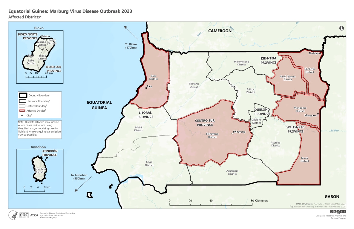
A noteworthy cluster of six cases of Marburg haemorrhagic fever (MHF) was identified in the Democratic Republic of Congo. One of the cases is the first infant Marburg fever patient ever documented. Three of six cases presented surprisingly mild symptoms. The results of epidemiological and virological investigations are compatible with person-to-person transmission through body fluids and with mother-to-child transmission while nurturing. The findings show that mild cases of MHF have to be expected during an outbreak and point out the difficulty to base patient management decisions on clinical case definitions alone. (+info)Characterization of monoclonal antibodies to Marburg virus (strain Musoke) glycoprotein and identification of two protective epitopes. (7/99)
Monoclonal antibodies (MAbs) reactive with Marburg virus (strain Musoke) were evaluated for both biological activity and specificity. Several of the Marburg virus- (MBGV) specific MAbs reduced the size and/or number of MBGV plaques in vitro. The ability of the MAbs to affect plaque formation in vitro was demonstrated to be specific for the glycoprotein (GP) of the strain of MBGV used for vaccination. Using deletion analysis and peptide mapping, the binding epitopes of several of these neutralizing MAbs were identified. Not unexpectedly, the epitopes were shown to lie in the most hypervariable and highly glycosylated region of MBGV GP. An analysis of the in vivo activity of several MAbs revealed that some antibodies provided substantial but incomplete protection of naive guinea pigs by passive transfer. These data suggest that neutralizing epitopes exist within MBGV GP but that induction of antibodies to these neutralizing epitopes may not be sufficient for protection from lethal infection. (+info)Risk factors for Marburg hemorrhagic fever, Democratic Republic of the Congo. (8/99)
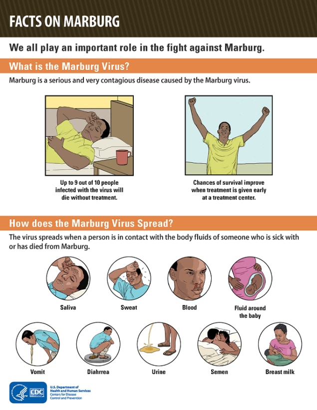
We conducted two antibody surveys to assess risk factors for Marburg hemorrhagic fever in an area of confirmed Marburg virus transmission in the Democratic Republic of the Congo. Questionnaires were administered and serum samples tested for Marburg-specific antibodies by enzyme-linked immunosorbent assay. Fifteen (2%) of 912 participants in a general village cross-sectional antibody survey were positive for Marburg immunoglobulin G antibody. Thirteen (87%) of these 15 were men who worked in the local gold mines. Working as a miner (odds ratio [OR] 13.9, 95% confidence interval [CI] 3.1 to 62.1) and receiving injections (OR 7.4, 95% CI 1.6 to 33.2) were associated with a positive antibody result. All 103 participants in a targeted antibody survey of healthcare workers were antibody negative. Primary transmission of Marburg virus to humans likely occurred via exposure to a still unidentified reservoir in the local mines. Secondary transmission appears to be less common with Marburg virus than with Ebola virus, the other known filovirus. (+info)Marburg Virus Disease (MVD) is an acute and often fatal viral hemorrhagic fever illness caused by the Marburg virus, a member of the filovirus family. It's a highly infectious disease that can be transmitted from human to human through direct contact with infected bodily fluids, tissues, or indirectly through contaminated surfaces and materials.
The incubation period for MVD ranges from 2 to 21 days, after which symptoms such as fever, chills, headache, muscle aches, severe malaise, and progressive weakness appear. Around the fifth day of illness, a maculopapular rash may occur, followed by diarrhea, nausea, vomiting, abdominal pain, and non-bloody stools. In some cases, patients may develop severe bleeding disorders, shock, liver failure, and multi-organ dysfunction, which can lead to death in 24-48 hours.
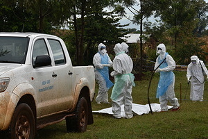
Currently, there are no approved vaccines or antiviral treatments for MVD, but supportive care is crucial for managing the symptoms of the disease. Preventive measures such as avoiding contact with infected individuals and their bodily fluids, wearing protective clothing, and practicing good hygiene can help prevent the spread of the virus.
According to the World Health Organization (WHO), Marburgviruses are toxiviral hemorrhagic fever-causing agents that belong to the Filoviridae family, which also includes Ebolaviruses. These enveloped, non-segmented, negative-stranded RNA viruses cause a severe and often fatal illness in humans and non-human primates. The Marburg virus was initially discovered in 1967, after simultaneous outbreaks occurred in laboratories in Marburg and Frankfurt, Germany, and in Belgrade, Yugoslavia (now Serbia).
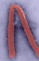
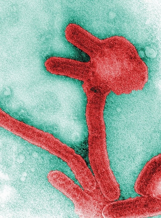
The virions of Marburgviruses are typically filamentous or U-shaped and measure approximately 80 nm in diameter. The genome consists of a single non-segmented, negative-sense RNA molecule that encodes seven structural proteins: nucleoprotein (NP), polymerase cofactor protein (VP35), matrix protein (VP40), glycoprotein (GP), transcription activator protein (VP30), RNA-dependent RNA polymerase (L), and a small hydrophobic protein (sVP24 or VP80).
Marburgviruses are primarily transmitted to humans through contact with the bodily fluids of infected animals, such as bats and non-human primates. Human-to-human transmission can occur via direct contact with infected individuals' blood, secretions, organs, or other bodily fluids, as well as through contaminated surfaces and materials.
The incubation period for Marburg virus disease (MVD) typically ranges from 2 to 21 days. Initial symptoms include fever, chills, headache, muscle aches, and general malaise. As the disease progresses, patients may develop severe watery diarrhea, abdominal pain, nausea, vomiting, and unexplained bleeding or bruising. In fatal cases, MVD can cause multi-organ failure, shock, and death, often within 7 to 14 days after symptom onset.
Currently, there are no approved vaccines or antiviral treatments specifically for Marburg virus infections. However, supportive care, such as fluid replacement, electrolyte management, and treatment of secondary infections, can help improve outcomes for MVD patients. Preventive measures, including the use of personal protective equipment (PPE) and proper infection control practices, are crucial to reducing the risk of transmission during outbreaks.
Filoviridae is a family of negative-sense, single-stranded RNA viruses that includes three genera: Ebolavirus, Marburgvirus, and Cuevavirus. These viruses are known to cause severe hemorrhagic fever in humans and nonhuman primates, with high fatality rates. The most well-known members of this family are Ebola virus and Marburg virus.
The virions of Filoviridae are filamentous, often having a "U," "6," or "hook" shape, and can be up to 14,000 nanometers in length. The genome of these viruses is non-segmented and contains seven genes that encode for structural proteins and enzymes necessary for replication.
Transmission of Filoviridae occurs through direct contact with infected bodily fluids or contaminated surfaces, and infection can result in a range of symptoms including fever, severe headache, muscle pain, weakness, fatigue, and hemorrhage. There are currently no approved vaccines or antiviral treatments for Filoviridae infections, although several are in development.
Ebolavirus is a genus of viruses in the family Filoviridae, order Mononegavirales. It is named after the Ebola River in the Democratic Republic of Congo (formerly Zaire), where the virus was first identified in 1976. There are six species of Ebolavirus, four of which are known to cause disease in humans: Zaire ebolavirus, Sudan ebolavirus, Bundibugyo ebolavirus, and Tai Forest ebolavirus (formerly Cote d'Ivoire ebolavirus). The fifth species, Reston ebolavirus, is known to cause disease in non-human primates and pigs, but not in humans. The sixth and most recently identified species, Bombali ebolavirus, has not been associated with any human or animal diseases.
Ebolaviruses are enveloped, negative-sense, single-stranded RNA viruses that cause a severe and often fatal hemorrhagic fever in humans and non-human primates. The virus is transmitted to people from wild animals and spreads in the human population through human-to-human transmission. Fruit bats of the Pteropodidae family are considered to be the natural host of Ebolavirus.
The symptoms of Ebolavirus disease (EVD) typically include fever, severe headache, muscle pain, weakness, fatigue, and sore throat, followed by vomiting, diarrhea, rash, impaired kidney and liver function, and in some cases, both internal and external bleeding. The case fatality rate of EVD is variable but has been historically high, ranging from 25% to 90% in past outbreaks depending on the species and the quality of medical care. There are no licensed specific treatments or vaccines available for EVD, although several promising candidates are currently under development.
**Hemorrhagic fevers, viral** are a group of severe, potentially fatal illnesses caused by viruses that affect the body's ability to regulate its blood vessels and clotting abilities. These viruses belong to several different families including *Filoviridae* (e.g., Ebola, Marburg), *Arenaviridae* (e.g., Lassa, Machupo), *Bunyaviridae* (e.g., Hantavirus, Crimean-Congo hemorrhagic fever virus) and *Flaviviridae* (e.g., Dengue, Yellow Fever).
The initial symptoms are non-specific and include sudden onset of fever, fatigue, muscle aches, joint pains, headache, and vomiting. As the disease progresses, it may lead to capillary leakage, internal and external bleeding, and multi-organ failure resulting in shock and death in severe cases.
The transmission of these viruses can occur through various means depending on the specific virus. For example, some are transmitted via contact with infected animals or their urine/feces (e.g., Hantavirus), others through insect vectors like ticks (Crimean-Congo hemorrhagic fever) or mosquitoes (Dengue, Yellow Fever), and yet others through direct contact with infected body fluids (Ebola, Marburg).
There are no specific treatments for most viral hemorrhagic fevers. However, some experimental antiviral drugs have shown promise in treating certain types of the disease. Supportive care, such as maintaining blood pressure, replacing lost fluids and electrolytes, and managing pain, is critical to improving outcomes. Prevention measures include avoiding areas where the viruses are common, using personal protective equipment when caring for infected individuals or handling potentially contaminated materials, and controlling insect vectors.
Sources: Centers for Disease Control and Prevention (CDC), World Health Organization (WHO).
Filoviridae infections refer to diseases caused by viruses belonging to the Filoviridae family, which includes Ebola virus and Marburg virus. These viruses are characterized by filamentous or threadlike shapes and can cause severe hemorrhagic fever in humans and primates. The infections are associated with high mortality rates, ranging from 25% to 90%, depending on the specific virus and strain.
Transmission of Filoviridae viruses occurs through direct contact with infected bodily fluids or contaminated surfaces. The initial symptoms of infection include fever, muscle pain, headache, and sore throat, followed by vomiting, diarrhea, rash, and impaired organ function. In severe cases, the disease can progress to hemorrhagic fever, characterized by internal and external bleeding, shock, and multi-organ failure.
Currently, there are no approved vaccines or antiviral treatments for Filoviridae infections, although several experimental therapies and vaccines are under development. Prevention measures include avoiding contact with infected individuals, practicing good hygiene, and using personal protective equipment when caring for sick patients.
Ebola Hemorrhagic Fever (EHF) is a severe, often fatal illness in humans. It is one of the five identified subtypes of the Ebolavirus. The virus is transmitted to people from wild animals and spreads in the human population through human-to-human transmission.
The early symptoms include sudden onset of fever, fatigue, muscle pain, headache and sore throat. This is followed by vomiting, diarrhea, rash, symptoms of impaired kidney and liver function, and in some cases, both internal and external bleeding.
Laboratory findings include low white blood cell and platelet counts and elevated liver enzymes.
The virus is introduced into the human population through close contact with the blood, secretions, organs or other bodily fluids of infected animals such as fruit bats, porcupines and non-human primates. Then it spreads in communities through human-to-human transmission via direct contact (through broken skin or mucous membranes) with the blood, secretions, organs or other bodily fluids of infected people, and with surfaces and materials contaminated with these fluids.
Healthcare workers have frequently been infected while treating patients with suspected or confirmed EVD due to a lack of adequate infection prevention and control measures.
There are currently no approved specific antiviral drugs or vaccines for Ebola. Several promising treatments and vaccine candidates are being evaluated.
Viral diseases are illnesses caused by the infection and replication of viruses in host organisms. These infectious agents are obligate parasites, meaning they rely on the cells of other living organisms to survive and reproduce. Viruses can infect various types of hosts, including animals, plants, and microorganisms, causing a wide range of diseases with varying symptoms and severity.
Once a virus enters a host cell, it takes over the cell's machinery to produce new viral particles, often leading to cell damage or death. The immune system recognizes the viral components as foreign and mounts an immune response to eliminate the infection. This response can result in inflammation, fever, and other symptoms associated with viral diseases.
Examples of well-known viral diseases include:
1. Influenza (flu) - caused by influenza A, B, or C viruses
2. Common cold - usually caused by rhinoviruses or coronaviruses
3. HIV/AIDS - caused by human immunodeficiency virus (HIV)
4. Measles - caused by measles morbillivirus
5. Hepatitis B and C - caused by hepatitis B virus (HBV) and hepatitis C virus (HCV), respectively
6. Herpes simplex - caused by herpes simplex virus type 1 (HSV-1) or type 2 (HSV-2)
7. Chickenpox and shingles - both caused by varicella-zoster virus (VZV)
8. Rabies - caused by rabies lyssavirus
9. Ebola - caused by ebolaviruses
10. COVID-19 - caused by severe acute respiratory syndrome coronavirus 2 (SARS-CoV-2)
Prevention and treatment strategies for viral diseases may include vaccination, antiviral medications, and supportive care to manage symptoms while the immune system fights off the infection.
Ross River virus (RRV) is an infectious disease caused by the Ross River virus, which is a type of alphavirus. It is transmitted to humans through the bite of infected mosquitoes, primarily Aedes vigilax, Culex annulirostris, and Culex australicus in Australia.
RRV is endemic to Australia, Papua New Guinea, and some islands in the Pacific Ocean. The symptoms of RRV include fever, rash, joint pain and swelling, muscle aches, fatigue, and headache, which can last for several weeks to months. In severe cases, it can lead to chronic arthritis and other long-term complications.
There is no specific treatment for RRV, and management typically involves relieving symptoms with rest, fluids, and pain relief medications. Preventive measures include avoiding mosquito bites by using insect repellent, wearing protective clothing, and staying indoors during peak mosquito activity hours.
The Democratic Republic of the Congo (DRC) is a country located in Central Africa. It is named after the Congo River, which flows through the country. The DRC is the second-largest country in Africa by area and the eleventh-largest in the world. It is home to a diverse population of more than 80 million people, making it one of the most populous countries on the continent.
The DRC is a democratic republic, which means that it is a form of government in which the people have the power to choose their leaders through free and fair elections. The country has a presidential system of government, in which the president serves as both the head of state and the head of government. The current president of the DRC is Félix Tshisekedi, who took office in January 2019.
The DRC is a federal republic, meaning that it is divided into several provinces, each with its own elected government. The country has a total of 26 provinces, which are further divided into districts and sectors.
The DRC is a member of various international organizations, including the United Nations, the African Union, and the Southern African Development Community. It is also a party to several international treaties and agreements, such as the Convention on International Trade in Endangered Species of Wild Fauna and Flora (CITES) and the Paris Agreement on climate change.
The DRC has a mixed economy, with both private and public sectors playing important roles. The country is rich in natural resources, including minerals such as copper, diamonds, gold, and tin. It also has large areas of fertile land that are suitable for agriculture. However, the DRC faces significant challenges, including poverty, corruption, and conflict. Despite these challenges, the country has made progress in recent years in terms of economic growth and development.