Maxillary Sinusitis
Maxillary Sinus
Bacteria, Aerobic
Porphyromonas
Stomatognathic Diseases
Sphenoid Sinusitis
Paranasal Sinus Diseases
Prevotella
Ethmoid Sinusitis
Bacteria, Anaerobic
Endoscopy
Frontal Sinusitis
Maxilla
Paranasal Sinuses
Maxillary Sinus Neoplasms
Cefuroxime
Cephalosporins
Suspensions
Prodrugs
Amoxicillin-Potassium Clavulanate Combination
Clavulanic Acids
Encyclopedias as Topic
Russia
Odontogenic Cysts
Midfacial complications of prolonged cocaine snorting. (1/68)
Acute and chronic ingestion of cocaine predisposes the abuser to a wide range of local and systemic complications. This article describes the case of a 38-year-old man whose chronic cocaine snorting resulted in the erosion of the midfacial anatomy and recurrent sinus infections. Previously published case reports specific to this problem are presented, as are the oral, systemic and behavioural effects of cocaine abuse. (+info)Calcification in chronic maxillary sinusitis: comparison of CT findings with histopathologic results. (2/68)
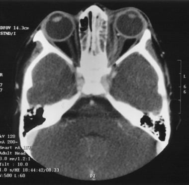
BACKGROUND AND PURPOSE: It is important to differentiate fungal from nonfungal sinusitis in order to determine the optimal treatment for chronic sinusitis. The purpose of this study was to describe the CT findings of calcifications in chronic fungal and nonfungal maxillary sinusitis. METHODS: Five hundred ten patients with pathologically proved chronic maxillary sinusitis were studied with unenhanced CT before undergoing sinonasal surgery. In 36 patients, the CT scans were reviewed retrospectively to ascertain the shape and location of intrasinus calcifications. RESULTS: Calcifications were found in 20 (51%) of 39 patients with fungal sinusitis and in 16 (3%) of 471 patients with nonfungal sinusitis. Direct histopathologic correlation was performed in two of 16 patients with nonfungal sinusitis who had intrasinus calcification. The location of intrasinus calcification was central in 95% of the patients with fungal sinusitis and peripheral in 81% of those with nonfungal sinusitis. Although calcifications with a nodular or linear shape were seen in both fungal and nonfungal sinusitis, fine punctate type calcifications were seen only in those with fungal sinusitis (50%) and round or eggshell type calcifications only in those with nonfungal sinusitis (19%). CONCLUSION: Intrasinus calcifications are different in location and shape between fungal and nonfungal maxillary sinusitis. Although intrasinus calcification is uncommon in nonfungal sinusitis, the CT finding of intrasinus calcification may be helpful for differentiating fungal from nonfungal maxillary sinusitis. (+info)Use of standard radiography to diagnose paranasal sinus disease of asthmatic children in Taiwan: comparison with computed tomography. (3/68)
Paranasal sinus disease and bronchial asthma are frequently associated. Computed tomography imaging is currently the most reliable method for confirming the diagnosis of sinusitis. Due to the cost and amount of radiation during computed tomography, our aim was to analyze whether standard radiography, under computed tomography-control, had a reasonable degree of confidence in the diagnosis of sinusitis. Fifty-three asthmatic patients (42 males and 11 females) with a mean age of 9 years (range 4-14) were enrolled. We evaluated the maxillary sinuses, ethmoidal sinuses, frontal sinuses, and sphenoidal sinuses using standard radiography (Waters' view, Caldwell view, and lateral view) and compared with computed tomography (coronal views), the latter served as a standard. Computed tomography (CT) showed paranasal sinusitis in 58% (31/53) of the asthmatic children. Compared with the results of computed tomography, standard radiography revealed a sensitivity of 81.1% and a specificity of 72.7% for maxillary sinusitis. The sensitivity and specificity for ethmoidal, frontal, and sphenoidal sinusitis were 51.8%, 84.8%; 47.3%, 87.2%; and 40.8%, 93.3%, respectively. In 21 (40%) of the 53 patients, discrepancies were seen between the interpretations of standard radiography c and those of CT scans. In patients with maxillary sinusitis, the correlation between standard radiography and CT was good. However, ethmoidal, frontal, and sphenoidal sinusitis were poorly demonstrated using radiography. Standard radiography can be recommended as a screening method for maxillary sinusitis, but it is not recommended for the diagnosis of other paranasal sinusitis. (+info)Maxillary sinusitis caused by medusoid form of Schizophyllum commune. (4/68)
We present a case of maxillary sinusitis in a diabetic female caused by the basidiomycete fungus Schizophyllum commune. Identification of the isolate was hampered by its atypical features. Subcultures formed sterile medusoid structures from nonclamped mycelia until spontaneous dikaryotization resulted in the development of characteristic fan-shaped fruiting bodies. Identification was confirmed by the presence of spicules formed on the hyphae and by vegetative compatibility with known isolates. (+info)Ventilator-associated sinusitis: microbiological results of sinus aspirates in patients on antibiotics. (5/68)
BACKGROUND: The efficacy of systemic antibiotics on the treatment of ventilator-associated infectious maxillary sinusitis (VAIMS) is debated. The objective of this study was to determine the etiologic diagnosis of VAIMS in patients receiving antibiotics. METHODS: Patients mechanically ventilated for more than or equal to 72 h, who had persistent fever while on antibiotics for more than or equal to 48 h, underwent computed tomography scan followed by transnasal puncture of involved maxillary sinuses. VAIMS was defined as follows: fever greater than or equal to 38 degrees C, radiographic signs (air fluid level or opacification of maxillary sinuses on computed tomography scan), and a quantitative culture of sinus aspirate yielding more than or equal to 103 colony-forming units/ml. RESULTS: Twenty-four patients had radiographic signs of sinusitis. The mean +/- SD prior durations of mechanical ventilation and antibiotic exposure were 9.5 +/- 4.7 days and 6 +/- 4 days, respectively. Six unilateral and nine bilateral VAIMS were diagnosed in 15 patients. The median number of etiologic organisms per patient was two (range, one to four). The bacteriologic cultures yielded gram-positive bacteria (n = 21), gram-negative bacteria (n = 22), and yeasts (n = 5). Forty percent of causative agents were susceptible to the antibiotics prescribed. Seven patients with VAIMS developed 10 concomitant infections: ventilator-associated pneumonia (n = 5), urinary tract infection (n = 3), catheter infections (n = 2). In all cases of ventilator-associated pneumonia, the implicated agents were the causative agents of VAIMS. CONCLUSION: In VAIMS patients on antibiotics, quantitative cultures of sinus aspirates may contribute to establish the diagnosis. The frequent recovery of microorganisms susceptible to the antimicrobial treatment administered suggests that therapy of VAIMS with systemic antibiotics may not be sufficient. (+info)Maxillary sinusitis caused by Actinomucor elegans. (6/68)
We report the first case of maxillary sinusitis caused by Actinomucor elegans in an 11-year-old patient. Histopathological and mycological examinations of surgical maxillary sinuses samples showed coenocytic hyphae characteristic of mucoraceous fungi. The fungi recovered had stolons and rhizoids, nonapophyseal and globose sporangia, and whorled branched sporangiophores and was identified as A. elegans. After surgical cleaning and chemotherapy with amphotericin B administered intravenously and by irrigation, the patient became asymptomatic and the mycological study results were negative. (+info)Rhinovirus RNA in the maxillary sinus epithelium of adult patients with acute sinusitis. (7/68)
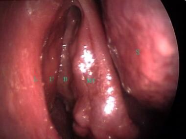
We used in situ hybridization for the detection of rhinovirus in maxillary sinus biopsy specimens obtained from 14 adult patients with acute sinusitis. In 7 specimens, rhinovirus RNA could be demonstrated in the maxillary sinus epithelium, thereby confirming the etiology of rhinovirus and the clinical suspicion of acute sinusitis. (+info)Rigid nasal endoscopy versus sinus puncture and aspiration for microbiologic documentation of acute bacterial maxillary sinusitis. (8/68)
Sinus puncture and aspiration is an invasive procedure that hinders patient enrollment in studies of acute bacterial maxillary sinusitis (ABMS). Pain and minor bleeding also limit its potential diagnostic utility in clinical practice. Cultures obtained by rigid nasal endoscopy were compared with those from sinus puncture and aspiration in 53 patients with ABMS; 46 patients were assessable. Considering recovery of Haemophilus influenzae, Moraxella catarrhalis, or Streptococcus pneumoniae from puncture and aspiration as the gold standard, endoscopy cultures demonstrated a sensitivity of 85.7% (95% confidence interval, 56.2-97.5), specificity of 90.6% (73.8-97.5), positive predictive value of 80% (51.4-94.7), negative predictive value of 93.5% (77.2-98.9), and accuracy of 89.1% (75.6-95.9). Ten adverse events related to puncture and aspiration occurred in 5 (9.6%) of 52 patients; there were no endoscopy-related adverse events. In our study, the largest to date, endoscopic sampling compared favorably with puncture and aspiration for identifying H. influenzae, M. catarrhalis, and S. pneumoniae in ABMS and produced less morbidity. (+info)Maxillary sinusitis is a medical condition characterized by inflammation or infection of the maxillary sinuses, which are air-filled cavities located in the upper part of the cheekbones. These sinuses are lined with mucous membranes that produce mucus to help filter and humidify the air we breathe.
When the maxillary sinuses become inflamed or infected, they can fill with fluid and pus, leading to symptoms such as:
* Pain or pressure in the cheeks, upper teeth, or behind the eyes
* Nasal congestion or stuffiness
* Runny nose or postnasal drip
* Reduced sense of smell or taste
* Headache or facial pain
* Fatigue or fever (in cases of bacterial infection)
Maxillary sinusitis can be caused by viruses, bacteria, or fungi, and may also result from allergies, structural abnormalities, or exposure to environmental irritants such as smoke or pollution. Treatment typically involves managing symptoms with over-the-counter remedies or prescription medications, such as decongestants, antihistamines, or antibiotics. In some cases, more invasive treatments such as sinus surgery may be necessary.
The maxillary sinuses, also known as the antrums of Highmore, are the largest of the four pairs of paranasal sinuses located in the maxilla bones. They are air-filled cavities that surround the nasolacrimal duct and are situated superior to the upper teeth and lateral to the nasal cavity. Each maxillary sinus is lined with a mucous membrane, which helps to warm, humidify, and filter the air we breathe. Inflammation or infection of the maxillary sinuses can result in conditions such as sinusitis, leading to symptoms like facial pain, headaches, and nasal congestion.
Sinusitis, also known as rhinosinusitis, is a medical condition characterized by inflammation of the paranasal sinuses, which are air-filled cavities located within the skull near the nose. The inflammation can be caused by viral, bacterial, or fungal infections, as well as allergies, structural issues, or autoimmune disorders.
In sinusitis, the mucous membranes lining the sinuses become swollen and may produce excess mucus, leading to symptoms such as nasal congestion, thick green or yellow nasal discharge, facial pain or pressure, reduced sense of smell, cough, fatigue, and fever.
Sinusitis can be classified into acute (lasting less than 4 weeks), subacute (lasting 4-12 weeks), chronic (lasting more than 12 weeks), or recurrent (multiple episodes within a year). Treatment options depend on the underlying cause and severity of symptoms, and may include antibiotics, nasal corticosteroids, decongestants, saline irrigation, and in some cases, surgery.
Aerobic bacteria are a type of bacteria that require oxygen to live and grow. These bacteria use oxygen as the final electron acceptor in their respiratory chain to generate energy in the form of ATP (adenosine triphosphate). Aerobic bacteria can be found in various environments, including soil, water, and the air, as well as on the surfaces of living things. Some examples of aerobic bacteria include species of Pseudomonas, Bacillus, and Staphylococcus.
It's worth noting that some bacteria can switch between aerobic and anaerobic metabolism depending on the availability of oxygen. These bacteria are called facultative anaerobes. In contrast, obligate anaerobes are bacteria that cannot tolerate oxygen and will die in its presence.
"Porphyromonas" is a genus of gram-negative, anaerobic bacteria that are commonly found in the human oral cavity and other areas of the body. One species, "Porphyromonas gingivalis," is a major contributor to chronic periodontitis, a severe form of gum disease. These bacteria are also associated with various systemic diseases, including atherosclerosis, rheumatoid arthritis, and aspiration pneumonia. The name "Porphyromonas" comes from the Greek words "porphyra," meaning purple, and "monas," meaning unit, referring to the bacteria's ability to produce porphyrins, which are pigments that can give a purple color to their colonies.
Cyclacillin is not a recognized or commonly used term in medicine or microbiology. It appears that you may have misspelled the name of an antibiotic. The correct spelling and medical definition are as follows:
Cloxacillin: A penicillinase-resistant antibiotic, closely related to dicloxacillin, used to treat infections caused by susceptible staphylococci, including beta-lactamase producing strains. It is commonly used for the treatment of skin and soft tissue infections.
Cloxacillin is a type of penicillin that resist breaking down by certain enzymes produced by bacteria (penicillinases). This allows cloxacillin to be effective against some bacteria that have become resistant to other types of penicillin.
Stomatognathic diseases are a group of disorders that affect the stomatognathic system, which includes the teeth, periodontal tissues, temporomandibular joints, muscles of mastication, and associated structures. These diseases can manifest as various symptoms such as pain, difficulty in chewing or swallowing, limited mouth opening, and abnormal jaw movements.
Some examples of stomatognathic diseases include temporomandibular disorders (TMD), oral mucosal diseases, dental caries, periodontal disease, oral cancer, and sleep-related breathing disorders. The diagnosis and management of these conditions often require a multidisciplinary approach involving dentists, oral surgeons, orthodontists, physicians, and other healthcare professionals.
Sphenoid sinusitis is a medical condition characterized by the inflammation or infection of the sphenoid sinuses, which are air-filled cavities located in the sphenoid bone at the center of the skull base, behind the eyes. These sinuses are relatively small and difficult to access, making infections less common than in other sinuses. However, when sphenoid sinusitis does occur, it can cause various symptoms such as headaches, facial pain, nasal congestion, fever, and vision problems. Sphenoid sinusitis may result from bacterial or fungal infections, allergies, or autoimmune disorders. Diagnosis typically involves a combination of clinical evaluation, imaging studies like CT scans, and sometimes endoscopic examination. Treatment options include antibiotics for bacterial infections, antifungal medications for fungal infections, nasal sprays, decongestants, pain relievers, and, in severe or recurrent cases, surgical intervention.
Paranasal sinus diseases refer to a group of medical conditions that affect the paranasal sinuses, which are air-filled cavities located within the skull near the nasal cavity. These sinuses include the maxillary, frontal, ethmoid, and sphenoid sinuses.
Paranasal sinus diseases can be caused by a variety of factors, including viral, bacterial, or fungal infections, allergies, structural abnormalities, or autoimmune disorders. Some common paranasal sinus diseases include:
1. Sinusitis: Inflammation or infection of the sinuses, which can cause symptoms such as nasal congestion, thick nasal discharge, facial pain or pressure, and reduced sense of smell.
2. Nasal polyps: Soft, benign growths that develop in the lining of the nasal passages or sinuses, which can obstruct airflow and cause difficulty breathing through the nose.
3. Sinonasal tumors: Abnormal growths that can be benign or malignant, which can cause symptoms such as nasal congestion, facial pain, and bleeding from the nose.
4. Sinus cysts: Fluid-filled sacs that form in the sinuses, which can cause symptoms similar to those of sinusitis.
5. Fungal sinusitis: Infection of the sinuses with fungi, which can cause symptoms such as nasal congestion, facial pain, and thick, discolored mucus.
Treatment for paranasal sinus diseases depends on the underlying cause and severity of the condition. Treatment options may include medications, such as antibiotics, antihistamines, or corticosteroids, as well as surgical intervention in more severe cases.
Preventella is a genus of Gram-negative, anaerobic, rod-shaped bacteria that are commonly found in the human oral cavity, gastrointestinal tract, and urogenital tract. They are part of the normal microbiota but can also be associated with various infections, particularly in individuals with compromised immune systems or underlying medical conditions.
Prevotella species have been implicated in a variety of diseases, including periodontal disease, dental caries, respiratory tract infections, bacteremia, soft tissue infections, and joint infections. They can also be found in association with abscesses, wound infections, and other types of infections, particularly in the head and neck region.
Prevotella species are generally resistant to antibiotics commonly used to treat anaerobic infections, such as clindamycin and metronidazole, making them difficult to eradicate. Therefore, accurate identification and susceptibility testing of Prevotella isolates is important for the appropriate management of infections caused by these organisms.
Ethmoid sinusitis is a medical condition that refers to the inflammation or infection of the ethmoid sinuses. The ethmoid sinuses are a pair of small, air-filled cavities located in the upper part of the nasal cavity, near the eyes. They are surrounded by delicate bone structures and are connected to the nasal cavity by narrow channels.
Ethmoid sinusitis can occur as a result of a viral, bacterial, or fungal infection, or it may be caused by allergies, environmental factors, or structural abnormalities in the nasal passages. When the ethmoid sinuses become inflamed or infected, they can cause symptoms such as:
* Nasal congestion or stuffiness
* Pain or pressure in the forehead, between the eyes, or in the cheeks
* Headaches or facial pain
* Thick, discolored nasal discharge
* Postnasal drip
* Coughing or sneezing
* Fever
* Fatigue
Ethmoid sinusitis can be acute (lasting for a short period of time) or chronic (persisting for several weeks or months). If left untreated, ethmoid sinusitis can lead to complications such as the spread of infection to other parts of the body, including the eyes and brain. Treatment for ethmoid sinusitis may include antibiotics, decongestants, nasal sprays, or surgery in severe cases.
Anaerobic bacteria are a type of bacteria that do not require oxygen to grow and survive. Instead, they can grow in environments that have little or no oxygen. Some anaerobic bacteria can even be harmed or killed by exposure to oxygen. These bacteria play important roles in many natural processes, such as decomposition and the breakdown of organic matter in the digestive system. However, some anaerobic bacteria can also cause disease in humans and animals, particularly when they infect areas of the body that are normally oxygen-rich. Examples of anaerobic bacterial infections include tetanus, gas gangrene, and dental abscesses.
Endoscopy is a medical procedure that involves the use of an endoscope, which is a flexible tube with a light and camera at the end, to examine the interior of a body cavity or organ. The endoscope is inserted through a natural opening in the body, such as the mouth or anus, or through a small incision. The images captured by the camera are transmitted to a monitor, allowing the physician to visualize the internal structures and detect any abnormalities, such as inflammation, ulcers, or tumors. Endoscopy can also be used for diagnostic purposes, such as taking tissue samples for biopsy, or for therapeutic purposes, such as removing polyps or performing minimally invasive surgeries.
Frontal sinusitis is a type of sinus infection that specifically involves the frontal sinuses, which are located in the forehead region above the eyes. The condition is characterized by inflammation and infection of the mucous membrane lining the frontal sinuses, leading to symptoms such as headaches, facial pain or pressure, nasal congestion, and thick nasal discharge.
Frontal sinusitis can be caused by viral, bacterial, or fungal infections, as well as structural issues like nasal polyps or deviated septum that obstruct the sinus drainage pathways. Treatment options for frontal sinitis may include antibiotics, nasal decongestants, corticosteroids, saline nasal irrigation, and in some cases, endoscopic sinus surgery to alleviate obstructions and improve sinus drainage.
The maxilla is a paired bone that forms the upper jaw in vertebrates. In humans, it is a major bone in the face and plays several important roles in the craniofacial complex. Each maxilla consists of a body and four processes: frontal process, zygomatic process, alveolar process, and palatine process.
The maxillae contribute to the formation of the eye sockets (orbits), nasal cavity, and the hard palate of the mouth. They also contain the upper teeth sockets (alveoli) and help form the lower part of the orbit and the cheekbones (zygomatic arches).
Here's a quick rundown of its key functions:
1. Supports the upper teeth and forms the upper jaw.
2. Contributes to the formation of the eye sockets, nasal cavity, and hard palate.
3. Helps shape the lower part of the orbit and cheekbones.
4. Partakes in the creation of important sinuses, such as the maxillary sinus, which is located within the body of the maxilla.
An acute disease is a medical condition that has a rapid onset, develops quickly, and tends to be short in duration. Acute diseases can range from minor illnesses such as a common cold or flu, to more severe conditions such as pneumonia, meningitis, or a heart attack. These types of diseases often have clear symptoms that are easy to identify, and they may require immediate medical attention or treatment.
Acute diseases are typically caused by an external agent or factor, such as a bacterial or viral infection, a toxin, or an injury. They can also be the result of a sudden worsening of an existing chronic condition. In general, acute diseases are distinct from chronic diseases, which are long-term medical conditions that develop slowly over time and may require ongoing management and treatment.
Examples of acute diseases include:
* Acute bronchitis: a sudden inflammation of the airways in the lungs, often caused by a viral infection.
* Appendicitis: an inflammation of the appendix that can cause severe pain and requires surgical removal.
* Gastroenteritis: an inflammation of the stomach and intestines, often caused by a viral or bacterial infection.
* Migraine headaches: intense headaches that can last for hours or days, and are often accompanied by nausea, vomiting, and sensitivity to light and sound.
* Myocardial infarction (heart attack): a sudden blockage of blood flow to the heart muscle, often caused by a buildup of plaque in the coronary arteries.
* Pneumonia: an infection of the lungs that can cause coughing, chest pain, and difficulty breathing.
* Sinusitis: an inflammation of the sinuses, often caused by a viral or bacterial infection.
It's important to note that while some acute diseases may resolve on their own with rest and supportive care, others may require medical intervention or treatment to prevent complications and promote recovery. If you are experiencing symptoms of an acute disease, it is always best to seek medical attention to ensure proper diagnosis and treatment.
Paranasal sinuses are air-filled cavities in the skull that surround the nasal cavity. There are four pairs of paranasal sinuses, including the maxillary, frontal, ethmoid, and sphenoid sinuses. These sinuses help to warm, humidify, and filter the air we breathe. They also contribute to our voice resonance and provide a slight cushioning effect for the skull. The openings of the paranasal sinuses lead directly into the nasal cavity, allowing mucus produced in the sinuses to drain into the nose. Infections or inflammation of the paranasal sinuses can result in conditions such as sinusitis.
Maxillary sinus neoplasms refer to abnormal growths or tumors that develop in the maxillary sinuses, which are located in the upper part of your cheekbones, below your eyes. These growths can be benign (non-cancerous) or malignant (cancerous).
Benign neoplasms may include conditions such as an osteoma (a benign bone tumor), a papilloma (a benign growth of the lining of the sinus), or a fibrous dysplasia (a condition where bone is replaced by fibrous tissue).
Malignant neoplasms, on the other hand, can be primary (originating in the maxillary sinuses) or secondary (spreading to the maxillary sinuses from another site in the body). Common types of malignant tumors that arise in the maxillary sinus include squamous cell carcinoma, adenocarcinoma, and mucoepidermoid carcinoma.
Symptoms of maxillary sinus neoplasms may include nasal congestion, nosebleeds, facial pain or numbness, vision changes, and difficulty swallowing or speaking. Treatment options depend on the type, size, and location of the tumor but may include surgery, radiation therapy, chemotherapy, or a combination of these approaches.
Cefuroxime is a type of antibiotic known as a cephalosporin, which is used to treat a variety of bacterial infections. It works by interfering with the bacteria's ability to form a cell wall, which is necessary for its survival. Without a functional cell wall, the bacteria are unable to grow and multiply, and are eventually destroyed by the body's immune system.
Cefuroxime is effective against many different types of bacteria, including both Gram-positive and Gram-negative organisms. It is often used to treat respiratory tract infections, urinary tract infections, skin and soft tissue infections, and bone and joint infections.
Like all antibiotics, cefuroxime should be used only under the direction of a healthcare provider, and it is important to take the full course of treatment as prescribed, even if symptoms improve before the medication is finished. Misuse of antibiotics can lead to the development of drug-resistant bacteria, which are more difficult to treat and can pose a serious threat to public health.
Cephalosporins are a class of antibiotics that are derived from the fungus Acremonium, originally isolated from seawater and cow dung. They have a similar chemical structure to penicillin and share a common four-membered beta-lactam ring in their molecular structure.
Cephalosporins work by inhibiting the synthesis of bacterial cell walls, which ultimately leads to bacterial death. They are broad-spectrum antibiotics, meaning they are effective against a wide range of bacteria, including both Gram-positive and Gram-negative organisms.
There are several generations of cephalosporins, each with different spectra of activity and pharmacokinetic properties. The first generation cephalosporins have a narrow spectrum of activity and are primarily used to treat infections caused by susceptible Gram-positive bacteria, such as Staphylococcus aureus and Streptococcus pneumoniae.
Second-generation cephalosporins have an expanded spectrum of activity that includes some Gram-negative organisms, such as Escherichia coli and Haemophilus influenzae. Third-generation cephalosporins have even broader spectra of activity and are effective against many resistant Gram-negative bacteria, such as Pseudomonas aeruginosa and Klebsiella pneumoniae.
Fourth-generation cephalosporins have activity against both Gram-positive and Gram-negative organisms, including some that are resistant to other antibiotics. They are often reserved for the treatment of serious infections caused by multidrug-resistant bacteria.
Cephalosporins are generally well tolerated, but like penicillin, they can cause allergic reactions in some individuals. Cross-reactivity between cephalosporins and penicillin is estimated to occur in 5-10% of patients with a history of penicillin allergy. Other potential adverse effects include gastrointestinal symptoms (such as nausea, vomiting, and diarrhea), neurotoxicity, and nephrotoxicity.
In the context of medical definitions, "suspensions" typically refers to a preparation in which solid particles are suspended in a liquid medium. This is commonly used for medications that are administered orally, where the solid particles disperse upon shaking and settle back down when left undisturbed. The solid particles can be made up of various substances such as drugs, nutrients, or other active ingredients, while the liquid medium is often water, oil, or alcohol-based.
It's important to note that "suspensions" in a medical context should not be confused with the term as it relates to pharmacology or physiology, where it may refer to the temporary stopping of a bodily function or the removal of something from a solution through settling or filtration.
Cefaclor is a type of antibiotic known as a second-generation cephalosporin. It works by interfering with the bacteria's ability to form a cell wall, which is necessary for its survival. Without a functional cell wall, the bacteria eventually die. Cefaclor is effective against a wide range of gram-positive and gram-negative bacteria, making it a broad-spectrum antibiotic.
Cefaclor is used to treat various types of bacterial infections, including respiratory tract infections (such as bronchitis and pneumonia), ear infections, skin infections, and urinary tract infections. It is available in both oral and intravenous forms.
Like all antibiotics, cefaclor should be used only to treat bacterial infections, as it is not effective against viral infections such as the common cold or flu. Overuse of antibiotics can lead to the development of antibiotic-resistant bacteria, which can make future infections more difficult to treat. It is important to take cefaclor exactly as directed by a healthcare professional and to complete the full course of treatment, even if symptoms improve before all of the medication has been taken.
A prodrug is a pharmacologically inactive substance that, once administered, is metabolized into a drug that is active. Prodrugs are designed to improve the bioavailability or delivery of a drug, to minimize adverse effects, or to target the drug to specific sites in the body. The conversion of a prodrug to its active form typically occurs through enzymatic reactions in the liver or other tissues.
Prodrugs can offer several advantages over traditional drugs, including:
* Improved absorption: Some drugs have poor bioavailability due to their chemical properties, which make them difficult to absorb from the gastrointestinal tract. Prodrugs can be designed with improved absorption characteristics, allowing for more efficient delivery of the active drug to the body.
* Reduced toxicity: By masking the active drug's chemical structure, prodrugs can reduce its interactions with sensitive tissues and organs, thereby minimizing adverse effects.
* Targeted delivery: Prodrugs can be designed to selectively release the active drug in specific areas of the body, such as tumors or sites of infection, allowing for more precise and effective therapy.
Examples of prodrugs include:
* Aspirin (acetylsalicylic acid), which is metabolized to salicylic acid in the liver.
* Enalapril, an angiotensin-converting enzyme (ACE) inhibitor used to treat hypertension and heart failure, which is metabolized to enalaprilat in the liver.
* Codeine, an opioid analgesic, which is metabolized to morphine in the liver by the enzyme CYP2D6.
It's important to note that not all prodrugs are successful, and some may even have unintended consequences. For example, if a patient has a genetic variation that affects the activity of the enzyme responsible for converting the prodrug to its active form, the drug may not be effective or may produce adverse effects. Therefore, it's essential to consider individual genetic factors when prescribing prodrugs.
The Amoxicillin-Potassium Clavulanate Combination is an antibiotic medication used to treat various infections caused by bacteria. This combination therapy combines the antibiotic amoxicillin with potassium clavulanate, which is a beta-lactamase inhibitor. The addition of potassium clavulanate helps protect amoxicillin from being broken down by certain types of bacteria that produce beta-lactamases, thus increasing the effectiveness of the antibiotic against a broader range of bacterial infections.
Amoxicillin is a type of penicillin antibiotic that works by inhibiting the synthesis of the bacterial cell wall, ultimately leading to bacterial death. However, some bacteria have developed enzymes called beta-lactamases, which can break down and inactivate certain antibiotics like amoxicillin. Potassium clavulanate is added to the combination to inhibit these beta-lactamase enzymes, allowing amoxicillin to maintain its effectiveness against a wider range of bacteria.
This combination medication is used to treat various infections, including skin and soft tissue infections, respiratory tract infections, urinary tract infections, and dental infections. It's essential to follow the prescribed dosage and duration as directed by a healthcare professional to ensure effective treatment and prevent antibiotic resistance.
Common brand names for this combination include Augmentin and Amoxiclav.
Clavulanic acid is not a medical condition, but rather an antibacterial compound that is often combined with certain antibiotics to increase their effectiveness against bacteria that have become resistant to the antibiotic alone. It works by inhibiting certain enzymes produced by bacteria that help them to resist the antibiotic, allowing the antibiotic to work more effectively.
Clavulanic acid is typically combined with antibiotics such as amoxicillin or ticarcillin to treat a variety of bacterial infections, including respiratory tract infections, urinary tract infections, and skin and soft tissue infections. It is important to note that clavulanate-containing medications should only be used under the direction of a healthcare provider, as misuse or overuse can contribute to antibiotic resistance.
Rhinitis is a medical condition characterized by inflammation and irritation of the nasal passages, leading to symptoms such as sneezing, runny nose, congestion, and postnasal drip. It can be caused by various factors, including allergies (such as pollen, dust mites, or pet dander), infections (viral or bacterial), environmental irritants (such as smoke or pollution), and hormonal changes. Depending on the cause, rhinitis can be classified as allergic rhinitis, non-allergic rhinitis, infectious rhinitis, or hormonal rhinitis. Treatment options vary depending on the underlying cause but may include medications such as antihistamines, decongestants, nasal sprays, and immunotherapy (allergy shots).
An encyclopedia is a comprehensive reference work containing articles on various topics, usually arranged in alphabetical order. In the context of medicine, a medical encyclopedia is a collection of articles that provide information about a wide range of medical topics, including diseases and conditions, treatments, tests, procedures, and anatomy and physiology. Medical encyclopedias may be published in print or electronic formats and are often used as a starting point for researching medical topics. They can provide reliable and accurate information on medical subjects, making them useful resources for healthcare professionals, students, and patients alike. Some well-known examples of medical encyclopedias include the Merck Manual and the Stedman's Medical Dictionary.
I'm sorry for any confusion, but "Russia" is not a medical term or concept. Russia is the largest country in the world by land area, located primarily in Asia with a smaller portion extending into Europe. It is a nation rich in history and culture, known for its diverse landscapes, from tundra and forests to subtropical beaches.
If you have any medical questions or terms that you would like me to define, please feel free to ask!
Odontogenic tumors are a group of neoplasms that originate from the dental tissues or their remnants, including the odontogenic epithelium, ectomesenchyme, and/or their derivatives. These tumors can be benign or malignant and may affect the jaw bones and surrounding structures. They can cause various symptoms, such as swelling, pain, loosening of teeth, and altered bite. The classification of odontogenic tumors includes a wide range of entities with different biological behaviors, clinical features, and treatment approaches. Accurate diagnosis is essential for proper management and prognosis.
Odontogenic cysts are a type of cyst that originates from the dental tissues or odontogenic apparatus. They are typically found in the jawbones, and can be classified as developmental or inflammatory in origin. Developmental odontogenic cysts arise from remnants of the tooth-forming structures, while inflammatory odontogenic cysts result from an infection or injury to a tooth.
The most common types of odontogenic cysts include:
1. Periapical cyst - an inflammatory cyst that forms at the tip of the root of a dead or non-vital tooth.
2. Dentigerous cyst - a developmental cyst that surrounds the crown of an unerupted or impacted tooth.
3. Follicular cyst - a type of dentigerous cyst that forms around the crown of an unerupted wisdom tooth.
4. Odontogenic keratocyst - a developmental cyst that arises from the dental lamina and has a high recurrence rate.
5. Lateral periodontal cyst - a rare, developmental cyst that forms in the periodontal ligament of a vital tooth.
Odontogenic cysts can cause various symptoms such as swelling, pain, or numbness in the affected area. They may also displace or resorb adjacent teeth. Diagnosis is typically made through radiographic imaging and histopathological examination of tissue samples obtained through biopsy. Treatment options include surgical excision, marsupialization (a procedure that creates an opening between the cyst and oral cavity), or enucleation (removal of the cyst lining).
 Surgical Treatment of Acute Maxillary Sinusitis: Overview, Surgery for Sinusitis Complications, Anesthesia
 Randomized controlled trial of 3 vs 10 days of trimethoprim/sulfamethoxazole for acute maxillary sinusitis
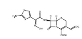
 Ceftin (cefuroxime) for Bacterial Infections: Uses, Dosage, Side Effects, Interactions, Warnings
 Intraoral dual wavelength laser diode therapy for chronic maxillary sinusitis - BUC
 Near-Infrared Imaging for Management of Chronic Maxillary Sinusitis > 논문 자료 | (주...
 Does Middle Meatal Antrostomy Prevent the Onset of Maxillary Sinusitis After Zygomatic Implant Placement?
 Antral lavage - Wikipedia
 Extensive complex odontoma in the maxillary sinus: an uncommon presentation as a cause of chronic sinusitis
 Ethmoid sinusitis: Causes, symptoms, and treatment
 Article - Billing and Coding: Allergy Skin Testing (A56559)
 October 2019 - Volume 30 - Issue 7 : Journal of Craniofacial Surgery
 Teen Dies After Sinus Infection: How To Tell If You Have Complications
 RX List database - use generic or medication brand name - GlobalRPH
 Penicillin V
- Phenoxymethylpenicillin
Summary Report | CureHunter
Penicillin V
- Phenoxymethylpenicillin
Summary Report | CureHunter Vitafon-T home therapy device: buy on Kalinka-Store
 Journal of Otorhinolaryngology, Hearing and Balance Medicine | An Open Access Journal from MDPI
 Kevin Ting, MD | Cardiology | Kelsey-Seybold Clinic
 Levaquin - Side Effects, Uses, Dosage, Overdose, Pregnancy, Alcohol | RxWiki
 Accuracy of signs, symptoms and blood tests for diagnosing acute bacterial rhinosinusitis and CT-confirmed acute rhinosinusitis...
 Plus it