Meningoencephalitis
Naegleria fowleri
Amebiasis
Central Nervous System Protozoal Infections
Angiostrongylus cantonensis
Meningitis, Cryptococcal
Amoeba
Cryptococcus neoformans
Cerebrospinal Fluid
Fatal Outcome
Meningitis, Listeria
Dog Diseases
Naegleria
Sandfly fever Naples virus
Meningitis, Aseptic
Acanthamoeba
Meningitis, Viral
Encephalitis, Tick-Borne
Brain
Cryptococcus gattii
Cerebrospinal Fluid Proteins
Encephalitis, Varicella Zoster
Leukoencephalitis, Acute Hemorrhagic
Animals, Zoo
Encephalitis, Viral
West Nile Fever
Meningitis
Association of simian virus 40 with a central nervous system lesion distinct from progressive multifocal leukoencephalopathy in macaques with AIDS. (1/429)
The primate polyomavirus SV40 is known to cause interstitial nephritis in primary infections and progressive multifocal leukoencephalopathy (PML) upon reactivation of a latent infection in SIV-infected macaques. We now describe a second central nervous system manifestation of SV40: a meningoencephalitis affecting cerebral gray matter, without demyelination, distinct from PML. Meningoencephalitis appears also to be a primary manifestation of SV40 infection and can be seen in conjunction with SV40-induced interstitial nephritis and pneumonitis. The difference in the lesions of meningoencephalitis and PML does not appear to be due to cellular tropism, as both oligodendrocytes and astrocytes are infected in PML and meningoencephalitis, as determined by in situ hybridization or immunohistochemistry for SV40 coupled with immunohistochemistry for cellular determinants. This is further supported by examination of SV40 nucleic acid sequences from the ori-enhancer and large-T-antigen regions, which reveals no tissue-or lesion-specific variation in SV40 sequences. (+info)Listeria monocytogenes and Escherichia coli septicemia and meningoencephalitis in a 7-day-old llama. (2/429)
Listeria monocytogenes and Escherichia coli were isolated from blood collected on presentation and tissues samples taken postmortem. Listeria monocytogenes was isolated from cerebrospinal fluid collected antemortem. The importance of passive transfer of immunity, the subtlety of neurologic signs in early meningitis, and considering blood-CSF penetration in antimicrobial selection are discussed. (+info)Non-purulent meningoencephalomyelitis of a Pacific striped dolphin (Lagenorhynchus obliquidens). The first evidence of morbillivirus infection in a dolphin at the Pacific Ocean around Japan. (3/429)
On March 22, 1998, a mature, male, hyposthenic Pacific striped dolphin (Lagenorhynchus obliquidens) was stranded at Aoshima Beach in Miyazaki prefecture, Japan. A necropsy performed 14 hr after death revealed mild diffuse congestion and edema of the leptomeninges and mild pulmonary atelectasis. Histopathologically, non-purulent inflammatory were observed throughout the cerebrum, thalamus, midbrain, pons, medulla oblongata, and spinal cord. Hematoxylin and eosin stain revealed no viral inclusion bodies. Immunohistochemistry using a monoclonal antibody against nucleoprotein of canine distemper virus (CDV-NP) revealed a number of CDV-NP-positive granular deposits in the cytoplasm and cell processes of the degenerating or intact neurons. The present paper is a first report of spontaneously occurred morbillivirus infection in a dolphin at the Pacific Ocean around Japan. (+info)Comparative efficacies of antibiotics in a rat model of meningoencephalitis due to Listeria monocytogenes. (4/429)
The antibacterial activities of amoxicillin-gentamicin, trovafloxacin, trimethoprim-sulfamethoxazole (TMP-SMX) and the combination of trovafloxacin with TMP-SMX were compared in a model of meningoencephalitis due to Listeria monocytogenes in infant rats. At 22 h after intracisternal infection, the cerebrospinal fluid was cultured to document meningitis, and the treatment was started. Treatment was instituted for 48 h, and efficacy was evaluated 24 h after administration of the last dose. All tested treatment regimens exhibited significant activities in brain, liver, and blood compared to infected rats receiving saline (P < 0.001). In the brain, amoxicillin plus gentamicin was more active than all of the other regimens, and trovafloxacin was more active than TMP-SMX (bacterial titers of 4.1 +/- 0.5 log10 CFU/ml for amoxicillin-gentamicin, 5.0 +/- 0.4 log10 CFU/ml for trovafloxacin, and 5.8 +/- 0.5 log10 CFU/ml for TMP-SMX; P < 0.05). In liver, amoxicillin-gentamicin and trovafloxacin were similarly active (2.8 +/- 0.8 and 2.7 +/- 0.8 log10 CFU/ml, respectively) but more active than TMP-SMX (4.4 +/- 0. 6 log10 CFU/ml; P < 0.05). The combination of trovafloxacin with TMP-SMX did not alter the antibacterial effect in the brain, but it did reduce the effect of trovafloxacin in the liver. Amoxicillin-gentamicin was the most active therapy in this study, but the activity of trovafloxacin suggests that further studies with this drug for the treatment of Listeria infections may be warranted. (+info)A diagnostic rule for tuberculous meningitis. (5/429)
Diagnostic confusion often exists between tuberculous meningitis and other meningoencephalitides. Newer diagnostic tests are unlikely to be available in many countries for some time. This study examines which clinical features and simple laboratory tests can differentiate tuberculous meningitis from other infections. Two hundred and thirty two children (110 tuberculous meningitis, 94 non-tuberculous meningitis, 28 indeterminate) with suspected meningitis and cerebrospinal fluid (CSF) pleocytosis were enrolled. Tuberculous meningitis was defined as positive CSF mycobacterial culture or acid fast bacilli stain, or basal enhancement or tuberculoma on computed tomography (CT) scan with clinical response to antituberculous treatment. Non-tuberculous meningitis was defined as positive CSF bacterial culture or Gram stain, or clinical response without antituberculous treatment. Thirty clinical/laboratory features of patients with tuberculous meningitis and non-tuberculous meningitis were compared by univariate and multiple logistic regression analysis. Five features were independently predictive of the diagnosis of tuberculous meningitis (p < 0.007): prodromal stage >/= 7 days, optic atrophy on fundal examination, focal deficit, abnormal movements, and CSF leucocytes < 50% polymorphs. When validated on another set of 128 patients, if at least one feature was present, sensitivity was 98.4% and, if three or more were present, specificity was 98.3%. This simple rule would be useful to physicians working in regions where tuberculosis is prevalent. (+info)Detection of antibodies to Brucella cytoplasmic proteins in the cerebrospinal fluid of patients with neurobrucellosis. (6/429)
The diagnosis of human neurobrucellosis usually relies on the detection of antibodies to Brucella lipopolysaccharide (LPS) in cerebrospinal fluid (CSF) by agglutination tests or enzyme-linked immunosorbent assay (ELISA). Here we describe the detection of immunoglobulin G (IgG) to cytoplasmic proteins (CP) of Brucella spp. by ELISA and Western blotting in seven CSF samples from five patients with neurobrucellosis. While IgG to CP (titers of 200 to 12, 800) and IgG to LPS (800 to 6,400) were found in the CSF of these patients, these antibodies were not detected in CSF samples from two patients who had systemic brucellosis without neurological involvement. The latter, however, had serum IgG and IgM to both LPS and CP. No reactivity to these antigens was found in CSF samples from 14 and 20 patients suffering from nonbrucellar meningitis and noninfectious diseases, respectively. These findings suggest that, in addition to its usefulness in the serological diagnosis of human systemic brucellosis, the ELISA with CP antigen can be used for the specific diagnosis of human neurobrucellosis. (+info)The clinical and epidemiological profile of tick-borne encephalitis in southern Germany 1994-98: a prospective study of 656 patients. (7/429)
Seven hundred and nine patients fell ill in southern Germany (Baden-Wurttemberg) after infection with the tick-borne encephalitis (TBE) virus between 1994 and 1998. Detailed clinical and epidemiological data on TBE were available for 656 patients. A biphasic course of the disease occurred in 485 patients (74%). TBE presented as meningitis in 320 patients (49%), as meningoencephalitis in 270 (41%) and as meningoencephalomyelitis in 66 (10%). Eight of the patients (1.2%) died from TBE. Four hundred and forty-five patients (68%) had noticed a tick bite and the first symptoms occurred, on average, 7 days later. The most frequent neurological symptoms were impairment of consciousness (31%), ataxia (18%) and paresis of the extremities (15%) and cranial nerves (11%). Laboratory investigations revealed leucocytosis in the peripheral blood in 224 out of 392 patients (74%), elevation of the erythrocyte sedimentation rate in 223 out of 245 (91%), increased C-reactive protein in 127 out of 155 (82%), pleocytosis in the CSF of all patients tested, damage of the blood-CSF barrier in 255 out of 322 (79%), abnormalities in EEG in 165 out of 214 (77%) and abnormalities in MRI in 18 out of 102 (18%). In general, adolescents up to 14 years of age had a more favourable course of the disease than adults. Of 230 patients who were re-examined at a later time, 53 (23%) had moderate or severe sequelae. Patients with sequelae presented more frequently (P < 0.001) with impaired consciousness (Glasgow Coma Scale < 7), ataxia, pareses of the extremities or cranial nerves, a need for assisted ventilation, abnormal findings in MRI, pleocytosis > 300 cells/microl and impairment of the blood-CSF barrier (total protein > 600 mg/l). In view of the severity of the illness and the high frequency of sequelae, active immunization against TBE is recommended for all subjects living in and travelling to areas of risk. Prevention of TBE by post-exposure prophylaxis with hyperimmunoglobulins is less effective and therefore should be performed only when absolutely necessary. (+info)Erythrocyte-aggregating relapsing fever spirochete Borrelia crocidurae induces formation of microemboli. (8/429)
The African relapsing fever spirochete Borrelia crocidurae forms aggregates with erythrocytes, resulting in a delayed immune response. Mice were infected with B. crocidurae and monitored during 50 days after infection. Spirochetes were observed extravascularly at day 2 after infection. Two days later, inflammatory responses, cell death, and tissue damage were evident. The pathologic responses in lungs and kidneys were similar, whereas the symptoms in the brains were delayed, with a less pronounced inflammatory response. Microemboli were found in the blood vessels, possibly a result of the erythrocyte aggregation. The B. crocidurae invasion emerged more rapidly than has been described for Lyme disease-causing Borrelia species. In addition to erythrocyte rosetting, the presence of extravascular B. crocidurae indicates a novel route for these bacteria to propagate and cause damage in the mammalian host. The histopathologic findings in this study may explain the clinical manifestations of human relapsing fever. (+info)Meningoencephalitis is a medical term that refers to an inflammation of both the brain (encephalitis) and the membranes covering the brain and spinal cord (meninges), known as the meninges. It is often caused by an infection, such as bacterial or viral infections, that spreads to the meninges and brain. In some cases, it can also be caused by other factors like autoimmune disorders or certain medications.
The symptoms of meningoencephalitis may include fever, headache, stiff neck, confusion, seizures, and changes in mental status. If left untreated, this condition can lead to serious complications, such as brain damage, hearing loss, learning disabilities, or even death. Treatment typically involves antibiotics for bacterial infections or antiviral medications for viral infections, along with supportive care to manage symptoms and prevent complications.
Naegleria fowleri is a free-living, thermophilic, and opportunistic protozoan parasite that causes the rare but often fatal primary amoebic meningoencephalitis (PAM) in humans. It's commonly found in warm freshwater bodies such as lakes, rivers, and hot springs, as well as inadequately chlorinated swimming pools and contaminated soil.
The life cycle of Naegleria fowleri includes three stages: trophozoite, flagellate, and cyst. The infective stage is the motile and feeding trophozoite, which enters the human body through the nasal passages during activities like swimming or diving in infected waters. Once inside the nose, it can migrate to the brain via the olfactory nerve, where it multiplies and causes extensive damage leading to severe inflammation and necrosis of the brain tissue.
The incubation period for PAM is typically between 1 to 14 days after exposure, with symptoms including sudden onset of fever, headache, nausea, vomiting, stiff neck, altered mental status, seizures, and hallucinations. Unfortunately, the infection progresses rapidly, often leading to death within 3 to 7 days post-symptom onset if left untreated.
Early diagnosis and prompt treatment with specific antimicrobial agents such as amphotericin B, miltefosine, rifampin, and azithromycin, along with supportive care, may improve the prognosis of PAM caused by Naegleria fowleri. However, due to its aggressive nature and rapid progression, the overall mortality rate remains high at around 95%. Preventive measures include avoiding water-related activities in warm freshwater bodies during peak temperature months and using nose clips while swimming or diving in suspected infected waters.
Amebiasis is defined as an infection caused by the protozoan parasite Entamoeba histolytica, which can affect the intestines and other organs. The infection can range from asymptomatic to symptomatic with various manifestations such as abdominal pain, diarrhea (which may be mild or severe), bloody stools, and fever. In some cases, it can lead to serious complications like liver abscess. Transmission of the parasite typically occurs through the ingestion of contaminated food or water.
Central nervous system (CNS) protozoal infections refer to diseases caused by protozoa that invade and infect the brain and spinal cord. These infections can lead to serious neurological symptoms and complications.
There are several types of protozoa that can cause CNS infections, including:
1. Toxoplasma gondii: This parasite is commonly found in cats and can be transmitted to humans through contact with infected cat feces or consumption of undercooked meat. In people with weakened immune systems, T. gondii can cause severe CNS symptoms such as seizures, confusion, and coma.
2. Naegleria fowleri: Also known as the "brain-eating amoeba," N. fowleri is a free-living protozoan found in warm freshwater environments. When people swim or dive in infected water, the amoeba can enter the body through the nose and travel to the brain, causing primary amoebic meningoencephalitis (PAM), a rare but often fatal CNS infection.
3. Acanthamoeba: Like N. fowleri, Acanthamoeba is a free-living protozoan found in freshwater and soil. It can cause a range of CNS infections, including granulomatous amoebic encephalitis (GAE), which typically affects people with weakened immune systems.
4. Trypanosoma brucei: This parasite is transmitted through the bite of infected tsetse flies and causes African sleeping sickness, a CNS infection that can lead to coma and death if left untreated.
5. Plasmodium falciparum: While not strictly a protozoan, P. falciparum is a parasite that causes malaria, a mosquito-borne disease that can cause severe CNS symptoms such as seizures, coma, and cerebral malaria.
Treatment for CNS protozoal infections depends on the specific type of infection and may include antiprotozoal medications, antibiotics, or supportive care to manage symptoms. Prevention measures include avoiding contact with infected animals or insects, practicing good hygiene, and using appropriate protective measures such as insect repellent or bed nets in areas where these infections are common.
Angiostrongylus cantonensis is a parasitic nematode, also known as the rat lungworm, which can cause eosinophilic meningitis in humans. The life cycle of this parasite involves rats as the definitive host and various mollusks, such as snails and slugs, as intermediate hosts. Humans can become accidentally infected by consuming raw or undercooked mollusks, contaminated vegetables, or through accidental ingestion of larvae present on produce. The parasite then migrates to the central nervous system, causing inflammation and potentially severe neurological symptoms.
Cryptococcal meningitis is a specific type of meningitis, which is an inflammation of the membranes covering the brain and spinal cord, known as the meninges. This condition is caused by the fungus Cryptococcus neoformans or Cryptococcus gattii.
In cryptococcal meningitis, the fungal cells enter the bloodstream and cross the blood-brain barrier, causing infection in the central nervous system. The immune system's response to the infection leads to inflammation of the meninges, resulting in symptoms such as headache, fever, neck stiffness, altered mental status, and sometimes seizures or focal neurological deficits.
Cryptococcal meningitis is a serious infection that can be life-threatening if left untreated. It primarily affects people with weakened immune systems, such as those with HIV/AIDS, organ transplant recipients, and individuals receiving immunosuppressive therapy for cancer or autoimmune diseases. Early diagnosis and appropriate antifungal treatment are crucial to improve outcomes in patients with cryptococcal meningitis.
An Amoeba is a type of single-celled organism that belongs to the kingdom Protista. It's known for its ability to change shape and move through its environment using temporary extensions of cytoplasm called pseudopods. Amoebas are found in various aquatic and moist environments, and some species can even live as parasites within animals, including humans.
In a medical context, the term "Amoeba" often refers specifically to Entamoeba histolytica, a pathogenic species that can cause amoebiasis, a type of infectious disease. This parasite typically enters the human body through contaminated food or water and can lead to symptoms such as diarrhea, stomach pain, and weight loss. In severe cases, it may invade the intestinal wall and spread to other organs, causing potentially life-threatening complications.
It's important to note that while many species of amoebas exist in nature, only a few are known to cause human disease. Proper hygiene practices, such as washing hands thoroughly and avoiding contaminated food and water, can help prevent the spread of amoebic infections.
'Cryptococcus neoformans' is a species of encapsulated, budding yeast that is an important cause of fungal infections in humans and animals. The capsule surrounding the cell wall is composed of polysaccharides and is a key virulence factor, allowing the organism to evade host immune responses. C. neoformans is found worldwide in soil, particularly in association with bird droppings, and can be inhaled, leading to pulmonary infection. In people with weakened immune systems, such as those with HIV/AIDS, hematological malignancies, or organ transplants, C. neoformans can disseminate from the lungs to other sites, most commonly the central nervous system (CNS), causing meningitis. The infection can also affect other organs, including the skin, bones, and eyes.
The diagnosis of cryptococcosis typically involves microscopic examination and culture of clinical specimens, such as sputum, blood, or cerebrospinal fluid (CSF), followed by biochemical and molecular identification of the organism. Treatment usually consists of a combination of antifungal medications, such as amphotericin B and fluconazole, along with management of any underlying immunodeficiency. The prognosis of cryptococcosis depends on various factors, including the patient's immune status, the extent and severity of infection, and the timeliness and adequacy of treatment.
Cerebrospinal fluid (CSF) is a clear, colorless fluid that surrounds and protects the brain and spinal cord. It acts as a shock absorber for the central nervous system and provides nutrients to the brain while removing waste products. CSF is produced by specialized cells called ependymal cells in the choroid plexus of the ventricles (fluid-filled spaces) inside the brain. From there, it circulates through the ventricular system and around the outside of the brain and spinal cord before being absorbed back into the bloodstream. CSF analysis is an important diagnostic tool for various neurological conditions, including infections, inflammation, and cancer.
Cryptococcosis is a fungal infection caused by the yeast-like fungus Cryptococcus neoformans or Cryptococcus gattii. It can affect people with weakened immune systems, such as those with HIV/AIDS, cancer, organ transplants, or long-term steroid use. The infection typically starts in the lungs and can spread to other parts of the body, including the brain (meningitis), causing various symptoms like cough, fever, chest pain, headache, confusion, and vision problems. Treatment usually involves antifungal medications, and the prognosis depends on the patient's immune status and the severity of the infection.
A fatal outcome is a term used in medical context to describe a situation where a disease, injury, or illness results in the death of an individual. It is the most severe and unfortunate possible outcome of any medical condition, and is often used as a measure of the severity and prognosis of various diseases and injuries. In clinical trials and research, fatal outcome may be used as an endpoint to evaluate the effectiveness and safety of different treatments or interventions.
"Listeria meningitis" is a type of bacterial meningitis caused by the pathogen *Listeria monocytogenes*. This gram-positive, facultatively anaerobic bacillus can cause severe invasive infections, particularly in pregnant women, newborns, older adults, and individuals with weakened immune systems. When the bacteria reach the central nervous system, they can cause meningitis, an inflammation of the membranes surrounding the brain and spinal cord. Symptoms may include fever, severe headache, neck stiffness, nausea, vomiting, confusion, and sensitivity to light. Early diagnosis and appropriate antibiotic treatment are crucial for managing Listeria meningitis and preventing potential complications.
There is no medical definition for "dog diseases" as it is too broad a term. However, dogs can suffer from various health conditions and illnesses that are specific to their species or similar to those found in humans. Some common categories of dog diseases include:
1. Infectious Diseases: These are caused by viruses, bacteria, fungi, or parasites. Examples include distemper, parvovirus, kennel cough, Lyme disease, and heartworms.
2. Hereditary/Genetic Disorders: Some dogs may inherit certain genetic disorders from their parents. Examples include hip dysplasia, elbow dysplasia, progressive retinal atrophy (PRA), and degenerative myelopathy.
3. Age-Related Diseases: As dogs age, they become more susceptible to various health issues. Common age-related diseases in dogs include arthritis, dental disease, cancer, and cognitive dysfunction syndrome (CDS).
4. Nutritional Disorders: Malnutrition or improper feeding can lead to various health problems in dogs. Examples include obesity, malnutrition, and vitamin deficiencies.
5. Environmental Diseases: These are caused by exposure to environmental factors such as toxins, allergens, or extreme temperatures. Examples include heatstroke, frostbite, and toxicities from ingesting harmful substances.
6. Neurological Disorders: Dogs can suffer from various neurological conditions that affect their nervous system. Examples include epilepsy, intervertebral disc disease (IVDD), and vestibular disease.
7. Behavioral Disorders: Some dogs may develop behavioral issues due to various factors such as anxiety, fear, or aggression. Examples include separation anxiety, noise phobias, and resource guarding.
It's important to note that regular veterinary care, proper nutrition, exercise, and preventative measures can help reduce the risk of many dog diseases.
Naegleria is a genus of free-living excavate protists, commonly found in warm freshwater such as lakes, rivers, and hot springs. It's also found in soil. The most notorious species within this genus is Naegleria fowleri, which is known to cause a rare but often fatal brain infection called primary amoebic meningoencephalitis (PAM) in humans. This occurs when the amoeba enters the nose and migrates to the brain through the olfactory nerve. It's important to note that this type of infection is extremely rare, but can be deadly if not treated promptly and effectively.
Strongylida infections are a group of parasitic diseases caused by roundworms that belong to the order Strongylida. These nematodes infect various hosts, including humans, causing different clinical manifestations depending on the specific species involved. Here are some examples:
1. Strongyloidiasis: This is an infection caused by the nematode Strongyloides stercoralis. The parasite can penetrate the skin and migrate to the lungs and small intestine, causing respiratory and gastrointestinal symptoms such as cough, wheezing, abdominal pain, and diarrhea. In immunocompromised individuals, the infection can become severe and disseminated, leading to systemic illness and even death.
2. Hookworm infections: The hookworms Ancylostoma duodenale and Necator americanus infect humans through skin contact with contaminated soil. The larvae migrate to the lungs and then to the small intestine, where they attach to the intestinal wall and feed on blood. Heavy infections can cause anemia, protein loss, and developmental delays in children.
3. Trichostrongyliasis: This is a group of infections caused by various species of nematodes that infect the gastrointestinal tract of humans and animals. The parasites can cause symptoms such as abdominal pain, diarrhea, and anemia.
4. Toxocariasis: This is an infection caused by the roundworms Toxocara canis or Toxocara cati, which infect dogs and cats, respectively. Humans can become infected through accidental ingestion of contaminated soil or food. The larvae migrate to various organs such as the liver, lungs, and eyes, causing symptoms such as fever, cough, abdominal pain, and vision loss.
Preventive measures for Strongylida infections include personal hygiene, proper sanitation, and avoidance of contact with contaminated soil or water. Treatment usually involves antiparasitic drugs such as albendazole or ivermectin, depending on the specific infection and severity of symptoms.
Sandfly Fever Naples Virus (SFNV) is an single-stranded RNA virus that belongs to the family Bunyaviridae and genus Phlebovirus. It is the causative agent of sandfly fever, also known as "pappataci fever," a disease transmitted to humans through the bite of infected female sandflies (Phlebotomus spp.). The virus was first isolated in Naples, Italy, hence its name.
The incubation period for sandfly fever Naples virus infection is typically 3-5 days, after which patients may experience sudden onset of symptoms including high fever, chills, severe headache, muscle and joint pain, and a transient skin rash. The disease is usually self-limiting, with symptoms resolving within 7-10 days, although some cases may be more severe and require hospitalization. There is no specific treatment for sandfly fever Naples virus infection, and management is primarily supportive. Prevention measures include the use of insect repellent and protective clothing to reduce exposure to sandfly bites.
Aseptic meningitis is a type of meningitis (inflammation of the membranes covering the brain and spinal cord) that is not caused by bacterial infection. Instead, it can be due to viral infections, fungal infections, or non-infectious causes such as certain medications, chemical irritants, or underlying medical conditions. In aseptic meningitis, the cerebrospinal fluid (CSF) analysis may show increased white blood cells, typically lymphocytes, but no bacterial growth on culture. Common viral causes include enteroviruses, herpes simplex virus, and varicella-zoster virus. Treatment depends on the underlying cause and may include supportive care, antiviral medications, or immunosuppressive therapy in some cases.
There is currently no medical definition for "Alzheimer vaccines" as there are no vaccines that have been approved for use in preventing or curing Alzheimer's disease. However, there are several experimental immunotherapy treatments being investigated in clinical trials. These therapies aim to stimulate the immune system to target and clear beta-amyloid plaques, which are a hallmark pathological feature of Alzheimer's disease.
One type of experimental immunotherapy is known as an active immunization approach, where a vaccine is used to stimulate the patient's own immune system to produce antibodies against beta-amyloid. An example of this approach is the AN1792 vaccine, which was tested in clinical trials but unfortunately showed significant side effects and did not demonstrate clinical benefits.
Another type of experimental immunotherapy is known as a passive immunization approach, where pre-made antibodies are given to the patient through infusions. Several monoclonal antibodies targeting beta-amyloid have been tested in clinical trials, with some showing promise in reducing beta-amyloid levels and slowing cognitive decline. However, further research is needed to determine their safety and efficacy before they can be approved for use as a treatment or prevention for Alzheimer's disease.
Acanthamoeba is a genus of free-living, ubiquitous amoebae found in various environments such as soil, water, and air. These microorganisms have a characteristic morphology with thin, flexible pseudopods and large, rounded cells that contain endospores. They are known to cause two major types of infections in humans: Acanthamoeba keratitis, an often painful and potentially sight-threatening eye infection affecting the cornea; and granulomatous amoebic encephalitis (GAE), a rare but severe central nervous system infection primarily impacting individuals with weakened immune systems.
Acanthamoeba keratitis typically occurs through contact lens wearers accidentally introducing the organism into their eyes, often via contaminated water sources or inadequately disinfected contact lenses and solutions. Symptoms include eye pain, redness, sensitivity to light, tearing, and blurred vision. Early diagnosis and treatment are crucial for preventing severe complications and potential blindness.
Granulomatous amoebic encephalitis is an opportunistic infection that affects people with compromised immune systems, such as those with HIV/AIDS, cancer, or organ transplant recipients. The infection spreads hematogenously (through the bloodstream) to the central nervous system, where it causes inflammation and damage to brain tissue. Symptoms include headache, fever, stiff neck, seizures, altered mental status, and focal neurological deficits. GAE is associated with high mortality rates due to its severity and the challenges in diagnosing and treating the infection effectively.
Prevention strategies for Acanthamoeba infections include maintaining good hygiene practices, regularly replacing contact lenses and storage cases, using sterile saline solution or disposable contact lenses, and avoiding swimming or showering while wearing contact lenses. Early detection and appropriate medical intervention are essential for managing these infections and improving patient outcomes.
Viral meningitis is a form of meningitis, which is an inflammation of the membranes (meninges) surrounding the brain and spinal cord. It is caused by viral infections, such as enteroviruses, herpesviruses, and HIV. The infection enters the body through the respiratory system or the gastrointestinal tract and then spreads to the central nervous system.
Symptoms of viral meningitis may include fever, headache, stiff neck, photophobia (intolerance to light), and altered mental status. In some cases, patients may also experience vomiting, seizures, or skin rash. However, viral meningitis is generally less severe than bacterial meningitis and has a lower mortality rate.
Most cases of viral meningitis resolve on their own within 7-10 days, and treatment typically involves supportive care such as hydration, pain relief, and fever reduction. Antibiotics are not effective against viruses, so they are not used to treat viral meningitis. In some cases, antiviral medications may be prescribed for certain types of viral meningitis, such as herpes simplex virus (HSV) meningitis.
Preventive measures include practicing good hygiene, such as washing hands frequently and avoiding close contact with people who are sick. There is also a vaccine available to protect against enterovirus D68, which can cause viral meningitis in some cases.
Bovine Herpesvirus 5 (BoHV-5), also known as Bovine Cytomegalovirus (BCMV), is a species of the Herpesviridae family that primarily infects cattle. It is a DNA virus that is characterized by its ability to establish lifelong latency in infected animals, causing persistent infection.
BoHV-5 is closely related to human cytomegalovirus (HCMV) and shares many biological and molecular characteristics with it. The virus primarily infects the respiratory tract and reproductive system of cattle, causing a variety of clinical signs including pneumonia, abortion, stillbirth, and the birth of weak calves.
Transmission of BoHV-5 occurs through direct contact with infected animals or their bodily fluids, such as saliva, nasal secretions, and reproductive tract secretions. The virus can also be spread through contaminated surfaces, feed, and water. Infection with BoHV-5 is often subclinical, meaning that many infected animals do not show any signs of disease.
There is no specific treatment for BoHV-5 infection, and prevention strategies such as vaccination and biosecurity measures are the primary means of controlling the spread of the virus in cattle populations.
Tick-borne encephalitis (TBE) is a viral infectious disease that causes inflammation of the brain (encephalitis). It is transmitted to humans through the bite of infected ticks, primarily of the Ixodes species. The TBE virus belongs to the family Flaviviridae and has several subtypes, with different geographical distributions.
The illness typically progresses in two stages:
1. An initial viremic phase, characterized by fever, headache, fatigue, muscle pain, and sometimes rash, which lasts about a week.
2. A second neurological phase, which occurs in approximately 20-30% of infected individuals, can manifest as meningitis (inflammation of the membranes surrounding the brain and spinal cord), encephalitis (inflammation of the brain), or meningoencephalitis (inflammation of both the brain and its membranes). Symptoms may include neck stiffness, severe headache, confusion, disorientation, seizures, and in severe cases, coma and long-term neurological complications.
Preventive measures include avoiding tick-infested areas, using insect repellents, wearing protective clothing, and promptly removing attached ticks. Vaccination is available and recommended for individuals living or traveling to TBE endemic regions. Treatment is primarily supportive, focusing on managing symptoms and addressing complications as they arise. There is no specific antiviral treatment for TBE.
The brain is the central organ of the nervous system, responsible for receiving and processing sensory information, regulating vital functions, and controlling behavior, movement, and cognition. It is divided into several distinct regions, each with specific functions:
1. Cerebrum: The largest part of the brain, responsible for higher cognitive functions such as thinking, learning, memory, language, and perception. It is divided into two hemispheres, each controlling the opposite side of the body.
2. Cerebellum: Located at the back of the brain, it is responsible for coordinating muscle movements, maintaining balance, and fine-tuning motor skills.
3. Brainstem: Connects the cerebrum and cerebellum to the spinal cord, controlling vital functions such as breathing, heart rate, and blood pressure. It also serves as a relay center for sensory information and motor commands between the brain and the rest of the body.
4. Diencephalon: A region that includes the thalamus (a major sensory relay station) and hypothalamus (regulates hormones, temperature, hunger, thirst, and sleep).
5. Limbic system: A group of structures involved in emotional processing, memory formation, and motivation, including the hippocampus, amygdala, and cingulate gyrus.
The brain is composed of billions of interconnected neurons that communicate through electrical and chemical signals. It is protected by the skull and surrounded by three layers of membranes called meninges, as well as cerebrospinal fluid that provides cushioning and nutrients.
'Cryptococcus gattii' is a species of encapsulated, yeast-like fungi belonging to the family Tremellaceae. It is an environmental pathogen that can cause pulmonary and central nervous system infections in humans and animals. The organism is typically found in soil and on trees in tropical and subtropical regions, but it has also been identified in temperate climates. Infection usually occurs through inhalation of the spores or desiccated yeast cells.
The disease caused by 'Cryptococcus gattii' is called cryptococcosis, which can manifest as a pulmonary infection (pneumonia) or a disseminated infection involving the central nervous system (meningitis). The symptoms of cryptococcosis may include cough, chest pain, fever, night sweats, weight loss, headache, stiff neck, confusion, and altered mental status.
Risk factors for developing cryptococcosis caused by 'Cryptococcus gattii' include underlying lung disease, immunosuppression (such as HIV/AIDS), and exposure to the fungus in endemic areas. Diagnosis typically involves microscopic examination of clinical specimens (e.g., sputum, cerebrospinal fluid) and culture isolation of the organism, followed by confirmation using biochemical or molecular methods. Treatment usually consists of antifungal therapy with agents such as amphotericin B and fluconazole.
Cerebrospinal fluid (CSF) proteins refer to the proteins present in the cerebrospinal fluid, which is a clear, colorless fluid that surrounds and protects the brain and spinal cord. The protein concentration in the CSF is much lower than that in the blood, and it contains a specific set of proteins that are produced by the brain, spinal cord, and associated tissues.
The normal range for CSF protein levels is typically between 15-45 mg/dL, although this can vary slightly depending on the laboratory's reference range. An elevation in CSF protein levels may indicate the presence of neurological disorders such as meningitis, encephalitis, multiple sclerosis, or Guillain-Barre syndrome. Additionally, certain conditions such as spinal cord injury, brain tumors, or neurodegenerative diseases can also cause an increase in CSF protein levels.
Therefore, measuring CSF protein levels is an important diagnostic tool for neurologists to evaluate various neurological disorders and monitor disease progression. However, it's essential to interpret the results of CSF protein tests in conjunction with other clinical findings and laboratory test results to make an accurate diagnosis.
Rhabditida is an order of nematode (roundworm) parasites that can infect humans and other animals. Rhabditida infections in humans are typically caused by the accidental ingestion or inhalation of infective stages of these parasites, which can be found in contaminated food, water, or soil.
The most common Rhabditida infection in humans is strongyloidiasis, which is caused by the nematode Strongyloides stercoralis. This parasite can infect the small intestine and cause symptoms such as abdominal pain, diarrhea, and skin rashes. In severe cases, strongyloidiasis can lead to a life-threatening condition called hyperinfection syndrome, in which large numbers of larvae invade various organs throughout the body.
Other Rhabditida species that can infect humans include Ancylostoma duodenale and Necator americanus, which cause hookworm infection, and Enterobius vermicularis, which causes pinworm infection.
Preventing Rhabditida infections involves practicing good hygiene, such as washing hands thoroughly with soap and water, avoiding contact with contaminated soil or feces, and cooking food thoroughly before eating it. Treatment for Rhabditida infections typically involves administering anti-parasitic medications to kill the parasites.
Encephalitis, Varicella Zoster is a type of encephalitis (inflammation of the brain) caused by the varicella-zoster virus, which also causes chickenpox and shingles. It typically occurs in individuals who have previously had chickenpox, and the virus remains dormant in the body and can reactivate later in life as shingles. In some cases, the virus can spread to the brain and cause encephalitis.
Symptoms of Varicella Zoster encephalitis may include fever, headache, confusion, seizures, and changes in consciousness. It is a serious condition that requires prompt medical attention and treatment with antiviral medications. Complications can include long-term neurological damage or even death.
It's important to note that not everyone who has shingles will develop encephalitis, but it is a potential complication of the infection. People who are at higher risk for developing Varicella Zoster encephalitis include those with weakened immune systems, such as people undergoing cancer treatment or those with HIV/AIDS.
Acute hemorrhagic leukoencephalitis (AHLE) is a rare and severe inflammatory disease of the central nervous system, characterized by extensive hemorrhage (bleeding) and destruction of the white matter in the brain. It is considered a hyperacute form of necrotizing vasculitis, which affects small blood vessels in the brain, leading to their rupture and subsequent bleeding into the surrounding white matter.
AHLE typically presents with sudden onset of symptoms, including fever, headache, altered mental status, seizures, focal neurological deficits, and signs of increased intracranial pressure. The condition can rapidly progress to coma and death within a few days if not promptly diagnosed and treated.
The exact cause of AHLE remains unclear; however, it is often associated with or preceded by an upper respiratory tract infection, suggesting a possible post-infectious immune-mediated etiology. Some cases have been linked to specific pathogens, such as influenza A virus and Mycoplasma pneumoniae.
Treatment typically involves high-dose corticosteroids, immunoglobulins, plasma exchange, and sometimes additional immunosuppressive therapies to control the inflammatory response. Supportive care, including management of increased intracranial pressure and prevention of complications, is also crucial for patient survival. Despite treatment, AHLE has a high mortality rate, and survivors often experience significant neurological sequelae.
"Animals, Zoo" is not a medical term. However, it generally refers to a collection of various species of wild animals kept in enclosures or exhibits for the public to view and learn about. These animals are usually obtained from different parts of the world and live in environments that attempt to simulate their natural habitats. Zoos play an essential role in conservation efforts, education, and research. They provide a unique opportunity for people to connect with wildlife and understand the importance of preserving and protecting endangered species and their ecosystems.
Viral encephalitis is a medical condition characterized by inflammation of the brain caused by a viral infection. The infection can be caused by various types of viruses, such as herpes simplex virus, enteroviruses, arboviruses (transmitted through insect bites), or HIV.
The symptoms of viral encephalitis may include fever, headache, stiff neck, confusion, seizures, and altered level of consciousness. In severe cases, it can lead to brain damage, coma, or even death. The diagnosis is usually made based on clinical presentation, laboratory tests, and imaging studies such as MRI or CT scan. Treatment typically involves antiviral medications, supportive care, and management of complications.
West Nile Fever is defined as a viral infection primarily transmitted to humans through the bite of infected mosquitoes. The virus responsible for this febrile illness, known as West Nile Virus (WNV), is maintained in nature between mosquito vectors and avian hosts. Although most individuals infected with WNV are asymptomatic, some may develop a mild, flu-like illness characterized by fever, headache, fatigue, body aches, skin rash, and swollen lymph glands. A minority of infected individuals, particularly the elderly and immunocompromised, may progress to severe neurological symptoms such as encephalitis (inflammation of the brain), meningitis (inflammation of the membranes surrounding the brain and spinal cord), or acute flaccid paralysis (sudden weakness in the limbs). The diagnosis is confirmed through laboratory tests, such as serological assays or nucleic acid amplification techniques. Treatment primarily focuses on supportive care, as there are no specific antiviral therapies available for West Nile Fever. Preventive measures include personal protection against mosquito bites and vector control strategies to reduce mosquito populations.
Meningitis is a medical condition characterized by the inflammation of the meninges, which are the membranes that cover the brain and spinal cord. This inflammation can be caused by various infectious agents, such as bacteria, viruses, fungi, or parasites, or by non-infectious causes like autoimmune diseases, cancer, or certain medications.
The symptoms of meningitis may include fever, headache, stiff neck, nausea, vomiting, confusion, and sensitivity to light. In severe cases, it can lead to seizures, coma, or even death if not treated promptly and effectively. Bacterial meningitis is usually more severe and requires immediate medical attention, while viral meningitis is often less severe and may resolve on its own without specific treatment.
It's important to note that meningitis can be a serious and life-threatening condition, so if you suspect that you or someone else has symptoms of meningitis, you should seek medical attention immediately.
Carnobacterium is a genus of Gram-positive, facultatively anaerobic bacteria that are commonly found in various environments such as water, soil, and decaying vegetation. Some species of Carnobacterium have been isolated from foods like fish, meat, and dairy products. These bacteria are non-pathogenic and generally considered to be harmless to humans. However, some species can cause spoilage of refrigerated foods due to their ability to grow at low temperatures.
The name "Carnobacterium" comes from the Latin word "carnis," which means meat, reflecting its association with meat products. The bacteria are typically rod-shaped and may form pairs or short chains. They produce lactic acid as a metabolic end product, which contributes to their ability to grow in foods with low pH levels.
While Carnobacterium species are not typically associated with human diseases, they have been studied for their potential probiotic properties. Some strains of Carnobacterium have been shown to inhibit the growth of pathogenic bacteria and may have beneficial effects on fish health. However, more research is needed to fully understand the potential benefits and risks of using these bacteria as probiotics in humans.
Encephalitis4
- and the medical suffix -itis, "inflammation"), also known as herpes meningoencephalitis, is a medical condition that simultaneously resembles both meningitis, which is an infection or inflammation of the meninges, and encephalitis, which is an infection or inflammation of the brain tissue. (wikipedia.org)
- Tick-borne encephalitis West Nile virus Measles Epstein-Barr virus Varicella-zoster virus Enterovirus Herpes simplex virus type 1 Herpes simplex virus type 2 Rabies virus Adenovirus, although meningoencephalitis is almost solely seen in heavily immunocompromised patients. (wikipedia.org)
- We aimed to identify the epidemiological and molecular characteristics of tick-borne encephalitis virus (TBEV) associated with fatal meningoencephalitis in Mongolia. (who.int)
- Baasandavga U, Badrakh B, Burged N, Davaajav O, Khurelsukh T, Barnes A, Ulaankhuu U, Nyamdorj T. A case series of fatal meningoencephalitis in Mongolia: epidemiological and molecular characteristics of tick-borne encephalitis virus. (who.int)
Meningitis2
- The initial symptoms of primary amebic meningoencephalitis (PAM) are indistinguishable from bacterial meningitis, while the symptoms of granulomatous amebic meningoencephalitis (GAE) can mimic a brain abscess or meningitis. (medscape.com)
- Whether parvoviruses are significant players in meningitis/meningoencephalitis? (rsu.lv)
Naegleria4
- Naegleria fowleri (percolozoa) Trypanosoma brucei (euglenozoa) Toxoplasma gondii (apicomplexa) Halicephalobus gingivalis This nematode is an exceptionally rare cause of meningoencephalitis. (wikipedia.org)
- Primary amebic meningoencephalitis is a rare, usually fatal, acute central nervous system (CNS) infection caused by Naegleria fowleri . (msdmanuals.com)
- Naegleria fowleri is a climate-sensitive, hot water-loving ameba found in freshwater lakes and hot springs that causes primary amebic meningoencephalitis (PAM), an almost universally fatal disease. (medscape.com)
- We do know for sure that Naegleria fowleri , the single-celled organism (it's not really an amoeba) that causes primary amebic meningoencephalitis, loves it some warm water. (grist.org)
20231
- The General Directorate of Health Surveillance in Paraguay, Dr. Guillermo Sequera reported Friday that 329 meningoencephalitis cases due to chikungunya have been reported in 2023 to date. (outbreaknewstoday.com)
Primary amoebic meningoence1
- was diagnosed with primary amoebic meningoencephalitis (PAM) - a disease that is almost always fatal. (archildrens.org)
Viral meningoencephalitis2
- [ 5 ] Fulminant, acute presentations mimicking bacterial or viral meningoencephalitis are typically caused by N fowleri . (medscape.com)
- They collected CSF samples from four cases with clinically suspected viral meningoencephalitis. (healthrising.org)
Etiology2
- Unfortunately, obtaining a definitive diagnosis requires analysis of a piece brain tissue and since this is not likely to occur in a living patient, the term "MUE" which stands for "meningoencephalitis of unknown etiology" is often used to include all the inflammatory brain conditions. (marvistavet.com)
- RV-related meningoencephalitis should be considered a possible etiology in adult meningoencephalitis patients. (encephalitisjournal.org)
Symptoms3
- The importance of understanding the symptoms of meningoencephalitis can't be overstated. (localquoter.net)
- It's one of the initial symptoms that could potentially suggest a patient may be developing meningoencephalitis. (localquoter.net)
- Symptoms of primary amebic meningoencephalitis begin within 1 to 2 weeks of exposure, sometimes with alteration of smell and taste. (msdmanuals.com)
Diagnosis1
- A diagnosis of necrotising meningoencephalitis was suggested from the clinical signs together with the results of computed tomography and cerebrospinal fluid examination. (avmi.net)
Autoimmune4
- NAIM or "Nonvasculitic autoimmune inflammatory meningoencephalitis" (NAIM). (wikipedia.org)
- Background: Nonvasculitic autoimmune inflammatory meningoencephalitis and Creutzfeldt-Jakob disease can present as rapidly progressive encephalopathies with similar clinical features. (elsevierpure.com)
- Slowing of background rhythm is an electroencephalographic characteristic shown by both, but persistent periodic sharp waves are more specific for Creutzfeldt-Jakob disease and have not been reported in nonvasculitic autoimmune inflammatory meningoencephalitis or related autoimmune meningoencephalitides. (elsevierpure.com)
- Our case is the first reported case of a patient with probable nonvasculitic autoimmune inflammatory meningoencephalitis and electroencephalographic periodic complexes suggestive of Creutzfeldt-Jakob disease. (elsevierpure.com)
Infection5
- HIV, a very small number of individuals exhibit meningoencephalitis at the primary stage of infection. (wikipedia.org)
- Amebic meningoencephalitis is an extremely rare and sporadic central nervous system (CNS) infection caused by free-living amoebae, mostly found in freshwater lakes and rivers. (medscape.com)
- The Centre for Health Protection (CHP) of the Department of Health is today (June 15) investigating a case of Enterovirus-71 (EV71) infection with meningoencephalitis complications involving a 17-year-old boy, and urges the public to be vigilant against the disease. (flutrackers.com)
- Amoebic meningoencephalitis is an infection of the brain and the membranes covering the brain (which are called the meninges). (sa.gov.au)
- Tuberculous meningoencephalitis is a manifestation of tuberculosis (TB) of the central nervous system due to infection by Mycobacterium tuberculosis that causes inflammation or swelling of the meninges (membranes around the brain and spinal cord) and the brain, which can occur even under appropriate antitubercular therapy. (codingahead.com)
Fatal1
- Primary amebic meningoencephalitis is rare, usually fatal. (msdmanuals.com)
Fever4
- Meningoencephalitis may be one of the severe complications of diseases originating from several Rickettsia species, such as Rickettsia rickettsii (agent of Rocky Mountain spotted fever (RMSF)), Rickettsia conorii, Rickettsia prowazekii (agent of epidemic louse-borne typhus), and Rickettsia africae. (wikipedia.org)
- This article reports meningoencephalitis-like manifestations, including fever, headache, neck resistance, seizures, and pleocytosis, accompanied by nausea and vomiting, in a patient with serum AQP4 antibody-positive area postrema syndrome (APS). (iasp-pain.org)
- A fever associated with meningoencephalitis is typically high, persistently over 100.4 degrees Fahrenheit. (localquoter.net)
- Rickettsial meningoencephalitis was diagnosed among these rickettsial fever patients in the presence of 2 of the following 3 features. (currentpediatrics.com)
Cryptococcus1
- The fungus, Cryptococcus neoformans, can be symptomatically manifested within the CNS as meningoencephalitis with hydrocephalus being a very characteristic finding due to the unique thick polysaccharide capsule of the organism. (wikipedia.org)
Complications1
- neurological complications, such as Guillain-Barré syndrome and meningoencephalitis, have been reported. (outbreaknewstoday.com)
Vasculitis1
- Other causes include granulomatous meningoencephalitis and vasculitis. (wikipedia.org)
Diseases2
- Meningoencephalitis is a rare, late-stage manifestation of tick-borne ricksettial diseases, such as RMSF and human monocytotropic ehrlichiosis (HME), caused by Ehrlichia chaffeensis (a species of rickettsiales bacteria). (wikipedia.org)
- Lyme borreliosis (LB) and tick-borne meningoencephalitis (TBE) are the most common vector-borne diseases transmitted by ticks. (gov.si)
Unboundmedicine.com2
- Nursing Central , nursing.unboundmedicine.com/nursingcentral/view/Tabers-Dictionary/746747/all/meningoencephalitis. (unboundmedicine.com)
- Anesthesia Central , anesth.unboundmedicine.com/anesthesia/view/GDT/619366/all/Parasitic_meningoencephalitis. (unboundmedicine.com)
Cerebrospinal fluid2
- Three patients were definite RV meningoencephalitis who had positive PCR results from cerebrospinal fluid. (encephalitisjournal.org)
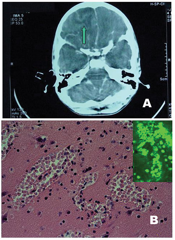
- Primary amebic meningoencephalitis is suspected based on history of swimming in fresh water, but confirmation is difficult because CT and routine cerebrospinal fluid (CSF) tests, although necessary to exclude other causes, are nonspecific. (msdmanuals.com)
Listeriosis2
- citation needed] Specific types include: Veterinarians have observed meningoencephalitis in animals infected with listeriosis, caused by the pathogenic bacteria L. monocytogenes. (wikipedia.org)
- This article presents a clinical case of lethal listeriosis meningoencephalitis, which developed within 7 days after the completion of the first cycle of alemtuzumab therapy. (nih.gov)
Signs1
- Signs of meningoencephalitis include unusual behavior, personality changes, nausea and thinking problems. (wikipedia.org)
Probable1
- The other seven patients were diagnosed with probable RV meningoencephalitis if they had positive PCR results in the sputum and negative results in other extensive workup. (encephalitisjournal.org)
Brain5
- L. monocytogenes meningoencephalitis has been documented to significantly increase the number of cytokines, such as IL-1β, IL-12, IL-15, leading to toxic effects on the brain. (wikipedia.org)
- Most people have never heard of granulomatous meningoencephalitis (GME) or any other form of central nervous system reticulosis until they have a dog with a brain disease that is gradually getting worse. (vin.com)
- This article discusses Tuberculoma of the brain and spinal cord, Tuberculous meningoencephalitis, Tuberculous neuritis, and Tuberculous polyneuropathy. (codingahead.com)
- It has yet to be confirmed by the CDC, but the U.S. may have just seen the year's fourth death from primary amebic meningoencephalitis, otherwise known as A FUCKING PARASITE EATS YOUR BRAIN. (grist.org)
- Although it primarily involves the brain, it often involves the meninges as well (meningoencephalitis). (medscape.com)
Bacterial1
- Inflammation in these areas due to bacterial, viral, or other infections leads to meningoencephalitis. (localquoter.net)
MeSH1
- Meningoencephalitis" is a descriptor in the National Library of Medicine's controlled vocabulary thesaurus, MeSH (Medical Subject Headings) . (wakehealth.edu)
Neurologic1
- However, most cases are mild, and mumps meningoencephalitis generally does not result in death or neurologic sequelae. (wikipedia.org)
Cases3
- In recent years, several cases of adult RV meningoencephalitis have begun to be reported. (encephalitisjournal.org)
- Adult cases of RV-related meningoencephalitis have been reported in a limited number of patients, predominantly those with influenza virus [ 2 ]. (encephalitisjournal.org)
- Maybe nothing is weird about the rise in cases of primary amebic meningoencephalitis. (grist.org)
Patients2
- Among a total of 661 patients in the registry, 10 adult patients were diagnosed with RV-related meningoencephalitis on RV multiplex polymerase chain reaction (PCR) screening test. (encephalitisjournal.org)
- A regimen should include miltefosine , an antileishmanial drug, which has been used in combination with other drugs in the successful treatment of patients with primary amebic meningoencephalitis. (msdmanuals.com)
Patient2
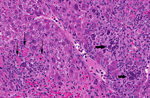
- Autopsy revealed Acanthamoeba species causing necrotizing meningoencephalitis, pneumonitis and adrenalitis in the first patient and causing necrotizing meningoencephalitis and dermatitis in the second patient. (psu.edu)
- Meningoencephalitis due to endogenous endophthalmitis by Klebsiella pneumoniae in a diabetic patient. (repositoriosalud.es)
Year1
- This graph shows the total number of publications written about "Meningoencephalitis" by people in this website by year, and whether "Meningoencephalitis" was a major or minor topic of these publications. (wakehealth.edu)
People1
- Below are the most recent publications written about "Meningoencephalitis" by people in Profiles. (wakehealth.edu)
Common1
- Mumps, a relatively common cause of meningoencephalitis. (wikipedia.org)