Monoclonal Gammopathy of Undetermined Significance
Paraproteinemias
Multiple Myeloma
Waldenstrom Macroglobulinemia
Hypergammaglobulinemia
Paraproteins
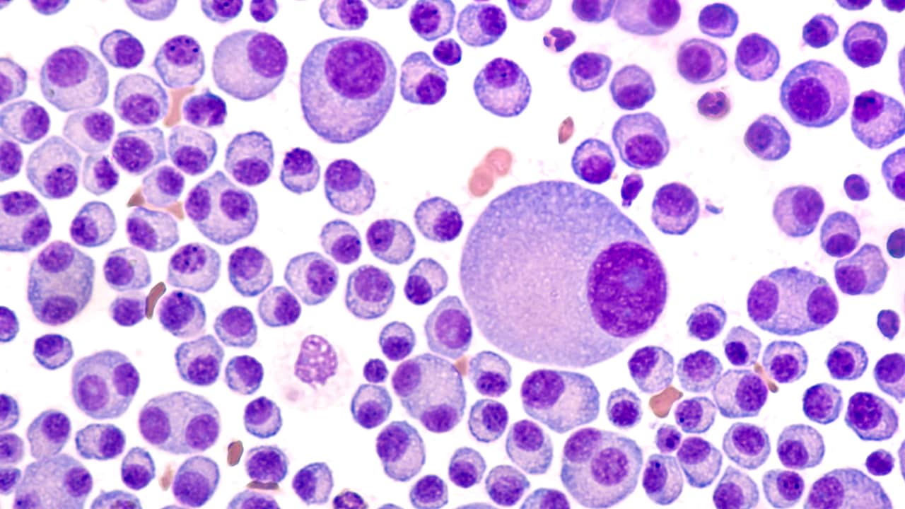
Plasma Cells
Leukemia, Plasma Cell
Vaginal Smears
POEMS Syndrome
Immunoglobulin lambda-Chains
Immunoglobulin kappa-Chains
Cervical Intraepithelial Neoplasia
Immunoglobulin M
Papanicolaou Test
Colposcopy
Polyneuropathies
Amyloidosis
Immunoglobulin Light Chains
Bone Marrow
Uterine Cervical Dysplasia
Neoplasms, Plasma Cell
Chromosomes, Human, Pair 13
Papillomavirus Infections
Neoplasms, Squamous Cell
Disease Progression
Necrobiotic Xanthogranuloma
Bence Jones Protein
Immunoglobulin A
Schnitzler Syndrome
Papillomaviridae
Precancerous Conditions
Cervix Uteri
Immunoglobulins
Cytodiagnosis
Peripheral Nervous System Diseases
Chromosomes, Human, Pair 14
Immunoglobulin G
Prognosis
Biopsy
Translocation, Genetic
Immunoglobulin Isotypes
Chromosomes, Human, Pair 4
Immunophenotyping
Blood Protein Disorders
Immunoglobulin Heavy Chains
Glomerulonephritis, Membranoproliferative
Risk Factors
Capillary Leak Syndrome
Plasmacytoma
In Situ Hybridization, Fluorescence
Sensitivity and Specificity
Chromosomes, Human, Pair 11
Clone Cells
Minnesota
Retrospective Studies
Myelin P0 Protein
B-Lymphocytes
Follow-Up Studies
Sweden
Chromosome Aberrations
beta 2-Microglobulin
Triage
Biopsy, Fine-Needle
Pyoderma Gangrenosum
Alphapapillomavirus
Bone Marrow Cells
Prevalence
Donor Selection
Colonialism
Case-Control Studies
Lymphoproliferative Disorders
Human papillomavirus 18
Demyelinating Diseases
Endometrial Hyperplasia
Flow Cytometry
Cohort Studies
Fluorescent Antibody Technique
Virology
Tumor Virus Infections
Bone marrow angiogenesis and mast cell density increase simultaneously with progression of human multiple myeloma. (1/109)
Immunohistochemical, cytochemical and ultrastructural data showing vivid angiogenesis and numerous mast cells (MCs) in the bone marrow of 24 patients with active multiple myeloma (MM) compared with 34 patients with non-active MM and 22 patients with monoclonal gammopathy of undetermined significance (MGUS) led us to hypothesize that angiogenesis parallels progression of MM, and that MCs participate in its induction via angiogenic factors in their secretory granules. (+info)Increased sialylation of oligosaccharides on IgG paraproteins--a potential new tumour marker in multiple myeloma. (2/109)
AIMS: To investigate whether changes in carbohydrate structure of IgG are related to malignancy and stage of disease in myeloma and monoclonal gammopathy of uncertain significance (MGUS). METHODS: 61 patients were studied at diagnosis: 14 with MGUS, nine with stage I multiple myeloma, 11 with stage II, 21 with stage III, and five with solitary plasmacytoma. IgG was extracted from serum by protein G affinity chromatography. Oligosaccharides were cleaved from the protein backbone enzymatically by N-glycosidase F. Oligosaccharide analysis was performed by high pressure anion exchange chromatography with pulsed electrochemical detection (HPAE-PED). RESULTS: Up to 15 oligosaccharide peaks were identified in three major fractions: neutral, monosialylated, and disialylated. Patients with myeloma showed an increase in the proportion of sialylated oligosaccharides in comparison with patients with MGUS. The ratio of neutral to sialylated oligosaccharides (N:S) was reduced at all stages of myeloma compared with MGUS: MGUS, 11.35; myeloma stage I, 7.6 (p = 0.047); stage II, 5.20 (p = 0.035); stage III, 3.60 (p = 0.0002); plasmacytoma, 7.5 (p = 0.046). The N:S ratio was independent of paraprotein concentration (r = 0.05). CONCLUSIONS: The ratio of neutral to sialylated oligosaccharides may act as a new marker of malignancy in IgG paraproteinaemia and warrants further investigation. (+info)Oligo-monoclonal immunoglobulins frequently develop during concurrent cytomegalovirus (CMV) and Epstein-Barr virus (EBV) infections in patients after renal transplantation. (3/109)
In the present study we report that the appearance of oligo-monoclonal immunoglobulins (oligoM-Igs) in the sera of transplanted individuals is concurrent with the detection of coincident active CMV infection and EBV replication. Eighty-four renal allograft patients were monitored with respect to CMV isolation, to CMV conventional serology and humoral response against the EBV trans-activator ZEBRA (an immediate-early antigen also called BZLF1). Titration of anti-ZEBRA antibodies (IgG and IgM) and amount of EBV DNA in serum were evaluated. Using the combination of four techniques (agarose gel electrophoresis, analytical isoelectric focusing, high resolution immunoelectrophoresis, immunofixation electrophoresis), oligoM-Igs were found in 25% of patients after allografting and significantly associated with rejection episodes (P < 0.001). Twenty out of 23 (86%) concurrent CMV/EBV infections were associated with serum oligoM-Igs (P < 0.001). One can thus reasonably assume that a sustained EBV replication following iatrogenic immunosuppression can promote the immunoglobulin heavy chain expression in EBV-infected B lymphocytes. The proliferation of immunoglobulin-secreting clones might occur after active CMV infection, through a transient over-immunosuppression or via immune subversion. (+info)Prognostic factors for malignant transformation in monoclonal gammopathy of undetermined significance and smoldering multiple myeloma. (4/109)
PURPOSE: To evaluate the natural history of monoclonal gammopathy of undetermined significance (MGUS) and smoldering multiple myeloma (SMM), identify early predictors of evolution, and assess whether associated conditions correlate with disease progression. PATIENTS AND METHODS: A total of 1,231 consecutive patients with either MGUS (n = 1,104) or SMM (n = 127) diagnosed from July 1975 to March 1998 were included in the study. Cumulative survival probability and cumulative probability of transformation into lymphoproliferative disease were calculated by means of the Kaplan-Meier estimator. Univariate and multivariate Cox models were used to identify possible predictors of malignant evolution. RESULTS: Cumulative transformation probability at 10 and 15 years was 14% and 30%, respectively. At a median follow-up of 65 months (range, 12 to 239 months), 64 MGUS cases (5.8%) evolved to multiple myeloma (MM) (n = 43), extramedullary plasmacytoma (n = 1), primary amyloidosis (n = 1), Waldenstrom's macroglobulinemia (n = 12), non-Hodgkin's lymphoma (n = 6), and B-chronic lymphocytic leukemia (n = 1). At a median follow-up of 72 months (range, 12 to 247 months), 25 SMMs (19.7%) evolved to overt MM. A lower evolution risk was observed in MGUS than in SMM (P <.0001). Greater than 5% marrow plasmacytosis, detectable Bence Jones proteinuria, polyclonal serum immunoglobulin reduction, and high erythrocyte sedimentation rate (ESR) were independent factors influencing MGUS transformation. SMM progression correlated with greater than 10% marrow plasma cells, detectable Bence Jones proteinuria, and immunoglobulin (Ig) A isotype. Neither concomitant diseases nor immunosuppression correlated with progression. CONCLUSION: Careful evaluation of marrow plasmacytosis, urinary paraprotein, background immunoglobulins, ESR, and paraprotein isotype might help identify at presentation patients with benign monoclonal gammopathies requiring stricter monitoring. (+info)Autologous peripheral blood stem cell transplantation for peripheral neuropathy secondary to monoclonal gammopathy of unknown significance. (5/109)
A 40-year-old patient presented with rapidly progressing peripheral neuropathy secondary to monoclonal gammopathy of unknown significance (MGUS). He became severely debilitated, being wheelchair-bound, despite treatment with chemotherapy, intravenous immunoglobulin and plasma exchange. He was subsequently treated with high-dose chemotherapy followed by autologous peripheral blood stem cell transplantation (PBSCT). He has made remarkable recovery at 12 months post transplantation. We propose that high-dose chemotherapy and autologous PBSC transplantation may have a role in the treatment of severe, progressive and treatment-resistant MGUS-related peripheral neuropathy. (+info)Serum reference intervals and diagnostic ranges for free kappa and free lambda immunoglobulin light chains: relative sensitivity for detection of monoclonal light chains. (6/109)
BACKGROUND: The detection of monoclonal free light chains (FLCs) is an important diagnostic aid for a variety of monoclonal gammopathies and is especially important in light-chain diseases, such as light-chain myeloma, primary systemic amyloidosis, and light-chain-deposition disease. These diseases are more prevalent in the elderly, and assays to detect and quantify abnormal amounts of FLCs require reference intervals that include elderly donors. METHODS: We used an automated immunoassay for FLCs and sera from a population 21-90 years of age. We used the calculated reference and diagnostic intervals to compare FLC results with those obtained by immunofixation (IFE) to detect low concentrations of monoclonal kappa and lambda FLCs in the sera of patients with monoclonal gammopathies. RESULTS: Serum kappa and lambda FLCs increased with population age, with an apparent change for those >80 years. This trend was lost when the FLC concentration was normalized to cystatin C concentration. The ratio of kappa FLC to lambda FLC (FLC K/L) did not exhibit an age-dependent trend. The diagnostic interval for FLC K/L was 0.26-1.65. The 95% reference interval for kappa FLC was 3.3-19.4 mg/L, and that for lambda FLC was 5.7-26.3 mg/L. Detection and quantification of monoclonal FLCs by nephelometry were more sensitive than IFE in serum samples from patients with primary systemic amyloidosis and light-chain-deposition disease. CONCLUSIONS: Reference and diagnostic intervals for serum FLCs have been developed for use with a new, automated immunoassay that makes the detection and quantification of monoclonal FLCs easier and more sensitive than with current methods. The serum FLC assay complements IFE and allows quantification of FLCs in light-chain-disease patients who have no detectable serum or urine M-spike. (+info)Expression of CD52 on plasma cells in plasma cell proliferative disorders. (7/109)
Multiple myeloma (MM) and primary systemic amyloidosis (AL) remain incurable disorders, and new treatments targeted to the malignant plasma cells are needed. Alemtuzumab is a humanized monoclonal antibody to CD52 and has activity in chronic lymphocytic leukemia. We examined the CD52 expression on CD45+ and CD45- plasma cell populations to evaluate the potential for using alemtuzumab for these disorders. Bone marrows from 61 patients (29 AL, 23 MM, and 9 MGUS [monoclonal gammopathies of undetermined significance]) were studied using 3-color (CD38/45/52) flow cytometry. Among those with MGUS, MM, and AL, 67%, 52%, and 35%, respectively, were positive for CD52 expression. The CD52 expression was predominantly confined to the clonal CD38+/CD45+ plasma cell fraction with median expression of 68%, 88%, and 82% in MGUS, MM, and AL, respectively, compared with 18%, 6%, and 9% among the CD45- plasma cell population. Clinical trials are warranted in these diseases to learn the therapeutic benefit of anti-CD52 immunotherapy. (+info)Immunochemical and clinical effects of immunosuppressive treatment in monoclonal IgM neuropathy. (8/109)
A pathogenic role of the M protein in monoclonal IgM neuropathy has been suggested. This is based among other things on a close relation between immunosuppressive treatment, lowered concentration of M protein, and clinical effect. We studied five patients with monoclonal IgM and antibodies to peripheral nerve myelin. The immunosuppressive treatment was beneficial in three of the patients. In three patients there was a relationship between antibody concentration and clinical effect (in one there was no change in antibody concentrations and correspondingly no change in clinical status, and in two patients clinical improvement corresponded to decreased antibody concentrations). In two patients, however, there was no clear correlation, since one patient improved despite increasing antibody concentrations and one patient did not improve despite a lowered antibody concentration. It is therefore possible that other mechanisms may contribute to the effect of treatment. (+info)Monoclonal gammopathy of undetermined significance (MGUS) is a medical condition characterized by the presence of a monoclonal protein, or M-protein, in the blood or urine, but without any signs or symptoms of related disorders. The M-protein is produced by a single clone of plasma cells, which are a type of white blood cell found in the bone marrow.
In MGUS, the level of M-protein is typically low (less than 3 grams per deciliter), and there are no signs of damage to organs such as the bones, kidneys, or nervous system. However, people with MGUS have a higher risk of developing certain related conditions, such as multiple myeloma, amyloidosis, or lymphoplasmacytic lymphoma, compared to those without MGUP.
MGUS is usually detected through routine blood or urine tests and is typically asymptomatic. However, in some cases, people with MGUS may experience symptoms such as fatigue, bone pain, or recurrent infections. If these symptoms occur, further testing may be necessary to determine if MGUS has progressed to a more serious condition.
It's important to note that MGUS is not a cancer itself, but rather a potential precursor to certain types of cancer. Regular monitoring with blood or urine tests and physical examinations is recommended for people diagnosed with MGUS to monitor for any changes that may indicate progression to a more serious condition.
Paraproteinemias refer to the presence of abnormal levels of paraproteins in the blood. Paraproteins are immunoglobulins (antibodies) produced by plasma cells, which are a type of white blood cell found in the bone marrow. In healthy individuals, paraproteins play a role in the immune system's response to infection and disease. However, in certain conditions, such as multiple myeloma, monoclonal gammopathy of undetermined significance (MGUS), and Waldenstrom macroglobulinemia, plasma cells produce excessive amounts of a single type of paraprotein, leading to its accumulation in the blood.
Paraproteinemias can cause various symptoms depending on the level of paraproteins present and their impact on organs and tissues. These symptoms may include fatigue, weakness, numbness or tingling in the extremities, bone pain, recurrent infections, and kidney problems. In some cases, paraproteinemias may not cause any symptoms and may only be detected during routine blood tests.
It is important to note that while paraproteinemias are often associated with plasma cell disorders, they can also occur in other conditions such as chronic inflammation or autoimmune diseases. Therefore, further testing and evaluation are necessary to determine the underlying cause of paraproteinemia and develop an appropriate treatment plan.
Multiple myeloma is a type of cancer that forms in a type of white blood cell called a plasma cell. Plasma cells help your body fight infection by producing antibodies. In multiple myeloma, cancerous plasma cells accumulate in the bone marrow and crowd out healthy blood cells. Rather than producing useful antibodies, the cancer cells produce abnormal proteins that can cause complications such as kidney damage, bone pain and fractures.
Multiple myeloma is a type of cancer called a plasma cell neoplasm. Plasma cell neoplasms are diseases in which there is an overproduction of a single clone of plasma cells. In multiple myeloma, this results in the crowding out of normal plasma cells, red and white blood cells and platelets, leading to many of the complications associated with the disease.
The abnormal proteins produced by the cancer cells can also cause damage to organs and tissues in the body. These abnormal proteins can be detected in the blood or urine and are often used to monitor the progression of multiple myeloma.
Multiple myeloma is a relatively uncommon cancer, but it is the second most common blood cancer after non-Hodgkin lymphoma. It typically occurs in people over the age of 65, and men are more likely to develop multiple myeloma than women. While there is no cure for multiple myeloma, treatments such as chemotherapy, radiation therapy, and stem cell transplantation can help manage the disease and its symptoms, and improve quality of life.
Waldenstrom macroglobulinemia is a type of rare cancer called a lymphoplasmacytic lymphoma. It is characterized by the uncontrolled growth of malignant white blood cells, specifically B lymphocytes or plasma cells, in the bone marrow and sometimes in other organs. These abnormal cells produce an excessive amount of a protein called macroglobulin, which can lead to the thickening of the blood and various symptoms associated with this condition.
The signs and symptoms of Waldenstrom macroglobulinemia may include fatigue, weakness, bruising or bleeding, frequent infections, numbness or tingling in the hands and feet, visual disturbances, and confusion or difficulty thinking. The diagnosis typically involves a combination of blood tests, bone marrow biopsy, imaging studies, and sometimes genetic testing to confirm the presence of the disease and determine its extent.
Treatment options for Waldenstrom macroglobulinemia depend on the severity of the symptoms and the stage of the disease. They may include chemotherapy, targeted therapy, immunotherapy, stem cell transplantation, or a combination of these approaches. Regular follow-up care is essential to monitor the progression of the disease and adjust treatment plans as needed.
Hypergammaglobulinemia is a medical condition characterized by an elevated level of gamma globulins (a type of immunoglobulins or antibodies) in the blood. These proteins are part of the body's immune system and help to fight off infections. However, when their levels become too high, it can indicate an underlying medical disorder.
There are several types of hypergammaglobulinemia, including:
1. Primary hypergammaglobulinemia: This is a rare condition that is present at birth or develops during early childhood. It is caused by genetic mutations that lead to overproduction of immunoglobulins.
2. Secondary hypergammaglobulinemia: This type is more common and is caused by an underlying medical condition, such as chronic infections, autoimmune disorders, or certain types of cancer.
Symptoms of hypergammaglobulinemia can vary depending on the cause and severity of the condition. They may include recurrent infections, fatigue, swelling of the lymph nodes, and joint pain. Treatment typically involves addressing the underlying cause of the condition, if possible, as well as managing symptoms and preventing complications.
Paraproteins, also known as M-proteins or monoclonal proteins, are immunoglobulins (antibodies) that are produced in abnormal amounts by a single clone of plasma cells. These proteins are typically produced in response to a stimulus such as an infection, but when they are produced in excessive and/or unusual forms, it can indicate the presence of a clonal disorder, such as multiple myeloma, Waldenstrom macroglobulinemia, or other related conditions.
Paraproteins can be detected in the blood or urine and are often used as a marker for disease progression and response to treatment. They can also cause various symptoms and complications, depending on their size, concentration, and location. These may include damage to organs such as the kidneys, nerves, and bones.
Plasma cells are a type of white blood cell that are derived from B cells (another type of white blood cell) and are responsible for producing antibodies. Antibodies are proteins that help the body to fight against infections by recognizing and binding to specific antigens, such as bacteria or viruses. Plasma cells are found in the bone marrow, spleen, and lymph nodes, and they play a crucial role in the immune system's response to infection.
Plasma cells are characterized by their large size, eccentric nucleus, and abundant cytoplasm filled with rough endoplasmic reticulum, which is where antibody proteins are synthesized and stored. When activated, plasma cells can produce and secrete large amounts of antibodies into the bloodstream and lymphatic system, where they can help to neutralize or eliminate pathogens.
It's worth noting that while plasma cells play an important role in the immune response, abnormal accumulations of these cells can also be a sign of certain diseases, such as multiple myeloma, a type of cancer that affects plasma cells.
Plasma cell leukemia (PCL) is a rare and aggressive type of cancer that involves the uncontrolled multiplication of malignant plasma cells in the bone marrow, blood, and sometimes in other organs. Plasma cells are a type of white blood cell that produces antibodies to help fight infections. In PCL, the malignant plasma cells produce abnormal antibodies called M-proteins or paraproteins, which can accumulate in various tissues and cause damage.
PCL is typically classified into two types: primary and secondary. Primary PCL is a distinct clinical entity that presents with more than 20% plasma cells in the bone marrow and/or blood. Secondary PCL is a complication of multiple myeloma, a more common type of plasma cell cancer, and occurs when the malignant plasma cells spread from the bone marrow to the blood.
The symptoms of PCL are similar to those of other types of leukemia and may include fatigue, weakness, weight loss, frequent infections, easy bruising or bleeding, and bone pain. Diagnosis of PCL typically involves a combination of clinical evaluation, laboratory tests, imaging studies, and bone marrow aspiration and biopsy. Treatment options for PCL may include chemotherapy, stem cell transplantation, radiation therapy, and targeted therapies. The prognosis for patients with PCL is generally poor, with a median survival time of less than one year.
A vaginal smear, also known as a Pap test or Pap smear, is a medical procedure in which a sample of cells is collected from the cervix (the lower part of the uterus that opens into the vagina) and examined under a microscope. The purpose of this test is to detect abnormal cells, including precancerous changes, that may indicate the presence of cervical cancer or other conditions such as infections or inflammation.
During the procedure, a speculum is inserted into the vagina to allow the healthcare provider to visualize the cervix. A spatula or brush is then used to gently scrape cells from the surface of the cervix. The sample is spread onto a microscope slide and sent to a laboratory for analysis.
Regular Pap smears are recommended for women as part of their routine healthcare, as they can help detect abnormalities at an early stage when they are more easily treated. The frequency of Pap smears may vary depending on age, medical history, and other factors. It is important to follow the recommendations of a healthcare provider regarding the timing and frequency of Pap smears.
Blood protein electrophoresis (BPE) is a laboratory test that separates and measures the different proteins in the blood, such as albumin, alpha-1 globulins, alpha-2 globulins, beta globulins, and gamma globulins. This test is often used to help diagnose or monitor conditions related to abnormal protein levels, such as multiple myeloma, macroglobulinemia, and other plasma cell disorders.
In this test, a sample of the patient's blood is placed on a special gel and an electric current is applied. The proteins in the blood migrate through the gel based on their electrical charge and size, creating bands that can be visualized and measured. By comparing the band patterns to reference ranges, doctors can identify any abnormal protein levels or ratios, which may indicate underlying medical conditions.
It's important to note that while BPE is a useful diagnostic tool, it should be interpreted in conjunction with other clinical findings and laboratory tests for accurate diagnosis and management of the patient's condition.
POEMS syndrome is a rare and complex disorder that affects multiple parts of the body. The name POEMS is an acronym that stands for the following symptoms: Polyneuropathy, Organomegaly, Endocrinopathy, Monoclonal gammopathy, and Skin changes.
Here's a brief definition of each component of the syndrome:
* Polyneuropathy: This refers to damage to the peripheral nerves that can cause symptoms such as numbness, tingling, pain, and weakness in the arms and legs.
* Organomegaly: This means enlargement of organs, such as the liver, spleen, or lymph nodes.
* Endocrinopathy: This refers to abnormalities in hormone-producing glands, which can lead to symptoms such as diabetes, low testosterone levels, and thyroid dysfunction.
* Monoclonal gammopathy: This is an abnormal production of a single type of immunoglobulin (a protein produced by the immune system) in the bone marrow.
* Skin changes: These can include skin thickening, darkening, or redness, as well as skin lesions.
POEMS syndrome is typically caused by an underlying plasma cell disorder, such as multiple myeloma or a related condition called Waldenstrom macroglobulinemia. Treatment for POEMS syndrome usually involves addressing the underlying plasma cell disorder, as well as managing specific symptoms of the syndrome.
Immunoglobulin lambda-chains (Igλ) are one type of light chain found in the immunoglobulins, also known as antibodies. Antibodies are proteins that play a crucial role in the immune system's response to foreign substances, such as bacteria and viruses.
Immunoglobulins are composed of two heavy chains and two light chains, which are interconnected by disulfide bonds. There are two types of light chains: kappa (κ) and lambda (λ). Igλ chains are one type of light chain that can be found in association with heavy chains to form functional antibodies.
Igλ chains contain a variable region, which is responsible for recognizing and binding to specific antigens, and a constant region, which determines the class of the immunoglobulin (e.g., IgA, IgD, IgE, IgG, or IgM).
In humans, approximately 60% of all antibodies contain Igλ chains, while the remaining 40% contain Igκ chains. The ratio of Igλ to Igκ chains can vary depending on the type of immunoglobulin and its function in the immune response.
Immunoglobulin kappa-chains are one of the two types of light chains (the other being lambda-chains) that make up an immunoglobulin molecule, also known as an antibody. These light chains combine with heavy chains to form the antigen-binding site of an antibody, which is responsible for recognizing and binding to specific antigens or foreign substances in the body.
Kappa-chains contain a variable region that differs between different antibodies and contributes to the diversity of the immune system's response to various antigens. They also have a constant region, which is consistent across all kappa-chains. Approximately 60% of all human antibodies contain kappa-chains, while the remaining 40% contain lambda-chains. The relative proportions of kappa and lambda chains can be used in diagnostic tests to identify clonal expansions of B cells, which may indicate a malignancy such as multiple myeloma or lymphoma.
Myeloma proteins, also known as monoclonal immunoglobulins or M-proteins, are entire or abnormal immunoglobulin (antibody) molecules produced by a single clone of plasma cells, which are malignant in the case of multiple myeloma and some related disorders. These proteins accumulate in the blood and/or urine and can cause damage to various organs and tissues.
In multiple myeloma, the excessive proliferation of these plasma cells leads to the overproduction of a single type of immunoglobulin or its fragments, which can be detected and quantified in serum and/or urine electrophoresis. The most common types of myeloma proteins are IgG and IgA, followed by light chains (Bence Jones proteins) and, less frequently, IgD and IgM.
The presence and levels of myeloma proteins are important diagnostic markers for multiple myeloma and related disorders, such as monoclonal gammopathy of undetermined significance (MGUS) and Waldenström macroglobulinemia. Regular monitoring of these proteins helps assess the disease's activity, response to treatment, and potential complications like kidney dysfunction or amyloidosis.
Cervical intraepithelial neoplasia (CIN) is a term used to describe the abnormal growth and development of cells on the surface of the cervix. These changes are usually caused by human papillomavirus (HPV) infection, which is a common sexually transmitted infection. CIN is not cancer, but it can develop into cancer if left untreated.
The term "intraepithelial" refers to the fact that the abnormal cells are found in the epithelium, or the lining of the cervix. The term "neoplasia" means abnormal growth or development of cells. CIN is further classified into three grades based on the severity of the cell changes:
* CIN 1: Mild dysplasia (abnormal cell growth) affecting the lower third of the epithelium.
* CIN 2: Moderate dysplasia affecting the lower two-thirds of the epithelium.
* CIN 3: Severe dysplasia or carcinoma in situ, which means that the abnormal cells are found in the full thickness of the epithelium and have a high risk of progressing to invasive cancer if not treated.
It's important to note that CIN can regress on its own without treatment, especially in younger women. However, some cases may progress to invasive cervical cancer if left untreated. Regular Pap testing is recommended to detect and monitor any abnormal cell changes in the cervix. If CIN is detected, further diagnostic procedures such as a colposcopy or biopsy may be performed to determine the extent of the abnormality and guide treatment decisions.
Monoclonal antibodies are a type of antibody that are identical because they are produced by a single clone of cells. They are laboratory-produced molecules that act like human antibodies in the immune system. They can be designed to attach to specific proteins found on the surface of cancer cells, making them useful for targeting and treating cancer. Monoclonal antibodies can also be used as a therapy for other diseases, such as autoimmune disorders and inflammatory conditions.
Monoclonal antibodies are produced by fusing a single type of immune cell, called a B cell, with a tumor cell to create a hybrid cell, or hybridoma. This hybrid cell is then able to replicate indefinitely, producing a large number of identical copies of the original antibody. These antibodies can be further modified and engineered to enhance their ability to bind to specific targets, increase their stability, and improve their effectiveness as therapeutic agents.
Monoclonal antibodies have several mechanisms of action in cancer therapy. They can directly kill cancer cells by binding to them and triggering an immune response. They can also block the signals that promote cancer growth and survival. Additionally, monoclonal antibodies can be used to deliver drugs or radiation directly to cancer cells, increasing the effectiveness of these treatments while minimizing their side effects on healthy tissues.
Monoclonal antibodies have become an important tool in modern medicine, with several approved for use in cancer therapy and other diseases. They are continuing to be studied and developed as a promising approach to treating a wide range of medical conditions.
Immunoglobulin M (IgM) is a type of antibody that is primarily found in the blood and lymph fluid. It is the first antibody to be produced in response to an initial exposure to an antigen, making it an important part of the body's primary immune response. IgM antibodies are large molecules that are composed of five basic units, giving them a pentameric structure. They are primarily found on the surface of B cells as membrane-bound immunoglobulins (mlgM), where they function as receptors for antigens. Once an mlgM receptor binds to an antigen, it triggers the activation and differentiation of the B cell into a plasma cell that produces and secretes large amounts of soluble IgM antibodies.
IgM antibodies are particularly effective at agglutination (clumping) and complement activation, which makes them important in the early stages of an immune response to help clear pathogens from the bloodstream. However, they are not as stable or long-lived as other types of antibodies, such as IgG, and their levels tend to decline after the initial immune response has occurred.
In summary, Immunoglobulin M (IgM) is a type of antibody that plays a crucial role in the primary immune response to antigens by agglutination and complement activation. It is primarily found in the blood and lymph fluid, and it is produced by B cells after they are activated by an antigen.
Uterine cervical neoplasms, also known as cervical cancer or cervical dysplasia, refer to abnormal growths or lesions on the lining of the cervix that have the potential to become cancerous. These growths are usually caused by human papillomavirus (HPV) infection and can be detected through routine Pap smears.
Cervical neoplasms are classified into different grades based on their level of severity, ranging from mild dysplasia (CIN I) to severe dysplasia or carcinoma in situ (CIN III). In some cases, cervical neoplasms may progress to invasive cancer if left untreated.
Risk factors for developing cervical neoplasms include early sexual activity, multiple sexual partners, smoking, and a weakened immune system. Regular Pap smears and HPV testing are recommended for early detection and prevention of cervical cancer.
The Papanicolaou (Pap) test, also known as the Pap smear, is a screening procedure for detecting precancerous and cancerous cells in the cervix. It involves collecting cells from the cervix and examining them under a microscope to look for any abnormalities. The test is typically recommended for women aged 21-65 as part of routine pelvic exams, with the frequency depending on age and risk factors.
The Pap test was developed by Georgios Papanikolaou in the early 20th century and has since become a widely used and important tool in preventing cervical cancer. The test is usually performed in a healthcare provider's office and takes only a few minutes to complete. It is a relatively simple, safe, and painless procedure that can help detect cervical abnormalities at an early stage, when they are most treatable.
Colposcopy is a medical procedure in which a colposcope, which is a type of microscope, is used to examine the cervix, vagina, and vulva for signs of disease or abnormalities. The colposcope allows the healthcare provider to see these areas in greater detail than is possible with the naked eye. During the procedure, the provider may take a small sample of tissue (biopsy) for further examination under a microscope.
Colposcopy is often used to investigate abnormal Pap test results or to follow up on women who have been diagnosed with certain types of cervical dysplasia (abnormal cell growth). It can also be used to diagnose and monitor other conditions, such as genital warts, inflammation, or cancer.
It is important to note that colposcopy is a diagnostic procedure and not a treatment. If abnormalities are found during the exam, additional procedures may be necessary to remove or treat them.
Polyneuropathy is a medical condition that refers to the damage or dysfunction of peripheral nerves (nerves outside the brain and spinal cord) in multiple areas of the body. These nerves are responsible for transmitting sensory, motor, and autonomic signals between the central nervous system and the rest of the body.
In polyneuropathies, this communication is disrupted, leading to various symptoms depending on the type and extent of nerve damage. Commonly reported symptoms include:
1. Numbness or tingling in the hands and feet
2. Muscle weakness and cramps
3. Loss of reflexes
4. Burning or stabbing pain
5. Balance and coordination issues
6. Increased sensitivity to touch
7. Autonomic dysfunction, such as bowel, bladder, or digestive problems, and changes in blood pressure
Polyneuropathies can be caused by various factors, including diabetes, alcohol abuse, nutritional deficiencies, autoimmune disorders, infections, toxins, inherited genetic conditions, or idiopathic (unknown) causes. The treatment for polyneuropathy depends on the underlying cause and may involve managing underlying medical conditions, physical therapy, pain management, and lifestyle modifications.
Amyloidosis is a medical condition characterized by the abnormal accumulation of insoluble proteins called amyloid in various tissues and organs throughout the body. These misfolded protein deposits can disrupt the normal function of affected organs, leading to a range of symptoms depending on the location and extent of the amyloid deposition.
There are different types of amyloidosis, classified based on the specific proteins involved:
1. Primary (AL) Amyloidosis: This is the most common form, accounting for around 80% of cases. It results from the overproduction and misfolding of immunoglobulin light chains, typically by clonal plasma cells in the bone marrow. The amyloid deposits can affect various organs, including the heart, kidneys, liver, and nervous system.
2. Secondary (AA) Amyloidosis: This form is associated with chronic inflammatory diseases, such as rheumatoid arthritis, tuberculosis, or familial Mediterranean fever. The amyloid fibrils are composed of serum amyloid A protein (SAA), an acute-phase reactant produced during the inflammatory response. The kidneys are commonly affected in this type of amyloidosis.
3. Hereditary or Familial Amyloidosis: These forms are caused by genetic mutations that result in the production of abnormal proteins prone to misfolding and amyloid formation. Examples include transthyretin (TTR) amyloidosis, fibrinogen amyloidosis, and apolipoprotein AI amyloidosis. These forms can affect various organs, including the heart, nerves, and kidneys.
4. Dialysis-Related Amyloidosis: This form is seen in patients undergoing long-term dialysis for chronic kidney disease. The amyloid fibrils are composed of beta-2 microglobulin, a protein that accumulates due to impaired clearance during dialysis. The joints and bones are commonly affected in this type of amyloidosis.
The diagnosis of amyloidosis typically involves a combination of clinical evaluation, imaging studies, and tissue biopsy with the demonstration of amyloid deposition using special stains (e.g., Congo red). Treatment depends on the specific type and extent of organ involvement and may include supportive care, medications to target the underlying cause (e.g., chemotherapy, immunomodulatory agents), and organ transplantation in some cases.
Immunoglobulin light chains are the smaller protein subunits of an immunoglobulin, also known as an antibody. They are composed of two polypeptide chains, called kappa (κ) and lambda (λ), which are produced by B cells during the immune response. Each immunoglobulin molecule contains either two kappa or two lambda light chains, in association with two heavy chains.
Light chains play a crucial role in the antigen-binding site of an antibody, where they contribute to the specificity and affinity of the interaction between the antibody and its target antigen. In addition to their role in immune function, abnormal production or accumulation of light chains can lead to various diseases, such as multiple myeloma and amyloidosis.
Bone marrow is the spongy tissue found inside certain bones in the body, such as the hips, thighs, and vertebrae. It is responsible for producing blood-forming cells, including red blood cells, white blood cells, and platelets. There are two types of bone marrow: red marrow, which is involved in blood cell production, and yellow marrow, which contains fatty tissue.
Red bone marrow contains hematopoietic stem cells, which can differentiate into various types of blood cells. These stem cells continuously divide and mature to produce new blood cells that are released into the circulation. Red blood cells carry oxygen throughout the body, white blood cells help fight infections, and platelets play a crucial role in blood clotting.
Bone marrow also serves as a site for immune cell development and maturation. It contains various types of immune cells, such as lymphocytes, macrophages, and dendritic cells, which help protect the body against infections and diseases.
Abnormalities in bone marrow function can lead to several medical conditions, including anemia, leukopenia, thrombocytopenia, and various types of cancer, such as leukemia and multiple myeloma. Bone marrow aspiration and biopsy are common diagnostic procedures used to evaluate bone marrow health and function.
Uterine cervical dysplasia is a condition characterized by abnormal cell growth on the lining of the cervix, which is the lower part of the uterus that connects to the vagina. It is also known as cervical intraepithelial neoplasia (CIN).
Cervical dysplasia can be caused by certain strains of human papillomavirus (HPV), a common sexually transmitted infection. The abnormal cells may develop into cancerous cells over time, although not all cases of cervical dysplasia will progress to cancer.
Cervical dysplasia is typically detected through a Pap test or HPV test, which are screening tests used to detect precancerous changes in the cervix. Depending on the severity and extent of the abnormal cells, treatment options may include close monitoring, surgical removal of the affected tissue, or more extensive surgery.
It is important for women to receive regular Pap tests and HPV tests as recommended by their healthcare provider to detect and treat cervical dysplasia early, before it has a chance to progress to cancer.
Plasma cell neoplasms are a type of cancer that originates from plasma cells, which are a type of white blood cell found in the bone marrow. These cells are responsible for producing antibodies to help fight off infections. When plasma cells become cancerous and multiply out of control, they can form a tumor called a plasmacytoma.
There are two main types of plasma cell neoplasms: solitary plasmacytoma and multiple myeloma. Solitary plasmacytoma is a localized tumor that typically forms in the bone, while multiple myeloma is a systemic disease that affects multiple bones and can cause a variety of symptoms such as bone pain, fatigue, and anemia.
Plasma cell neoplasms are diagnosed through a combination of tests, including blood tests, imaging studies, and bone marrow biopsy. Treatment options depend on the stage and extent of the disease, but may include radiation therapy, chemotherapy, and stem cell transplantation.
Human chromosome pair 13 consists of two rod-shaped structures present in the nucleus of each cell in the human body. Each chromosome is made up of DNA tightly coiled around histone proteins, forming a complex structure called a chromatin.
Chromosomes carry genetic information in the form of genes, which are sequences of DNA that code for specific traits and functions. Human cells typically have 23 pairs of chromosomes, for a total of 46 chromosomes. Chromosome pair 13 is one of the autosomal pairs, meaning it is not a sex chromosome (X or Y).
Chromosome pair 13 contains several important genes that are associated with various genetic disorders, such as cri-du-chat syndrome and Phelan-McDermid syndrome. Cri-du-chat syndrome is caused by a deletion of the short arm of chromosome 13 (13p), resulting in distinctive cat-like crying sounds in infants, developmental delays, and intellectual disabilities. Phelan-McDermid syndrome is caused by a deletion or mutation of the terminal end of the long arm of chromosome 13 (13q), leading to developmental delays, intellectual disability, absent or delayed speech, and autistic behaviors.
It's important to note that while some genetic disorders are associated with specific chromosomal abnormalities, many factors can contribute to the development and expression of these conditions, including environmental influences and interactions between multiple genes.
Papillomavirus infections are a group of diseases caused by various types of human papillomaviruses (HPVs). These viruses infect the skin and mucous membranes, and can cause benign growths such as warts or papillomas, as well as malignant growths like cervical cancer.
There are more than 100 different types of HPVs, and they can be classified into low-risk and high-risk types based on their potential to cause cancer. Low-risk HPV types, such as HPV-6 and HPV-11, commonly cause benign genital warts and respiratory papillomas. High-risk HPV types, such as HPV-16 and HPV-18, are associated with an increased risk of developing cancer, including cervical, anal, penile, vulvar, and oropharyngeal cancers.
HPV infections are typically transmitted through sexual contact, and most sexually active individuals will acquire at least one HPV infection during their lifetime. In many cases, the immune system is able to clear the virus without any symptoms or long-term consequences. However, persistent high-risk HPV infections can lead to the development of cancer over time.
Prevention measures for HPV infections include vaccination against high-risk HPV types, safe sex practices, and regular screening for cervical cancer in women. The HPV vaccine is recommended for both boys and girls aged 11-12 years old, and can also be given to older individuals up to age 45 who have not previously been vaccinated or who have not completed the full series of shots.
Squamous cell neoplasms are abnormal growths or tumors that originate from squamous cells, which are flat, scale-like cells that make up the outer layer of the skin and the lining of mucous membranes. These neoplasms can be benign (noncancerous) or malignant (cancerous). When malignant, they are called squamous cell carcinomas.
Squamous cell carcinomas often develop in areas exposed to excessive sunlight or ultraviolet radiation, such as the skin, lips, and mouth. They can also occur in other areas of the body, including the cervix, anus, and lungs. Risk factors for developing squamous cell carcinoma include fair skin, a history of sunburns, exposure to certain chemicals or radiation, and a weakened immune system.
Symptoms of squamous cell carcinomas may include rough or scaly patches on the skin, a sore that doesn't heal, a wart-like growth, or a raised bump with a central depression. Treatment for squamous cell carcinomas typically involves surgical removal of the tumor, along with radiation therapy or chemotherapy in some cases. Early detection and treatment can help prevent the spread of the cancer to other parts of the body.
Disease progression is the worsening or advancement of a medical condition over time. It refers to the natural course of a disease, including its development, the severity of symptoms and complications, and the impact on the patient's overall health and quality of life. Understanding disease progression is important for developing appropriate treatment plans, monitoring response to therapy, and predicting outcomes.
The rate of disease progression can vary widely depending on the type of medical condition, individual patient factors, and the effectiveness of treatment. Some diseases may progress rapidly over a short period of time, while others may progress more slowly over many years. In some cases, disease progression may be slowed or even halted with appropriate medical interventions, while in other cases, the progression may be inevitable and irreversible.
In clinical practice, healthcare providers closely monitor disease progression through regular assessments, imaging studies, and laboratory tests. This information is used to guide treatment decisions and adjust care plans as needed to optimize patient outcomes and improve quality of life.
Necrobiotic xanthogranuloma (NXG) is a rare, progressive inflammatory disorder characterized by the formation of distinctive granulomatous lesions in the skin and occasionally in various visceral organs. The term "necrobiotic" refers to the presence of necrotic tissue within the lesions, while "xanthogranuloma" indicates the accumulation of foamy histiocytes (a type of white blood cell) and multinucleated giant cells.
The pathogenesis of NXG is not entirely understood, but it has been associated with monoclonal gammopathies, particularly those involving the M-protein IgG4. The disease typically affects middle-aged to older adults, with a slight female predominance.
Clinically, NXG presents as yellowish-red or violaceous papules, plaques, or nodules, often located on the periorbital area, eyelids, and less frequently on the trunk and extremities. The lesions may be asymptomatic or associated with mild pruritus, burning sensation, or pain. In some cases, NXG can lead to serious complications such as sight-threatening ocular involvement, kidney dysfunction, or neurological symptoms due to systemic dissemination.
Histopathologically, NXG lesions are characterized by the presence of necrobiotic areas surrounded by palisading histiocytes, T lymphocytes, and multinucleated giant cells, often with a cholesterol clefts and calcifications. The accumulation of foamy histiocytes is a hallmark feature of NXG.
The diagnosis of NXG is based on the clinical presentation, histopathological findings, and laboratory investigations to exclude associated conditions such as monoclonal gammopathies or other systemic disorders. Treatment options for NXG include topical and intralesional corticosteroids, systemic immunosuppressive agents, and targeted therapies against the underlying clonal gammopathy if present. Regular follow-up is essential to monitor disease progression and response to treatment.
Bence Jones protein is a type of immunoglobulin light chain that can be detected in the urine or blood of some patients with certain diseases, most notably multiple myeloma. It's named after Henry Bence Jones, a 19th-century English physician who first described it.
These proteins are produced by malignant plasma cells, which are a type of white blood cell found in the bone marrow. In multiple myeloma, these cancerous cells multiply and produce abnormal amounts of immunoglobulins, leading to the overproduction of Bence Jones proteins.
When these proteins are excreted in the urine, they can cause damage to the kidneys, leading to kidney dysfunction or failure. Therefore, the detection of Bence Jones protein in the urine can be a sign of multiple myeloma or other related diseases. However, it's important to note that not all patients with multiple myeloma will have Bence Jones proteins in their urine.
Immunoglobulin A (IgA) is a type of antibody that plays a crucial role in the immune function of the human body. It is primarily found in external secretions, such as saliva, tears, breast milk, and sweat, as well as in mucous membranes lining the respiratory and gastrointestinal tracts. IgA exists in two forms: a monomeric form found in serum and a polymeric form found in secretions.
The primary function of IgA is to provide immune protection at mucosal surfaces, which are exposed to various environmental antigens, such as bacteria, viruses, parasites, and allergens. By doing so, it helps prevent the entry and colonization of pathogens into the body, reducing the risk of infections and inflammation.
IgA functions by binding to antigens present on the surface of pathogens or allergens, forming immune complexes that can neutralize their activity. These complexes are then transported across the epithelial cells lining mucosal surfaces and released into the lumen, where they prevent the adherence and invasion of pathogens.
In summary, Immunoglobulin A (IgA) is a vital antibody that provides immune defense at mucosal surfaces by neutralizing and preventing the entry of harmful antigens into the body.
Schnitzler Syndrome is a rare autoinflammatory disorder characterized by the recurrent occurrence of erythema (skin rash), often resembling chronic urticaria, and arthralgia or arthritis (joint pain or inflammation). It is typically associated with monoclonal gammopathy, usually of IgM type. Other common features may include fever, lymphadenopathy (swollen lymph nodes), hepatosplenomegaly (enlarged liver and spleen), bone pain, and fatigue. The exact cause of Schnitzler Syndrome is not known, but it is thought to be related to an abnormal immune response. Treatment typically involves the use of medications that suppress the immune system, such as steroids or biologic agents.
Papillomaviridae is a family of small, non-enveloped DNA viruses that primarily infect the epithelial cells of mammals, birds, and reptiles. The name "papillomavirus" comes from the Latin word "papilla," which means nipple or small projection, reflecting the characteristic wart-like growths (papillomas) that these viruses can cause in infected host tissues.
The family Papillomaviridae includes more than 200 distinct papillomavirus types, with each type being defined by its specific DNA sequence. Human papillomaviruses (HPVs), which are the most well-studied members of this family, are associated with a range of diseases, from benign warts and lesions to malignant cancers such as cervical, anal, penile, vulvar, and oropharyngeal cancers.
Papillomaviruses have a circular, double-stranded DNA genome that is approximately 8 kbp in size. The viral genome encodes several early (E) proteins involved in viral replication and oncogenesis, as well as late (L) proteins that form the viral capsid. The life cycle of papillomaviruses is tightly linked to the differentiation program of their host epithelial cells, with productive infection occurring primarily in the differentiated layers of the epithelium.
In summary, Papillomaviridae is a family of DNA viruses that infect epithelial cells and can cause a variety of benign and malignant diseases. Human papillomaviruses are a significant public health concern due to their association with several cancer types.
A precancerous condition, also known as a premalignant condition, is a state of abnormal cellular growth and development that has a higher-than-normal potential to progress into cancer. These conditions are characterized by the presence of certain anomalies in the cells, such as dysplasia (abnormal changes in cell shape or size), which can indicate an increased risk for malignant transformation.
It is important to note that not all precancerous conditions will eventually develop into cancer, and some may even regress on their own. However, individuals with precancerous conditions are often at a higher risk of developing cancer compared to the general population. Regular monitoring and appropriate medical interventions, if necessary, can help manage this risk and potentially prevent or detect cancer at an early stage when it is more treatable.
Examples of precancerous conditions include:
1. Dysplasia in the cervix (cervical intraepithelial neoplasia or CIN)
2. Atypical ductal hyperplasia or lobular hyperplasia in the breast
3. Actinic keratosis on the skin
4. Leukoplakia in the mouth
5. Barrett's esophagus in the digestive tract
Regular medical check-ups, screenings, and lifestyle modifications are crucial for individuals with precancerous conditions to monitor their health and reduce the risk of cancer development.
The cervix uteri, often simply referred to as the cervix, is the lower part of the uterus (womb) that connects to the vagina. It has an opening called the external os through which menstrual blood exits the uterus and sperm enters during sexual intercourse. During childbirth, the cervix dilates or opens to allow for the passage of the baby through the birth canal.
Vasodilator agents are pharmacological substances that cause the relaxation or widening of blood vessels by relaxing the smooth muscle in the vessel walls. This results in an increase in the diameter of the blood vessels, which decreases vascular resistance and ultimately reduces blood pressure. Vasodilators can be further classified based on their site of action:
1. Systemic vasodilators: These agents cause a generalized relaxation of the smooth muscle in the walls of both arteries and veins, resulting in a decrease in peripheral vascular resistance and preload (the volume of blood returning to the heart). Examples include nitroglycerin, hydralazine, and calcium channel blockers.
2. Arterial vasodilators: These agents primarily affect the smooth muscle in arterial vessel walls, leading to a reduction in afterload (the pressure against which the heart pumps blood). Examples include angiotensin-converting enzyme (ACE) inhibitors, angiotensin receptor blockers (ARBs), and direct vasodilators like sodium nitroprusside.
3. Venous vasodilators: These agents primarily affect the smooth muscle in venous vessel walls, increasing venous capacitance and reducing preload. Examples include nitroglycerin and other organic nitrates.
Vasodilator agents are used to treat various cardiovascular conditions such as hypertension, heart failure, angina, and pulmonary arterial hypertension. It is essential to monitor their use carefully, as excessive vasodilation can lead to orthostatic hypotension, reflex tachycardia, or fluid retention.
Immunoglobulins (Igs), also known as antibodies, are glycoprotein molecules produced by the immune system's B cells in response to the presence of foreign substances, such as bacteria, viruses, and toxins. These Y-shaped proteins play a crucial role in identifying and neutralizing pathogens and other antigens, thereby protecting the body against infection and disease.
Immunoglobulins are composed of four polypeptide chains: two identical heavy chains and two identical light chains, held together by disulfide bonds. The variable regions of these chains form the antigen-binding sites, which recognize and bind to specific epitopes on antigens. Based on their heavy chain type, immunoglobulins are classified into five main isotypes or classes: IgA, IgD, IgE, IgG, and IgM. Each class has distinct functions in the immune response, such as providing protection in different body fluids and tissues, mediating hypersensitivity reactions, and aiding in the development of immunological memory.
In medical settings, immunoglobulins can be administered therapeutically to provide passive immunity against certain diseases or to treat immune deficiencies, autoimmune disorders, and other conditions that may benefit from immunomodulation.
Cytodiagnosis is the rapid, initial evaluation and diagnosis of a disease based on the examination of individual cells obtained from a body fluid or tissue sample. This technique is often used in cytopathology to investigate abnormalities such as lumps, bumps, or growths that may be caused by cancerous or benign conditions.
The process involves collecting cells through various methods like fine-needle aspiration (FNA), body fluids such as urine, sputum, or washings from the respiratory, gastrointestinal, or genitourinary tracts. The collected sample is then spread onto a microscope slide, stained, and examined under a microscope for abnormalities in cell size, shape, structure, and organization.
Cytodiagnosis can provide crucial information to guide further diagnostic procedures and treatment plans. It is often used as an initial screening tool due to its speed, simplicity, and cost-effectiveness compared to traditional histopathological methods that require tissue biopsy and more extensive processing. However, cytodiagnosis may not always be able to distinguish between benign and malignant conditions definitively; therefore, additional tests or follow-up evaluations might be necessary for a conclusive diagnosis.
Peripheral Nervous System (PNS) diseases, also known as Peripheral Neuropathies, refer to conditions that affect the functioning of the peripheral nervous system, which includes all the nerves outside the brain and spinal cord. These nerves transmit signals between the central nervous system (CNS) and the rest of the body, controlling sensations, movements, and automatic functions such as heart rate and digestion.
PNS diseases can be caused by various factors, including genetics, infections, toxins, metabolic disorders, trauma, or autoimmune conditions. The symptoms of PNS diseases depend on the type and extent of nerve damage but often include:
1. Numbness, tingling, or pain in the hands and feet
2. Muscle weakness or cramps
3. Loss of reflexes
4. Decreased sensation to touch, temperature, or vibration
5. Coordination problems and difficulty with balance
6. Sexual dysfunction
7. Digestive issues, such as constipation or diarrhea
8. Dizziness or fainting due to changes in blood pressure
Examples of PNS diseases include Guillain-Barre syndrome, Charcot-Marie-Tooth disease, diabetic neuropathy, and peripheral nerve injuries. Treatment for these conditions varies depending on the underlying cause but may involve medications, physical therapy, lifestyle changes, or surgery.
Human chromosome pair 14 consists of two rod-shaped structures present in the nucleus of human cells, which contain genetic material in the form of DNA and proteins. Each member of the pair contains a single very long DNA molecule that carries an identical set of genes and other genetic elements, totaling approximately 105 million base pairs. These chromosomes play a crucial role in the development, functioning, and reproduction of human beings.
Chromosome 14 is one of the autosomal chromosomes, meaning it is not involved in determining the sex of an individual. It contains around 800-1,000 genes that provide instructions for producing various proteins responsible for numerous cellular functions and processes. Some notable genes located on chromosome 14 include those associated with neurodevelopmental disorders, cancer susceptibility, and immune system regulation.
Human cells typically have 23 pairs of chromosomes, including 22 autosomal pairs (numbered 1-22) and one pair of sex chromosomes (XX for females or XY for males). Chromosome pair 14 is the eighth largest autosomal pair in terms of its total length.
It's important to note that genetic information on chromosome 14, like all human chromosomes, can vary between individuals due to genetic variations and mutations. These differences contribute to the unique characteristics and traits found among humans.
Immunoglobulin G (IgG) is a type of antibody, which is a protective protein produced by the immune system in response to foreign substances like bacteria or viruses. IgG is the most abundant type of antibody in human blood, making up about 75-80% of all antibodies. It is found in all body fluids and plays a crucial role in fighting infections caused by bacteria, viruses, and toxins.
IgG has several important functions:
1. Neutralization: IgG can bind to the surface of bacteria or viruses, preventing them from attaching to and infecting human cells.
2. Opsonization: IgG coats the surface of pathogens, making them more recognizable and easier for immune cells like neutrophils and macrophages to phagocytose (engulf and destroy) them.
3. Complement activation: IgG can activate the complement system, a group of proteins that work together to help eliminate pathogens from the body. Activation of the complement system leads to the formation of the membrane attack complex, which creates holes in the cell membranes of bacteria, leading to their lysis (destruction).
4. Antibody-dependent cellular cytotoxicity (ADCC): IgG can bind to immune cells like natural killer (NK) cells and trigger them to release substances that cause target cells (such as virus-infected or cancerous cells) to undergo apoptosis (programmed cell death).
5. Immune complex formation: IgG can form immune complexes with antigens, which can then be removed from the body through various mechanisms, such as phagocytosis by immune cells or excretion in urine.
IgG is a critical component of adaptive immunity and provides long-lasting protection against reinfection with many pathogens. It has four subclasses (IgG1, IgG2, IgG3, and IgG4) that differ in their structure, function, and distribution in the body.
Prognosis is a medical term that refers to the prediction of the likely outcome or course of a disease, including the chances of recovery or recurrence, based on the patient's symptoms, medical history, physical examination, and diagnostic tests. It is an important aspect of clinical decision-making and patient communication, as it helps doctors and patients make informed decisions about treatment options, set realistic expectations, and plan for future care.
Prognosis can be expressed in various ways, such as percentages, categories (e.g., good, fair, poor), or survival rates, depending on the nature of the disease and the available evidence. However, it is important to note that prognosis is not an exact science and may vary depending on individual factors, such as age, overall health status, and response to treatment. Therefore, it should be used as a guide rather than a definitive forecast.
A biopsy is a medical procedure in which a small sample of tissue is taken from the body to be examined under a microscope for the presence of disease. This can help doctors diagnose and monitor various medical conditions, such as cancer, infections, or autoimmune disorders. The type of biopsy performed will depend on the location and nature of the suspected condition. Some common types of biopsies include:
1. Incisional biopsy: In this procedure, a surgeon removes a piece of tissue from an abnormal area using a scalpel or other surgical instrument. This type of biopsy is often used when the lesion is too large to be removed entirely during the initial biopsy.
2. Excisional biopsy: An excisional biopsy involves removing the entire abnormal area, along with a margin of healthy tissue surrounding it. This technique is typically employed for smaller lesions or when cancer is suspected.
3. Needle biopsy: A needle biopsy uses a thin, hollow needle to extract cells or fluid from the body. There are two main types of needle biopsies: fine-needle aspiration (FNA) and core needle biopsy. FNA extracts loose cells, while a core needle biopsy removes a small piece of tissue.
4. Punch biopsy: In a punch biopsy, a round, sharp tool is used to remove a small cylindrical sample of skin tissue. This type of biopsy is often used for evaluating rashes or other skin abnormalities.
5. Shave biopsy: During a shave biopsy, a thin slice of tissue is removed from the surface of the skin using a sharp razor-like instrument. This technique is typically used for superficial lesions or growths on the skin.
After the biopsy sample has been collected, it is sent to a laboratory where a pathologist will examine the tissue under a microscope and provide a diagnosis based on their findings. The results of the biopsy can help guide further treatment decisions and determine the best course of action for managing the patient's condition.
Translocation, genetic, refers to a type of chromosomal abnormality in which a segment of a chromosome is transferred from one chromosome to another, resulting in an altered genome. This can occur between two non-homologous chromosomes (non-reciprocal translocation) or between two homologous chromosomes (reciprocal translocation). Genetic translocations can lead to various clinical consequences, depending on the genes involved and the location of the translocation. Some translocations may result in no apparent effects, while others can cause developmental abnormalities, cancer, or other genetic disorders. In some cases, translocations can also increase the risk of having offspring with genetic conditions.
Immunoglobulins, also known as antibodies, are proteins produced by the immune system to recognize and neutralize foreign substances like pathogens or antigens. The term "immunoglobulin isotypes" refers to the different classes of immunoglobulins that share a similar structure but have distinct functions and properties.
There are five main isotypes of immunoglobulins in humans, namely IgA, IgD, IgE, IgG, and IgM. Each isotype has a unique heavy chain constant region (CH) that determines its effector functions, such as binding to Fc receptors, complement activation, or protection against pathogens.
IgA is primarily found in external secretions like tears, saliva, and breast milk, providing localized immunity at mucosal surfaces. IgD is expressed on the surface of B cells and plays a role in their activation and differentiation. IgE is associated with allergic responses and binds to mast cells and basophils, triggering the release of histamine and other mediators of inflammation.
IgG is the most abundant isotype in serum and has several subclasses (IgG1, IgG2, IgG3, and IgG4) that differ in their effector functions. IgG can cross the placenta, providing passive immunity to the fetus. IgM is the first antibody produced during an immune response and is primarily found in the bloodstream, where it forms large pentameric complexes that are effective at agglutination and complement activation.
Overall, immunoglobulin isotypes play a crucial role in the adaptive immune response, providing specific and diverse mechanisms for recognizing and neutralizing foreign substances.
Human chromosome pair 4 consists of two rod-shaped structures present in the nucleus of each cell in the human body. Each member of the pair is a single chromosome, and they are identical or very similar in length and gene content. Chromosomes are made up of DNA, which contains genetic information, and proteins that package and organize the DNA.
Human chromosomes are numbered from 1 to 22, with chromosome pair 4 being one of the autosomal pairs, meaning it is not a sex chromosome (X or Y). Chromosome pair 4 is a medium-sized pair and contains an estimated 1,800-2,000 genes. These genes provide instructions for making proteins that are essential for various functions in the body, such as development, growth, and metabolism.
Abnormalities in chromosome pair 4 can lead to genetic disorders, including Wolf-Hirschhorn syndrome, which is caused by a deletion of part of the short arm of chromosome 4, and 4p16.3 microdeletion syndrome, which is caused by a deletion of a specific region on the short arm of chromosome 4. These conditions can result in developmental delays, intellectual disability, physical abnormalities, and other health problems.
Immunophenotyping is a medical laboratory technique used to identify and classify cells, usually in the context of hematologic (blood) disorders and malignancies (cancers), based on their surface or intracellular expression of various proteins and antigens. This technique utilizes specific antibodies tagged with fluorochromes, which bind to the target antigens on the cell surface or within the cells. The labeled cells are then analyzed using flow cytometry, allowing for the detection and quantification of multiple antigenic markers simultaneously.
Immunophenotyping helps in understanding the distribution of different cell types, their subsets, and activation status, which can be crucial in diagnosing various hematological disorders, immunodeficiencies, and distinguishing between different types of leukemias, lymphomas, and other malignancies. Additionally, it can also be used to monitor the progression of diseases, evaluate the effectiveness of treatments, and detect minimal residual disease (MRD) during follow-up care.
Blood protein disorders refer to a group of medical conditions that affect the production or function of proteins in the blood. These proteins are crucial for maintaining the proper functioning of the body's immune system, transporting nutrients, and preventing excessive bleeding. Some examples of blood protein disorders include:
1. Hemophilia: A genetic disorder caused by a deficiency or absence of clotting factors in the blood, leading to prolonged bleeding and poor clot formation.
2. Von Willebrand disease: A genetic disorder characterized by abnormal or deficient von Willebrand factor, which is necessary for platelet function and proper clotting.
3. Dysproteinemias: Abnormal levels of certain proteins in the blood, such as immunoglobulins (antibodies) or paraproteins, which can indicate underlying conditions like multiple myeloma or macroglobulinemia.
4. Hypoproteinemia: Low levels of total protein in the blood, often caused by liver disease, malnutrition, or kidney disease.
5. Hyperproteinemia: Elevated levels of total protein in the blood, which can be caused by dehydration, inflammation, or certain types of cancer.
6. Hemoglobinopathies: Genetic disorders affecting the structure and function of hemoglobin, a protein found in red blood cells that carries oxygen throughout the body. Examples include sickle cell anemia and thalassemia.
7. Disorders of complement proteins: Abnormalities in the complement system, which is a group of proteins involved in the immune response, can lead to conditions like autoimmune disorders or recurrent infections.
Treatment for blood protein disorders varies depending on the specific condition and its severity but may include medications, transfusions, or other medical interventions.
Immunoglobulin heavy chains are proteins that make up the framework of antibodies, which are Y-shaped immune proteins. These heavy chains, along with light chains, form the antigen-binding sites of an antibody, which recognize and bind to specific foreign substances (antigens) in order to neutralize or remove them from the body.
The heavy chain is composed of a variable region, which contains the antigen-binding site, and constant regions that determine the class and function of the antibody. There are five classes of immunoglobulins (IgA, IgD, IgE, IgG, and IgM) that differ in their heavy chain constant regions and therefore have different functions in the immune response.
Immunoglobulin heavy chains are synthesized by B cells, a type of white blood cell involved in the adaptive immune response. The genetic rearrangement of immunoglobulin heavy chain genes during B cell development results in the production of a vast array of different antibodies with unique antigen-binding sites, allowing for the recognition and elimination of a wide variety of pathogens.
A bone marrow examination is a medical procedure in which a sample of bone marrow, the spongy tissue inside bones where blood cells are produced, is removed and examined. This test is used to diagnose or monitor various conditions affecting blood cell production, such as infections, leukemia, anemia, and other disorders of the bone marrow.
The sample is typically taken from the hipbone (iliac crest) or breastbone (sternum) using a special needle. The procedure may be done under local anesthesia or with sedation to minimize discomfort. Once the sample is obtained, it is examined under a microscope for the presence of abnormal cells, changes in cell size and shape, and other characteristics that can help diagnose specific conditions. Various stains, cultures, and other tests may also be performed on the sample to provide additional information.
Bone marrow examination is an important diagnostic tool in hematology and oncology, as it allows for a detailed assessment of blood cell production and can help guide treatment decisions for patients with various blood disorders.
Membranoproliferative Glomerulonephritis (MPGN) is a type of glomerulonephritis, which is a group of kidney disorders characterized by inflammation and damage to the glomeruli, the tiny blood vessels in the kidneys responsible for filtering waste and excess fluids from the blood.
MPGN is specifically characterized by thickening of the glomerular basement membrane and proliferation (increased number) of cells in the mesangium, a region within the glomerulus. This condition can be primary or secondary to other diseases such as infections, autoimmune disorders, or monoclonal gammopathies.
MPGN is typically classified into three types based on the pattern of injury seen on electron microscopy: Type I, Type II (Dense Deposit Disease), and Type III. Each type has distinct clinical features, laboratory findings, and treatment approaches. Symptoms of MPGN may include hematuria (blood in urine), proteinuria (protein in urine), hypertension (high blood pressure), edema (swelling), and eventually progress to chronic kidney disease or end-stage renal disease if left untreated.
Medical Definition:
"Risk factors" are any attribute, characteristic or exposure of an individual that increases the likelihood of developing a disease or injury. They can be divided into modifiable and non-modifiable risk factors. Modifiable risk factors are those that can be changed through lifestyle choices or medical treatment, while non-modifiable risk factors are inherent traits such as age, gender, or genetic predisposition. Examples of modifiable risk factors include smoking, alcohol consumption, physical inactivity, and unhealthy diet, while non-modifiable risk factors include age, sex, and family history. It is important to note that having a risk factor does not guarantee that a person will develop the disease, but rather indicates an increased susceptibility.
Capillary leak syndrome (CLS) is a rare, but serious condition characterized by the abnormal leakage of plasma from the bloodstream into surrounding tissues. This occurs due to increased permeability of the capillary walls, which are the smallest blood vessels in the body that connect arterioles and venules, allowing for the exchange of nutrients, waste products, and gases between the blood and the tissues.
In CLS, the leakage of plasma leads to a rapid loss of intravascular volume, resulting in hypotension (low blood pressure), hemoconcentration (increased concentration of red blood cells due to reduced plasma volume), and edema (swelling) in various parts of the body. The fluid shift from the bloodstream to the tissues can also cause organ dysfunction and failure if not promptly treated.
The exact causes of capillary leak syndrome are not fully understood, but it can be associated with certain medical conditions, such as infections, autoimmune disorders, medications, or cancer. In some cases, CLS may occur without an identifiable underlying cause, known as idiopathic capillary leak syndrome.
Treatment for capillary leak syndrome typically involves supportive care to maintain blood pressure, replace lost fluids and electrolytes, and manage any organ dysfunction. Medications such as corticosteroids, immunoglobulins, or vasopressors may be used depending on the severity of the condition and the presence of underlying causes. In severe cases, extracorporeal membrane oxygenation (ECMO) or other intensive care interventions might be necessary to support organ function and ensure adequate blood flow.
A plasmacytoma is a discrete tumor mass that is composed of neoplastic plasma cells, which are a type of white blood cell found in the bone marrow. Plasmacytomas can be solitary (a single tumor) or multiple (many tumors), and they can develop in various locations throughout the body.
Solitary plasmacytoma is a rare cancer that typically affects older adults, and it usually involves a single bone lesion, most commonly found in the vertebrae, ribs, or pelvis. In some cases, solitary plasmacytomas can also occur outside of the bone (extramedullary plasmacytoma), which can affect soft tissues such as the upper respiratory tract, gastrointestinal tract, or skin.
Multiple myeloma is a more common and aggressive cancer that involves multiple plasmacytomas in the bone marrow, leading to the replacement of normal bone marrow cells with malignant plasma cells. This can result in various symptoms such as bone pain, anemia, infections, and kidney damage.
The diagnosis of plasmacytoma typically involves a combination of imaging studies, biopsy, and laboratory tests to assess the extent of the disease and determine the appropriate treatment plan. Treatment options for solitary plasmacytoma may include surgery or radiation therapy, while multiple myeloma is usually treated with chemotherapy, targeted therapy, immunotherapy, and/or stem cell transplantation.
In situ hybridization, fluorescence (FISH) is a type of molecular cytogenetic technique used to detect and localize the presence or absence of specific DNA sequences on chromosomes through the use of fluorescent probes. This technique allows for the direct visualization of genetic material at a cellular level, making it possible to identify chromosomal abnormalities such as deletions, duplications, translocations, and other rearrangements.
The process involves denaturing the DNA in the sample to separate the double-stranded molecules into single strands, then adding fluorescently labeled probes that are complementary to the target DNA sequence. The probe hybridizes to the complementary sequence in the sample, and the location of the probe is detected by fluorescence microscopy.
FISH has a wide range of applications in both clinical and research settings, including prenatal diagnosis, cancer diagnosis and monitoring, and the study of gene expression and regulation. It is a powerful tool for identifying genetic abnormalities and understanding their role in human disease.
Sensitivity and specificity are statistical measures used to describe the performance of a diagnostic test or screening tool in identifying true positive and true negative results.
* Sensitivity refers to the proportion of people who have a particular condition (true positives) who are correctly identified by the test. It is also known as the "true positive rate" or "recall." A highly sensitive test will identify most or all of the people with the condition, but may also produce more false positives.
* Specificity refers to the proportion of people who do not have a particular condition (true negatives) who are correctly identified by the test. It is also known as the "true negative rate." A highly specific test will identify most or all of the people without the condition, but may also produce more false negatives.
In medical testing, both sensitivity and specificity are important considerations when evaluating a diagnostic test. High sensitivity is desirable for screening tests that aim to identify as many cases of a condition as possible, while high specificity is desirable for confirmatory tests that aim to rule out the condition in people who do not have it.
It's worth noting that sensitivity and specificity are often influenced by factors such as the prevalence of the condition in the population being tested, the threshold used to define a positive result, and the reliability and validity of the test itself. Therefore, it's important to consider these factors when interpreting the results of a diagnostic test.
Human chromosome pair 11 consists of two rod-shaped structures present in the nucleus of each cell in the human body. Each member of the pair is a single chromosome, and together they contain the genetic material that is inherited from both parents. They are located on the eleventh position in the standard karyotype, which is a visual representation of the 23 pairs of human chromosomes.
Chromosome 11 is one of the largest human chromosomes and contains an estimated 135 million base pairs. It contains approximately 1,400 genes that provide instructions for making proteins, as well as many non-coding RNA molecules that play a role in regulating gene expression.
Chromosome 11 is known to contain several important genes and genetic regions associated with various human diseases and conditions. For example, it contains the Wilms' tumor 1 (WT1) gene, which is associated with kidney cancer in children, and the neurofibromatosis type 1 (NF1) gene, which is associated with a genetic disorder that causes benign tumors to grow on nerves throughout the body. Additionally, chromosome 11 contains the region where the ABO blood group genes are located, which determine a person's blood type.
It's worth noting that human chromosomes come in pairs because they contain two copies of each gene, one inherited from the mother and one from the father. This redundancy allows for genetic diversity and provides a backup copy of essential genes, ensuring their proper function and maintaining the stability of the genome.
A clone is a group of cells that are genetically identical to each other because they are derived from a common ancestor cell through processes such as mitosis or asexual reproduction. Therefore, the term "clone cells" refers to a population of cells that are genetic copies of a single parent cell.
In the context of laboratory research, cells can be cloned by isolating a single cell and allowing it to divide in culture, creating a population of genetically identical cells. This is useful for studying the behavior and characteristics of individual cell types, as well as for generating large quantities of cells for use in experiments.
It's important to note that while clone cells are genetically identical, they may still exhibit differences in their phenotype (physical traits) due to epigenetic factors or environmental influences.
I'm sorry for any confusion, but "Minnesota" is not a medical term or concept. It is a state located in the Midwestern United States, known for its cold winters, beautiful lakes, and friendly people. If you have any questions about medical terms or concepts, I would be happy to help!
Retrospective studies, also known as retrospective research or looking back studies, are a type of observational study that examines data from the past to draw conclusions about possible causal relationships between risk factors and outcomes. In these studies, researchers analyze existing records, medical charts, or previously collected data to test a hypothesis or answer a specific research question.
Retrospective studies can be useful for generating hypotheses and identifying trends, but they have limitations compared to prospective studies, which follow participants forward in time from exposure to outcome. Retrospective studies are subject to biases such as recall bias, selection bias, and information bias, which can affect the validity of the results. Therefore, retrospective studies should be interpreted with caution and used primarily to generate hypotheses for further testing in prospective studies.
Myelin P0 protein, also known as P0 or MPZ (myelin protein zero), is a major structural component of the myelin sheath in the peripheral nervous system. The myelin sheath is a multilayered membrane that surrounds and insulates nerve fibers to increase the speed of electrical impulse transmission.
P0 protein is a transmembrane glycoprotein, which means it spans the lipid bilayer of the myelin membrane and has sugar molecules (glycans) attached to it. It plays a crucial role in maintaining the compact structure of the myelin sheath by forming homodimers that interact with each other through their extracellular domains, creating tight junctions between the apposing layers of the myelin membrane.
P0 protein also contributes to the stability and integrity of the myelin sheath by interacting with other myelin proteins, such as connexin 32 and peripheral myelin protein 22 (PMP22). Mutations in the MPZ gene can lead to various peripheral neuropathies, including Charcot-Marie-Tooth disease type 1B and Dejerine-Sottas syndrome.
B-lymphocytes, also known as B-cells, are a type of white blood cell that plays a key role in the immune system's response to infection. They are responsible for producing antibodies, which are proteins that help to neutralize or destroy pathogens such as bacteria and viruses.
When a B-lymphocyte encounters a pathogen, it becomes activated and begins to divide and differentiate into plasma cells, which produce and secrete large amounts of antibodies specific to the antigens on the surface of the pathogen. These antibodies bind to the pathogen, marking it for destruction by other immune cells such as neutrophils and macrophages.
B-lymphocytes also have a role in presenting antigens to T-lymphocytes, another type of white blood cell involved in the immune response. This helps to stimulate the activation and proliferation of T-lymphocytes, which can then go on to destroy infected cells or help to coordinate the overall immune response.
Overall, B-lymphocytes are an essential part of the adaptive immune system, providing long-lasting immunity to previously encountered pathogens and helping to protect against future infections.
Follow-up studies are a type of longitudinal research that involve repeated observations or measurements of the same variables over a period of time, in order to understand their long-term effects or outcomes. In medical context, follow-up studies are often used to evaluate the safety and efficacy of medical treatments, interventions, or procedures.
In a typical follow-up study, a group of individuals (called a cohort) who have received a particular treatment or intervention are identified and then followed over time through periodic assessments or data collection. The data collected may include information on clinical outcomes, adverse events, changes in symptoms or functional status, and other relevant measures.
The results of follow-up studies can provide important insights into the long-term benefits and risks of medical interventions, as well as help to identify factors that may influence treatment effectiveness or patient outcomes. However, it is important to note that follow-up studies can be subject to various biases and limitations, such as loss to follow-up, recall bias, and changes in clinical practice over time, which must be carefully considered when interpreting the results.
Viral DNA refers to the genetic material present in viruses that consist of DNA as their core component. Deoxyribonucleic acid (DNA) is one of the two types of nucleic acids that are responsible for storing and transmitting genetic information in living organisms. Viruses are infectious agents much smaller than bacteria that can only replicate inside the cells of other organisms, called hosts.
Viral DNA can be double-stranded (dsDNA) or single-stranded (ssDNA), depending on the type of virus. Double-stranded DNA viruses have a genome made up of two complementary strands of DNA, while single-stranded DNA viruses contain only one strand of DNA.
Examples of dsDNA viruses include Adenoviruses, Herpesviruses, and Poxviruses, while ssDNA viruses include Parvoviruses and Circoviruses. Viral DNA plays a crucial role in the replication cycle of the virus, encoding for various proteins necessary for its multiplication and survival within the host cell.
I'm sorry for any confusion, but "Sweden" is not a medical term. It is a country located in northern Europe. If you have any questions about medical terminology or concepts, I would be happy to help answer those!
Chromosome aberrations refer to structural and numerical changes in the chromosomes that can occur spontaneously or as a result of exposure to mutagenic agents. These changes can affect the genetic material encoded in the chromosomes, leading to various consequences such as developmental abnormalities, cancer, or infertility.
Structural aberrations include deletions, duplications, inversions, translocations, and rings, which result from breaks and rearrangements of chromosome segments. Numerical aberrations involve changes in the number of chromosomes, such as aneuploidy (extra or missing chromosomes) or polyploidy (multiples of a complete set of chromosomes).
Chromosome aberrations can be detected and analyzed using various cytogenetic techniques, including karyotyping, fluorescence in situ hybridization (FISH), and comparative genomic hybridization (CGH). These methods allow for the identification and characterization of chromosomal changes at the molecular level, providing valuable information for genetic counseling, diagnosis, and research.
Beta-2 microglobulin (β2M) is a small protein that is a component of the major histocompatibility complex class I molecule, which plays a crucial role in the immune system. It is found on the surface of almost all nucleated cells in the body and is involved in presenting intracellular peptides to T-cells for immune surveillance.
β2M is produced at a relatively constant rate by cells throughout the body and is freely filtered by the glomeruli in the kidneys. Under normal circumstances, most of the filtrated β2M is reabsorbed and catabolized in the proximal tubules of the nephrons. However, when the glomerular filtration rate (GFR) is decreased, as in chronic kidney disease (CKD), the reabsorption capacity of the proximal tubules becomes overwhelmed, leading to increased levels of β2M in the blood and its subsequent appearance in the urine.
Elevated serum and urinary β2M levels have been associated with various clinical conditions, such as CKD, multiple myeloma, autoimmune disorders, and certain infectious diseases. Measuring β2M concentrations can provide valuable information for diagnostic, prognostic, and monitoring purposes in these contexts.
Triage is a medical term that refers to the process of prioritizing patients based on the severity of their condition or illness, and the resources available. The goal of triage is to ensure that the most critical patients receive care first, which can help reduce morbidity and mortality in emergency situations. This process is typically used in settings where there are more patients than can be treated immediately, such as during mass casualty incidents or in busy emergency departments. Triage nurses or doctors quickly assess each patient's condition, often using a standardized system, to determine the urgency of their medical needs and allocate resources accordingly.
A fine-needle biopsy (FNB) is a medical procedure in which a thin, hollow needle is used to obtain a sample of cells or tissue from a suspicious or abnormal area in the body, such as a lump or mass. The needle is typically smaller than that used in a core needle biopsy, and it is guided into place using imaging techniques such as ultrasound, CT scan, or MRI.
The sample obtained during an FNB can be used to diagnose various medical conditions, including cancer, infection, or inflammation. The procedure is generally considered safe and well-tolerated, with minimal risks of complications such as bleeding, infection, or discomfort. However, the accuracy of the diagnosis depends on the skill and experience of the healthcare provider performing the biopsy, as well as the adequacy of the sample obtained.
Overall, FNB is a valuable diagnostic tool that can help healthcare providers make informed decisions about treatment options and improve patient outcomes.
Pyoderma gangrenosum is a rare, inflammatory skin condition that typically begins as a small pustule or blister, which then rapidly progresses to form painful ulcers with a characteristic violaceous (bluish-purple) undermined border. The etiology of pyoderma gangrenosum is not entirely clear, but it's often associated with an underlying systemic disease, such as inflammatory bowel disease, rheumatoid arthritis, or hematologic disorders.
The pathophysiology of pyoderma gangrenosum involves a dysregulated immune response and neutrophil-mediated tissue damage. Diagnosis is often based on the clinical presentation and exclusion of other conditions with similar lesions. Treatment typically includes systemic immunosuppressive therapy, such as corticosteroids, cyclosporine, or biologic agents, along with local wound care to promote healing and prevent infection.
It's important to note that pyoderma gangrenosum can be a challenging condition to manage, and a multidisciplinary approach involving dermatologists, internists, and surgeons may be necessary for optimal care.
Alphapapillomavirus is a genus of Papillomaviridae, a family of small, non-enveloped DNA viruses that infect the skin and mucous membranes of humans and other animals. Members of this genus are known to cause various types of benign and malignant tumors in humans, including skin warts, genital warts, and cancers of the cervix, anus, penis, vulva, and oropharynx.
The Alphapapillomavirus genus is further divided into several species, each containing multiple types or strains of the virus. Some of the most well-known and studied types of Alphapapillomavirus include:
* Human papillomavirus (HPV) type 16 and 18, which are associated with a high risk of cervical cancer and other anogenital cancers
* HPV type 6 and 11, which are commonly found in genital warts and recurrent respiratory papillomatosis
* HPV types 31, 33, 45, 52, and 58, which are also associated with an increased risk of cervical cancer and other malignancies.
Preventive measures such as vaccination against high-risk HPV types have been shown to significantly reduce the incidence of cervical cancer and other HPV-related diseases. Regular screening for cervical cancer and other precancerous lesions is also an important part of prevention and early detection.
Bone marrow cells are the types of cells found within the bone marrow, which is the spongy tissue inside certain bones in the body. The main function of bone marrow is to produce blood cells. There are two types of bone marrow: red and yellow. Red bone marrow is where most blood cell production takes place, while yellow bone marrow serves as a fat storage site.
The three main types of bone marrow cells are:
1. Hematopoietic stem cells (HSCs): These are immature cells that can differentiate into any type of blood cell, including red blood cells, white blood cells, and platelets. They have the ability to self-renew, meaning they can divide and create more hematopoietic stem cells.
2. Red blood cell progenitors: These are immature cells that will develop into mature red blood cells, also known as erythrocytes. Red blood cells carry oxygen from the lungs to the body's tissues and carbon dioxide back to the lungs.
3. Myeloid and lymphoid white blood cell progenitors: These are immature cells that will develop into various types of white blood cells, which play a crucial role in the body's immune system by fighting infections and diseases. Myeloid progenitors give rise to granulocytes (neutrophils, eosinophils, and basophils), monocytes, and megakaryocytes (which eventually become platelets). Lymphoid progenitors differentiate into B cells, T cells, and natural killer (NK) cells.
Bone marrow cells are essential for maintaining a healthy blood cell count and immune system function. Abnormalities in bone marrow cells can lead to various medical conditions, such as anemia, leukopenia, leukocytosis, thrombocytopenia, or thrombocytosis, depending on the specific type of blood cell affected. Additionally, bone marrow cells are often used in transplantation procedures to treat patients with certain types of cancer, such as leukemia and lymphoma, or other hematologic disorders.
In the field of medicine, "time factors" refer to the duration of symptoms or time elapsed since the onset of a medical condition, which can have significant implications for diagnosis and treatment. Understanding time factors is crucial in determining the progression of a disease, evaluating the effectiveness of treatments, and making critical decisions regarding patient care.
For example, in stroke management, "time is brain," meaning that rapid intervention within a specific time frame (usually within 4.5 hours) is essential to administering tissue plasminogen activator (tPA), a clot-busting drug that can minimize brain damage and improve patient outcomes. Similarly, in trauma care, the "golden hour" concept emphasizes the importance of providing definitive care within the first 60 minutes after injury to increase survival rates and reduce morbidity.
Time factors also play a role in monitoring the progression of chronic conditions like diabetes or heart disease, where regular follow-ups and assessments help determine appropriate treatment adjustments and prevent complications. In infectious diseases, time factors are crucial for initiating antibiotic therapy and identifying potential outbreaks to control their spread.
Overall, "time factors" encompass the significance of recognizing and acting promptly in various medical scenarios to optimize patient outcomes and provide effective care.
Vaginal neoplasms refer to abnormal growths or tumors in the vagina. These growths can be benign (non-cancerous) or malignant (cancerous). The two main types of vaginal neoplasms are:
1. Vaginal intraepithelial neoplasia (VAIN): This is a condition where the cells on the inner lining of the vagina become abnormal but have not invaded deeper tissues. VAIN can be low-grade or high-grade, depending on the severity of the cell changes.
2. Vaginal cancer: This is a malignant tumor that arises from the cells in the vagina. The two main types of vaginal cancer are squamous cell carcinoma and adenocarcinoma. Squamous cell carcinoma is the most common type, accounting for about 85% of all cases.
Risk factors for vaginal neoplasms include human papillomavirus (HPV) infection, smoking, older age, history of cervical cancer or precancerous changes, and exposure to diethylstilbestrol (DES) in utero. Treatment options depend on the type, stage, and location of the neoplasm but may include surgery, radiation therapy, chemotherapy, or a combination of these approaches.
Prevalence, in medical terms, refers to the total number of people in a given population who have a particular disease or condition at a specific point in time, or over a specified period. It is typically expressed as a percentage or a ratio of the number of cases to the size of the population. Prevalence differs from incidence, which measures the number of new cases that develop during a certain period.
Donor selection is the process of evaluating and choosing potential organ, tissue, or stem cell donors based on various medical and non-medical criteria to ensure the safety and efficacy of the transplantation. The goal of donor selection is to identify a compatible donor with minimal risk of rejection and transmission of infectious diseases while also considering ethical and legal considerations.
Medical criteria for donor selection may include:
1. Age: Donors are typically required to be within a certain age range, depending on the type of organ or tissue being donated.
2. Blood type and human leukocyte antigen (HLA) typing: Compatibility between the donor's and recipient's blood types and HLA markers is crucial to reduce the risk of rejection.
3. Medical history: Donors must undergo a thorough medical evaluation, including a review of their medical history, physical examination, and laboratory tests to assess their overall health and identify any potential risks or contraindications for donation.
4. Infectious disease screening: Donors are tested for various infectious diseases, such as HIV, hepatitis B and C, syphilis, and cytomegalovirus (CMV), among others, to ensure they do not transmit infections to the recipient.
5. Tissue typing: For organ transplants, tissue typing is performed to assess the compatibility of the donor's and recipient's major histocompatibility complex (MHC) antigens, which play a significant role in the immune response and rejection risk.
Non-medical criteria for donor selection may include:
1. Consent: Donors must provide informed consent for organ or tissue donation, and their next of kin or legal representative may be involved in the decision-making process for deceased donors.
2. Legal considerations: There are specific laws and regulations governing organ and tissue donation that must be followed, such as age restrictions, geographical proximity between the donor and recipient, and cultural or religious beliefs.
3. Ethical considerations: Donor selection should adhere to ethical principles, such as fairness, respect for autonomy, and non-maleficence, to ensure that the process is transparent, equitable, and free from coercion or exploitation.
Colonialism, in a medical context, can refer to the process by which colonial powers imposed their own medical practices and systems upon the colonized peoples. This could include the introduction of new diseases (through forced contact or migration), the spread of infectious diseases due to poor living conditions and lack of access to healthcare, and the imposition of Western medical theories and treatments on non-Western cultures. Colonialism also had a profound impact on the social determinants of health, such as poverty, education, and housing, which further exacerbated health disparities between colonizers and the colonized. Additionally, colonial powers often used medicine as a tool of control and domination, for example by forcing indigenous peoples to undergo medical procedures or experiments without their consent.
A case-control study is an observational research design used to identify risk factors or causes of a disease or health outcome. In this type of study, individuals with the disease or condition (cases) are compared with similar individuals who do not have the disease or condition (controls). The exposure history or other characteristics of interest are then compared between the two groups to determine if there is an association between the exposure and the disease.
Case-control studies are often used when it is not feasible or ethical to conduct a randomized controlled trial, as they can provide valuable insights into potential causes of diseases or health outcomes in a relatively short period of time and at a lower cost than other study designs. However, because case-control studies rely on retrospective data collection, they are subject to biases such as recall bias and selection bias, which can affect the validity of the results. Therefore, it is important to carefully design and conduct case-control studies to minimize these potential sources of bias.
Lymphoproliferative disorders (LPDs) are a group of diseases characterized by the excessive proliferation of lymphoid cells, which are crucial components of the immune system. These disorders can arise from both B-cells and T-cells, leading to various clinical manifestations ranging from benign to malignant conditions.
LPDs can be broadly classified into reactive and neoplastic categories:
1. Reactive Lymphoproliferative Disorders: These are typically triggered by infections, autoimmune diseases, or immunodeficiency states. They involve an exaggerated response of the immune system leading to the excessive proliferation of lymphoid cells. Examples include:
* Infectious mononucleosis (IM) caused by Epstein-Barr virus (EBV)
* Lymph node enlargement due to various infections or autoimmune disorders
* Post-transplant lymphoproliferative disorder (PTLD), which occurs in the context of immunosuppression following organ transplantation
2. Neoplastic Lymphoproliferative Disorders: These are malignant conditions characterized by uncontrolled growth and accumulation of abnormal lymphoid cells, leading to the formation of tumors. They can be further classified into Hodgkin lymphoma (HL) and non-Hodgkin lymphoma (NHL). Examples include:
* Hodgkin lymphoma (HL): Classical HL and nodular lymphocyte-predominant HL
* Non-Hodgkin lymphoma (NHL): Various subtypes, such as diffuse large B-cell lymphoma, follicular lymphoma, mantle cell lymphoma, and Burkitt lymphoma
It is important to note that the distinction between reactive and neoplastic LPDs can sometimes be challenging, requiring careful clinical, histopathological, immunophenotypic, and molecular evaluations. Proper diagnosis and classification of LPDs are crucial for determining appropriate treatment strategies and predicting patient outcomes.
Human papillomavirus 18 (HPV-18) is a specific type of human papillomavirus (HPV), which is a group of more than 200 related viruses. HPV is named for the warts (papillomas) some types can cause.
HPV-18 is one of the high-risk types of HPV that are linked to several types of cancer, including cervical, anal, vaginal, vulvar, and oropharyngeal (throat) cancers. HPV-18 along with HPV-16 are responsible for about 70% of all cervical cancers.
HPV is passed from one person to another during skin-to-skin contact, usually during sexual activity. Most sexually active people will have an HPV infection at some point in their lives, but most will never know it because the virus often causes no symptoms and goes away on its own. However, when HPV doesn't go away, it can cause serious health problems, including cancer.
There are vaccines available to protect against HPV-18 and other high-risk types of HPV. The Centers for Disease Control and Prevention (CDC) recommends that all boys and girls get the HPV vaccine at age 11 or 12, but it can be given as early as age 9 and until age 26 for those who have not yet received it. The vaccine is most effective when given before becoming sexually active.
Demyelinating diseases are a group of disorders that are characterized by damage to the myelin sheath, which is the protective covering surrounding nerve fibers in the brain, optic nerves, and spinal cord. Myelin is essential for the rapid transmission of nerve impulses, and its damage results in disrupted communication between the brain and other parts of the body.
The most common demyelinating disease is multiple sclerosis (MS), where the immune system mistakenly attacks the myelin sheath. Other demyelinating diseases include:
1. Acute Disseminated Encephalomyelitis (ADEM): An autoimmune disorder that typically follows a viral infection or vaccination, causing widespread inflammation and demyelination in the brain and spinal cord.
2. Neuromyelitis Optica (NMO) or Devic's Disease: A rare autoimmune disorder that primarily affects the optic nerves and spinal cord, leading to severe vision loss and motor disability.
3. Transverse Myelitis: Inflammation of the spinal cord causing damage to both sides of one level (segment) of the spinal cord, resulting in various neurological symptoms such as muscle weakness, numbness, or pain, depending on which part of the spinal cord is affected.
4. Guillain-Barré Syndrome: An autoimmune disorder that causes rapid-onset muscle weakness, often beginning in the legs and spreading to the upper body, including the face and breathing muscles. It occurs when the immune system attacks the peripheral nerves' myelin sheath.
5. Central Pontine Myelinolysis (CPM): A rare neurological disorder caused by rapid shifts in sodium levels in the blood, leading to damage to the myelin sheath in a specific area of the brainstem called the pons.
These diseases can result in various symptoms, such as muscle weakness, numbness, vision loss, difficulty with balance and coordination, and cognitive impairment, depending on the location and extent of the demyelination. Treatment typically focuses on managing symptoms, modifying the immune system's response, and promoting nerve regeneration and remyelination when possible.
Endometrial hyperplasia is a condition in which the lining of the uterus (endometrium) becomes thickened due to an overgrowth of cells. This occurs as a result of excessive estrogen stimulation without adequate progesterone to balance it. The thickening of the endometrium can range from mild to severe, and in some cases, it may lead to the development of abnormal or precancerous cells.
There are different types of endometrial hyperplasia, including simple hyperplasia, complex hyperplasia, and atypical hyperplasia. Simple hyperplasia has an increased number of glands but no significant architectural distortion, while complex hyperplasia shows crowded glands with architectural complexity. Atypical hyperplasia is a more serious condition characterized by the presence of abnormal cells, which can increase the risk of developing endometrial cancer if left untreated.
The primary symptoms of endometrial hyperplasia include irregular menstrual periods, heavy or prolonged bleeding, and postmenopausal bleeding. The diagnosis typically involves a transvaginal ultrasound and an endometrial biopsy to evaluate the tissue sample for cell changes. Treatment options depend on the type and severity of hyperplasia, as well as the patient's age and overall health. Hormonal therapy, progestin-based medications, or a hysterectomy (surgical removal of the uterus) may be recommended to manage this condition.
Flow cytometry is a medical and research technique used to measure physical and chemical characteristics of cells or particles, one cell at a time, as they flow in a fluid stream through a beam of light. The properties measured include:
* Cell size (light scatter)
* Cell internal complexity (granularity, also light scatter)
* Presence or absence of specific proteins or other molecules on the cell surface or inside the cell (using fluorescent antibodies or other fluorescent probes)
The technique is widely used in cell counting, cell sorting, protein engineering, biomarker discovery and monitoring disease progression, particularly in hematology, immunology, and cancer research.
A cohort study is a type of observational study in which a group of individuals who share a common characteristic or exposure are followed up over time to determine the incidence of a specific outcome or outcomes. The cohort, or group, is defined based on the exposure status (e.g., exposed vs. unexposed) and then monitored prospectively to assess for the development of new health events or conditions.
Cohort studies can be either prospective or retrospective in design. In a prospective cohort study, participants are enrolled and followed forward in time from the beginning of the study. In contrast, in a retrospective cohort study, researchers identify a cohort that has already been assembled through medical records, insurance claims, or other sources and then look back in time to assess exposure status and health outcomes.
Cohort studies are useful for establishing causality between an exposure and an outcome because they allow researchers to observe the temporal relationship between the two. They can also provide information on the incidence of a disease or condition in different populations, which can be used to inform public health policy and interventions. However, cohort studies can be expensive and time-consuming to conduct, and they may be subject to bias if participants are not representative of the population or if there is loss to follow-up.
The Fluorescent Antibody Technique (FAT) is a type of immunofluorescence assay used in laboratory medicine and pathology for the detection and localization of specific antigens or antibodies in tissues, cells, or microorganisms. In this technique, a fluorescein-labeled antibody is used to selectively bind to the target antigen or antibody, forming an immune complex. When excited by light of a specific wavelength, the fluorescein label emits light at a longer wavelength, typically visualized as green fluorescence under a fluorescence microscope.
The FAT is widely used in diagnostic microbiology for the identification and characterization of various bacteria, viruses, fungi, and parasites. It has also been applied in the diagnosis of autoimmune diseases and certain cancers by detecting specific antibodies or antigens in patient samples. The main advantage of FAT is its high sensitivity and specificity, allowing for accurate detection and differentiation of various pathogens and disease markers. However, it requires specialized equipment and trained personnel to perform and interpret the results.
Virology is the study of viruses, their classification, and their effects on living organisms. It involves the examination of viral genetic material, viral replication, how viruses cause disease, and the development of antiviral drugs and vaccines to treat or prevent virus infections. Virologists study various types of viruses that can infect animals, plants, and microorganisms, as well as understand their evolution and transmission patterns.
Bone diseases is a broad term that refers to various medical conditions that affect the bones. These conditions can be categorized into several groups, including:
1. Developmental and congenital bone diseases: These are conditions that affect bone growth and development before or at birth. Examples include osteogenesis imperfecta (brittle bone disease), achondroplasia (dwarfism), and cleidocranial dysostosis.
2. Metabolic bone diseases: These are conditions that affect the body's ability to maintain healthy bones. They are often caused by hormonal imbalances, vitamin deficiencies, or problems with mineral metabolism. Examples include osteoporosis, osteomalacia, and Paget's disease of bone.
3. Inflammatory bone diseases: These are conditions that cause inflammation in the bones. They can be caused by infections, autoimmune disorders, or other medical conditions. Examples include osteomyelitis, rheumatoid arthritis, and ankylosing spondylitis.
4. Degenerative bone diseases: These are conditions that cause the bones to break down over time. They can be caused by aging, injury, or disease. Examples include osteoarthritis, avascular necrosis, and diffuse idiopathic skeletal hyperostosis (DISH).
5. Tumors and cancers of the bone: These are conditions that involve abnormal growths in the bones. They can be benign or malignant. Examples include osteosarcoma, chondrosarcoma, and Ewing sarcoma.
6. Fractures and injuries: While not strictly a "disease," fractures and injuries are common conditions that affect the bones. They can result from trauma, overuse, or weakened bones. Examples include stress fractures, compound fractures, and dislocations.
Overall, bone diseases can cause a wide range of symptoms, including pain, stiffness, deformity, and decreased mobility. Treatment for these conditions varies depending on the specific diagnosis but may include medication, surgery, physical therapy, or lifestyle changes.
A tumor virus infection is a condition in which a person's cells become cancerous or transformed due to the integration and disruption of normal cellular functions by a viral pathogen. These viruses are also known as oncoviruses, and they can cause tumors or cancer by altering the host cell's genetic material, promoting uncontrolled cell growth and division, evading immune surveillance, and inhibiting apoptosis (programmed cell death).
Examples of tumor viruses include:
1. DNA tumor viruses: These are double-stranded DNA viruses that can cause cancer in humans. Examples include human papillomavirus (HPV), hepatitis B virus (HBV), and Merkel cell polyomavirus (MCV).
2. RNA tumor viruses: Also known as retroviruses, these single-stranded RNA viruses can cause cancer in humans. Examples include human T-cell leukemia virus type 1 (HTLV-1) and human immunodeficiency virus (HIV).
Tumor virus infections are responsible for approximately 15-20% of all cancer cases worldwide, making them a significant public health concern. Prevention strategies, such as vaccination against HPV and HBV, have been shown to reduce the incidence of associated cancers.
Antibody specificity refers to the ability of an antibody to bind to a specific epitope or antigenic determinant on an antigen. Each antibody has a unique structure that allows it to recognize and bind to a specific region of an antigen, typically a small portion of the antigen's surface made up of amino acids or sugar residues. This highly specific binding is mediated by the variable regions of the antibody's heavy and light chains, which form a pocket that recognizes and binds to the epitope.
The specificity of an antibody is determined by its unique complementarity-determining regions (CDRs), which are loops of amino acids located in the variable domains of both the heavy and light chains. The CDRs form a binding site that recognizes and interacts with the epitope on the antigen. The precise fit between the antibody's binding site and the epitope is critical for specificity, as even small changes in the structure of either can prevent binding.
Antibody specificity is important in immune responses because it allows the immune system to distinguish between self and non-self antigens. This helps to prevent autoimmune reactions where the immune system attacks the body's own cells and tissues. Antibody specificity also plays a crucial role in diagnostic tests, such as ELISA assays, where antibodies are used to detect the presence of specific antigens in biological samples.
 Monoclonal gammopathy of undetermined significance - Wikipedia
 Monoclonal Gammopathy of Undetermined Significance | ACP Online
 Monoclonal Gammopathies of Undetermined Significance (MGUS): Practice Essentials, Pathophysiology, Epidemiology
 Chronic Neutrophilic Leukemia Associated with Monoclonal Gammopathy of Undetermined Significance | Acta Haematologica | Karger...
 A Study of the Natural History of Monoclonal Gammopathy of Undetermined Significance (MGUS) Over Time - Mayo Clinic
 Monoclonal gammopathy of undetermined significance (MGUS): where is the hidden danger? Definition and work-up | springermedizin...
 Genome-wide meta-analysis of monoclonal gammopathy of undetermined significance (MGUS) identifies risk loci impacting IRF-6 |...
 Monoclonal gammopathy of undetermined significance | Osmosis
 Dental and MGUS Coverage Updates - WTC Health Program
 Monoclonal Gammopathy of Undetermined Significance | Current Medical Diagnosis & Treatment 2023 | AccessMedicine | McGraw Hill...
 Monoclonal Gammopathy of Undetermined Significance | Profiles RNS
 'Understanding MGUS: Monoclonal Gammopathy of Undetermined Significance' - Longmore...
 Multiple Myeloma Risk Factors: Research, Statistics, and More
 Monoclonal Gammopathy of Undetermined Significance (MGUS) - Blood Disorders - MSD Manual Consumer Version
 Leukocytoclastic vasculitis associated with immunoglobulin A lambda monoclonal gammopathy of undetermined significance: A case...
 Monoclonal gammopathy of undetermined significance and risk of infections: a population-based study<...
 Extensive thromboembolic event as initial presentation of monoclonal gammopathy of undetermined significance. - Université de...
 VA New Jersey Health Care | Vietnam Veterans Day Commemorative PACT Act Event | Veterans Affairs
 Vol. 17 No. 4 (2022)
| Sudan Journal of Medical Sciences
Vol. 17 No. 4 (2022)
| Sudan Journal of Medical Sciences
 Rafael Fonseca, M.D. - 医生与医务人员 - 妙佑医疗国际
 Saad Z. Usmani, MD, MBA, FACP - MSK Myeloma Specialist & Cellular Therapist