Moyamoya Disease
Cerebral Revascularization
Cerebral Angiography
Temporal Arteries
Magnetic Resonance Angiography
Meninges
Carotid Artery, Internal
Cerebral Hemorrhage
Tomography, Emission-Computed, Single-Photon
Middle Cerebral Artery
Circle of Willis
Posterior Cerebral Artery
Photophobia
Arterial Occlusive Diseases
Collateral Circulation
Anastomosis, Surgical
Ischemic Attack, Transient
Cerebral Infarction
Intracranial Hemorrhages
Angiography, Digital Subtraction
Carotid Artery, External
Internet
Cardiopulmonary Bypass
India
Postoperative Complications
Heart Defects, Congenital
Brain Ischemia
Ischemia
Encyclopedias as Topic
Anemia, Sickle Cell
Unconsciousness
Myocardial Ischemia
Cylindrical or T-shaped silicone rubber stents for microanastomosis--technical note. (1/313)
The ostium of the recipient artery and the orifice of the donor artery must be clearly visualized for the establishment of microvascular anastomosis. Specially designed colored flexible cylindrical or T-shaped silicone rubber stents were made in various sizes (400 or 500 microns diameter and 5 mm length) and applied to bypass surgery in patients with occlusive cerebrovascular disease such as moyamoya disease and internal carotid artery occlusion. The colored flexible stents facilitated confirmation of the ostium of the artery even in patients with moyamoya disease and allowed precise microvascular anastomosis without problems caused by the stent. (+info)The relationship between cerebral infarction and angiographic characteristics in childhood moyamoya disease. (2/313)
BACKGROUND AND PURPOSE: In childhood-onset moyamoya disease, the angiographic disease process of stenoocclusive lesions is progressive, and cerebral infarctions often develop as a result of ischemia. Our purpose was to determine how the severity of stenoocclusive lesions in the anterior and posterior circulations affects the distribution of cerebral infarction in patients with childhood-onset moyamoya disease. METHODS: In 69 patients with childhood-onset moyamoya disease, angiograms were reviewed for stenoocclusive lesions, and CT scans, MR images, or both were reviewed for the sites and extent of cerebral infarction. The relationship between the angiographic and CT/MR findings was examined. RESULTS: The prevalence and degree of stenoocclusive lesions of the posterior cerebral artery (PCA) significantly correlated with the extent of lesions around the terminal portion of the internal carotid artery (ICA). The prevalence of infarction significantly correlated with the degree of stenoocclusive changes of both the ICA and PCA. Infarctions tended to be distributed in the anterior borderzone in less-advanced cases, while in more advanced cases lesions were additionally found posteriorly in the territory of the middle cerebral artery, the posterior borderzone, and the PCA territory. CONCLUSION: Our results indicate that progressive changes of the anterior and posterior circulations are associated with the distribution of cerebral infarction, culminating in a patchily disseminated or honeycomb pattern of infarction on CT and MR studies in late stages of the disease. (+info)Direct anastomotic bypass for cerebrovascular moyamoya disease. (3/313)
Therapeutic result and pitfalls in the surgical treatment of cerebrovascular moyamoya disease are evaluated. During the recent 15 years, 268 patients with moyamoya disease have been treated in our clinic. Among them, 238 patients showed ischemic symptoms. Superficial temporal artery to middle cerebral artery anastomoses combined with temporal muscle grafting (encephalo-myo-synangiosis) were performed for most of the cases. Complete remission and clinical improvement were obtained in 34.0% and 64.2% of the patients, respectively. Symptomatic aggravation due to ischemic complication followed the operation in five patients (1.9%). Normocapnic control during general anesthesia with sufficient hydration is essential to avoid perioperative ischemic complications. Omental graft was performed in 16 patients. In 13 patients, omental graft was performed for the progressing ischemia in the posterior cerebral artery or anterior cerebral artery distribution. In the other three patients, omental graft was used for marked brain atrophy. (+info)An indirect revascularization method in the surgical treatment of moyamoya disease--various kinds of indirect procedures and a multiple combined indirect procedure. (4/313)
The indirect non-anastomotic bypass procedures for moyamoya disease are herein reviewed, and our multiple combined indirect procedure, i.e. a fronto-parieto-temporal combined indirect bypass procedure, is also introduced. Direct procedures such as superficial temporal artery-middle cerebral artery anastomosis are able to form collaterals with a high reliability, but these procedures are often difficult to technically perform in small children, and complications, when they occur, tend to be severe. Indirect procedures, such as encephalo-duro-arterio-synangiosis (EDAS), encephalo-myo-synangiosis (EMS), and encephalo-myo-arterio-synangiosis (EMAS) etc., are safe and easy and also successfully form collaterals especially in children with moyamoya disease. However, there are a few drawbacks with such procedures. They do not always form sufficient collaterals. The area where the original EDAS using the posterior branch of the superficial temporal artery can be done is also limited. Moreover, because the area covered by each single procedure is small, the collateral formation obtained by a single procedure is not always satisfactory. For these reasons we developed a fronto-temporoparietal combined indirect bypass procedure for child patients in order to overcome these problems. This multiple combined indirect procedure can cover a wider area of the ischemic brain through the EMAS in the frontal and the EDAS and EMS in the temporo-parietal regions. It is also safe and easy to perform, and one or two of these three procedures form sufficient collaterals with a relatively high reliability. This technique is described and the results are presented. (+info)Moyamoya disease showing atypical angiographic findings--two case reports. (5/313)
A 7-year-old boy and a 10-year-old girl presented with moyamoya disease showing atypical angiographic findings. In these cases, the internal carotid artery (ICA) had a tapering occlusion just distal to the origin of the ophthalmic artery, whereas the top of the ICA was not occluded and was retrogradely supplied through the posterior communicating artery from the posterior circulation. Surgical treatment resolved the symptoms in both patients. Moyamoya disease may include a number of variant types not showing all the characteristic angiographic findings of moyamoya disease. (+info)High incidence of persistent primitive arteries in moyamoya and quasi-moyamoya diseases. (6/313)
This study investigated the incidences of persistent primitive arteries in patients with moyamoya disease, unilateral moyamoya disease, and quasi-moyamoya disease. Cerebral angiograms of 50 patients (39 moyamoya disease patients, 6 unilateral moyamoya disease patients, and 5 quasi-moyamoya disease patients) were retrospectively reviewed. There were 35 females and 15 males, aged from 3 to 63 years (mean 27.4 years). Persistent primitive carotid-basilar artery anastomoses were observed in three patients: primitive hypoglossal artery in one moyamoya disease patient, primitive trigeminal artery variant in one unilateral moyamoya disease patient, and an anastomosis between the accessory meningeal artery and the anterosuperior cerebellar artery in one quasi-moyamoya disease patient. The ophthalmic artery originated from the middle meningeal artery in three moyamoya and two quasi-moyamoya disease patients. The incidence of the persistent primitive arteries is significantly higher in patients with moyamoya disease (10.7%) and quasi-moyamoya disease (60%) than in patients with other disease (0.67%) (p < 0.001), so congenital factors may be important in the pathogenesis of moyamoya disease. (+info)Posterior circulation abnormalities in moyamoya disease : a radiological study. (7/313)
Moyamoya disease (MMD) is an uncommon entity outside Japan. Though the clinical and radiological features are well described, involvement of the posterior circulation has not been highlighted. Out of 10 patients of MMD studied, the posterior circulation was involved in 9 (3 bilateral, 6 unilateral). The P1 segment was most commonly affected. Interestingly, no infarcts were seen in the territory of the posterior circulation in any patient. Five patients showed recent haemorrhages on scan. It was thalamic haemorrhage in four and subarachnoid in one patient. The posterior circulation is frequently involved in MMD as evident on angiography. However, ischaemic events of the posterior circulation are not frequent, as the posterior circulation acts as collateral pathway for the diseased anterior circulation till later stages of the disease. (+info)Myocardial infarction with Moyamoya disease and pituitary gigantism in a young female patient. (8/313)
Myocardial infarction is very rare in young female patients with systemic vascular disorders. Moyamoya disease is a cerebrovascular disease associated with an abnormal vascular network. This report presents a 19-year-old female patient who suffered from chest pain and exertional dyspnea for 2 months prior to admission. She had a history of Moyamoya disease and pituitary gigantism since childhood. Her ejection fraction on echocardiogram was 20% and a perfusion defect with partial reversibility in the anterior wall was demonstrated on stress single photon emission computed tomography (SPECT). Diagnostic coronary angiogram revealed critical stenosis in the middle left anterior descending artery, which was treated by coronary stenting. Her subjective symptoms were relieved and the perfusion defect seen on SPECT decreased after coronary intervention. (+info)Moyamoya Disease is a rare, progressive cerebrovascular disorder characterized by the narrowing or occlusion (blockage) of the internal carotid artery and its main branches. The name "moyamoya" means "puff of smoke" in Japanese and describes the look of the tangle of tiny vessels formed to compensate for the blockage. Over time, these fragile vessels can become less effective or rupture, leading to transient ischemic attacks (mini-strokes), strokes, bleeding in the brain, or cognitive decline. The exact cause of moyamoya disease is unknown, but it may be associated with genetic factors and certain medical conditions such as Down syndrome, neurofibromatosis type 1, and sickle cell anemia. Treatment options include surgical procedures to improve blood flow to the brain.
Cerebral revascularization is a surgical procedure aimed at restoring blood flow to the brain. This is often performed in cases where there is narrowing or blockage of the cerebral arteries, a condition known as cerebrovascular disease. The most common type of cerebral revascularization is called carotid endarterectomy, which involves removing plaque buildup from the carotid artery in the neck to improve blood flow to the brain. Another type is extracranial-intracranial bypass, where a new connection is created between an external carotid artery and an intracranial artery to bypass a blockage.
Cerebral angiography is a medical procedure that involves taking X-ray images of the blood vessels in the brain after injecting a contrast dye into them. This procedure helps doctors to diagnose and treat various conditions affecting the blood vessels in the brain, such as aneurysms, arteriovenous malformations, and stenosis (narrowing of the blood vessels).
During the procedure, a catheter is inserted into an artery in the leg and threaded through the body to the blood vessels in the neck or brain. The contrast dye is then injected through the catheter, and X-ray images are taken to visualize the blood flow through the brain's blood vessels.
Cerebral angiography provides detailed images of the blood vessels in the brain, allowing doctors to identify any abnormalities or blockages that may be causing symptoms or increasing the risk of stroke. Based on the results of the cerebral angiography, doctors can develop a treatment plan to address these issues and prevent further complications.
Meningeal arteries refer to the branches of the major cerebral arteries that supply blood to the meninges, which are the protective membranes covering the brain and spinal cord. These arteries include:
1. The middle meningeal artery, a branch of the maxillary artery, which supplies the dura mater in the cranial cavity.
2. The anterior and posterior meningeal arteries, branches of the internal carotid and vertebral arteries, respectively, that supply blood to the dura mater in the anterior and posterior cranial fossae.
3. The vasorum nervorum, small arteries that arise from the spinal branch of the ascending cervical artery and supply the spinal meninges.
These arteries play a crucial role in maintaining the health and integrity of the meninges and the central nervous system they protect.
Temporal arteries are the paired set of arteries that run along the temples on either side of the head. They are branches of the external carotid artery and play a crucial role in supplying oxygenated blood to the scalp and surrounding muscles. One of the most common conditions associated with temporal arteries is Temporal Arteritis (also known as Giant Cell Arteritis), which is an inflammation of these arteries that can lead to serious complications like vision loss if not promptly diagnosed and treated.
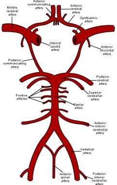
Cerebral arteries refer to the blood vessels that supply oxygenated blood to the brain. These arteries branch off from the internal carotid arteries and the vertebral arteries, which combine to form the basilar artery. The major cerebral arteries include:
1. Anterior cerebral artery (ACA): This artery supplies blood to the frontal lobes of the brain, including the motor and sensory cortices responsible for movement and sensation in the lower limbs.
2. Middle cerebral artery (MCA): The MCA is the largest of the cerebral arteries and supplies blood to the lateral surface of the brain, including the temporal, parietal, and frontal lobes. It is responsible for providing blood to areas involved in motor function, sensory perception, speech, memory, and vision.
3. Posterior cerebral artery (PCA): The PCA supplies blood to the occipital lobe, which is responsible for visual processing, as well as parts of the temporal and parietal lobes.
4. Anterior communicating artery (ACoA) and posterior communicating arteries (PComAs): These are small arteries that connect the major cerebral arteries, forming an important circulatory network called the Circle of Willis. The ACoA connects the two ACAs, while the PComAs connect the ICA with the PCA and the basilar artery.
These cerebral arteries play a crucial role in maintaining proper brain function by delivering oxygenated blood to various regions of the brain. Any damage or obstruction to these arteries can lead to serious neurological conditions, such as strokes or transient ischemic attacks (TIAs).
Magnetic Resonance Angiography (MRA) is a non-invasive medical imaging technique that uses magnetic fields and radio waves to create detailed images of the blood vessels or arteries within the body. It is a type of Magnetic Resonance Imaging (MRI) that focuses specifically on the circulatory system.
MRA can be used to diagnose and evaluate various conditions related to the blood vessels, such as aneurysms, stenosis (narrowing of the vessel), or the presence of plaques or tumors. It can also be used to plan for surgeries or other treatments related to the vascular system. The procedure does not use radiation and is generally considered safe, although people with certain implants like pacemakers may not be able to have an MRA due to safety concerns.
The meninges are the protective membranes that cover the brain and spinal cord. They consist of three layers: the dura mater (the outermost, toughest layer), the arachnoid mater (middle layer), and the pia mater (the innermost, delicate layer). These membranes provide protection and support to the central nervous system, and contain blood vessels that supply nutrients and remove waste products. Inflammation or infection of the meninges is called meningitis, which can be a serious medical condition requiring prompt treatment.
The internal carotid artery is a major blood vessel that supplies oxygenated blood to the brain. It originates from the common carotid artery and passes through the neck, entering the skull via the carotid canal in the temporal bone. Once inside the skull, it branches into several smaller vessels that supply different parts of the brain with blood.
The internal carotid artery is divided into several segments: cervical, petrous, cavernous, clinoid, and supraclinoid. Each segment has distinct clinical significance in terms of potential injury or disease. The most common conditions affecting the internal carotid artery include atherosclerosis, which can lead to stroke or transient ischemic attack (TIA), and dissection, which can cause severe headache, neck pain, and neurological symptoms.
It's important to note that any blockage or damage to the internal carotid artery can have serious consequences, as it can significantly reduce blood flow to the brain and lead to permanent neurological damage or even death. Therefore, regular check-ups and screening tests are recommended for individuals at high risk of developing vascular diseases.
A cerebral hemorrhage, also known as an intracranial hemorrhage or intracerebral hemorrhage, is a type of stroke that results from bleeding within the brain tissue. It occurs when a weakened blood vessel bursts and causes localized bleeding in the brain. This bleeding can increase pressure in the skull, damage nearby brain cells, and release toxic substances that further harm brain tissues.
Cerebral hemorrhages are often caused by chronic conditions like hypertension (high blood pressure) or cerebral amyloid angiopathy, which weakens the walls of blood vessels over time. Other potential causes include trauma, aneurysms, arteriovenous malformations, illicit drug use, and brain tumors. Symptoms may include sudden headache, weakness, numbness, difficulty speaking or understanding speech, vision problems, loss of balance, and altered level of consciousness. Immediate medical attention is required to diagnose and manage cerebral hemorrhage through imaging techniques, supportive care, and possible surgical interventions.
Cerebrovascular circulation refers to the network of blood vessels that supply oxygenated blood and nutrients to the brain tissue, and remove waste products. It includes the internal carotid arteries, vertebral arteries, circle of Willis, and the intracranial arteries that branch off from them.
The internal carotid arteries and vertebral arteries merge to form the circle of Willis, a polygonal network of vessels located at the base of the brain. The anterior cerebral artery, middle cerebral artery, posterior cerebral artery, and communicating arteries are the major vessels that branch off from the circle of Willis and supply blood to different regions of the brain.
Interruptions or abnormalities in the cerebrovascular circulation can lead to various neurological conditions such as stroke, transient ischemic attack (TIA), and vascular dementia.
Emission-Computed Tomography, Single-Photon (SPECT) is a type of nuclear medicine imaging procedure that generates detailed, three-dimensional images of the distribution of radioactive pharmaceuticals within the body. It uses gamma rays emitted by a radiopharmaceutical that is introduced into the patient's body, and a specialized gamma camera to detect these gamma rays and create tomographic images. The data obtained from the SPECT imaging can be used to diagnose various medical conditions, evaluate organ function, and guide treatment decisions. It is commonly used to image the heart, brain, and bones, among other organs and systems.
The Middle Cerebral Artery (MCA) is one of the main blood vessels that supplies oxygenated blood to the brain. It arises from the internal carotid artery and divides into several branches, which supply the lateral surface of the cerebral hemisphere, including the frontal, parietal, and temporal lobes.
The MCA is responsible for providing blood flow to critical areas of the brain, such as the primary motor and sensory cortices, Broca's area (associated with speech production), Wernicke's area (associated with language comprehension), and the visual association cortex.
Damage to the MCA or its branches can result in a variety of neurological deficits, depending on the specific location and extent of the injury. These may include weakness or paralysis on one side of the body, sensory loss, language impairment, and visual field cuts.
The Circle of Willis is a circulatory arrangement in the brain where the major arteries that supply blood to the brain converge to form an almost circular structure. It is named after Thomas Willis, an English physician who first described it in 1664.
This circle is formed by the joining of the two internal carotid arteries, which divide into the anterior cerebral and middle cerebral arteries, with the basilar artery, which arises from the vertebral arteries. These vessels anastomose, or connect, to form a polygon-like structure at the base of the brain.
The Circle of Willis plays a crucial role in maintaining adequate blood flow to the brain, as it allows for collateral circulation. If one of the arteries that make up the circle becomes blocked or narrowed, blood can still reach the affected area through the other vessels in the circle. This helps to minimize the risk of stroke and other neurological disorders.
The Posterior Cerebral Artery (PCA) is one of the major arteries that supplies blood to the brain. It is a branch of the basilar artery, which is formed by the union of the two vertebral arteries. The PCA supplies oxygenated blood to the occipital lobe (responsible for visual processing), the temporal lobe (involved in auditory and memory functions), and the thalamus and midbrain (relay station for sensory and motor signals).
The PCA has two segments: the precommunicating segment (P1) and the postcommunicating segment (P2). The P1 segment runs posteriorly along the cerebral peduncle, while the P2 segment courses around the midbrain to reach the occipital lobe.
Atherosclerosis, embolism, or other vascular conditions can affect the PCA and lead to a variety of neurological symptoms, including visual loss, memory impairment, and difficulty with language processing.
Photophobia is a condition characterized by an abnormal sensitivity to light. It's not a fear of light, despite the name suggesting otherwise. Instead, it refers to the discomfort or pain felt in the eyes due to exposure to light, often leading to a strong desire to avoid light. This can include both natural and artificial light sources.
The severity of photophobia can vary greatly among individuals. Some people may only experience mild discomfort in bright light conditions, while others may find even moderate levels of light intolerable. It can be a symptom of various underlying health issues, including eye diseases or disorders like uveitis, keratitis, corneal abrasions, or optic neuritis, as well as systemic conditions such as migraines, meningitis, or certain medications that increase light sensitivity.
Arterial occlusive diseases are medical conditions characterized by the blockage or narrowing of the arteries, which can lead to a reduction in blood flow to various parts of the body. This reduction in blood flow can cause tissue damage and may result in serious complications such as tissue death (gangrene), organ dysfunction, or even death.
The most common cause of arterial occlusive diseases is atherosclerosis, which is the buildup of plaque made up of fat, cholesterol, calcium, and other substances in the inner lining of the artery walls. Over time, this plaque can harden and narrow the arteries, restricting blood flow. Other causes of arterial occlusive diseases include blood clots, emboli (tiny particles that travel through the bloodstream and lodge in smaller vessels), inflammation, trauma, and certain inherited conditions.
Symptoms of arterial occlusive diseases depend on the location and severity of the blockage. Common symptoms include:
* Pain, cramping, or fatigue in the affected limb, often triggered by exercise and relieved by rest (claudication)
* Numbness, tingling, or weakness in the affected limb
* Coldness or discoloration of the skin in the affected area
* Slow-healing sores or wounds on the toes, feet, or legs
* Erectile dysfunction in men
Treatment for arterial occlusive diseases may include lifestyle changes such as quitting smoking, exercising regularly, and eating a healthy diet. Medications to lower cholesterol, control blood pressure, prevent blood clots, or manage pain may also be prescribed. In severe cases, surgical procedures such as angioplasty, stenting, or bypass surgery may be necessary to restore blood flow.
Collateral circulation refers to the alternate blood supply routes that bypass an obstructed or narrowed vessel and reconnect with the main vascular system. These collateral vessels can develop over time as a result of the body's natural adaptation to chronic ischemia (reduced blood flow) caused by various conditions such as atherosclerosis, thromboembolism, or vasculitis.
The development of collateral circulation helps maintain adequate blood flow and oxygenation to affected tissues, minimizing the risk of tissue damage and necrosis. In some cases, well-developed collateral circulations can help compensate for significant blockages in major vessels, reducing symptoms and potentially preventing the need for invasive interventions like revascularization procedures. However, the extent and effectiveness of collateral circulation vary from person to person and depend on factors such as age, overall health status, and the presence of comorbidities.
Surgical anastomosis is a medical procedure that involves the connection of two tubular structures, such as blood vessels or intestines, to create a continuous passage. This technique is commonly used in various types of surgeries, including vascular, gastrointestinal, and orthopedic procedures.
During a surgical anastomosis, the ends of the two tubular structures are carefully prepared by removing any damaged or diseased tissue. The ends are then aligned and joined together using sutures, staples, or other devices. The connection must be secure and leak-free to ensure proper function and healing.
The success of a surgical anastomosis depends on several factors, including the patient's overall health, the location and condition of the structures being joined, and the skill and experience of the surgeon. Complications such as infection, bleeding, or leakage can occur, which may require additional medical intervention or surgery.
Proper postoperative care is also essential to ensure the success of a surgical anastomosis. This may include monitoring for signs of complications, administering medications to prevent infection and promote healing, and providing adequate nutrition and hydration.
A Transient Ischemic Attack (TIA), also known as a "mini-stroke," is a temporary period of symptoms similar to those you'd get if you were having a stroke. A TIA doesn't cause permanent damage and is often caused by a temporary decrease in blood supply to part of your brain, which may last as little as five minutes.
Like an ischemic stroke, a TIA occurs when a clot or debris blocks blood flow to part of your nervous system. However, unlike a stroke, a TIA doesn't leave lasting damage because the blockage is temporary.
Symptoms of a TIA can include sudden onset of weakness, numbness or paralysis in your face, arm or leg, typically on one side of your body. You could also experience slurred or garbled speech, or difficulty understanding others. Other symptoms can include blindness in one or both eyes, dizziness, or a severe headache with no known cause.
Even though TIAs usually last only a few minutes, they are a serious condition and should not be ignored. If you suspect you or someone else is experiencing a TIA, seek immediate medical attention. TIAs can be a warning sign that a full-blown stroke is imminent.
Cerebral infarction, also known as a "stroke" or "brain attack," is the sudden death of brain cells caused by the interruption of their blood supply. It is most commonly caused by a blockage in one of the blood vessels supplying the brain (an ischemic stroke), but can also result from a hemorrhage in or around the brain (a hemorrhagic stroke).
Ischemic strokes occur when a blood clot or other particle blocks a cerebral artery, cutting off blood flow to a part of the brain. The lack of oxygen and nutrients causes nearby brain cells to die. Hemorrhagic strokes occur when a weakened blood vessel ruptures, causing bleeding within or around the brain. This bleeding can put pressure on surrounding brain tissues, leading to cell death.
Symptoms of cerebral infarction depend on the location and extent of the affected brain tissue but may include sudden weakness or numbness in the face, arm, or leg; difficulty speaking or understanding speech; vision problems; loss of balance or coordination; and severe headache with no known cause. Immediate medical attention is crucial for proper diagnosis and treatment to minimize potential long-term damage or disability.
Intracranial hemorrhage (ICH) is a type of stroke caused by bleeding within the brain or its surrounding tissues. It's a serious medical emergency that requires immediate attention and treatment. The bleeding can occur in various locations:
1. Epidural hematoma: Bleeding between the dura mater (the outermost protective covering of the brain) and the skull. This is often caused by trauma, such as a head injury.
2. Subdural hematoma: Bleeding between the dura mater and the brain's surface, which can also be caused by trauma.
3. Subarachnoid hemorrhage: Bleeding in the subarachnoid space, which is filled with cerebrospinal fluid (CSF) and surrounds the brain. This type of ICH is commonly caused by the rupture of an intracranial aneurysm or arteriovenous malformation.
4. Intraparenchymal hemorrhage: Bleeding within the brain tissue itself, which can be caused by hypertension (high blood pressure), amyloid angiopathy, or trauma.
5. Intraventricular hemorrhage: Bleeding into the brain's ventricular system, which contains CSF and communicates with the subarachnoid space. This type of ICH is often seen in premature infants but can also be caused by head trauma or aneurysm rupture in adults.
Symptoms of intracranial hemorrhage may include sudden severe headache, vomiting, altered consciousness, confusion, seizures, weakness, numbness, or paralysis on one side of the body, vision changes, or difficulty speaking or understanding speech. Rapid diagnosis and treatment are crucial to prevent further brain damage and potential long-term disabilities or death.
Digital subtraction angiography (DSA) is a medical imaging technique used to visualize the blood vessels and blood flow within the body. It combines the use of X-ray technology with digital image processing to produce detailed images of the vascular system.
In DSA, a contrast agent is injected into the patient's bloodstream through a catheter, which is typically inserted into an artery in the leg and guided to the area of interest using fluoroscopy. As the contrast agent flows through the blood vessels, X-ray images are taken at multiple time points.
The digital subtraction process involves taking a baseline image without contrast and then subtracting it from subsequent images taken with contrast. This allows for the removal of background structures and noise, resulting in clearer images of the blood vessels. DSA can be used to diagnose and evaluate various vascular conditions, such as aneurysms, stenosis, and tumors, and can also guide interventional procedures such as angioplasty and stenting.
Neurosurgical procedures are operations that are performed on the brain, spinal cord, and peripheral nerves. These procedures are typically carried out by neurosurgeons, who are medical doctors with specialized training in the diagnosis and treatment of disorders of the nervous system. Neurosurgical procedures can be used to treat a wide range of conditions, including traumatic injuries, tumors, aneurysms, vascular malformations, infections, degenerative diseases, and congenital abnormalities.
Some common types of neurosurgical procedures include:
* Craniotomy: A procedure in which a bone flap is temporarily removed from the skull to gain access to the brain. This type of procedure may be performed to remove a tumor, repair a blood vessel, or relieve pressure on the brain.
* Spinal fusion: A procedure in which two or more vertebrae in the spine are fused together using bone grafts and metal hardware. This is often done to stabilize the spine and alleviate pain caused by degenerative conditions or spinal deformities.
* Microvascular decompression: A procedure in which a blood vessel that is causing pressure on a nerve is repositioned or removed. This type of procedure is often used to treat trigeminal neuralgia, a condition that causes severe facial pain.
* Deep brain stimulation: A procedure in which electrodes are implanted in specific areas of the brain and connected to a battery-operated device called a neurostimulator. The neurostimulator sends electrical impulses to the brain to help alleviate symptoms of movement disorders such as Parkinson's disease or dystonia.
* Stereotactic radiosurgery: A non-invasive procedure that uses focused beams of radiation to treat tumors, vascular malformations, and other abnormalities in the brain or spine. This type of procedure is often used for patients who are not good candidates for traditional surgery due to age, health status, or location of the lesion.
Neurosurgical procedures can be complex and require a high degree of skill and expertise. Patients considering neurosurgical treatment should consult with a qualified neurosurgeon to discuss their options and determine the best course of action for their individual situation.
The external carotid artery is a major blood vessel in the neck that supplies oxygenated blood to the structures of the head and neck, excluding the brain. It originates from the common carotid artery at the level of the upper border of the thyroid cartilage, then divides into several branches that supply various regions of the head and neck, including the face, scalp, ears, and neck muscles.
The external carotid artery has eight branches:
1. Superior thyroid artery: Supplies blood to the thyroid gland, larynx, and surrounding muscles.
2. Ascending pharyngeal artery: Supplies blood to the pharynx, palate, and meninges of the brain.
3. Lingual artery: Supplies blood to the tongue and floor of the mouth.
4. Facial artery: Supplies blood to the face, nose, lips, and palate.
5. Occipital artery: Supplies blood to the scalp and muscles of the neck.
6. Posterior auricular artery: Supplies blood to the ear and surrounding muscles.
7. Maxillary artery: Supplies blood to the lower face, nasal cavity, palate, and meninges of the brain.
8. Superficial temporal artery: Supplies blood to the scalp, face, and temporomandibular joint.
The external carotid artery is an essential structure for maintaining adequate blood flow to the head and neck, and any damage or blockage can lead to serious medical conditions such as stroke or tissue necrosis.
Cardiac surgical procedures are operations that are performed on the heart or great vessels (the aorta and vena cava) by cardiothoracic surgeons. These surgeries are often complex and require a high level of skill and expertise. Some common reasons for cardiac surgical procedures include:
1. Coronary artery bypass grafting (CABG): This is a surgery to improve blood flow to the heart in patients with coronary artery disease. During the procedure, a healthy blood vessel from another part of the body is used to create a detour around the blocked or narrowed portion of the coronary artery.
2. Valve repair or replacement: The heart has four valves that control blood flow through and out of the heart. If one or more of these valves become damaged or diseased, they may need to be repaired or replaced. This can be done using artificial valves or valves from animal or human donors.
3. Aneurysm repair: An aneurysm is a weakened area in the wall of an artery that can bulge out and potentially rupture. If an aneurysm occurs in the aorta, it may require surgical repair to prevent rupture.
4. Heart transplantation: In some cases, heart failure may be so severe that a heart transplant is necessary. This involves removing the diseased heart and replacing it with a healthy donor heart.
5. Arrhythmia surgery: Certain types of abnormal heart rhythms (arrhythmias) may require surgical treatment. One such procedure is called the Maze procedure, which involves creating a pattern of scar tissue in the heart to disrupt the abnormal electrical signals that cause the arrhythmia.
6. Congenital heart defect repair: Some people are born with structural problems in their hearts that require surgical correction. These may include holes between the chambers of the heart or abnormal blood vessels.
Cardiac surgical procedures carry risks, including bleeding, infection, stroke, and death. However, for many patients, these surgeries can significantly improve their quality of life and longevity.
I'm sorry for any confusion, but "Internet" is a term that pertains to the global network of interconnected computers and servers that enable the transmission and reception of data via the internet protocol (IP). It is not a medical term and does not have a specific medical definition. If you have any questions related to medicine or health, I'd be happy to try to help answer them for you!
Cardiopulmonary bypass (CPB) is a medical procedure that temporarily takes over the functions of the heart and lungs during major heart surgery. It allows the surgeon to operate on a still, bloodless heart.
During CPB, the patient's blood is circulated outside the body with the help of a heart-lung machine. The machine pumps the blood through a oxygenator, where it is oxygenated and then returned to the body. This bypasses the heart and lungs, hence the name "cardiopulmonary bypass."
CPB involves several components, including a pump, oxygenator, heat exchanger, and tubing. The patient's blood is drained from the heart through cannulas (tubes) and passed through the oxygenator, where it is oxygenated and carbon dioxide is removed. The oxygenated blood is then warmed to body temperature in a heat exchanger before being pumped back into the body.
While on CPB, the patient's heart is stopped with the help of cardioplegia solution, which is infused directly into the coronary arteries. This helps to protect the heart muscle during surgery. The surgeon can then operate on a still and bloodless heart, allowing for more precise surgical repair.
After the surgery is complete, the patient is gradually weaned off CPB, and the heart is restarted with the help of electrical stimulation or medication. The patient's condition is closely monitored during this time to ensure that their heart and lungs are functioning properly.
While CPB has revolutionized heart surgery and allowed for more complex procedures to be performed, it is not without risks. These include bleeding, infection, stroke, kidney damage, and inflammation. However, with advances in technology and technique, the risks associated with CPB have been significantly reduced over time.
A "periodical" in the context of medicine typically refers to a type of publication that is issued regularly, such as on a monthly or quarterly basis. These publications include peer-reviewed journals, magazines, and newsletters that focus on medical research, education, and practice. They may contain original research articles, review articles, case reports, editorials, letters to the editor, and other types of content related to medical science and clinical practice.
As a "Topic," periodicals in medicine encompass various aspects such as their role in disseminating new knowledge, their impact on clinical decision-making, their quality control measures, and their ethical considerations. Medical periodicals serve as a crucial resource for healthcare professionals, researchers, students, and other stakeholders to stay updated on the latest developments in their field and to share their findings with others.
I'm sorry for any confusion, but "India" is not a medical term or concept. It is a country in South Asia, the second-most populous country in the world, known for its rich history, diverse culture, and numerous contributions to various fields including medicine. If you have any questions related to medical topics, I would be happy to help answer them!
Postoperative complications refer to any unfavorable condition or event that occurs during the recovery period after a surgical procedure. These complications can vary in severity and may include, but are not limited to:
1. Infection: This can occur at the site of the incision or inside the body, such as pneumonia or urinary tract infection.
2. Bleeding: Excessive bleeding (hemorrhage) can lead to a drop in blood pressure and may require further surgical intervention.
3. Blood clots: These can form in the deep veins of the legs (deep vein thrombosis) and can potentially travel to the lungs (pulmonary embolism).
4. Wound dehiscence: This is when the surgical wound opens up, which can lead to infection and further complications.
5. Pulmonary issues: These include atelectasis (collapsed lung), pneumonia, or respiratory failure.
6. Cardiovascular problems: These include abnormal heart rhythms (arrhythmias), heart attack, or stroke.
7. Renal failure: This can occur due to various reasons such as dehydration, blood loss, or the use of certain medications.
8. Pain management issues: Inadequate pain control can lead to increased stress, anxiety, and decreased mobility.
9. Nausea and vomiting: These can be caused by anesthesia, opioid pain medication, or other factors.
10. Delirium: This is a state of confusion and disorientation that can occur in the elderly or those with certain medical conditions.
Prompt identification and management of these complications are crucial to ensure the best possible outcome for the patient.
Congenital heart defects (CHDs) are structural abnormalities in the heart that are present at birth. They can affect any part of the heart's structure, including the walls of the heart, the valves inside the heart, and the major blood vessels that lead to and from the heart.
Congenital heart defects can range from mild to severe and can cause various symptoms depending on the type and severity of the defect. Some common symptoms of CHDs include cyanosis (a bluish tint to the skin, lips, and fingernails), shortness of breath, fatigue, poor feeding, and slow growth in infants and children.
There are many different types of congenital heart defects, including:
1. Septal defects: These are holes in the walls that separate the four chambers of the heart. The two most common septal defects are atrial septal defect (ASD) and ventricular septal defect (VSD).
2. Valve abnormalities: These include narrowed or leaky valves, which can affect blood flow through the heart.
3. Obstruction defects: These occur when blood flow is blocked or restricted due to narrowing or absence of a part of the heart's structure. Examples include pulmonary stenosis and coarctation of the aorta.
4. Cyanotic heart defects: These cause a lack of oxygen in the blood, leading to cyanosis. Examples include tetralogy of Fallot and transposition of the great arteries.
The causes of congenital heart defects are not fully understood, but genetic factors and environmental influences during pregnancy may play a role. Some CHDs can be detected before birth through prenatal testing, while others may not be diagnosed until after birth or later in childhood. Treatment for CHDs may include medication, surgery, or other interventions to improve blood flow and oxygenation of the body's tissues.
Brain ischemia is the medical term used to describe a reduction or interruption of blood flow to the brain, leading to a lack of oxygen and glucose delivery to brain tissue. This can result in brain damage or death of brain cells, known as infarction. Brain ischemia can be caused by various conditions such as thrombosis (blood clot formation), embolism (obstruction of a blood vessel by a foreign material), or hypoperfusion (reduced blood flow). The severity and duration of the ischemia determine the extent of brain damage. Symptoms can range from mild, such as transient ischemic attacks (TIAs or "mini-strokes"), to severe, including paralysis, speech difficulties, loss of consciousness, and even death. Immediate medical attention is required for proper diagnosis and treatment to prevent further damage and potential long-term complications.
Ischemia is the medical term used to describe a lack of blood flow to a part of the body, often due to blocked or narrowed blood vessels. This can lead to a shortage of oxygen and nutrients in the tissues, which can cause them to become damaged or die. Ischemia can affect many different parts of the body, including the heart, brain, legs, and intestines. Symptoms of ischemia depend on the location and severity of the blockage, but they may include pain, cramping, numbness, weakness, or coldness in the affected area. In severe cases, ischemia can lead to tissue death (gangrene) or organ failure. Treatment for ischemia typically involves addressing the underlying cause of the blocked blood flow, such as through medication, surgery, or lifestyle changes.
An encyclopedia is a comprehensive reference work containing articles on various topics, usually arranged in alphabetical order. In the context of medicine, a medical encyclopedia is a collection of articles that provide information about a wide range of medical topics, including diseases and conditions, treatments, tests, procedures, and anatomy and physiology. Medical encyclopedias may be published in print or electronic formats and are often used as a starting point for researching medical topics. They can provide reliable and accurate information on medical subjects, making them useful resources for healthcare professionals, students, and patients alike. Some well-known examples of medical encyclopedias include the Merck Manual and the Stedman's Medical Dictionary.
Sickle cell anemia is a genetic disorder that affects the hemoglobin in red blood cells. Hemoglobin is responsible for carrying oxygen throughout the body. In sickle cell anemia, the hemoglobin is abnormal and causes the red blood cells to take on a sickle shape, rather than the normal disc shape. These sickled cells are stiff and sticky, and they can block blood vessels, causing tissue damage and pain. They also die more quickly than normal red blood cells, leading to anemia.
People with sickle cell anemia often experience fatigue, chronic pain, and jaundice. They may also have a higher risk of infections and complications such as stroke, acute chest syndrome, and priapism. The disease is inherited from both parents, who must both be carriers of the sickle cell gene. It primarily affects people of African descent, but it can also affect people from other ethnic backgrounds.
There is no cure for sickle cell anemia, but treatments such as blood transfusions, medications to manage pain and prevent complications, and bone marrow transplantation can help improve quality of life for affected individuals. Regular medical care and monitoring are essential for managing the disease effectively.
Unconsciousness is a state of complete awareness where a person is not responsive to stimuli and cannot be awakened. It is often caused by severe trauma, illness, or lack of oxygen supply to the brain. In medical terms, it is defined as a lack of response to verbal commands, pain, or other stimuli, indicating that the person's brain is not functioning at a level necessary to maintain wakefulness and awareness.
Unconsciousness can be described as having different levels, ranging from drowsiness to deep coma. The causes of unconsciousness can vary widely, including head injury, seizure, stroke, infection, drug overdose, or lack of oxygen supply to the brain. Depending on the cause and severity, unconsciousness may last for a few seconds or continue for an extended period, requiring medical intervention and treatment.
Myocardial ischemia is a condition in which the blood supply to the heart muscle (myocardium) is reduced or blocked, leading to insufficient oxygen delivery and potential damage to the heart tissue. This reduction in blood flow typically results from the buildup of fatty deposits, called plaques, in the coronary arteries that supply the heart with oxygen-rich blood. The plaques can rupture or become unstable, causing the formation of blood clots that obstruct the artery and limit blood flow.
Myocardial ischemia may manifest as chest pain (angina pectoris), shortness of breath, fatigue, or irregular heartbeats (arrhythmias). In severe cases, it can lead to myocardial infarction (heart attack) if the oxygen supply is significantly reduced or cut off completely, causing permanent damage or death of the heart muscle. Early diagnosis and treatment of myocardial ischemia are crucial for preventing further complications and improving patient outcomes.
Otorhinolaryngologic diseases, also known as ear, nose, and throat (ENT) diseases, refer to a group of medical conditions that affect the ears, nose, and/or throat. These specialized areas are closely related both anatomically and functionally, and disorders in one area can often have impacts on the others.
Here are some examples of otorhinolaryngologic diseases categorized by the affected area:
1. Otologic diseases - affecting the ear:
* Otitis media (ear infection)
* Otitis externa (swimmer's ear)
* Tinnitus (ringing in the ears)
* Hearing loss
* Meniere's disease (inner ear disorder causing vertigo, tinnitus, and hearing loss)
* Acoustic neuroma (noncancerous tumor on the vestibular nerve)
2. Rhinologic diseases - affecting the nose:
* Allergic rhinitis (hay fever)
* Non-allergic rhinitis
* Sinusitis (sinus infection)
* Deviated septum
* Nasal polyps
* Epistaxis (nosebleed)
3. Laryngologic diseases - affecting the throat and voice box:
* Laryngitis (inflammation of the larynx, causing hoarseness or voice loss)
* Vocal cord nodules or polyps
* Reflux laryngitis (acid reflux irritating the throat)
* Subglottic stenosis (narrowing of the airway below the vocal cords)
* Laryngeal cancer
4. Common otorhinolaryngologic diseases:
* Tonsillitis (inflammation of the tonsils, often causing sore throat and difficulty swallowing)
* Adenoiditis (inflammation of the adenoids, commonly seen in children)
* Obstructive sleep apnea (OSA, a disorder characterized by pauses in breathing during sleep)
* Pharyngitis (inflammation of the pharynx or throat)
Otorhinolaryngologists, also known as ENT specialists, diagnose and treat these conditions. They may use various methods such as physical examination, imaging studies, endoscopy, and laboratory tests to determine the best course of treatment for each individual patient.
 Moyamoya disease - Wikipedia
 Moyamoya Disease: Background, Etiology, Epidemiology
 Moyamoya disease - Diagnosis and treatment - Mayo Clinic
 Medical Management in Moyamoya Disease | IntechOpen
 Cerebral hyperperfusion syndrome after revascularization surgery in patients with moyamoya disease
Effects of surgical revascularization on peripheral artery aneurysms in moyamoya disease: report of three cases
 Moya Moya Disease
Executive dysfunction in adults with moyamoya disease is associated with increased diffusion in frontal white matter | Journal...
Blog - tagged "Moyamoya disease" - vt-fiddle
 Flow-Mediated Dilatation of the Brachial Artery for Assessing Endothelial Dysfunction in Children with Moyamoya Disease. -...
Reply to: Moyamoya disease and systemic sclerosis (MoSys syndrome): a combination of two rare entities: comment to the authors
 New Jersey Moyamoya Disease Treatment | RWJBarnabas Health
 Delayed posterior circulation insufficiency in pediatric moyamoya disease. | Read by QxMD
 Moya Moya Disease
 Moyamoya disease - Everything2.com
Moyamoya Disease: Background, Etiology, Epidemiology
 Moyamoya disease symptoms - Newstrendline.Com
 New Book: Moyamoya Disease Update - Neurosurgery
Five-Year Changes in Cognitive Function and Their Predictor in Adult Moyamoya Disease<...
 Moyamoya Disease and Syndrome | David Altschul, MD
Omental flap transplantation for Moyamoya disease - Neurosurgery
 Moyamoya syndrome in hemoglobin E-beta thalassemia: A rare presentation and association Doctor P N, Choudhari A, Verma M,...
Medicine and Technology: Reading about Moyamoya Disease in the NEJM
Commentary: Clinical course of unilateral moyamoya disease<...
Plus it
 Moyamoya disease: A clinical spectrum, literature review and case series from a tertiary care hospital in Pakistan | BMC...
Brain ischemia - Wikipedia
Neurologic Disease and Pregnancy: Overview, General Considerations, New-Onset Neurologic Complications
Disease Variant Landscape of a Large Multiethnic Population of Moyamoya Patients by Exome Sequencing - McMaster Experts
Puff of smoke6
- Moyamoya means "puff of smoke" in Japanese and is used to describe the tangled appearance of tiny vessels compensating for the blockage. (nih.gov)
- The name "moyamoya" means "puff of smoke" in Japanese and describes the look of the tangled vessels that form to compensate for the blockage. (nih.gov)
- On conventional angiography, these collateral vessels have the appearance of a "puff of smoke" (described as "もやもや (moyamoya)" in Japanese). (wikipedia.org)
- These networks, visualized by a particular test called an angiogram, resemble puffs of smoke, which is how the condition got its name: "moyamoya" is an expression meaning "something hazy like a puff of smoke" in Japanese. (medlineplus.gov)
- Cerebral angiogram shows the puff-of-smoke (moyamoya) collaterals at the base of the brain. (findexpertmd.com)
- The term moyamoya is Japanese for "puff of smoke. (prismahealth.org)
Syndrome17
- Moyamoya syndrome is a related term that refers to cases of Moyamoya disease that occur in association with other conditions or risk factors, such as neurofibromatosis, tuberculosis meningitis, sickle cell disease, leptospirosis, brain tumors, Sturge-Weber syndrome, and tuberous sclerosis. (nih.gov)
- Moyamoya syndrome is unilateral arterial constriction, or occurs when one of the several specified conditions is also present. (wikipedia.org)
- Patients with Down syndrome, sickle cell anemia, neurofibromatosis type 1, congenital heart disease, fibromuscular dysplasia, activated protein C resistance, or head trauma can develop moyamoya malformations. (wikipedia.org)
- These individuals are said to have moyamoya syndrome. (medlineplus.gov)
- Long-term outcome in children with moyamoya syndrome after cranial revascularization by pial synangiosis. (medscape.com)
- Moyamoya disease and syndrome. (medscape.com)
- BackgroundMoyamoya is a rare progressive cerebral arteriopathy, occurring as an isolated phenomenon (moyamoya disease, MMD) or associated with other conditions (moyamoya syndrome, MMS), responsible for 6-10% of all childhood strokes and transient ischemic attacks (TIAs). (unibo.it)
- Having a family history of the disease increases your risk, as well as having other medical conditions such as sickle cell anemia and Down syndrome. (prismahealth.org)
- Moyamoya on one side of the brain only is called moyamoya syndrome. (prismahealth.org)
- Serum apolipoprotein E ( APOE ) levels were compared in 33 MMD patients and 28 Moyamoya syndrome (MMS) patients to investigate the potential of APOE to be as an MMD biomarker . (bvsalud.org)
- Wegner, F , Müller-Ladner, U & Meier, FMP 2017, ' Reply to: Moyamoya disease and systemic sclerosis (MoSys syndrome): A combination of two rare entities - comment to the authors ', Clinical and Experimental Rheumatology , Jg. (uni-luebeck.de)
- Most attention in Down syndrome (trisomy 21)is directed toward imaging to detect gastrointestinal anomalies in the early postnatal period and toward imaging congenital heart disease, which may be present at birth and may remain throughout the patient's lifetime. (medscape.com)
- Approximately 50% of infants with Down syndrome have congenital heart disease, most commonly atrioventricular septal defect and ventricular septal defect. (medscape.com)
- Because Down syndrome is associated with a high risk of congenital heart disease, there is an increased risk of cardioembolic events, such as moyamoya disease, which has been found to occur in patients with Down syndrome at a rate 3 times that of the general population. (medscape.com)
- Adults with Down syndrome are at increased risk for Alzheimer disease, especially by their 5th decade. (medscape.com)
- 15. Management of glioblastoma in an NF1 patient with moyamoya syndrome: a case report. (nih.gov)
- A very rare hereditary neurological dysmorphic syndrome with characteristics of moyamoya disease, short stature of postnatal onset and stereotypical facial dysmorphism. (cdc.gov)
Cerebrovascular diseases13
- The relationship of SUA with cerebrovascular diseases is controversial [ 9 - 11 ]. (hindawi.com)
- In addition, data from several studies suggest that triglycerides (TGs) are a risk factor for cerebrovascular diseases, including carotid stenosis and intracranial artery stenosis [ 14 , 15 ]. (hindawi.com)
- Overall, SUA and TGs are extremely important in the pathological and physiological processes of cerebrovascular diseases. (hindawi.com)
- Cerebrovascular Diseases , 28 (3), 247-257. (tmu.edu.tw)
- The interventional neuroradiology service at Rhode Island Hospital offers treatment for a broad range of cerebrovascular diseases, including aneurysms, vascular malformations and ischemic stroke, using minimally invasive techniques combined with high-end imaging technology. (lifespan.org)
- People don't often think of children having strokes, but some cerebrovascular diseases can cause a stroke, even in children. (childrens.com)
- At Children's Health℠, we know how cerebrovascular diseases in children differ from those in adults. (childrens.com)
- We are uniquely equipped with top-notch technology, expert providers and key partnerships to treat cerebrovascular diseases - and help prevent stroke in children. (childrens.com)
- Cerebrovascular diseases affect the blood vessels and blood flow in the brain. (childrens.com)
- Although cerebrovascular diseases are rare in children, they increase the risk of brain damage and may lead to a stroke. (childrens.com)
- Our specialists have expertise in diagnosing cerebrovascular diseases in their early stages and providing advanced treatment before they become life-threatening. (childrens.com)
- What are the signs and symptoms of cerebrovascular diseases and pediatric stroke? (childrens.com)
- Cerebrovascular Diseases , 36 (1), 19-25. (elsevierpure.com)
Ischemic11
- In children, the first symptom of moyamoya disease is often stroke or recurrent transient ischemic attacks (TIAs), also known as "mini-strokes," that are frequently accompanied by muscular weakness or paralysis affecting one side of the body. (nih.gov)
- Moyamoya disease (MMD) is a type of chronic cerebrovascular occlusion disease that frequently occurs in East Asian populations, including pediatric and adult patients, and may lead to ischemic or hemorrhagic stroke, headaches, epilepsy, or transient ischemic attack [ 1 ]. (hindawi.com)
- Moyamoya disease is characterized by progressive stenosis or occlusion of the intracranial portion of the internal carotid artery and their proximal branches, resulting in ischemic or hemorrhagic stroke with high rate of disability and even death. (nih.gov)
- The superficial temporal artery is a useful donor artery for direct bypass surgery for moyamoya disease and can provide sufficient blood supply to the ischemic cerebral territory 1) . (neurosurgery.directory)
- Collateral artery formation from the extracranial carotid artery to ischemic brain tissue determines the clinical success of superficial temporal artery (STA) to middle cerebral artery (MCA) bypass surgery in adult patients with moyamoya disease , but postoperative collateral formation (PCF) after STA-MCA bypass surgery is unpredictable. (neurosurgery.directory)
- Data for consecutive patients who underwent STA-MCA MVB from 2000-2019 due to moyamoya/moyamoya-like disease, complex intracranial aneurysm s, or intractable brain ischemia due to internal carotid artery or middle cerebral artery occlusive disease with repeated ischemic events were retrospectively analyzed under a waiver of informed consent. (neurosurgery.directory)
- Methods: Twenty-three moyamoya disease patients with ischemic presentation who received revascularization with complete angiography and xenon CT during a minimum of 3 years' clinical follow-up were enrolled. (tmu.edu.tw)
- Ischemic stroke in children in course of moyamoya disease - case report. (medscimonit.com)
- Moyamoya disease - A rare disease of the carotid arteries in which they progressively narrow and potentially close, resulting in ischemic or hemorrhagic stroke. (stroke.org)
- There are increasing evidences for an association between migraine and vascular diseases and, in particular, between migraine and ischemic stroke, subclinical brain lesions, cardiac events, and vascular mortality. (j-stroke.org)
- Our team treats conditions such as cerebral aneurysm, arteriovenous malformations (AVMs), carotid stenosis, intracranial stenosis, acute ischemic stroke, spinal arteriovenous malformations, arteriovenous fistulae, moyamoya disease and cavernous malformation among other neurovascular diseases. (cooperhealth.org)
Internal caroti3
- The disease causes constrictions primarily in the internal carotid artery, and often extends to the middle and anterior cerebral arteries, branches of the internal carotid artery inside the skull. (wikipedia.org)
- A bilateral steno occlusive disease of the intra cranial internal carotid artery. (neuroradiologycases.com)
- If the symptoms are caused by moyamoya, the scan will reveal that there are tiny moyamoya blood vessels, and the internal carotid artery is not healthy. (prismahealth.org)
Stroke13
- Moyamoya disease is a rare condition caused by blocked arteries in the brain that lead to stroke-like symptoms. (prismahealth.org)
- More specifically, Dr. Sun is investigating novel monitoring and stroke prevention techniques in children with moyamoya disease, which is a rare disease that places affected children and young adults at high risk of stroke. (hopkinsmedicine.org)
- The goal of Dr. Sun's research is to improve outcomes and quality of life of individuals affected by stroke and moyamoya disease. (hopkinsmedicine.org)
- What are the types of Cerebrovascular Disease and Stroke? (childrens.com)
- How are Cerebrovascular Disease and Stroke diagnosed? (childrens.com)
- Symptoms may include headache, seizure, stroke and degenerative brain disease. (stroke.org)
- Neuroinflammation has a key role in the onset and/or progression of several CNS disorders, including acute brain injuries such as stroke and traumatic brain injury (TBI), and chronic neurodegenerative diseases such as Parkinson's disease (PD), Alzheimer's disease (AD), Huntington's disease (HD), Multiple sclerosis (MS), and Amyotrophic Lateral Sclerosis (ALS). (biomedcentral.com)
- She has a long term expertise in Cerebrovascular disease and particularly heritable and rare causes of stroke as CADASIL, Moyamoya Disease, Fabry disease and Cerebral Amyloid Angiopathy (CAA). (biomedcentral.com)
- Neuroinflammation has a key role in the onset and/or progression of several neurological disorders, including stroke, Parkinson's disease (PD), Alzheimer's disease (AD), Huntington's condition, Multi-sclerosis (MS), and Amyotrophic Lateral Sclerosis (ALS). (biomedcentral.com)
- Under the influence of exogenous and endogenous factors (trauma, stroke, chronic infections, disease related proteins like Aβ, Tau/p-Tau or α-syn,), the activation of microglia triggers several signal transduction pathways, including phosphoinositide 3-kinase/protein kinase B (PI3K/AKT), mitogen-activated protein kinase (MAPK) and mammalian target of rapamycin (mTOR), leading to NF-κB activation. (biomedcentral.com)
- Our neurointerventional team will be able to treat emergent and non-emergent forms of cerebrovascular disease, including acute stroke of any type, cerebral aneurysms, carotid artery disease, and other serious conditions affecting the brain and spinal cord, faster than ever before. (cooperhealth.org)
- Moyamoya is a rare neurological disease in which the blood vessels that supply blood to the brain narrow, greatly reducing blood flow and putting the child at risk for stroke. (creditunionskidsatheart.org)
- Studies show that a patient with Moyamoya has a 66-90% chance of having a stroke within 5 years of diagnosis. (creditunionskidsatheart.org)
Pediatric Moyamoya Disease1
- Long-term follow-up of pediatric moyamoya disease treated by combined direct-indirect revascularization surgery: single institute experience with surgical and perioperative management. (qxmd.com)
20231
- On May 6, 2023, help us raise awareness of Moyamoya disease and stop the strokes! (creditunionskidsatheart.org)
Cases of moyamoya3
- About 10% of cases of moyamoya disease are familial, and some cases result from specific genetic mutations. (wikipedia.org)
- The global moyamoya disease drugs market is estimated to grow on the back of increasing cases of moyamoya disease especially in females under 20, along with increasing awareness about various neurological disorders. (newshunt360.com)
- 16. [Three Cases of Moyamoya Disease with a History of Kawasaki Disease]. (nih.gov)
Vascular5
- In short, the authors report that moyamoya disease likely occurs due to a number of factors (e.g., differences in vascular anatomy) that ultimately contribute to broad cerebral blood vessel occlusion and consequent shifts in vessel connections to try to provide blood for the compromised brain. (wikipedia.org)
- 7 with complex aneurysms (22%) and 3 with vascular occlusive disease (9%) underwent unilateral bypass. (neurosurgery.directory)
- Moyamoya disease is a vascular disorder in which the carotid artery in the skull becomes narrow, reducing blood flow to your brain, causing symptoms, such as, seizures, visual disturbances, cognitive delays, and others. (newshunt360.com)
- State-of-the-art 3D neuroimaging provides a powerful means of accurately diagnosing and understanding an individual's vascular disease. (lifespan.org)
- history of any of the following: myocardial infarction, coronary artery bypass, coronary angioplasty or stenting, carotid endarterectomy or stenting, or peripheral vascular surgery for atherosclerotic disease. (who.int)
Bilateral3
- Moyamoya disease is a rare progressive cerebrovascular disease characterized by bilateral stenosis of vasculature of the Circle of Willis, specifically the distal internal carotid arteries, that leads to extensive collateral circulation. (jefferson.edu)
- CT angiography of the head and neck with contrast has demonstrated severe narrowing of the bilateral supraclinoid internal carotid arteries, which suggests moyamoya disease. (medscape.com)
- 2. [A case of adult moyamoya disease showing fulminant clinical course associated with progression from unilateral to bilateral involvement]. (nih.gov)
Collaterals1
- To try to make up for the lack of blood flow in the brain, small blood vessels called collaterals, or moyamoya vessels, form over time. (prismahealth.org)
Stenosis4
- The disease moyamoya, which is a Japanese mimetic word, gets its characteristic name due to the appearance of smoke on relevant angiographs resultant from the tangle of tiny vessels in response to stenosis. (wikipedia.org)
- Moyamoya is a Japanese word for a "puff" or "cloud of smoke" , and it has been used to refer to an extensive basal cerebral network of small anastomotic vessels at the base of the brain around and distal to the circle of Willis secondary to segmental stenosis or occlusion of the terminal parts of both internal carotid arteries. (neuroradiologycases.com)
- Carotid stenosis is the full or partial occlusion of the carotid artery by atherosclerotic disease. (lifespan.org)
- The best management for a particular patient's disease depends on the degree of the stenosis (occlusion) and the quality of the available medical and surgical options. (lifespan.org)
Vasculopathy2
- Cognitive functions in children and adults with Moyamoya vasculopathy: a systematic review and meta-Analysis. (medscape.com)
- Moyamoya disease is an uncommon progressive cerebral vasculopathy that is more frequently seen in the Asian population. (smj.org.sg)
RNF2137
- Over the last six decades since the disease was first described, pathogenesis of moyamoya disease remained elusive, although the gene ring finger protein 213 (RNF213) has been implicated. (wikipedia.org)
- Changes in the RNF213 gene involved in moyamoya disease replace single protein building blocks (amino acids) in the RNF213 protein. (medlineplus.gov)
- The effect of these changes on the function of the RNF213 protein is unknown, and researchers are unsure how the changes contribute to the narrowing of blood vessels or the characteristic blood vessel growth of moyamoya disease. (medlineplus.gov)
- A genome-wide association study identifies RNF213 as the first Moyamoya disease gene. (medscape.com)
- Ma J, Liu Y, Ma L, Huang S, Li H, You C. RNF213 polymorphism and Moyamoya disease: A systematic review and meta-analysis. (medscape.com)
- The R4810K variant of ring finger protein 213 ( RNF213 ) was identified as a strong genetic susceptibility factor for moyamoya disease (MMD) in the East Asian population [ 1 , 2 ]. (j-stroke.org)
- The RNF213 R4810K variant is associated with significantly early age of onset and the severity of clinical disease phenotypes [ 3 - 5 ]. (j-stroke.org)
Neurosurgery1
- from the Department of Neurosurgery, Fudan University, Shanghai , China performed a combined revascularization procedure in 111 patients with different types and stages of moyamoya disease . (neurosurgery.directory)
Abstract1
- abstract = "The origin of moyamoya disease remains unknown. (elsevierpure.com)
Arteries11
- Moyamoya disease is a rare, progressive cerebrovascular disorder caused by blocked arteries at the base of the brain in an area called the basal ganglia. (nih.gov)
- Without surgery, the majority of individuals with moyamoya disease will experience mental decline and multiple strokes because of the progressive narrowing of arteries. (nih.gov)
- Moyamoya disease is a rare, progressive, blood vessel disease caused by blocked arteries at the base of the brain in an area called the basal ganglia. (nih.gov)
- Moyamoya disease is a disease in which certain arteries in the brain are constricted. (wikipedia.org)
- Moyamoya disease is a disorder of blood vessels in the brain, specifically the internal carotid arteries and the arteries that branch from them. (medlineplus.gov)
- Moyamoya disease (MMD) is a rare, chronic vaso-occlusive disease affecting the arteries of the Circle of Willis, leading to the development of characteristic collateral vessels. (iasp-pain.org)
- Moyamoya disease is a genetic disease where the carotid arteries in the brain become narrow over time. (prismahealth.org)
- For example, if someone is brought to the emergency room after a fall and a scan of the head could reveals the carotid arteries have moyamoya. (prismahealth.org)
- Fibromuscular dysplasia, commonly called FMD, is a disease that causes one or more arteries in the body to have abnormal cell development in the artery wall. (rarediseases.org)
- Some patients with FMD may have no symptoms at all but are diagnosed with this disease when a physician hears a noise over one of the arteries due to disturbed or turbulent blood flow within the vessel. (rarediseases.org)
- Cerebral vasculitis related to neoplasms - Diseases of the arteries may rarely complicate systemic tumors. (stroke.org)
Revascularization5
- Conclusions: Progressive steno-occlusive change in the PCA after revascularization is associated with a reduction in LMC blood flow and cerebral ischemia in moyamoya patients. (tmu.edu.tw)
- Long-term outcomes of moyamoya disease (MMD) patients post revascularization are not well documented. (cns.org)
- Due to the rarity of moyamoya disease, the number of patients who had revascularization surgeries, and very little is known on the long-term outcome for these patients in the literature, patients always have many uncertainties and unanswered questions for their future. (cns.org)
- This long-term outcome data on patients with moyamoya disease post revascularization would help with the patients' long-term expectation of their wellbeing. (cns.org)
- By the conclusion of this session, participants should be able to: 1) Describe the long term physical, functional and social outcome of moyamoya patients post revascularization surgery. (cns.org)
Angiography1
- The authors report on a case of de novo development of moyamoya disease in a middle-aged female whose cerebral angiography demonstrated no abnormal findings 5 years previously. (elsevierpure.com)
Intracerebral hemorrhage1
- Without treatment, moyamoya disease can be fatal as the result of intracerebral hemorrhage (bleeding within the brain). (nih.gov)
Neurology1
- BMC Neurology and BMC Neuroscience are calling for submissions to our Collection on Neuroinflammation and Brain Disease. (biomedcentral.com)
Incidence5
- Moyamoya disease was first described in Japan and is found in individuals around the world, although its incidence is higher in Asian countries than in Europe or North America. (nih.gov)
- In the United States moyamoya has an incidence rate of 0.086 per 100,000. (wikipedia.org)
- Moyamoya disease was originally described in Japanese populations but is present in a variety of ethnicities.3,5,6 In Japan, the incidence per 100,000 patient years is between 0.35 to 0.943 with a male: female ratio of 1:1.87. (jefferson.edu)
- The incidence of Moyamoya disease (MMD) in Europe is not well known. (neurosurgery.directory)
- These families are not from Japan or Asia, whereas in general the incidence of moyamoya disease is highest in Japan and other Asian countries, in comparison with other parts of the world. (cdc.gov)
Epidemiology2
- Moyamoya Disease: Epidemiology, Clinical Features, and Diagnosis. (medscape.com)
- This review encapsulates current advances of moyamoya disease on the aspects of epidemiology, etiology, clinical features, imaging diagnosis and treatment. (nih.gov)
Short stature1
- 608796). See also MYMY4 (300845), an X-linked recessive syndromic disorder characterized by moyamoya disease, short stature, hypergonadotropic hypogonadism, and facial dysmorphism, and linked to q25.3, on chromosome 17. (wikipedia.org)
Arteriovenous1
- The authors report the unique case of a 6-year-old African-American girl with sickle cell disease (SCD) and an associated moyamoya arteriopathy who developed a de novo arteriovenous malformation (AVM) of the cerebral circulation. (elsevierpure.com)
Collateral1
- Objective: It has been noted that the posterior circulation serves as an important source of collateral blood supply in moyamoya disease. (tmu.edu.tw)
Gene3
Familial1
- Inheritance pattern of familial moyamoya disease: autosomal dominant mode and genomic imprinting. (medscape.com)
Unilateral2
Neurovascular disease1
- There were no surgical complications, no perioperative mortality, and no death from complications related to neurovascular disease at late follow-up. (neurosurgery.directory)
Steno occlusive disease1
- Ooi YC, Laiwalla AN, Liou R, Gonzalez NR . Angiographic structural differentiation between native arteriogenesis and therapeutic synangiosis in intracranial arterial steno-occlusive disease . (cedars-sinai.edu)
Adult3
- Clinical features, surgical treatment, and long-term outcome in adult patients with moyamoya disease. (medscape.com)
- Adult patients with moyamoya disease undergoing the STA-MCA bypass surgery between January 2013 and December 2020 at a single institution were retrospectively or prospectively enrolled in this observational study. (neurosurgery.directory)
- The nomogram exhibits high accuracy in predicting good PCF after the STA-MCA bypass surgery in adult patients with moyamoya disease and may allow surgeons to better evaluate preoperatively candidacy for successful bypass surgery 3) . (neurosurgery.directory)
20181
- A total of 261 Chinese patients with newly diagnosed moyamoya disease were recruited from a hospital in China from 24 March 2013 to 24 December 2018. (hindawi.com)
Sickle cell a1
- Sickle cell disease (also called sickle cell anemia) - A genetic disorder that causes abnormal hemoglobin (the substance in red blood cells that carries oxygen to all the body's organs). (stroke.org)
Blood vessel1
- Some people have the blood vessel changes characteristic of moyamoya disease in addition to features of another disorder, such as neurofibromatosis type 1 , sickle cell disease , or Graves' disease . (medlineplus.gov)
Symptoms9
- When Do Symptoms of Moyamoya disease Begin? (nih.gov)
- Symptoms of this disease may start to appear at any time in life. (nih.gov)
- The age symptoms may begin to appear differs between diseases. (nih.gov)
- The symptoms from some diseases may begin at any age. (nih.gov)
- The types of symptoms experienced, and their intensity, may vary among people with this disease. (nih.gov)
- The symptoms of moyamoya disease often worsen over time if the condition is not treated. (medlineplus.gov)
- What are the symptoms of moyamoya disease? (prismahealth.org)
- Moyamoya is sometimes found by accident in a person with no symptoms. (prismahealth.org)
- Some people with this disease do not have symptoms or findings on a physical examination. (rarediseases.org)
Vessels8
- For unknown reasons, people with moyamoya disease have elevated levels of proteins involved in cell and tissue growth, including the growth of blood vessels (angiogenesis). (medlineplus.gov)
- An excess of these proteins could account for the growth of new blood vessels characteristic of moyamoya disease. (medlineplus.gov)
- MMD is a type of cerebrovascular disease characterized by chronic progressive steno-occlusion of the cerebral vessels. (hindawi.com)
- If moyamoya disease is suspected, the diagnosis is made using various imaging studies, the first being a CAT scan angiogram which looks at the blood vessels in the brain. (prismahealth.org)
- 4. [A patient with Moyamoya-like vessels after radiation therapy for a tumor in the basal ganglia]. (nih.gov)
- Fabry disease - A rare genetic disease that causes lipid deposits in the inner lining of blood vessels. (stroke.org)
- Cerebral vasculitis related to infection - Several infectious diseases may cause inflammation of cerebral blood vessels, including meningovascular syphilis, tuberculous meningitis, other bacterial meningitis, fungal meningoencephalitides, neurocysticercosis, varicella-zoster virus encephalitis, HIV and hepatitis C virus. (stroke.org)
- This is a progressive disease and blood vessels will continue to narrow over time. (creditunionskidsatheart.org)
Disorders3
- Consider participating in a clinical trial so clinicians and scientists can learn more about moyamoya disease and related disorders. (nih.gov)
- The organization and its supporters are committed to funding research activities aimed at the prevention, treatment, and/or cure of pediatric neurological diseases, including Moyamoya disease, Cerebral Palsy, pediatric epilepsy and other seizure disorders, and pediatric brain cancer. (creditunionskidsatheart.org)
- [ 1 ] beginning with a summary of key general considerations, proceeding to a review of the main neurologic complications that may arise in pregnant people who were previously free of neurologic disease, and concluding with a discussion of individual neurologic disorders in the context of pregnancy. (medscape.com)
Disorder3
- Clinical research uses human volunteers to help researchers learn more about a disorder and perhaps find better ways to safely detect, treat, or prevent disease. (nih.gov)
- ConclusionsMoyamoya is a severe condition that may affect young children and frequently cause cerebrovascular events throughout the disease course, but may also manifest with multiple and non-cerebrovascular clinical phenotypes including headache (isolated or associated with other manifestations), seizures, and movement disorder. (unibo.it)
- Of the predicted and known human E3 ligase genes, we found ~13% were mutated in a neurological disorder with 83 total genes representing 70 different types of neurological diseases. (frontiersin.org)
Patients8
- Increased Thyroid Function and Elevated Thyroid Autoantibodies in Pediatric Patients With Moyamoya Disease: A Case-Control Study. (medscape.com)
- Therefore, the specific objective of this study was to investigate whether serum uric acid was independently related to triglycerides in Chinese patients with newly diagnosed moyamoya disease after adjusting for other covariates. (hindawi.com)
- In patients with moyamoya disease, there seemed to be a positive association between serum uric acid and triglycerides. (hindawi.com)
- Very high concentration of basic fibroblast growth factor (bFGF) with high angiogenic activity in Csf samples of patients typical imaging findings of Moyamoya disease. (neuroradiologycases.com)
- Combined with a temporal muscle patch can deliver a higher total clinical curative rate for patients with moyamoya disease and can alleviate their coma 2) . (neurosurgery.directory)
- Data from 243 patients with moyamoya disease who underwent the STA-MCA bypass surgery were analyzed to build the nomogram. (neurosurgery.directory)
- 2) Discuss the outcome of headache and hypertension post extra cranial-intracranial bypass for moyamoya patients. (cns.org)
- Congenital heart disease is the second leading cause of death among these patients, and early diagnosis is essential to improve outcomes. (medscape.com)
Clinical Features1
- Epidemiological and clinical features of moyamoya disease in the USA. (neurosurgery.directory)
Genetics1
- The genetics of moyamoya disease are not well understood. (medlineplus.gov)
Bypass surgery1
- Ultimately, the patient underwent extracranial-intracranial (EC-IC) bypass surgery for treatment of Moyamoya. (iasp-pain.org)
Patient2
- Our sub-specialized physicians are trained in the management of these diseases, and know whether and when one should intervene, and what type of treatment is likely to be most effective and most safe for a specific patient. (lifespan.org)
- The findings demonstrate that the distinct presentations of Moyamoya are associated with patient age, gender, and race. (neurosurgery.directory)
Occurs2
- For unknown reasons, moyamoya disease occurs twice as often in females as in males. (medlineplus.gov)
- The onset of the angiographically apparent changes of typical moyamoya disease occurs in childhood, but de novo development of the disease has not been confirmed angiographically. (elsevierpure.com)
20201
- Birkeland P, Tharmabalan V, Lauritsen J, Ganesan V, Bjarkam CR, von Weitzel-Mudersbach P. Moyamoya disease in a european setting: A danish population-based study [published online ahead of print, 2020 Jul 15]. (neurosurgery.directory)
Neurofibromatosis1
- 12. Neurofibromatosis type 1 association with moyamoya disease. (nih.gov)
Rare7
- Questions about rare diseases? (nih.gov)
- Many rare diseases have limited information. (nih.gov)
- Moyamoya disease (MMD) is a rare occlusive cerebrovascular disease that mainly presents in children as cerebral ischemia. (qxmd.com)
- We constructed a comprehensive atlas of disrupted E3 ligase genes in common (CND) and rare neurological diseases (RND). (frontiersin.org)
- Finally, when Sarah turned sixteen, she was admitted to Great Ormond Street Hospital where the doctors diagnosed her with the rare disease, Moyamoya. (brokenfrontier.com)
- Scroll down to the News section below to learn how CU Kids at Heart advocated for state recognition and how you can help us raise awareness of this rare neurological disease and view the May 6 activities . (creditunionskidsatheart.org)
- This is a significant win for those living with this rare disease and their families, for the medical community seeking a cure, and for the CU Kids at Heart family. (creditunionskidsatheart.org)
Congenital3
- Moyamoya disease can be either congenital or acquired. (wikipedia.org)
- Conventional radiography remains the mainstay of imaging in the immediate postnatal period and in the older child with suspected congenital heart disease, infection, GI anomalies, or skeletal anomalies (see the images below). (medscape.com)
- However, the results do not permit a specific diagnosis of congenital heart disease. (medscape.com)