Palatal Neoplasms
Maxillary Sinus Neoplasms
Salivary Glands, Minor
Vimentin
Cavernous Sinus
S100 Proteins
Glial Fibrillary Acidic Protein
Analysis of the CAVEOLIN-1 gene at human chromosome 7q31.1 in primary tumours and tumour-derived cell lines. (1/94)
We identified CAVEOLIN-1 as a candidate for a tumour suppressor gene mapping to human chromosome 7q31.1. A number of studies suggest that caveolin could function as a tumour suppressor. Expression of caveolin, and in turn the number of caveolae within a cell, are inversely correlated with the transforming ability of numerous oncoproteins, including H-ras, v-abl, and bcr-abl, and caveolin is a major transformation-dependent substrate of v-src. Heterologous expression of caveolin has been shown to abrogate anchorage-independent growth and induce apoptosis in transformed fibroblasts and also to suppress anchorage-independent growth in human mammary carcinoma cells. We have analysed the status and expression of the human CAVEOLIN-1 gene in primary tumours and tumour-derived cell lines. We found no evidence for mutation of CAVEOLIN-1 in human cancers. Additionally, we found that while the first two exons of CAVEOLIN-1 are associated with a CpG island, this is not methylated in either primary tumours or in tumour-derived cell lines in which Caveolin-1 expression is low or undetectable. The level of expression of Caveolin-1 does not correlate with loss of heterozygosity at the CAVEOLIN-1 locus in these same cell lines. Contrary to other published studies, we have shown that CAVEOLIN-1 is not expressed in normal breast ductal epithelial cells in vivo. CAVEOLIN-1 is however highly expressed in breast myoepithelial cells and its expression is retained in tumours derived from breast myoepithelium. Together our data refute a role for CAVEOLIN-1 as a breast tumour suppressor gene in vivo. (+info)The mammary myoepithelial cell--Cinderella or ugly sister? (2/94)
The breast myoepithelial cell is the Cinderella of mammary biology. Although its contribution to benign and some malignant pathologies is recognised, it has been largely neglected in molecular and biological studies. The reason for this has been the perception that its role in normal physiology is confined to lactation and the belief that most breast cancers arise from luminal epithelial cells. This review presents our perspective on its broader biological significance and its potential use as a model system for understanding breast carcinogenesis. (+info)Pulmonary epithelial-myoepithelial tumor of unproven malignant potential: report of a case and review of the literature. (3/94)
Epithelial-myoepithelial tumors of the lung are rare neoplasms whose biological behavior and clinical course still remain to be defined. A case of epithelial-myoepithelial tumor of the lung arising from bronchial mucosa-submucosa and occurring as a polypoid lesion of the upper left bronchus in a 47-year-old man is reported. The tumor did not infiltrate the cartilaginous wall of the bronchus and showed a biphasic histological appearance with a double layering of epithelial and myoepithelial cells. Myoepithelial spindle cells with eosinophilic cytoplasm were also observed. Mitotic figures were very rare and necrosis absent. Immunohistochemical study for epithelial and muscular markers confirmed the presence of a double-cell component in the tumor, namely epithelial and myoepithelial. The patient is alive and well, with no evidence of recurrent or metastatic disease 6 months after surgery. On the basis of the present case and the six previously reported cases, we suggest using the noncommittal term pulmonary epithelial-myoepithelial tumor of unproven malignant potential (PEMTUMP) for this type of neoplasm. In addition, we first introduce p63 as a novel marker for highlighting the myoepithelial cells of the respiratory tract and speculate on the role of these cells in the development of this unusual tumor. (+info)CGH analysis of ductal carcinoma of the breast with basaloid/myoepithelial cell differentiation. (4/94)
2-18% of ductal carcinoma-No Special Type (NST) are reported to express basal cell keratin 14 and such tumours may have a different metastatic pattern and prognosis. We performed immunohistochemistry for cytokeratins 19 (luminal) and 14 (basal) on 92 ductal carcinoma-NST. Those tumours showing CK14 expression were further characterized by immunohistochemistry for myoepithelial cell phenotype and analysed by comparative genomic hybridization. The 7 cases of ductal carcinoma-NST exhibiting a basal cell phenotype were all grade III tumours and showed a molecular cytogenetic profile similar to more conventional myoepithelial cell carcinomas. Therefore it appears that grade III invasive ductal carcinomas contain a subset of tumours with specific morphological and cytogenetic characteristics, and probably prognosis for the patient. (+info)Radiologic-pathologic correlation of unusual lingual masses: Part II: benign and malignant tumors. (5/94)
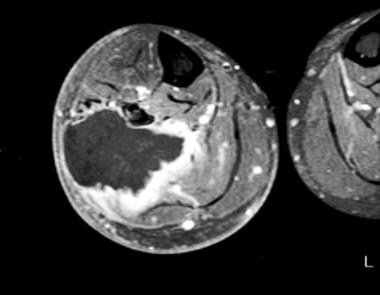
Because the tongue is superficially located and the initial manifestation of most diseases occurring there is mucosal change, lingual lesions can be easily accessed and diagnosed without imaging analysis. Some lingual neoplasms, however, may manifest as a submucosal bulge and be located in a deep portion of the tongue, such as its base; their true characteristics and extent may be recognized only on cross-sectional images such as those obtained by CT or MRI. Some uncommon tongue neoplasms may have characteristic radiologic features, thus permitting quite specific radiologic diagnosis. Lipomas typically manifest at both CT and MR imaging as homogeneous nonenhancing lesions. Relative to subcutaneous fat they are isoattenuating on CT images, and all MR sequences show them as isointense. Due to the paramagnetic properties of melanin, metastases from melanotic melanoma usually demonstrate high signal intensity on T1-weighted MR images and low signal intensity on T2-weighted images. Although the radiologic findings for other submucosal neoplasms are nonspecific, CT and MR imaging can play an important role in the diagnostic work-up of these unusual tumors. Delineation of the extent of the tumor, and recognition and understanding of the spectrum of imaging and the pathologic features of these lesions, often help narrow the differential diagnosis. (+info)Cytology of myoepithelial carcinoma of the salivary gland. (6/94)
BACKGROUND: Myoepithelial carcinoma, also know as malignant myoepithelioma, is rare in the salivary gland, and its cytologic features have rarely been reported. DESIGN: Four cases of myoepithelial carcinoma with cytology were retrieved from the archives of the Pathology Departments of two academic institutes. In three cases, the specimens were obtained by fine needle aspiration biopsy (FNA); the remaining case was a bench aspiration performed on the surgically resected specimen at the time of intra-operative consultation. The cytologic features were reviewed and correlated with the histology. RESULTS: The four patients with myoepithelial carcinoma (two men and two women) ranged in age from 48 to 64 years. Three cases arose from the parotid gland, and the remaining case was a recurrent tumor in the minor salivary glands of the hard palate. The aspirates of two cases consisted of predominantly spindle cells, one predominantly epithelioid/plasmacytoid cells, and one with a mixture of both spindle and epithelioid/plasmacytoid cells. Cellular pleomorphism was noted in two cases and mitotic figures in three cases. Two cases were cytologically diagnosed as malignant spindle cell neoplasm, not otherwise specified. The FNA of the recurrent tumor was diagnosed as consistent with the previous malignancy. The remaining case was interpreted as a pleomorphic adenoma with atypia. CONCLUSIONS: The cytologic features of myoepithelial carcinoma are diverse and may lack overt features of malignancy. Pathologists should be aware of this entity when evaluating cytologic specimen of salivary gland mass. (+info)A case of myoepithelial carcinoma displaying biallelic inactivation of the tumour suppressor gene APC in a patient with familial adenomatous polyposis. (7/94)
Familial adenomatous polyposis (FAP) is an autosomal dominant disorder caused by mutation of the APC gene. It is characterised by the appearance of hundreds to thousands of colorectal adenomas in adolescence and the subsequent development of colorectal cancer. Various extracolonic malignancies are associated with FAP, including desmoids and neoplasms of the stomach, duodenum, pancreas, liver, and brain. We present a family affected by FAP with an exon 14 APC mutation displaying two rare extracolonic lesions, a hepatoblastoma and a myoepithelial carcinoma. The hepatoblastoma was found in a male patient aged 2 years. The second lesion, a myoepithelial carcinoma of the right cheek, was found in a female patient aged 14 years. Inactivation of the normal APC allele was demonstrated in this lesion by loss of heterozygosity analysis, thus implicating APC in the initiation or progression of this neoplasm. This is the first reported case of this lesion in a family affected by FAP. (+info)Myoepithelial carcinoma of the salivary glands: behavior and management. (8/94)
OBJECTIVE: To investigate the biological behavior and proper management of myoepithelial carcinomas of salivary glands. METHODS: Twenty-seven cases of myoepithelial carcinoma of salivary glands were retrospectively studied and their detailed clinical and follow-up data were presented. RESULTS: The subjects consisted of 17 men and 10 women aged 16 to 73 years (mean age: 51 years). The parotid gland was the most common site (n = 14) of cancer. Clinical features included extensive local growth, invasion of the surrounding tissues, infrequent cervical lymph node metastasis but high rates of distant metastasis, frequent/multiple recurrences and poor prognosis. CONCLUSIONS: Myoepithelial carcinomas of the salivary gland should be classified as high-grade malignancies. Early and radical surgery with close follow-up are essential for achieving favorable outcomes. Radiotherapy appears to be non-sensitive and elective neck dissection is generally unnecessary. (+info)Myoepithelioma is a very rare, benign (non-cancerous) tumor that arises from the myoepithelial cells, which are found in various glands throughout the body, including salivary glands, sweat glands, and mammary glands. These tumors typically appear as slow-growing, painless masses. While they are usually benign, some myoepitheliomas can become malignant (cancerous) and invasive, leading to more serious health concerns. Treatment for myoepithelioma typically involves surgical removal of the tumor.
Palatal neoplasms refer to abnormal growths or tumors that occur on the palate, which is the roof of the mouth. These growths can be benign (non-cancerous) or malignant (cancerous). Benign neoplasms are typically slower growing and less likely to spread, while malignant neoplasms are more aggressive and can invade nearby tissues and organs.
Palatal neoplasms can have various causes, including genetic factors, environmental exposures, and viral infections. They may present with symptoms such as mouth pain, difficulty swallowing, swelling or lumps in the mouth, bleeding, or numbness in the mouth or face.
The diagnosis of palatal neoplasms typically involves a thorough clinical examination, imaging studies, and sometimes biopsy to determine the type and extent of the growth. Treatment options depend on the type, size, location, and stage of the neoplasm but may include surgery, radiation therapy, chemotherapy, or a combination of these approaches. Regular follow-up care is essential to monitor for recurrence or spread of the neoplasm.
Maxillary sinus neoplasms refer to abnormal growths or tumors that develop in the maxillary sinuses, which are located in the upper part of your cheekbones, below your eyes. These growths can be benign (non-cancerous) or malignant (cancerous).
Benign neoplasms may include conditions such as an osteoma (a benign bone tumor), a papilloma (a benign growth of the lining of the sinus), or a fibrous dysplasia (a condition where bone is replaced by fibrous tissue).
Malignant neoplasms, on the other hand, can be primary (originating in the maxillary sinuses) or secondary (spreading to the maxillary sinuses from another site in the body). Common types of malignant tumors that arise in the maxillary sinus include squamous cell carcinoma, adenocarcinoma, and mucoepidermoid carcinoma.
Symptoms of maxillary sinus neoplasms may include nasal congestion, nosebleeds, facial pain or numbness, vision changes, and difficulty swallowing or speaking. Treatment options depend on the type, size, and location of the tumor but may include surgery, radiation therapy, chemotherapy, or a combination of these approaches.
Salivary gland neoplasms refer to abnormal growths or tumors that develop in the salivary glands. These glands are responsible for producing saliva, which helps in digestion, lubrication of food and maintaining oral health. Salivary gland neoplasms can be benign (non-cancerous) or malignant (cancerous).
Benign neoplasms are slow-growing and typically do not spread to other parts of the body. They may cause symptoms such as swelling, painless lumps, or difficulty swallowing if they grow large enough to put pressure on surrounding tissues.
Malignant neoplasms, on the other hand, can be aggressive and have the potential to invade nearby structures and metastasize (spread) to distant organs. Symptoms of malignant salivary gland neoplasms may include rapid growth, pain, numbness, or paralysis of facial nerves.
Salivary gland neoplasms can occur in any of the major salivary glands (parotid, submandibular, and sublingual glands) or in the minor salivary glands located throughout the mouth and throat. The exact cause of these neoplasms is not fully understood, but risk factors may include exposure to radiation, certain viral infections, and genetic predisposition.
Minor salivary glands are numerous small exocrine glands that produce saliva and are distributed throughout the oral cavity, nasal cavity, pharynx, larynx, and paranasal sinuses. They are classified as "minor" due to their smaller size compared to the three pairs of major salivary glands (parotid, submandibular, and sublingual). The minor salivary glands are primarily mucous glands, although some contain serous cells. They are responsible for producing approximately 5-10% of the total saliva in the mouth. These glands help moisten the oral cavity, protect the mucosal lining, and facilitate speaking, chewing, and swallowing.
Vimentin is a type III intermediate filament protein that is expressed in various cell types, including mesenchymal cells, endothelial cells, and hematopoietic cells. It plays a crucial role in maintaining cell structure and integrity by forming part of the cytoskeleton. Vimentin is also involved in various cellular processes such as cell division, motility, and intracellular transport.
In addition to its structural functions, vimentin has been identified as a marker for epithelial-mesenchymal transition (EMT), a process that occurs during embryonic development and cancer metastasis. During EMT, epithelial cells lose their polarity and cell-cell adhesion properties and acquire mesenchymal characteristics, including increased migratory capacity and invasiveness. Vimentin expression is upregulated during EMT, making it a potential target for therapeutic intervention in cancer.
In diagnostic pathology, vimentin immunostaining is used to identify mesenchymal cells and to distinguish them from epithelial cells. It can also be used to diagnose certain types of sarcomas and carcinomas that express vimentin.
Parotid neoplasms refer to abnormal growths or tumors in the parotid gland, which is the largest of the salivary glands and is located in front of the ear and extends down the neck. These neoplasms can be benign (non-cancerous) or malignant (cancerous).
Benign parotid neoplasms are typically slow-growing, painless masses that may cause facial asymmetry or difficulty in chewing or swallowing if they become large enough to compress surrounding structures. The most common type of benign parotid tumor is a pleomorphic adenoma.
Malignant parotid neoplasms, on the other hand, are more aggressive and can invade nearby tissues and spread to other parts of the body. They may present as rapidly growing masses that are firm or fixed to surrounding structures. Common types of malignant parotid tumors include mucoepidermoid carcinoma, adenoid cystic carcinoma, and squamous cell carcinoma.
The diagnosis of parotid neoplasms typically involves a thorough clinical evaluation, imaging studies such as CT or MRI scans, and fine-needle aspiration biopsy (FNAB) to determine the nature of the tumor. Treatment options depend on the type, size, and location of the neoplasm but may include surgical excision, radiation therapy, and chemotherapy.
The cavernous sinus is a venous structure located in the middle cranial fossa, which is a depression in the skull that houses several important nerves and blood vessels. The cavernous sinus is situated on either side of the sphenoid bone, near the base of the skull, and it contains several important structures:
* The internal carotid artery, which supplies oxygenated blood to the brain
* The abducens nerve (cranial nerve VI), which controls lateral movement of the eye
* The oculomotor nerve (cranial nerve III), which controls most of the muscles that move the eye
* The trochlear nerve (cranial nerve IV), which controls one of the muscles that moves the eye
* The ophthalmic and maxillary divisions of the trigeminal nerve (cranial nerve V), which transmit sensory information from the face and head
The cavernous sinus is an important structure because it serves as a conduit for several critical nerves and blood vessels. However, it is also vulnerable to various pathological conditions such as thrombosis (blood clots), infection, tumors, or aneurysms, which can lead to serious neurological deficits or even death.
S100 proteins are a family of calcium-binding proteins that are involved in the regulation of various cellular processes, including cell growth and differentiation, intracellular signaling, and inflammation. They are found in high concentrations in certain types of cells, such as nerve cells (neurons), glial cells (supporting cells in the nervous system), and skin cells (keratinocytes).
The S100 protein family consists of more than 20 members, which are divided into several subfamilies based on their structural similarities. Some of the well-known members of this family include S100A1, S100B, S100 calcium-binding protein A8 (S100A8), and S100 calcium-binding protein A9 (S100A9).
Abnormal expression or regulation of S100 proteins has been implicated in various pathological conditions, such as neurodegenerative diseases, cancer, and inflammatory disorders. For example, increased levels of S100B have been found in the brains of patients with Alzheimer's disease, while overexpression of S100A8 and S100A9 has been associated with the development and progression of certain types of cancer.
Therefore, understanding the functions and regulation of S100 proteins is important for developing new diagnostic and therapeutic strategies for various diseases.
Glial Fibrillary Acidic Protein (GFAP) is a type of intermediate filament protein that is primarily found in astrocytes, which are a type of star-shaped glial cells in the central nervous system (CNS). These proteins play an essential role in maintaining the structural integrity and stability of astrocytes. They also participate in various cellular processes such as responding to injury, providing support to neurons, and regulating the extracellular environment.
GFAP is often used as a marker for astrocytic activation or reactivity, which can occur in response to CNS injuries, neuroinflammation, or neurodegenerative diseases. Elevated GFAP levels in cerebrospinal fluid (CSF) or blood can indicate astrocyte damage or dysfunction and are associated with several neurological conditions, including traumatic brain injury, stroke, multiple sclerosis, Alzheimer's disease, and Alexander's disease.
Immunohistochemistry (IHC) is a technique used in pathology and laboratory medicine to identify specific proteins or antigens in tissue sections. It combines the principles of immunology and histology to detect the presence and location of these target molecules within cells and tissues. This technique utilizes antibodies that are specific to the protein or antigen of interest, which are then tagged with a detection system such as a chromogen or fluorophore. The stained tissue sections can be examined under a microscope, allowing for the visualization and analysis of the distribution and expression patterns of the target molecule in the context of the tissue architecture. Immunohistochemistry is widely used in diagnostic pathology to help identify various diseases, including cancer, infectious diseases, and immune-mediated disorders.
No data available that match "myoepithelioma"
 Myoepithelioma of the head and neck - Wikipedia
 Myoepithelioma Pathology: Definition, Epidemiology, Location
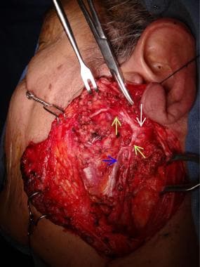
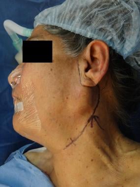
 Myoepithelioma preauricular: literature review and case report
 Tony NG | University of British Columbia, Vancouver | UBC | Department of Pathology and Laboratory Medicine | Research profile
Model Details
 Page 1 | Search Results | Acta Cytologica | Karger Publishers
 Joshua H. Mo - Search Results - PubMed
 Sarcoma and Bone Cancer Types | Dana-Farber Cancer Institute
 Gary M. Kupfer, MD| Pediatric Hematology Oncology, Pediatric Hematology | MedStar Health
 Non-cancerous lung tumours | Canadian Cancer Society
Strain Summary
 How to pronounce Xishong | HowToPronounce.com
 JPMA - Journal Of Pakistan Medical Association
 Salivary glands - Libre Pathology
Goats diagnosed with neoplasia display variability in clinical presentations, treatments, and outcomes in: Journal of the...
 Vol 9, No 1 (2022)
Model Details
Rafi Kabarriti - Publications - Albert Einstein College of Medicine
 Cases Journal | Articles
 Vol. 4 No. 04 (2014)
| National Journal of Medical Research
Vol. 4 No. 04 (2014)
| National Journal of Medical Research
Code System Concept
 PPT - Salivary Gland Neoplasms PowerPoint Presentation, free download - ID:144325
Bio2Vec
 Surgical Outcome of Extracapsular Dissection of Benign Parotid Gland Tumor: A Comparative Study to Superficial Parotidectomy
 Deakin University / All Locations
Bio2Vec
Mathangi K - Research output - Manipal Academy of Higher Education, Manipal, India
Faculty of Engineering - Research output - Okayama University
病理學科 - 研究成果 - 臺北醫學大學
 Cancer Genome Interpreter - Identification of therapeutically actionable genomic alterations in tumors
Myoepithelial6
- When malignant, which is exceedingly rare, they are known as malignant myoepithelioma or Myoepithelial carcinoma, and they account for 1% of the salivary tumors with poor prognosis. (wikipedia.org)
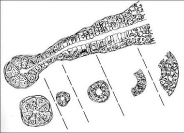
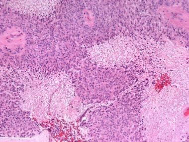
- Myoepithelioma is a benign salivary gland tumor that consists entirely of myoepithelial cells with variable cellular morphologies including spindle, epithelioid, plasmacytoid, or clear cells. (medscape.com)
- Histologically, benign myoepithelioma is composed of myoepithelial cells and treatment consists of surgical removal of the lesion with low rate of recurrence after complete removal. (bvsalud.org)
- 5. Lakhani S.R., O'Hare M.J., Monaghan P., Winehouse J., Gazet J.C., Sloane J.P. (1995) Malignant myoepithelioma (myoepithelial carcinoma) of the breast: a detailed cytokeratin study. (nordicmubio.com)
- Myoepithelial carcinoma, also known as malignant myoepithelioma, is mainly encountered in the salivary glands and, at a lower incidence, in the sweat glands (skin), breast, soft tissue, bone and very rarely in the lung. (asianarchpath.com)
- Salivary gland tumors with myoepithelial differentiation such as adenoid cystic carcinoma (ACC), PA, myoepithelioma, basal cell adenoma (BCA), basal cell adenocarcinoma (BCAC), and myoepithelial carcinoma are to be considered in the differential diagnoses. (ijhnp.org)
Malignant myoepithelioma1
- Michal M, Skálová A, Simpson RHW, Rychterová V, Leivo I: Clear cell malignant myoepithelioma of the salivary glands. (biopticka.cz)
Pleomorphic adenoma2
- The main differential diagnosis of myoepithelioma is the pleomorphic adenoma. (bvsalud.org)
- Oncocytic pleomorphic adenoma and myoepithelioma with novel gene fusions in a subset of cases. (patologie.cz)
Benign3
- Myoepithelioma of the head and neck, also myoepithelioma, is a salivary gland tumour of the head and neck that is usually benign. (wikipedia.org)
- Objective: this work allow to demonstrate by case report and literature review the comportment of benign myoepithelioma. (bvsalud.org)
- Case report: this work reported a case of benign myoepithelioma located in the preauricular region, removed surgically and followed for eight years without signs of recurrence, as well as the clinical and microscopic characteristics of this disease. (bvsalud.org)
Tumors3
- A myoepithelioma can be composed of one or a mixture of the above cell types, and a variable stromal component can be seen in these tumors. (medscape.com)
- Final considerations: although rare, tumors like myoepithelioma can happen in rotine evaluations. (bvsalud.org)
- Introduction Dermatofibrosarcoma protuberans, elastofibroma dorsi, desmoid-type fibromatosis and myoepithelioma belong to the category of rare soft tissue tumors. (evereth.pl)
Parotid1
- Skálová A, Michal M: Biphasic myoepithelioma of parotid gland with collagenous crystalloids. (biopticka.cz)
Soft Tissue1
- Myoepithelioma of Soft Tissue With Both Squamous and Adipocytic Metaplasia. (uci.edu)
Literature1
- Myoepithelioma of the APG is much rarer than PA, and to date, only 5 cases have been sporadically reported in the English literature. (sagepub.com)
Pleomorphic1
- The main differential diagnosis of myoepithelioma is the pleomorphic adenoma. (bvsalud.org)
Parotid gland1
- Lee M, Nam S, Choi H, Choi J, Moon K, Koh J. Myoepithelioma of parotid gland presenting as infra-auricular subcutaneous mass. (ijorl.com)
Minor salivary glands2
Salivary gland3
- Myoepithelioma of the head and neck, also myoepithelioma, is a salivary gland tumour of the head and neck that is usually benign. (wikipedia.org)
- Cytogenetic analysis of a primary salivary gland myoepithelioma. (nih.gov)
- 32. Salivary gland myoepithelioma with focal capsular invasion. (nih.gov)
Benign myoepithelioma3
- Objective: this work allow to demonstrate by case report and literature review the comportment of benign myoepithelioma. (bvsalud.org)
- Case report: this work reported a case of benign myoepithelioma located in the preauricular region, removed surgically and followed for eight years without signs of recurrence, as well as the clinical and microscopic characteristics of this disease. (bvsalud.org)
- Hunt K, Stevens M, Abdelsayed R, Nguyen C. Benign myoepithelioma of floor of mouth with mandibular involvement: a case report and literature review. (ijorl.com)
Tumor1
- The tumor shows wide morphologic and cytologic diversity in a similar way to its benign counterpart, myoepithelioma, with evidence of malignant change. (medscape.com)
Ductal1
- Among investigators, the level of "tolerance" for the presence of epithelial ductal elements in a myoepithelioma is variable and controversial (see Microscopic Findings ). (medscape.com)
Cystic1
- 35. [Cystic myoepithelioma. (nih.gov)
Case3
- Souliou C, Tzermpos F, Argyris P, Tosios K. Plasmacytoid Myoepithelioma of the Hard Palate: Case Report. (ijorl.com)
- Ramesh D, Khong G, Sumathi V. A Case of Myoepithelioma of the Nasal Cavity. (ijorl.com)
- Yadav A, Nadarajah J, Chandrashekhara S, Tambade V, Acharya S. Myoepithelioma of the soft palate: a case report. (ijorl.com)