Myositis, Inclusion Body
Inclusion Bodies
Inclusion Bodies, Viral
Polymyositis
Orbital Myositis
Dermatomyositis
Nervous System Autoimmune Disease, Experimental
Histidine-tRNA Ligase
Orbital Pseudotumor
Protein Refolding
Escherichia coli
Muscle, Skeletal
Sarcocystis
Muscle Weakness
Intranuclear Inclusion Bodies
Osteitis Deformans
Solubility
Distal Myopathies
Molecular Sequence Data
Microscopy, Electron
Boidae
Autoantibodies
Amino Acid Sequence
Muscle Fibers, Skeletal
Biopsy
Cloning, Molecular
Chymosin
Expression of the costimulatory molecule BB-1, the ligands CTLA-4 and CD28, and their mRNA in inflammatory myopathies. (1/153)
To examine if the muscle fibers in patients with inflammatory myopathies have the potential to behave as antigen presenting cells (APCs), we investigated the expression of costimulatory molecules BB-1, B7-1 (CD80), and B7-2 (CD86), and their counterreceptors, CD28 or CTLA-4 (CD152), in the muscle biopsies of patients with polymyositis (PM), PM associated with human immunodeficiency virus infection (HIV-PM), sporadic inclusion body myositis (s-IBM), dermatomyositis (DM), and normal or disease controls. The expression of the B7 family of molecules on the muscle fibers was limited to BB-1. In PM, HIV-PM, and s-IBM, but not the disease controls, the nonnecrotic, MHC-class I-expressing muscle fibers, invaded or not by CD8+ T cells, had prominent membrane expression of BB-1. Several of the BB-1-positive fibers bound strongly in a cell-to-cell contact with their CD28 or CTLA-4 ligands on the autoinvasive CD8+ T cells, as confirmed by confocal microscopy. By reverse transcription-polymerase chain reaction, the expression of CD28 and CTLA-4 was up-regulated in PM, HIV-PM, and s-IBM, but not the controls. Because the BB-1-positive fibers expressed MHC-class I antigen and bound to up-regulated counterreceptors CD28 and CTLA-4 on the autoinvasive CD8+ T cells only in PM, HIV-PM, and s-IBM, the BB-1 molecule in these diseases should have a functional role in antigen presentation and T cell differentiation. These findings complement recent studies and suggest that in PM, HIV-PM, and s-IBM the muscle fibers are not only targets of CD8+ cytotoxic T cells but may also behave as "professional" APC. (+info)A case of inclusion body myositis with benign monoclonal gammopathy successfully responding to repeated immunoabsorption. (2/153)
A 69 year old woman with inclusion body myositis is described. She presented with benign monoclonal gammopathy. She was resistant to steroid therapy, but responded to repeated immunoabsorption. Up to now, there has been no established therapy for inclusion body myositis, including IVIg. It is suggested that immunoabsorption could be an alternative therapy for inclusion body myositis, when it was accompanied by immunological abnormality. (+info)Synergistic effect of beta-amyloid protein and interferon gamma on nitric oxide production by C2C12 muscle cells. (3/153)
Nitric oxide (NO) is an important mediator of diverse physiological and pathological responses. NO-induced oxidative stress has been proposed in the pathogenesis of muscle tissue damage in inclusion-body myositis (IBM), which is characterized by deposition of beta-amyloid protein (Abeta) in vacuolated muscle fibres. To determine whether Abeta can induce NO production in skeletal muscle, we stimulated C2C12 mouse skeletal muscle cells in vitro with Abeta[1-42] or Abeta[25-35] peptides in the presence or absence of interferon gamma (IFN-gamma). Neither Abeta peptides nor IFN-gamma were able to stimulate nitrite (NO(2)(-)) production by C2C12 cells when given alone. However, combination of IFN-gamma with either Abeta[1-42] or Abeta[25-35] resulted in significant NO(2)(-) release into cell-free supernatants. Northern blot analysis of RNA obtained from Abeta/IFN-gamma-stimulated C2C12 cells revealed increased mRNA accumulation of inducible nitric oxide synthase (iNOS). Moreover, approximately 4% of muscle cells incubated with Abeta peptides and IFN-gamma showed ultrastructural features of DNA fragmentation. These findings, taken together, indicate that the association of Abeta with IFN-gamma stimulates NO(2)(-) production via induction of iNOS gene expression in skeletal muscle cells, with occasional evidence for nuclear changes suggesting apoptotic morphology. These data further support a role for Abeta deposition in the pathogenesis of postulated oxidative damage in IBM. (+info)Sporadic inclusion body myositis correlates with increased expression and cross-linking by transglutaminases 1 and 2. (4/153)
Sporadic inclusion body myositis (SIBM) is characterized by vacuolar degeneration of muscle fibers and intrafiber clusters of paired helical filaments with abnormal amyloid deposition. Because of their potential involvement in other degenerative disorders, we have examined the expression of transglutaminases (TGases) in normal and SIBM tissues. We report that at least two different enzymes, the ubiquitous TGase 2 as well as the TGase 1 enzyme, are present in muscle tissues. However, in comparison with normal tissue, the expression of TGases 1 and 2 was increased 2.5- and 4-fold in SIBM, accompanied by about a 20-fold higher total TGase activity. By immunohistochemical staining, in normal muscle, TGase 2 expression was restricted to some endomysial connective tissue elements, whereas TGase 1 and beta-amyloid proteins were not detectable. In SIBM muscle, both TGases 1 and 2 as well as amyloid proteins were brightly expressed and co-localized in the vacuolated muscle fibers, but none of these proteins colocalized with inflammatory cell markers. Next, we isolated high molecular weight insoluble proteins from SIBM muscle tissue and showed that they were cross-linked by about 6 residues/1000 residues of the isopeptide bond. Furthermore, by amino acid sequencing of solubilized tryptic peptides, they contain amyloid and skeletal muscle proteins. Together, these findings suggest that elevated expression of TGases 1 and 2 participate in the formation of insoluble amyloid deposits in SIBM tissue and in this way may contribute to progressive and debilitating muscle disease. (+info)Paired helical filaments of inclusion-body myositis muscle contain RNA and survival motor neuron protein. (5/153)
Sporadic inclusion-body myositis (s-IBM) is the most common progressive muscle disease of older persons. Pathologically, the muscle biopsy manifests various degrees of inflammation and specific vacuolar degeneration of muscle fibers characterized by paired helical filaments (PHFs) composed of phosphorylated tau. IBM vacuolated fibers also contain accumulations of several other Alzheimer-characteristic proteins. Molecular mechanisms leading to formation of the PHFs and accumulations of proteins in IBM muscle are not known. We report that the abnormal muscle fibers of IBM contained (i) acridine-orange-positive RNA inclusions that colocalized with the immunoreactivity of phosphorylated tau and (ii) survival motor neuron protein immunoreactive inclusions, which by immuno-electron microscopy were confined to paired helical filaments. This study demonstrates two novel components of the IBM paired helical filaments, which may lead to better understanding of their pathogenesis. (+info)Association of active extracellular signal-regulated protein kinase with paired helical filaments of inclusion-body myositis muscle suggests its role in inclusion-body myositis tau phosphorylation. (6/153)
The possible role of extracellular signal-regulated kinase (ERK) in the pathogenesis of inclusion-body myositis (IBM) was investigated by immunostaining the active phosphorylated form of ERK in muscle biopsies of six IBM and 14 control patients. Between 80% and 90% of IBM vacuolated muscle fibers contained well-defined ERK-immunoreactive inclusions, which were co-localized by light microscopy, with phosphorylated tau in 70 to 80% of those fibers. Immunoelectronmicroscopy colocalized ERK to small amorphous tufts adjacent to the muscle fiber paired-helical filaments. Strong ERK immunoreactivity was also present at the postsynaptic domain of all human neuromuscular junctions. Our study suggests 1) that ERK, a signal transducer, might play a role in IBM pathogenesis, including participation in the pathological phosphorylation of IBM tau; and 2) that signal transduction abnormalities may be a component of the IBM pathogenic cascade. Our novel immunolocalization of ERK at the postsynaptic domain of human neuromuscular junctions supports a role in transcription of junctional-protein genes. The ERK localized in nonjunctional regions of IBM fibers may underlie the known pathological up-regulation of junctional proteins there. (+info)Clonal restriction of T-cell receptor expression by infiltrating lymphocytes in inclusion body myositis persists over time. Studies in repeated muscle biopsies. (7/153)
Inclusion body myositis (IBM) is an inflammatory myopathy characterized immunohistologically by prominent invasion of the non-necrotic, MHC-I class antigen-expressing muscle fibres by CD8+ cytotoxic T cells. If the autoinvasive CD8+ T cells are recruited specifically to the muscle and play a primary pathogenetic role in the disease, a clonal restriction persisting over time should be anticipated. In this study, we analysed the T-cell receptor (TCR) gene usage by endomysial T lymphocytes in three sequential muscle biopsies from three different IBM patients over a 19-22 month period using immunohistochemistry, reverse transcription-polymerase chain reaction (RT-PCR) and sequence analysis of the complementarity determining region (CDR3) of the amplified TCRs. We found that CD8+ T lymphocytes persist in the endomysial infiltrates in all biopsies during a 19-22 month period. The most frequently detected TCRs were the V beta 3, V beta 5.1, V beta 6.7 and V beta 13 gene families, and several of the autoinvasive CD8+ T cells expressed the TCRs V beta 6.7 and V beta 5.1. A restricted usage of the examined V beta 6 gene family was found to persist in the complementarity CDR3 determining region of the autoinvasive T cells over the 22 month period. Identical V beta 6 CDR3 gene arrangements were also found in the multiple muscle biopsies from two of the three IBM patients. The results indicate that in IBM there is a restricted expression of the TCR gene families among the autoinvasive T lymphocytes with homologies in the CDR3 region that persist over the course of the disease. A continuous, antigen-driven T-cell response is prominent in the muscle of patients with IBM. (+info)Sporadic inclusion body myositis in a patient with human T cell leukemia virus type 1-associated myelopathy. (8/153)
Sporadic inclusion body myositis is a disease of unknown pathogenesis in which a viral etiology has long been suspected. We report a case that occurred in a patient with human T cell leukemia virus type 1-associated myelopathy. The diagnosis was confirmed by histopathological studies of the deltoid muscle. Nucleic acids amplification and in situ hybridization indicated the presence of integrated proviral DNA and viral mRNA transcripts in the lesions. (+info)Myositis is a medical term that refers to inflammation of the muscle tissue. This condition can cause various symptoms, including muscle weakness, pain, swelling, and stiffness. There are several types of myositis, such as polymyositis, dermatomyositis, and inclusion body myositis, which have different causes and characteristics.
Polymyositis is a type of myositis that affects multiple muscle groups, particularly those close to the trunk of the body. Dermatomyositis is characterized by muscle inflammation as well as a skin rash. Inclusion body myositis is a less common form of myositis that typically affects older adults and can cause both muscle weakness and wasting.
The causes of myositis vary depending on the type, but they can include autoimmune disorders, infections, medications, and other medical conditions. Treatment for myositis may involve medication to reduce inflammation, physical therapy to maintain muscle strength and flexibility, and lifestyle changes to manage symptoms and prevent complications.
Inclusion body myositis (IBM) is a rare inflammatory muscle disease characterized by progressive weakness and wasting (atrophy) of skeletal muscles. The term "inclusion body" refers to the presence of abnormal protein accumulations within muscle fibers, which are observed under a microscope during muscle biopsy. These inclusions are primarily composed of aggregated forms of amyloid-β and tau proteins, similar to those found in neurodegenerative disorders like Alzheimer's disease.
IBM typically affects individuals over 50 years old, and it is more common in men than women. The disease usually starts with weakness in the wrist and finger flexors, making it difficult to perform tasks such as gripping, buttoning shirts, or lifting objects. Over time, the weakness spreads to other muscle groups, including the thigh muscles (quadriceps), resulting in difficulty climbing stairs or rising from a seated position.
The exact cause of inclusion body myositis remains unclear; however, both immune-mediated and degenerative mechanisms are believed to contribute to its pathogenesis. Currently, there is no cure for IBM, and treatment options are primarily aimed at managing symptoms and improving quality of life. Immunosuppressive medications may be used to target the inflammatory component of the disease; however, their efficacy varies among patients. Physical therapy and exercise programs can help maintain muscle strength and function as much as possible.
Inclusion bodies are abnormal, intracellular accumulations or aggregations of various misfolded proteins, protein complexes, or other materials within the cells of an organism. They can be found in various tissues and cell types and are often associated with several pathological conditions, including infectious diseases, neurodegenerative disorders, and genetic diseases.
Inclusion bodies can vary in size, shape, and location depending on the specific disease or condition. Some inclusion bodies have a characteristic appearance under the microscope, such as eosinophilic (pink) staining with hematoxylin and eosin (H&E) histological stain, while others may require specialized stains or immunohistochemical techniques to identify the specific misfolded proteins involved.
Examples of diseases associated with inclusion bodies include:
1. Infectious diseases: Some viral infections, such as HIV, hepatitis B and C, and herpes simplex virus, can lead to the formation of inclusion bodies within infected cells.
2. Neurodegenerative disorders: Several neurodegenerative diseases are characterized by the presence of inclusion bodies, including Alzheimer's disease (amyloid-beta plaques and tau tangles), Parkinson's disease (Lewy bodies), Huntington's disease (Huntingtin aggregates), and amyotrophic lateral sclerosis (TDP-43 and SOD1 inclusions).
3. Genetic diseases: Certain genetic disorders, such as Danon disease, neuronal intranuclear inclusion disease, and some lysosomal storage disorders, can also present with inclusion bodies due to the accumulation of abnormal proteins or metabolic products within cells.
The exact role of inclusion bodies in disease pathogenesis remains unclear; however, they are often associated with cellular dysfunction, oxidative stress, and increased inflammation, which can contribute to disease progression and neurodegeneration.
Inclusion bodies, viral are typically described as intracellular inclusions that appear as a result of viral infections. These inclusion bodies consist of aggregates of virus-specific proteins, viral particles, or both, which accumulate inside the host cell's cytoplasm or nucleus during the replication cycle of certain viruses.
The presence of inclusion bodies can sometimes be observed through histological or cytological examination using various staining techniques. Different types of viruses may exhibit distinct morphologies and locations of these inclusion bodies, which can aid in the identification and diagnosis of specific viral infections. However, it is important to note that not all viral infections result in the formation of inclusion bodies, and their presence does not necessarily indicate active viral replication or infection.
Polymyositis is defined as a rare inflammatory disorder that causes muscle weakness and inflammation (swelling) of the muscles. It primarily affects the skeletal muscles, which are the muscles responsible for voluntary movements such as walking, talking, and swallowing. The onset of polymyositis can occur at any age but is most commonly seen in adults between 31 to 60 years old, with women being slightly more affected than men.
The exact cause of polymyositis remains unknown; however, it is believed to be an autoimmune disorder, where the body's immune system mistakenly attacks its own muscle tissue. Certain factors such as genetics, viral infections, and exposure to certain drugs may contribute to the development of this condition.
Polymyositis can cause various symptoms, including:
- Progressive muscle weakness and wasting, particularly affecting the proximal muscles (those closest to the trunk of the body) such as the hips, thighs, shoulders, and upper arms.
- Difficulty climbing stairs, lifting objects, or rising from a seated position.
- Fatigue and stiffness, especially after periods of inactivity.
- Joint pain and swelling.
- Difficulty swallowing or speaking.
- Shortness of breath due to weakened respiratory muscles.
Diagnosis of polymyositis typically involves a combination of medical history, physical examination, laboratory tests, electromyography (EMG), and muscle biopsy. Treatment usually includes medications such as corticosteroids and immunosuppressants to reduce inflammation and control the immune response. Physical therapy may also be recommended to help maintain muscle strength and flexibility.
If left untreated, polymyositis can lead to significant disability and complications, including respiratory failure, malnutrition, and cardiovascular disease. Early diagnosis and treatment are crucial for improving outcomes and preventing long-term complications.
Orbital myositis is a medical condition characterized by inflammation of the extraocular muscles, which are the muscles responsible for eye movement. These muscles are located within the orbit, the bony cavity that contains and protects the eye. Orbital myositis can cause symptoms such as painful eye movements, double vision, redness, swelling, and decreased visual acuity.
The condition is often associated with other systemic inflammatory or autoimmune disorders, such as rheumatoid arthritis, granulomatosis with polyangiitis (GPA), and sarcoidosis. However, it can also occur as an isolated phenomenon, known as idiopathic orbital myositis.
Diagnosis of orbital myositis typically involves a combination of clinical examination, imaging studies such as MRI or CT scans, and blood tests to evaluate for underlying systemic conditions. Treatment usually includes corticosteroids to reduce inflammation and alleviate symptoms, as well as addressing any underlying systemic disorders if present.
Dermatomyositis is a medical condition characterized by inflammation and weakness in the muscles and skin. It is a type of inflammatory myopathy, which means that it causes muscle inflammation and damage. Dermatomyositis is often associated with a distinctive rash that affects the skin around the eyes, nose, mouth, fingers, and toes.
The symptoms of dermatomyositis can include:
* Progressive muscle weakness, particularly in the hips, thighs, shoulders, and neck
* Fatigue
* Difficulty swallowing or speaking
* Skin rash, which may be pink or purple and is often accompanied by itching
* Muscle pain and tenderness
* Joint pain and swelling
* Raynaud's phenomenon, a condition that affects blood flow to the fingers and toes
The exact cause of dermatomyositis is not known, but it is believed to be related to an autoimmune response in which the body's immune system mistakenly attacks healthy tissue. Treatment for dermatomyositis typically involves medications to reduce inflammation and suppress the immune system, as well as physical therapy to help maintain muscle strength and function.
A nervous system autoimmune disease, experimental, refers to a type of disorder in which the immune system mistakenly attacks healthy nerves or tissues in the nervous system. This category includes conditions that are currently being researched and have not yet been fully proven or accepted by the medical community as definitive diseases.
In an autoimmune disease, the body's immune system produces antibodies and activates immune cells (such as T-cells) to attack and destroy foreign substances, such as bacteria and viruses. However, in an experimental nervous system autoimmune disease, the immune system mistakenly identifies normal nerves or nerve tissues as harmful and attacks them. This can lead to damage or destruction of the nerves, resulting in various neurological symptoms.
Examples of experimental nervous system autoimmune diseases may include conditions such as MOG antibody-associated disease (MOGAD) or anti-NMDA receptor encephalitis, which are still being studied and have not yet been fully recognized by the medical community. It is important to note that while these conditions are considered experimental, they can still cause significant harm and should be treated with appropriate medical interventions.
Histidine-tRNA ligase is an enzyme involved in the process of protein synthesis, specifically during the step of translation. Its primary function is to catalyze the attachment of the amino acid histidine to its corresponding transfer RNA (tRNA) molecule. This enzyme does this by forming a ester bond between the carboxyl group of histidine and the 3'-hydroxyl group of the tRNA, creating a charged histidine-tRNA complex.
The histidine-tRNA ligase enzyme plays a crucial role in maintaining the accuracy of protein synthesis, as it ensures that only the correct amino acid is attached to its specific tRNA. This helps to prevent errors in the genetic code and contributes to the proper folding and functioning of proteins.
The systematic name for this enzyme is "histidine:tRNA(His) ligase (AMP-forming)" and it belongs to the family of ligases, specifically the aminoacyl-tRNA ligases. The gene that encodes this enzyme in humans is known as HARS1 (Histidyl-tRNA Synthetase 1). Defects or mutations in this gene can lead to various genetic disorders, such as histidinemia and Charcot-Marie-Tooth disease.
Orbital pseudotumor, also known as orbital inflammatory syndrome or idiopathic orbital inflammation, is a non-specific term used to describe a group of conditions characterized by inflammation in the orbit (the bony cavity surrounding the eye) without any identifiable cause. It is not a true tumor, but rather an inflammatory reaction that can mimic the symptoms and signs of a tumor.
The condition can affect people of any age, although it is more common in middle-aged adults. The exact cause of orbital pseudotumor is unknown, but it is believed to be related to an abnormal immune response or inflammation triggered by various factors such as infections, trauma, or autoimmune disorders.
Symptoms of orbital pseudotumor may include eye pain, redness, swelling, protrusion of the eyeball (proptosis), double vision, and decreased vision. Diagnostic tests such as imaging studies (CT or MRI scans) and biopsy may be used to rule out other causes of orbital inflammation. Treatment typically involves corticosteroids to reduce inflammation, although other immunosuppressive medications may be necessary in severe cases. In some cases, the condition may resolve on its own without treatment.
Protein refolding is the process by which a denatured or misfolded protein reverts to its native, functional three-dimensional structure. Proteins are complex molecules that perform a wide range of functions within living organisms. Their function is heavily dependent on their unique three-dimensional shape, which is determined by the specific sequence of amino acids that make up the protein.
When proteins are exposed to certain environmental conditions, such as changes in temperature, pH, or the presence of denaturing agents, they can lose their native conformation and become denatured or misfolded. This can result in the loss of protein function and, in some cases, the formation of aggregates that can be toxic to cells.
Protein refolding is a crucial process for maintaining proper protein function within cells. It involves several steps:
1. Unfolding: The denatured or misfolded protein must first be unfolded into its linear amino acid sequence. This can be accomplished through various methods, such as exposure to chemical denaturants or changes in pH.
2. Renaturation: Once the protein is unfolded, it can begin to refold into its native conformation. This process is often facilitated by chaperone proteins, which help to stabilize the protein and prevent aggregation during the refolding process.
3. Folding: The protein must then fold into its correct three-dimensional structure. This is a complex process that involves the formation of specific bonds between amino acids, as well as the interaction with other molecules in the cell.
4. Quality control: Once the protein has folded, it must be checked for correct folding and function. Misfolded proteins may be targeted for degradation by the cell's quality control mechanisms.
Protein refolding is a critical process that occurs naturally within cells, but it can also be studied in vitro (outside of the cell) using various techniques. Understanding the mechanisms of protein refolding is important for developing therapies for diseases caused by protein misfolding and aggregation, such as Alzheimer's disease and Parkinson's disease.
Protein renaturation is the process of restoring the native, functional structure of a protein that has been denatured due to exposure to external stressors such as changes in temperature, pH, or the addition of chemical agents. Denaturation causes proteins to lose their unique three-dimensional structure, which is essential for their proper function. Renaturation involves slowly removing these stressors and allowing the protein to refold into its original configuration, restoring its biological activity. This process can be facilitated by various techniques, including dialysis, dilution, or the addition of specific chemical chaperones.
'Escherichia coli' (E. coli) is a type of gram-negative, facultatively anaerobic, rod-shaped bacterium that commonly inhabits the intestinal tract of humans and warm-blooded animals. It is a member of the family Enterobacteriaceae and one of the most well-studied prokaryotic model organisms in molecular biology.
While most E. coli strains are harmless and even beneficial to their hosts, some serotypes can cause various forms of gastrointestinal and extraintestinal illnesses in humans and animals. These pathogenic strains possess virulence factors that enable them to colonize and damage host tissues, leading to diseases such as diarrhea, urinary tract infections, pneumonia, and sepsis.
E. coli is a versatile organism with remarkable genetic diversity, which allows it to adapt to various environmental niches. It can be found in water, soil, food, and various man-made environments, making it an essential indicator of fecal contamination and a common cause of foodborne illnesses. The study of E. coli has contributed significantly to our understanding of fundamental biological processes, including DNA replication, gene regulation, and protein synthesis.
Skeletal muscle, also known as striated or voluntary muscle, is a type of muscle that is attached to bones by tendons or aponeuroses and functions to produce movements and support the posture of the body. It is composed of long, multinucleated fibers that are arranged in parallel bundles and are characterized by alternating light and dark bands, giving them a striped appearance under a microscope. Skeletal muscle is under voluntary control, meaning that it is consciously activated through signals from the nervous system. It is responsible for activities such as walking, running, jumping, and lifting objects.
Protein folding is the process by which a protein molecule naturally folds into its three-dimensional structure, following the synthesis of its amino acid chain. This complex process is determined by the sequence and properties of the amino acids, as well as various environmental factors such as temperature, pH, and the presence of molecular chaperones. The final folded conformation of a protein is crucial for its proper function, as it enables the formation of specific interactions between different parts of the molecule, which in turn define its biological activity. Protein misfolding can lead to various diseases, including neurodegenerative disorders such as Alzheimer's and Parkinson's disease.
Sarcocystis is a genus of intracellular parasitic protozoa that belongs to the phylum Apicomplexa. These microscopic organisms are known to infect both animals and humans, causing a variety of symptoms depending on the specific species involved and the immune status of the host.
Sarcocystis spp. have a complex life cycle involving two hosts: an intermediate host, which is typically a herbivorous animal, and a definitive host, which is usually a carnivorous or omnivorous animal. The parasites form cysts, known as sarcocysts, in the muscles of the intermediate host, which are then ingested by the definitive host during feeding.
In humans, Sarcocystis spp. can cause two main types of infections: intestinal and muscular. Intestinal infection occurs when humans accidentally ingest undercooked or raw meat containing Sarcocystis cysts. The parasites then invade the human's intestinal wall, causing symptoms such as diarrhea, abdominal pain, and fever.
Muscular infection, on the other hand, is caused by the ingestion of water or food contaminated with sporocysts shed in the feces of infected definitive hosts. This type of infection is relatively rare in humans and typically causes mild symptoms such as muscle pain, weakness, and fever.
It's worth noting that while Sarcocystis spp. can cause illness in humans, they are not usually considered a significant public health concern. Proper cooking of meat and good hygiene practices can help prevent infection with these parasites.
Muscle weakness is a condition in which muscles cannot develop the expected level of physical force or power. This results in reduced muscle function and can be caused by various factors, including nerve damage, muscle diseases, or hormonal imbalances. Muscle weakness may manifest as difficulty lifting objects, maintaining posture, or performing daily activities. It is essential to consult a healthcare professional for proper diagnosis and treatment of muscle weakness.
Recombinant proteins are artificially created proteins produced through the use of recombinant DNA technology. This process involves combining DNA molecules from different sources to create a new set of genes that encode for a specific protein. The resulting recombinant protein can then be expressed, purified, and used for various applications in research, medicine, and industry.
Recombinant proteins are widely used in biomedical research to study protein function, structure, and interactions. They are also used in the development of diagnostic tests, vaccines, and therapeutic drugs. For example, recombinant insulin is a common treatment for diabetes, while recombinant human growth hormone is used to treat growth disorders.
The production of recombinant proteins typically involves the use of host cells, such as bacteria, yeast, or mammalian cells, which are engineered to express the desired protein. The host cells are transformed with a plasmid vector containing the gene of interest, along with regulatory elements that control its expression. Once the host cells are cultured and the protein is expressed, it can be purified using various chromatography techniques.
Overall, recombinant proteins have revolutionized many areas of biology and medicine, enabling researchers to study and manipulate proteins in ways that were previously impossible.
Muscular diseases, also known as myopathies, refer to a group of conditions that affect the functionality and health of muscle tissue. These diseases can be inherited or acquired and may result from inflammation, infection, injury, or degenerative processes. They can cause symptoms such as weakness, stiffness, cramping, spasms, wasting, and loss of muscle function.
Examples of muscular diseases include:
1. Duchenne Muscular Dystrophy (DMD): A genetic disorder that results in progressive muscle weakness and degeneration due to a lack of dystrophin protein.
2. Myasthenia Gravis: An autoimmune disease that causes muscle weakness and fatigue, typically affecting the eyes and face, throat, and limbs.
3. Inclusion Body Myositis (IBM): A progressive muscle disorder characterized by muscle inflammation and wasting, typically affecting older adults.
4. Polymyositis: An inflammatory myopathy that causes muscle weakness and inflammation throughout the body.
5. Metabolic Myopathies: A group of inherited disorders that affect muscle metabolism, leading to exercise intolerance, muscle weakness, and other symptoms.
6. Muscular Dystonias: Involuntary muscle contractions and spasms that can cause abnormal postures or movements.
It is important to note that muscular diseases can have a significant impact on an individual's quality of life, mobility, and overall health. Proper diagnosis and treatment are crucial for managing symptoms and improving outcomes.
Intranuclear inclusion bodies are abnormal, rounded structures found within the nucleus of a cell. They are composed of aggregated proteins or other cellular components and can be associated with various viral infections and certain genetic disorders. These inclusion bodies can interfere with normal nuclear functions, leading to cell damage and contributing to the pathogenesis of diseases such as cytomegalovirus infection, rabies, and some forms of neurodegenerative disorders like polyglutamine diseases. The presence of intranuclear inclusion bodies is often used in diagnostic pathology to help identify specific underlying conditions.
Osteitis deformans, also known as Paget's disease of bone, is a chronic disorder of the bone characterized by abnormal turnover and remodeling of the bone. In this condition, the bone becomes enlarged, thickened, and deformed due to excessive and disorganized bone formation and resorption.
The process begins when the bone-remodeling cycle is disrupted, leading to an imbalance between the activity of osteoclasts (cells that break down bone) and osteoblasts (cells that form new bone). In Paget's disease, osteoclasts become overactive and increase bone resorption, followed by an overzealous response from osteoblasts, which attempt to repair the damage but do so in a disorganized manner.
The affected bones can become weakened, prone to fractures, and may cause pain, deformities, or other complications such as arthritis, hearing loss, or neurological symptoms if the skull or spine is involved. The exact cause of Paget's disease remains unknown, but it is believed that genetic and environmental factors play a role in its development.
Early diagnosis and treatment can help manage the symptoms and prevent complications associated with osteitis deformans. Treatment options include medications to slow down bone turnover, pain management, and orthopedic interventions when necessary.
An Aviadenovirus is a type of virus that belongs to the family *Adenoviridae* and the genus *Aviadenovirus*. These viruses primarily infect avian species, such as birds, and can cause a variety of diseases. The genome of an Aviadenovirus is double-stranded DNA. Some species of Aviadenoviruses have been known to cause respiratory and reproductive problems in poultry, leading to significant economic losses in the poultry industry. It's important to note that Aviadenoviruses are not known to infect or cause disease in humans.
Solubility is a fundamental concept in pharmaceutical sciences and medicine, which refers to the maximum amount of a substance (solute) that can be dissolved in a given quantity of solvent (usually water) at a specific temperature and pressure. Solubility is typically expressed as mass of solute per volume or mass of solvent (e.g., grams per liter, milligrams per milliliter). The process of dissolving a solute in a solvent results in a homogeneous solution where the solute particles are dispersed uniformly throughout the solvent.
Understanding the solubility of drugs is crucial for their formulation, administration, and therapeutic effectiveness. Drugs with low solubility may not dissolve sufficiently to produce the desired pharmacological effect, while those with high solubility might lead to rapid absorption and short duration of action. Therefore, optimizing drug solubility through various techniques like particle size reduction, salt formation, or solubilization is an essential aspect of drug development and delivery.
Distal myopathies are a group of rare genetic muscle disorders that primarily affect the muscles of the hands, feet, and lower legs. These myopathies are characterized by progressive weakness and wasting (atrophy) of the distal muscles, which are located further from the center of the body. The onset of symptoms can vary widely, ranging from early childhood to late adulthood.
There are several different types of distal myopathies, each caused by mutations in specific genes that affect muscle function. Some common forms include:
1. Nonaka Distal Myopathy: This form is caused by mutations in the GNE gene and typically presents in the third or fourth decade of life with weakness and wasting of the ankle dorsiflexors, foot extensors, and wrist and finger extensors.
2. Miyoshi Distal Myopathy: This form is caused by mutations in the DYSF gene and affects the calf muscles initially, followed by weakness in other distal muscles over time.
3. Welander Distal Myopathy: This form is caused by mutations in the TIA1 gene and typically presents in adulthood with weakness and wasting of the hand and forearm muscles.
4. Laing Distal Myopathy: This form is caused by mutations in the CAV3 gene and affects the anterior compartment of the lower leg, resulting in foot drop and weakness of the ankle dorsiflexors.
5. Gowers Distal Myopathy: This form is caused by mutations in the HNRNPDL gene and typically presents in adulthood with weakness and wasting of the hand and forearm muscles, as well as foot drop.
There is no cure for distal myopathies, but treatment can help manage symptoms and improve quality of life. Physical therapy, bracing, and orthotics may be used to support weakened muscles and maintain mobility. In some cases, medications such as corticosteroids or immunosuppressants may be prescribed to reduce muscle inflammation and slow disease progression.
Molecular sequence data refers to the specific arrangement of molecules, most commonly nucleotides in DNA or RNA, or amino acids in proteins, that make up a biological macromolecule. This data is generated through laboratory techniques such as sequencing, and provides information about the exact order of the constituent molecules. This data is crucial in various fields of biology, including genetics, evolution, and molecular biology, allowing for comparisons between different organisms, identification of genetic variations, and studies of gene function and regulation.
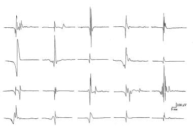
Electron microscopy (EM) is a type of microscopy that uses a beam of electrons to create an image of the sample being examined, resulting in much higher magnification and resolution than light microscopy. There are several types of electron microscopy, including transmission electron microscopy (TEM), scanning electron microscopy (SEM), and reflection electron microscopy (REM).
In TEM, a beam of electrons is transmitted through a thin slice of the sample, and the electrons that pass through the sample are focused to form an image. This technique can provide detailed information about the internal structure of cells, viruses, and other biological specimens, as well as the composition and structure of materials at the atomic level.
In SEM, a beam of electrons is scanned across the surface of the sample, and the electrons that are scattered back from the surface are detected to create an image. This technique can provide information about the topography and composition of surfaces, as well as the structure of materials at the microscopic level.
REM is a variation of SEM in which the beam of electrons is reflected off the surface of the sample, rather than scattered back from it. This technique can provide information about the surface chemistry and composition of materials.
Electron microscopy has a wide range of applications in biology, medicine, and materials science, including the study of cellular structure and function, disease diagnosis, and the development of new materials and technologies.
In the context of human anatomy, the thigh is the part of the lower limb that extends from the hip to the knee. It is the upper and largest portion of the leg and is primarily composed of the femur bone, which is the longest and strongest bone in the human body, as well as several muscles including the quadriceps femoris (front thigh), hamstrings (back thigh), and adductors (inner thigh). The major blood vessels and nerves that supply the lower limb also pass through the thigh.
Boidae is a family of snakes, also known as boas. This family includes many different species of large, non-venomous snakes found in various parts of the world, particularly in Central and South America, Africa, and Asia. Boas are known for their strong bodies and muscular tails, which they use to constrict their prey before swallowing it whole. Some well-known members of this family include the anaconda, the python, and the boa constrictor.
Autoantibodies are defined as antibodies that are produced by the immune system and target the body's own cells, tissues, or organs. These antibodies mistakenly identify certain proteins or molecules in the body as foreign invaders and attack them, leading to an autoimmune response. Autoantibodies can be found in various autoimmune diseases such as rheumatoid arthritis, lupus, and thyroiditis. The presence of autoantibodies can also be used as a diagnostic marker for certain conditions.
Adenoviridae infections refer to diseases caused by members of the Adenoviridae family of viruses, which are non-enveloped, double-stranded DNA viruses. These viruses can infect a wide range of hosts, including humans, animals, and birds. In humans, adenovirus infections can cause a variety of symptoms, depending on the specific type of virus and the age and immune status of the infected individual.
Common manifestations of adenovirus infections in humans include:
1. Respiratory illness: Adenoviruses are a common cause of respiratory tract infections, such as bronchitis, pneumonia, and croup. They can also cause conjunctivitis (pink eye) and pharyngoconjunctival fever.
2. Gastrointestinal illness: Some types of adenoviruses can cause diarrhea, vomiting, and abdominal pain, particularly in children and immunocompromised individuals.
3. Genitourinary illness: Adenoviruses have been associated with urinary tract infections, hemorrhagic cystitis, and nephritis.
4. Eye infections: Epidemic keratoconjunctivitis is a severe form of conjunctivitis caused by certain adenovirus types.
5. Central nervous system infections: Adenoviruses have been linked to meningitis, encephalitis, and other neurological disorders, although these are rare.
Transmission of adenoviruses typically occurs through respiratory droplets, contaminated surfaces, or contaminated water. Preventive measures include good hygiene practices, such as handwashing and avoiding close contact with infected individuals. There is no specific treatment for adenovirus infections, but supportive care can help alleviate symptoms. In severe cases or in immunocompromised patients, antiviral therapy may be considered.
An amino acid sequence is the specific order of amino acids in a protein or peptide molecule, formed by the linking of the amino group (-NH2) of one amino acid to the carboxyl group (-COOH) of another amino acid through a peptide bond. The sequence is determined by the genetic code and is unique to each type of protein or peptide. It plays a crucial role in determining the three-dimensional structure and function of proteins.
Skeletal muscle fibers, also known as striated muscle fibers, are the type of muscle cells that make up skeletal muscles, which are responsible for voluntary movements of the body. These muscle fibers are long, cylindrical, and multinucleated, meaning they contain multiple nuclei. They are surrounded by a connective tissue layer called the endomysium, and many fibers are bundled together into fascicles, which are then surrounded by another layer of connective tissue called the perimysium.
Skeletal muscle fibers are composed of myofibrils, which are long, thread-like structures that run the length of the fiber. Myofibrils contain repeating units called sarcomeres, which are responsible for the striated appearance of skeletal muscle fibers. Sarcomeres are composed of thick and thin filaments, which slide past each other during muscle contraction to shorten the sarcomere and generate force.
Skeletal muscle fibers can be further classified into two main types based on their contractile properties: slow-twitch (type I) and fast-twitch (type II). Slow-twitch fibers have a high endurance capacity and are used for sustained, low-intensity activities such as maintaining posture. Fast-twitch fibers, on the other hand, have a higher contractile speed and force generation capacity but fatigue more quickly and are used for powerful, explosive movements.
A biopsy is a medical procedure in which a small sample of tissue is taken from the body to be examined under a microscope for the presence of disease. This can help doctors diagnose and monitor various medical conditions, such as cancer, infections, or autoimmune disorders. The type of biopsy performed will depend on the location and nature of the suspected condition. Some common types of biopsies include:
1. Incisional biopsy: In this procedure, a surgeon removes a piece of tissue from an abnormal area using a scalpel or other surgical instrument. This type of biopsy is often used when the lesion is too large to be removed entirely during the initial biopsy.
2. Excisional biopsy: An excisional biopsy involves removing the entire abnormal area, along with a margin of healthy tissue surrounding it. This technique is typically employed for smaller lesions or when cancer is suspected.
3. Needle biopsy: A needle biopsy uses a thin, hollow needle to extract cells or fluid from the body. There are two main types of needle biopsies: fine-needle aspiration (FNA) and core needle biopsy. FNA extracts loose cells, while a core needle biopsy removes a small piece of tissue.
4. Punch biopsy: In a punch biopsy, a round, sharp tool is used to remove a small cylindrical sample of skin tissue. This type of biopsy is often used for evaluating rashes or other skin abnormalities.
5. Shave biopsy: During a shave biopsy, a thin slice of tissue is removed from the surface of the skin using a sharp razor-like instrument. This technique is typically used for superficial lesions or growths on the skin.
After the biopsy sample has been collected, it is sent to a laboratory where a pathologist will examine the tissue under a microscope and provide a diagnosis based on their findings. The results of the biopsy can help guide further treatment decisions and determine the best course of action for managing the patient's condition.
Molecular cloning is a laboratory technique used to create multiple copies of a specific DNA sequence. This process involves several steps:
1. Isolation: The first step in molecular cloning is to isolate the DNA sequence of interest from the rest of the genomic DNA. This can be done using various methods such as PCR (polymerase chain reaction), restriction enzymes, or hybridization.
2. Vector construction: Once the DNA sequence of interest has been isolated, it must be inserted into a vector, which is a small circular DNA molecule that can replicate independently in a host cell. Common vectors used in molecular cloning include plasmids and phages.
3. Transformation: The constructed vector is then introduced into a host cell, usually a bacterial or yeast cell, through a process called transformation. This can be done using various methods such as electroporation or chemical transformation.
4. Selection: After transformation, the host cells are grown in selective media that allow only those cells containing the vector to grow. This ensures that the DNA sequence of interest has been successfully cloned into the vector.
5. Amplification: Once the host cells have been selected, they can be grown in large quantities to amplify the number of copies of the cloned DNA sequence.
Molecular cloning is a powerful tool in molecular biology and has numerous applications, including the production of recombinant proteins, gene therapy, functional analysis of genes, and genetic engineering.
Chymosin, also known as rennin or rennet, is a proteolytic enzyme that is naturally present in the stomachs of ruminant animals such as cows, goats, and sheep. It plays an essential role in the digestion of milk in these animals by curdling or coagulating the milk protein casein, which helps in the separation of solid curds from liquid whey during the process of stomach digestion.
In the context of food production, chymosin is often used as a coagulant in the manufacturing of cheese and other dairy products. Traditionally, rennet was obtained by extracting it from the fourth stomach chamber (abomasum) of young calves, but nowadays, most commercial chymosin is produced through microbial fermentation using genetically modified bacteria or yeast that have been engineered to produce this enzyme. This method of production allows for a more consistent and animal-friendly source of chymosin for industrial applications.
The primary function of chymosin in cheese making is to catalyze the coagulation of casein, leading to the formation of a curd that can be further processed into various types of cheese. The enzyme specifically cleaves a bond in the casein protein called Phe105-Met106, resulting in the formation of para-κ-casein and paracaseinompholine, which then interact to form the curd. This reaction is crucial for initiating the cheese making process, as it allows for the separation of solid curds from liquid whey, which can then be pressed, aged, and transformed into a wide variety of cheese styles.
A muscle is a soft tissue in our body that contracts to produce force and motion. It is composed mainly of specialized cells called muscle fibers, which are bound together by connective tissue. There are three types of muscles: skeletal (voluntary), smooth (involuntary), and cardiac. Skeletal muscles attach to bones and help in movement, while smooth muscles are found within the walls of organs and blood vessels, helping with functions like digestion and circulation. Cardiac muscle is the specific type that makes up the heart, allowing it to pump blood throughout the body.
Recombinant fusion proteins are artificially created biomolecules that combine the functional domains or properties of two or more different proteins into a single protein entity. They are generated through recombinant DNA technology, where the genes encoding the desired protein domains are linked together and expressed as a single, chimeric gene in a host organism, such as bacteria, yeast, or mammalian cells.
The resulting fusion protein retains the functional properties of its individual constituent proteins, allowing for novel applications in research, diagnostics, and therapeutics. For instance, recombinant fusion proteins can be designed to enhance protein stability, solubility, or immunogenicity, making them valuable tools for studying protein-protein interactions, developing targeted therapies, or generating vaccines against infectious diseases or cancer.
Examples of recombinant fusion proteins include:
1. Etaglunatide (ABT-523): A soluble Fc fusion protein that combines the heavy chain fragment crystallizable region (Fc) of an immunoglobulin with the extracellular domain of the human interleukin-6 receptor (IL-6R). This fusion protein functions as a decoy receptor, neutralizing IL-6 and its downstream signaling pathways in rheumatoid arthritis.
2. Etanercept (Enbrel): A soluble TNF receptor p75 Fc fusion protein that binds to tumor necrosis factor-alpha (TNF-α) and inhibits its proinflammatory activity, making it a valuable therapeutic option for treating autoimmune diseases like rheumatoid arthritis, ankylosing spondylitis, and psoriasis.
3. Abatacept (Orencia): A fusion protein consisting of the extracellular domain of cytotoxic T-lymphocyte antigen 4 (CTLA-4) linked to the Fc region of an immunoglobulin, which downregulates T-cell activation and proliferation in autoimmune diseases like rheumatoid arthritis.
4. Belimumab (Benlysta): A monoclonal antibody that targets B-lymphocyte stimulator (BLyS) protein, preventing its interaction with the B-cell surface receptor and inhibiting B-cell activation in systemic lupus erythematosus (SLE).
5. Romiplostim (Nplate): A fusion protein consisting of a thrombopoietin receptor agonist peptide linked to an immunoglobulin Fc region, which stimulates platelet production in patients with chronic immune thrombocytopenia (ITP).
6. Darbepoetin alfa (Aranesp): A hyperglycosylated erythropoiesis-stimulating protein that functions as a longer-acting form of recombinant human erythropoietin, used to treat anemia in patients with chronic kidney disease or cancer.
7. Palivizumab (Synagis): A monoclonal antibody directed against the F protein of respiratory syncytial virus (RSV), which prevents RSV infection and is administered prophylactically to high-risk infants during the RSV season.
8. Ranibizumab (Lucentis): A recombinant humanized monoclonal antibody fragment that binds and inhibits vascular endothelial growth factor A (VEGF-A), used in the treatment of age-related macular degeneration, diabetic retinopathy, and other ocular disorders.
9. Cetuximab (Erbitux): A chimeric monoclonal antibody that binds to epidermal growth factor receptor (EGFR), used in the treatment of colorectal cancer and head and neck squamous cell carcinoma.
10. Adalimumab (Humira): A fully humanized monoclonal antibody that targets tumor necrosis factor-alpha (TNF-α), used in the treatment of various inflammatory diseases, including rheumatoid arthritis, psoriasis, and Crohn's disease.
11. Bevacizumab (Avastin): A recombinant humanized monoclonal antibody that binds to VEGF-A, used in the treatment of various cancers, including colorectal, lung, breast, and kidney cancer.
12. Trastuzumab (Herceptin): A humanized monoclonal antibody that targets HER2/neu receptor, used in the treatment of breast cancer.
13. Rituximab (Rituxan): A chimeric monoclonal antibody that binds to CD20 antigen on B cells, used in the treatment of non-Hodgkin's lymphoma and rheumatoid arthritis.
14. Palivizumab (Synagis): A humanized monoclonal antibody that binds to the F protein of respiratory syncytial virus, used in the prevention of respiratory syncytial virus infection in high-risk infants.
15. Infliximab (Remicade): A chimeric monoclonal antibody that targets TNF-α, used in the treatment of various inflammatory diseases, including Crohn's disease, ulcerative colitis, rheumatoid arthritis, and ankylosing spondylitis.
16. Natalizumab (Tysabri): A humanized monoclonal antibody that binds to α4β1 integrin, used in the treatment of multiple sclerosis and Crohn's disease.
17. Adalimumab (Humira): A fully human monoclonal antibody that targets TNF-α, used in the treatment of various inflammatory diseases, including rheumatoid arthritis, psoriatic arthritis, ankylosing spondylitis, Crohn's disease, and ulcerative colitis.
18. Golimumab (Simponi): A fully human monoclonal antibody that targets TNF-α, used in the treatment of rheumatoid arthritis, psoriatic arthritis, ankylosing spondylitis, and ulcerative colitis.
19. Certolizumab pegol (Cimzia): A PEGylated Fab' fragment of a humanized monoclonal antibody that targets TNF-α, used in the treatment of rheumatoid arthritis, psoriatic arthritis, ankylosing spondylitis, and Crohn's disease.
20. Ustekinumab (Stelara): A fully human monoclonal antibody that targets IL-12 and IL-23, used in the treatment of psoriasis, psoriatic arthritis, and Crohn's disease.
21. Secukinumab (Cosentyx): A fully human monoclonal antibody that targets IL-17A, used in the treatment of psoriasis, psoriatic arthritis, and ankylosing spondylitis.
22. Ixekizumab (Taltz): A fully human monoclonal antibody that targets IL-17A, used in the treatment of psoriasis and psoriatic arthritis.
23. Brodalumab (Siliq): A fully human monoclonal antibody that targets IL-17 receptor A, used in the treatment of psoriasis.
24. Sarilumab (Kevzara): A fully human monoclonal antibody that targets the IL-6 receptor, used in the treatment of rheumatoid arthritis.
25. Tocilizumab (Actemra): A humanized monoclonal antibody that targets the IL-6 receptor, used in the treatment of rheumatoid arthritis, systemic juvenile idiopathic arthritis, polyarticular juvenile idiopathic arthritis, giant cell arteritis, and chimeric antigen receptor T-cell-induced cytokine release syndrome.
26. Siltuximab (Sylvant): A chimeric monoclonal antibody that targets IL-6, used in the treatment of multicentric Castleman disease.
27. Satralizumab (Enspryng): A humanized monoclonal antibody that targets IL-6 receptor alpha, used in the treatment of neuromyelitis optica spectrum disorder.
28. Sirukumab (Plivensia): A human monoclonal antibody that targets IL-6, used in the treatment
 Inclusion body myositis
Inflammatory myopathy
Exercise therapy for idiopathic inflammatory myopathies
HLA-DR52
Bimagrumab
Niemann-Pick disease, type C
Rochester Epidemiology Project
Bafilomycin
Beevor's sign
Hereditary inclusion body myopathy
Ronald Sukenick
Deaths in January 2017
Foot drop
Robert Rodman
Mike Krukow
Martin Shubik
Proteinopathy
Rawleigh Warner Jr.
Blood flow restriction training
Deaths in July 2004
Deaths in June 2013
APBB1
Dick Edell
HLA-DR17
Polymyositis
Stuart Long
HLA-DR18
Frampton Forgets the Words
Skeletal muscle
Myopathy
Inclusion body myositis - Wikipedia
 Inclusion body myositis | Bartleby
 Sporadic Inclusion Body Myositis: Practice Essentials, Background, Pathophysiology
 Sporadic inclusion body myositis: new insights and potential therapy
098 Developing new therapeutic strategies for inclusion body myositis | Journal of Neurology, Neurosurgery & Psychiatry
 Myositis: Symptoms, Causes, and Treatment
Inclusion body myositis - wikidoc
 RheumaKnowledgy » Inclusion Body Myositis
 Inclusion Body Myositis - Symptoms | Cure IBM
 Inclusion Body Myositis - Myositis Association Australia
 inclusion body myositis News and Features - MuseWire
Inclusion body myositis (IBM) - Causes | Muscular Dystrophy UK
A Garden in Bethlehem PA: Inclusion Body Myositis (IBM)
 Intravenous Immunoglobulin Use for Neurologic Diseases | Article | NursingCenter
Types of myositis - Myositis UK
Simply Stated: Inclusion Body Myositis, Revisited - Quest | Muscular Dystrophy Association
Immunohistochemical Phenotype of T Cells Invading Muscle in Inclusion Body Myositis<...
Sporadic inclusion body myositis - a myodegenerative disease or an inflammatory myopathy<...
ArboCat Virus: Keuraliba (KEUV)
 Quest Magazine | Muscular Dystrophy Association
 Quadriceps strength is a sensitive marker of disease progression in sporadic inclusion body myositis - R Discovery
 Myositis Causes, Symptoms, and Treatments | UPMC
 NCA - Seat Elevation Systems as an Accessory to Power Wheelchairs (Group 3) (CAG-00461N) - Decision Memo
 N.M. Code R. § 7.34.2.7 - DEFINITIONS | State Regulations | US Law | LII / Legal Information Institute
 The NT5C1A Antibody Test and its Role in the Diagnosis of Inclusion Body Myositis - Myositis Support and Understanding
 Current status of clinical outcome measures in inclusion body myositis: a systematised review. | Clin Exp Rheumatol;41(2): 370...
 ICGNMD Publications | International Centre for Genomic Medicine in Neuromuscular Diseases - UCL - University College London
 Abcuro to Present Late-Breaking Abstract at American College of Rheumatology Convergence 2021 of ABC008 Inclusion Body Myositis...
Sporadic inclusion body myositis in Japanese is associated with the MHC ancestral haplotype 52.1 - Fingerprint - the UWA...
Inclusion body myositis with human immunodeficiency virus infection: Four cases with clonal expansion of viral-specific T cells...
Sporadic inclusion18
- Inclusion body myositis (IBM) (/maɪoʊˈsaɪtɪs/) (sometimes called sporadic inclusion body myositis, sIBM) is the most common inflammatory muscle disease in older adults. (wikipedia.org)
- The term "sporadic inclusion body myositis" (sIBM) was introduced as a way to refer to IBM to avoid confusion with hIBM. (wikipedia.org)
- Sporadic inclusion body myositis (s-IBM) and hereditary inclusion body myopathies (h-IBM) encompass a group of disorders sharing the common pathological finding of vacuoles and filamentous inclusions. (medscape.com)
- Sporadic inclusion body myositis (s-IBM) has been traditionally classified as one of the idiopathic inflammatory myopathies along with dermatomyositis (DM) and polymyositis (PM). However, the pathologic findings of sporadic inclusion body myositis (s-IBM) involve both inflammatory and degenerative characteristics, and the true primary pathogenesis of the disease remains a subject of significant debate. (medscape.com)
- To describe new insights and developments in the pathogenesis, diagnosis and treatment of sporadic inclusion body myositis (IBM). (nih.gov)
- Sporadic inclusion body myositis ( sIBM ) is an inflammatory muscle disease , characterized by slowly progressive weakness and wasting of the distal and proximal muscles, most apparent in the muscles of the arms and legs . (wikidoc.org)
- In sporadic inclusion body myositis [MY-oh-sigh-tis] muscle, two processes, one autoimmune and the other degenerative, appear to occur in the muscle cells in parallel. (wikidoc.org)
- Sporadic inclusion body myositis (sIBM) is an insidious late-onset progressive myopathy that typically affects patients over the age of 50. (johnshopkins.edu)
- Weihl, CC & Mammen, AL 2017, ' Sporadic inclusion body myositis - a myodegenerative disease or an inflammatory myopathy ', Neuropathology and Applied Neurobiology , vol. 43, no. 1, pp. 82-91. (johnshopkins.edu)
- There are currently no effective treatments to restore the muscle function in sporadic inclusion body myositis. (researcher.life)
- The goal of this study consisted in defining the functional pattern of patients with sporadic inclusion body myositis and to follow its change over a 9-month period to determine the most sensitive outcome measures for future clinical trials. (researcher.life)
- Twenty-two patients with definite sporadic inclusion body myositis were assessed using clinical and functional scales. (researcher.life)
- Knee extension strength seems to be the most relevant marker of disease progression in sporadic inclusion body myositis when measured with suitable dynamometry. (researcher.life)
- Sporadic inclusion body myositis (IBM) is a debilitating idiopathic inflammatory myopathy (IIM) which affects hand function, ambulation , and swallowing . (bvsalud.org)
- Objective: Sporadic inclusion body myositis (sIBM), a common adult-onset myositis, is characterized by an antigen-driven inflammatory tesponse and vacuolar degeneration. (elsevierpure.com)
- Pruitt, J. N. 2nd, Showalter, C. J. & Engel, A. G. Sporadic inclusion body myositis: counts of different types of abnormal fibers. (nature.com)
- Magnetic resonance imaging pattern recognition in sporadic inclusion-body myositis. (nature.com)
- Idiopathic inflammatory myopathies comprise a heterogeneous group of disorders, including polymyositis, dermatomyositis and sporadic inclusion body myositis (s-IBM). (bmj.com)
Dermatomyositis5
- Dermatomyositis (DM) is the easiest form of myositis to diagnose due to the purple-red rashes in the shape of the heliotrope flower . (healthline.com)
- Necrotizing myopathy is a more newly defined form of myositis with muscle weakness similar to that of dermatomyositis and polymyositis. (upmc.com)
- To shed light on this debilitating aspect of the condition, let us consider an illustrative example: imagine a middle-aged woman named Sarah who has been diagnosed with dermatomyositis, a specific subtype of myositis. (myositissupportgroup.org)
- Autoimmune myositis causes inflammation and weakness in the muscles (polymyositis) or in the skin and muscles (dermatomyositis). (msdmanuals.com)
- The ANA is often positive, but anemia is not a common finding for some reason in myositis, and inflammatory markers are often normal in dermatomyositis. (medscape.com)
Myopathy12
- IBM stands for "inclusion body myositis: not "inclusion body myopathy. (wikipedia.org)
- The term inclusion body myositis was originally used by Yunis and Samaha in 1971 for a case of myopathy that phenotypically suggested chronic polymyositis but showed cytoplasmic vacuoles and inclusions on muscle biopsy . (medscape.com)
- There are also several very rare forms of hereditary inclusion body myopathy (hIBM) that are linked to specific genetic defects and that are passed on from generation to generation, each inherited in different ways. (wikidoc.org)
- See hereditary inclusion body myopathy . (wikidoc.org)
- The differential diagnosis of inclusion body myositis includes other types of IIM as well as other endocrinologic, metabolic, infectious, and toxic etiologies (see Myopathy ). (rheumaknowledgy.com)
- Inclusion body myositis (IBM) is an inflammatory myopathy of aged people with poor response to therapy. (elsevierpure.com)
- Muscle biopsy, often utilized in the diagnosis, demonstrates a chronic myopathy with mixed pathologies harbouring intramyofiber protein inclusions and endomysial inflammation. (johnshopkins.edu)
- Dr. Soltanzadeh is an expert in myotonic dystrophies types 1 and 2, inclusion body myopathies including GNE myopathy and sporadic IBM, limb girdle muscular dystrophies, statin myopathies including the statin-triggered autoimmune necrotizing myopathy (HMG CR Ab associated myositis), myositis, dystrophinopathies, myasthenia gravis, amyotrophic lateral sclerosis, and neuropathies. (uclahealth.org)
- Dr. Soltanzadeh received his M.D. from Tehran University of Medical Sciences, where his thesis focused on hereditary inclusion body myopathy (GNE myopathy). (uclahealth.org)
- On the basis of these findings, we made a diagnosis of chronic destructive myopathy with inflammation (including eosinophils), with differential diagnoses of inclusion body myositis and parasitic myositis. (cdc.gov)
- Today we wanted to bring you some quick tips about diagnosing myositis and myopathy. (medscape.com)
- But I did not have nearly the framework that I do now after hearing the episode Myositis and Myopathy with Dr Lisa Christopher-Stine. (medscape.com)
Polymyositis4
- Polymyositis (PM) begins with muscle weakness in the muscles closest to the trunk of the body and then expands from there. (healthline.com)
- Inclusion body myositis (IBM) and polymyositis can normally be distinguished on the basis of clinical features. (nature.com)
- 18F]Florbetapir positron emission tomography: identification of muscle amyloid in inclusion body myositis and differentiation from polymyositis. (nature.com)
- For instance, consider the case of Mr. Johnson, a 45-year-old man diagnosed with polymyositis, one of the subtypes of autoimmune myositis. (myositissupportgroup.org)
Myopathies8
- IBM is often confused with an entirely different class of diseases, called hereditary inclusion body myopathies (hIBM). (wikipedia.org)
- Multiple genetic diseases that feature inclusion bodies have been grouped into "hereditary inclusion body myopathies (hIBM). (wikipedia.org)
- Inclusion Body Myositis ,or IBM, is one of many muscle diseases known as inflammatory myopathies, which causes slowly progressing muscular atrophy and weakness(NINDS IBM ,2014,para 1). (bartleby.com)
- Notwithstanding, IBM is distinct from the Hereditary Inclusion Body Myopathies (hIBM). (myositis.org.au)
- Inclusion-body myositis (IBM) is one of the most common disabling inflammatory myopathies in older adults, but its underlying cause is poorly understood.IBM is characterized by progressive muscle weakness and wasting. (mda.org)
- Progressive myopathies characterized by the presence of inclusion bodies on muscle biopsy. (nih.gov)
- IBMA merges with the National Myositis Association (NMA), expands to include all forms of myopathies and changes its name to the Myositis Association of America (MAA). (myositis.org)
- For example, autoimmune myopathies such as inclusion body myositis cause secondary mitochondrial DNA deletions and ragged-red fibers. (umdf.org)
Types of myositis4
- Symptoms of toxic myositis are similar to those of other types of myositis. (healthline.com)
- Discover more about the different types of myositis and possible further complications. (myositis.org.uk)
- What Are the Different Types of Myositis? (upmc.com)
- Symptoms start more slowly than they do for other types of myositis. (upmc.com)
Autoimmune Myositis10
- Joint pain is a common symptom experienced by individuals with autoimmune myositis, a group of inflammatory muscle diseases characterized by chronic muscle weakness and inflammation. (myositissupportgroup.org)
- Understanding the underlying mechanisms causing joint pain in autoimmune myositis is crucial for effective management of this symptom. (myositissupportgroup.org)
- Symptoms of joint pain in autoimmune myositis can vary in intensity and location. (myositissupportgroup.org)
- To diagnose joint involvement in autoimmune myositis, healthcare professionals may perform a thorough physical examination, review medical history, and order additional tests. (myositissupportgroup.org)
- Treatment options for joint pain in autoimmune myositis aim to reduce inflammation, manage pain, and improve joint function. (myositissupportgroup.org)
- As always, it is essential for individuals experiencing joint pain associated with autoimmune myositis to consult with their healthcare provider for an accurate diagnosis and appropriate treatment plan tailored to their specific needs. (myositissupportgroup.org)
- In autoimmune myositis, the body's immune cells mistakenly attack healthy tissues such as muscles and joints instead of foreign invaders. (myositissupportgroup.org)
- Autoimmune myositis usually occurs in adults aged 40 to 60 or in children aged 5 to 15 years. (msdmanuals.com)
- The cause of autoimmune myositis is unknown. (msdmanuals.com)
- The symptoms of autoimmune myositis are similar for people of all ages, but the muscle inflammation often appears to develop more abruptly in children than in adults. (msdmanuals.com)
Ragged red fi1
- On light microscopic analysis, there is an endomysial accumulation of mononuclear cells, particularly CD8+ T cells (similar to the findings in PM). Other characteristic findings in muscle include vacuoles lined with basophilic granules, eosinophilic inclusions, abnormal microtubular filaments in nuclear and cytoplasmic inclusions, and ragged red fibers. (rheumaknowledgy.com)
People living with m1
- In the United States, there are an estimated 1,600 to 3,200 new cases per year and 50,000 to 75,000 people living with myositis. (healthline.com)
Filamentous inclusions2
- Degeneration is characterized by the appearance of holes, deposits of abnormal proteins, and filamentous inclusions in the muscle fibers. (wikipedia.org)
- The degeneration aspect is characterized by the appearance of holes in the muscle (vacuoles), deposits of amyloid-related proteins within the cells and filamentous inclusions (hence the name inclusion body myositis) of abnormal proteins. (wikidoc.org)
Muscle biopsy2
- Nonetheless, muscle biopsy with electron microscopic analysis is required to make a diagnosis of inclusion body myositis. (rheumaknowledgy.com)
- In such instances, inclusion body myositis should be suspected, and muscle biopsy repeated or reviewed with emphasis on electron microscopic findings. (rheumaknowledgy.com)
Forms of myositis3
- Similar to the other forms of myositis, JM is characterized by muscle weakness and skin rashes. (healthline.com)
- TMA begins work on publishing a book on juvenile forms of myositis with the help of medical experts and families of those with JM. (myositis.org)
- TMA publishes "Myositis and You", comprehensive 466-page text addressing the entire spectrum of issues and challenges related to juvenile forms of myositis. (myositis.org)
Neurology2
- Matsubara, S , Suzuki, S & Komori, T 2022, ' Immunohistochemical Phenotype of T Cells Invading Muscle in Inclusion Body Myositis ', Journal of Neuropathology and Experimental Neurology , vol. 81, no. 10, pp. 825-835. (elsevierpure.com)
- The IBM clinical trial is being led by Professor Merrilee Needham, a recognized leader in the myositis field, head of the Myositis Research Group at The Perron Institute, and Foundation Chair of Neurology at the Fiona Stanley Hospital in Australia. (abcuro.com)
Intranuclear1
- Both forms feature intracytoplasmic and intranuclear inclusions in muscle tissue. (nih.gov)
Pathologic1
- Although clinical, pathologic, and laboratory assessments were inconclusive, a new, nested PCR-coupled sequencing method enabled the unequivocal diagnosis of myositis caused by the enigmatic nematode Haycocknema perplexum . (cdc.gov)
Autoantibodies1
- The syndrome is essentially the presence of specific autoantibodies in the body. (myositis.org.uk)
20181
- On May 9, 2018, Dr. Kevin Dooley, a retired ophthalmologist who has the rare disease inclusion body myositis , joined us live for a patient-led video education session titled, "The NT5C1A Antibody Test and its Role in the Diagnosis of Inclusion Body Myositis. (understandingmyositis.org)
Inflammation of the muscles2
- Myositis is a chronic, progressive inflammation of the muscles. (healthline.com)
- The definition of myositis is inflammation of the muscles. (upmc.com)
Idiopathic1
- Inclusion body myositis is a form of idiopathic inflammatory myositis (IIM)of unknown etiology. (rheumaknowledgy.com)
Juvenile3
- Juvenile myositis (JM) occurs in children under 18 . (healthline.com)
- Conference offers activities for juvenile myositis (JM) parents, families, and patients. (myositis.org)
- TMA sponsors first national conference solely for juvenile myositis in Washington, D.C. (myositis.org)
Prognosis1
- To learn more about the symptoms and prognosis of IBM, visit Simply Stated: What is Inclusion Body Myositis? . (mdaquest.org)
Clinical Trials3
- A prospective natural history study of inclusion body myositis: implications for clinical trials. (rheumaknowledgy.com)
- We're committed to executing on our clinical trials including our registrational trial in inclusion body myositis. (businesswire.com)
- The company is conducting two clinical trials in rare diseases: a Phase 2/3 registrational trial evaluating ABC008 in inclusion body myositis (IBM,) and a Phase 1/2 trial evaluating ABC008 in T cell large granular lymphocytic leukemia. (businesswire.com)
Vacuoles1
- The 'inclusion body' refers to a histological finding of rimmed vacuoles in muscle tissue. (wikipedia.org)
Patients12
- Often the diagnosis of inclusion body myositis is made when patients with presumed PM or DM either do not respond to steroid therapy or display atypical manifestations (e.g., asymmetry, distal muscle weakness, peripheral neuropathy). (rheumaknowledgy.com)
- However, because of the poor response to corticosteroid or immunosuppressive therapies, many physicians do not pursue aggressive or alternative therapies in patients with inclusion body myositis with little or no response to corticosteroids. (rheumaknowledgy.com)
- Inclusion body myositis is a slowly progressive disorder, and patients are less likely to respond to therapy than those with other types of IIM. (rheumaknowledgy.com)
- Swallowing difficulty, known as dysphagia , eventually affects many patients, but is an uncommon early symptom of inclusion body myositis. (cureibm.org)
- Sleep apnea may be common in inclusion body myositis patients. (cureibm.org)
- There are rare reports of IBM patients with trouble breathing because of weak muscles, but the vast majority of inclusion body myositis patients do not have prominent respiratory symptoms. (cureibm.org)
- Muscle biopsies of patients diagnosed with IBM reveal multiple "inclusion bodies" containing cellular material of dead tissue. (mda.org)
- Ubiquitinated inclusions in inclusion-body myositis patients are immunoreactive for cathepsin D but not beta-amyloid. (nature.com)
- The cumulative risk of incident connective tissue disorder in patients with myositis was significantly increased. (bmj.com)
- Furthermore, certain biological mechanisms exacerbate joint pain in myositis patients. (myositissupportgroup.org)
- TMA publishes special comprehensive issues of The OutLook that cover products for myositis patients and treatment options and developments. (myositis.org)
- United States Congress grants TMA an appropriation of $170,000 to fund a myositis patient registry to better track myositis patients and treatments. (myositis.org)
Symptoms6
- Inclusion body myositis symptoms are due to muscle weakness, usually beginning in middle-age or later, and worsen gradually over several years. (cureibm.org)
- Most autoimmune diseases are complex conditions meaning those with myositis may experience other symptoms that don't appear to be part of the disease itself. (myositis.org.uk)
- Myositis can cause problems in muscle groups and other symptoms throughout the body. (upmc.com)
- What Are the Symptoms and Complications of Myositis? (upmc.com)
- Myositis symptoms and complications vary by type and severity. (upmc.com)
- This article aims to provide an informative overview of joint pain in body myositis, discussing its causes, symptoms, diagnosis approaches, and potential treatment options. (myositissupportgroup.org)
Body's1
- Overview of Autoimmune Disorders of Connective Tissue In an autoimmune disorder, antibodies or cells produced by the body attack the body's own tissues. (msdmanuals.com)
Proteins1
- proteinopathic adj ), or proteopathy , protein conformational disorder , or protein misfolding disease , is a class of diseases in which certain proteins become structurally abnormal, and thereby disrupt the function of cells , tissues and organs of the body. (wikipedia.org)
Diagnose1
- How Do You Diagnose Myositis? (upmc.com)
Syndrome1
- You can see isolated weakness, or weakness with mild pain, but myositis doesn't present as an isolated pain syndrome. (medscape.com)
Treatment5
- Herbal can be an effective Inclusion Body Myositis Treatment for a variety of conditions, either as an assistant to standard Western meds or in solitude. (blogspot.com)
- The particular Inclusion Body Myositis Treatment recommended by your expert will rely on the seriousness and kind of issues the nearness of other medical conditions and antagonistic responses to previous treatment. (blogspot.com)
- Myositis UK is a small national charity providing information to those affected by myositis and funding research to improve diagnosis and treatment. (myositis.org.uk)
- Myositis can cause health problems that can lead to death without proper treatment. (upmc.com)
- Abcuro will use the proceeds from the financing to complete a Phase 2/3 registrational clinical trial evaluating ABC008, a first-in-class monoclonal antibody targeting killer cell lectin like receptor G1 (KLRG1), for the treatment of inclusion body myositis (IBM). (businesswire.com)
Pathogenesis1
- Greenberg, S. A. Inclusion body myositis: clinical features and pathogenesis. (nature.com)
Disorders1
- These disorders result in muscle inflammation (myositis), disabling muscle weakness, and occasionally tenderness. (msdmanuals.com)
Clumps3
- When viewed under a microscope, the muscle cells of people with IBM contain abnormal protein clumps called inclusion bodies, which give the condition its name. (myositis.org.au)
- These clumps are the "inclusion bodies" for which the disease is named. (mdaquest.org)
- Micrograph of immunostained α-synuclein (brown) in Lewy bodies (large clumps) and Lewy neurites (thread-like structures) in the cerebral cortex of a patient with Lewy body disease , a synucleinopathy . (wikipedia.org)
Treatments2
- Treatments help to prevent muscle loss and heart, lung, and other problems myositis can cause. (upmc.com)
- Several new trials exploring new treatments for myositis are initiated. (myositis.org)
Findings1
- Inclusion in the update does not necessarily represent the views of the Centers for Disease Control and Prevention nor does it imply endorsement of the article's methods or findings. (cdc.gov)
Illness2
- SAN DIEGO, Calif. (MuseWire) - Based out of mostly sunny San Diego, Calif., Mr. Mike Shirk, an artist and former ad exec, is living with an illness called inclusion body myositis. (musewire.com)
- Is myositis a serious illness? (upmc.com)
Chronic1
- Inclusion body myositis (IBM) is a rare, progressive muscle disease characterized by chronic muscle inflammation and weakness. (mdaquest.org)
Mild1
- [ 3 ] Individual responses, functional improvement, or mild regional improvement in strength have been reported, but sustained remission and improvement in whole-body strength have not been demonstrated. (medscape.com)
Muscles4
- The Microscopic Anatomy of Muscles 650 muscles operate the body. (bartleby.com)
- The muscle weakness is more prominent in smaller muscles and is asymmetrical, with one side of the body affected more than the other. (healthline.com)
- The weakness typically occurs in the shoulders and hips but can affect muscles symmetrically throughout the body. (msdmanuals.com)
- Because muscles close to the center of the body are affected most, tasks such as lifting the arms above the shoulders, climbing stairs, and getting out of a chair or off of a toilet seat can become very difficult. (msdmanuals.com)
Focal1
- 2,3 Gadolinium contrast is not required unless fasciitis, focal myositis or a cystic or solid mass lesion are of primary concern. (the-rheumatologist.org)
Common4
- Inclusion body myositis has distinct differences from the more common forms of IIM, PM, and DM . (rheumaknowledgy.com)
- Inclusion body myositis is more common in older people. (upmc.com)
- Myositis can start at any age, but inclusion body myositis is more common after age 50. (upmc.com)
- But inclusion body myositis is more common in men. (upmc.com)